Archives of Cardiovascular Disease (2008) 101, 333—342 Disponible en ligne sur www.sciencedirect.com CLINICAL RESEARCH Combined transplantation of endothelial progenitor cells and mesenchymal stem cells into a rat model of isoproterenol-induced myocardial injury La transplantation combinée des cellules progénitrices endothéliales et mésenchymateuses améliore la fonction cardiaque dans un modèle d’atteinte myocardique induite par isprotereol chez le rat Xin Zhang, Meng Wei ∗ , Wei Zhu, Beibei Han The Division of Cardiology, Shanghai Sixth Hospital, Shanghai Jiao Tong University School of Medicine, 600, Yishan Road, 200233 Shanghai, PR China Received 16 February 2008; accepted 9 May 2008 Available online 25 June 2008 KEYWORDS Isoproterenol; Stem cells; Transplantation; Endothelial; Mesenchymal; Cardiac function Summary Background. — Endothelial progenitor cells (EPCs) and mesenchymal stem cells (MSCs) have different biological properties, but their potential for synergy in the treatment of injured myocardium has not been studied extensively. Aim. — To determine if outcome could be improved by simultaneously transplanting MSCs and EPCs into a rat model of isoproterenol (ISO)-induced injured myocardium. Methods. — Four weeks after ISO injection, 50 rats were separated randomly into five groups (n = 10 per group) and allocated to receive a saline injection (control group), 200 L medium alone, 200 L medium plus 2 × 10 6 EPCs, 200 L medium plus 2 × 10 6 MSCs, or 200 L medium plus a combination of 1 × 10 6 EPCs and 1 × 10 6 MSCs. Abbreviations: Ang-2, Angiopoietin-2; b-FGF, Basic fibroblast growth factor; BMNC, Bone marrow mononuclear cell; Dil-ac-LDL, Dil- labelled acetylated low-density lipoprotein; DMSO, Dimethyl sulphoxide; EGM-2, Endothelial cell growth medium-2; ELISA, Enzyme-linked immunosorbent assay; EPC, Endothelial progenitor cell; FISH, Fluorescence in situ hybridization; FITC-UEA-1, Fluorescein isothiocyanate- labelled Ulex europaeus agglutinin-1; GAPDH, Glyceraldehyde-3-phosphate dehydrogenase; ISO, Isoproterenol; LVEDD, Left ventricular end-diastolic diameter; LVEDP, Left ventricular end-diastolic pressure; MSC, Mesenchymal stem cells; MTT, 3-(4,5-dimethylthiazol-2-yl)- 2,5-diphenyltetrazolium bromide; RT-PCR, Reverse transcriptase polymerase chain reaction; VEGF, Vascular endothelial growth factor; vWF, von Willebrand factor. ∗ Corresponding author. Fax: +8621 54481564 E-mail address: [email protected] (M. Wei). 1875-2136/$ — see front matter © 2008 Elsevier Masson SAS. All rights reserved. doi:10.1016/j.acvd.2008.05.002

Welcome message from author
This document is posted to help you gain knowledge. Please leave a comment to let me know what you think about it! Share it to your friends and learn new things together.
Transcript

Archives of Cardiovascular Disease (2008) 101, 333—342
Disponib le en l igne sur www.sc iencedi rec t .com
CLINICAL RESEARCH
Combined transplantation of endothelial progenitorcells and mesenchymal stem cells into a rat model ofisoproterenol-induced myocardial injury
La transplantation combinée des cellules progénitrices endothéliales etmésenchymateuses améliore la fonction cardiaque dans un modèle d’atteintemyocardique induite par isprotereol chez le rat
Xin Zhang, Meng Wei ∗, Wei Zhu, Beibei Han
The Division of Cardiology, Shanghai Sixth Hospital, Shanghai Jiao Tong University School ofMedicine, 600, Yishan Road, 200233 Shanghai, PR China
Received 16 February 2008; accepted 9 May 2008Available online 25 June 2008
KEYWORDSIsoproterenol;Stem cells;Transplantation;Endothelial;Mesenchymal;
SummaryBackground. — Endothelial progenitor cells (EPCs) and mesenchymal stem cells (MSCs) havedifferent biological properties, but their potential for synergy in the treatment of injuredmyocardium has not been studied extensively.Aim. — To determine if outcome could be improved by simultaneously transplanting MSCs andEPCs into a rat model of isoproterenol (ISO)-induced injured myocardium.
Cardiac function Methods. — Four weeks after ISO injection, 50 rats were separated randomly intofive groups (n = 10 per group) and allocated to receive a saline injection (controlgroup), 200 �L medium alone, 200 �L medium plus 2 × 10⁶ EPCs, 200 �L medium plus2 × 106 MSCs, or 200 �L medium plus a combination of 1 × 10⁶ EPCs and 1 × 10⁶ MSCs.
Abbreviations: Ang-2, Angiopoietin-2; b-FGF, Basic fibroblast growth factor; BMNC, Bone marrow mononuclear cell; Dil-ac-LDL, Dil-labelled acetylated low-density lipoprotein; DMSO, Dimethyl sulphoxide; EGM-2, Endothelial cell growth medium-2; ELISA, Enzyme-linkedimmunosorbent assay; EPC, Endothelial progenitor cell; FISH, Fluorescence in situ hybridization; FITC-UEA-1, Fluorescein isothiocyanate-labelled Ulex europaeus agglutinin-1; GAPDH, Glyceraldehyde-3-phosphate dehydrogenase; ISO, Isoproterenol; LVEDD, Left ventricularend-diastolic diameter; LVEDP, Left ventricular end-diastolic pressure; MSC, Mesenchymal stem cells; MTT, 3-(4,5-dimethylthiazol-2-yl)-2,5-diphenyltetrazolium bromide; RT-PCR, Reverse transcriptase polymerase chain reaction; VEGF, Vascular endothelial growth factor;vWF, von Willebrand factor.
∗ Corresponding author. Fax: +8621 54481564E-mail address: [email protected] (M. Wei).
1875-2136/$ — see front matter © 2008 Elsevier Masson SAS. All rights reserved.doi:10.1016/j.acvd.2008.05.002

334 X. Zhang et al.
Echocardiography and invasive catheterization were performed to evaluate dynamic changesin cardiac performance, 12 weeks after treatment administration.Results. — Transplanted cells were detected in myocardial tissue by fluorescence in situhybridization, indicating either differentiation or integration into cardiac tissue cells. The groupof rats that received both EPCs and MSCs had an increased level of angiogenic growth factorsexpression, less collagen deposition, fewer apoptotic cells and an improved regional myocardialblood flow compared with the other groups; these effects resulted in greater enhancement ofcardiac function in that group.Conclusion. — Transplantation of EPCs combined with MSCs may represent a novel and effi-cient therapeutic strategy for enhancing regional myocardial blood flow and improving cardiacfunction in injured myocardium.© 2008 Elsevier Masson SAS. All rights reserved.
MOTS CLÉSIsoproterenol ;Cellules souches ;Transplantation ;Endothélial ;Mésenchymateux ;Fonction cardiaque
RésuméJustification. — Les cellules progénitrices endothéliales et cellules souches mésenchymateusesont des propriétés différentes, mais ont la faculté d’être synergiques dans le traitement del’atteinte myocardique, qui n’a pas été étudié à ce jour de facon exhaustive.Objectifs. — Déterminer si le suivi pouvait être amélioré par la transplantation simultanée decellules endothéliales et de cellules souches mésenchymateuses dans un modèle de rats ayantune ischémie myocardique induite par l’isoproterenol.Méthode. — Quatre semaines après l’injection d’isoproterenol, 50 rats ont été séparés de faconrandomisée en cinq groupes (n = 10 par groupe), assignés pour recevoir une injection de sérumsalé (groupe témoin), 200 �L de médium seulement, 200 �L de médium avec 2 × 10⁶ cellulesendothéliales, 200 �L de médium plus de 2 × 10⁶ cellules souches mésenchymateuses ou 200 �Lmédium plus une combinaison de 106 cellules endothéliales et 106 cellules souches mésenchy-mateuses. L’échographie et le cathétérisme ont été réalisés pour évaluer des modificationshémodynamiques dans la performance myocardique, 12 semaines après l’administration dutraitement.Résultats. — Les cellules transplantées ont été détectées dans le tissu myocardique par fluores-cence et hybridation in situ indiquant soit la différenciation ou l’intégration dans les celluleset les tissus cardiaques. Le groupe de rats ayant recu les cellules endothéliales et les cellulessouches mésenchymateuses avaient une augmentation de l’expression du facteur de croissanceangiogénique, moins de défaut de tissu collagène, moins de cellules apoptotiques et une amélio-ration de la perfusion myocardique régionale, comparativement aux autres groupes. Ces essaisont abouti à une amélioration plus importante de la fonction cardiaque dans chacun des groupes.Conclusion. — La transplantation de cellules endothéliales associées à des cellules souchesmésenchymateuses peut représenter une thérapeutique nouvelle et efficace pour augmenterle flux myocardique régional et améliorer la fonction cardiaque en cas d’atteinte myocardique.
. All
B
CmoOttciict
it[i
w[b(horaopto
a
© 2008 Elsevier Masson SAS
ackground
ardiovascular disease is the leading cause of morbidity andortality, not only in the Western world, but also in devel-
ping countries, according to a report from the World Healthrganization [1]. With the exception of heart transplanta-ion, current therapeutic strategies, which aim to enablehe heart to survive and work at a fraction of its originalapacity, have limitations. The regeneration of function-ng cardiac tissue is a desirable goal in the treatment ofnjured myocardium, and the biological properties of stemells mean that they may have an important role to play inhis regard [2].
Mesenchymal stem cells (MSCs) have been shown to
nduce myocardial regeneration and improve cardiac func-ion when injected directly into infarcted myocardium3]. However, although global left ventricular function hadmproved at four weeks after transplantation, this benefitaspb
rights reserved.
as no longer present at six months after transplantation4], indicating that the cells were dysfunctional — proba-ly due to ischaemic conditions. Endothelial progenitor cellsEPCs) have been shown to release angiogenic factors andence augment the cardiac performance in an animal modelf acute myocardial infarction [2], probably by improvingegional circulation [2,5]. However, the bone marrow EPCsre unable to differentiate into cardiomyocytes [6]. Basedn the evidence that MSCs and EPCs have different biologicalroperties, we hypothesized that the combined transplan-ation of MSCs and EPCs into injured myocardium mightvercome these inherent problems and enhance outcome.
Most stem-cell therapy researches have been done inmodel of myocardial infarction induced by coronary lig-
tion. Isoproterenol (ISO)-induced myocardial injury is atandard model for the investigation of pharmacologicalrotective effects against ischaemic reperfusion injury [7],ut is seldom used for stem-cell therapy research; given

d me
sW(p
aDto(rMn
O
Tsti(1r2aa2mlcpton1
S
Fitws22m(
aubeiw
E
Combined transplantation of endothelial progenitor cells an
its well-maintained coronary vasculature, we thought thatthis model might provide transplanted stem cells with abetter homing environment than the traditional model ofmyocardial infarction induced by coronary ligation. There-fore, using a rat model of ISO-induced myocardial injury, weaimed to test whether simultaneous transplantation of MSCsand EPCs could enhance angiogenic signals in patchy areasof myocardial infarction, improve the survival rate of trans-planted cells and hence augment the cardiac performance.
Methods
Induction of myocardial injury
Seventy female rats (150—220 g) received ISO 250 mg/kg perday by inguinal subcutaneous injection on two consecutivedays. The second injection was delivered at the oppositeinguinal area to avoid local necrosis by ISO [8]. The rats werehoused in cages under close monitoring for four weeks, thenimpaired cardiac function was verified by echocardiographicexamination. Only rats with a notable decrease (>20% com-pared with baseline) in ejection fraction were enrolled forfurther study.
Isolation and culture of stem cells
Fifty male rats (250—300 g) were used as bone mar-row mononuclear cell (BMNC) donors. Bone marrow wascollected from the femur and tibia and placed in phosphate-buffered saline. Samples were separated using Ficoll-Paque(1.077 g/mL, Huajing, China) density gradient centrifuga-tion. EPCs were cultured in dishes coated with fibronectin(10 ug/mL, Sigma, USA). Culture medium (endothelial cellgrowth medium-2 [EGM-2], Clonetics, USA) was replacedtwice weekly. The MSC expansion method has beendescribed previously [9]. Briefly, BMNCs were cultured inDulbecco’s modified Eagle’s medium (Gibco, UAS), sup-plemented with 10% fetal bovine serum (Gibco, UAS),L-glutamine (2 mmol/L) and penicillin (100 U/mL). Non-adherent cells were removed with each medium change.
Phenotyping of cultured stem cells
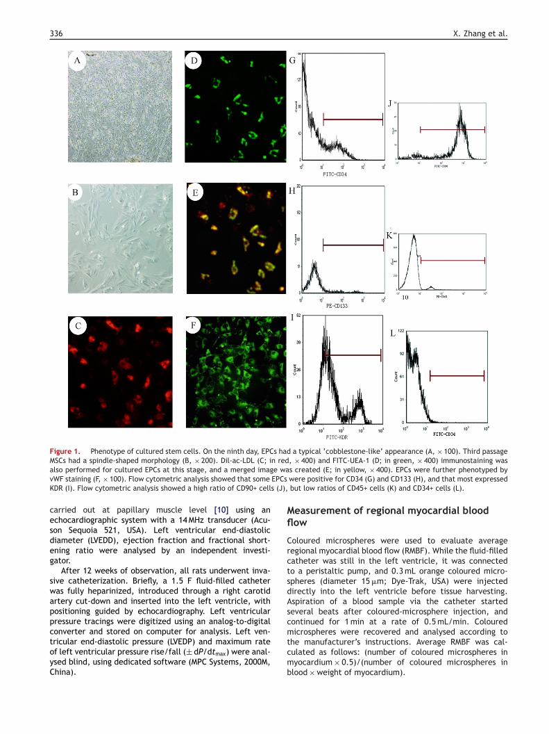
By the ninth day, EPCs had replicated rapidly, displaying atypical ‘cobblestone-like’ appearance (Fig. 1A). Third pas-sage MSCs had a spindle-shaped morphology (Fig. 1B).
At this stage, cultured EPCs were double stained forDil-labelled acetylated low-density lipoprotein (ac-LDL,10 ug/mL, Biomedical Technologies) and fluorescein isothio-cyanate (FITC)-labelled Ulex europaeus agglutinin-1 (UEA-1,1 mg/mL, Sigma), at 37 ◦C for 4 and 1 h, respectively. Thecells took up Dil-ac-LDL (shown in red at 570 nm, Fig. 1C) andwere simultaneously stained positive for FITC-UEA-1 (shownin green at 490 nm, Fig. 1D). Using Photoshop software (ver-sion 7.0) a merged image was created from Fig. 1C andD, with double-positive staining shown in yellow (Fig. 1E),
demonstrating typical EPC properties. The numbers of yel-low and red cells were counted using 10 high-power fieldsof a fluorescent microscope (OLYMPUS IX71-A12FL, Japan).The purity of the EPCs was calculated as follows: (numberof yellow cells/number of red cells) ×100%.e
AId
senchymal stem cells 335
The phenotype of the EPC culture was further demon-trated by immunofluorescence staining, targeting vonillebrand factor (vWF, Biodesign International, USA)
Fig. 1F). More than 83.7% of cultured cells were doubleositive and expressed vWF.
Cultured EPCs and MSCs were analysed by fluorescence-ctivated cell sorting (FACScan flow cytometer, Bectonickinson), with isotype-identical antibodies serving as con-rols. Flow-cytometric analysis showed that on the ninth dayf culture, EPCs expressed CD34 (16.5%, Fig. 1G), CD13313.9%, Fig. 1H) and vascular endothelial growth factoreceptor (VEGF; KDR; 57.8%, Fig. 1I), while third passageSCs were strongly positive for CD90 (90.8%, Fig. 1J), butegative for CD45 (4.6%, Fig. 1K) and CD34 (0.8%, Fig. 1L).
ptimizing stem-cell ratio
o achieve better proliferative activity, the first pas-age EPCs obtained on the ninth day of culture and thehird passage MSCs were cultured in serum-free mediumn 96-well culture plates (200 �L/well) at ratios of 1:21 × 104 EPCs with 2 × 10⁴ MSCs), 1:1 (1.5 × 10⁴ EPCs with.5 × 104 MSCs) and 2:1 (2 × 10⁴ EPCs with 1 × 10⁴ MSCs),espectively. After 72 h, 10 �L of 3-(4,5-dimethylthiazol--yl)-2,5-diphenyltetrazolium bromide (MTT) (5 g/L) weredded to each well and the plates were incubated fornother 4 h. The cell preparation was then mixed with00 �L DMSO and shaken for 10 min, and optical density waseasured at 570 nm. MTT staining showed that most pro-
iferative activity was achieved when EPCs and MSCs wereultured at the 1:1 ratio (optical density = 0.65 ± 0.03 com-ared with 0.51 ± 0.05 for the 1:2 ratio and 0.43 ± 0.05 forhe 2:1 ratio; P < 0.05, respectively). Therefore, on the dayf cell transplantation, first passage EPCs obtained on theinth day were mixed with third passage MSCs at a ratio of:1 (1 × 10⁶ EPCs and 1 × 10⁶ MSCs) for delivery.
tem-cell administration
our weeks after injection of ISO, rats with apparentmpaired cardiac performance (> 20% decrease in ejec-ion fraction) validated by echocardiography examinationere randomized into five groups, and allocated to receive
aline injection (control group), 200 �L medium alone (EGM-group), 200 �L medium plus 2 × 10⁶ EPCs (EPC group),
00 �L medium plus 2 × 10⁶ MSCs (MSC group), or 200 �Ledium plus a combination of 1 × 10⁶ EPCs and 1 × 10⁶ MSCs
EPC/MSC group).The rats were intubated (with appropriate anaesthesia),
nd ventilated with room air (60 cycles/min) with a tidal vol-me of 1 mL/kg (DW-2000, Shanghai). The heart was exposedy a left thoracotomy. Cultured stem cells were injectedvenly into the left ventricle wall, and the chest was closedn layers. The rats were monitored closely for further 12eeks.
chocardiographic and haemodynamic
valuationt baseline before ISO injection, four weeks afterSO injection and 12 weeks after cell therapy, two-imensional echocardiography and M-mode imaging were
336 X. Zhang et al.
Figure 1. Phenotype of cultured stem cells. On the ninth day, EPCs had a typical ‘cobblestone-like’ appearance (A, × 100). Third passageMSCs had a spindle-shaped morphology (B, × 200). Dil-ac-LDL (C; in red, × 400) and FITC-UEA-1 (D; in green, × 400) immunostaining wasa e wav EPCsK s (J),
cesdeg
swappctoyC
Mfl
CrctsdAsc
lso performed for cultured EPCs at this stage, and a merged imagWF staining (F, × 100). Flow cytometric analysis showed that someDR (I). Flow cytometric analysis showed a high ratio of CD90+ cell
arried out at papillary muscle level [10] using anchocardiographic system with a 14 MHz transducer (Acu-on Sequoia 521, USA). Left ventricular end-diastoliciameter (LVEDD), ejection fraction and fractional short-ning ratio were analysed by an independent investi-ator.
After 12 weeks of observation, all rats underwent inva-ive catheterization. Briefly, a 1.5 F fluid-filled catheteras fully heparinized, introduced through a right carotidrtery cut-down and inserted into the left ventricle, withositioning guided by echocardiography. Left ventricularressure tracings were digitized using an analog-to-digital
onverter and stored on computer for analysis. Left ven-ricular end-diastolic pressure (LVEDP) and maximum ratef left ventricular pressure rise/fall (± dP/dtmax) were anal-sed blind, using dedicated software (MPC Systems, 2000M,hina).mtcmb
s created (E; in yellow, × 400). EPCs were further phenotyped bywere positive for CD34 (G) and CD133 (H), and that most expressedbut low ratios of CD45+ cells (K) and CD34+ cells (L).
easurement of regional myocardial bloodow
oloured microspheres were used to evaluate averageegional myocardial blood flow (RMBF). While the fluid-filledatheter was still in the left ventricle, it was connectedo a peristaltic pump, and 0.3 mL orange coloured micro-pheres (diameter 15 �m; Dye-Trak, USA) were injectedirectly into the left ventricle before tissue harvesting.spiration of a blood sample via the catheter startedeveral beats after coloured-microsphere injection, andontinued for 1 min at a rate of 0.5 mL/min. Coloured
icrospheres were recovered and analysed according tohe manufacturer’s instructions. Average RMBF was cal-ulated as follows: (number of coloured microspheres inyocardium × 0.5)/(number of coloured microspheres inlood × weight of myocardium).

d mesenchymal stem cells 337
instructions, on tissue slices selected at random. Tissue sec-tions were examined microscopically at × 400 magnificationand greater than or equal to 100 cells were counted in 10fields (OLYMPUS BH2, Japan). The apoptotic index was cal-culated as follows: (number of apoptotic cells/total numberof cells) × 100%.
Quantification of angiogenic growth factorprotein-expression levels
To assess the level of growth factor secretion, the randomly-selected heart tissue slices were crushed in ethanoic acidand centrifuged, and the supernatant was collected. VEGF(RD, USA), basic fibroblast growth factor (b-FGF, RD, USA)and angiopoietin-2 (Ang-2, ADL, USA) were measured byELISA assay, according to the manufacturer’s instructions.
Quantification of Sry gene and angiogenicgrowth factor mRNA expression levels
Two-step real time RT-PCR, using SYBR® Green (Molecu-lar Probes) as a dye, was carried out to analyse levels ofVEGF, b-FGF, Ang-2 and Sry gene mRNA expression. Trizon(Gibco BRL,CA) was used to isolate mRNA, which was treatedwith DNase I and reverse-transcribed with random hex-amers (Invitrogen), using Moloney murine leukaemia virus.Glyceraldehyde-3-phosphate dehydrogenase (GAPDH) mRNAfrom the same tissue lysate was used as an internal control.The primer pairs for PCR amplification, targeting each spe-cific gene, were designed using Primer Express 1.5 software(Applied Biosystems); the sequences are shown in Table 1.Real time RT-PCR was performed using the ABI 7000 system(Applied Biosystems, USA) with the following cycle condi-tions: 45 cycles of 30 s at 95 ◦C for denaturation, 45 s at 59 ◦Cfor annealing and 50 s at 72 ◦C for extension. Using ABI Prism7300 SDS Software, ��CT methods were used to quantifythe mRNA expression level of each target gene.
Statistical analysis
Numerical values are presented as the mean ± standarddeviation (S.D.). Comparisons between the five groups weremade using a one-way analysis of variance, followed bythe Scheffe-multiple-comparison test. Analyses were per-formed using SPSS version 10.0 statistical software (SPSS
Table 1 Primer pairs for angiogenic growth factors andthe Sry gene.
Gene Primers
GAPDH F 5′GGCATCCTGACCCTGAAGTA 3′
R 5′GGGGTGTTGAAGGTCTCAAA 3′
rat Sry gene F 5′ GGCTTCAAAGTAGATTAGTTGGG 3′
R 5′ ATGCATTCATGGGGCGCTTGAC 3′
b-FGF F 5′ CGACCCACACGTCAAACTA 3′
R 5′ AGCAGCCGTCCATCTTCCT 3′
Combined transplantation of endothelial progenitor cells an
Tissue harvesting
At the end of the 12-week observation period, the ratswere sacrificed with an overdose of anaesthetic (ketamine300 mg/kg) and the hearts were harvested. The left ven-tricle was sliced, perpendicular to the septum, into foursections, each approximately 4—5 mm thick. Because ofthe patchiness of the areas of myocardial infarction, andto avoid sample selection bias, sections were selectedrandomly for further analysis, which included immunostain-ing, RMBF, enzyme-linked immunosorbent assay (ELISA) andreal-time reverse transcriptase-polymerase chain reaction(RT-PCR). Tissue slices for RMBF and immunostaining werekept in 4% formalin, and those for ELISA and real-time RT-PCR were snap frozen and stored at −80 ◦C.
Assessment of engraftment and celldifferentiation
Transplanted male cells in female rat hearts were detectedusing fluorescence in situ hybridization (FISH) on paraffin-embedded tissue slices selected at random. A probe specificfor the rat Y chromosome Sry gene was synthesized (HaoYangCo, China), with the sequence 5′-ATAGT GTGTA GGTTGTTGTC CCATT GCAGC-3′. Sections were deparaffinized anddenatured at 95 ◦C for 10 min, chilled on ice for 5 min,then incubated with the probe at 37 ◦C for 12 h. Nucleistained with 5′-FAM-NHS (flavo-green) were detected at490 nm.
Engrafted Sry-positive cells were phenotyped usingimmunofluorescence staining with an antibody againsteither cardiac troponin T (monoclonal mouse anti-rat,Serotec, British) or vWF (rabbit polyclonal, Dako, Den-mark), further characterizing the transplanted stem cellson the adjacent slice of FISH tissue section. Phycoerythrin-conjugated IgG antibody (BD, USA) was used as a secondaryantibody.
Evaluation of myocardial fibrosis
Cardiac muscle fibrosis was detected using Masson’strichrome staining of tissue slices selected at random. Foreach slice, 10 randomly selected fields were captured (mag-nification × 100) and images were digitized and analysedwith a digital image analyser (MIQAS, Qiuwei Co, China).
Quantification of myocardial capillary density
Capillaries in the myocardium were semiquantified usingimmunohistochemical staining of endothelial cells with apolyclonal rabbit anti-vWF (Dako, Denmark), in tissue slicesselected at random. A capillary vessel was defined as havinga diameter < 20 �m. The number of capillaries was countedunder a light microscope (magnification × 250, OLYMPUSBH2, Japan) for 10 random fields in each transverse sliceand presented as the mean number of blood vessels per unitarea (preset at mm2).
Detection and quantification of apoptotic cells
Apoptotic cells were detected using tunnel staining (Roche,Germany), performed according to the manufacturer’s
VEGF F 5′ GTTCGAGGAAAGGGAAAGGGTC 3′
R 5′ GCGAGTCTGTGTTTTTGCAGGA 3′
Ang-2 F 5′GATGGCAGCGTTGATTTTCA 3′
R 5′ACATGCATCAAACCACTGGC 3′

338 X. Zhang et al.
Figure 2. FISH was performed on tissue slices selected at random from each group. No Sry-positive cells were present in the control group( tecte( 1, × 4× lso po
Is
R
Rm
Frdddwsv7tttf7
Et
Tttwa
c(tf
t0ofgt
C
AdfsaegEtsaai
A, × 400) or the EGM-2 group (B, × 400). Sry-positive cells were deD, × 200) and the EPC group (E, × 400). Some Sry-positive cells (D
400). Some Sry-positive cells (E1, × 400) in the EPC group were a
nc. Chicago, USA). A P-value less than 0.05 was consideredtatistically significant.
esults
at model of isoproterenol-inducedyocardial injury
ifty-five of 70 female rats survived ISO injection. Fiveats were excluded from the study because of insufficientecrease in cardiac performance. Increased LVEDD withecreased ejection fraction and fractional shortening wasemonstrated by echocardiography in 50 female rats, foureeks after ISO injection (LVEDD, pre 0.45 ± 0.05 cm ver-
us post 0.67 ± 0.04 cm; ejection fraction, pre 77.47 ± 1.97%ersus post 56.16 ± 4.23%; fractional shortening, pre3.20 ± 6.30% versus post 36.69 ± 2.23%; P < 0.05, respec-ively). These 50 rats were divided randomly into the fivereatment groups (10 per group). After stem-cell transplan-ation, the final numbers in each treatment group were asollows: control, 6; EGM-2, 7; MSC, 7; EPC, 8; and EPC/MSC,.
ngraftment and differentiation ofransplanted cells
welve weeks after stem-cell transplantation, FISH revealedhat Sry-positive cells were not present in heart tissue inhe control group (Fig. 2A) or the EGM-2 group (Fig. 2B), butere present in heart tissue in the EPC/MSC group (Fig. 2C)nd the MSC group (Fig. 2D). In the EPC group, Sry-positive
wanta
d in cardiac tissue in the EPC/MSC group (C, × 400), the MSC group00) in the MSC group were also positive for cardiac troponin T (D2sitive for vWF (E2, × 400).
ells were found in blood vessels (Fig. 2E). Sry-positive cellsFig. 2D1) from the MSC group were shown to express cardiacroponin T (Fig. 2D2). Similarly, Sry-positive cells (Fig. 2E1)rom the EPC group were shown to express vWF (Fig. 2E2).
Real time RT-PCR analysis showed that expression ofhe Sry gene was higher in the EPC/MSC group (��CT,
.32 ± 0.04%) than in the EPC group (0.21 ± 0.07%; P < 0.05)r the MSC group (0.19 ± 0.06%; P < 0.05). There was no dif-erence in Sry gene expression between the EPC and MSCroups (P > 0.05). Sry gene expression was not detectable inhe EGM-2 and control groups.
ardiac function and haemodynamics
s summarized in Table 2, the EGM-2 and control groupsid not show a significant improvement in cardiac per-ormance, 12 weeks after cell transplantation. However,imilar degrees of improvement were seen in the MSCnd EPC groups, with regard to fractional shortening,jection fraction and LVEDD (P < 0.05, respectively). Thereatest improvement in cardiac function was seen in thePC/MSC group (P < 0.05) although the differences betweenhe EPC/MSC group and the MSC and EPC groups were nottatistically different in terms of LVEDD (P > 0.05). Intrac-rdiac pressure recordings showed that the maximum risend fall in left ventricular pressure (±dP/dtmax) and LVEDPmproved significantly in the MSC and EPC groups compared
ith the control and EGM-2 groups (P < 0.05, respectively),nd that the difference in improvement was even more pro-ounced in the EPC/MSC group (P < 0.05, respectively); thesehree variables were not significantly different in the EPCnd MSC groups (P > 0.05, respectively).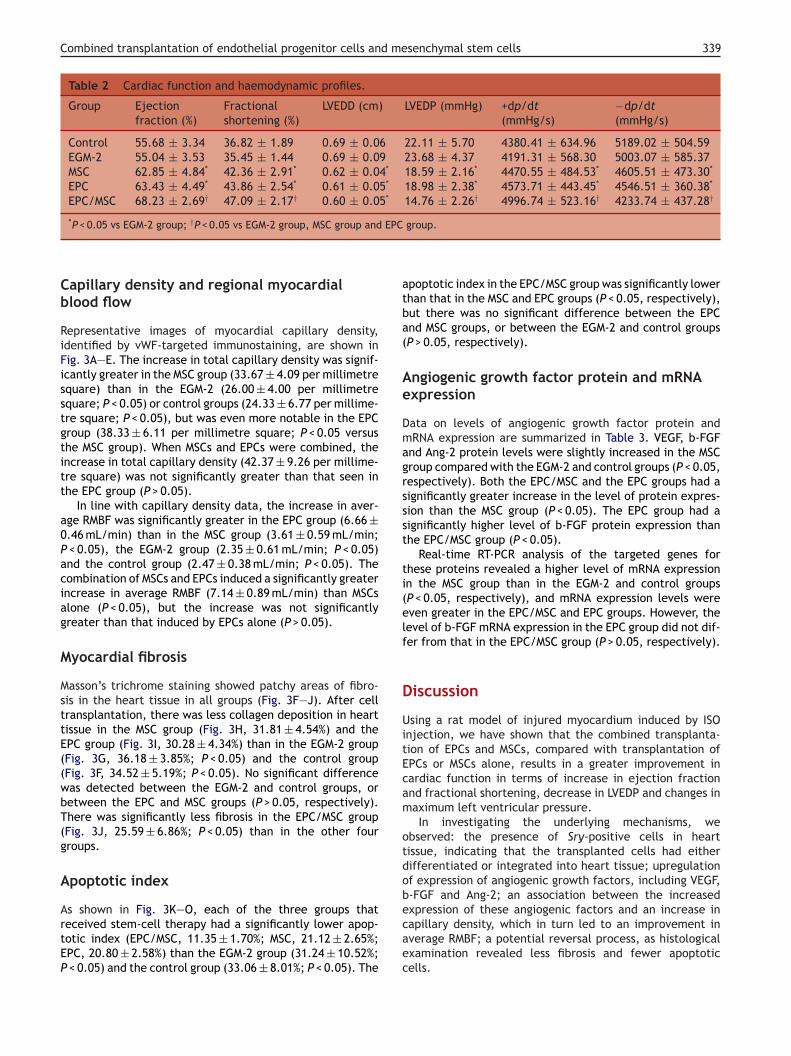
Combined transplantation of endothelial progenitor cells and mesenchymal stem cells 339
Table 2 Cardiac function and haemodynamic profiles.
Group Ejectionfraction (%)
Fractionalshortening (%)
LVEDD (cm) LVEDP (mmHg) +dp/dt(mmHg/s)
−dp/dt(mmHg/s)
Control 55.68 ± 3.34 36.82 ± 1.89 0.69 ± 0.06 22.11 ± 5.70 4380.41 ± 634.96 5189.02 ± 504.59EGM-2 55.04 ± 3.53 35.45 ± 1.44 0.69 ± 0.09 23.68 ± 4.37 4191.31 ± 568.30 5003.07 ± 585.37MSC 62.85 ± 4.84* 42.36 ± 2.91* 0.62 ± 0.04* 18.59 ± 2.16* 4470.55 ± 484.53* 4605.51 ± 473.30*
EPC 63.43 ± 4.49* 43.86 ± 2.54* 0.61 ± 0.05* 18.98 ± 2.38* 4573.71 ± 443.45* 4546.51 ± 360.38*
EPC/MSC 68.23 ± 2.69† 47.09 ± 2.17† 0.60 ± 0.05* 14.76 ± 2.26† 4996.74 ± 523.16† 4233.74 ± 437.28†
EPC
atba(
Ae
Dmagrssst
ti(elf
D
UitEcam
otdob
*P < 0.05 vs EGM-2 group; †P < 0.05 vs EGM-2 group, MSC group and
Capillary density and regional myocardialblood flow
Representative images of myocardial capillary density,identified by vWF-targeted immunostaining, are shown inFig. 3A—E. The increase in total capillary density was signif-icantly greater in the MSC group (33.67 ± 4.09 per millimetresquare) than in the EGM-2 (26.00 ± 4.00 per millimetresquare; P < 0.05) or control groups (24.33 ± 6.77 per millime-tre square; P < 0.05), but was even more notable in the EPCgroup (38.33 ± 6.11 per millimetre square; P < 0.05 versusthe MSC group). When MSCs and EPCs were combined, theincrease in total capillary density (42.37 ± 9.26 per millime-tre square) was not significantly greater than that seen inthe EPC group (P > 0.05).
In line with capillary density data, the increase in aver-age RMBF was significantly greater in the EPC group (6.66 ±0.46 mL/min) than in the MSC group (3.61 ± 0.59 mL/min;P < 0.05), the EGM-2 group (2.35 ± 0.61 mL/min; P < 0.05)and the control group (2.47 ± 0.38 mL/min; P < 0.05). Thecombination of MSCs and EPCs induced a significantly greaterincrease in average RMBF (7.14 ± 0.89 mL/min) than MSCsalone (P < 0.05), but the increase was not significantlygreater than that induced by EPCs alone (P > 0.05).
Myocardial fibrosis
Masson’s trichrome staining showed patchy areas of fibro-sis in the heart tissue in all groups (Fig. 3F—J). After celltransplantation, there was less collagen deposition in hearttissue in the MSC group (Fig. 3H, 31.81 ± 4.54%) and theEPC group (Fig. 3I, 30.28 ± 4.34%) than in the EGM-2 group(Fig. 3G, 36.18 ± 3.85%; P < 0.05) and the control group(Fig. 3F, 34.52 ± 5.19%; P < 0.05). No significant differencewas detected between the EGM-2 and control groups, orbetween the EPC and MSC groups (P > 0.05, respectively).There was significantly less fibrosis in the EPC/MSC group(Fig. 3J, 25.59 ± 6.86%; P < 0.05) than in the other fourgroups.
Apoptotic index
As shown in Fig. 3K—O, each of the three groups thatreceived stem-cell therapy had a significantly lower apop-totic index (EPC/MSC, 11.35 ± 1.70%; MSC, 21.12 ± 2.65%;EPC, 20.80 ± 2.58%) than the EGM-2 group (31.24 ± 10.52%;P < 0.05) and the control group (33.06 ± 8.01%; P < 0.05). The
ecaec
group.
poptotic index in the EPC/MSC group was significantly lowerhan that in the MSC and EPC groups (P < 0.05, respectively),ut there was no significant difference between the EPCnd MSC groups, or between the EGM-2 and control groupsP > 0.05, respectively).
ngiogenic growth factor protein and mRNAxpression
ata on levels of angiogenic growth factor protein andRNA expression are summarized in Table 3. VEGF, b-FGF
nd Ang-2 protein levels were slightly increased in the MSCroup compared with the EGM-2 and control groups (P < 0.05,espectively). Both the EPC/MSC and the EPC groups had aignificantly greater increase in the level of protein expres-ion than the MSC group (P < 0.05). The EPC group had aignificantly higher level of b-FGF protein expression thanhe EPC/MSC group (P < 0.05).
Real-time RT-PCR analysis of the targeted genes forhese proteins revealed a higher level of mRNA expressionn the MSC group than in the EGM-2 and control groupsP < 0.05, respectively), and mRNA expression levels wereven greater in the EPC/MSC and EPC groups. However, theevel of b-FGF mRNA expression in the EPC group did not dif-er from that in the EPC/MSC group (P > 0.05, respectively).
iscussion
sing a rat model of injured myocardium induced by ISOnjection, we have shown that the combined transplanta-ion of EPCs and MSCs, compared with transplantation ofPCs or MSCs alone, results in a greater improvement inardiac function in terms of increase in ejection fractionnd fractional shortening, decrease in LVEDP and changes inaximum left ventricular pressure.In investigating the underlying mechanisms, we
bserved: the presence of Sry-positive cells in heartissue, indicating that the transplanted cells had eitherifferentiated or integrated into heart tissue; upregulationf expression of angiogenic growth factors, including VEGF,-FGF and Ang-2; an association between the increased
xpression of these angiogenic factors and an increase inapillary density, which in turn led to an improvement inverage RMBF; a potential reversal process, as histologicalxamination revealed less fibrosis and fewer apoptoticells.
340 X. Zhang et al.
F —E),c tainina
awaoucasIwamco
Iafdttae
p
igure 3. Immunostaining against vWF to identify capillaries (Aollagen stained in green, myocardium stained in red) and Tunnel snd EPC/MSC groups, respectively.
The pathological process of ISO-induced myocardial dam-ge is characterized by patchy areas of myocardial infarctionith well-maintained coronary vasculature. This model haslso been shown to exhibit dose-dependent progressionf postmyocardial infarction remodelling [8]. Although thenderlying mechanism is not fully understood, there areertain similarities with traditional myocardial infarctionttributable to ischaemic coronary disease, as oxidativetress is the main trigger for cardiac injury [7]. Using thisSO-induced myocardial injury model for the first time, we
ere able to demonstrate that the combination of EPCsnd MSCs may induce neovascularization and hence aug-ent cardiac performance. Our reasons for undertakingombined transplantation of EPCs and MSCs were basedn the different biological properties of these stem cells.
dpnww
Table 3 Angiogenic growth factor protein and mRNA expressi
Group VEGF
protein (pg/mg) mRNA (%) protein (pg/m
Control 110.03 ± 4.31 2.91 ± 1.53 426.87 ± 25.4EGM-2 121.38 ± 2.93 3.21 ± 1.10 415.30 ± 37.6MSC 134.48 ± 4.88* 4.93 ± 2.10* 545.50 ± 65.7EPC 154.25 ± 32.91† 7.97 ± 2.11† 972.41 ± 204.EPC/MSC 185.75 ± 13.20‡ 27.05 ± 1.84‡ 732.85 ± 51.2
*P < 0.05 vs EGM-2 group; †P < 0.05 vs EGM-2 group and MSC group; ‡P <
Masson’s trichrome staining to detect myocardial fibrosis (F—J;g to detect apoptotic cells (K—O) in the control, EGM-2, MSC, EPC
mproved myocardial contractility has been observed inclinical trial where both EPCs and MSCs were used
or transcoronary transplantation in patients with myocar-ial infarction [11]. The mechanism by which combinedransplantation of EPCs and MSCs improves cardiac func-ion significantly is not fully understood, but we havecquired a number of pieces of evidence that support thisffect.
Firstly, we found evidence of transplanted cells incor-orated into cardiac tissue. Sry-positive cells may have
ifferentiated into cardiac tissue cells, as they exhibitedositive immunostaining for troponin T, although we can-ot exclude the possibility that these MSCs had mergedith pre-existing cardiac tissue cells. The same phenomenonas observed for EPCs implanted into cardiac tissue, ason.
b-FGF Ang-2
g) mRNA (%) protein (ng/mg) mRNA (%)
6 11.86 ± 3.37 1.18 ± 0.24 9.93 ± 6.796 12.46 ± 2.14 1.01 ± 0.10 9.81 ± 4.836* 17.81 ± 5.65* 1.31 ± 0.10* 12.84 ± 4.10*
21† 38.66 ± 14.50† 1.46 ± 0.05† 18.40 ± 6.97†
5‡ 36.43 ± 12.68† 1.73 ± 0.15‡ 32.01 ± 13.80‡
0.05 vs EGM-2 group, MSC group and EPC group.

d me
ttcdtittfmtial
R
[
[
[
Combined transplantation of endothelial progenitor cells an
Sry-positive cells detected by FISH were also positive forimmunofluorescence staining against vWF in the EPC group.
In addition, the levels of angiogenic growth factor proteinand mRNA expression were higher in the EPC and EPC/MSCgroups than in MSC group. The factors in question (VEGF,b-FGF and Ang-2) have been shown to augment neovascular-ization and hence increase capillary density [12]. b-FGF is apowerful mitogen that stimulates the migration and prolifer-ation of various vascular cell types, including smooth-musclecells, endothelial cells and fibroblasts [13]. Ang-2 can stim-ulate the degradation of the base membrane by proteasessecreted by activated endothelial cells, which aids migra-tion, proliferation, and the formation of solid endothelialcell sprouts into the stromal space [14]. We were surprisedto observe a higher level of b-FGF protein expression in theEPC group than in the EPC/MSC group, but a similar levelof b-FGF mRNA expression in these two groups. This dif-ference in post-translational regulation might be due to theinteraction between EPCs and MSCs; the detailed underlyingmechanism requires further investigation.
The beneficial effect of the increase in angiogenic fac-tors was evidenced by the higher Sry gene expression levelsdetected by real time RT-PCR, indicating a synergetic effectof the combination therapy in enhancing either engraftmentrate or possibly proliferative activity.
Finally, we observed the lowest level of fibrosis depo-sition in cardiac tissue in the EPC/MSC group, indicating apotential reversal of cardiomyopathy. We did not investi-gate the mechanism underlying this effect but propose thatit may have been associated with the paracrine effects ofthe angiogenic factors secreted by transplanted cells andthe lower numbers of apoptotic cells.
Recently, Suuronen et al. reported that the transplanta-tion of EPCs resulted in better cardiac function, increasedarteriole density and less myocardial fibrosis in a rat modelof myocardial infarction than the transplantation of MSCs,or EPCs plus MSCs [15]. These results are strikingly differentfrom our data, and can be attributed to a number of factors:a much shorter observation period post-transplantation; theuse of far fewer EPCs and MSCs; a relatively superior baselinecardiac performance before cell transplantation (relativelysmaller decrease in cardiac function compared with baselinebefore coronary ligation); and the use of a different animalmodel, involving different pathological processes.
Given the immunogenic properties of EPCs, it is advisablethat the immunosuppressant cyclosporine A is adminis-tered. However, it is still uncertain whether cyclosporineA enhances the engraftment rate of cell therapy [16], andone report has shown that it can even suppress the differ-entiation process [17].
In conclusion, our study has shown for the first timethat the intramyocardial infusion of culture-expanded EPCstogether with MSCs may represent a novel and more effi-cient therapeutic strategy for the treatment of ISO-inducedcardiomyopathy.
Limitations of the study
Our study was limited by the small sample size. In addi-tion, we should have attempted to use BMNCs as controlcells. Also, we were unable to label all the stem cells before
[
senchymal stem cells 341
ransplantation, to enable us to identify the cells in vivo;his made the evaluation of engrafted stem cells more diffi-ult. The ISO-induced cardiac injury model also has its ownrawback, characterized by patchy areas of necrosis ratherhan a fixed risk area, which can make results difficult tonterpret. Nevertheless, the data showed that the combinedransplantation of EPCs and MSCs into a rat model of isopro-erenol (ISO)-induced injured myocardium improved cardiacunction after 12 weeks. We excluded all rats with a drop inyocardial performance of less or equal to 20% to minimize
he variability inherent in this animal model. Further studys warranted to elucidate detailed underlying mechanisms,nd an extended observation period is required to validateong-term outcome.
eferences
[1] Lopez AD, Murray CC. The global burden of disease,1990—2020. Nat Med 1998;4(11):1241—3.
[2] Schuh A, Liehn EA, Sasse A, et al. Transplantation of endothelialprogenitor cells improves neovascularization and left ventric-ular function after myocardial infarction in a rat model. BasicRes Cardiol 2008;103(1):69—77.
[3] Berry MF, Engler AJ, Woo YJ, et al. Mesenchymal stemcell injection after myocardial infarction improvesmyocardial compliance. Am J Physiol Heart Circ Physiol2006;290(6):H2196—203.
[4] Dai W, Hale SL, Martin BJ, et al. Allogeneic mesenchymal stemcell transplantation in postinfarcted rat myocardium: short-and long-term effects. Circulation 2005;112(2):214—23.
[5] Kudo FA, Nishibe T, Nishibe M, et al. Autologous transplanta-tion of peripheral blood endothelial progenitor cells (CD34+)for therapeutic angiogenesis in patients with critical limbischemia. Int Angiol 2003;22(4):344—8.
[6] Gruh I, Beilner J, Blomer U, et al. No evidence of trans-differentiation of human endothelial progenitor cells intocardiomyocytes after coculture with neonatal rat cardiomy-ocytes. Circulation 2006;113(10):1326—34.
[7] Karthikeyan K, Sarala Bai BR, Niranjali Devaraj S. Grapeseed proanthocyanidins ameliorates isoproterenol-inducedmyocardial injury in rats by stabilizing mitochondrial and lyso-somal enzymes: an in vivo study. Life Sci 2007;81(23—24):1615—21.
[8] Teerlink JR, Pfeffer JM, Pfeffer MA. Progressive ventricu-lar remodeling in response to diffuse isoproterenol-inducedmyocardial necrosis in rats. Circ Res 1994;75(1):105—13.
[9] Pittenger MF, Mackay AM, Beck SC, et al. Multilineagepotential of adult human mesenchymal stem cells. Science1999;284(5411):143—7.
10] Sahn DJ, DeMaria A, Kisslo J, et al. Recommendationsregarding quantitation in M-mode echocardiography: resultsof a survey of echocardiographic measurements. Circulation1978;58(6):1072—83.
11] Katritsis DG, Sotiropoulou PA, Karvouni E, et al. Transcoro-nary transplantation of autologous mesenchymal stem cellsand endothelial progenitors into infarcted human myocardium.Catheter Cardiovasc Interv 2005;65(3):321—9.
12] Gong B, Asimakis GK, Chen Z, et al. Whole-body hyperthermiainduces up-regulation of vascular endothelial growth factoraccompanied by neovascularization in cardiac tissue. Life Sci
2006;79(19):1781—8.13] Nakamae A, Sunagawa T, Ishida O, et al. Acceleration ofsurgical angiogenesis in necrotic bone with a single injec-tion of fibroblast growth factor-2 (FGF-2). J Orthop Res2004;22(3):509—13.

3
[
[
[
42
14] Dal Monte M, Cammalleri M, Martini D, et al. Antiangiogenicrole of somatostatin receptor 2 in a model of hypoxia-inducedneovascularization in the retina: results from transgenic mice.
Invest Ophthalmol Vis Sci 2007;48(8):3480—9.15] Suuronen EJ, Price J, Veinot JP, et al. Comparative effects ofmesenchymal progenitor cells, endothelial progenitor cells, ortheir combination on myocardial infarct regeneration and car-diac function. J Thorac Cardiovasc Surg 2007;134(5):1249—58.
[
X. Zhang et al.
16] Zeng L, Hu Q, Wang X, et al. Bioenergetic and functional con-sequences of bone marrow-derived multipotent progenitor celltransplantation in hearts with postinfarction left ventricular
remodeling. Circulation 2007;115(14):1866—75.17] Davies WR, Wang S, Oi K, et al. Cyclosporine decreases vascu-lar progenitor cell numbers after cardiac transplantation andattenuates progenitor cell growth in vitro. J Heart Lung Trans-plant 2005;24(11):1868—77.
Related Documents