Downloaded from www.medrech.com “Screening for preeclampsia by uterine artery Doppler Velocimetry in the late first trimester of pregnancy” Genovese F. et al., Med. Res. Chron., 2014, 1 (1), 60-72 Medico Research Chronicles, 2014 60 Submitted on: October 2014 Accepted on: October 2014 For Correspondence Email ID: SCREENING FOR PREECLAMPSIA BY UTERINE ARTERY DOPPLER VELOCIMETRY IN THE LATE FIRST TRIMESTER OF PREGNANCY F. Genovese, M. Stracquadanio, A. Ingala, L. Ciotta, A. D’Agati, C. Pafumi, A. Carbonaro, M. A. Palumbo Institute of Obstetric and Gynecological Pathology Santo Bambino Hospital, University of Catania, Catania, Italy Original Research Article Abstract: The Authors prospectively determined the uterine artery Doppler velocimetry (UADV) in 108 women with history of hypertensive disorder of pregnancy (HDP) between the 10 th and 13 th week of pregnancy to identify those at higher risk for recurrent preeclampsia and to evaluate the efficacy of prophylactic pharmacological treatment in this subgroup of patients. Patients with abnormal flow values (RI > 0.58 and any notching) were immediately started on aspirin 100 mg/daily, replaced by daily subcutaneous injection of low molecular weight heparin from the 24th week till delivery. Concerning the maternal morbidity (expressed by abnormal both mean blood pressure and 24-hour proteinuria throughout pregnancy), the Authors found that the average pregnancy course in 20 (18.5%) prophylactic treated patients with altered 1st trimester UADV (group A) was better than in their previous pregnancies, however regarding the above parameters no significant difference (P > 0.05) was found between the patients treated and another cohort of patients (n =20), also extrapolated from the same pool of patients at risk for HDP, but with normal I trimester UADV studies (group B) who were not prophylactic treated. Both groups of patients were followed up from the time of Doppler studies till 6 weeks post partum. The study suggests that UADV in the late first trimester of pregnancy is useful in reducing the risk of preeclampsia in patients with history of HDP, allowing the prompt institution of prophylactic treatment in selected patients. 1. Introduction Preeclampsia (PE) is a multisystemic disorder that provokes a great number of maternal deaths worldwide. [1] It is recognized as the 3rd cause of direct maternal mortality. PE risk factors are: elevated body mass index, maternal age extremes and Afro-American ethnicity; besides, some diseases such as diabetes and chronic hypertension also significantly increase the risk. [2-3] Today it’s well known that preeclampsia is a disease affecting the vascular endotelium. A regularly evolving pregnancy is characterized by a preserved endothelial function and a progressive reduction in the resistance of uterine vessels, due to the placentation process. An inadequate

Welcome message from author
This document is posted to help you gain knowledge. Please leave a comment to let me know what you think about it! Share it to your friends and learn new things together.
Transcript
Downloaded from www.medrech.com
“Screening for preeclampsia by uterine artery Doppler Velocimetry in the late first trimester of pregnancy”
Genovese F. et al., Med. Res. Chron., 2014, 1 (1), 60-72
Med
ico R
esearc
h C
hro
nic
les, 2014
60
Submitted on: October 2014 Accepted on: October 2014 For Correspondence Email ID:
SCREENING FOR PREECLAMPSIA BY UTERINE ARTERY DOPPLE R VELOCIMETRY IN THE LATE FIRST TRIMESTER OF PREGNANC Y
F. Genovese, M. Stracquadanio, A. Ingala, L. Ciotta, A. D’Agati, C. Pafumi, A. Carbonaro, M. A.
Palumbo Institute of Obstetric and Gynecological Pathology Santo Bambino Hospital, University of
Catania, Catania, Italy
Original Research Article
Abstract: The Authors prospectively determined the uterine artery Doppler velocimetry (UADV) in 108 women with history of hypertensive disorder of pregnancy (HDP) between the 10th and 13th week of pregnancy to identify those at higher risk for recurrent preeclampsia and to evaluate the efficacy of prophylactic pharmacological treatment in this subgroup of patients. Patients with abnormal flow values (RI > 0.58 and any notching) were immediately started on aspirin 100 mg/daily, replaced by daily subcutaneous injection of low molecular weight heparin from the 24th week till delivery. Concerning the maternal morbidity (expressed by abnormal both mean blood pressure and 24-hour proteinuria throughout pregnancy), the Authors found that the average pregnancy course in 20 (18.5%) prophylactic treated patients with altered 1st trimester UADV (group A) was better than in their previous pregnancies, however regarding the above parameters no significant difference (P > 0.05) was found between the patients treated and another cohort of patients (n =20), also extrapolated from the same pool of patients at risk for HDP, but with normal I trimester UADV studies (group B) who were not prophylactic treated. Both groups of patients were followed up from the time of Doppler studies till 6 weeks post partum. The study suggests that UADV in the late first trimester of pregnancy is useful in reducing the risk of preeclampsia in patients with history of HDP, allowing the prompt institution of prophylactic treatment in selected patients. 1. Introduction Preeclampsia (PE) is a multisystemic disorder that provokes a great number of maternal deaths worldwide. [1] It is recognized as the 3rd cause of direct maternal mortality. PE risk factors are: elevated body mass index, maternal age extremes and Afro-American ethnicity; besides, some diseases such as diabetes and
chronic hypertension also significantly increase the risk. [2-3] Today it’s well known that preeclampsia is a disease affecting the vascular endotelium. A regularly evolving pregnancy is characterized by a preserved endothelial function and a progressive reduction in the resistance of uterine vessels, due to the placentation process. An inadequate
Downloaded from www.medrech.com
“Screening for preeclampsia by uterine artery Doppler Velocimetry in the late first trimester of pregnancy”
Genovese F. et al., Med. Res. Chron., 2014, 1 (1), 60-72
Med
ico R
esearc
h C
hro
nic
les, 2014
61
trophoblastic invasion and the consequent poor placentation are the key mechanisms of PE physiopathology. [4-7] The systemic endothelial dysfunction, leading to thrombotic phenomenon in the microcirculation, reduces the perfusion of tissues and organs, including the placenta itself, creating an even more hypoxemic environment. Chronic placental hypoxia causes both oxidative stress with consequent placental apoptosis and necrosis and also an increased expression of proinflammatory, antiangiogenic, and angiogenic factors, amplifying the systemic endothelial impairment. A good endothelial function and an optimal vascular dilatation are extremely important for a healthy gestation, without these conditions both maternal and fetal prognosis is compromised. [8] According to the recent Literature [2], perinatal and maternal morbidity and mortality due to pre-eclampsia may be reduced through an early diagnosis and treatment. Prediction of early pre-eclampsia (clinical symptoms before 34 weeks of pregnancy) is possible by searching for some circulating factors PAI (Plasminogen Activator Inhibitor) -1, Fibronectin, PlGF (Placental Growth Factor), VEGF (Vascular Endothelial Growth Factor) and other substances and/or by determining the uterine arteries Doppler velocimetry during the second trimester. The use of UtA-PI alone in the second trimester of pregnancy allows detecting almost 95% of all cases of early PE [10], the uterine arteries bilateral notch is able to outline not only patients that subsequently develop PE, but also those whose pregnancies will be complicated by intrauterine growth restriction (IUGR). [11-12] Determination of Doppler indices, during the late 1st trimester of pregnancy (from 11+0 to 13+6 gestational weeks) may be
even more important in the management of pregnant patients with history of preeclampsia, [9] because it allows an earlier identification of an unbalance between proangiogenic substances, such as PlGF and others, and antiangiogenic factors, such as sVEGFR (Soluble Vascular Endothelium Growth Factor Receptor) -1 and others. A part from the predictive value of the above circulating factors, there is no doubt that a lot of evidence in the Literature support an association between abnormal Doppler indices both in the I and II trimester of pregnancy and adverse pregnancy outcome especially in patient at risk. At this point a question raises spontaneously: what can the clinician do in the presence of abnormal UAD studies to improve pregnancy outcome, besides increasing maternal and fetal surveillance? A good number of medical treatments have been attempted in these cases with controversial results. The aim of this study is not only to conduct a careful quantitative and morphological analysis of the pulsatile waves of uterine arteries in patients with history of HDP (Hypertensive Disorders in Pregnancy) and to eventually submit them to appropriate therapy, but also to evaluate the efficacy of such treatment. 2. Materials & Methods The Authors did a prospective study using 2 cohorts of patients: A and B, extrapolated from a pool of 108 patients with history of hypertensive disorders in pregnancy (HDP), examined at the outpatient clinic of “Ospedale Santo Bambino” in Catania from January 2007 till December 2012.The patients’ mean age and BMI (body mass index) were respectively 30 ± 0,4 years and 25 ± 0,2 kg/m2 ; all women underwent Doppler flowmetry of the uterine arteries between 10 and 13 weeks of pregnancy, using a 3.5 MHz multi frequency curvilinear

Downloaded from www.medrech.com
“Screening for preeclampsia by uterine artery Doppler Velocimetry in the late first trimester of pregnancy”
Genovese F. et al., Med. Res. Chron., 2014, 1 (1), 60-72
Med
ico R
esearc
h C
hro
nic
les, 2014
62
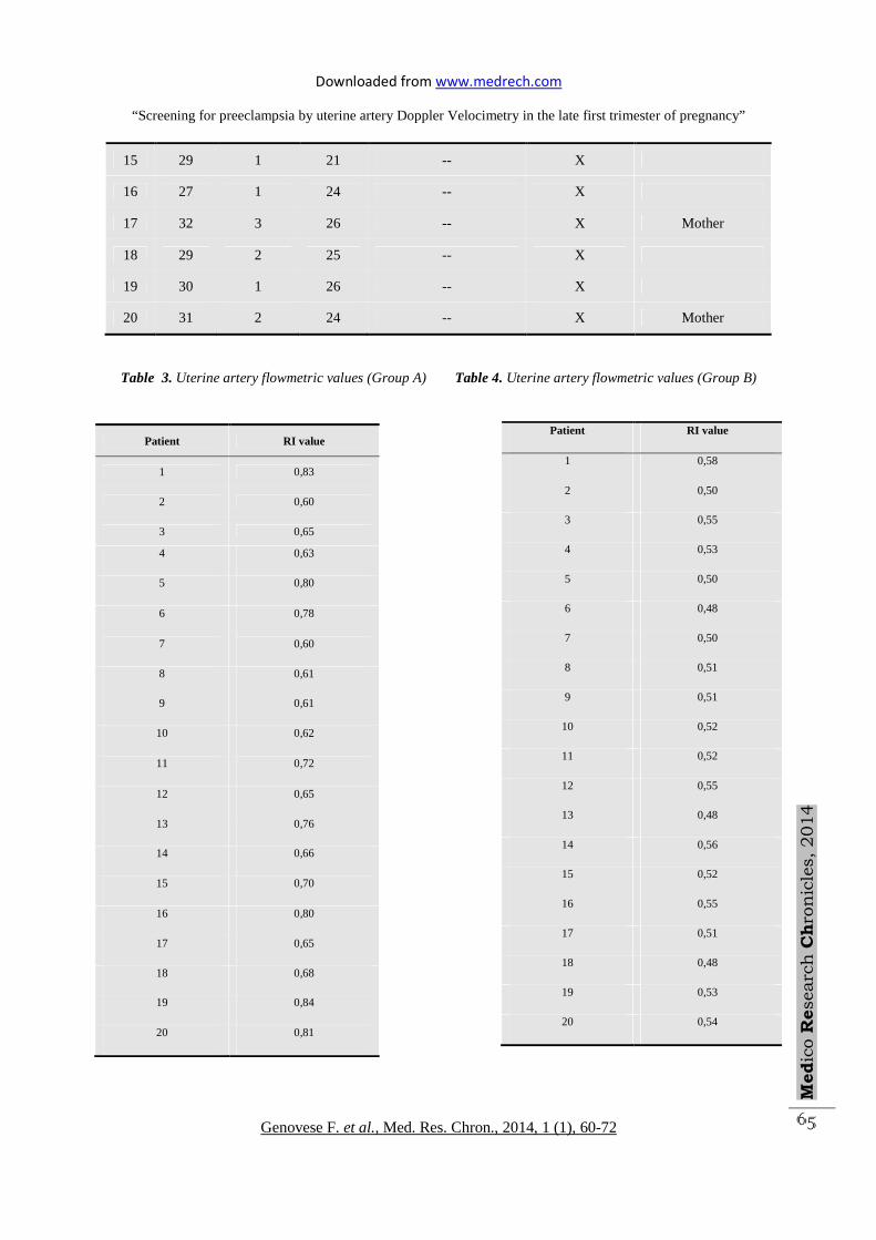
probe. The right and left uterine arteries were identified in an oblique plane of the pelvis at the crossover with the external iliac arteries and the Doppler signals were registered. The Doppler indices outlined, after the appearance of 3 similar consecutive waveforms, were: resistance index and any notching. Resistance index (RI) is defined as peak systolic flow minus end diastolic flow divided by peak systolic flow (A – B) / A. The diastolic notching is expressed by a characteristic waveform indicating decreased early diastolic flow in the uterine artery compared with later diastolic flow. The diastolic wave reduction, resistance index (RI) increase, the image of a "notch” between the systolic and diastolic components would be the expression of increased uterine resistance, which usually represents the first stage in the increase of peripheral resistance (fig.1, fig.2). The examination, if done by an experienced sonographer, is relatively simple and non-invasive, repeatable, and therefore it takes place in a very short time. An increased RI (> 0.58) and a characteristic unit and/or bilateral notch were found (tab. 3) in 20
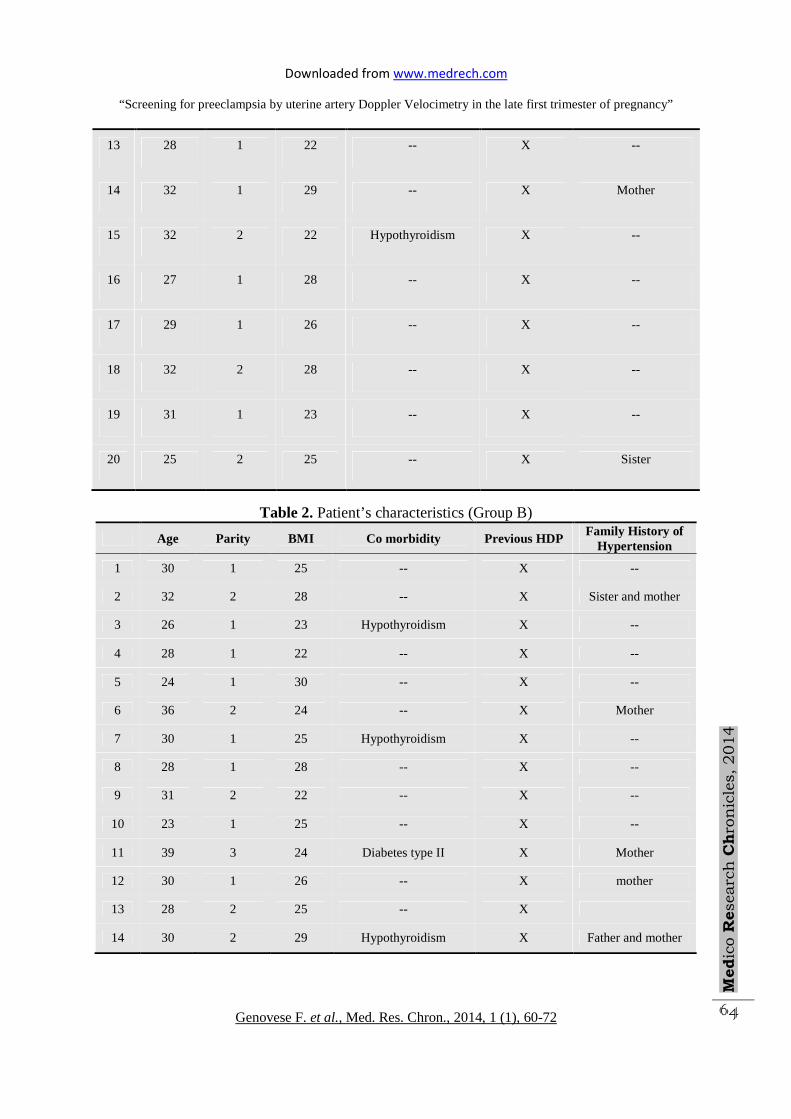
patients (group A, tab. 1), 18,5 % of the total number of women examined, prenatal charts attesting mean B.P. and 24-proteinuria during the pregnancy complicated by the hypertensive disorder were obtained. Among the remaining 88 patients without flowmetric alterations, 20 patients with obstetrical history, age and BMI similar to group A patients, were selected and used as a control cohort defined as group B (tab. 2, 4). Group A patients were started to close clinical and ultrasound follow-up (every 15 days) for the assessment of fetal and maternal wellbeing from the late first trimester till delivery and treated with aspirin (100 mg daily) from the time of ultrasound examination to the 24th week, when they were switched to subcutaneous low molecular weight heparin (4.000 I.U. daily) till delivery and after. During the puerperal hospital stay and 6 weeks postpartum they were also re-evaluated for signs and symptoms of hypertensive disorders. Group B patients, although not subjected to any prophylactic treatment, were also followed up in the same manner as group A patients.
Figure 1. Normal uterine artery Doppler flowmetry
“Screening for preeclampsia by uterine artery Doppler Velocimetry in the late first trimester of pregnancy
Genovese F
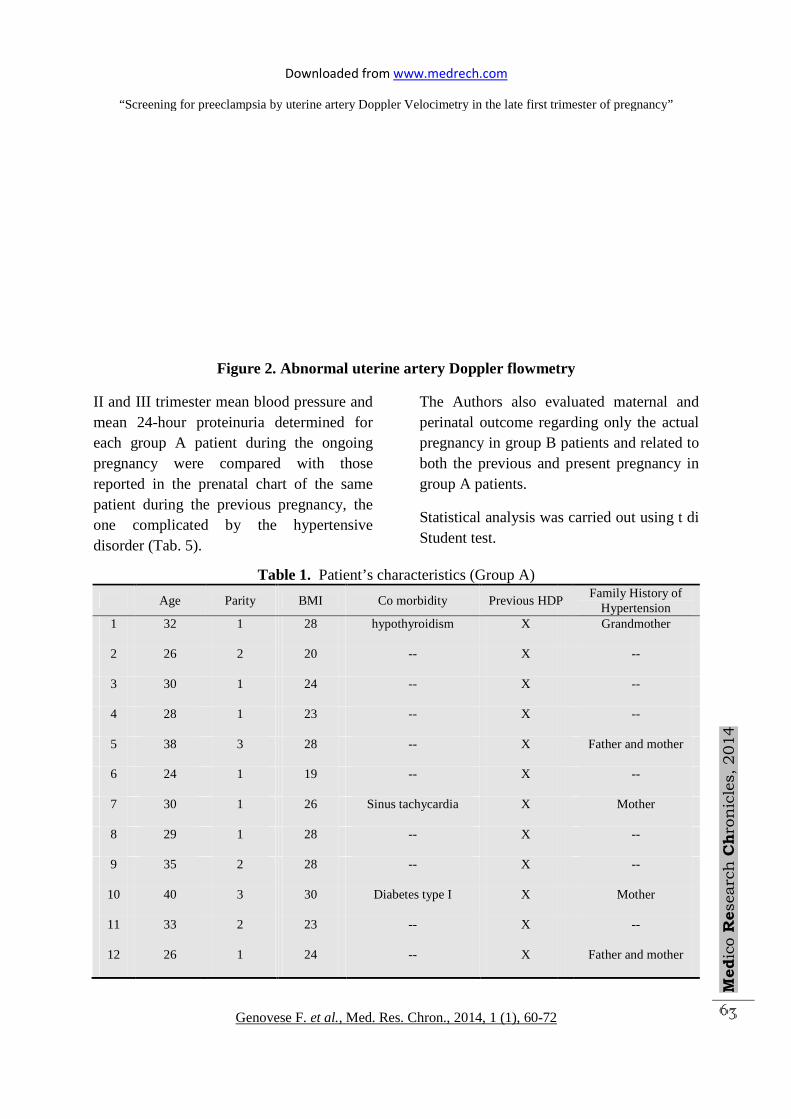
Figure 2. Abnormal uterine artery Doppler flowmetry
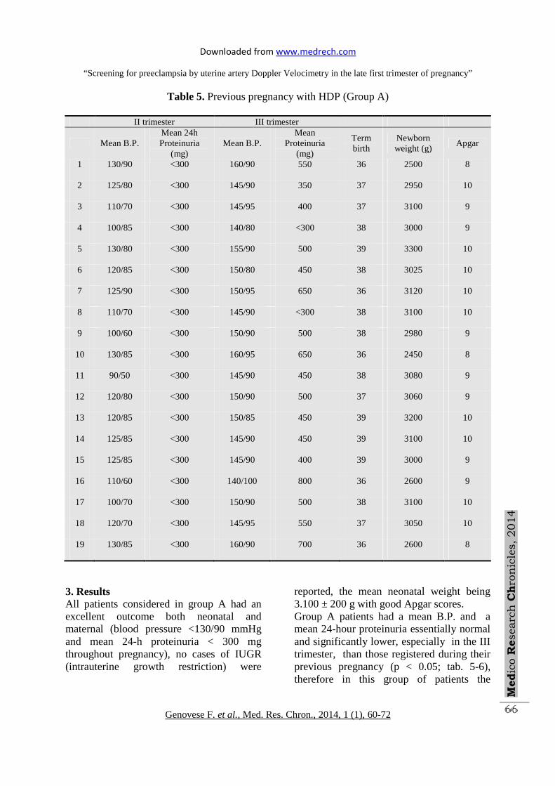
II and III trimester mean blood pressure and mean 24-hour proteinuria determined each group A patient during the ongoing pregnancy were compared with reported in the prenatal chart of the same patient during the previous pregnancy, the one complicated by the hypertensive disorder (Tab. 5).
Table 1.
Age Parity
1 32 1
2 26 2
3 30 1
4 28 1
5 38 3
6 24 1
7 30 1
8 29 1
9 35 2
10 40 3
11 33 2
12 26 1
Downloaded from www.medrech.com
Screening for preeclampsia by uterine artery Doppler Velocimetry in the late first trimester of pregnancy
Genovese F. et al., Med. Res. Chron., 2014, 1 (1), 60-72
Figure 2. Abnormal uterine artery Doppler flowmetry
II and III trimester mean blood pressure and hour proteinuria determined for
group A patient during the ongoing pregnancy were compared with those
in the prenatal chart of the same the previous pregnancy, the
cated by the hypertensive
The Authors also evaluated maternal and perinatal outcome regarding only the actual pregnancy in group B both the previous and present pregnancy in group A patients.
Statistical analysis was carried out using t di Student test.
Table 1. Patient’s characteristics (Group A)
BMI Co morbidity Previous HDP
28 hypothyroidism X
20 -- X
24 -- X
23 -- X
28 -- X
19 -- X
26 Sinus tachycardia X
28 -- X
28 -- X
30 Diabetes type I X
23 -- X
24 -- X
Screening for preeclampsia by uterine artery Doppler Velocimetry in the late first trimester of pregnancy”
Med
ico R
esearc
h C
hro
nic
les, 2014
63
Figure 2. Abnormal uterine artery Doppler flowmetry
The Authors also evaluated maternal and perinatal outcome regarding only the actual pregnancy in group B patients and related to both the previous and present pregnancy in
s was carried out using t di
HDP Family History of
Hypertension Grandmother
--
--
--
Father and mother
--
Mother
--
--
Mother
--
Father and mother
Downloaded from www.medrech.com
“Screening for preeclampsia by uterine artery Doppler Velocimetry in the late first trimester of pregnancy”
Genovese F. et al., Med. Res. Chron., 2014, 1 (1), 60-72
Med
ico R
esearc
h C
hro
nic
les, 2014
64
13 28 1 22 -- X --
14 32 1 29 -- X Mother
15 32 2 22 Hypothyroidism X --
16 27 1 28 -- X --
17 29 1 26 -- X --
18 32 2 28 -- X --
19 31 1 23 -- X --
20 25 2 25 -- X Sister
Table 2. Patient’s characteristics (Group B)
Age Parity BMI Co morbidity Previous HDP Family History of
Hypertension
1 30 1 25 -- X --
2 32 2 28 -- X Sister and mother
3 26 1 23 Hypothyroidism X --
4 28 1 22 -- X --
5 24 1 30 -- X --
6 36 2 24 -- X Mother
7 30 1 25 Hypothyroidism X --
8 28 1 28 -- X --
9 31 2 22 -- X --
10 23 1 25 -- X --
11 39 3 24 Diabetes type II X Mother
12 30 1 26 -- X mother
13 28 2 25 -- X
14 30 2 29 Hypothyroidism X Father and mother
Downloaded from www.medrech.com
“Screening for preeclampsia by uterine artery Doppler Velocimetry in the late first trimester of pregnancy”
Genovese F. et al., Med. Res. Chron., 2014, 1 (1), 60-72
Med
ico R
esearc
h C
hro
nic
les, 2014
65
15 29 1 21 -- X
16 27 1 24 -- X
17 32 3 26 -- X Mother
18 29 2 25 -- X
19 30 1 26 -- X
20 31 2 24 -- X Mother
Table 3. Uterine artery flowmetric values (Group A) Table 4. Uterine artery flowmetric values (Group B)
Patient RI value
1 0,83
2 0,60
3 0,65
4 0,63
5 0,80
6 0,78
7 0,60
8 0,61
9 0,61
10 0,62
11 0,72
12 0,65
13 0,76
14 0,66
15 0,70
16 0,80
17 0,65
18 0,68
19 0,84
20 0,81
Patient RI value
1 0,58
2 0,50
3 0,55
4 0,53
5 0,50
6 0,48
7 0,50
8 0,51
9 0,51
10 0,52
11 0,52
12 0,55
13 0,48
14 0,56
15 0,52
16 0,55
17 0,51
18 0,48
19 0,53
20 0,54
Downloaded from www.medrech.com
“Screening for preeclampsia by uterine artery Doppler Velocimetry in the late first trimester of pregnancy”
Genovese F. et al., Med. Res. Chron., 2014, 1 (1), 60-72
Med
ico R
esearc
h C
hro
nic
les, 2014
66
Table 5. Previous pregnancy with HDP (Group A)
II trimester III trimester
Mean B.P. Mean 24h Proteinuria
(mg) Mean B.P.
Mean Proteinuria
(mg)
Term birth
Newborn weight (g)
Apgar
1 130/90 <300 160/90 550 36 2500 8
2 125/80 <300 145/90 350 37 2950 10
3 110/70 <300 145/95 400 37 3100 9
4 100/85 <300 140/80 <300 38 3000 9
5 130/80 <300 155/90 500 39 3300 10
6 120/85 <300 150/80 450 38 3025 10
7 125/90 <300 150/95 650 36 3120 10
8 110/70 <300 145/90 <300 38 3100 10
9 100/60 <300 150/90 500 38 2980 9
10 130/85 <300 160/95 650 36 2450 8
11 90/50 <300 145/90 450 38 3080 9
12 120/80 <300 150/90 500 37 3060 9
13 120/85 <300 150/85 450 39 3200 10
14 125/85 <300 145/90 450 39 3100 10
15 125/85 <300 145/90 400 39 3000 9
16 110/60 <300 140/100 800 36 2600 9
17 100/70 <300 150/90 500 38 3100 10
18 120/70 <300 145/95 550 37 3050 10
19 130/85 <300 160/90 700 36 2600 8
3. Results All patients considered in group A had an excellent outcome both neonatal and maternal (blood pressure <130/90 mmHg and mean 24-h proteinuria < 300 mg throughout pregnancy), no cases of IUGR (intrauterine growth restriction) were
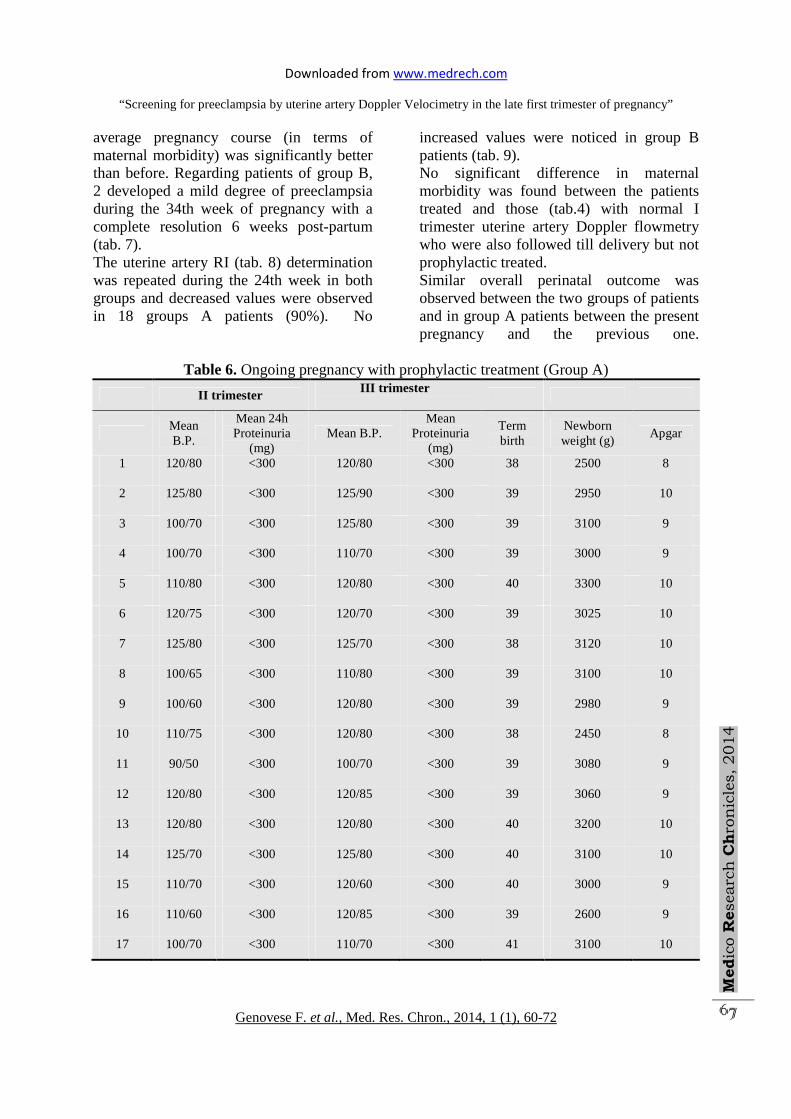
reported, the mean neonatal weight being 3.100 ± 200 g with good Apgar scores. Group A patients had a mean B.P. and a mean 24-hour proteinuria essentially normal and significantly lower, especially in the III trimester, than those registered during their previous pregnancy (p < 0.05; tab. 5-6), therefore in this group of patients the
Downloaded from www.medrech.com
“Screening for preeclampsia by uterine artery Doppler Velocimetry in the late first trimester of pregnancy”
Genovese F. et al., Med. Res. Chron., 2014, 1 (1), 60-72
Med
ico R
esearc
h C
hro
nic
les, 2014
67
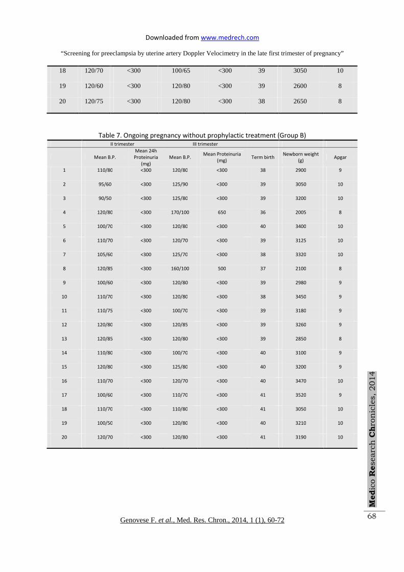
average pregnancy course (in terms of maternal morbidity) was significantly better than before. Regarding patients of group B, 2 developed a mild degree of preeclampsia during the 34th week of pregnancy with a complete resolution 6 weeks post-partum (tab. 7). The uterine artery RI (tab. 8) determination was repeated during the 24th week in both groups and decreased values were observed in 18 groups A patients (90%). No
increased values were noticed in group B patients (tab. 9). No significant difference in maternal morbidity was found between the patients treated and those (tab.4) with normal I trimester uterine artery Doppler flowmetry who were also followed till delivery but not prophylactic treated. Similar overall perinatal outcome was observed between the two groups of patients and in group A patients between the present pregnancy and the previous one.
Table 6. Ongoing pregnancy with prophylactic treatment (Group A)
II trimester III trimester
Mean B.P.
Mean 24h Proteinuria
(mg) Mean B.P.
Mean Proteinuria
(mg)
Term birth
Newborn weight (g)
Apgar
1 120/80 <300 120/80 <300 38 2500 8
2 125/80 <300 125/90 <300 39 2950 10
3 100/70 <300 125/80 <300 39 3100 9
4 100/70 <300 110/70 <300 39 3000 9
5 110/80 <300 120/80 <300 40 3300 10
6 120/75 <300 120/70 <300 39 3025 10
7 125/80 <300 125/70 <300 38 3120 10
8 100/65 <300 110/80 <300 39 3100 10
9 100/60 <300 120/80 <300 39 2980 9
10 110/75 <300 120/80 <300 38 2450 8
11 90/50 <300 100/70 <300 39 3080 9
12 120/80 <300 120/85 <300 39 3060 9
13 120/80 <300 120/80 <300 40 3200 10
14 125/70 <300 125/80 <300 40 3100 10
15 110/70 <300 120/60 <300 40 3000 9
16 110/60 <300 120/85 <300 39 2600 9
17 100/70 <300 110/70 <300 41 3100 10
Downloaded from www.medrech.com
“Screening for preeclampsia by uterine artery Doppler Velocimetry in the late first trimester of pregnancy”
Genovese F. et al., Med. Res. Chron., 2014, 1 (1), 60-72
Med
ico R
esearc
h C
hro
nic
les, 2014
68
18 120/70 <300 100/65 <300 39 3050 10
19 120/60 <300 120/80 <300 39 2600 8
20 120/75 <300 120/80 <300 38 2650 8
Table 7. Ongoing pregnancy without prophylactic treatment (Group B) II trimester III trimester
Mean B.P.
Mean 24h
Proteinuria
(mg)
Mean B.P. Mean Proteinuria
(mg) Term birth
Newborn weight
(g) Apgar
1 110/80 <300 120/80 <300 38 2900 9
2 95/60 <300 125/90 <300 39 3050 10
3 90/50 <300 125/80 <300 39 3200 10
4 120/80 <300 170/100 650 36 2005 8
5 100/70 <300 120/80 <300 40 3400 10
6 110/70 <300 120/70 <300 39 3125 10
7 105/60 <300 125/70 <300 38 3320 10
8 120/85 <300 160/100 500 37 2100 8
9 100/60 <300 120/80 <300 39 2980 9
10 110/70 <300 120/80 <300 38 3450 9
11 110/75 <300 100/70 <300 39 3180 9
12 120/80 <300 120/85 <300 39 3260 9
13 120/85 <300 120/80 <300 39 2850 8
14 110/80 <300 100/70 <300 40 3100 9
15 120/80 <300 125/80 <300 40 3200 9
16 110/70 <300 120/70 <300 40 3470 10
17 100/60 <300 110/70 <300 41 3520 9
18 110/70 <300 110/80 <300 41 3050 10
19 100/50 <300 120/80 <300 40 3210 10
20 120/70 <300 120/80 <300 41 3190 10

Downloaded from www.medrech.com
“Screening for preeclampsia by uterine artery Doppler Velocimetry in the late first trimester of pregnancy”
Genovese F. et al., Med. Res. Chron., 2014, 1 (1), 60-72
Med
ico R
esearc
h C
hro
nic
les, 2014
69
Table 8. Uterine artery flowmetric values at 24th week during therapy (Group A) Patient RI value
1 0,80
2 0,57
3 0,55
4 0,56
5 0,58
6 0,57
7 0,55
8 0,54
9 0,52
10 0,58
11 0,57
12 0,67
13 0,57
14 0,55
15 0,58
16 0,57
17 0,51
18 0,52
19 0,53
20 0,54
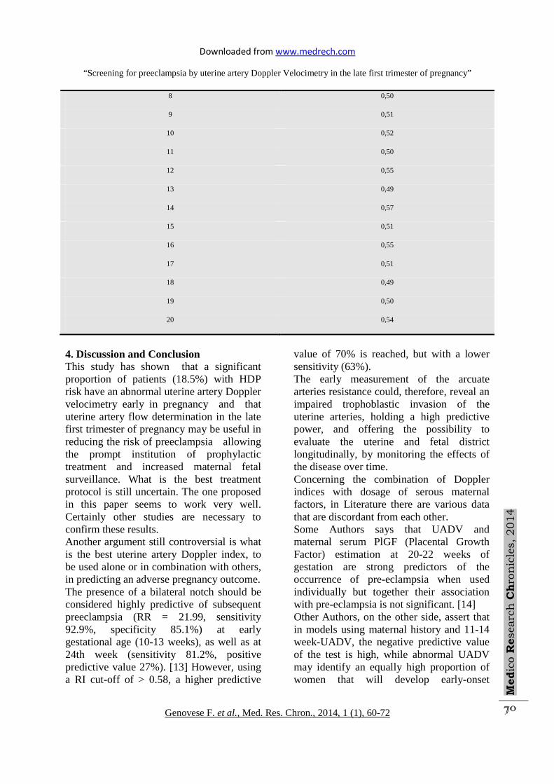
Table 9. Uterine artery flowmetric values at 24th (Group B) Patient RI value
1 0,57
2 0,51
3 0,55
4 0,54
5 0,52
6 0,50
7 0,52
Downloaded from www.medrech.com
“Screening for preeclampsia by uterine artery Doppler Velocimetry in the late first trimester of pregnancy”
Genovese F. et al., Med. Res. Chron., 2014, 1 (1), 60-72
Med
ico R
esearc
h C
hro
nic
les, 2014
70
8 0,50
9 0,51
10 0,52
11 0,50
12 0,55
13 0,49
14 0,57
15 0,51
16 0,55
17 0,51
18 0,49
19 0,50
20 0,54
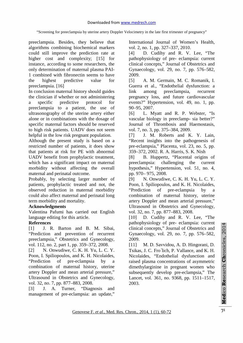
4. Discussion and Conclusion This study has shown that a significant proportion of patients (18.5%) with HDP risk have an abnormal uterine artery Doppler velocimetry early in pregnancy and that uterine artery flow determination in the late first trimester of pregnancy may be useful in reducing the risk of preeclampsia allowing the prompt institution of prophylactic treatment and increased maternal fetal surveillance. What is the best treatment protocol is still uncertain. The one proposed in this paper seems to work very well. Certainly other studies are necessary to confirm these results. Another argument still controversial is what is the best uterine artery Doppler index, to be used alone or in combination with others, in predicting an adverse pregnancy outcome. The presence of a bilateral notch should be considered highly predictive of subsequent preeclampsia (RR = 21.99, sensitivity 92.9%, specificity 85.1%) at early gestational age (10-13 weeks), as well as at 24th week (sensitivity 81.2%, positive predictive value 27%). [13] However, using a RI cut-off of > 0.58, a higher predictive
value of 70% is reached, but with a lower sensitivity (63%). The early measurement of the arcuate arteries resistance could, therefore, reveal an impaired trophoblastic invasion of the uterine arteries, holding a high predictive power, and offering the possibility to evaluate the uterine and fetal district longitudinally, by monitoring the effects of the disease over time. Concerning the combination of Doppler indices with dosage of serous maternal factors, in Literature there are various data that are discordant from each other. Some Authors says that UADV and maternal serum PlGF (Placental Growth Factor) estimation at 20-22 weeks of gestation are strong predictors of the occurrence of pre-eclampsia when used individually but together their association with pre-eclampsia is not significant. [14] Other Authors, on the other side, assert that in models using maternal history and 11-14 week-UADV, the negative predictive value of the test is high, while abnormal UADV may identify an equally high proportion of women that will develop early-onset
Downloaded from www.medrech.com
“Screening for preeclampsia by uterine artery Doppler Velocimetry in the late first trimester of pregnancy”
Genovese F. et al., Med. Res. Chron., 2014, 1 (1), 60-72
Med
ico R
esearc
h C
hro
nic
les, 2014
71
preeclampsia. Besides, they believe that algorithms combining biochemical markers could still improve the prediction rate at higher cost and complexity; [15] for instance, according to some researchers, the only determination of maternal plasma PAI-1 combined with fibronectin seems to have the highest predictive value for preeclampsia. [16] In conclusion maternal history should guides the clinician if whether or not administering a specific predictive protocol for preeclampsia to a patient, the use of ultrasonography of the uterine artery either alone or in combinations with the dosage of specific maternal factors should be reserved to high risk patients. UADV does not seem helpful in the low risk pregnant population. Although the present study is based on a restricted number of patients, it does show that patients at risk for PE with abnormal UADV benefit from prophylactic treatment, which has a significant impact on maternal morbidity without affecting the overall maternal and perinatal outcome. Probably, by selecting larger number of patients, prophylactic treated and not, the observed reduction in maternal morbidity could also affect maternal and perinatal long term morbidity and mortality. Acknowledgments Valentina Pafumi has carried out English language editing for this article. References [1] J. R. Barton and B. M. Sibai, “Prediction and prevention of recurrent preeclampsia,” Obstetrics and Gynecology, vol. 112, no. 2, part 1, pp. 359–372, 2008. [2] N. Onwudiwe, C. K. H. Yu, L. C. Y. Poon, I. Spiliopoulos, and K. H. Nicolaides, “Prediction of pre-eclampsia by a combination of maternal history, uterine artery Doppler and mean arterial pressure,” Ultrasound in Obstetrics and Gynecology, vol. 32, no. 7, pp. 877–883, 2008. [3] J. A. Turner, “Diagnosis and management of pre-eclampsia: an update,”
International Journal of Women’s Health, vol. 2, no. 1, pp. 327–337, 2010. [4] D. Cudihy and R. V. Lee, “The pathophysiology of pre- eclampsia: current clinical concepts,” Journal of Obstetrics and Gynaecology, vol. 29, no. 7, pp. 576–582, 2009. [5] A. M. Germain, M. C. Romanik, I. Guerra et al., “Endothelial dysfunction: a link among preeclampsia, recurrent pregnancy loss, and future cardiovascular events?” Hypertension, vol. 49, no. 1, pp. 90–95, 2007. [6] L. Myatt and R. P. Webster, “Is vascular biology in preeclamp- sia better?” Journal of Thrombosis and Haemostasis, vol. 7, no. 3, pp. 375–384, 2009. [7] J. M. Roberts and K. Y. Lain, “Recent insights into the pathogenesis of pre-eclampsia,” Placenta, vol. 23, no. 5, pp. 359–372, 2002. R. A. Harris, S. K. Nish [8] B. Huppertz, “Placental origins of preeclampsia: challenging the current hypothesis,” Hypertension, vol. 51, no. 4, pp. 970– 975, 2008. [9] N. Onwudiwe, C. K. H. Yu, L. C. Y. Poon, I. Spiliopoulos, and K. H. Nicolaides, “Prediction of pre-eclampsia by a combination of maternal history, uterine artery Doppler and mean arterial pressure,” Ultrasound in Obstetrics and Gynecology, vol. 32, no. 7, pp. 877–883, 2008. [10] D. Cudihy and R. V. Lee, “The pathophysiology of pre- eclampsia: current clinical concepts,” Journal of Obstetrics and Gynaecology, vol. 29, no. 7, pp. 576–582, 2009. [11] M. D. Savvidou, A. D. Hingorani, D. Tsikas, J. C. Fro ̈lich, P. Vallance, and K. H. Nicolaides, “Endothelial dysfunction and raised plasma concentrations of asymmetric dimethylarginine in pregnant women who subsequently develop pre-eclampsia,” The Lancet, vol. 361, no. 9368, pp. 1511–1517, 2003.
Downloaded from www.medrech.com
“Screening for preeclampsia by uterine artery Doppler Velocimetry in the late first trimester of pregnancy”
Genovese F. et al., Med. Res. Chron., 2014, 1 (1), 60-72
Med
ico R
esearc
h C
hro
nic
les, 2014
72
[12] Augusto Henriques Fulgeˆncio Branda �o, Ludmila Maria Guimara �es
Pereiraet al. Comparative Study of Endothelial Function andUterine Artery Doppler Velocimetry between Pregnant Women with or without Preeclampsia Development. Journal of Pregnancy Volume 2012, Article ID 909315, 5 pages
[13] Harrington K, Cooper D, Lees C, Hecher K, Campbell S. Doppler ultrasound of the uterine arteries: the importance of bilateral notching in the prediction of pre-eclampsia, placental abruption or delivery of a small-for-gestational-age baby. Ultrasound Obstet Gynecol 1996; 7: 182-8
[14] Macé G, Cynober E, Carbonne B.Ultrasound markers for the detection of women at risk of developing pre-eclampsia.ClinChem Lab Med. 2011 Dec 13.
[15] Carbillon L.First trimester uterine artery Doppler for the prediction of preeclampsia and foetal growth restriction. J Matern Fetal Neonatal Med. 2011 Aug 30
[16] Bodova K B, Biringer K, Dokus K, Ivankova J, Stasko J, Danko J. Fibronectin, plasminogen activator inhibitor type 1 (PAI-1) and uterine artery Doppler velocimetry as markers of preeclampsia. Dis Markers. 2011; 30 (4):191-6.
Related Documents