Experimental Parasitology 113 (2006) 16–23 www.elsevier.com/locate/yexpr 0014-4894/$ - see front matter © 2005 Elsevier Inc. All rights reserved. doi:10.1016/j.exppara.2005.12.001 Schistosoma mekongi: The in vitro eVect of praziquantel and artesunate on the adult Xuke Wannee Jiraungkoorskul a,¤ , Somphong Sahaphong a,b , Prasert Sobhon c , Suda Riengrojpitak a , Niwat Kangwanrangsan a a Department of Pathobiology, Faculty of Science, Mahidol University, Rama VI Road, Bangkok 10400, Thailand b Mahidol University International College, Mahidol University, Salaya Campus, Nakhonpathom 73170, Thailand c Department of Anatomy, Faculty of Science, Mahidol University, Rama VI Road, Bangkok 10400, Thailand Received 23 August 2005; received in revised form 1 December 2005; accepted 1 December 2005 Available online 17 January 2006 Abstract The eYcacy and tolerance of 80 g/ml praziquantel (PZQ) and 40 g/ml artesunate (ATS) against adult stage Schistosoma mekongi in vitro were investigated after 3, 6, 12, and 24 h incubation by monitoring worm motility and compared tegumental changes using scan- ning electron microscopy (SEM). Thirty mice were infected with S. mekongi cercaria for 49 days. Adult worms were collected by perfusion method and prepared for in vitro study. Contraction and decreased motor activity were observed after as little as 3 h incubation with PZQ and ATS. Some of the worms were immobile 12 h after exposure, and died within 24 h. The tegument of S. mekongi showed severe swell- ing, vacuolization and disruption, fusion of the tegumental ridges, collapse and peeling. After 12–24 h incubation, PZQ induced similar but they less severe, tegumental changes to those observed after exposure to ATS. The direct observation of the Xuke motility and SEM study suggest that ATS is more eVective than PZQ in causing tegumental damage in adult S. mekongi, and provides a basis for subsequent clinical trials. © 2005 Elsevier Inc. All rights reserved. Index Descriptors: Schistosoma mekongi; Praziquantel; Artesunate; Scanning electron microscopy 1. Introduction Schistosomiasis is a disease caused by the blood Xuke Schistosoma spp. (Class: Trematoda, Family: Schistosomat- idae). It is estimated that 200 million people in the world are currently aVected by this disease (WHO, 2002). There are Wve species that can parasitize humans, two of which are endemic in Asia, i.e., Schistosoma japonicum and Schis- tosoma mekongi. Schistosoma mekongi is restricted to areas of the Mekong River Basin, i.e., Lao People’s Democratic Republic and Thailand (Urbani et al., 2002). Only a few studies had been reported in the scientiWc literature on this schistosome species until 1992 when a focus was rediscovered in the province of Kracheh, Northeast Cambodia (Stich et al., 1999). Transmission is seasonal during the dry season, from February to April in Cambodia, and from March to June both in Lao People’s Democratic Republic and Thai- land (Urbani et al., 2002). Eggs are excreted by infected people, and after reaching a body of freshwater, eggs hatch to release miracidia that must infect the intermediate host snail for reproduction. Cercariae are then shed by the snail and penetrate the intact skin of humans to complete the life cycle. In the second half of the 20th century a number of diVer- ent antischistosomal drugs were discovered having diVerent modes of actions. Praziquantel (PZQ), a pyrazinoisoquino- line, is very eVective against schistosomiasis infections (WHO, 2002). It causes severe spasms and paralysis of worm musculature by a speciWc eVect on the cell membrane permeability, vacuolization, and disintegration of the * Corresponding author. Fax: +66 02 354 7158. E-mail address: [email protected] (W. Jiraungkoorskul).

Welcome message from author
This document is posted to help you gain knowledge. Please leave a comment to let me know what you think about it! Share it to your friends and learn new things together.
Transcript
Experimental Parasitology 113 (2006) 16–23
www.elsevier.com/locate/yexpr
Schistosoma mekongi: The in vitro eVect of praziquantel and artesunate on the adult Xuke
Wannee Jiraungkoorskul a,¤, Somphong Sahaphong a,b, Prasert Sobhon c, Suda Riengrojpitak a, Niwat Kangwanrangsan a
a Department of Pathobiology, Faculty of Science, Mahidol University, Rama VI Road, Bangkok 10400, Thailandb Mahidol University International College, Mahidol University, Salaya Campus, Nakhonpathom 73170, Thailand
c Department of Anatomy, Faculty of Science, Mahidol University, Rama VI Road, Bangkok 10400, Thailand
Received 23 August 2005; received in revised form 1 December 2005; accepted 1 December 2005Available online 17 January 2006
Abstract
The eYcacy and tolerance of 80 �g/ml praziquantel (PZQ) and 40 �g/ml artesunate (ATS) against adult stage Schistosoma mekongiin vitro were investigated after 3, 6, 12, and 24 h incubation by monitoring worm motility and compared tegumental changes using scan-ning electron microscopy (SEM). Thirty mice were infected with S. mekongi cercaria for 49 days. Adult worms were collected by perfusionmethod and prepared for in vitro study. Contraction and decreased motor activity were observed after as little as 3 h incubation with PZQand ATS. Some of the worms were immobile 12 h after exposure, and died within 24 h. The tegument of S. mekongi showed severe swell-ing, vacuolization and disruption, fusion of the tegumental ridges, collapse and peeling. After 12–24 h incubation, PZQ induced similarbut they less severe, tegumental changes to those observed after exposure to ATS. The direct observation of the Xuke motility and SEMstudy suggest that ATS is more eVective than PZQ in causing tegumental damage in adult S. mekongi, and provides a basis for subsequentclinical trials.© 2005 Elsevier Inc. All rights reserved.
Index Descriptors: Schistosoma mekongi; Praziquantel; Artesunate; Scanning electron microscopy
1. Introduction
Schistosomiasis is a disease caused by the blood XukeSchistosoma spp. (Class: Trematoda, Family: Schistosomat-idae). It is estimated that 200 million people in the worldare currently aVected by this disease (WHO, 2002). Thereare Wve species that can parasitize humans, two of whichare endemic in Asia, i.e., Schistosoma japonicum and Schis-tosoma mekongi. Schistosoma mekongi is restricted to areasof the Mekong River Basin, i.e., Lao People’s DemocraticRepublic and Thailand (Urbani et al., 2002). Only a fewstudies had been reported in the scientiWc literature on thisschistosome species until 1992 when a focus was rediscovered
* Corresponding author. Fax: +66 02 354 7158.E-mail address: [email protected] (W. Jiraungkoorskul).
0014-4894/$ - see front matter © 2005 Elsevier Inc. All rights reserved. doi:10.1016/j.exppara.2005.12.001
in the province of Kracheh, Northeast Cambodia (Stichet al., 1999). Transmission is seasonal during the dry season,from February to April in Cambodia, and from March toJune both in Lao People’s Democratic Republic and Thai-land (Urbani et al., 2002). Eggs are excreted by infectedpeople, and after reaching a body of freshwater, eggs hatchto release miracidia that must infect the intermediate hostsnail for reproduction. Cercariae are then shed by the snailand penetrate the intact skin of humans to complete the lifecycle.
In the second half of the 20th century a number of diVer-ent antischistosomal drugs were discovered having diVerentmodes of actions. Praziquantel (PZQ), a pyrazinoisoquino-line, is very eVective against schistosomiasis infections(WHO, 2002). It causes severe spasms and paralysis ofworm musculature by a speciWc eVect on the cell membranepermeability, vacuolization, and disintegration of the
W. Jiraungkoorskul et al. / Experimental Parasitology 113 (2006) 16–23 17
tegument (Xiao et al., 1985). However, there is considerableconcern about the development of PZQ resistance (Sang-ster, 2001). Thus, there is a pressing need to look for newsynthetic or natural antischistosomal drugs.
Qinghaosu (artemisinin) is an antimalarial drug, derivedfrom Artemisia annual L. (QACRG, 1979). Qinghaosu andits derivatives, such as artesunate, artemether, and arteetherare safe, potent, well-tolerated compounds that are used asWrst-line antimalarial therapy in many tropical and sub-tropical countries. They also display antischistosomal prop-erties. Studies in animal models have shown thatartemether and artesunate are also eVective against S.japonicum (Li et al., 1996; Xiao et al., 1995), Schistosomamansoni (Xiao and Catto, 1989) Schistosoma haematobium(Xiao et al., 2000a), and S. mekongi (Jiraungkoorskul et al.,2005a). The purpose of this study was to investigate theeVects of praziquantel and artesunate on worm motilityand the tegument in adult S. mekongi in vitro using scan-ning electron microscopy. It is hoped that this may improveknowledge of the eVectiveness of these drugs in treatingAsian schistosomiasis and be of use in developing betterdrugs against this disease.
2. Materials and methods
2.1. Snail
The freshwater snails Neotricula aperta were collectedfrom the Mekong River in Ubon Ratchathani Province,Thailand. They were maintained for the life cycle ofS. mekongi in the Malacology Unit, Department of Biol-ogy, Faculty of Science, Mahidol University, Bangkok,Thailand.
2.2. Mice
ICR male mice were bred at the National LaboratoryAnimal Center, Nakhon Pathom Province, Thailand. Mice,6 weeks old, mean weight 26.82§ 1.01 g, were used in thisexperiment. The animals were housed in shoe box typecages (18£ 30£ 13 cm) containing sterile wood shavingbedding in a strictly hygienic conventional animal room atthe Faculty of Science, Mahidol University. Standard diet(Perfect companion, Bangkok, Thailand) and tap waterwere available ad libitum. Room temperature was kept at22–25 °C with relative humidity of 60–70% and a 12:12 hlight–dark cycle was maintained throughout. All experi-mental animals used in this study were approved followingGuidelines for the Care and use of Laboratory Animals,Mahidol University, authorized by the Animal Care andUse Committee, Faculty of Science, Mahidol University.
2.3. Experimental design
Mice (nD30) were infected individually with 40S. mekongi cercariae shed from experimentally infectedN. aperta, after exposure to artiWcial light for at least 4 h, by
the looping method (Sornmani et al., 1973). Forty-ninedays postinfection, mice were sacriWced by overinhalationof ether. Adult Xukes were collected from mice by perfusionusing 0.1 M citrate in 0.15 M NaCl solution (Sornmaniet al., 1973). After washing Xukes several times with normalsaline and selecting for the healthy ones with normalmacroscopic structure and good motility, they were kept inculture medium 199 (M-5017, Sigma, USA, Lot No.053K83051) containing antibiotics (penicillin 50 IU/ml;streptomycin 50 mg/ml) until incubation experimentsbegan.
One hundred and twenty adult Xukes were randomlyassigned to three equally sized groups: group 1 was the con-trol group; group 2 was treated with 80�g/ml PZQ (AtlanticLaboratory, Bangkok, Thailand, Lot No. 000491); group 3was treated with 40�g/ml ATS (Atlantic Laboratory, Bang-kok, Thailand, Lot No. 040105). The drugs were initially pre-pared as a stock solution in dimethyl sulphoxide (DMSO)and then added to culture medium containing antibiotics (asdescribed above) to give a maximum solvent concentrationof 0.1% (v/v). The concentration used in vitro was chosen tocorrespond to the therapeutic concentration of these drugs(Becker et al., 1980). The Xukes in culture medium were keptin the incubator with 5% CO2 at 37 °C. After 3, 6, 12, and 24 hincubation, motility, tegumental alterations, and parasite sur-vival were assessed by examination under the OlympusSZ-ST stereomicroscope (Tokyo, Japan).
2.4. Motility criteria
Motility was scored using the following criteria: 3 (mov-ing whole body), 2 (moving only parts of body), 1 (immo-bile but not dead, unstained with vital dye), and 0(immobile, stained with vital dye) (Kiuchi et al., 1987).
2.5. Specimen preparation for scanning electron microscopy analysis
Worms were Wxed in 2.5% glutaraldehyde-phosphatebuVer (0.1 mol/L, pH 7.4) at 4 °C for 24 h and post-Wxed in1% osmium tetroxide for 1 h. They were dehydratedthrough a graded series of ethanol, dried in a Hitachi HCP-2 critical point dryer using liquid carbon dioxide as a tran-sitional medium. After drying, they were mounted on alu-minium stubs and coated with platinum and paladium inan ion-sputtering apparatus, Hitachi E-102, at 10–15 mAfor 6 min. They were examined and photographed in aHitachi scanning electron microscope S-2500 (HitachiHigh-Technologies, Hitachi-Naka City, Japan), operatingat 15 kV (Humason, 1972).
3. Results
3.1. Motility observation
All Xukes in the control group remained active, withwhole body movement (scoreD 3) throughout the
18 W. Jiraungkoorskul et al. / Experimental Parasitology 113 (2006) 16–23
experiment. Adult worms (70 and 100% in PZQ and ATStreated groups, respectively) were signiWcantly contractedimmediately and moving only parts of body (scoreD2)within 3 h. In the PZQ treated group, 2 of 10 (20%) becamecontracted and immobile (scoreD 1) and 3 of 10 (30%) died(scoreD0) after 12 and 24 h incubation, respectively. Themotility of the Xukes in the ATS treated group decreasedwith longer exposure. Fifty percents of Xukes were immo-bile (scoreD1) after 12 h incubation and all were dead(scoreD0) after 24 h (Table 1).
3.2. Scanning electron microscopy studies
3.2.1. Control groupThe surface topography of S. mekongi as studied by
SEM has been reported in great details in previous studies(Sobhon et al., 1984; Sobhon and Upatham, 1990; Vongpa-yabal et al., 1982). In brief, the adult male worm is thickerand shorter than the female worm and had a longitudinalcleft, the gynaecophoral canal, in which the female is heldduring copulation. The tegument of the adult male wormcan be divided into three parts: (1) the anterior part whichis the area from the oral sucker to the beginning of thegynaecophoral canal; (2) the middle part which is the larg-est portion of the body containing most of the gynaecoph-oral canal normally occupied by a female; (3) the posteriorpart which comprises the area behind the region where thebody of the female usually emerges from the gynaecophoralcanal. There are numerous large trabeculae on the tegu-ment covering the middle part of the male body (Figs. 1Aand B). The surface of adult female worm is also dividedinto three parts, similar to those described in male (Figs. 2Aand B).
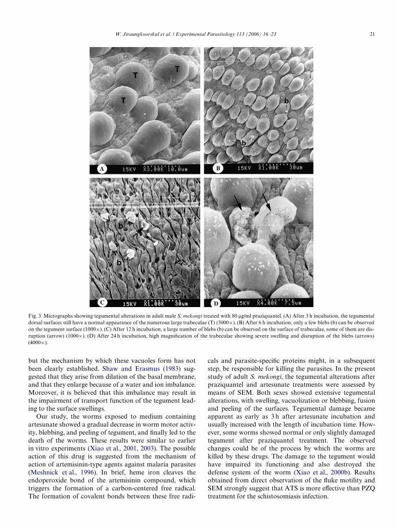
3.2.2. EVects of praziquantelAdult worms incubated in a medium containing with
80 �g/ml PZQ, contracted immediately. After 3–6 h incuba-tion, adult worms showed a few blebs and normal appear-ance of the dorsal surfaces of the tegument (Figs. 3A andB). After the 12 h, worms became contracted and immobile,assuming a shrunk and sometimes tightly curved appear-ance (Figs. 1C and D). The male worms showed changes inthe trabeculae, namely swelling with a few blebs around the
Table 1In vitro experiment, motility scores of control, and treated Xukes with pra-ziquantel (PZQ) and artesunate (ATS) at diVerent hours post-incubation
3, moving whole body; 2, moving only parts of body; 1, immobile(unstained with vital dye); 0, immobile and death (stained with vital dye).¤ The mean diVerence was signiWcant when compared the control group
(p 6 0.05).
Groups Percent of Xukes (%) in motility scores after incubation
3 h 6 h 12 h 24 h
3 2 1 0 3 2 1 0 3 2 1 0 3 2 1 0
1. Control 100 100 100 1002. PZQ 30 70¤ 30 70¤ 30 50 20 30 40 30¤
3. ATS 100¤ 80¤ 20 50 50 100¤
trabeculae (Fig. 3C). Severity increased after 24 h incuba-tion, the trabeculae showing severe swelling, vacuolization,and disruption of the blebs (Fig. 3D). In female worms,slight to moderate peeling of the dorsal surfaces was seen(Figs. 2C and D).
3.2.3. EVects of artesunateAdult worms incubated in a medium containing with
40 �g/ml ATS, contracted immediately and continuously ina similar manner to PZQ exposure, but showed a somewhatgreater severity (Figs. 1E and F). Decrease motor activitywas observed as early as 3 h incubation. Some of the wormsbecame immobile 6 h after exposure, and most died within24 h. All worms showed moderate to severe focal damage ofthe dorsal surfaces of the tegument. In female worms, themost prominent change was severe swelling, extensive peel-ing, and erosion of the middle dorsal surfaces of the tegu-ment (Figs. 2E and F). The intensity of response increasedwith the length of incubation time. In male worms, thelesions were similar to those with praziquantel, but weresevere. After 3–6 h incubation, numerous small blebs wereprotruded from the surface of the trabeculae (Figs. 4A andB). After 12 h incubation, the formation of blebs hadincreased; a large number of blebs could be observed on thesurface of the trabeculae (Fig. 4C). After 24 h incubation,severe swelling was usually accompanied by vacuolizationand disruption (Figs. 4D).
4. Discussion
The objective of this in vitro experiment was to comparethe eYcacy of PZQ and ATS against adult S. mekongi. Thetolerance against each drug was determined by scoring theXuke motility after 3, 6, 12, and 24 h incubation as shown inTable 1. ATS had severe eVects, and sudden paralysis of theXukes was observed. Further exposure led to the death ofthe parasites after 24 h incubation. In contrast, the PZQexerted a moderate eVect on the motility of the adultS. mekongi. This was similar to previous reports of in vitroexperiments (Jiraungkoorskul et al., 2005b).
PZQ was introduced as a novel anthelminthic for thetreatment of cestode and trematode infections in animals.This drug has two major eVects. First, the stimulation ofmotor activity by the inXux of calcium from externalsources causes spasms and paralysis of the muscles (Paxet al., 1978). Second, there is damage to the tegumentrevealed by the formation of vacuoles and later vesicles(Irie et al., 1989). Transmission electron microscope obser-vations indicated that the ultrastructural changes of thetegument induced by PZQ occur in the following sequence:depolymerization of the microtrabecular network, followedby vacuolization and then erosion of the surface (Sobhonand Upatham, 1990). These eVects impair the function ofthe muscle and the tegument structure that result in thedeath of the parasite. Vacuolization of the tegument hasbeen reported to be the direct and primary eVect of PZQ inS. mansoni (Becker et al., 1980; Shaw and Erasmus, 1983),

W. Jiraungkoorskul et al. / Experimental Parasitology 113 (2006) 16–23 19
Fig. 1. Micrographs of adult male S. mekongi. (A) Control, showing the normal body surface (50£). (B) Control, higher magniWcation of the tegument ofthe middle part showing the normal appearance of the numerous large trabeculae (T) (500£). (C) Male treated with praziquantel, showing contracted andtightly curved appearance (70£). (D) Male treated with praziquantel, higher magniWcation of the tegument of the middle part showing a few blebs (b)around the trabeculae (T) (500£). (E) Male treated with artesunate, showing more contraction and shortening of whole body (80£). (F) Male treated withartesunate, higher magniWcation of the tegument of the middle part showing numerous blebs (b) around the trabeculae (T) (500£).

20 W. Jiraungkoorskul et al. / Experimental Parasitology 113 (2006) 16–23
showing swelling, extensive peeling and erosion (arrows) (5000£).
Fig. 2. Micrographs of the adult female S. mekongi. (A) Control, the dorsal surface of the tegument covering the middle part of the body appears as a Xatsheet pitted with minute holes (1000£). (B) Control, higher magniWcation of the dorsal tegument appearing highly invaginated with small holes (h) andlong narrow clefts (C) (5000£). (C) Female treated with praziquantel, showing slight to moderate peeling (1500£). (D) Female treated with praziquantel,higher magniWcation of the middle dorsal tegument showing loosing of the clefts (C) and slight to moderate peeling (arrows) (5000£). (E) Female treatedwith artesunate, showing fusion of tegumental ridges (1200£). (F) Female treated with artesunate, higher magniWcation of the middle dorsal tegument
W. Jiraungkoorskul et al. / Experimental Parasitology 113 (2006) 16–23 21
but the mechanism by which these vacuoles form has notbeen clearly established. Shaw and Erasmus (1983) sug-gested that they arise from dilation of the basal membrane,and that they enlarge because of a water and ion imbalance.Moreover, it is believed that this imbalance may result inthe impairment of transport function of the tegument lead-ing to the surface swellings.
Our study, the worms exposed to medium containingartesunate showed a gradual decrease in worm motor activ-ity, blebbing, and peeling of tegument, and Wnally led to thedeath of the worms. These results were similar to earlierin vitro experiments (Xiao et al., 2001, 2003). The possibleaction of this drug is suggested from the mechanism ofaction of artemisinin-type agents against malaria parasites(Meshnick et al., 1996). In brief, heme iron cleaves theendoperoxide bond of the artemisinin compound, whichtriggers the formation of a carbon-centered free radical.The formation of covalent bonds between these free radi-
cals and parasite-speciWc proteins might, in a subsequentstep, be responsible for killing the parasites. In the presentstudy of adult S. mekongi, the tegumental alterations afterpraziquantel and artesunate treatments were assessed bymeans of SEM. Both sexes showed extensive tegumentalalterations, with swelling, vacuolization or blebbing, fusionand peeling of the surfaces. Tegumental damage becameapparent as early as 3 h after artesunate incubation andusually increased with the length of incubation time. How-ever, some worms showed normal or only slightly damagedtegument after praziquantel treatment. The observedchanges could be of the process by which the worms arekilled by these drugs. The damage to the tegument wouldhave impaired its functioning and also destroyed thedefense system of the worm (Xiao et al., 2000b). Resultsobtained from direct observation of the Xuke motility andSEM strongly suggest that ATS is more eVective than PZQtreatment for the schistosomiasis infection.
Fig. 3. Micrographs showing tegumental alterations in adult male S. mekongi treated with 80 �g/ml praziquantel. (A) After 3 h incubation, the tegumentaldorsal surfaces still have a normal appearance of the numerous large trabeculae (T) (3000£). (B) After 6 h incubation, only a few blebs (b) can be observedon the tegument surface (1000£). (C) After 12 h incubation, a large number of blebs (b) can be observed on the surface of trabeculae, some of them are dis-ruption (arrow) (1000£). (D) After 24 h incubation, high magniWcation of the trabeculae showing severe swelling and disruption of the blebs (arrows)(4000£).

22 W. Jiraungkoorskul et al. / Experimental Parasitology 113 (2006) 16–23
Acknowledgments
This study was funded by the Thailand Research Fund(TRF) and the Commission on Higher Education: the NewResearchers Grant 2003.
References
Becker, B., Mehlhorn, H., Andrews, P., Thomas, H., Eckert, J., 1980. Lightand electron microscopic studies on the eVect of praziquantel on Schis-tosoma mansoni, Dicrocoelium dendriticum, and Fasciola hepatica(Trematoda) in vitro. Zeitschrift fur Parasitenkunde–ParasitologyResearch 63, 113–128.
Humason, G.L., 1972. Animal tissue techniques, third ed. W.H. Freeman,San Francisco.
Irie, Y., Utsunomiya, H., Tanaka, M., Ohmae, H., Nara, T., Yasuraoka, K.,1989. Schistosoma japonicum and S. mansoni: ultrastructural damage inthe tegument and reproductive organs after treatment with levo- and
dextro-praziquantel. American Journal of Tropical Medicine andHygiene 41, 204–211.
Jiraungkoorskul, W., Sahaphong, S., Sobhon, P., Riengrojpitak, S., Kang-wanrangsan, N., 2005a. EVects of praziquantel and artesunate on thetegument of adult Schistosoma mekongi harboured in mice. Parasitol-ogy International 54, 177–183.
Jiraungkoorskul, W., Sahaphong, S., Tansatit, T., Kangwanrangsan, N.,Pipatshukiat, S., 2005b. Eurytrema pancreaticum: the in vitro eVect ofpraziquantel and triclabendazole on the adult Xuke. Experimental Par-asitology 111, 172–177.
Kiuchi, F., Miyashita, N., Tsuda, Y., Kondo, K., Yoshimura, H., 1987.Studies on crude drugs eVective on visceral larva Migrans. I. IdentiWca-tion of larvicidal principles in betel nuts. Chemical and PharmaceuticalBulletin 35, 2880–2886.
Li, S., Wu, L., Liu, Z., 1996. Studies on prophylactic eVect of artesunate onschistosomiasis japonica. Chinese Medical Journal 109, 848–853.
Meshnick, S.R., Taylor, T.E., Kamchonwongpaisan, S., 1996. Artemisininand the antimalarial endoperoxides: from herbal remedy to targetedchemotherapy. Microbiology Review 60, 301–315.
Fig. 4. Micrographs showing tegumental alterations in adult male S. mekongi treated with 40 �g/ml artesunate. (A) After 3 h incubation, small blebs (b) haveformed in the area between trabeculae (T) (5000£). (B) After 6 h incubation, the formation of blebs have increased, a large number of blebs (b) can be observedon the surface of trabeculae (T) (5000£). (C) After 12 h incubation, the tegument showing severe swelling, vacuolization, and disruption (arrows) (3500£). (D)After 24 h incubation, the tegument showing severe swelling and increased number of blebs (b) accompanied by vacuolization and disruption (arrows) (1500£).
W. Jiraungkoorskul et al. / Experimental Parasitology 113 (2006) 16–23 23
Pax, R., Bennett, T.L., Fetterer, R., 1978. A benzodiazepine derivative andpraziquantel: eVects on musculature of Schistosoma mansoni and Schis-tosoma japonicum. Naunyn Schiedbergs Archives of Pharmacology304, 309–315.
Qinghaosu Antimalarial Coordinating Research Group (QACRG),1979. Antimalarial studies on Qingaosu. Chinese Medical Journal 92,811–816.
Sangster, N.C., 2001. Managing parasiticide resistance. Veterinary Parasi-tology 98, 89–109.
Shaw, M.K., Erasmus, D.A., 1983. Schistosoma mansoni: the eVects of asubcurative dose of praziquantel on the ultrastructure of wormsin vitro. Zeitschrift fur Parasitenkunde 69, 73–90.
Sobhon, P., Upatham, E.S., McLaren, D., 1984. Topography and ultra-structure of the tegument of adult Schistosoma mekongi. Parasitology89, 511–521.
Sobhon, P., Upatham, E.S., 1990. Snail hosts, life-cycle, and tegumentalstructure of oriental schistosomes. UNDP/World Bank/WHO SpecialProgramme for Research and Training in Tropical Diseases (TDR),Geneva, Switzerland.
Sornmani, S., Kitikoon, V., Schneider, C.R., Harinasuta, C., Pathamma-wong, O., 1973. Mekong schistosomiasis: 1. Life cycle of Schistosomajaponicum, Mekong strain in the laboratory. Southeast Asian Journalof Tropical Medicine Public Health 4, 218–225.
Stich, A.H., Biays, S., Odermatt, P., Men, C., Saem, C., Sokha, K., Ly, C.S.,Legros, P., Philips, M., Lormand, J.D., Tanner, M., 1999. Foci of Schis-tosomiasis mekongi, northern Cambodia: II. Distribution of infectionand morbidity. Tropical Medicine and International Health 4, 674–685.
Urbani, C., Sinoun, M., Socheat, D., Pholsena, K., Strandgaard, H.,Odermatt, P., Hatz, C., 2002. Epidemiology and control of mekongischistosomiasis. Acta Tropica 82, 157–168.
Vongpayabal, P., Sobhon, P., Upatham, E.S., Wanichanon, C., Mitranond,V., Tanphaichitr, N., Tumbel, V.E.C., 1982. Scanning electron micro-scopic study of the tegumental surface of adult Schistosoma mekongi.Parasitology 85, 325–332.
World Health Organization, 2002. Prevention and control of schistosomi-asis and soil-transmitted helminthiasis: report of a WHO Expert Com-mittee. WHO Technical Report Series No. 912, Geneva.
Xiao, S.H., Catto, B.A., Webster, L.T., 1985. EVects of praziquantel ondiVerent developmental stages of Schistosoma mansoni in vitro andin vivo. Journal of Infectious Disease 151, 1130–1137.
Xiao, S.H., Catto, B.A., 1989. In vitro and in vivo studies of the eVect ofartemether on Schistosoma mansoni. Antimicrobial Agents and Che-motherapy 33, 1557–1562.
Xiao, S.H., You, J.Q., Yang, Y.Q., Wang, C.Z., 1995. Experimental studieson early treatment of schistosomal infection with artemether. South-east Asian Journal of Tropical Medicine Public Health 26, 306–318.
Xiao, S.H., Utzinger, J., Chollet, J., Endriss, Y., N’Goran, E.K., Tanner, M.,2000a. EVect of artemether against Schistosoma haematobium in experimen-tally infected hamsters. International Journal of Parasitology 30, 1001–1006.
Xiao, S.H., Shen, B.G., Chollet, J., Tanner, M., 2000b. Tegumental changesin 21-day-old Schistosoma mansoni harboured in mice treated withartemether. Acta Tropica 75, 341–348.
Xiao, S.H., Chollet, J., Utzinger, J., Matile, H., Mei, J.Y., Tanner, M., 2001.Artemether administered together with haemin damages schistosomesin vitro. Transactions of the Royal Society of Tropical Medicine andHygiene 95, 67–71.
Xiao, S.H., Wu, Y.L., Tanner, M., Wu, W.M., Utzinger, J., Mei, J.Y.,Scorneaux, B., Chollet, J., Zhai, Z., 2003. Schistosoma japonicum:in vitro eVects of artemether combined with haemin depend on cultiva-tion media and appraisal of artemether products appearing in themedia. Parasitology Research 89, 459–466.
Related Documents