ALS-MES 11.0.2 Scanning Transmission Soft X-ray Microscopy Tolek Tyliszczak Advanced Light Source

Welcome message from author
This document is posted to help you gain knowledge. Please leave a comment to let me know what you think about it! Share it to your friends and learn new things together.
Transcript
ALS-MES 11.0.2
Scanning Transmission Soft X-ray Microscopy
Tolek Tyliszczak Advanced Light Source

ALS-MES 11.0.2 zone plates and achieved resolution
12 nm lines Si/Mo
20 nm zone plate 25 nm zone plate 17 nm zone plate
10 nm lines Si/Mo 20 nm (vertical) Au lines
9 nm lines Si/Mo Imaged by 17 nm zone plate
All images taken at 700 eV Structures have equal line/space nominal dimensions
Dimensions of half periods are quoted
ALS-MES 11.0.2
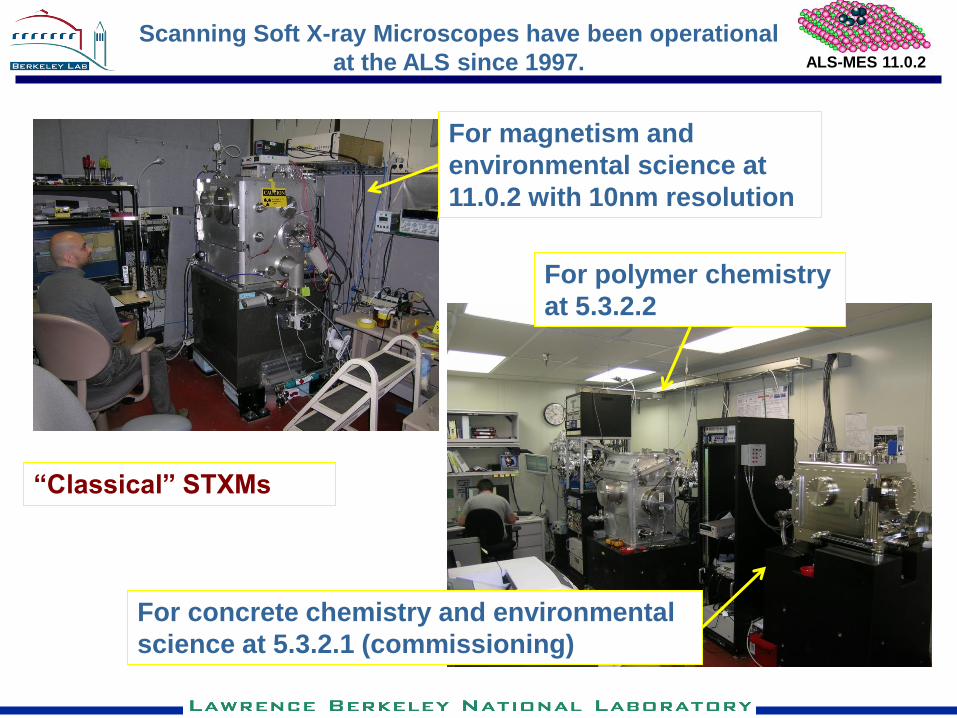
Scanning Soft X-ray Microscopes have been operational
at the ALS since 1997.
For magnetism and
environmental science at
11.0.2 with 10nm resolution
For polymer chemistry
at 5.3.2.2
For concrete chemistry and environmental
science at 5.3.2.1 (commissioning)
“Classical” STXMs
ALS-MES 11.0.2
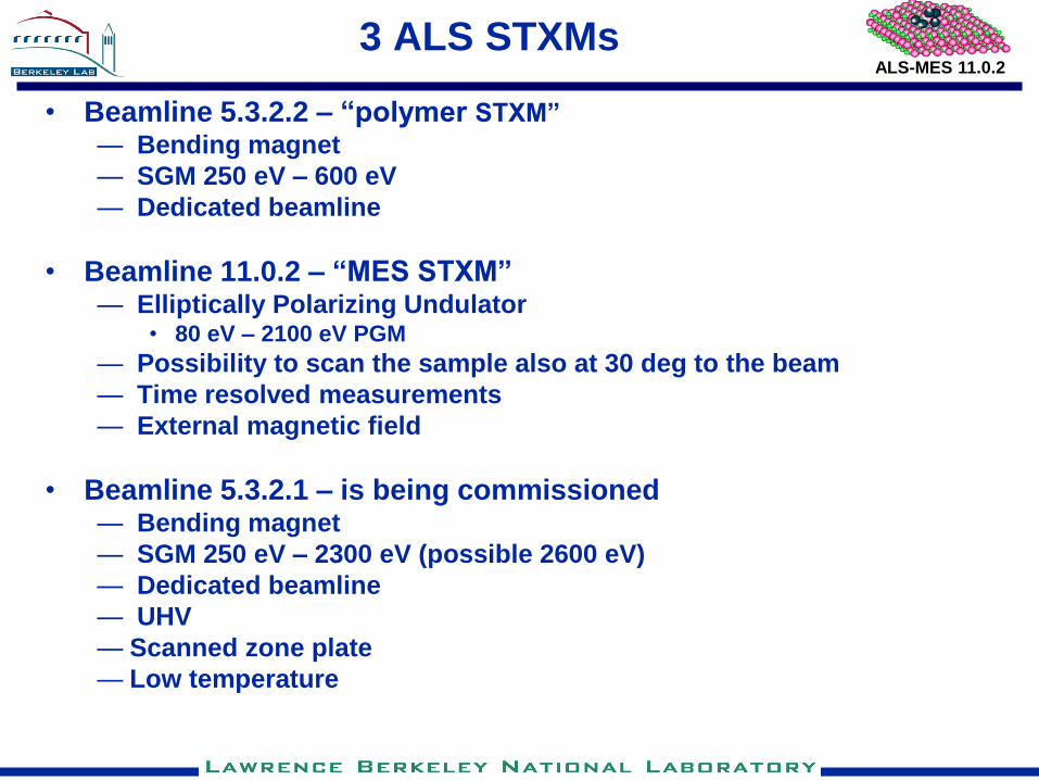
3 ALS STXMs
• Beamline 5.3.2.2 – “polymer STXM”
— Bending magnet
— SGM 250 eV – 600 eV
— Dedicated beamline
• Beamline 11.0.2 – “MES STXM” — Elliptically Polarizing Undulator
• 80 eV – 2100 eV PGM
— Possibility to scan the sample also at 30 deg to the beam
— Time resolved measurements
— External magnetic field
• Beamline 5.3.2.1 – is being commissioned — Bending magnet
— SGM 250 eV – 2300 eV (possible 2600 eV)
— Dedicated beamline
— UHV
— Scanned zone plate
— Low temperature
ALS-MES 11.0.2
Electrons
mono mirror
4-jaw slits
Vertical deflection Horizontal deflection
Rotating endstations
4
3
1
2
KB MIRROR
mono grating
EPU gap EPU Z
Polarization
M201 Micro mirror
mono body
M212 Bend 1, bend 2
Spectro slits
Spectro mirror
Micro slits
Spectro mirror vessel
M213 Bend 1, bend 2
width, height
width, height horz, vert deflection
horz, vert deflection shield wall
beamline diagnostics
M211
M221
STXM
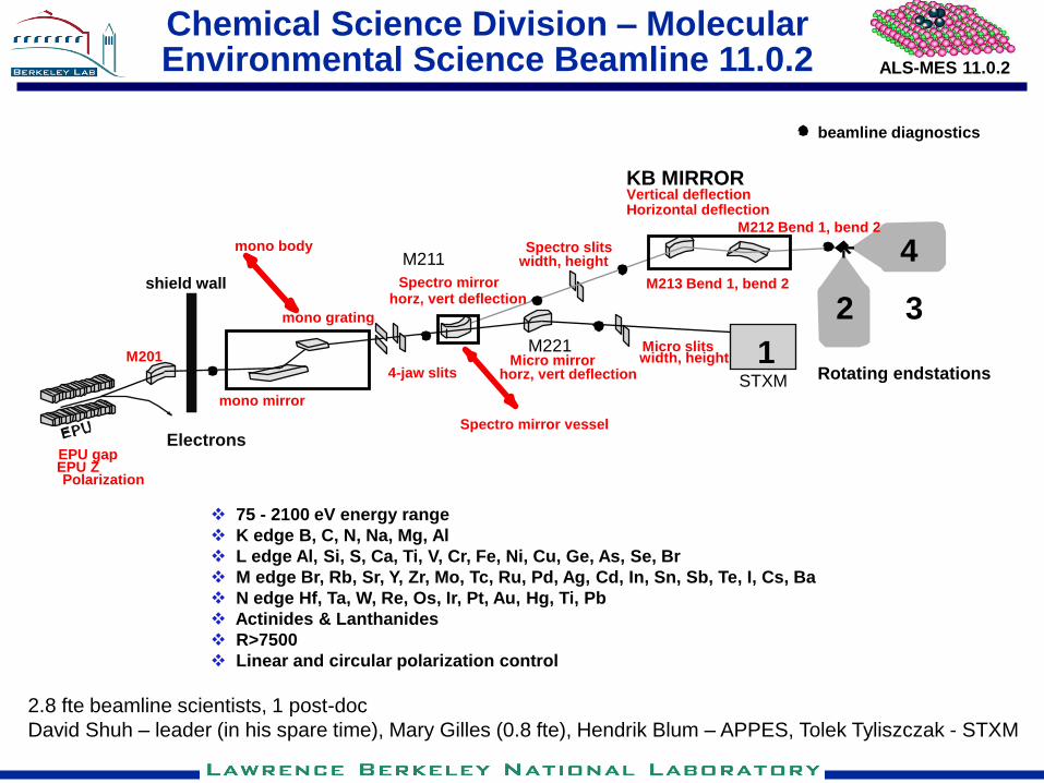
Chemical Science Division – Molecular Environmental Science Beamline 11.0.2
75 - 2100 eV energy range
K edge B, C, N, Na, Mg, Al
L edge Al, Si, S, Ca, Ti, V, Cr, Fe, Ni, Cu, Ge, As, Se, Br
M edge Br, Rb, Sr, Y, Zr, Mo, Tc, Ru, Pd, Ag, Cd, In, Sn, Sb, Te, I, Cs, Ba
N edge Hf, Ta, W, Re, Os, Ir, Pt, Au, Hg, Ti, Pb
Actinides & Lanthanides
R>7500
Linear and circular polarization control
2.8 fte beamline scientists, 1 post-doc
David Shuh – leader (in his spare time), Mary Gilles (0.8 fte), Hendrik Blum – APPES, Tolek Tyliszczak - STXM
ALS-MES 11.0.2
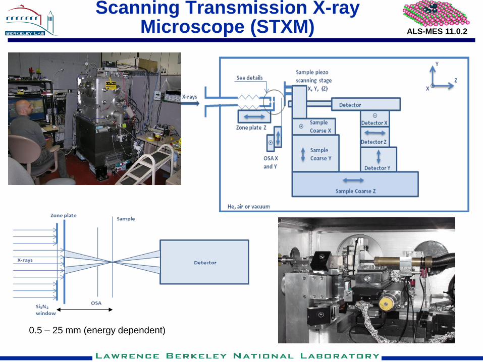
Scanning Transmission X-ray Microscope (STXM)
0.5 – 25 mm (energy dependent)
ALS-MES 11.0.2
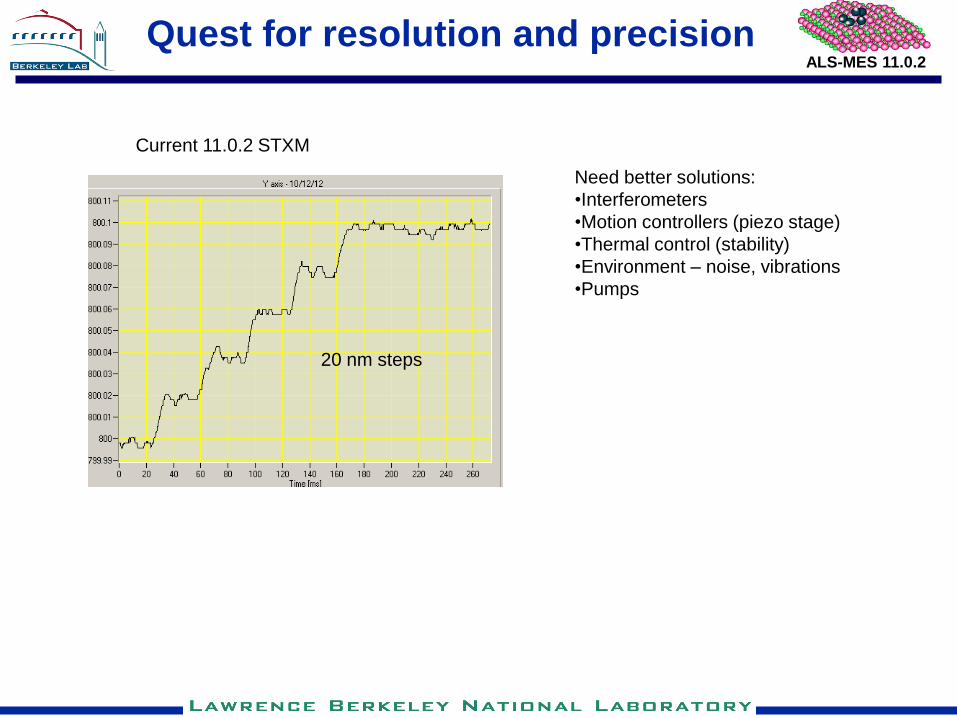
Quest for resolution and precision
20 nm steps
Current 11.0.2 STXM
Need better solutions:
•Interferometers
•Motion controllers (piezo stage)
•Thermal control (stability)
•Environment – noise, vibrations
•Pumps
ALS-MES 11.0.2
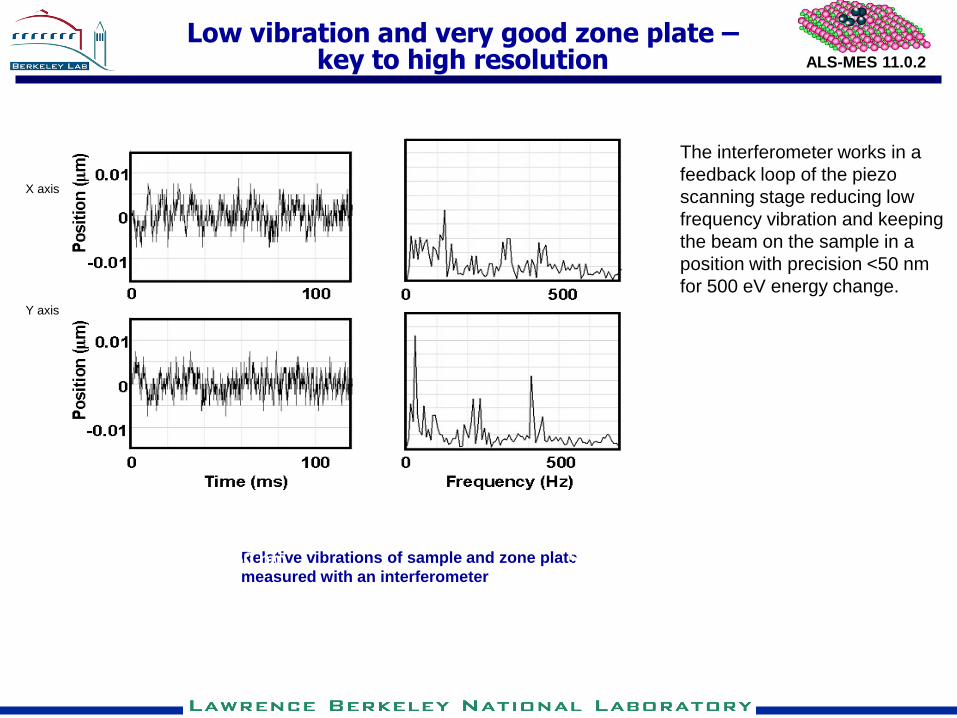
Low vibration and very good zone plate – key to high resolution
Relative vibrations of sample and zone plate
measured with an interferometer
X axis
Y axis
50 nm 50 nm
The interferometer works in a
feedback loop of the piezo
scanning stage reducing low
frequency vibration and keeping
the beam on the sample in a
position with precision <50 nm
for 500 eV energy change.
ALS-MES 11.0.2
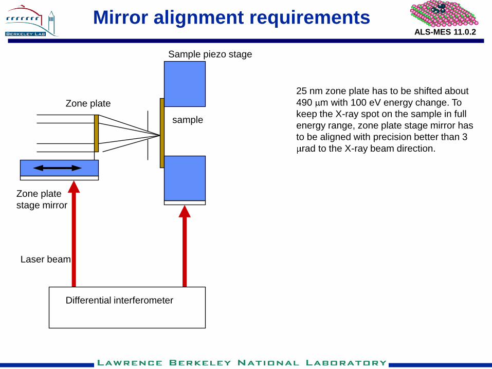
Mirror alignment requirements
Differential interferometer
Zone plate
sample
Sample piezo stage
Zone plate
stage mirror
Laser beam
25 nm zone plate has to be shifted about
490 m with 100 eV energy change. To
keep the X-ray spot on the sample in full
energy range, zone plate stage mirror has
to be aligned with precision better than 3
rad to the X-ray beam direction.
ALS-MES 11.0.2
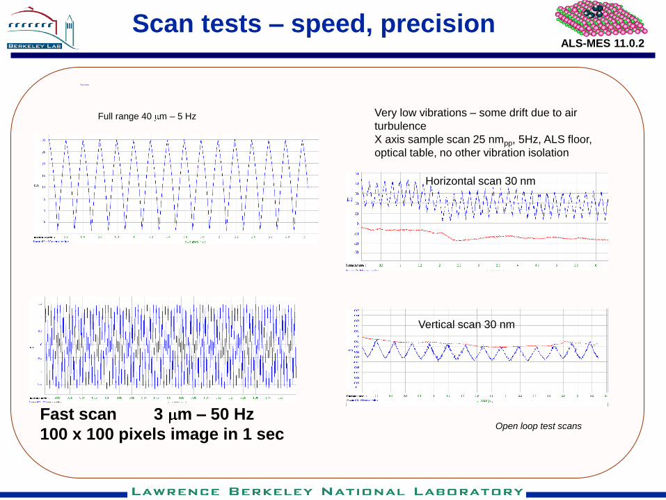
Scan tests – speed, precision
Test scans
Fast scan 3 m – 50 Hz
100 x 100 pixels image in 1 sec
Full range 40 m – 5 Hz Very low vibrations – some drift due to air
turbulence
X axis sample scan 25 nmpp, 5Hz, ALS floor,
optical table, no other vibration isolation
Zone Plate reference mirror
Open loop test scans
Horizontal scan 30 nm
Vertical scan 30 nm
ALS-MES 11.0.2
Ambient Pressure Scanning Photoemission Microscopy (In development)
Expected outcome: Photoelectron spectrometer with <100 nm spatial resolution, operating at gas pressures > 10 Torr in 280 eV – 1600 eV X-ray energy range, using a single zone plate.
Realization: Scanning zone plate microscope module for existing ambient pressure photoemission spectrometer
Challenges: zone plate illumination, vibrations, precision of scanning, precision of moving the zone plate along beam direction ( >10mm at < 200 nm run out), confined space
ALS-MES 11.0.2
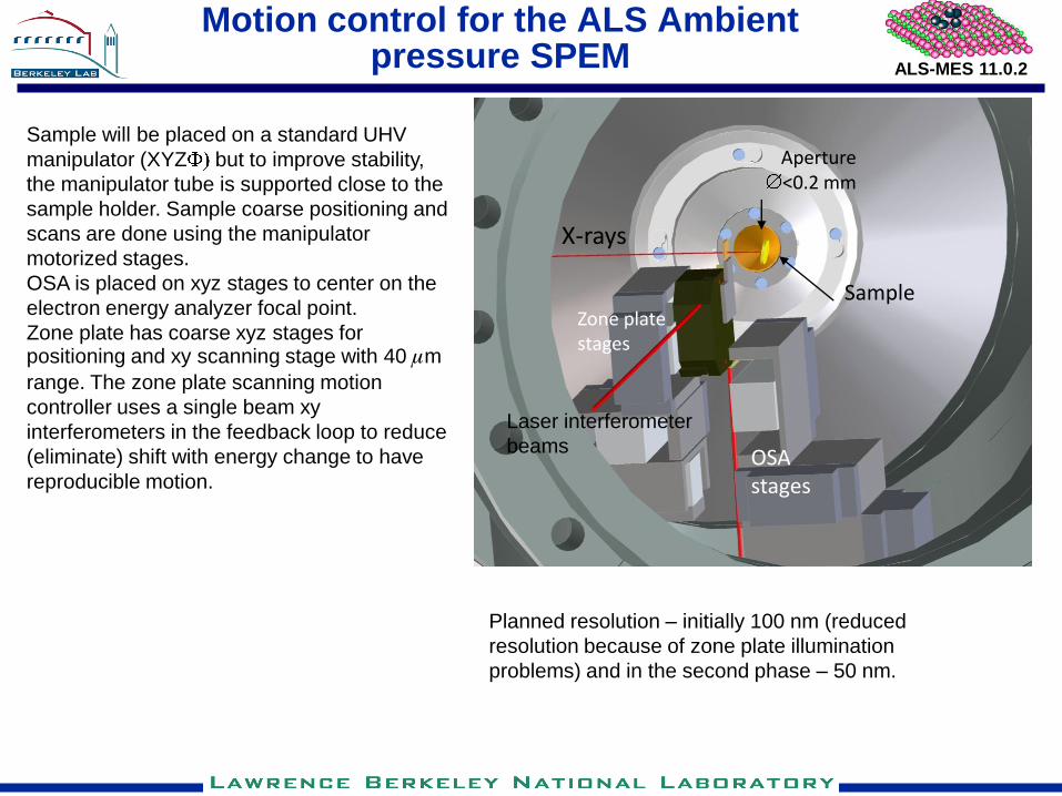
Motion control for the ALS Ambient pressure SPEM
Sample
X-rays
OSA stages
Zone plate stages
Aperture <0.2 mm
Laser interferometer
beams
Sample will be placed on a standard UHV
manipulator (XYZ but to improve stability,
the manipulator tube is supported close to the
sample holder. Sample coarse positioning and
scans are done using the manipulator
motorized stages.
OSA is placed on xyz stages to center on the
electron energy analyzer focal point.
Zone plate has coarse xyz stages for positioning and xy scanning stage with 40 mm
range. The zone plate scanning motion
controller uses a single beam xy
interferometers in the feedback loop to reduce
(eliminate) shift with energy change to have
reproducible motion.
Planned resolution – initially 100 nm (reduced
resolution because of zone plate illumination
problems) and in the second phase – 50 nm.
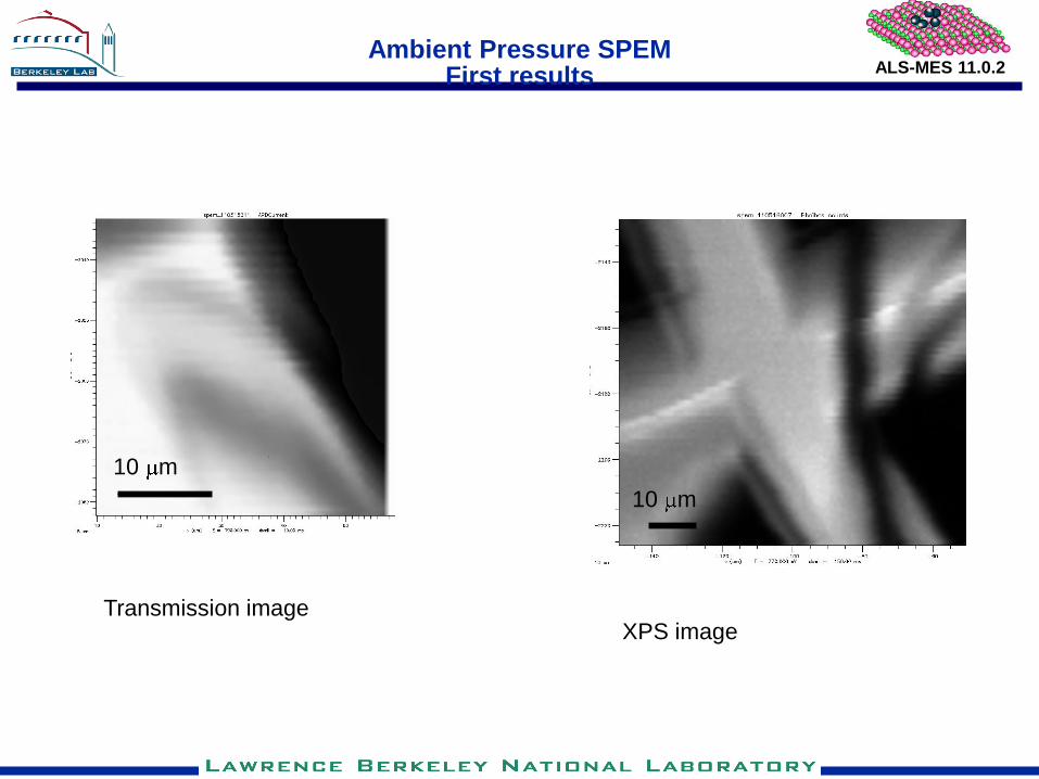
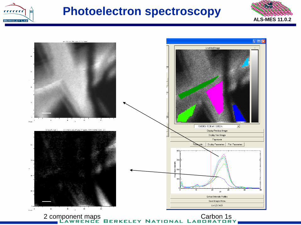
ALS-MES 11.0.2 Ambient Pressure SPEM
First results
XPS image Transmission image
10 m
10 m
ALS-MES 11.0.2
Photoelectron spectroscopy
Carbon 1s 2 component maps

ALS-MES 11.0.2
Au Pt C
Photoelectron spectroscopy Au, Pt

ALS-MES 11.0.2
Zone plates and working distance EY
Working distance
DR [nm] D [ m] Working distance Energy [eV]
at 300 eV 300 700 1800
45 240 700 2810 6790 17460
25 240 350 1467 3423 8802
17 120 130 570 1330 3420
Focal length [mm]
Electron energy analyzer lens
EY in transmission
ALS-MES 11.0.2
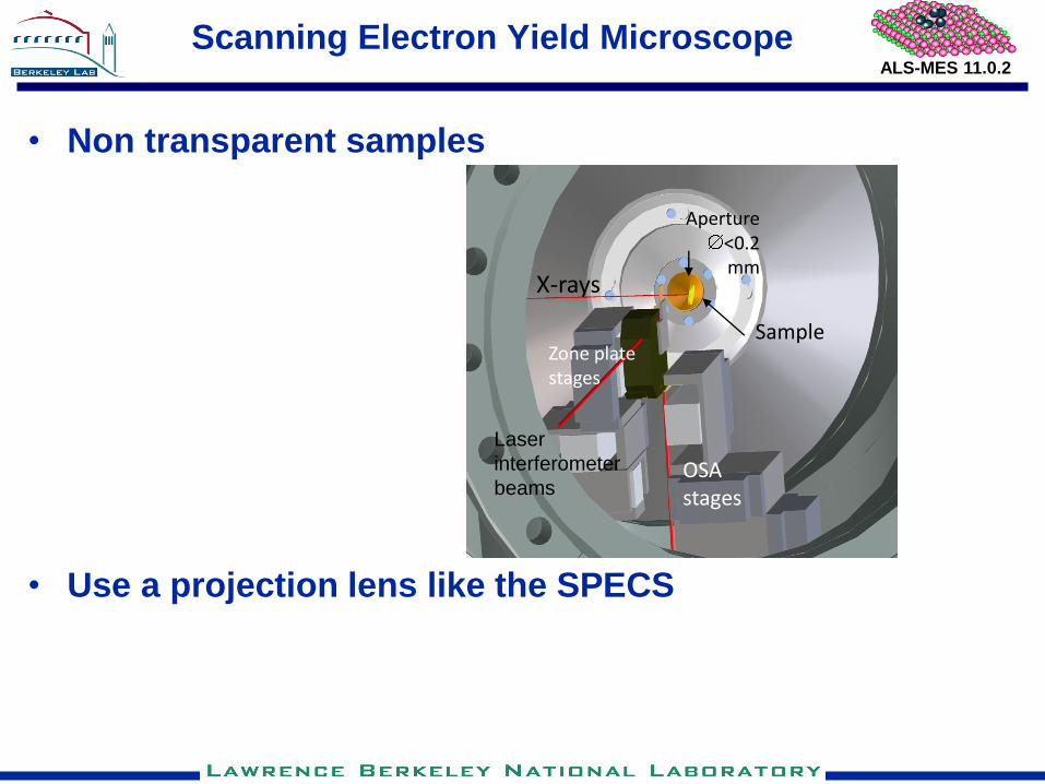
Scanning Electron Yield Microscope
• Use a projection lens like the SPECS
• Non transparent samples
Sample
X-rays
OSA stages
Zone plate stages
Aperture <0.2 mm
Laser
interferometer
beams
ALS-MES 11.0.2
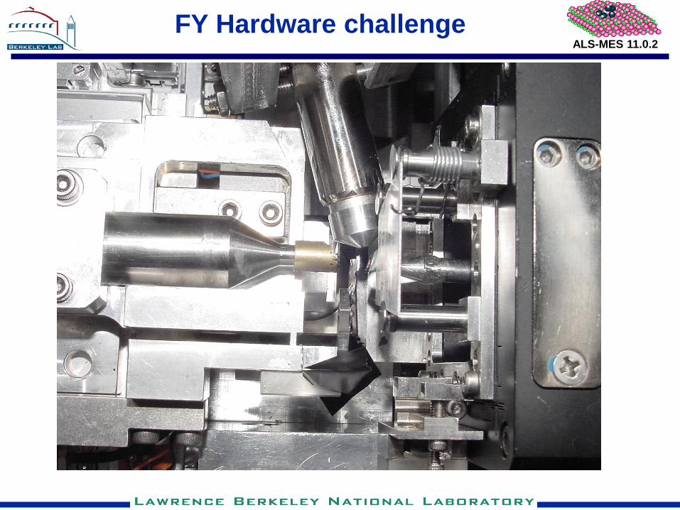
FY Hardware challenge
ALS-MES 11.0.2
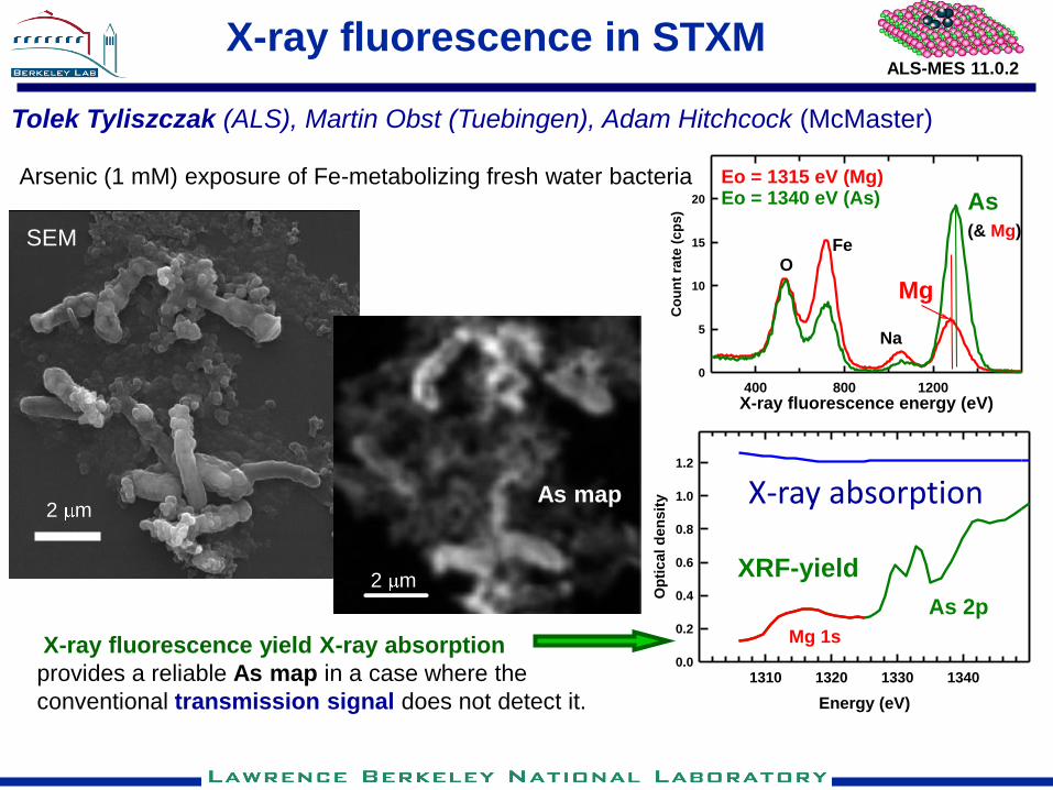
X-ray fluorescence in STXM
Energy (eV)
1310 1320 1330 1340
Op
tic
al
den
sit
y0.0
0.2
0.4
0.6
0.8
1.0
1.2
Mg 1s
As 2p
X-ray absorption
X-ray fluorescence energy (eV)400 800 1200
Co
un
t ra
te (
cp
s)
0
5
10
15
20
OFe
Na
Mg
As (& Mg)
Eo = 1315 eV (Mg)Eo = 1340 eV (As)
2 m
Tolek Tyliszczak (ALS), Martin Obst (Tuebingen), Adam Hitchcock (McMaster)
Arsenic (1 mM) exposure of Fe-metabolizing fresh water bacteria
2 m
SEM
X-ray fluorescence yield X-ray absorption
provides a reliable As map in a case where the
conventional transmission signal does not detect it.
As map
XRF-yield

ALS-MES 11.0.2
Fluorescence yields in soft X-ray range
K edge L edge
Are the FY STXM measurements feasible?

ALS-MES 11.0.2
Expected count rate
• Flux = 108 photons/s
• FY = 1%
• Collection angle (10 mm sample-detector, 30 mm2
detector) = 0.6 %
• Detection time (dwell time) = 100 ms
If you have thick sample (total absorption)
600 photons / pixel
• What detection limits we can expect?
ALS-MES 11.0.2
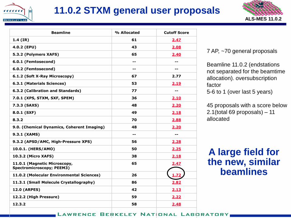
11.0.2 STXM general user proposals
Beamline % Allocated Cutoff Score
1.4 (IR) 61 2.47
4.0.2 (EPU) 43 2.08
5.3.2 (Polymers XAFS) 65 2.40
6.0.1 (Femtosecond) -- --
6.0.2 (Femtosecond) -- --
6.1.2 (Soft X-Ray Microscopy) 67 2.77
6.3.1 (Materials Sciences) 53 2.19
6.3.2 (Calibration and Standards) 77 --
7.0.1 (XPS, STXM, SXF, SPEM) 36 2.10
7.3.3 (SAXS) 48 2.20
8.0.1 (SXF) 49 2.18
8.3.2 70 2.88
9.0. (Chemical Dynamics, Coherent Imaging) 48 2.20
9.3.1 (XAMS) -- --
9.3.2 (APSD/AMC, High-Pressure XPS) 56 2.28
10.0.1. (HERS/AMO) 50 2.25
10.3.2 (Micro XAFS) 38 2.18
11.0.1 (Magnetic Microscopy, Spectromicroscopy; PEEM3)
65 2.47
11.0.2 (Molecular Environmental Sciences) 26 1.72
11.3.1 (Small Molecule Crystallography) 86 2.82
12.0 (ARPES) 42 2.13
12.2.2 (High Pressure) 59 2.22
12.3.2 58 2.48
7 AP, ~70 general proposals
Beamline 11.0.2 (endstations
not separated for the beamtime
allocation). oversubscription
factor
5-6 to 1 (over last 5 years)
45 proposals with a score below
2.1(total 69 proposals) – 11
allocated
A large field for the new, similar
beamlines
ALS-MES 11.0.2
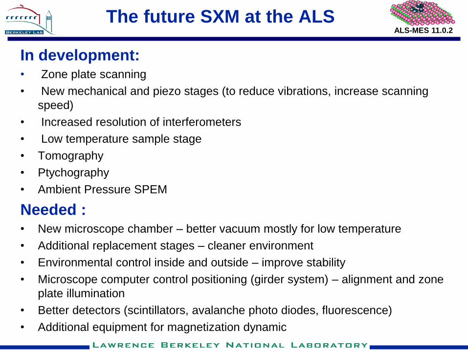
The future SXM at the ALS
In development: • Zone plate scanning
• New mechanical and piezo stages (to reduce vibrations, increase scanning
speed)
• Increased resolution of interferometers
• Low temperature sample stage
• Tomography
• Ptychography
• Ambient Pressure SPEM
Needed : • New microscope chamber – better vacuum mostly for low temperature
• Additional replacement stages – cleaner environment
• Environmental control inside and outside – improve stability
• Microscope computer control positioning (girder system) – alignment and zone
plate illumination
• Better detectors (scintillators, avalanche photo diodes, fluorescence)
• Additional equipment for magnetization dynamic

ALS-MES 11.0.2
SXM at the NSLS II
Obvious: high performance microscope but there are
important question
• One microscope or 2-3
• Omnibus or dedicated/specialized microscopes
• Geared towards physics or multidiscipline general user
instrument
• Low temperature – LN2 or LHe
• Environmental cells
Do not sell it cheap – it should be the best.

ALS-MES 11.0.2
Publications:
• 34 publications
— STXM - 18
Successful, multidiscipline beamline and
endstation
A model for many beamline around the world

ALS-MES 11.0.2
STXM 11.0.2 status and development in progress
11.0.2 STXM- still best in the world
• Energy 90 eV – 2000 eV with resolving power > 6000
• Spatial resolution - can resolve 10 nm spaced lines (in 1st order)
• Time resolved measurements with 70 ps resolution
• Internal electromagnets (in plane and perpendicular field) for static
magnetization measurements
• Normal and rotated (30 deg) scanning for polarization measurements
• Flexible sample mounting
•Fluorescence detection
In development
• Zone plate scanning
• New mechanical and piezo stages (to reduce vibrations, increase scanning
speed)
• Increased resolution of interferometers
• Low temperature sample stage
•Tomography
17 nm zone plate
10 nm lines Si/Mo
Best working resolution of all X-ray microscopes
ALS-MES 11.0.2
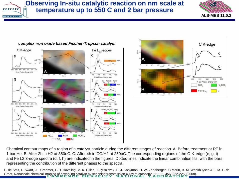
Observing In-situ catalytic reaction on nm scale at temperature up to 550 C and 2 bar pressure
Chemical contour maps of a region of a catalyst particle during the different stages of reaction. A: Before treatment at RT in
1 bar He. B: After 2h in H2 at 350oC. C: After 4h in CO/H2 at 250oC. The corresponding regions of the O K-edge (e, g, i)
and Fe L2,3-edge spectra (d, f, h) are indicated in the figures. Dotted lines indicate the linear combination fits, with the bars
representing the contribution of the different phases to the spectra.
complex iron oxide based Fischer-Tropsch catalyst
E. de Smit, I. Swart, J. . Creemer, G.H. Hoveling, M. K. Gilles, T.Tyliszczak, P. J. Kooyman, H. W. Zandbergen, C.Morin, B. M. Weckhuysen & F. M. F. de
Groot, Nanoscale chemical imaging of a working catalyst by scanning transmission X-ray microscopy, Nature, 456, 222-225, (2008).
ALS-MES 11.0.2
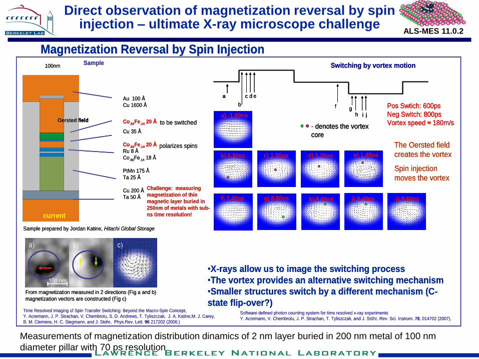
Direct observation of magnetization reversal by spin injection – ultimate X-ray microscope challenge
Magnetization Reversal by Spin Injection
Sample prepared by Jordan Katine, Hitachi Global Storage
Software defined photon counting system for time resolved x-ray experiments
Y. Acremann, V. Chembrolu, J. P. Strachan, T. Tyliszczak, and J. Stöhr, Rev. Sci. Instrum. 78, 014702 (2007).
Time Resolved Imaging of Spin Transfer Switching: Beyond the Macro-Spin Concept,
Y. Acremann, J. P. Strachan, V. Chembrolu, S. D. Andrews, T. Tyliszczak, J. A. Katine,M. J. Carey,
B. M. Clemens, H. C. Siegmann, and J. Stohr, Phys.Rev. Lett. 96 217202 (2006.)
Switching by vortex motion
current
Oersted field
Au 100 Å
Cu 1600 Å
Co.86Fe.14 20 Å
Cu 35 Å
Co.86Fe.14 20 Å
Ru 8 Å
Co.86Fe.14 18 Å
PtMn 175 Å
Ta 25 Å
Cu 200 Å
Ta 50 Å
to be switched
polarizes spins
100nm
Challenge: measuring
magnetization of thin
magnetic layer buried in
250nm of metals with sub-
ns time resolution!
Sample
From magnetization measured in 2 directions (Fig a and b)
magnetization vectors are constructed (Fig c)
•X-rays allow us to image the switching process
•The vortex provides an alternative switching mechanism
•Smaller structures switch by a different mechanism (C-
state flip-over?)
a
b
c
f gha) -1.00ns
b) 0.80ns c) 1.00ns d) 1.20ns e) 1.40ns
f) 7.40ns g) 8.80ns h) 9.00ns
d e
- denotes the vortex
core
i) 9.40ns j) 9.60ns
Pos Swtich: 600ps
Neg Switch: 800ps
Vortex speed ≈ 180m/s
i j
The Oersted field
creates the vortex
Spin injection
moves the vortex
Magnetization Reversal by Spin Injection
Sample prepared by Jordan Katine, Hitachi Global Storage
Software defined photon counting system for time resolved x-ray experiments
Y. Acremann, V. Chembrolu, J. P. Strachan, T. Tyliszczak, and J. Stöhr, Rev. Sci. Instrum. 78, 014702 (2007).
Time Resolved Imaging of Spin Transfer Switching: Beyond the Macro-Spin Concept,
Y. Acremann, J. P. Strachan, V. Chembrolu, S. D. Andrews, T. Tyliszczak, J. A. Katine,M. J. Carey,
B. M. Clemens, H. C. Siegmann, and J. Stohr, Phys.Rev. Lett. 96 217202 (2006.)
Switching by vortex motion
current
Oersted field
current
Oersted field
Au 100 Å
Cu 1600 Å
Co.86Fe.14 20 Å
Cu 35 Å
Co.86Fe.14 20 Å
Ru 8 Å
Co.86Fe.14 18 Å
PtMn 175 Å
Ta 25 Å
Cu 200 Å
Ta 50 Å
to be switched
polarizes spins
100nm
Challenge: measuring
magnetization of thin
magnetic layer buried in
250nm of metals with sub-
ns time resolution!
Sample
From magnetization measured in 2 directions (Fig a and b)
magnetization vectors are constructed (Fig c)
•X-rays allow us to image the switching process
•The vortex provides an alternative switching mechanism
•Smaller structures switch by a different mechanism (C-
state flip-over?)
a
b
c
f gha) -1.00ns
b) 0.80ns c) 1.00ns d) 1.20ns e) 1.40ns
f) 7.40ns g) 8.80ns h) 9.00ns
d e
- denotes the vortex
core
i) 9.40ns j) 9.60ns
Pos Swtich: 600ps
Neg Switch: 800ps
Vortex speed ≈ 180m/s
i j
The Oersted field
creates the vortex
Spin injection
moves the vortex
aa
b
c
f gha) -1.00ns
b) 0.80ns c) 1.00ns d) 1.20ns e) 1.40ns
f) 7.40ns g) 8.80ns h) 9.00ns
d e
- denotes the vortex
core
i) 9.40ns j) 9.60ns
Pos Swtich: 600ps
Neg Switch: 800ps
Vortex speed ≈ 180m/s
i j
The Oersted field
creates the vortex
Spin injection
moves the vortex
Measurements of magnetization distribution dinamics of 2 nm layer buried in 200 nm metal of 100 nm
diameter pillar with 70 ps resolution
ALS-MES 11.0.2
Magnetization dynamic
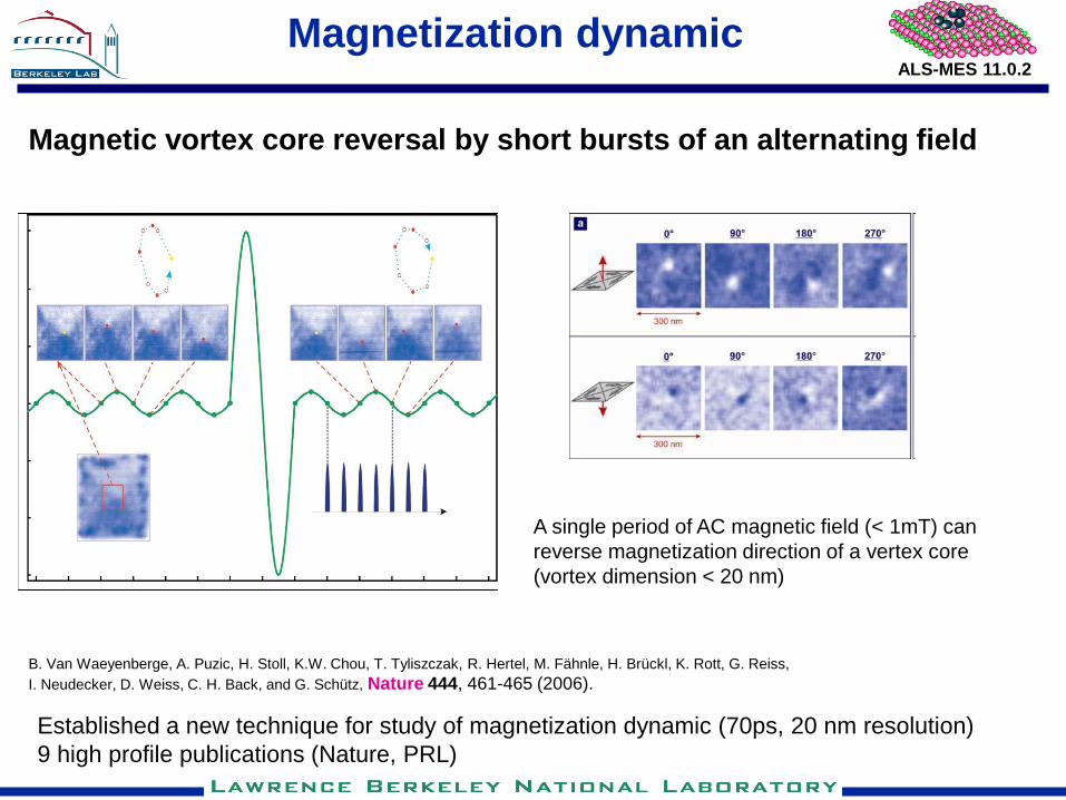
Magnetic vortex core reversal by short bursts of an alternating field
B. Van Waeyenberge, A. Puzic, H. Stoll, K.W. Chou, T. Tyliszczak, R. Hertel, M. Fähnle, H. Brückl, K. Rott, G. Reiss,
I. Neudecker, D. Weiss, C. H. Back, and G. Schütz, Nature 444, 461-465 (2006).
Established a new technique for study of magnetization dynamic (70ps, 20 nm resolution)
9 high profile publications (Nature, PRL)
A single period of AC magnetic field (< 1mT) can
reverse magnetization direction of a vertex core
(vortex dimension < 20 nm)
ALS-MES 11.0.2
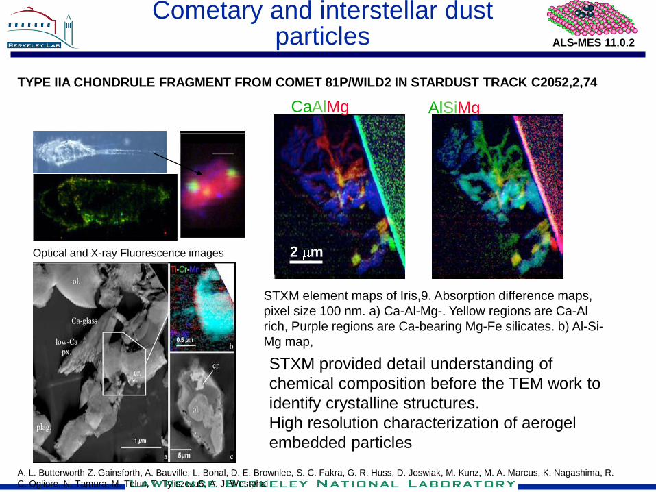
Cometary and interstellar dust particles
TYPE IIA CHONDRULE FRAGMENT FROM COMET 81P/WILD2 IN STARDUST TRACK C2052,2,74
Optical and X-ray Fluorescence images
CaAlMg
2 m
AlSiMg
STXM element maps of Iris,9. Absorption difference maps,
pixel size 100 nm. a) Ca-Al-Mg-. Yellow regions are Ca-Al
rich, Purple regions are Ca-bearing Mg-Fe silicates. b) Al-Si-
Mg map,
STXM provided detail understanding of
chemical composition before the TEM work to
identify crystalline structures.
High resolution characterization of aerogel
embedded particles
A. L. Butterworth Z. Gainsforth, A. Bauville, L. Bonal, D. E. Brownlee, S. C. Fakra, G. R. Huss, D. Joswiak, M. Kunz, M. A. Marcus, K. Nagashima, R.
C. Ogliore, N. Tamura, M. Telus, T. Tyliszcza5, A. J. Westphal
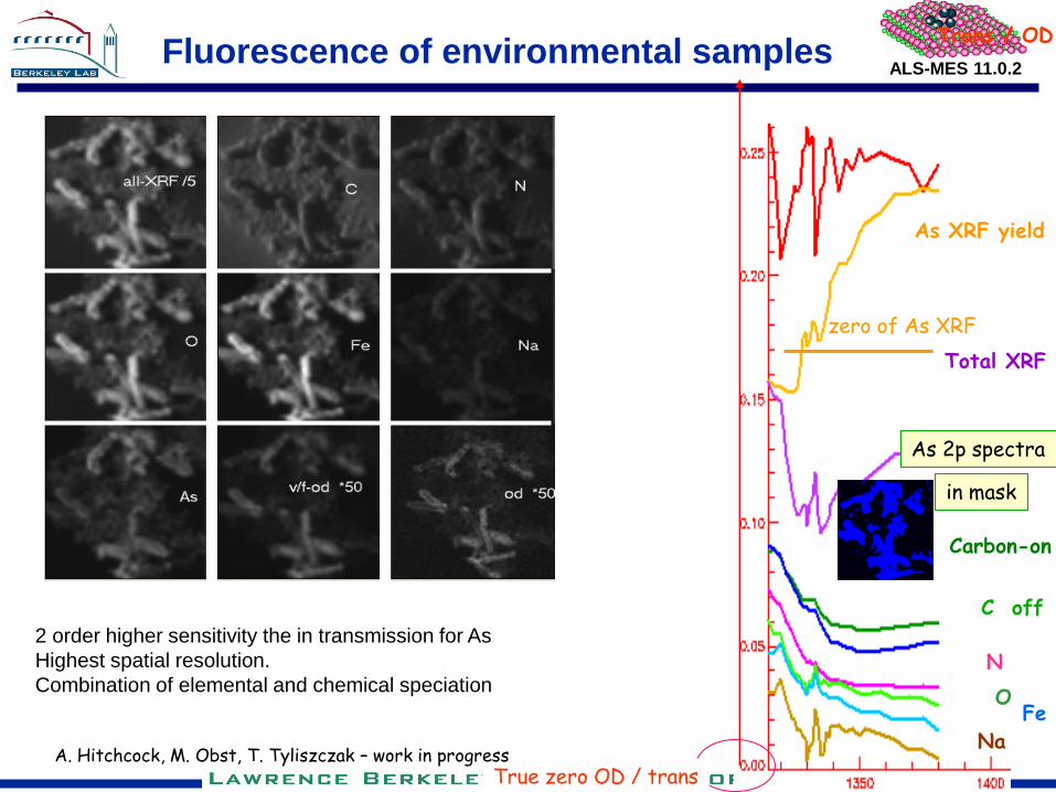
ALS-MES 11.0.2 Fluorescence of environmental samples
As XRF yield
Trans / OD
Total XRF
Carbon-on
C off
N
O Fe
Na
True zero OD / trans
As 2p spectra
in mask
zero of As XRF
A. Hitchcock, M. Obst, T. Tyliszczak – work in progress
2 order higher sensitivity the in transmission for As
Highest spatial resolution.
Combination of elemental and chemical speciation
Related Documents