Institut für Tierwissenschaften, Abt. Tierzucht und Tierhaltung der Rheinischen Friedrich – Wilhelms – Universität Bonn Effect of sub-clinical endometritis on miRNAs expression profile of endometrial and oviductal epithelium and its implication of early embryonic development I n a u g u r a l – D i s s e r t a t i o n zur Erlangung des Grades Doktor der Agrarwissenschaft (Dr. agr.) der Landwirtschaftlichen Fakultät der Rheinischen Friedrich – Wilhelms – Universität Bonn von Sally Rashad Elsaid Ibrahim aus Kairo, Ägypten

Welcome message from author
This document is posted to help you gain knowledge. Please leave a comment to let me know what you think about it! Share it to your friends and learn new things together.
Transcript

Institut für Tierwissenschaften, Abt. Tierzucht und Tierhaltung
der Rheinischen Friedrich – Wilhelms – Universität Bonn
Effect of sub-clinical endometritis on miRNAs expression profile of endometrial
and oviductal epithelium and its implication of early embryonic development
I n a u g u r a l – D i s s e r t a t i o n
zur
Erlangung des Grades
Doktor der Agrarwissenschaft
(Dr. agr.)
der
Landwirtschaftlichen Fakultät
der
Rheinischen Friedrich – Wilhelms – Universität Bonn
von
Sally Rashad Elsaid Ibrahim
aus
Kairo, Ägypten

Referent : Prof. Dr. Karl Schellander Koreferent: Prof. Dr. agr. Brigitte Petersen Tag der mündlichen Prüfung: 09 March 2015 Erscheinungsjahr: 2015

D edicated to D edicated to D edicated to D edicated to m y beloved m y beloved m y beloved m y beloved M otherM otherM otherM other, , , , m y m y m y m y lovely lovely lovely lovely sistersistersistersisters N ihal s N ihal s N ihal s N ihal & & & &
B asm aB asm aB asm aB asm a, , , , and and and and m ym ym ym y lovely lovely lovely lovely brothers M oham m ed brothers M oham m ed brothers M oham m ed brothers M oham m ed &&&& A bdel A bdel A bdel A bdel
R ahm anR ahm anR ahm anR ahm an
IIIIn m em ory n m em ory n m em ory n m em ory of of of of m y fatherm y fatherm y fatherm y father

Effect of sub-clinical endometritis on miRNAs expression profile of endometrial
and oviductal epithelium and its implication of early embryonic development
Understanding the molecular mechanisms associated with regulation of inflammatory
responses during female genital tract infection is one step forward for development of
diagnostic and therapeutic strategies in bovine reproduction. Therefore, the aim of this
thesis was to investigate post-transcriptional regulation of inflammatory immune
response genes during LPS treatment in bovine oviductal, endometrial cells and
embryos. For this, two studies were conducted. In the first study, the mRNA expression
analysis of inflammatory response genes (TNFα & IL1β) was performed in primary
bovine oviductal cell culture and co-cultured blastocysts after minimum dose of LPS
treatment in vitro. In the second study, the alterations of let-7 miRNAs expression were
addressed in primary bovine endometrial cells after LPS challenge (with clinical dose of
3.0 µg/ml or a sub-clinical dose of 0.5 µg/ml), as well as functional study of let-7
miRNAs using gain and loss of function. While LPS treatment resulted in significantly
up-regulation of pro-inflammatory cytokines (TNFα & IL1β) and stress response genes
(SOD & CAT) in bovine oviductal cell and co-cultured blastocysts, the expression level
of essential elements like OVGP1 and IGF2 was reduced in the challenged group
compared to the untreated control. Interestingly, the over-expression of these pro-
inflammatory cytokines in bovine oviductal cells was associated with aberrant
expression of their potential regulatory miRNAs (miR-155, miR-146a, miR-223, miR-
21, miR-16 and miR-215). Furthermore, blastocysts co-cultured with oviductal cells in
the presence of LPS showed reduced mitochondrial distribution pattern, higher ROS and
apoptotic cells. A minimum dose of LPS challenge resulted in changes in relative
abundance of let-7 miRNAs in a time dependent-manner, where the peak expression of
let-7a reached at 6h, while let-7e, let-f and let-7i peaked at 24h post treatment.
Overexpression of let-7a inhibited pro-inflammatory cytokines (TNFα & IL6) on
mRNAs as well as protein levels, while the let-7a inhibitor (antagonist) resulted in an
increase in the expression of the same genes. The mRNAs and protein levels of TNFα,
IL6 have shown a clear suppression upon transfection with let-7f inhibitor. In
conclusion, infections in endometrial or oviductal microenvironment resulted in
aberrant expression of genes and miRNAs which support the role of regulatory let-7
miRNAs during bovine uterine infection by fine-tuning inflammatory cytokines.

Einfluss der subklinischen Endometritis auf miRNA-Expressionsprofile im Epithel
von Endometrium und Eileiter sowie die Konsequenzen für die embryonale
Frühentwicklung
Die Klärung von molekularen Mechanismen, die die Regulation einer
Entzündungsreaktion während Infektionen des weiblichen Genitaltraktes begleiten, ist
entscheidend für die Entwicklung von diagnostischen und therapeutischen
Behandlungsmethoden in der bovinen Reproduktion. Daher war das Ziel dieser Studie
die post-transkriptionale Regulation von Genen der entzündungsbedingten
Immunreaktionen zu untersuchen.
Dazu wurden innerhalb von zwei Forschungsansätzen bovine Zellen des Eileiters und
des Endometriums sowie präimplantative Embryonen mit LPS behandelt. In der ersten
Studie wurde die mRNA Expression von Genen der Entzündungsreaktion (TNFα &
IL1β) in kultivierten primären bovinen Eileiterzellen und co-kultivierten Blastozysten in
vitro gemessen, nachdem diese mit einer minimalen Menge an LPS behandelt wurden.
In einem zweiten Experiment wurden die Veränderungen der let-7 miRNAs Expression
untersucht. Zu diesem Zweck wurden primäre bovine Zellen des Endometriums mit
LPS behandelt (klinische Dosis (3.0 µg/ml) oder subklinische Dosis (0.5 µg/ml)) sowie
eine funktionale Studie durchgeführt, um mögliche Funktionsnutzen und -verluste durch
let-7 miRNAs zu untersuchen.
Die Behandlung mit LPS führte zur signifikanten Hochregulierung von pro-
inflammatorischen Zytokinen (TNFα & IL1β) und Genen der Stressantwort (SOD &
CAT) in bovinen Eileiterzellen und co-kultivierten Blastozysten. Die
Expressionsniveaus von essentiellen Genen wie OVGP1 und IGF2 waren im Vergleich
zur unbehandelten Kontrolle reduziert. Interessanterweise ging die Überexpression
dieser pro-inflammatorischen Zytokine in den Eileiterzellen mit einer abweichenden
Expression ihrer potentiell regulierenden miRNAs (miR-155, miR-146a, miR-223, miR-
21, miR-16 und miR-215) einher. Darüber hinaus zeigten die Blastozysten, die mit den
Eileiterzellen co-kultiviert wurden, bei der Behandlung mit LPS veränderte Muster der
mitochondrialen Verteilung sowie erhöhte Anteile an ROS und apoptotischen Zellen.
Der Einsatz einer minimalen Menge an LPS führte zu einer zeitabhängigen
Veränderung der relativen Abundanz der let-7 miRNAs, wobei die höchste Expression
von let-7a nach 6h erreicht war, während let-7e, let-f und let-7i eine maximale
Abundanz 24h nach der Behandlung zeigten. Die Überexpression von let-7a hemmte
das mRNA- sowie das Proteinniveau der pro-inflammatorischen Zytokine, wohingegen
der let-7a Inhibitor (Antagonist) zu einer Steigerung der Expression der gleichen Gene
führte. Die mRNA- und Proteinniveaus von TNFα und IL6 zeigten eine deutliche
Suppression nach der Transfektion mit dem let-7f Inhibitor.
Abschließend lässt sich folgern, dass eine Infektion im Endometrium oder im Eileiter zu
Veränderungen der Expressionen von Genen und miRNAs führte, welches die
regulative Bedeutung von let-7 miRNAs auf inflammatorische Zytokine während einer
Infektion des bovinen Uterus unterstützt.

V
Contents
Abstract III
Zusammenfassung IV
List of abbreviations VII
List of tables X
List of figures XI
Chapter 1 General overview
1.1 Introduction 1
1.1.2 Endometritis 2
1.1.3 Oviduct 4
1.1.4 The role of immunomediators and other molecules in
successful pregnancy 5
1.1.5 MiRNAs 7
1.2 Rationale and objectives 10
1.3 Materials and methods 11
1.3.1.1 Total RNA isolation from BOEC and endometrial
cells 11
1.3.1.2 Total RNA isolation from embryos (blastocyst stage) 11
1.3.2 First strand cDNA synthesis for large and small RNA 11
1.3.3 Quantitative real-time PCR (qRT-PCR) 12
1.3.4 Western immunoblotting 12
1.3.5 Luciferase reporter constructions and luciferase assay 12
1.3.6 Experimental design 13
1.3.7 Statistical analysis 14
1.4 Results 15
1.4.1
Changes in expression of genes associated with
inflammatory immune response and physiological
function of BOEC after LPS challenge
15
1.4.2 Temporal pattern of miRNAs potentially targeting
inflammatory immune response genes 15

VI
1.4.3 Effect of LPS treated BOEC on co-cultured embryos 16
1.4.4
LPS challenge induced alterations in let-7 miRNAs
and their target genes expression in primary bovine
endometrial cells in vitro
16
1.4.5 Pathways interaction between center genes and
targeting microRNA 16
1.4.6
Effect of functional modulation of let-7 miRNAs on
pro-inflammatory cytokines in LPS challenged
endometrial stromal cells
17
1.5 Conclusions 18
1.6 References 21
Chapter 2 29-68
Expression pattern of inflammatory response genes
and their regulatory microRNAs in bovine oviductal
cells in response to lipopolysaccharide: Implication
for early embryonic development
Chapter 3 69-103
The regulatory role of let-7 miRNAs family during
bovine clinical and sub-clinical endometritis

VII
List of abbreviations 3´UTR : Three prime untranslated region
AA : Arachidonic acid
ACTB : β-actin
Ago : Argonaute protein
ARE : Adenylate-uridylate-rich elements
ARE-BPs : ARE-binding proteins
BOEC : bovine oviductal epithelial cell culture
C. elegans : Caenorhabditis elegans
CASP3 : Caspase 3, poptosis-related cysteine peptidase
CD45 : Pan-leukocyte marker
cDNA : Complementray DNA
CL : Corpus luteum
CLSM : Confocal laser scanning microscope
CO2 : Carbondioxide
COCs : Cumulus oocyte complexes
COX 1&2 : Cyclooxygenases
Ct : Threshold cycle
DAPI : 4’,6-Diamidin-2’-phenylindoldihydrochlorid
DEGs : Differential expressed genes
DNA : Deoxyribonucleic acid
DNase : Deoxyribonuclease
dNTP : Deoxyribonucleoside triphosphate
DTT : Dithiothreitol
E. coli : Escherichia coli
EGF : Epidermal growth factor
ELISA : Enzyme-linked immunosorbent assay
FBS : Fetal bovine serum
FIG : Figure
FITC : Fluoresceinisothiocyanat
FSH : Follicle stimulating hormone
GAPDH : Glyceraldehyde 3-phosphate dehydrogenase

VIII
GPX : Glutathione peroxidase
HRP : Horseradish peroxidase
ICC : Immunocytochemistry
ICM : Inner cell mass
IGF2 : Insulin-like growth factor 2
IGFs : Insulin-like growth factor
IL6 : Interleukin 6
IL8 : Interleukin 8
INOS : Nitric oxide synthase, inducible
IPA : Ingenuity pathway analysis
IVF : In vitro fertilization
IVM : In vitro maturationRryanodine-receptor gene
IVP : In vitro production
kDa : Kilo Dalton
Let-7 : Lethal-7
LPS : Lipopolysaccharides
MFE : Minimal free energy
MiRNAs : MicroRNAs
mRNA : Messenger RNA
NFκB : Nuclear factor-kappa B
OD : Optical density
OVGP1 : Oviductal glycoprotein 1
PBL : Peripheral blood lymphocytes
PBS : Phosphate-buffered saline
PBS− : Phosphate buffer saline Ca2+/Mg2+ free
PCR : Polymerase chain reaction
PDGF : Platelet-derived growth factor
PGFS : Prostaglandin F synthase
PGs : Prostaglandins
PID : Pelvic inflammatory disease
PLA2 : Phospholipase A2
pre-miRNA : Precursor miRNA
pri-miRNA : Primary transcript

IX
PTGES : Prostaglandin E synthase
qRT-PCR : Quantitative real-time polymerase chain reaction
RISC : RNA-induced silencing complex
RNA : Ribonucleic acid
ROS : Reactive oxygen species
rpm : Revoulution per minute
RT-PCR : Reverse transcriptase polymerase chain reaction
S.D. : Standard deviation
SDPR : Serum deprivation response
SDS-PAGE : Sodium dodecyl sulfate polyacrylamide gel electrophoresis
SE : Sub-clinical endometritis
SOD : Superoxide dismutase
SOF : Synthetic oviductal fluid
TGFB1I1 : Transforming growth factor beta 1 induced transcript 1
TGFβ : Transforming growth factor-β
TGFβ1 : Transforming growth factor, beta 1
Th1 : Pro-inflammatory mediators
Th2 : Anti-inflammatory mediators
TLRs : Toll-like receptors
TNF : Tumor necrosis factor
TRAF6 : TNF receptor associated factor 6
TTP : Tristetraprolin
TUNEL : Terminal deoxinucleotil transferase uracil nick end labeling
V/V : Volume per volume
VEGF : Vascular endothelial growth factor
18S : 18S ribosomal RNA

X
List of tables
2.1 Chapter 2
Table 1: List of primers that were used for semi-quantitative PCR analysis
of target genes
54
Table 2: The list of miRNAs with the corresponding mature sequences
amplified using quantitative real time PCR
55
Table 3: Developmental rates of presumptive zygotes which were cultured
either in SOF or co-cultured with BOECs, with/without LPS
55
3.1 Chapter 3
Table 1:
List of primers that were used for real-time PCR analysis of
target genes
94

XI
List of figures
2.1 Chapter 2
Figure 1: The viability of oviduct epithelial cells 24 hours after LPS
challenge. ***: p < 0.001.
56
Figure 2: Real-time PCR analysis of candidate inflammatory
response genes in oviduct epithelial cells after LPS
challenge. *: p < 0.05, **: p < 0.01, ***: p < 0.001.
56
Figure 3: Temporal pattern of immune and stress response genes in
BOEC at different time points after LPS challenge. *; p <
0.05, **; p < 0.01, ***; p < 0.001.
57
Figure 4: The relative abundance of selected regulatory miRNAs in
BOEC after challenge by LPS. *; p < 0.05, ***; p < 0.001.
58
Figure 5: Protein expression analysis in BOEC after 24 hours LPS
challenge. Enzyme-linked immunosorbent assay (ELISA)
of TNFα and prostaglandin E2 (PGE2) to prostaglandin F2α
(PGF2α) ratio in cell culture supernatant (A). The level of
OVGP1, TGFβ1 and TNFα immunoreactive proteins in
oviduct epithelial cells (B). ***; p < 0.001.
59
Figure 6: Quantitative expression analysis of genes related to
inflammation (NFκB, LIF, CSF1 and TNFα), growth factor
(IGF1), apoptosis (CASP3), marker for embryo quality &
competence (CTSB) and stress response (SOD and CAT)
in bovine blastocysts produced after culture in SOF media
or co-culture BOEC with/without LPS. *; p < 0.05, **; p <
0.01, ***; p < 0.001.
60
Figure 7: Pattern of active mitochondria as detected by staining with
MitoTracker red in bovine blastocyst produced after co-
culture LPS. A, B, C & D show stained nuclei using DAPI
with blue fluorescence. A1, B1, C1& D1 show
mitochondria stained with MitoTracker red and A2, B2, C2
& D2 indicate merged images. Original magnification 40×
61

XII
Figure 8: Detection of apoptotic nuclei in bovine blastocysts using
TUNEL assay. Representative images of TUNEL assay to
assess the level of apoptosis in blastocyst, which were
produced either from SOF media or co-culture with BOEC
with/without LPS (A). The number of individual cells that
were TUNEL positive was counted in each blastocyst and
is represented as the average number of cells that are
TUNEL positive per blastocyst (B). **; p < 0.01.
62
Figure 9: Hypothetical presentation of mechanisms of response
during exposure of bovine oviduct to LPS and subsequent
influence on early embryo development during pregnancy.
Hypothetical embryo images are taken from (Oron and
Ivanova 2012).
63
Supplementary
Figure 1:
The potential regulatory miRNAs and their sequence
alignment with the binding sites of 3´ UTR of candidate
genes.
64
Supplementary
Figure 2:
Expression profile of miR-146a and its target genes
(TRAF6 and IL1β). RT-PCR of miR-155 and its target
genes (IL1β, CASP3 and IGF2) in BOEC after LPS
challenge for 48 hours. Both miRNAs and their target
genes show different dynamic patterns at different time
points, where the peak of both miRNAs was at 6h after
LPS stimulation.
65
Supplementary
Figure 3:
Real-time PCR of miR-16 and its target genes (TNFα and
IGF2), miR-223 and its target gene (IGF2), and miR-215
and its target gene (INOS) in BOEC after LPS challenge
for 48 hours. All miRNAs reached their peaks at 6h after
LPS stimulation. On the other hand, some genes revealed
the same trend and/or reciprocal of miRNAs.
66
Supplementary
Figure 4:
Pattern of miR-21 expression profiling and its target genes
(TNFα, TGFβ1, OVGP1 and IGF2) in BOEC after LPS
challenge for 48 hours. MiR-21 reached to peak at 12h
then gradually reduced post LPS challenge. Both OVGP1
67

XIII
and IGF2 shown peaks only in untreated groups. In
contrast, TNF and TGFβ1 provide clear peaks in
challenged groups.
Supplementary
Figure 5:
ROS generation was detected by fluorescent probe
H2DCFDA in bovine blastocyst on day 7. ROS production
in embryo co-cultured with BOEC without or with LPS, (A
& B) respectively. ROS production in bovine blastocysts
which were cultured in SOF media without or with LPS, (C
& D) respectively. Scale bars represent 100 µm.
68
3.1 Chapter 3
Figure 1: Relative expression of let-7 miRNAs in primary bovine
endometrial epithelial and stromal fibroblast cells after
LPS (3.0 & 0.5 µg/ml) challenge for 24h. *; p < 0.05, **; p
< 0.01, ***; p < 0.001.
95
Figure 2: Enzyme-linked immunosorbent assay (ELISA) analysis of
elaborated TNFα, IL6 and the prostaglandin E2:
prostaglandin F2α ratio in the cell culture supernatant in
primary endometrial cells challenged by clinical and
subclinical doses of LPS. *; p < 0.05, **; p < 0.01, ***; p
< 0.001.
95
Figure 3: Temporal pattern of let-7 miRNAs in primary endometrial
stromal cells after LPS (0.5 µg/ml) challenge for 48h.
Characterization of the time- and dose-dependent response
of let-7a, let-7c, let-7e, let-7f and let-7i were determined by
real-time PCR.
96
Figure 4: Let-7a and let-7f are directly targeting TNFα, TGFβ1I1 and
SDPR genes. (A) Luciferase activity of reporters
containing either 3´ UTR of TNFα or TGFβ1I1 or SDPR in
stromal endometrial cells transfected with miRNA negative
control (scramble) or let-7a mimics. (B) Luciferase activity
of reporters containing either 3´ UTR of TNFα or TGFβ1I1
or SDPR in stromal endometrial cells transfected with
96

XIV
miRNA negative control (scramble) or let-7f mimics. *; p
< 0.05, **; p < 0.01.
Figure 5: Relative abundance of let-7 miRNAs after family
inhibitors transfection. ***; p < 0.001.
97
Figure 6: The expression pattern of let-7 miRNAs after inhibition or
over-expression. (A) Expression profiling of let-7a miRNA
in primary stromal fibroblast endometrial cells transfection
of stromal cells with inhibitor or mimic or scramble for
48h. (B) Characterization of let-7f after transfection of
stromal cells with inhibitor or mimic or scramble for 48h.
***; p < 0.001.
97
Figure 7: Functional modulation of let-7a revealed clear alterations
in selected inflammatory cytokines and other target genes
expression in LPS treated cells. (A) TNFα, TGFβ1I1 and
SDPR expression showed significantly reciprocal pattern
upon transfection with let-7a mimic (M) or inhibitor (I) or
scramble (Scr). (B) The changes in IL6, NFκB and CASP3
expression profile after transfection with let-7a mimic or
inhibitor or scramble. *; p < 0.05, **; p < 0.01, ***; p <
0.001.
98
Figure 8: Effect of let-7f modulation on expression of candidate
inflammatory cytokines and other target genes in LPS
challenged stromal cells. (A) Alterations of TNFα,
TGFβ1I1 and SDPR expression profile upon transfection
with let-7f mimic (M) or inhibitor (I) or scramble (Scr). (B)
Relative abundance of IL6, NFκB and CASP3 mRNAs
after transfection with let-7f mimic or inhibitor or
scramble. *; p < 0.05, **; p < 0.01, ***; p < 0.001.
99
Figure 9: Direct/indirect effect of let-7a or let-7f transfection on
NFκB and TTP proteins. (A) The indicated proteins were
detected by Western blot from cell lysate of LPS
challenged stomal cells after transfection with let-7a mimic
(M) or inhibitor (I) or scramble (Scr). (B) Immunoreactive
100

XV
bands of indicated protein from cell lysate of LPS treated
stromal cells after transfection with let-7f mimic or
inhibitor or scramble, using Western blot.
Supplementary
Figure 1:
The potential regulatory let-7a and let-7f miRNAs and
their sequence alignment with the binding sites of 3´ UTR
(A) TNFα, (B) TGFβ1I1 and (C) SDPR of candidate genes.
101
Supplementary
Figure 2:
Protein expression analysis in LPS challenged stomal cells.
(A) ELISA analysis of TNFα and IL6 after transfection
with let-7a mimic or inhibitor or scramble. (B) ELISA
analysis of TNFα and IL6 after transfection with let-7f
mimic or inhibitor or scramble. (C) prostaglandin E2
(PGE2) to prostaglandin F2α (PGF2α) ratio in cell culture
supernatant upon transfection with let-7a mimic or
inhibitor or scramble. *; p < 0.05, **; p < 0.01, ***; p <
0.001.
102
Supplementary
Figure 3:
LPS regulates let-7 miRNAs expression in primary
endometrial stromal cells. (A) Luciferase activity of
reporters containing either 3´ UTR of TNFα or TGFβ1I1 or
SDPR in cells transfected with miRNA negative control
(scramble) or let-7a mimics (M) or let-7a inhibitors (I) or
let-7a inhibitors+LPS. (B) Luciferase activity of reporters
containing either 3´ UTR of TNFα or TGFβ1I1 or SDPR in
cells transfected with miRNA negative control (scramble)
or let-7f mimics or let-7f inhibitors or let-7f
inhibitors+LPS. *; p < 0.05, **; p < 0.01, ***; p < 0.001.
103

Chapter 1 ‒ General overview
1
1.1 Introduction
Fertility is a multi-factorial trait and its deterioration has been caused by a network of
genetic, environmental and managerial factors and their complex interactions make it
difficult to determine the exact reason for this decline. The key for an optimal fertility in
females is a healthy environment of reproductive tract. A healthy and a sterile
reproductive milieu is the basis for high submission and conception rates and early
embryonic development (Kaufmann et al. 2009, Kaufmann 2010, Walsh et al. 2011).
Inflammatory process of uterine tissue is one of the most common reproductive
disorders in bovine, which could hamper the implantation and development of the
embryo. Innate immune responses to pathogens are the driving force for bacterial
removal, regulation of inflammation, and keeping of uterine homeostasis (Herath et al.
2006, Herath et al. 2009, Sheldon et al. 2002).
Based on fertilization rates of 90%, embryonic and fetal mortality rates are
approximately 40% and 56%, while calving rates are approximately 55% and 40% in
moderate and high yielding dairy cows, respectively (Diskin and Morris 2008). The
very early embryo remains in the oviduct for 4–5 days after ovulation before travelling
into the uterus. It was found that the causes of very early embryo mortality (0–7 days)
focus on the early embryos inability to develop as a consequence of poor oocyte quality
or an inadequate oviduct/uterine environment (Walsh et al. 2011). In addition, uterine
function is compromised by the presence of pathogenic bacteria which can cause
embryonic death and abortion (Sheldon et al. 2006). Advances in knowledge about
infection and immunity in the female genital tract should be exploited to develop new
treatments and prevention strategies for female infertility.
However, the expression of molecules such as cytokines, proteases, chemokines,
growth factors and extracellular matrix in female genital tract must be optimal, precise,
and synchronized to generate a timely progression of their signalling pathways (Pan and
Chegini 2008). It was demonstrated that the alterations in the expression of these
mediators seem to be responsible for inappropriate tissue repairing, defect of
implantation process, and other uterine or oviductal abnormalities (Abal et al. 2006,
Makker and Singh 2006, Prat et al. 2007). Moreover, resolution of the endometrial
inflammatory response is necessary for reparative mechanisms to take place (Jabbour et
al. 2006).

Chapter 1 ‒ General overview
2
MicroRNAs (miRNAs) may function at multiple hierarchical levels of gene regulatory
networks, from targeting hundreds of effector genes incompatible with the differentiated
state controlling the levels of global regulators of transcription and alternative pre-
mRNA splicing. This multistage regulation can enable individual miRNA to profoundly
affect the gene expression program of differentiated cells (Makeyev and Maniatis
2008). The product of some of these genes acting in an autocrine/paracrine and
interactive manners are known to regulate many events such as inflammatory immune
responses, cellular differentiation, apoptosis, and tissue remodeling. The fine-tuning
regulation of the expression of these genes is fundamental in directing these processes
for normal female fertility (Pan and Chegini 2008).
1.1.2 Endometritis
The mammalian uterus is usually a sterile environment but it is readily contaminated
with bacteria during coitus or parturition (Herath et al. 2009). The prevalence of uterine
infections range from pelvic inflammatory disease (PID) to chronic endometritis and
infertility, also previous studies showed that prevalence of sub-clinical endometritis
(SE) in the range between 12‒94% (Gilbert et al. 2005, Hammon et al. 2006,
Kasimanickam et al. 2004, Kasimanickam et al. 2005). In bovine, bacterial
contamination of the uterine lumen is ubiquitous after parturition, and up to 40% of
animals develop PID and 20% have endometritis. Infection of the endometrium with
Escherichia coli (E. coli) precedes infection by other pathogens, and is associated with
the severity of PID and the impact on female fertility (Sheldon et al. 2002, Williams et
al. 2007).
Sub-clinical endometritis is characterized by inflammation of the endometrium,
which associated with a remarkable reduction in reproductive performance, and the
absence of signs of clinical endometritis (Sheldon et al. 2009a, Sheldon et al. 2009b).
Sub-clinical disease is diagnosed by measuring the proportion of neutrophils present in
a sample collected by flushing the uterine lumen, using a cytobrush or measuring
uterine fluid by transrectal ultrasonography (Gilbert et al. 2005). A cow with sub-
clinical endometritis had >18% neutrophils in uterine cytology samples collected 20–33
days postpartum, or >10% neutrophils at 34–47 days postpartum (Sheldon et al. 2009b),
and the incidence is 37–74% (Gilbert et al. 2005, Lincke et al. 2007). Animals with sub-

Chapter 1 ‒ General overview
3
clinical disease also have more days open, take longer to conceive, have lower
conception rates and are culled more than normal animals. Typical conception rates are
half that of normal animals (Kasimanickam et al. 2004). The endometrial cells appear to
have a key role in innate immune defence of the female genital tract and that also lead
to modulation of endocrine function and persistence of neutrophils in the endometrium
in the absence of bacteria, which is the primary characteristic of sub-clinical
endometritis (Gilbert et al. 2005, Kasimanickam et al. 2004, Sheldon et al. 2009b). The
cost of uterine disease and the associated infertility was estimated to be $650 million
per annum in the United States (Cronin et al. 2012, Sheldon et al. 2009a)
Bacterial infections of the endometrium provoke a marked and persistent
inflammatory response, with secretion of chemokines & cytokines, an influx of
neutrophils, and disruption of the integrity of the epithelium (Sheldon et al. 2006,
Subandrio et al. 2000, Zerbe et al. 2003). Subsequently, these changes in uterine tissue
perturb its function and influence the transport of spermatozoa and implantation of the
zygote (Achache and Revel 2006). In previous study by (Hill and Gilbert 2008), they
observed that the exposure to an inflamed environment has been reduced the number of
trophectoderm cells around the embryo.
Resolution of the endometrial inflammatory response is necessary for reparative
mechanisms to take place (Pan and Chegini 2008), where the endometrial regenerative
process is initiated by an inflammatory reaction followed by a rapid cell proliferation,
angiogenesis, differentiation and tissue remodelling (Girling and Rogers 2005, Jabbour
et al. 2006, Makker and Singh 2006). The efficiency of the uterus to resolve an infection
depends on its ability to recognize and respond to microbial ligands (Foldi et al. 2006,
Williams et al. 2005). After infection, an immune response is initiated by cell surface
receptors as toll-like receptors (TLRs) (Chapwanya et al. 2009), which detect pathogens
and initiate signaling pathways that activate molecules as nuclear factor-kappa B
(NFκB) (Cronin et al. 2012, O'Neill 2006). NFκB is a crucial transcription factor that
controls the release of a pro-inflammatory immune response through activation of genes
encoding cytokines like interleukin 6 (IL6), tumor necrosis factor (TNF) and
chemokines as interleukin 8 (IL8), (Bonizzi and Karin 2004).

Chapter 1 ‒ General overview
4
1.1.3 Oviduct
Bovine oviduct also referred to as bovine fallopian tube, which is a highly specialized
organ and it assumes one of the most fundamental roles during different stages of
female reproductivity (Abe and Hoshi 1997, Rottmayer et al. 2006). In mammals,
oviduct function is more than a simple conduit for the transport of ova, spermatozoa or
developing embryos between ovary and uterus (Georgiou et al. 2005). The functional
oviduct is an active organ that maintains and modulates a dynamic fluid-filled milieu,
whereas oviductal fluid provides a suitable environment for ovum, sperm and early
embryonic development (Aviles et al. 2010, Leese et al. 2001). The epithelium of the
oviduct consists of ciliated and secretory cells (Yaniz et al. 2000). The ciliated cells
play an important role in the transport of oocytes and embryos, while the secretory cells
produce and release specific secretory materials (Abe 1996, Hagiwara et al. 1997, Odor
and Blandau 1973). Together with a selective transudate of serum (Hugentobler et al.
2008, Leese 1988), these secretions form the oviductal fluid. Some of these secretory
products associate with the gametes and/or the embryo and may play important roles in
embryonic development and sperm function (Hunter, 1994; Malette et al., 1995;
Gandolfi, 1995). It has been reported that in vitro development of embryos is improved
by co-culture with oviductal cells (Nagao et al. 1994, Tavares LM 2011). Therefore, it
seems likely that oviductal epithelial cells are deeply involved in the reproductive and
developmental events that occur in the oviduct.
In bovine species, the fertilization process occurs in ampulla, which is the
second portion of the fallopian tube (Ellington 1991, Rizos et al. 2010). The first
cleavage to the 2-cell stage occurs approximately 1 to 2 days after fertilization and
between days 3‒4 after fertilization (8- to 16-cell stage), the embryo moves from the
oviduct to the uterus (Hunter 2012). Between days 5‒6, the embryo reaches the 16- to
32-cell stage and the cells begin to form intimate junctions (Boni et al. 1999), forming
a compact ball of cells termed the morula. During the early postfertilization period,
several vital developmental events happen in the embryo including; the first cleavage
division, the embryonic genome activation, and morula compaction. These events are
started in the oviduct, and the absence of assistance from the oviduct may compromise
the developmental ability of embryo in bovine (Ellington 1991, Lechniak et al. 2008,
Rizos et al. 2010). Furthermore, gametes as well as the early embryo undergo epigenetic

Chapter 1 ‒ General overview
5
changes in oviduct. These epigenetic changes involve histone modifications and DNA
methylation, which play a crucial role in the regulation of both nonimprinted and
imprinted genes. Therefore, environmental and metabolic stimuli from the oviduct
could have a remarkable impact on further embryonic development and prenatal and
postnatal phenotype (Ulbrich et al. 2010, Wrenzycki et al. 2005). It was indicated that
oviduct secrets a plethora of essential elements such as growth factors [insulin-like
growth factor (IGFs), epidermal growth factor (EGF), and platelet-derived growth
factor (PDGF)], (Lei and Rao 1992, Thibodeaux et al. 1993, Winger et al. 1997, Xia et
al. 1996). In addition, other molecules that have precise role in the regulation of oviduct
contractions include endothelin (Rosselli et al. 1994), nitric oxide synthase (Rosselli et
al. 1996), vascular endothelial growth factor (VEGF), (Wijayagunawardane et al. 2005)
and prostaglandins (Wijayagunawardane et al. 1998), which have been identified as
important regulatory factors of oviductal motility and embryo transport. Moreover, it
was demonstrated that oviduct expressed genes encoding antioxidant enzymes as
superoxide dismutase (SOD) and glutathione peroxidase (GPX), (Harvey et al. 1995).
Expressions of these genes prepare an optimal maternal environment as well as reduce
the deleterious effect of free oxygen radicals as reactive oxygen species (ROS), and
subsequently support early embryonic development (Lapointe and Bilodeau 2003,
Ulbrich et al. 2010).
1.1.4 The role of immunomediators and other molecules in successful pregnancy
It is generally accepted that successful pregnancy requires a delicate balance between
pro- and anti-inflammatory orchestra (Chen et al. 2012, Singh et al. 2011). This process
is highly fascinating and sophisticated, which result from numerous interactions among
the receptive uterus, blastocyst and immunohormonal mediators (Saito 2000, Thellin et
al. 2000).
Cytokines are multifunctional water-soluble, signaling proteins and
glycoproteins (Holloway et al. 2002), where their biological actions are mediated
through specific cell surface receptors and work as potent intercellular signals
regulating functions of female reproduction as well as embryo–maternal interactions
(Singh et al. 2011). These cytokines are not only secreted by the embryo, but also by
peripheral blood lymphocytes (PBL), macrophages, oviductal, and endometrial cells
(Holloway et al. 2002, Schafer-Somi 2003, Singh et al. 2011). Cytokine expression may

Chapter 1 ‒ General overview
6
be regulated by other cytokines and they may stimulate other cells as well (Schafer-
Somi 2003). Moreover, cytokines could be directly/indirectly regulated by microRNAs
(Asirvatham et al. 2009, McCoy 2011). In mammals, it was reported that the aberrant
regulation of cytokines expression and their signaling resulting in an absolute or partial
failure of implantation and abnormal placental formation (Guzeloglu-Kayisli et al.
2009). However, inflammation is necessary for successful implantation, but excessive
inflammation can cause embryo resorption, so adequate balance for Th1/Th2 immunity
is necessary for successful pregnancy (Saito et al. 2010). Furthermore, it was observed
that Th1-type immunity in recurrent spontaneous abortion and pregnancy disorders
(Piccinni et al. 1998, Raghupathy 1997). However, Th2-dominant immunity was also
observed in recurrent abortion cases (Bates et al. 2002, Chaouat et al. 2003). Thus,
Th1/Th2 paradigm is now enough to address mechanisms behind early embryonic
deaths and unsuccessful pregnancy (Saito et al. 2010).
The term growth factor like IGFs, EGF, and transforming growth factor-β
(TGFβ), stands for a family of secreted signaling molecules have ability to induce
mammalian cells proliferation and differentiation (Singh et al. 2011). These factors bind
to specific cell surface receptors and upon ligand binding, the receptors further initiate
signaling cascade (Dey et al. 2004, Jones et al. 2006, Rechler and Nissley 1985).
Similarly, prostaglandins (PGs) play a crucial role in female reproduction
(Poyser 1995, Swangchan-Uthai et al. 2012); they are synthesized from membrane
phospholipids that release arachidonic acid (AA) via phospholipase A2 (PLA2) action
(Godkin et al. 2008). Prostaglandin F synthase (PGFS) and prostaglandin E synthase
(PTGES) produce PGF2α and PGE2, respectively (Smith et al. 1996). Both uterine
epithelial and stromal fibroblast cells produce both PGE2 and PGF2α (Asselin et al.
1996). Cyclooxygenases (COX-1 and COX-2) are important enzymes, which are
responsible for the synthesis of various PGs. The expression pattern of both Cox-1 &
Cox-2 genes in mouse uterus during preimplantation stage reveals the crucial role of
PGs in implantation and subsequently pregnancy process. Recently, it was reported that
steroid hormones regulate the expression of COX-2 (St-Louis et al. 2010).

Chapter 1 ‒ General overview
7
1.1.5 MiRNAs
MiRNAs term were formally introduced in 2001 (Lagos-Quintana et al. 2001, Lau et al.
2001). However, only in 1993 Lee et al. discovered that the lin-14 gene is regulated by a
short 22-nt RNA encoded by the lin-4 gene in C. elegans. Hinske and colleagues found
that 42.6% miRNAs are located within intronic regions, 5.3% within exonic regions and
the remaining 52.1% are intergenic miRNAs by mapping miRNA genomic coordinates
to genomic position of all annotated human genes. Intriguingly, genomic distribution of
miRNAs in other surveyed species with well-annotated protein-coding genes was show
to be similar to the one in humans (Hinske et al. 2010). Animal miRNAs are
phylogenetically conserved (∼55% of C. elegans miRNAs have homologs in humans),
and mammalian miRNA genes also have multiple isoforms (paralogs), which are
probably generated by duplication (the human let-7 gene accounts for 8 different
isoforms distributed across 11 genomic loci) (Hertel et al. 2012). On the basis of
miRNAs genomic organization, miRNAs can be divided in two main classes; intergenic
miRNAs are independent transcription units, while intragenic miRNAs are located
inside a host gene and transcribed in the same orientation (Lee et al. 2004). Intergenic
miRNA genes can be mono-, bi-, or polycistronic (Di Leva et al. 2014, Lee et al. 2002).
Intragenic miRNAs are generally located in the introns of their host genes, but a small
percentage is located in exons and the host genes can be coding or noncoding (Di Leva
et al. 2014, Rodriguez et al. 2004). A new class of intronic miRNAs (called ‘mirtrons’)
has been derived from short hairpin introns via a non-classical miRNA pathway and
constitutes 5-10% of miRNA genes in invertebrates and vertebrates (Berezikov et al.
2007, Glazov et al. 2008). The discovery of miRNAs and their target mRNAs has
revealed novel mechanisms regulating gene expression beyond the central dogma.
MiRNAs belong to noncoding small RNAs, which are not translated into proteins (Sun
et al. 2010). The biogenesis of miRNA is a multistep process that can be divided into
transcription, nuclear cropping, export to the cytoplasm, and cytoplasmic dicing (Fatima
et al. 2014, Kim 2005).
Genes encoding for miRNAs are transcribed from DNA to produce a primary
transcript (pri-miRNA) that is processed into a shorter precursor miRNA (pre-miRNA),
which is further processed into a mature, single-stranded miRNA that is 18 to 24
nucleotides long (Axtell et al. 2011, Sun et al. 2010). If the binding of the mature

Chapter 1 ‒ General overview
8
miRNA to the three prime untranslated region (3´UTR) “seed sequence” exhibits
perfect base pairing, the mRNA transcript is degraded and mRNA translation does not
occur (Jackson and Standart 2007). When base pair binding homology between miRNA
and 3´UTR of the mRNA is imperfect, mRNA translation is inhibited (Saxena et al.
2003). While the majority of the literature supports the notion that miRNAs inhibit
translation, there is some evidence that miRNAs can actually enhance translation
through alterations in the Argo component of the RNA-induced silencing complex
(RISC), (Bhattacharyya et al. 2006, Vasudevan and Steitz 2007). Although miRNAs
appear to primarily regulate translation in an inhibitory fashion, they also may enhance
translation in certain biological scenarios such as starvation conditions or cellular stress
(Nothnick 2012, Raychaudhuri 2012).
For animal miRNA-target prediction, most computational approaches depend on
the algorithm match between seed-region and 3´UTR, which recognizes the high-
complementarity region in the 3´UTR to score among different web-sites and enumerate
putative gene targets (Krek et al. 2005, Sun et al. 2010). In addition, other biological
features such as binding affinities via calculation of minimal free energy (MFE) of the
miRNA/its target gene, accessibility of target sites, and conservation of target sites are
incorporated into the computational methods for identifying miRNA targets to increase
accuracy and to avoid non-random hits (John et al. 2004, Kiriakidou et al. 2004, Lewis
et al. 2005, Sun et al. 2010).
Let-7 comprises 1-5% of mammalian genome, which is represented one of the
most abundant classes of genes regulators (Bentwich et al. 2005, Berezikov et al. 2005,
Jerome et al. 2007). There are 14 and 13 different let-7 family members in mouse and
human, respectively (Roush and Slack 2008). Whereas, let-7a-1, let-7a-2, let-7a-3, let-
7b, let-7c, let-7d, let-7e, let-7f-1, let-7f-2, let-7g, let-7i, miR-98, and miR-202 are found
on different chromosomes in human (He and Wang 2012, Ruby et al. 2006). Let-7
family consists of eight essentially identical mature let-7 miRNAs, which are co-
expressed in many immune cell types (Kuchen et al. 2010). These findings are puzzling
and suggest that let-7 miRNAs have a crucial role in different biological activities
(Chen et al. 2013). It has been reported that let-7a, let-7b, let-7c, let-7d, let-7f, and let-7i
expressed in human endometrium (Pan et al. 2007). Some members of let-7 miRNAs
were down-regulated in response to different infections as Salmonella Typhimurium,
Cryptosporidium parvum, Helicobacter pylori and lipopolysaccharides (LPS), either in

Chapter 1 ‒ General overview
9
immune or non-immune cells (Chen et al. 2007, Eulalio et al. 2012, Teng et al. 2013).
Thus, let-7 miRNAs seem a common denominator in the response of both phagocytic
(macrophages) and non-phagocytic (epithelial) cells (Eulalio et al. 2012, Schulte et al.
2011).

Chapter 1 ‒ General overview
10
1.2 Rationale and objectives
Here, we hypothesized that changes of oviductal milieu due to LPS challenge could
perturb oviductal function as well as early embryonic development in bovine (chapter
2), and the aberrant expression of let-7 family and mRNA of endometrium due to
induced inflammation using LPS, could abrogate uterine function (chapter 3).
Therefore, objectives were:
1. Address the changes in inflammatory response genes and their potential
regulatory miRNAs expression profile in oviductal epithelial cells after LPS
challenge, and to investigate the subsequent effect of inflamed environment on
embryos viability (chapter 2).
2. Study the alterations in let-7 miRNAs and their target (mRNA and its product)
expression profile in endometrial cells (both primary epithelial and stromal
fibroblast) due to in vitro LPS challenge, as well as to identify the role of let-7
miRNAs in inflammatory immune response of uterine tissue against bacterial
infection (chapter 3).

Chapter 1 ‒ General overview
11
1.3 Materials and methods
In order to accomplish our goals, in the current thesis we conducted two different
primary cell cultures; (I) bovine oviductal epithelial cell culture (BOEC) and (II)
endometrial cells (both epithelium and stromal fibroblast cells). The details of materials
and methods that have been used can be found in the relevant chapters of this thesis.
Here, the main concept of methods and their approaches is addressed.
1.3.1.1 Total RNA isolation from BOEC and endometrial cells
Total RNA (involved large and small RNA) was isolated from BOEC and endometrial
cells using the miRNeasy mini kit (Qiagen, Hilden, Germany). Genomic DNA
contamination was removed by performing on column DNA digestion using RNase-free
DNase (Qiagen, Hilden, Germany). At the end total RNA was eluted by adding RNase
free water to the membrane of the spin colmun and total RNA concentration and purity
were assessed using NanoDrop ND-1000 spectrophotometer (Thermo Scientific,
Schwerte, Germany).
1.3.1.2 Total RNA isolation from embryos (blastocyst stage)
RNA isolation has been done using the PicoPureTM RNA isolation kit (Arcturs, Munich,
Germany). Genomic DNA contamination was removed by performing on column DNA
digestion using RNase-free DNase (Qiagen, Hilden, Germany). Later total RNA was
eluted by adding 11 µl of elution buffer to the membrane of the spin colmun, then
concentration and purity of total RNA were evaluated using NanoDrop ND-1000
spectrophotometer (Thermo Scientific, Schwerte, Germany).
1.3.2 First strand cDNA synthesis for large and small RNA
The cDNA of small RNA was synthesized from the isolated total RNA using the
miScript II RT kit (Qiagen, Hilden, Germany), where 5 µl of total RNA (with 50 ng of
input RNA) samples were mixed with reverse-transcription master. Reaction incubation
was performed at 37°C for 60 min followed by heating at 95°C for 5 min to inactivate
miScript Reverse Transcriptase. According to the amount of small RNA used for RT-
PCR, the resulting cDNA samples were diluted before use as a template for miRNA
qPCR assay. The cDNA for large RNA was synthesized from the isolated total RNA

Chapter 1 ‒ General overview
12
using SuperScript® II (Invitrogen, CA, USA), then thermocycler programmed was
adjusted at 42°C, 90 minutes; 75°C 15 minutes and hold at 4°C. The synthesised cDNA
was confirmed in a PCR reaction using GAPDH primer and kept at -20°C until use.
1.3.3 Quantitative real-time PCR (qRT-PCR)
The real-time PCR reaction were performed in a 20 µl reaction volume containing
miScript SYBR® Green PCR kit or using SYBR Green/ ROX Mix (Qiagen, Hilden,
Germany) for miRNAs or mRNAs, respectively. Real-time PCR was performed in a
StepOnePlus™ Real-Time PCR System (Applied Biosystems). Melting curve analysis
was constructed to verify the presence of specific amplification and for the absence of
primer dimer. The data were analyzed by the comparative threshold cycle (Ct) method
and normalization was done using geometric mean of at least two endogenous controls,
where GAPDH, β-actin (ACTB) and 18S were for mRNAs, and 5S, U6 and SNORD48
were for miRNAs as endogenous references.
1.3.4 Western immunoblotting
Proteins from lysates of cultured cells were normalized to 1 mg/ml using a NanoDrop
ND-8000 spectrophotometer (Thermo scientific, Schwerte, Germany) and separated (10
µg/lane) using gradient gel 4-18% (vol/vol) SDS-PAGE. After electrophoresis, proteins
were transferred to nitrocellulose membrane (Thermo Scientific, Schwerte, Germany)
using the omniPAGE electroblotting system (Cleaver Scientific, Rugby, UK).
Membranes were incubated with different antibodies separately (dilution rate and
antibodies detailed can be found in the respective chapters). Membranes were then
incubated in secondary horseradish peroxidase- conjugated antibody (Santa Cruz
Biotechnology, INC, Germany). The immunoreactive protein bands were visualized
using enhanced chemiluminescence Clarity Western ECL Substrate (Bio-Rad, Munich,
Germany). Densitometric quantification of immunoreactive bands was carried out using
Quantity One analysis software (Bio-Rad, Munich, Germany).
1.3.5 Luciferase reporter constructions and luciferase assay
The 3´ UTRs of TNFα, transforming growth factor beta 1 induced transcript 1
(TGFB1I1) and serum deprivation response (SDPR) sequences encompassing the
predicted binding sites of both let-7a and let-7f, respectively, were designed using

Chapter 1 ‒ General overview
13
SnapGene Viewer 2.3.5. Then, the amplified PCR products were cloned into pmirGLO
Dual-Luciferase miRNA Target Expression Vector (Promega, WI, USA). Cloning of
the right sequence was confirmed by sequencing from the constructed plasmid vector.
In addition, we used mutated sequence of 3´ UTRs of TNFα, TGFB1I1 and SDPR
sequences encompassing the predicted binding sites of both let-7a and let-7f,
respectively. Luciferase activity was determined using Dual-Luciferase® Reporter
Assay System (Promega, WI, USA) and the Lmax microplate luminometer (Centro LB
960, Berthold Technologies, Germany). Renilla luciferase activity was used to
normalize transfection efficiency.
1.3.6 Experimental design
Other sub-experiments have been done to:
1. Detect DNA fragmentation in blastocysts cultured either in normal SOF or co-
cultured with BOEC and challenged with/without LPS by TUNEL assay
(terminal deoxinucleotil transferase uracil nick end labeling).
2. Measure reactive oxygen species (ROS) production in bovine blastocyst stage
embryos (day 7) cultured either in normal SOF or co-cultured with BOEC and
challenged with/without LPS.
3. Check the pattern of mitochondrial distribution in bovine blastocyst on day 7
cultured either in normal SOF or co-cultured with BOEC and challenged
with/without LPS.

Chapter 1 ‒ General overview
14
1.3.7 Statistical analysis
Data of miRNAs and mRNAs expression profiling were analyzed using Student’s t test.
The values shown in graphs are presented as the mean ± standard deviation (SD) of at
least three independent experiments each done in quadruplicate, p-values < 0.05 were
considered statistically significant.

Chapter 1 ‒ General overview
15
1.4 Results
The most important findings are briefly summarized below. Detailed explanations can
be found in the respective chapters of this thesis.
1.4.1 Changes in expression of genes associated with inflammatory immune response
and physiological function of BOEC after LPS challenge
In the first experiment (Chapter 2), the relative abundance of inflammatory immune
response genes as IL1β, TNFα and transforming growth factor, beta 1 (TGFβ1) was
significantly increased (0.001 ≤ p ≤ 0.01) and the expression level of secretory essential
elements like oviductal glycoprotein 1 (OVGP1) and IGF2 was significantly decreased
in the challenged group compared with the control group. Furthermore, the stimulation
of primary BOEC with minimum dose of LPS significantly up-regulated the expression
of TLR4 and its accessory molecules. These results revealed that minimum dose of LPS
can have a profound effect on transcriptome profile of primary bovine oviduct epithelial
cells.
1.4.2 Temporal pattern of miRNAs potentially targeting inflammatory immune response
genes
We identified the potential regulatory miRNAs (miR-155, miR-146a, miR-223, miR-21,
miR-16 and miR-215) targeting the candidate genes in oviductal epithelial cells, using
bioinformatics tools. Then we checked alignment between seed region and 3´ UTR of
selected candidate genes. Moreover, we observed the dynamics pattern of microRNAs
expression level in BOEC after LPS challenge at different time points (0, 3, 6, 12, 24
and 48h). Interestingly, all miRNAs except miR-21 were significantly increased at 6h
after LPS treatment. The expression level of some miRNAs was found to show a
reciprocal pattern to their target genes (IL1β, TGFβ1 and TNFα), whereas the
expression level of some miRNAs were found to have a similar pattern to their target
genes (IGF2 and OVGP1). The overall results showed that miR-155, miR-146a, miR-
223, miR-21, miR-16 and miR-215 have shown a clear suppression in the challenged
group after BOEC treated with LPS for 24h (Chapter 2).

Chapter 1 ‒ General overview
16
1.4.3 Effect of LPS treated BOEC on co-cultured embryos
In the second experiment (chapter 2), the cleavage rate of cultured or co-cultured
embryos was not affected significantly among groups namely: embryo+LPS free media
(83.75%), embryo+LPS (85.38%), BOECs+embryo (86.20%) and BOEC+embryo+LPS
(88.00%). On the other hand, embryos challenged with LPS with or without BOEC,
resulted in significantly lower blastocyst rates (15.66±7.78, 25.60±6.84, respectively),
than unchallenged embryos (22.59±10.98, 37.51±9.47, respectively).
1.4.4 LPS challenge induced alterations in let-7 miRNAs and their target gene
expression in primary bovine endometrial cells in vitro
In the current thesis (chapter 3), LPS was shown to induce alterations in let-7 miRNAs
family expression profiling in both epithelial and stromal cells, whereas some let-7
members were down-regulated in both cell types as let-7a, other members were
expressed in an opposite pattern in both cell types as let-7e. Next, we examined the
expression profile of candidate genes that are targeted by let-7 miRNAs after LPS
challenge. The expression of TNFα, IL6, IL1β, NFκB, caspase 3, apoptosis-related
cysteine peptidase (CASP3), and inducible nitric oxide synthase (INOS) were
significantly increased in a dose-dependant manner in challenged cells compared to
untreated control and in both epithelial and stromal cells. In contrast, SDPR and
TGFB1I1 were significantly reduced in LPS treated cells compared to untreated control
cells, in epithelial and stromal endometrial cells as well.
1.4.5 Pathways interaction between center genes and targeting microRNA
The list of differential expressed miRNAs as well as the differential expressed genes
(DEGs) of the in vivo study was uploaded into the Ingenuity Pathway Analysis (IPA) to
uncover common pathways, and to identify the biological functions & canonical
pathway between miRNAs and their potential target genes. Networks of the genes were
then algorithmically generated based on their connectivity. The significance of the
association between the data set and the canonical pathway was calculated as the ratio
of the number of genes from the data set that mapped to the pathway divided by the

Chapter 1 ‒ General overview
17
total number of genes that mapped to the canonical pathway. Thus, we proposed that
contents resulting from functional interpretation of the correlation between a limited
number of miRNAs and their top inversely correlated mRNA targets, could identify a
distinct function of candidate molecules, with results comparable to the global analysis
(chapter 3).
1.4.6 Effect of functional modulation of let-7 miRNAs on pro-inflammatory cytokines
in LPS challenged endometrial stromal cells
To assess the potential role of let-7 miRNAs in inflammatory immune response of
endometrial stromal cells, we examined the effect of let-7a on TNFα and IL6 in stromal
cells using gain- and loss-of-function experiments. Overexpression of let-7a inhibited
pro-inflammatory cytokines like TNFα and IL6 on mRNAs as well as protein levels.
Interestingly, TGFβ1I1, SDPR and NFκB mRNAs show a reciprocal pattern upon
transfection with let-7a mimic or inhibitor. Furthermore, luciferase activity of reporters
containing either 3´ UTR of TNFα or TGFβ1I1 or SDPR were significantly decreased
upon let-7a mimic transfection in stromal endometrial cells. So TNFα, TGFβ1I1 and
SDPR were identified as novel let-7 miRNAs targets. Moreover, we investigated
whether down-regulation of the let-7 miRNAs in LPS challenged primary endometrial
stromal cells in turn may elevate the activities of the reporters. For this, TNFα,
TGFβ1I1 and SDPR 3´ UTR reporters with the intact or a mutated let-7 binding sites
were transfected into primary endometrial stromal cells, and upon treatment with LPS;
TNFα, TGFβ1I1 and SDPR reporter activities were elevated, while regulation was lost
upon the mutated let-7 binding-site. These findings indicated that let-7a may regulate
the expression of the secretory pro-inflammatory cytokines (TNFα and IL6) in LPS
challenged endometrial stromal cells (chapter 3).

Chapter 1 ‒ General overview
18
1.5 Conclusions
The mechanisms that regulate the mucosal immune system against bacterial infection in
the bovine oviduct and their subsequent effect on early embryonic development have
received only little attention. This could be due to the difficulties of conducting in vivo
experiments. Therefore, we established and optimized an in vitro model as a tool to
investigate inflammatory immune response of primary bovine oviductal cells.
In this thesis, we have evidenced that BOEC immediately recognized low LPS
dose through TLR4 and its accessory molecules, which displayed a clear dynamic
pattern at different time points post LPS stimulation. Moreover, the minimum dose of
LPS stimulated TLR4 and its downstream genes, NFκB, IL1β and TNFα expression,
switched off PGF2α production, and subsequently increased PGE2/PGF2α ratio. This
increased PGE2 production resulted in proliferation of infected epithelial cells (Fukata et
al. 2006). In addition, all miRNAs were clearly reduced after LPS challenge. These
results are similar to a recent report (Teng et al. 2013) that mentioned miRNA
regulations in mammalian host cells challenged with various microbial pathogens, such
as let-7 miRNAs were significantly decreased in patients with H.pylori infection. Also,
we observed dynamic pattern of miRNAs expression at different time points.
Interestingly, all selected miRNAs except miR-21 reached to peak at 6h after LPS
stimulation. So it seems that certain miRNA functions may only be revealed at a
specific concentration of an environmental trigger. The aberrant expression of
inflammatory cytokines as TNFα was observed in blastocysts either cultured or co-
cultured with LPS. In addition, LPS potentiated the release of reactive oxygen through
TNFα induced ROS production, which is known to activate NF-κB (Kastl et al. 2014).
Beside the pro-inflammatory actions of TNFα, recently it was observed that the release
of TNFα is associated with an increased oxidative stress (An et al. 2012, Manna et al.
1998) and it serves a role of ROS as second messenger to activate signaling pathways
and lead to alterations in gene expression (Droge 2002, Weinberg et al. 2010). We
noticed a clear suppression of IGF1 expression in groups challenged with LPS, and this
could be related to increased apoptosis and decreased blastocyst quality. Similarly, the
previous studies demonstrated that the perturbed IGF signalling pathway within the
oviduct affects embryo development and blastocyst cell number (Neira et al. 2010,
Yilmaz et al. 2012). Collectively, we concluded that the minimum dose of LPS had a

Chapter 1 ‒ General overview
19
clear effect on pro-inflammatory mediators expression profiles and their potential
regulatory targeting miRNAs, which may disturb oviduct function. These alterations in
pattern of mitochondrial distribution were associated with higher ROS generation and
apoptotic cells in blastocysts. Moreover, the aberrant changes in blastocyst
transcriptome profile after LPS treatment may lead to a defective genomic imprinting
and subsequent a less viable embryo.
Additionally, we addressed a comprehensive investigation of the let-7 miRNAs
in bovine endometrial cells after LPS challenge with two doses as model for clinical and
sub-clinical endometritis. We found that the evolutionarily conserved let-7 miRNAs
family were aberrant regulated in endometrial cells after LPS challenge. Furthermore,
LPS stimulation activated NFκB signaling and led to the release of a plethora of pro-
inflammatory cytokines like TNFα, IL1β and IL6. Also, these changes in LPS
challenged endometrial cells were associated with an increased PGE2/PGF2α ratio, and
all these alterations contributed to induced inflammatory immune response in
endometrial cells post LPS treatment. Our findings were similar to the findings of
previous studies (Herath et al. 2009, Xu et al. 2014). Notably, the dysregulation of let-7
miRNAs family expression were associated with over-expression of pro-inflammatory
cytokines as IL1β and obvious changes in other genes expression that might be
potentially regulated by let-7 miRNAs family as SDPR, TGFβ1I1, CASP3, NFκB,
TLR4 and INOS. Persistent inflammation is linked clinically and epidemiologically to
bovine infertility (Cheong et al. 2011), and the proper regulation of pro-inflammatory
cytokines appears to play an important role in maintaining uterine function and uterine
homeostasis as well. So far, little is known about the role of let-7 miRNAs family in
bovine endometritis. Therefore, we suppose that there is an intimate link between
aberrant regulation of let-7 miRNAs and persistent inflammation in bovine endometrial
cells through post-transcriptional regulation of genes related to inflammatory immune
response. Our results provided primary evidence that let-7 miRNAs family is involved
in the regulation of pro-inflammatory cytokines as TNFα and IL6, which are key
modulators of the local inflammatory immune response in LPS challenged endometrial
cells, either in direct or indirect manner, using gain and loss of function.
In particular, this thesis highlights for the first time that let-7 miRNAs family have a
precise role in bovine endometrium, where LPS induced a remarkable suppression of

Chapter 1 ‒ General overview
20
let-7 miRNAs expression, and subsequently resulted in an increased in pro-
inflammatory cytokines level as TNFα, IL6, NFκB, TGFβ1I1 and SDPR either in direct
or indirect manner. To our knowledge, this is the first study showing that TNFα,
TGFβ1I1 and SDPR were identified as novel let-7 miRNAs family targets and may
have vital role in inflammatory immune response in LPS challenged bovine endometrial
cells.

Chapter 1 ‒ General overview
21
1.6 References
Abal M, Planaguma J, Gil-Moreno A, Monge M, Gonzalez M, Baro T, Garcia A, Castellvi J, Ramon YCS, Xercavins J, Alameda F, Reventos J (2006): Molecular pathology of endometrial carcinoma: transcriptional signature in endometrioid tumors. Histol Histopathol 21, 197-204 Abe H (1996): The mammalian oviductal epithelium: regional variations in cytological and functional aspects of the oviductal secretory cells. Histol Histopathol 11, 743-768 Abe H, Hoshi H (1997): Bovine oviductal epithelial cells: their cell culture and applications in studies for reproductive biology. Cytotechnology 23, 171-183 Achache H, Revel A (2006): Endometrial receptivity markers, the journey to successful embryo implantation. Hum Reprod Update 12, 731-746 An L, Wang X, Cederbaum AI (2012): Cytokines in alcoholic liver disease. Arch Toxicol 86, 1337-1348 Asirvatham AJ, Magner WJ, Tomasi TB (2009): miRNA regulation of cytokine genes. Cytokine 45, 58-69 Asselin E, Goff AK, Bergeron H, Fortier MA (1996): Influence of sex steroids on the production of prostaglandins F2 alpha and E2 and response to oxytocin in cultured epithelial and stromal cells of the bovine endometrium. Biol Reprod 54, 371-379 Aviles M, Gutierrez-Adan A, Coy P (2010): Oviductal secretions: will they be key factors for the future ARTs? Mol Hum Reprod 16, 896-906 Axtell MJ, Westholm JO, Lai EC (2011): Vive la difference: biogenesis and evolution of microRNAs in plants and animals. Genome Biol 12, 221 Bates MD, Quenby S, Takakuwa K, Johnson PM, Vince GS (2002): Aberrant cytokine production by peripheral blood mononuclear cells in recurrent pregnancy loss? Hum Reprod 17, 2439-2444 Bentwich I, Avniel A, Karov Y, Aharonov R, Gilad S, Barad O, Barzilai A, Einat P, Einav U, Meiri E, Sharon E, Spector Y, Bentwich Z (2005): Identification of hundreds of conserved and nonconserved human microRNAs. Nat Genet 37, 766-770 Berezikov E, Chung WJ, Willis J, Cuppen E, Lai EC (2007): Mammalian mirtron genes. Mol Cell 28, 328-336 Berezikov E, Guryev V, van de Belt J, Wienholds E, Plasterk RH, Cuppen E (2005): Phylogenetic shadowing and computational identification of human microRNA genes. Cell 120, 21-24 Bhattacharyya SN, Habermacher R, Martine U, Closs EI, Filipowicz W (2006): Relief of microRNA-mediated translational repression in human cells subjected to stress. Cell 125, 1111-1124 Boni R, Tosti E, Roviello S, Dale B (1999): Intercellular communication in in vivo- and in vitro-produced bovine embryos. Biol Reprod 61, 1050-1055 Bonizzi G, Karin M (2004): The two NF-kappaB activation pathways and their role in innate and adaptive immunity. Trends Immunol 25, 280-288 Chaouat G, Ledee-bataille N, Zourbas S, Dubanchet S, Sandra O, Martal J, Ostojojic S, Frydman R (2003): Implantation: can immunological parameters of implantation failure be of interest for pre-eclampsia? J Reprod Immunol 59, 205-217 Chapwanya A, Meade KG, Doherty ML, Callanan JJ, Mee JF, O'Farrelly C (2009): Histopathological and molecular evaluation of Holstein-Friesian cows postpartum: toward an improved understanding of uterine innate immunity. Theriogenology 71, 1396-1407

Chapter 1 ‒ General overview
22
Chen CZ, Schaffert S, Fragoso R, Loh C (2013): Regulation of immune responses and tolerance: the microRNA perspective. Immunol Rev 253, 112-128 Chen SJ, Liu YL, Sytwu HK (2012): Immunologic regulation in pregnancy: from mechanism to therapeutic strategy for immunomodulation. Clin Dev Immunol 2012, 258391 Chen XM, Splinter PL, O'Hara SP, LaRusso NF (2007): A cellular micro-RNA, let-7i, regulates Toll-like receptor 4 expression and contributes to cholangiocyte immune responses against Cryptosporidium parvum infection. J Biol Chem 282, 28929-28938 Cheong SH, Nydam DV, Galvao KN, Crosier BM, Gilbert RO (2011): Cow-level and herd-level risk factors for subclinical endometritis in lactating Holstein cows. J Dairy Sci 94, 762-770 Cronin JG, Turner ML, Goetze L, Bryant CE, Sheldon IM (2012): Toll-like receptor 4 and MYD88-dependent signaling mechanisms of the innate immune system are essential for the response to lipopolysaccharide by epithelial and stromal cells of the bovine endometrium. Biol Reprod 86, 51 Dey SK, Lim H, Das SK, Reese J, Paria BC, Daikoku T, Wang H (2004): Molecular cues to implantation. Endocr Rev 25, 341-373 Di Leva G, Garofalo M, Croce CM (2014): MicroRNAs in cancer. Annu Rev Pathol 9, 287-314 Diskin MG, Morris DG (2008): Embryonic and early foetal losses in cattle and other ruminants. Reprod Domest Anim 43 Suppl 2, 260-267 Droge W (2002): Free radicals in the physiological control of cell function. Physiol Rev 82, 47-95 Ellington JE (1991): The bovine oviduct and its role in reproduction: a review of the literature. Cornell Vet 81, 313-328 Eulalio A, Schulte L, Vogel J (2012): The mammalian microRNA response to bacterial infections. RNA Biol 9, 742-750 Fatima A, Waters S, O'Boyle P, Seoighe C, Morris DG (2014): Alterations in hepatic miRNA expression during negative energy balance in postpartum dairy cattle. BMC Genomics 15, 28 Foldi J, Kulcsar M, Pecsi A, Huyghe B, de Sa C, Lohuis JA, Cox P, Huszenicza G (2006): Bacterial complications of postpartum uterine involution in cattle. Anim Reprod Sci 96, 265-281 Fukata M, Chen A, Klepper A, Krishnareddy S, Vamadevan AS, Thomas LS, Xu R, Inoue H, Arditi M, Dannenberg AJ, Abreu MT (2006): Cox-2 is regulated by Toll-like receptor-4 (TLR4) signaling: Role in proliferation and apoptosis in the intestine. Gastroenterology 131, 862-877 Georgiou AS, Sostaric E, Wong CH, Snijders AP, Wright PC, Moore HD, Fazeli A (2005): Gametes alter the oviductal secretory proteome. Mol Cell Proteomics 4, 1785-1796 Gilbert RO, Shin ST, Guard CL, Erb HN, Frajblat M (2005): Prevalence of endometritis and its effects on reproductive performance of dairy cows. Theriogenology 64, 1879-1888 Girling JE, Rogers PA (2005): Recent advances in endometrial angiogenesis research. Angiogenesis 8, 89-99 Glazov EA, Cottee PA, Barris WC, Moore RJ, Dalrymple BP, Tizard ML (2008): A microRNA catalog of the developing chicken embryo identified by a deep sequencing approach. Genome Res 18, 957-964

Chapter 1 ‒ General overview
23
Godkin JD, Roberts MP, Elgayyar M, Guan W, Tithof PK (2008): Phospholipase A2 regulation of bovine endometrial (BEND) cell prostaglandin production. Reprod Biol Endocrinol 6, 44 Guzeloglu-Kayisli O, Kayisli UA, Taylor HS (2009): The role of growth factors and cytokines during implantation: endocrine and paracrine interactions. Semin Reprod Med 27, 62-79 Hagiwara H, Aoki T, Ohwada N, Fujimoto T (1997): Development of striated rootlets during ciliogenesis in the human oviduct epithelium. Cell Tissue Res 290, 39-42 Hammon DS, Evjen IM, Dhiman TR, Goff JP, Walters JL (2006): Neutrophil function and energy status in Holstein cows with uterine health disorders. Vet Immunol Immunopathol 113, 21-29 Harvey MB, Arcellana-Panlilio MY, Zhang X, Schultz GA, Watson AJ (1995): Expression of genes encoding antioxidant enzymes in preimplantation mouse and cow embryos and primary bovine oviduct cultures employed for embryo coculture. Biol Reprod 53, 532-540 He Y, Wang ZJ (2012): Let-7 microRNAs and Opioid Tolerance. Front Genet 3, 110 Herath S, Fischer DP, Werling D, Williams EJ, Lilly ST, Dobson H, Bryant CE, Sheldon IM (2006): Expression and function of Toll-like receptor 4 in the endometrial cells of the uterus. Endocrinology 147, 562-570 Herath S, Lilly ST, Fischer DP, Williams EJ, Dobson H, Bryant CE, Sheldon IM (2009): Bacterial lipopolysaccharide induces an endocrine switch from prostaglandin F2alpha to prostaglandin E2 in bovine endometrium. Endocrinology 150, 1912-1920 Hertel J, Bartschat S, Wintsche A, Otto C, Stadler PF (2012): Evolution of the let-7 microRNA family. RNA Biol 9, 231-241 Hill J, Gilbert R (2008): Reduced quality of bovine embryos cultured in media conditioned by exposure to an inflamed endometrium. Aust Vet J 86, 312-316 Hinske LC, Galante PA, Kuo WP, Ohno-Machado L (2010): A potential role for intragenic miRNAs on their hosts' interactome. BMC Genomics 11, 533 Holloway AF, Rao S, Shannon MF (2002): Regulation of cytokine gene transcription in the immune system. Mol Immunol 38, 567-580 Hugentobler SA, Humpherson PG, Leese HJ, Sreenan JM, Morris DG (2008): Energy substrates in bovine oviduct and uterine fluid and blood plasma during the oestrous cycle. Mol Reprod Dev 75, 496-503 Hunter RH (2012): Components of oviduct physiology in eutherian mammals. Biol Rev Camb Philos Soc 87, 244-255 Jabbour HN, Kelly RW, Fraser HM, Critchley HO (2006): Endocrine regulation of menstruation. Endocr Rev 27, 17-46 Jackson RJ, Standart N (2007): How do microRNAs regulate gene expression? Sci STKE 2007, re1 Jerome T, Laurie P, Louis B, Pierre C (2007): Enjoy the Silence: The Story of let-7 MicroRNA and Cancer. Curr Genomics 8, 229-233 John B, Enright AJ, Aravin A, Tuschl T, Sander C, Marks DS (2004): Human MicroRNA targets. PLoS Biol 2, e363 Jones RL, Stoikos C, Findlay JK, Salamonsen LA (2006): TGF-beta superfamily expression and actions in the endometrium and placenta. Reproduction 132, 217-232 Kasimanickam R, Duffield TF, Foster RA, Gartley CJ, Leslie KE, Walton JS, Johnson WH (2005): The effect of a single administration of cephapirin or cloprostenol on the reproductive performance of dairy cows with subclinical endometritis. Theriogenology 63, 818-830

Chapter 1 ‒ General overview
24
Kasimanickam R, Duffield TF, Foster RA, Gartley CJ, Leslie KE, Walton JS, Johnson WH (2004): Endometrial cytology and ultrasonography for the detection of subclinical endometritis in postpartum dairy cows. Theriogenology 62, 9-23 Kastl L, Sauer SW, Ruppert T, Beissbarth T, Becker MS, Suss D, Krammer PH, Gulow K (2014): TNF-alpha mediates mitochondrial uncoupling and enhances ROS-dependent cell migration via NF-kappaB activation in liver cells. FEBS Lett 588, 175-183 Kaufmann T (2010): Clinical and subclinical endometritis in dairy cattle: Prevalence, Indicators, and Therapy. Kaufmann TB, Drillich M, Tenhagen BA, Forderung D, Heuwieser W (2009): Prevalence of bovine subclinical endometritis 4h after insemination and its effects on first service conception rate. Theriogenology 71, 385-391 Kim VN (2005): MicroRNA biogenesis: coordinated cropping and dicing. Nat Rev Mol Cell Biol 6, 376-385 Kiriakidou M, Nelson PT, Kouranov A, Fitziev P, Bouyioukos C, Mourelatos Z, Hatzigeorgiou A (2004): A combined computational-experimental approach predicts human microRNA targets. Genes Dev 18, 1165-1178 Krek A, Grun D, Poy MN, Wolf R, Rosenberg L, Epstein EJ, MacMenamin P, da Piedade I, Gunsalus KC, Stoffel M, Rajewsky N (2005): Combinatorial microRNA target predictions. Nat Genet 37, 495-500 Kuchen S, Resch W, Yamane A, Kuo N, Li Z, Chakraborty T, Wei L, Laurence A, Yasuda T, Peng S, Hu-Li J, Lu K, Dubois W, Kitamura Y, Charles N, Sun HW, Muljo S, Schwartzberg PL, Paul WE, O'Shea J, Rajewsky K, Casellas R (2010): Regulation of microRNA expression and abundance during lymphopoiesis. Immunity 32, 828-839 Lagos-Quintana M, Rauhut R, Lendeckel W, Tuschl T (2001): Identification of novel genes coding for small expressed RNAs. Science 294, 853-858 Lapointe J, Bilodeau JF (2003): Antioxidant defenses are modulated in the cow oviduct during the estrous cycle. Biol Reprod 68, 1157-1164 Lau NC, Lim LP, Weinstein EG, Bartel DP (2001): An abundant class of tiny RNAs with probable regulatory roles in Caenorhabditis elegans. Science 294, 858-862 Lechniak D, Pers-Kamczyc E, Pawlak P (2008): Timing of the first zygotic cleavage as a marker of developmental potential of mammalian embryos. Reprod Biol 8, 23-42 Lee RC, Feinbaum RL, Ambros V (1993): The C. elegans heterochronic gene lin-4 encodes small RNAs with antisense complementarity to lin-14. Cell 75, 843-845 Lee Y, Jeon K, Lee JT, Kim S, Kim VN (2002): MicroRNA maturation: stepwise processing and subcellular localization. EMBO J 21, 4663-4670 Lee Y, Kim M, Han J, Yeom KH, Lee S, Baek SH, Kim VN (2004): MicroRNA genes are transcribed by RNA polymerase II. EMBO J 23, 4051-4060 Leese HJ (1988): The formation and function of oviduct fluid. J Reprod Fertil 82, 843-856 Leese HJ, Tay JI, Reischl J, Downing SJ (2001): Formation of Fallopian tubal fluid: role of a neglected epithelium. Reproduction 121, 339-346 Lei ZM, Rao CV (1992): Expression of epidermal growth factor (EGF) receptor and its ligands, EGF and transforming growth factor-alpha, in human fallopian tubes. Endocrinology 131, 947-957 Lewis BP, Burge CB, Bartel DP (2005): Conserved seed pairing, often flanked by adenosines, indicates that thousands of human genes are microRNA targets. Cell 120, 15-20

Chapter 1 ‒ General overview
25
Lincke A, Drillich M, Heuwieser W (2007): Subclinical endometritis in dairy cattle and its effect on fertility a review of recent publications. Berl Munch Tierarztl Wochenschr 120, 245-250 Makeyev EV, Maniatis T (2008): Multilevel regulation of gene expression by microRNAs. Science 319, 1789-1790 Makker A, Singh MM (2006): Endometrial receptivity: clinical assessment in relation to fertility, infertility, and antifertility. Med Res Rev 26, 699-746 Manna SK, Zhang HJ, Yan T, Oberley LW, Aggarwal BB (1998): Overexpression of manganese superoxide dismutase suppresses tumor necrosis factor-induced apoptosis and activation of nuclear transcription factor-kappaB and activated protein-1. J Biol Chem 273, 13245-13254 McCoy CE (2011): The role of miRNAs in cytokine signaling. Front Biosci (Landmark Ed) 16, 2161-2171 Nagao Y, Saeki K, Hoshi M, Kainuma H (1994): Effects of oxygen concentration and oviductal epithelial tissue on the development of in vitro matured and fertilized bovine oocytes cultured in protein-free medium. Theriogenology 41, 681-687 Neira JA, Tainturier D, Pena MA, Martal J (2010): Effect of the association of IGF-I, IGF-II, bFGF, TGF-beta1, GM-CSF, and LIF on the development of bovine embryos produced in vitro. Theriogenology 73, 595-604 Nothnick WB (2012): The role of micro-RNAs in the female reproductive tract. Reproduction 143, 559-576 O'Neill LA (2006): How Toll-like receptors signal: what we know and what we don't know. Curr Opin Immunol 18, 3-9 Odor DL, Blandau RJ (1973): EGG transport over the fimbrial surface of the rabbit oviduct under experimental conditions. Fertil Steril 24, 292-300 Pan Q, Chegini N (2008): MicroRNA signature and regulatory functions in the endometrium during normal and disease states. Semin Reprod Med 26, 479-493 Pan Q, Luo X, Toloubeydokhti T, Chegini N (2007): The expression profile of micro-RNA in endometrium and endometriosis and the influence of ovarian steroids on their expression. Mol Hum Reprod 13, 797-806 Piccinni MP, Beloni L, Livi C, Maggi E, Scarselli G, Romagnani S (1998): Defective production of both leukemia inhibitory factor and type 2 T-helper cytokines by decidual T cells in unexplained recurrent abortions. Nat Med 4, 1020-1024 Poyser NL (1995): The control of prostaglandin production by the endometrium in relation to luteolysis and menstruation. Prostaglandins Leukot Essent Fatty Acids 53, 147-195 Prat J, Gallardo A, Cuatrecasas M, Catasus L (2007): Endometrial carcinoma: pathology and genetics. Pathology 39, 72-87 Raghupathy R (1997): Th1-type immunity is incompatible with successful pregnancy. Immunol Today 18, 478-482 Raychaudhuri S (2012): MicroRNAs overexpressed in growth-restricted rat skeletal muscles regulate the glucose transport in cell culture targeting central TGF-beta factor SMAD4. PLoS One 7, e34596 Rechler MM, Nissley SP (1985): The nature and regulation of the receptors for insulin-like growth factors. Annu Rev Physiol 47, 425-442 Rizos D, Ramirez MA, Pintado B, Lonergan P, Gutierrez-Adan A (2010): Culture of bovine embryos in intermediate host oviducts with emphasis on the isolated mouse oviduct. Theriogenology 73, 777-785

Chapter 1 ‒ General overview
26
Rodriguez A, Griffiths-Jones S, Ashurst JL, Bradley A (2004): Identification of mammalian microRNA host genes and transcription units. Genome Res 14, 1902-1910 Rosselli M, Dubey RK, Rosselli MA, Macas E, Fink D, Lauper U, Keller PJ, Imthurn B (1996): Identification of nitric oxide synthase in human and bovine oviduct. Mol Hum Reprod 2, 607-612 Rosselli M, Imthurn B, Macas E, Keller PJ (1994): Endothelin production by bovine oviduct epithelial cells. J Reprod Fertil 101, 27-30 Rottmayer R, Ulbrich SE, Kolle S, Prelle K, Neumueller C, Sinowatz F, Meyer HH, Wolf E, Hiendleder S (2006): A bovine oviduct epithelial cell suspension culture system suitable for studying embryo-maternal interactions: morphological and functional characterization. Reproduction 132, 637-648 Roush S, Slack FJ (2008): The let-7 family of microRNAs. Trends Cell Biol 18, 505-516 Ruby JG, Jan C, Player C, Axtell MJ, Lee W, Nusbaum C, Ge H, Bartel DP (2006): Large-scale sequencing reveals 21U-RNAs and additional microRNAs and endogenous siRNAs in C. elegans. Cell 127, 1193-1207 Saito S (2000): Cytokine network at the feto-maternal interface. J Reprod Immunol 47, 87-103 Saito S, Nakashima A, Shima T, Ito M (2010): Th1/Th2/Th17 and regulatory T-cell paradigm in pregnancy. Am J Reprod Immunol 63, 601-610 Saxena S, Jonsson ZO, Dutta A (2003): Small RNAs with imperfect match to endogenous mRNA repress translation. Implications for off-target activity of small inhibitory RNA in mammalian cells. J Biol Chem 278, 44312-44319 Schafer-Somi S (2003): Cytokines during early pregnancy of mammals: a review. Anim Reprod Sci 75, 73-94 Schulte LN, Eulalio A, Mollenkopf HJ, Reinhardt R, Vogel J (2011): Analysis of the host microRNA response to Salmonella uncovers the control of major cytokines by the let-7 family. EMBO J 30, 1977-1989 Sheldon IM, Cronin J, Goetze L, Donofrio G, Schuberth HJ (2009a): Defining postpartum uterine disease and the mechanisms of infection and immunity in the female reproductive tract in cattle. Biol Reprod 81, 1025-1032 Sheldon IM, Lewis GS, LeBlanc S, Gilbert RO (2006): Defining postpartum uterine disease in cattle. Theriogenology 65, 1516-1530 Sheldon IM, Noakes DE, Rycroft AN, Pfeiffer DU, Dobson H (2002): Influence of uterine bacterial contamination after parturition on ovarian dominant follicle selection and follicle growth and function in cattle. Reproduction 123, 837-845 Sheldon IM, Price SB, Cronin J, Gilbert RO, Gadsby JE (2009b): Mechanisms of infertility associated with clinical and subclinical endometritis in high producing dairy cattle. Reprod Domest Anim 44 Suppl 3, 1-9 Singh M, Chaudhry P, Asselin E (2011): Bridging endometrial receptivity and implantation: network of hormones, cytokines, and growth factors. J Endocrinol 210, 5-14 Smith WL, Garavito RM, DeWitt DL (1996): Prostaglandin endoperoxide H synthases (cyclooxygenases)-1 and -2. J Biol Chem 271, 33157-33160 St-Louis I, Singh M, Brasseur K, Leblanc V, Parent S, Asselin E (2010): Expression of COX-1 and COX-2 in the endometrium of cyclic, pregnant and in a model of pseudopregnant rats and their regulation by sex steroids. Reprod Biol Endocrinol 8, 103

Chapter 1 ‒ General overview
27
Subandrio AL, Sheldon IM, Noakes DE (2000): Peripheral and intrauterine neutrophil function in the cow: the influence of endogenous and exogenous sex steroid hormones. Theriogenology 53, 1591-1608 Sun W, Julie Li YS, Huang HD, Shyy JY, Chien S (2010): microRNA: a master regulator of cellular processes for bioengineering systems. Annu Rev Biomed Eng 12, 1-27 Swangchan-Uthai T, Lavender CR, Cheng Z, Fouladi-Nashta AA, Wathes DC (2012): Time course of defense mechanisms in bovine endometrium in response to lipopolysaccharide. Biol Reprod 87, 135 Tavares LM FW, Oliveira VP, Nichi M, Assumpcao ME, Visintin JA (2011): In vitro development of bovine embryos cultured under different fetal calf serum concentrations and cell types. Braz. J. Vet. Res. Anim. Sci., São Paulo 48, 38-45 Teng GG, Wang WH, Dai Y, Wang SJ, Chu YX, Li J (2013): Let-7b is involved in the inflammation and immune responses associated with Helicobacter pylori infection by targeting Toll-like receptor 4. PLoS One 8, e56709 Thellin O, Coumans B, Zorzi W, Igout A, Heinen E (2000): Tolerance to the foeto-placental 'graft': ten ways to support a child for nine months. Curr Opin Immunol 12, 731-737 Thibodeaux JK, Del Vecchio RP, Hansel W (1993): Role of platelet-derived growth factor in development of in vitro matured and in vitro fertilized bovine embryos. J Reprod Fertil 98, 61-66 Ulbrich SE, Zitta K, Hiendleder S, Wolf E (2010): In vitro systems for intercepting early embryo-maternal cross-talk in the bovine oviduct. Theriogenology 73, 802-816 Vasudevan S, Steitz JA (2007): AU-rich-element-mediated upregulation of translation by FXR1 and Argonaute 2. Cell 128, 1105-1118 Walsh SW, Williams EJ, Evans AC (2011): A review of the causes of poor fertility in high milk producing dairy cows. Anim Reprod Sci 123, 127-138 Weinberg F, Hamanaka R, Wheaton WW, Weinberg S, Joseph J, Lopez M, Kalyanaraman B, Mutlu GM, Budinger GR, Chandel NS (2010): Mitochondrial metabolism and ROS generation are essential for Kras-mediated tumorigenicity. Proc Natl Acad Sci U S A 107, 8788-8793 Wijayagunawardane MP, Kodithuwakku SP, Yamamoto D, Miyamoto A (2005): Vascular endothelial growth factor system in the cow oviduct: a possible involvement in the regulation of oviductal motility and embryo transport. Mol Reprod Dev 72, 511-520 Wijayagunawardane MP, Miyamoto A, Cerbito WA, Acosta TJ, Takagi M, Sato K (1998): Local distributions of oviductal estradiol, progesterone, prostaglandins, oxytocin and endothelin-1 in the cyclic cow. Theriogenology 49, 607-618 Williams EJ, Fischer DP, Noakes DE, England GC, Rycroft A, Dobson H, Sheldon IM (2007): The relationship between uterine pathogen growth density and ovarian function in the postpartum dairy cow. Theriogenology 68, 549-559 Williams EJ, Fischer DP, Pfeiffer DU, England GC, Noakes DE, Dobson H, Sheldon IM (2005): Clinical evaluation of postpartum vaginal mucus reflects uterine bacterial infection and the immune response in cattle. Theriogenology 63, 102-117 Winger QA, de los Rios P, Han VK, Armstrong DT, Hill DJ, Watson AJ (1997): Bovine oviductal and embryonic insulin-like growth factor binding proteins: possible regulators of "embryotrophic" insulin-like growth factor circuits. Biol Reprod 56, 1415-1423 Wrenzycki C, Herrmann D, Lucas-Hahn A, Gebert C, Korsawe K, Lemme E, Carnwath JW, Niemann H (2005): Epigenetic reprogramming throughout preimplantation

Chapter 1 ‒ General overview
28
development and consequences for assisted reproductive technologies. Birth Defects Res C Embryo Today 75, 1-9 Xia P, Han VK, Viuff D, Armstrong DT, Watson AJ (1996): Expression of insulin-like growth factors in two bovine oviductal cultures employed for embryo co-culture. J Endocrinol 149, 41-53 Xu Y, Jin H, Yang X, Wang L, Su L, Liu K, Gu Q, Xu X (2014): MicroRNA-93 inhibits inflammatory cytokine production in LPS-stimulated murine macrophages by targeting IRAK4. FEBS Lett 588, 1692-1698 Yaniz JL, Lopez-Gatius F, Santolaria P, Mullins KJ (2000): Study of the functional anatomy of bovine oviductal mucosa. Anat Rec 260, 268-278 Yilmaz O, Calka J, Bukowski R, Zalecki M, Wasowicz K, Jaroszewski JJ, Markiewicz W, Bulbul A, Ucar M (2012): Nitric oxide in the bovine oviduct: influence on contractile activity and nitric oxide synthase isoforms localization. Theriogenology 77, 1312-1327 Zerbe H, Schuberth HJ, Engelke F, Frank J, Klug E, Leibold W (2003): Development and comparison of in vivo and in vitro models for endometritis in cows and mares. Theriogenology 60, 209-223

Chapter 2
29
Chapter 2 (Plos One PONE-D-14-35766R2 - [EMID:1d643e9e6a99d05b])
Expression pattern of inflammatory response genes and their regulatory
microRNAs in bovine oviductal cells in response to lipopolysaccharide: Implication
for early embryonic development
Sally Ibrahim, Dessie Salilew-Wondim, Franca Rings, Michael Hoelker, Christiane
Neuhoff, Ernst Tholen, Christian Looft, Karl Schellander, Dawit Tesfaye*
Institute of Animal Science, Animal Breeding and Husbandry Group, University of
Bonn, Endenicher Allee 15, Bonn, Germany
* Correspondence: Dawit Tesfaye, PhD Institute of Animal Science Dept. of Animal Breeding and Husbandry Endenicher Allee 15 53115 Bonn, Germany E-mail: [email protected] Tel. ++49-228-732286 Fax ++49-228-732284
Chapter 2
30
Abstract
In the present study, we used an in vitro model to investigate the response of the oviduct
with respect to inflammatory mediators and their regulatory microRNAs in case of
bacterial infection and subsequent association with embryo survival. For this, we
conducted two experiments. In the first experiment, cultured primary bovine oviductal
cells (BOECs) were challenged with lipopolysaccharide (LPS) for 24h and the temporal
expression pattern of inflammatory mediators and their regulatory microRNAs were
measured at 0, 3, 6, 12, 24 and 48h after LPS treatment. Intriguingly, the temporal
patterns of all miRNAs except miR-21 were a significantly up-regulating at 6h after
LPS treatment. Whereas, we observed significantly overexpression of pro-inflammatory
mediators as tumor necrosis factor alpha (TNFα) and interleukin-1 beta (IL1β) after LPS
challenge for 24h. On the other hand, the expression level of essential element like
oviductal glycoprotein 1 (OVGP1) and insulin-like growth factor 2 (IGF2) was
significantly decreased in challenged groups compared with control. Moreover, miR-
155, miR-146a, miR-223, miR-21, miR-16 and miR-215 have shown a clear
suppression in the challenged group after LPS treatment. In the 2nd experiment there
were four groups of blastocysts produced namely: embryo+LPS free media,
embryo+LPS, BOECs+embryo and BOECs+embryo+LPS. The suboptimal oviduct
environment due to LPS challenge is found to have a significant influence on the
expression of inflammatory response genes (TNFα & CSF1), stress response genes
(SOD and CAT), mitochondrial activity, reactive oxygen species (ROS) accumulation
and apoptotic level either in cultured or co-cultured blastocysts. Collectively, LPS
challenge led to aberrant changes in oviductal transcriptome profile, which could lead to
a suboptimal environment for embryo development.
Key words: bovine oviduct, lipopolysaccharide, microRNAs, embryo co-culture

Chapter 2
31
Introduction
In cattle, bacterial contamination of the uterine lumen is ubiquitous after parturition, and
up to 40% of animals develop pelvic inflammatory disease (PID) and 20% have
endometritis [1,2]. Infection of the endometrium with Escherichia coli (E. coli)
precedes infection by other pathogenic bacteria and viruses, which paves the way for
other pathogens to cause endometrial damage and disrupt ovarian cycles, including
extended luteal phases [3]. The cost of female reproductive disorders and the associated
infertility was estimated to be $650 million per annum in the United States [4,5].
Oviduct is the female genital organ at which oocyte maturation, sperm
capacitation, fertilization and transport of gametes and embryos is occurring
(maturation, capacitation etc do not occur in other organ) [6]; the disturbance of oviduct
function due to pathogenic infection could result early embryonic loses and infertility.
The surface of the oviduct and other female reproductive systems are lined with mucosa
which can act as a physical barrier against the outside environment and participates in
both innate and acquired immune defence [7,8]. The mucus faces the challenge of
different antigenic/inflammatory stimuli arising during mating or parturition [9,10]. The
effect of endometritis on ovarian function is a well documented fact in which higher and
moderate level of LPS concentrations were found in follicular fluid collected from post
partum cows with clinical and subclinical endometritis, respectively compared with the
normal ones [9]. As an intermediate organ between the uterus and the ovary, there is a
higher chance that the oviduct will be under the influence of LPS. Despite the mucosal
role in immune defence of the genital tract, little is known about the mechanism of
bovine oviduct epithelial cell activation by pathogens or about the receptors and
secondary mediators involved in this response. It is well established that Toll-like
receptors (TLRs) have important roles in detecting pathogens and in initiating

Chapter 2
32
inflammatory responses that subsequently prime specific adaptive immune responses
during infection [11]. It has also been recognized that dysregulation of this process is a
hallmark of inflammatory and autoimmune diseases [12]. TLR4, in association with
accessory molecules MD-2 and CD14 are required for Gram-negative bacterial LPS
detection. Binding of TLR4 to LPS initiates a cascade of signaling events that evokes
the production of cytokines and pro-inflammatory mediators [13]. Although the
functional role of TLR4 in the intact endometrium and oviduct of human, mouse and in
the intact endometrium of bovine has been explored, less is known about its role in
bovine oviduct [10,14,15].
To date, the early embryonic death in bovine is a poorly understood
phenomenon and this is attributed to the paradoxical biological and immunological
cellular activities involved in this process. It was estimated that up to 40% of total
embryonic losses occur between days 0−7 of pregnancy in cattle [16-18]. In addition, in
laboratory and domestic farm animals, it is becoming increasingly clear that the
oviducts play a critical role in the development of the zygote and during the early
cleavage stages‒a phase that embraces the transition from maternal to embryonic
regulation of the genome. In some manner yet to be explained, the exposure to the
oviduct milieu acts to promote an integrated unfolding and expression of the embryonic
gene programme [19].
Here, we report the establishment and optimization of an in vitro model, as a
tool to test whether TLR4 and its accessory molecules (CD14/MyD88) are essential for
the response to minimum dose of LPS by bovine oviduct epithelial cells. In addition to
decipher the molecular & cellular changes mediated by LPS on oviduct function and to
investigate subsequent effects on early embryonic development in bovine will enable us
to draw the association between oviductal infection and early embryonic death in

Chapter 2
33
bovine. The aim of the current study was (I) to address the changes in inflammatory
response mRNAs and their targeting miRNAs expression profile in oviductal epithelial
cells after LPS challenge, (II) to investigate the subsequent effect of challenged
oviductal cells on co-cultured embryos viability. For this, we have selected candidate
inflammatory and stress response genes namely: tumor necrosis factor alpha (TNFα),
interleukin-1 beta (IL1β), TNF receptor associated factor 6 (TRAF6), insulin-like
growth factor 2 (IGF2), oviductal glycoprotein 1 (OVGP1), caspase 3, apoptosis-related
cysteine peptidase (CASP3), transforming growth factor, beta 1 (TGFβ1), superoxide
dismutase 1 (SOD) and glutathione peroxidase 4 (GPX4), which could have a crucial
role in oviductal function as well as immune response against bacterial infection.
Moreover, we have investigated their potential regulatory miRNAs including miR-16,
miR-21, miR-223, miR-215, miR-146a and miR-155 in primary bovine oviductal
epithelial cells exposed to minimum dose of LPS.
Material and methods
All chemicals and reagents for in vitro culture were purchased from Sigma Aldrich
unless otherwise stated. Plastic dishes, four-well plates, and tubes were obtained from
Nunc (Thermo Scientific). All chemicals for reverse transcription were acquired from
Invitrogen (Life Technologies). The whole experiment was replicated at least three
independent biological replicates each done in quadruplicate.
The use of in vitro produced bovine embryos for this experiment was approved
by the Animal Welfare committee of the University of Bonn with proposition number
84-02.05.20.12.075. But all the other experiments were conducted using oviductal cells
and ovaries collected as by-products of the local abattoirs (Bernhard Frenken GmbH

Chapter 2
34
Vieh- und Fleischhandel Schlachthof Düren and EFM Euskirchener Fleisch
MarktGmbH) which are slaughtering animals for local meat consumption.
Experiment 1: Effect of in vitro LPS challenge on bovine oviductal epithelial cells
Bovine oviductal epithelial cell culture (BOEC)
The collection and culture of oviductal cells were performed as described previously
[20]. Briefly, oviductal tissue ipsilateral to corpus luteum (CL) was collected from five
cows after slaughter and transported to the lab in phosphate buffer saline without
Ca2+/Mg2+ (PBS−) supplemented with 2% Penicillin-Streptomycin (Gibco®, Karlsruhe;
Germany). Upon arrival in the laboratory, the oviducts were rinsed in 70% ethanol
followed by three times washing in PBS−. The oviduct ampulla was gently squeezed in
a stripping motion with forceps to obtain epithelial cells. After repeated pipetted, the
cell suspension was passed twice through a 23 gauge (G) syringe needle and then
incubated for 30 min to allow the fibroblast cells to adhere to the culture dish. The cells
were then washed with fresh culture medium and allowed to sediment for 25 min. Then
cells were seeded in a 24‒well culture plate in a concentration of 106 cells per well in
800 µl culture medium. An aliquot of the cell suspension was used for cell count. The
proportion of alive cells prior seeding was assessed by Trypan blue staining (Gibco®,
Karlsruhe; Germany). The cells were cultured at 38°C in a humidified atmosphere with
5% CO2 in air. Culture medium was changed every 2‒3 days, until the cells attained
85‒90% confluency.
Cell culture treatment
After cells reached confluency, one group of cells were challenged with LPS and
untreated groups were used as a control. The cells were challenged with arachidonic

Chapter 2
35
acid (AA, 100 µM), (Sigma, Munich; Germany) and with 0.5 µg/ml of Ultra-pure LPS
(InvivoGen, San Diego; USA), the LPS concentrations at a level found in the uterine
lumen during sub-clinical infected animals [9]. The cells were harvested at one time
point (after 24h) from LPS challenge as well as at different time points (0, 3, 6, 12, 24
and 48h) from LPS challenge to observe temporal expression pattern of miRNAs. The
supernatant and harvested cells were kept in -80oC for molecular analysis. In addition,
the cell culture supernatant was collected for measurement of TNFα and prostaglandins
(PGE2 & PGF2α) by using specific bovine ELISA kits. The absence of immune cells in
the oviduct epithelial cell cultures was confirmed by PCR for the CD45 pan-leukocyte
marker as previously described [21,22] (data not shown). Cell viability was assessed by
using WST-1 Cell Proliferation Assay Kit (Cayman Chemical, Michigan; USA).
RNA isolation and cDNA synthesis
Total RNA was isolated from the cells using the miRNeasy® Mini kit (Qiagen, Hilden;
Germany) according to manufacture’s protocol. In order to remove possible
contaminations of genomic DNA, the extracted RNA was subjected to on-column DNA
digestion by using RNase free DNase set on-column (Qiagen, Hilden; Germany). The
cDNA for gene expression analysis was synthesised from the isolated total RNA using
SuperScript® II (Invitrogen, CA; USA). Briefly, maximum volume of 10 µl RNA from
each replicate of the treatment was co-incubated with 0.5 µl of 100 µM Oligo(dT)15
(Promega, WI; USA) and 0.5 µl of Random Primer (Promega, WI; USA) at 70°C for 3
min then chilled on ice for 2 min. Reverse transcription was performed in a 20 µl
mixture of 4 µl 5x first strand buffer, 2 µl 0.1 M DTT, 1 µl 10 mM dNTP, 0.7 µl
Superscript II reverse transcriptase (Invitrogen, CA; USA), 0.3 µl RNasin (Promega,
WI; USA) and RNase free water was added to the RNA mixture in a PCR strip and run

Chapter 2
36
in a thermocycler programmed at 42°C, 90 minutes; 75°C 15 minutes and hold at 4°C.
The cDNA was synthesis for small RNA from the isolated total RNA using the miScript
II RT kit (Qiagen, Hilden; Germany) following manufacturer’s protocols. Briefly, 2 µl
of total RNA samples were mixed with reverse-transcription master, which composed
of 4 µl 5x miScript HiSpec, 2 µl 10x Nucleics Mix, 2 µl miScript Reverse Transcriptase
Mix in a 20 µl reaction volume. Reaction incubation was performed at 37°C for 60 min
followed by heating at 95°C for 5 min to inactivate miScript Reverse Transcriptase.
Depending on the amount of total RNA used for RT-PCR, the resulting cDNA samples
were diluted before use as a template for miRNA quantitative real-time PCR assay.
Quantitative real-time PCR analysis of selected candidate genes
Gene specific primers were designed using Primer3 Program version 4.0
(http://primer3.ut.ee/) [23]. Details of primers are described in (Table 1). The
specificity of each primer amplicon was evaluated by sequencing the PCR products
using GenomeLab™ GeXP Genetic Analysis System (Beckman Coulter). Quantitative
real-time PCR of mRNAs was performed in a StepOnePlus™ Real-Time PCR System
(Applied Biosystems, Foster City; CA), using SYBR Green/ ROX Mix (Qiagen, Hilden;
Germany), with the following program: 95°C for 3 min, 40 cycles at 95°C for 15 s, 60°C
for 1 min and 95°C for 1 min. Melting curve was evaluated at the end of the run to
observe the specificity of the amplification. The data was analyzed by the comparative
threshold cycle (∆Ct) method and normalization was performed using geometric mean
of GAPDH, β-actin (ACTB) and 18S.

Chapter 2
37
Quantitative real-time PCR analysis of selected candidate miRNAs
The differential expressed genes in BOECs due to LPS challenge were uploaded into
miRNA prediction tools namely: DIANA-microT v3.0
(http://diana.cslab.ece.ntua.gr/microT/) and miRecords (http://mirecords.biolead.org/).
Then we filtered the miRNA hits on the basis their potential relevance for physiological
function and immune response of oviduct at least in four different search algorithms,
and thus, miR-16, miR-21, miR-223, miR-215, miR-146a and miR-155 were identified
as a potential miRNAs targeting our genes of interest. Interestingly, miR-16, miR-21,
miR-223 and miR-215 were reported by [24], where these miRNAs are differentially
expressed between healthy and sub-clinical endometritis cows, but miR-146a and miR-
155 are indicated to be endotoxin-responsive genes [25,26]. Sequence specific miRNA
primers were used to quantify the candidate miRNAs with the corresponding mature
sequences listed in (Table 2). In addition, a functional annotation analysis was
performed using DAVID Bioinformatics Resource (http://david.abcc.ncifcrf.gov/).
Quantitative real-time PCR of these miRNAs was performed using sequence specific
miRNA primer sets and miScript SYBR® Green PCR kit (Qiagen, Hilden; Germany).
Prior to real-time PCR profiling, 200 µl RNase-free water was added to each 20 µl
reverse-transcription reaction. A PCR master mix was prepared using 2 µl of cDNA
template, 12.5 µl of 2x QuantiTect SYBR Green PCR Master mix, 2.5 µl of 10x
miScript Universal primers, 2.5 µl of 10x miScript Primer Assay. RNase-free water was
added to final volume 25 µl. Quantitative real-time PCR was performed in a
StepOnePlus™ Real-Time PCR System (Applied Biosystems, Foster City; CA). The
thermal cycling conditions were initial activation step 15 min at 95°C followed by 3-
step cycling; denaturation 15 s at 94°C, annealing 30 s at 55°C and extension 30 s at
70°C (40 cycles). Melting curve analysis was constructed to verify the presence of
Chapter 2
38
specific amplification. The data was analysed by the comparative threshold cycle
method and normalization was done using geometric mean of the two endogenous
controls [5S and U6 snRNA (Exiqon, Vedbaek; Denmark)].
ELISA for TNFα and PGs (PGE2 and PGF2α) concentrations in the culture media
The concentration of TNFα and PGs were measured in the cell culture supernatant from
LPS challenge and control cell groups at different time points using commercially
available bovine specific TNFα (Bethyl Laboratories, Montgomery; USA) and PGE2
&PGF2α (Oxford Biomedical Research, Oxford; USA), respectively, following the
manufacturer’s instructions. The optical density (OD) value was detected using ELISA
microplate reader (Labequip Ltd, Ontario; Canada) at 450 nm wavelengths for TNFα
and at 650 nm for PGs using an ELISA microplate reader (Labequip Ltd, Ontario;
Canada).
Immunoblotting
Proteins from lysates of cultured cells were normalized to 1 mg/ml using a NanoDrop
ND-8000 spectrophotometer and separated (10 µg/lane) using gradient gel 4‒18%
(vol/vol) SDS-PAGE. After electrophoresis, proteins were transferred to nitrocellulose
membrane (Whatman- Protran®, Rodgau; Germany). Membranes were incubated with
antibodies for TNFα (1:400), (LifeSpan Biosciences; North America), OVGP1 (1:200),
and TGFβ1 (1:200), (Santa Cruz Biotechnology, CA; USA) separately for the same
membrane by using stripping buffer (mild stripping) according to Abcam’s protocol
(http://www.abcam.com/ps/pdf/protocols/Stripping%20for%20reprobing.pdf). Protein
loading was evaluated and normalized by examining GAPDH protein levels using a
GAPDH antibody (Santa Cruz Biotechnology, CA; USA). Densitometric quantification

Chapter 2
39
of immunoreactive bands was carried out using Quantity One analysis software (Bio-
Rad, Munich; Germany).
Experiment 2: Effect of LPS challenge of BOECs on embryo development and quality
In vitro production of embryos
Bovine ovaries were collected from local slaughterhouse and transported to the
laboratory in 0.9% physiological saline solution 37°C within 1‒3 h of slaughter. Ovaries
were dipped in 70% ethanol then washed 2‒3 times in PBS−. Cumulus oocyte
complexes (COCs) were then aspirated from antral follicles (2‒8 mm in diameter), only
COCs with a homogenous cytoplasm and surrounded by at least three layers of compact
cumulus cells were used for in vitro maturation, in a group of 50 in modified TCM199
culture media supplemented with 4.4 mM HEPES, 33.9 mM NaCHO3, 0.2 mM sodium
pyruvate, 50 mg/ml gentamicin, 10 µl/ml FSH (Folltropin, Vetrepharm) and 12% ECS.
After maturation, COCs were co-incubated with concentration of 2×10⁶ sperms/ml for
in vitro fertilization in F-TALP as indicated previously [27]. Subsequently, presumptive
zygotes were denuded by repeated pipetting and transferred to SOF culture medium
[28,29] supplemented with 10% ECS. Thereafter, presumptive zygotes were transferred
into wells containing 400 µl of SOF medium in four-well dishes (Thermo Fisher
Scientific, Roskilde; Denmark) alone or with a BOECs (isolation and seeding
mentioned before at experiment (1) of material and methods), twenty-five presumptive
zygotes (n=25‒30/group) were allocated according to the experimental design. Each
group of sample namely; embryo+LPS free media, embryo+LPS, BOECs+embryo and
BOEC+embryo+LPS, were cultured under mineral oil at 38.7°C until the blastocyst
stage. The maturation, fertilization and cultural procedures were performed under 20%
oxygen level. Then blastocysts were kept at -80°C for further analysis.

Chapter 2
40
RNA isolation, cDNA synthesis and real-time-PCR of selected genes from different
embryo groups
Expression of some candidate genes related to inflammation, growth factor, apoptosis,
embryo quality & competence, and stress response in bovine blastocyst cultured either
in SOF media or co-culture with BOECs with or without LPS was measured by qRT-
PCR. For this, total RNA was isolated from three independent biological replicates from
each group of blastocysts, using a picopure® RNA isolation kit (Arcturs, CA; USA)
according to methods recommended by the manufacturer. Purified RNA was transcribed
into cDNA immediately using First Strand cDNA Synthesis Kit (Thermo scientific,
Schwerte; Germany). Then real-time PCR was performed in a StepOnePlus™ Real-
Time PCR System (Applied Biosystems, Foster City; CA), using SYBR Green/ ROX
Mix (Qiagen, Hilden; Germany). The thermocycler program was: 95°C for 3 min, 40
cycles at 95°C for 15 s, 60°C for 1 min and 95°C for 1 min. The data was analyzed by
the comparative threshold cycle (∆Ct) method and normalization was done using
geometric mean of the GAPDH and β-actin (ACTB).
Measurement of ROS
ROS level in blastocyst stage embryos in experiment 2 was performed using the
H2DCFDA fluorescent probe (6-carboxy- 2’,7’-dichlorodihydrofluorescin diacetate),
(Life technologies, Darmstadt; Germany). Fifteen blastocysts from each group were
incubated with 400 µl of 5 µM H2DCFDA for 20 minutes in dark at 37°C. Then
embryos were washed twice in PBS and the images were captured immediately under
inverted microscope (Leica DM IRB, Germany) using green fluorescence filter.

Chapter 2
41
Analysis of mitochondrial distribution
We visualized the pattern of mitochondrial distribution in bovine blastocyst on day 7
cultured either in normal SOF or co-cultured with BOECs, challenged with or without
LPS, using MitoTracker Red CMXRos (Invitrogen, Darmstadt;, Germany). Ten
blastocysts from each group were incubated with 200nM of MitoTracker red dye for 45
minutes followed by twice washing with PBS- and fixed with 4% formaldehyde in 4°C
for overnight. Fixed specimens were mounted on the slide with VECTASHIELD
Mounting Medium with DAPI (Vector Laboratories, Burlingame; CA). The stained
embryos were examined under a confocal laser scanning microscope, CLSM LSM-780
(Carl Zeiss, Germany), at 40 x magnification.
Detection of DNA fragmentation by TUNEL assay
In situ cell death was detected using Tunel assay (Roche®, Mannheim; Germany), as
described by Paula-Lopes and Hansen [29,30]. Briefly, after embryos were fixed and
permeabilized, all specimens were incubated in micro-drops of the TUNEL Kit
containing 10% of the enzymatic solution (deoxinucleotidil terminal transferase
enzyme) with 90% of the marking solution (2´-deoxyuridine 5´-triphosphate-
dUTP+fluorescein isothiocyanate-conjugated-FITC) for 1h in a humid chamber at 37°C
in the dark. Whereas, a positive control was performed by treating samples with 1
IU/µL of DNase (Promega, WI; USA) and negative control was incubated in micro-
drops containing only marking solution. After washing, control and experimental
samples were stained with Hoechst 33342 (Sigma, Munich; Germany) and mounted in
glycerol on histological slides and observed under a fluorescence microscope. Nuclei
with green fluorescence (FITC) were considered TUNEL positive (fragmented DNA).
Hoechst 33342 stained all healthy and apoptotic cells.

Chapter 2
42
Data analysis
Statistical analysis of expression data was performed using Student’s t test. The values
shown in graphs are presented as the mean ± standard deviation (SD) of at least three
independent experiments each done in quadruplicate, p-values < 0.05 were considered
statistically significant. GraphPad Prism 5.0 was used for data plotting.
Results
Effect of LPS on viability of oviduct epithelial cells
The oviduct epithelial cells viability was determined after the cells were challenged
with minimal dose of LPS for 24h. We found the cells viability were significantly
higher (p < 0.001) in control group compared with the challenged group (Fig. 1).
Changes in expression of genes associated with inflammation and immunological
response of BOECs after LPS challenge
The relative abundance of IL1β, TRAF6, TNFα, CASP3, TGFβ1 and SOD was
increased significantly (0.001 ≤ p ≤0.01) and the expression level of OVGP1 and IGF2
was decreased significantly in the challenged group compared with control (Fig. 2).
Furthermore, stimulation of primary BOECs with minimum dose of LPS significantly
increased the expression of TLR4 and its accessory molecules (CD14/MyD88).
Moreover, the expression level of the stress response genes; SOD was significantly up-
regulated (Fig. 3), whereas the level of GPX4 was reduced after LPS challenge at 24h
(data not shown). These results revealed that minimum dose of LPS can have a
profound effect on transcriptome profile of primary bovine oviduct epithelial cells.

Chapter 2
43
Temporal pattern of miRNAs potentially targeting inflammatory response genes
We identified the potential regulatory miRNAs (miR-155, miR-146a, miR-223, miR-21,
miR-16 and miR-215) targeting the candidate genes in oviductal epithelial cells, using
bioinformatics tools. Then we checked alignment between seed region and 3´ UTR of
selected candidate genes (Supplemental Fig. 1). Furthermore, we observed the dynamics
pattern of microRNAs expression level in BOECs after LPS challenge at different time
points (0, 3, 6, 12, 24 and 48h). Surprisingly, all miRNAs except miR-21 were
significantly increased at 6h after LPS treatment. The expression level of some miRNAs
was found to show a reciprocal pattern to their target genes (TRAF6, IL1β, TGFβ1 and
TNFα), whereas the expression level of some miRNAs were found to have a similar
pattern to their target genes (IGF2, OVGP1 and INOS), (Supplemental Fig. 2‒4). The
overall results showed that miR-155, miR-146a, miR-223, miR-21, miR-16 and miR-
215 have shown a clear inhibition in challenged group after BOECs treated with LPS
for 24h (Fig. 4).
Protein level in primary BOECs in response to LPS
We have determined the level of TNFα and PGE2/PGF2α ratio in cell culture supernatant
at different time points post-LPS treatment. TNFα level was significantly increased in
LPS treated group compared with untreated control and the PGE2/PGF2α ratio was
significantly higher in the challenged group (Fig. 5A). The OVGP1 immunoreactive
protein was lower after LPS challenge compared with untreated controls. On the other
hand, as evidenced by a clear band, TGFβ1 and TNFα were found to be higher in
treated group compared with the control (Fig. 5B).

Chapter 2
44
Effect of LPS treated BOECs on co-cultured embryos
The cleavage rate of cultured or co-cultured embryos was not affected significantly
among groups namely; embryo+LPS free media (83.75%), embryo+LPS (85.38%),
BOECs+embryo (86.20%) and BOEC+embryo+LPS (88.00%). On the other hand,
embryos challenged with LPS with or without BOECs, resulted in significantly lower
blastocyst rates (15.66±7.78, 25.60±6.84, respectively), than those unchallenged
embryos (22.59±10.98, 37.51±9.47, respectively) (Table 3).
Alterations in relative abundance of mRNA in co-cultured bovine blastocyst
Here we quantified some candidate genes related to inflammation (NFκB, LIF, CSF1
and TNFα), growth factor (IGF1), apoptosis (CASP3), marker for embryo quality &
competence (CTSB) and stress response (SOD and CAT), in bovine blastocyst cultured
either in SOF media or co-culture with BOECs with or without LPS. The inflammatory
response genes (NFκB, LIF, CSF1 and TNFα) were significantly increased in
challenged embryos with LPS. Notably, stress response genes as SOD and CAT were
significantly higher expressed in LPS treated groups compared with untreated.
Furthermore, embryos quality gene (CTSB) and apoptotic gene (CASP3) were up-
regulated in embryos challenged with LPS. Only IGF1 was up-regulated in untreated
groups (Fig. 6).
Mitochondrial distribution and ROS accumulation in co-cultured bovine blastocyst
In order to gain insight whether LPS could alter the distribution pattern of mitochondria
in bovine blastocyst, day 7 bovine blastocysts were produced either from SOF media or
co-culture with BOECs with/without LPS, then incubated with MitoTracker red. We
observed inadequate distribution of mitochondria and decreased mitochondrial

Chapter 2
45
functional efficiency, which was associated with higher ROS production in LPS treated
groups compared with untreated controls. So we suggest that LPS has a deteriorated
effect on mitochondria, which could compromise further embryonic development (Fig.
7 and supplemental Fig. 5).
Detection and quantification of apoptosis
Distributions of the TUNEL-positive nuclei were higher in LPS challenged compared
with unchallenged groups (Fig. 8A). In addition to control embryos, embryos co-
cultured with BOECs in the absence of LPS displayed very few apoptotic nuclei per
embryo (5.6% and 2.8%, respectively). In contrast, blastocysts cultured or co-cultured
with BOECs in the presence of LPS displayed a significant increase in the percentage of
apoptotic nuclei per embryo (11.01% and 4.81%, respectively), (Fig. 8B). Therefore,
LPS induces apoptosis in preimplantation embryos.
Discussion
The oviduct is a sterile milieu in its nature but it is readily contaminated with pathogens
via uterus, peritoneal cavity and follicular fluid [9,19,31]. For this, the oviduct should
be equipped with an efficient and strictly controlled immune system that would
maintain optimal conditions for fertilization and early embryo development. Local
immune responses, regulated by the secretions of epithelial cells, form a part of the
mucosal innate immunity, recently are termed “epimmunome” [32]. Despite extensive
studies demonstrating the negative effect of clinical or sub-clinical endometritis on
bovine fertility with respect to molecular genetic aspects of uterine tissue in in vivo or in
vitro models, the functional understanding of bacterial infection on oviductal function
has remained elusive.

Chapter 2
46
In the current study, the in vitro approach has been used to challenge bovine oviductal
epithelial cells with minimum dose of LPS (0.5 µg/ml), to elucidate the effect of
oviductal infection on expression of inflammation related genes and their regulatory
miRNAs, and subsequent influence on embryo development and quality. Here we have
evidenced that BOECs immediately recognized low LPS dose through TLR4 and its
accessory molecules (MyD88 and CD14), which displayed a clear dynamic pattern at
different time points post LPS stimulation. In addition to stimulate TLR4 and its
downstream genes CD14, MyD88, nuclear factor kappa B (NFκB), IL1β and TNFα
expression, LPS switched off PGF2α production, thus leading to an increase of the
PGE2/PGF2α ratio. This increased PGE2 production resulted in proliferation of infected
epithelial cells [33].
LPS treatment blocked oviductal function by suppression the expression of
OVGP1 and IGF2; these genes similar to components of the maternal environment that
are necessary for optimal embryonic development, increased blastocyst cell number and
birth of a healthy calf [34,35]. In contrast, pro-inflammatory mediators such as TNF,
IL1β, TGFβ1, apoptotic gene (CASP3) and stress response genes (SOD & GPX4) were
up-regulated in challenged cells [36]. Taken together, these results suggest the existence
of an early signaling system to respond to infection in the BOECs.
Accumulative evidences suggest that alterations in the expression of pro-
inflammatory mediators seem to be responsible for inappropriate tissue regeneration,
embryo implantation failure and other reproductive disorders [37,38]. Also, resolution
of the inflammatory response is necessary for reparative mechanisms to take place [39].
It has become clear that miRNAs are instrumental players in the arena of mammalian
inflammatory responses. So far, the role miRNAs in bovine oviduct against bacterial
infection especially Gram negative bacteria, is unknown. Therefore, we checked some

Chapter 2
47
selected miRNAs, which are targeting most of expressed genes in BOECs in response to
LPS stimulation. Herein, all miRNAs were decreased after LPS challenge. These results
are similar to a recent report [40] that mentioned miRNA regulations in mammalian
host cells challenged with various microbial pathogens, such as let-7 were significant
decreased in patients with H.pylori infection. Moreover, we observed dynamic pattern
of miRNAs expression at different time points. Intriguingly, all selected miRNAs
except miR-21 reached to peak at 6h after LPS stimulation. So it seems that certain
miRNA functions may only be revealed at a specific concentration of an environmental
trigger. Furthermore, this might hold true for miRNA controlled pathways that are
related to immune response [41].
Cytokines are important immunoregulatory mediators at the mammalian
maternal-fetal interface. An improper balance of the pro- and anti-inflammatory
cytokines (Th1 and Th2, respectively) is known to play a role in the intrauterine
infection pathway [42,43]. Here, we checked embryonic development and quality in
terms of cleavage rate, blastocyst rate, mitochondrial activity, ROS accumulation and
apoptosis, after LPS treatment. No significant differences were observed in cleavage
rate among groups, but blastocyst rate was obviously decreased in challenged groups.
These results are consistent with previous reports [18,44,45], which showed that
exposure of embryos to improper surrounding environment lead to accumulation of free
radicals, thus resulting in lower embryo quality, survival and a delay in embryonic
development.
In the present study, we found that NFκB, LIF, CSF1, TNFα were over-
expressed in blastocysts produced in the presence of LPS. Aberrant expression of these
inflammatory cytokine and increased NFκB expression are some of the molecular
factors that contribute to immune response disorders [46]. Notably, LPS showed

Chapter 2
48
modulation the expression of different cytokines like TNFα and growth factors like
CSF1 [37]. In addition, LPS potentiated the release of reactive oxygen through TNFα-
induced ROS production, which is known to activate NF-κB [47]. Beside the pro-
inflammatory actions of TNFα, recently it was observed that the release of TNFα is
associated with an increased oxidative stress [48,49] and it serves a role of ROS as
second messenger to activate signaling pathways and lead to alterations in gene
expression [50,51].
A recent study by [52] suggested that mitochondrial dynamics are an important
constituent of cellular quality control and function. Moreover, mitochondrial ROS are
important for modulating immunoreactions as part of the innate immune system through
NFκB [53,54]. Furthermore, maintaining mitochondrial functions with respect to energy
production and apoptosis is crucial for cellular quality and development [55]. Similarly,
here we demonstrated significant alterations in mitochondrial distribution patterns in
embryos challenged with LPS. Moreover, these alterations were associated with higher
ROS production in LPS treated groups. Also, we examined expression of the stress
response genes (SOD and CAT) in blastocysts. The produced blastocyst in LPS treated
groups showed higher abundance of SOD and CAT and this was accompanied by higher
ROS generation. So we suggested LPS induced a remarkable increase in SOD and CAT
mRNA levels, which were insufficient to scavenge the whole produced ROS, whereas
LPS and cytokines could act synergistically to evoke more ROS [56].
Apoptosis is known to be associated with the quality and viability of mammalian
embryos at preimplantation stages and it may more likely occur because of suboptimal
conditions [57-59]. In the current study, LPS elicited a series of signal transduction
events that evoke numerous biochemical mediators, including cytokines (TNFα and
CSF1) and toxic free radicals. Successful pregnancy requires a delicate balance between

Chapter 2
49
pro-inflammatory (Th1) and anti-inflammatory molecules (Th2), to maintain maternal
immune system integrity, while preventing rejection of the embryo [60]. Therefore, the
disturbance of these mediators showed an inhibitory effect on cell growth or
proliferation and enhanced apoptosis in LPS treated groups. In agreement with previous
studies [61,62], we found that CTSB and CASP3 expression increased in blastocysts
challenged with LPS and this was associated with inferior embryos quality.
Furthermore, we observed a clear suppression of IGF1 expression in groups challenged
with LPS and this could be related to increased apoptosis and decreased blastocyst
quality. Similarly, the previous studies demonstrated that the perturbed IGF signalling
pathway within the oviduct affects embryo development and blastocyst cell number
[8,35].
Taken together the previous mentioned mechanisms could be implicated in female
infertility and early embryonic death, we illustrated it through a schematic drawing
model (Fig. 9). These findings shed a new light on relevance of inflammatory condition
induced by LPS in the oviduct milieu and subsequent early embryo development. It
indicated a balance among immune mediators, mother and embryo that could act
dependently and synergistically, and is one of the most elegant and fascinating
interactions in first cross-talk, which takes place in oviduct between mother and embryo
to initiate and maintain the embryonic development and subsequent implantation
process. Meanwhile, disturbance of that delicate balance between pro-inflammatory
mediators (Th1) and anti-inflammatory mediators (Th2) may be reflected on dynamic
function of mitochondria through over-production of ROS and subsequently increased
apoptosis during early embryonic development.
Chapter 2
50
References
1. Williams EJ, Fischer DP, Noakes DE, England GC, Rycroft A, et al. (2007) The relationship between uterine pathogen growth density and ovarian function in the postpartum dairy cow. Theriogenology 68: 549-559.
2. Herath S, Lilly ST, Fischer DP, Williams EJ, Dobson H, et al. (2009) Bacterial lipopolysaccharide induces an endocrine switch from prostaglandin F2alpha to prostaglandin E2 in bovine endometrium. Endocrinology 150: 1912-1920.
3. Sheldon IM, Noakes DE, Rycroft AN, Pfeiffer DU, Dobson H (2002) Influence of uterine bacterial contamination after parturition on ovarian dominant follicle selection and follicle growth and function in cattle. Reproduction 123: 837-845.
4. Cronin JG, Turner ML, Goetze L, Bryant CE, Sheldon IM (2012) Toll-like receptor 4 and MYD88-dependent signaling mechanisms of the innate immune system are essential for the response to lipopolysaccharide by epithelial and stromal cells of the bovine endometrium. Biol Reprod 86: 51.
5. Sheldon IM, Cronin J, Goetze L, Donofrio G, Schuberth HJ (2009) Defining postpartum uterine disease and the mechanisms of infection and immunity in the female reproductive tract in cattle. Biol Reprod 81: 1025-1032.
6. Hunter RH (2003) Reflections upon sperm-endosalpingeal and sperm-zona pellucida interactions in vivo and in vitro. Reprod Domest Anim 38: 147-154.
7. Herath S, Lilly ST, Santos NR, Gilbert RO, Goetze L, et al. (2009) Expression of genes associated with immunity in the endometrium of cattle with disparate postpartum uterine disease and fertility. Reprod Biol Endocrinol 7: 55.
8. Yilmaz O, Calka J, Bukowski R, Zalecki M, Wasowicz K, et al. (2012) Nitric oxide in the bovine oviduct: influence on contractile activity and nitric oxide synthase isoforms localization. Theriogenology 77: 1312-1327.
9. Herath S, Williams EJ, Lilly ST, Gilbert RO, Dobson H, et al. (2007) Ovarian follicular cells have innate immune capabilities that modulate their endocrine function. Reproduction 134: 683-693.
10. Carletti MZ, Christenson LK (2009) MicroRNA in the ovary and female reproductive tract. J Anim Sci 87: E29-38.
11. O'Neill LA (2006) How Toll-like receptors signal: what we know and what we don't know. Curr Opin Immunol 18: 3-9.
12. Cook DN, Pisetsky DS, Schwartz DA (2004) Toll-like receptors in the pathogenesis of human disease. Nat Immunol 5: 975-979.
13. Murphy AJ, Guyre PM, Pioli PA (2010) Estradiol suppresses NF-kappa B activation through coordinated regulation of let-7a and miR-125b in primary human macrophages. J Immunol 184: 5029-5037.
14. Horne AW, Stock SJ, King AE (2008) Innate immunity and disorders of the female reproductive tract. Reproduction 135: 739-749.
15. Wira CR, Fahey JV, Sentman CL, Pioli PA, Shen L (2005) Innate and adaptive immunity in female genital tract: cellular responses and interactions. Immunol Rev 206: 306-335.
16. Humblot P (2001) Use of pregnancy specific proteins and progesterone assays to monitor pregnancy and determine the timing, frequencies and sources of embryonic mortality in ruminants. Theriogenology 56: 1417-1433.
17. Diskin MG, Morris DG (2008) Embryonic and early foetal losses in cattle and other ruminants. Reprod Domest Anim 43 Suppl 2: 260-267.

Chapter 2
51
18. Walsh SW, Williams EJ, Evans AC (2011) A review of the causes of poor fertility in high milk producing dairy cows. Anim Reprod Sci 123: 127-138.
19. Hunter RH (2012) Components of oviduct physiology in eutherian mammals. Biol Rev Camb Philos Soc 87: 244-255.
20. Rottmayer R, Ulbrich SE, Kolle S, Prelle K, Neumueller C, et al. (2006) A bovine oviduct epithelial cell suspension culture system suitable for studying embryo-maternal interactions: morphological and functional characterization. Reproduction 132: 637-648.
21. Herath S, Fischer DP, Werling D, Williams EJ, Lilly ST, et al. (2006) Expression and function of Toll-like receptor 4 in the endometrial cells of the uterus. Endocrinology 147: 562-570.
22. Harris SG, Padilla J, Koumas L, Ray D, Phipps RP (2002) Prostaglandins as modulators of immunity. Trends Immunol 23: 144-150.
23. Rozen S, Skaletsky H (2000) Primer3 on the WWW for general users and for biologist programmers. Methods Mol Biol 132: 365-386.
24. Hailemariam D, Ibrahim S, Hoelker M, Drillich M, Heuwieser W, et al. (2013) MicroRNA-regulated molecular mechanism underlying bovine subclinical endometritis. Reprod Fertil Dev: 898-913.
25. Tili E, Michaille JJ, Cimino A, Costinean S, Dumitru CD, et al. (2007) Modulation of miR-155 and miR-125b levels following lipopolysaccharide/TNF-alpha stimulation and their possible roles in regulating the response to endotoxin shock. J Immunol 179: 5082-5089.
26. Schulte LN, Westermann AJ, Vogel J (2013) Differential activation and functional specialization of miR-146 and miR-155 in innate immune sensing. Nucleic Acids Res 41: 542-553.
27. Parrish JJ, Krogenaes A, Susko-Parrish JL (1995) Effect of bovine sperm separation by either swim-up or Percoll method on success of in vitro fertilization and early embryonic development. Theriogenology 44: 859-869.
28. Holm P, Booth PJ, Schmidt MH, Greve T, Callesen H (1999) High bovine blastocyst development in a static in vitro production system using SOFaa medium supplemented with sodium citrate and myo-inositol with or without serum-proteins. Theriogenology 52: 683-700.
29. Sudano MJ, Paschoal DM, Rascado Tda S, Magalhaes LC, Crocomo LF, et al. (2011) Lipid content and apoptosis of in vitro-produced bovine embryos as determinants of susceptibility to vitrification. Theriogenology 75: 1211-1220.
30. Paula-Lopes FF, Hansen PJ (2002) Heat shock-induced apoptosis in preimplantation bovine embryos is a developmentally regulated phenomenon. Biol Reprod 66: 1169-1177.
31. Kowsar R, Hambruch N, Liu J, Shimizu T, Pfarrer C, et al. (2013) Regulation of innate immune function in bovine oviduct epithelial cells in culture: the homeostatic role of epithelial cells in balancing TH1/TH2 response. J Reprod Dev 59: 470-478.
32. Swamy M, Jamora C, Havran W, Hayday A (2010) Epithelial decision makers: in search of the 'epimmunome'. Nat Immunol 11: 656-665.
33. Fukata M, Chen A, Klepper A, Krishnareddy S, Vamadevan AS, et al. (2006) Cox-2 is regulated by Toll-like receptor-4 (TLR4) signaling: Role in proliferation and apoptosis in the intestine. Gastroenterology 131: 862-877.
34. Killian GJ (2004) Evidence for the role of oviduct secretions in sperm function, fertilization and embryo development. Anim Reprod Sci 82-83: 141-153.

Chapter 2
52
35. Neira JA, Tainturier D, Pena MA, Martal J (2010) Effect of the association of IGF-I, IGF-II, bFGF, TGF-beta1, GM-CSF, and LIF on the development of bovine embryos produced in vitro. Theriogenology 73: 595-604.
36. Hvid M, Baczynska A, Deleuran B, Fedder J, Knudsen HJ, et al. (2007) Interleukin-1 is the initiator of Fallopian tube destruction during Chlamydia trachomatis infection. Cell Microbiol 9: 2795-2803.
37. Jaiswal YK, Chaturvedi MM, Deb K (2006) Effect of bacterial endotoxins on superovulated mouse embryos in vivo: is CSF-1 involved in endotoxin-induced pregnancy loss? Infect Dis Obstet Gynecol 2006: 32050.
38. Chapwanya A, Meade KG, Doherty ML, Callanan JJ, Mee JF, et al. (2009) Histopathological and molecular evaluation of Holstein-Friesian cows postpartum: toward an improved understanding of uterine innate immunity. Theriogenology 71: 1396-1407.
39. Pan Q, Chegini N (2008) MicroRNA signature and regulatory functions in the endometrium during normal and disease states. Semin Reprod Med 26: 479-493.
40. Teng GG, Wang WH, Dai Y, Wang SJ, Chu YX, et al. (2013) Let-7b is involved in the inflammation and immune responses associated with Helicobacter pylori infection by targeting Toll-like receptor 4. PLoS One 8: e56709.
41. Eulalio A, Schulte L, Vogel J (2012) The mammalian microRNA response to bacterial infections. RNA Biol 9: 742-750.
42. McGregor JA, French JI (2000) Bacterial vaginosis in pregnancy. Obstet Gynecol Surv 55: S1-19.
43. Raghupathy R, Kalinka J (2008) Cytokine imbalance in pregnancy complications and its modulation. Front Biosci 13: 985-994.
44. Guerin P, El Mouatassim S, Menezo Y (2001) Oxidative stress and protection against reactive oxygen species in the pre-implantation embryo and its surroundings. Hum Reprod Update 7: 175-189.
45. Rizos D, Lonergan P, Boland MP, Arroyo-Garcia R, Pintado B, et al. (2002) Analysis of differential messenger RNA expression between bovine blastocysts produced in different culture systems: implications for blastocyst quality. Biol Reprod 66: 589-595.
46. Schetter AJ, Heegaard NH, Harris CC (2010) Inflammation and cancer: interweaving microRNA, free radical, cytokine and p53 pathways. Carcinogenesis 31: 37-49.
47. Kastl L, Sauer SW, Ruppert T, Beissbarth T, Becker MS, et al. (2014) TNF-alpha mediates mitochondrial uncoupling and enhances ROS-dependent cell migration via NF-kappaB activation in liver cells. FEBS Lett 588: 175-183.
48. Manna SK, Zhang HJ, Yan T, Oberley LW, Aggarwal BB (1998) Overexpression of manganese superoxide dismutase suppresses tumor necrosis factor-induced apoptosis and activation of nuclear transcription factor-kappaB and activated protein-1. J Biol Chem 273: 13245-13254.
49. An L, Wang X, Cederbaum AI (2012) Cytokines in alcoholic liver disease. Arch Toxicol 86: 1337-1348.
50. Droge W (2002) Free radicals in the physiological control of cell function. Physiol Rev 82: 47-95.
51. Weinberg F, Hamanaka R, Wheaton WW, Weinberg S, Joseph J, et al. (2010) Mitochondrial metabolism and ROS generation are essential for Kras-mediated tumorigenicity. Proc Natl Acad Sci U S A 107: 8788-8793.

Chapter 2
53
52. Park J, Choi H, Min JS, Park SJ, Kim JH, et al. (2013) Mitochondrial dynamics modulate the expression of pro-inflammatory mediators in microglial cells. J Neurochem 127: 221-232.
53. Kasahara E, Sekiyama A, Hori M, Hara K, Takahashi N, et al. (2011) Mitochondrial density contributes to the immune response of macrophages to lipopolysaccharide via the MAPK pathway. FEBS Lett 585: 2263-2268.
54. West AP, Brodsky IE, Rahner C, Woo DK, Erdjument-Bromage H, et al. (2011) TLR signalling augments macrophage bactericidal activity through mitochondrial ROS. Nature 472: 476-480.
55. Chan DC (2006) Mitochondria: dynamic organelles in disease, aging, and development. Cell 125: 1241-1252.
56. Sugino N, Telleria CM, Gibori G (1998) Differential regulation of copper-zinc superoxide dismutase and manganese superoxide dismutase in the rat corpus luteum: induction of manganese superoxide dismutase messenger ribonucleic acid by inflammatory cytokines. Biol Reprod 59: 208-215.
57. Makarevich AV, Markkula M (2002) Apoptosis and cell proliferation potential of bovine embryos stimulated with insulin-like growth factor I during in vitro maturation and culture. Biol Reprod 66: 386-392.
58. Hardy K (1999) Apoptosis in the human embryo. Rev Reprod 4: 125-134. 59. Makarevich AV MP, Lukac N, Pivko J (2008) Apoptosis detection as a tool for the
determination of animal embryo quality. Slovak J Anim Sci 41: 153 - 159. 60. Walker CG, Meier S, Littlejohn MD, Lehnert K, Roche JR, et al. (2010) Modulation
of the maternal immune system by the pre-implantation embryo. BMC Genomics 11: 474.
61. Altmae S, Reimand J, Hovatta O, Zhang P, Kere J, et al. (2012) Research resource: interactome of human embryo implantation: identification of gene expression pathways, regulation, and integrated regulatory networks. Mol Endocrinol 26: 203-217.
62. Balboula AZ, Yamanaka K, Sakatani M, Hegab AO, Zaabel SM, et al. (2010) Intracellular cathepsin B activity is inversely correlated with the quality and developmental competence of bovine preimplantation embryos. Mol Reprod Dev 77: 1031-1039.
63. Oron E, Ivanova N (2012) Cell fate regulation in early mammalian development. Phys Biol 9: 045002.

Chapter 2
54
Table 1. List of primers that were used for semi-quantitative PCR analysis of target genes
Gene name Accession no. Primer sequence (5′→3′) Annealing
temperature
(◦C)
CAT NM_001035386 F: AGCCAGAAGAGAAACCCTCA R: CTGCCTCTCCATTTGCATTA
53
TGFβ1 NM_000660 F: CACGTGGAGCTGTACCAGAA R: GCGAAAGCCCTCTATTTCCT
55
IGF1 NM_001077828 F: TTGCACTTCAGAAGCAATGG R: ACTGGAGAGCATCCACCAAC
54
INOS NM_001076799 F: TGTTCAGCTGTGCCTTCAAC R: AAAGCGCAGAACTGAGGGTA
55
CASP3 NM_001077840 F: TGCCACTGTATCAGGGAACA R: TGCTCAGCACAAACATCACA
52
TRAF6 NM_001034661 F: CCCAGGCTGTTCAGACTTTA R: CATACATGCTCTGGGTTTCC
53
CD14 NM_174008 F: TATCGTGGACAACAGGAGGT R: GCGTAGCGCTAGATATTGGA
54
TLR4 NM_174198 F: AGAGCCACTTCTGGTCACAG R: TAAAGCTCAGGTCCAGCATC
55
MYD88 NM_001014382 F: CCTCTCATCTGCCTTTTTGA R: GCCCCAGAAAGAAAGACTTC
53
CD45 NM_001206523 F: CAAAGAGCCCAGGAAGTACA R: GTTGATCTCCACAATCACA
51
IL1β NM_174093 F: CCTTGGGTATCAAGGACAAG R: CGATTTGAGAAGTGCTGATG
53
SOD NM_174615 F: CCTTGGGTATCAAGGACAAG R: CGATTTGAGAAGTGCTGATG
50
TNFα NM_173966 F: CTTCCACCCCCTTGTTCCT R: AGGCGATCTCCCTTCTCCA
55
IGF2 XM_005227270 F: GCCCTGCTGGAGACTTACTG R: GGTGACTCTTGGCCTCTCTG
54
OVGP1 NM_001080216
F: CTCTGCACCCACCTGGTATT R: GCGATCACTGAACTGACGAA
54
GAPDH NM_001034034 F: ACCCAGAAGACTGTGGATGG R: ACGCCTGCTTCACCACCTT
57
18S NR_036642 F: CGCAGCTAGGAATAATGGAA R: TCTGATCGTCTTCGAACCTC
53
ACTB NM_173979 F: GGCATTCACGAAACTACCTT R: CAATCCACACGGAGTACTTG
53
NFκB1 NM_001076409 F: AATTTGGGAAGGATTTGGAG R: CTGTCGTTTCCTTTGCACTT
55
CTSB NM_174031 F: TCCCCATAGACGAACTGTGT R: TCTCAGATCTGTCCCACTCC
55
LIF NM_173931 F: AGACCAGAAGGTCCTCAACC R: ACAGCCCAGCTTCTTCTTCT
55

Chapter 2
55
CSF1 XM_005204070 F: CTTTGTCACTGGGATGGAAG R: TTTCCTGATCCAGAGAGTGC
55
Table 2. The list of miRNAs with the corresponding mature sequences amplified using quantitative real time PCR.
MiR name Accession no. Mature miRNA sequence (5′→3′)
bta-miR-16b MIMAT0003525 UAGCAGCACGUAAAUAUUGGC bta-miR-21 MIMAT0003528 UAGCUUAUCAGACUGAUGUUGACU bta-miR-223 MIMAT0009270 UGUCAGUUUGUCAAAUACCCCA bta-miR-215 MIMAT0003797 AUGACCUAUGAAUUGACAGACA bta-miR-146a MIMAT0009236 UGAGAACUGAAUUCCAUAGGUUGU bta-miR-155 MIMAT0009241 UUAAUGCUAAUCGUGAUAGGGGU
Table 3. Developmental rates of presumptive zygotes which were cultured either in SOF or co-cultured with BOECs, with/without LPS
Group Matured oocytes n
Embryonic development
Cleavage rate Blastocyst rate
n % Mean±SD
Embryo culture in SOF 517 433 83.75 22.59±10.98a
Embryo culture in SOF+LPS 520 444 85.38 155.66±7.78b
Co-culure BOECs+ embryo 522 450 86.20 37.51±9.47c
Co-culure BOECs+ embryo+LPS 525 462 88.00 25.60±6.84d
a,b,c,d Values with different superscripts within columns differ significantly.

Chapter 2
56
Figure 1. The viability of oviduct epithelial cells 24 hours after LPS challenge. ***: p <
0.001.
Figure 2. Real-time PCR analysis of candidate inflammatory response genes in oviduct
epithelial cells after LPS challenge. *: p < 0.05, **: p < 0.01, ***: p < 0.001.

Chapter 2
57
Figure 3. Temporal pattern of immune and stress response genes in BOEC at different
time points after LPS challenge. *; p < 0.05, **; p < 0.01, ***; p < 0.001.

Chapter 2
58
Figure 4. The relative abundance of selected regulatory miRNAs in BOEC after
challenge by LPS. *; p < 0.05, ***; p < 0.001.

Chapter 2
59
Figure 5. Protein expression analysis in BOEC after 24 hours LPS challenge. Enzyme-
linked immunosorbent assay (ELISA) of TNFα and prostaglandin E2 (PGE2)
to prostaglandin F2α (PGF2α) ratio in cell culture supernatant (A). The level of
OVGP1, TGFβ1 and TNFα immunoreactive proteins in oviduct epithelial
cells (B). ***; p < 0.001.

Chapter 2
60
Figure 6. Quantitative expression analysis of genes related to inflammation (NFκB,
LIF, CSF1 and TNFα), growth factor (IGF1), apoptosis (CASP3), marker
for embryo quality & competence (CTSB) and stress response (SOD and
CAT) in bovine blastocysts produced after culture in SOF media or co-
culture BOEC with/without LPS. *; p < 0.05, **; p < 0.01, ***; p < 0.001.

Chapter 2
61
Figure 7. Pattern of active mitochondria as detected by staining with MitoTracker red
in bovine blastocyst produced after co-culture LPS. A, B, C & D show
stained nuclei using DAPI with blue fluorescence. A1, B1, C1& D1 show
mitochondria stained with MitoTracker red and A2, B2, C2 & D2 indicate
merged images. Original magnification 40×

Chapter 2
62
Figure 8. Detection of apoptotic nuclei in bovine blastocysts using TUNEL assay.
Representative images of TUNEL assay to assess the level of apoptosis in
blastocyst, which were produced either from SOF media or co-culture with
BOEC with/without LPS (A). The number of individual cells that were
TUNEL positive was counted in each blastocyst and is represented as the
average number of cells that are TUNEL positive per blastocyst (B). **; p <
0.01.

Chapter 2
63
Figure 9. Hypothetical presentation of mechanisms of response during exposure of
bovine oviduct to LPS and subsequent influence on early embryo
development during pregnancy. Hypothetical embryo images are taken from
[63]. BOEC are sensitive to minimum dose of LPS. They initiated innate
immune response after TLR4 recognized LPS, then BOEC secreted a
plethora of cytokines and chemokines like TNFα and IL1β. LPS could have
a direct or indirect effect on mitochondria dynamic function in embryo at
blastocyst stage, which were associated with higher ROS production as well
as higher apoptotic cells. Aberrant regulation of miRNAs may perturb a
delicate balance between Th1 and Th2 and subsequently abrogate first
cross-talk between mother and embryo in oviduct.

Chapter 2
64
Supplementary Figure 1. The potential regulatory miRNAs and their sequence
alignment with the binding sites of 3´ UTR of candidate
genes.

Chapter 2
65
Supplementary Figure 2. Expression profile of miR-146a and its target genes (TRAF6
and IL1β). RT-PCR of miR-155 and its target genes (IL1β,
CASP3 and IGF2) in BOEC after LPS challenge for 48
hours. Both miRNAs and their target genes show different
dynamic patterns at different time points, where the peak of
both miRNAs was at 6h after LPS stimulation then gradually
decrease.

Chapter 2
66
Supplementary Figure 3. Real-time PCR of miR-16 and its target genes (TNFα and
IGF2), miR-223 and its target gene (IGF2), and miR-215 and
its target gene (INOS) in BOEC after LPS challenge for 48
hours. All miRNAs reached their peaks at 6h after LPS
stimulation. On the other hand, some genes revealed the same
trend and/or reciprocal of miRNAs.

Chapter 2
67
Supplementary Figure 4. Pattern of miR-21 expression profiling and its target genes
(TNFα, TGFβ1, OVGP1 and IGF2) in BOEC after LPS
challenge for 48 hours. MiR-21 reached to peak at 12h then
gradually reduced post LPS challenge. Both OVGP1 and
IGF2 shown peaks only in untreated groups. In contrast,
TNF and TGFβ1 provide clear peaks in challenged groups.

Chapter 2
68
Supplementary Figure 5. ROS generation was detected by fluorescent probe
H2DCFDA in bovine blastocyst on day 7. ROS
production in embryo co-cultured with BOEC without or
with LPS, (A & B) respectively. ROS production in
bovine blastocysts which were cultured in SOF media
without or with LPS, (C & D) respectively. Scale bars
represent 100 µm.

Chapter 3
69
Chapter 3
The regulatory role of let-7 miRNAs family during bovine clinical and sub-clinical
endometritis
Sally Ibrahim, Dessie Salilew-Wondim, Michael Hoelker, Christian Looft, Ernst Tholen,
Christine Große-Brinkhaus, Karl Schellander, Christiane Neuhoff, Dawit Tesfaye*
Institute of Animal Science, Animal Breeding and Husbandry Group, University of
Bonn, Germany
* Correspondence: Dawit Tesfaye, PhD Institute of Animal Science Dept. of Animal Breeding and Husbandry Endenicher Allee 15 53115 Bonn, Germany E-mail: [email protected] Tel. ++49-228-732286 Fax ++49-228-732284
Chapter 3
70
Abstract
The proper function of uterine tissue is regulated by multiple types of transcriptome,
which are activated in time of physiological condition dependant manner. The post-
transcriptional control of these transcripts is recently getting research attention. Among
these the let-7 miRNAs are potential regulatory non-coding RNA involved in post-
transcriptional regulation of genes in various physiological processes. Here, we
conducted two experiments. In the first experiment, we challenged primary bovine
endometrial cells with clinical (3.0 µg/ml) and sub-clinical (0.5 µg/ml) doses of
lipopolysaccharide (LPS) for 24h. In the 2nd experiment, we have investigated the
potential role of let-7 miRNAs (let-7a and let-7f) using gain and loss of function and
validated some selected genes (TNFα, TGFB1I1 and SDPR) by reporter assay. Let-7
miRNAs were dysregulated during inflammatory condition induced by LPS.
Intriguingly, targeting let-7 miRNAs, which act as rheostats for inflammatory immune
response coincided with down-regulation of pro-inflammatory cytokines. Moreover, let-
7a and let-7f directly target novel genes (TNFα, TGFβ1I1 and SDPR). Our results
confirmed the role of let-7 miRNAs in bovine endometritis and generates a novel
biological hypothesis that LPS could alter let-7 expression and subsequently was
associated with an increase of pro-inflammatory cytokine level by directly/indirectly
targeting the TNFα, IL6. Therefore, these alterations could abrogate uterine
homeostasis.
Running title: Involvement of let-7 miRNAs in uterine infection

Chapter 3
71
Introduction
The mammalian uterine milieu is usually a sterile environment, but in cattle bacterial
contamination of the uterine lumen is ubiquitous after parturition and up to 40% of
animals develop pelvic inflammatory disease (PID) and 20% have endometritis.
Infection of the endometrium with Escherichia coli (E. coli) is a predisposing factor for
infection by other pathogens and is associated with the severity of PID as well as a clear
impact on female fertility [1,2]. The costs of female reproductive disorders and the
associated infertility were estimated to be $650 million per annum in the United States
[3,4]. Innate immune responses to pathogens are the driving force for eradication of
pathogens, regulation of inflammation, and maintenance of uterine homeostasis [5]. The
assignment of the immune system is to provide protection from invading pathogens, and
this system appears to be a pivotal for successful implantation and maintenance of
pregnancy [6]. The endometrial cells appear to have a key role in innate immune
defence of the female genital tract, and this lead to modulation of endocrine function
[7]. The innate immune system, including epithelial cells plays a vital role in the first
line of defense against foreign infection by recognizing the conserved components of
microorganisms through toll-like receptors (TLRs) [8].
Inflammation is highly regulated and dampening inflammation is crucial to
maintain homeostasis, where persistent and unhalted inflammation explains the
pathogenesis of many inflammatory diseases [9,10]. Resolution of inflammation is
partly achieved by regulating mRNA stability of pro-inflammatory cytokines at the
post-transcriptional level. Whereas, these transcripts often contain adenylate-uridylate-
rich elements (AU-rich) sequences within the 3′-untranslated region (3′-UTR) that are
regulated and recognized either by AU-rich element (ARE)-binding proteins like
tristetraprolin (TTP), which is one of the best-characterized ARE-binding proteins, or

Chapter 3
72
by microRNAs [11-13]. Recently, it has been demonstrated that the aberrant expression
of TTP and let-7 often result in human disease [14]. Nuclear factor kappa-light-chain-
enhancer of activated B cells (NF-κB) represents a central transcriptional factor, which
is involved in inflammatory immune response, stress, cellular differentiation &
proliferation and apoptosis. The activation of NF-κB can be induced by different stimuli
and subsequently initiated complex signaling pathways [15].
Lin-4 and the lethal-7 (let-7) are the first two known miRNAs, which were
originally discovered in the nematode Caenorhabditis elegans and control the timing of
cell division and differentiation. Let-7 and its family members are polycistronic, highly
conserved in animal phylogenic tree [16], and misregulation of let-7 leads to a less
differentiated cellular state and the development of cell-based diseases such as cancer
[17]. Recently in humans, 10 mature subtypes of the let-7 family have been identified
namely; let-7a, let-7b, let-7c, let-7d, let-7e, let-7f, let-7g, let-7i, miR-98 and miR-202, in
which mature let-7a and let-7f were produced by precursor sequences (let-7a-1, let-7a-
2, let-7a-3; let-7f-1, let-7f-2), [18]. Let-7 miRNAs have the potential to regulate IL-10
[19], also let-7i regulates toll-like receptor 4 (TLR4) expression [20,21]. In normal
physiology, let-7 is primarily involved in development, cell adhesion and gene
regulation. The let-7 family members can differ from each other by as little as a single
nucleotide, making the ability to differentiate between forms with single-nucleotide
differences important [22].
Recently, we have investigated both miRNAs and genes transcriptome profile in
clinically and sub-clinically endometritis cows compared to their healthy counterparts
Salilew-Wondim et al. (unpublished data). In that study, a global suppression of let-7
family members (let-7a, let-7b, let-7d, let-7e, let-7f and let-7i) was observed in the
uterine biopsy of clinical and sub-clinical endometritis cows compared to healthy ones.

Chapter 3
73
Potential target genes of let-7 miRNAs were screened using miRecords
(http://mirecords.biolead.org/) and common genes from the results of array experiment
and in silico analysis were selected for further investigation. We found that tumor
necrosis factor alpha (TNFα), interleukin 6 (IL6), transforming growth factor beta 1
induced transcript 1 (TGFβ1I1) and serum deprivation response (SDPR), which were
differentially expressed in clinical and sub-clinical diseased cows, were found to be
potentially targeted by let-7 family miRNAs.
However, how let-7 family modulates this robust immune response of the bovine
endometrium to bacterial inflammation induced by E. coli is poorly understood. We
hypothesized that aberrant regulation of let-7 miRNAs could account as a functional
link between perturbed transcriptome of endometrial cells with respect to inflammatory
immune response between disease and healthy uterine environment. The aim of the
present study was (i) to investigate the response of cultured endometrial cells to
bacterial LPS with respect to the expression of let-7 family miRNAs, and (ii) to
elucidate the role of let-7 miRNAs in modulation the expression of inflammatory
response genes post LPS stimulation in bovine endometrium. This study evidenced the
aberrant expression of let-7 miRNAs during bacterial infection and subsequently
elucidated the fine-tuning role of let-7 family members.
Materials and methods
The current study was conducted two parts. Part 1 of the experiment has been carried
out to investigate the expression pattern of let-7 family members in primary bovine
endometrial cells (both epithelial and stromal fibroblast cells) after LPS stimulation with
two different doses, which simulated clinical and sub-clinical inflammatory conditions
under in vitro conditions. For better understanding the role of let-7 miRNAs in

Chapter 3
74
inflammatory immune response of uterine tissue, part 2 was designed for functional
study, as well as to check temporal pattern of let-7 family members in response to LPS
treatment. The whole experiment was replicated in three or more biological pools for
further analysis.
Experiment 1
Isolation and culture of primary endometrial cell culture
As described previous by [23] with simple modification, bovine uteri were collected
from postpubertal nonpregnant animals with no evidence of genital disease or microbial
infection at a local abattoir and kept on ice until further processing in the laboratory. We
determined the stage of the reproductive cycle by observation of ovarian morphology,
and genital tracts with an ovarian stage I corpus luteum were selected for endometrial
culture [24]. The endometrium from the horn ipsilateral to the corpus luteum was cut
into smaller pieces and placed into PBS without Ca2+/Mg2+ (PBS−), tissue was digested
in 25 ml sterile digestive solution, made by dissolving 50 mg trypsin III (Gibco®,
Karlsruhe; Germany), 50 mg collagenase II (Sigma, Munich; Germany), and 10 µl
deoxyribonuclease I (Qiagen, Hilden; Germany) in 100 ml PBS−. After 1.5 h incubation
in a shaking water bath at 37°C, the cell suspension was filtered through a 40-µm mesh
to remove undigested material, then the filtrate was resuspended in washing medium
comprised of Dulbecco's Modified Eagle Medium: Nutrient Mixture F-12 (DMEM/F-
12), (Gibco®, Karlsruhe; Germany) with 10% fetal bovine serum (Gibco®, Karlsruhe;
Germany). The suspension was centrifuged at 100 ×g for 10 min then washed two times
in washing medium. The cells were resuspended in DMEM/F-12 containing 10% fetal
bovine serum, 10 µl/ml penicillinstreptomycin (Gibco®, Karlsruhe; Germany) and 10
µl/ml Fungizol (Gibco®, Karlsruhe; Germany). The cells were plated at a density of 1 ×

Chapter 3
75
105 cells/ml in 24-well plates. To obtain separate stromal and epithelial cell populations,
the cell suspension was removed after 18h from seeding, which allowed attachment of
stromal cells selectively [23]. The removed cell suspension was then replated and
incubated allowing epithelial cells to adhere [25]. Stromal and epithelial cell
populations were distinguished by cell morphology as previously described [23], and by
immunocytochemistry (ICC) for cytokeratin 18 and vimentin, as markers for epithelial
cells and stromal fibroblast, respectively (data not shown) [26,27]. The culture media
were changed every 48h until the cells have reached confluency. Cell cultures were
maintained at 37°C, 5% CO2 in air, in a humidified incubator.
Experimental treatment
Confluent stromal and epithelial cells were challenged with arachidonic acid (AA, 100
µM), (Sigma, Munich; Germany) and two doses of ultra pure LPS from E. coli 0111:B4
strain- TLR4 ligand [clinical dose (3.0) and sub-clinical dose (0.5) µg/ml], (InvivoGen,
San Diego; USA) for 24h. The LPS concentrations reflected those found in the uterine
lumen of clinical and sub-clinical infected animals, respectively [28,29], whereas 24h
time points were selected on the basis of PGE2 maximal concentrations according to [5].
Cell viability was assessed by using WST-1 Cell Proliferation Assay Kit (Cayman
Chemical, Michigan; USA). The cell culture supernatant was collected from each well
for measurement of PGE2, PGF2α, TNFα and IL6 by ELISA. The supernatant and
harvested cells were kept in -80°C until further use. The absence of immune cells in the
primary epithelial cell and stromal cultures were confirmed by absence of the pan-
leukocyte marker (CD45) using PCR as previously described [5], (data not shown).

Chapter 3
76
Experiment 2
Temporal pattern of let-7 family members post LPS treatment
To observe temporal expression pattern of let-7 family in primary endometrial stromal
cells, the confluent cells were stimulated with LPS (0.5 µg/ml), then cells were
harvested at different time points 0, 3, 6, 12, 24 and 48h. The harvested cells were kept
in -80oC till further analysis.
Target prediction and selection of candidates
The target genes of let-7 miRNAs family were predicted using miRecords
(http://mirecords.biolead.org/), and the corresponding target hits were further filtered on
the basis of their score and free energy as well as their potential relevance for
physiological function and immune response of endometrium at least in four different
search algorithms. A functional annotation analysis was performed using DAVID
Bioinformatics Resource (http://david.abcc.ncifcrf.gov/) and Ingenuity® Pathway
Analysis (http://www.ingenuity.com/). In addition, an online resource to investigate
AU-rich elements in mRNA UTR sequences (http://rna.tbi.univie.ac.at/cgi-
bin/AREsite.cgi) was used.
Validation of miRNAs: mRNAs interaction
Plasmids containing 3´ UTR of TNF alpha, TGFβ1I1 and SDPR sequences
encompassing the predicted binding sites of both let-7a and let-7f, respectively, were
designed using SnapGene Viewer 2.3.5. Primer sequences of TNF 3´ UTR (Forward:
5´- GCGAGCTCTTTATTATTTATTTATTTACTAA-3´ and Reverse: 5´-
GCTCTAGATCAGCATCATTTAGACAACTCGA-3´), TGFβ1I1 3´ UTR (Forward:
5´-TCTAGAAAAAAAAAAAAAAAAGAGCTCCACTTTGTTCCCA-3´ and Reverse:

Chapter 3
77
5´-TCTAGATTTTTTTTTTTTTTTTTTTTTTTTTTGACGTGGAATGTGT-3´) and
SDPR 3´ UTR (Forward: 5´-GAGCTCCATTTAGAAATCCAAGACTCATTCTTT -3´
Reverse: 5´-TCTAGAACATGCAGATAGTTTCGCTCCT-3´). The mutant sequence
was 5´-…CAAGCGGCCGCAAGGAGACACAAACTAAGGGCTTAATTGTG…-3´.
The luciferase assay was performed by transfecting primary bovine endometrial stromal
cells with 100 ng of plasmid and 10 nM relevant miRNA mimics or negative-control
using Lipofectamine® RNAiMAX Reagent (Invitrogen, CA; USA). Luciferase activity
was determined using Dual-Luciferase® Reporter Assay System (Promega, WI; USA)
and the Lmax microplate luminometer. Renilla luciferase activity was used to normalize
transfection efficiency.
Modulation of let-7 miRNAs by overexpression or inhibition
Confluent primary endometrial stromal fibroblast cells were transfected in 6-well plates
using Lipofectamine® RNAiMAX Reagent (Invitrogen, CA; USA). About 240,000 cells
were transfected with 150 pmole let-7 family inhibitors oligonucleotides hsa-let-7 FI-1
and hsa-let-7 FI-3 (Exiqon, Vedbaek, Denmark) or 5 nmole Syn-bta-let-7a-5p mimic
(Qiagen, Hilden; Germany) or 15 pmole hsa-let-7f-5p mimic (Exiqon, Vedbaek;
Denmark) or their irrelevant control (Exiqon, Vedbaek; Denmark) or BLOCK-iT™
Alexa Fluor® Red Fluorescent Oligo (Invitrogen, CA; USA) and cultured for 48h,
following the manufactures instructions. Then transfected cells were challenged with
LPS (0.5 µg/ml), and further incubated for 24h. Cells were harvested for further
analysis.
Chapter 3
78
RNA isolation and cDNA synthesis
Total RNA was isolated from the cells using the miRNeasy® Mini kit (Qiagen, Hilden;
Germany) according to manufacture’s instructions. On-column genomic DNA digestion
was performed using RNase free DNase set on-column (Qiagen, Hilden; Germany).
The cDNA was synthesised for small RNA from the isolated total RNA using the
miScript II RT kit (Exiqon, Vedbaek; Denmark) following manufacturer’s instructions,
as well as cDNA was synthesised for large RNA from the isolated total RNA using
SuperScript® II (Invitrogen, CA; USA) according to manufacture’s instructions.
Quantitative real-time PCR analysis of miRNAs
The quantification of let-7 family members (let-7a, let-7c, let-7d, let-7e, let-7f and let-
7i) was performed using sequence specific miRNA qPCR primer sets and miScript
SYBR® Green PCR kit (Exiqon, Vedbaek; Denmark). Quantitative real-time PCR of
miRNAs were performed in a StepOnePlus™ Real-Time PCR System (Applied
Biosystems, Foster City; CA), the data were analyzed by the comparative threshold
cycle (∆Ct) method and normalization was done using the geometric mean of the three
endogenous controls 5S, U6 and SNORD48.
Quantitative real-time PCR analysis of target mRNAs
Here, we have selected candidate inflammatory immune response gene as interleukin 6
(IL6), and other genes TNFα, TGFβ1I1, SDPR, NF-κB and caspase 3, apoptosis-related
cysteine peptidase (CASP3), which could be potentially targeted by let-7 miRNAs and
may have a crucial role in inflammatory immune response of bovine endometrial cells
against bacterial infection. Primers used for this study, were designed using Primer3
Program version 4.0.0 (http://primer3.ut.ee/), [30]. Details of the primers are described
in the table 1. Quantitative real-time PCR of mRNAs were performed in a

Chapter 3
79
StepOnePlus™ Real-Time PCR System (Applied Biosystems, Foster City; CA), the
data were analyzed by the comparative threshold cycle (∆Ct) method and normalization
was done using the geometric mean of the housekeeping genes GAPDH, β-actin
(ACTB) and 18S.
ELISA for TNFα, IL6 and prostaglandins (PGs) concentrations in the supernatant
The concentrations of cytokines (TNFα and IL6) were measured in the cultured
supernatant from control and LPS stimulated groups using commercially available
specific Bovine ELISA (Bethyl Laboratories, Montgomery; USA and Uscn Life Science
Inc., Wuhan Hubei; China, respectively). Additionally, the concentrations of PGs were
measured in the cultured supernatant from control and LPS treated groups using
commercially available specific Bovine ELISA, PGE2 & PGF2α (Oxford Biomedical
Research, Oxford; USA), following the manufacturer’s instructions. The optical density
(OD) value was detected using ELISA microplate reader (Labequip Ltd, Ontario;
Canada), using 450 nm for both cytokines and 650 nm for PGE2 & PGF2α.
Analysis of target proteins
Protein was isolated from lysates of cultured cells according to Qiagen User-Developed
Protocol. Next protein was normalized to 1 mg/ml using a NanoDrop ND-8000
spectrophotometer and separated (10 µg/lane) using gradient gel 4‒18% (vol/vol) SDS-
PAGE. After electrophoresis, proteins were transferred to nitrocellulose membrane
(Whatman- Protran®, Rodgau; Germany). Membranes were incubated with antibodies
for TNFα (1:400), IL6 (1:500) (LifeSpan Biosciences; North America), P65 (1:500) and
P50 (1:400), (Abcam, Cambridge; UK), TTP (1:400) and IKKβ (1:400), (Santa Cruz
Biotechnology, CA; USA) separately for the same membrane by using stripping buffer

Chapter 3
80
(mild stripping) according to Abcam’s protocol (http://www. abcam.com/ ps/ pdf/
protocols/ Stripping%20for%20reprobing.pdf). Protein loading was evaluated and
normalized by examining GAPDH protein levels using a GAPDH antibody (Santa Cruz
Biotechnology, CA; USA). Densitometric quantification of immunoreactive bands was
carried out using Quantity One analysis software (Bio-Rad, Munich; Germany).
Data analysis
Statistical analysis of expression data was performed using Student’s t test. The values
shown in graphs are presented as the mean ± standard deviation (SD) of at least three
independent experiments each done in quadruplicate, p-values < 0.05 were considered
statistically significant. GraphPad Prism 5.0 was used for data plotting.
Results
LPS challenge induced alterations in expression of let-7 miRNAs in primary bovine
endometrial cells in vitro
To gain insight into the biological activity of let-7 miRNAs, we assessed the expression
of let-7 miRNAs after LPS challenge using quantitative real-time PCR. In primary
endometrial epithelial cells challenged with clinical dose of LPS (3 µg/ml), let-7a was
significantly decreased, but let-7c was significantly increased. The challenged primary
endometrial epithelial cells with sub-clinical dose of LPS (0.5 µg/ml) showed
significantly decreased in the expression of let-7a, let-7e and let-7i, while let-7d was
significantly increased. In primary endometrial stromal fibroblast cells challenged with
clinical dose of LPS (3 µg/ml), let-7a, let-7c, let-7d and let-7e were significantly down-
regulated. The challenged primary endometrial stromal fibroblast cells with sub-clinical
dose of LPS (0.5 µg/ml) showed a significant suppression of let-7a and let-7c

Chapter 3
81
expression level, and an increased level of let-7e, let-7f and let-7i (Fig.1). So, LPS was
shown to induce clear alterations in let-7 miRNAs expression profile in both epithelial
and stromal cells.
Inflammatory immune response of primary endometrial epithelial and stromal
fibroblast cells after LPS challenge
In order to confirm the response of cells to LPS treatment, we measured some
inflammatory mediators in cell culture supernatant. We found that the challenged
endometrial cells exhibited an increased level of pro-inflammatory cytokines namely;
TNFα, IL6 in dose-dependent manner, where higher LPS dose evoked higher release of
pro-inflammatory cytokines. Moreover, PGE2: PGF2α ratio was higher in the challenged
cells compared to untreated control cells (Fig. 2).
Temporal pattern of let-7 miRNAs in response to LPS at different time points
To elucidate time-dependant expression of let-7 miRNAs in primary bovine endometrial
stromal fibroblast cells, we monitored the expression of the let-7 family after LPS
challenge at different time points. The whole let-7 members have been increased upon
LPS stimulation, only let-7a peaked at 6h, and then gradually decreased, while let-7e,
let-f and let-7i peaked at 24h (Fig. 3).
Let-7a and let-7f regulate the expression of TNFα, TGFβ1I1 and SDPR
We hypothesized that the 3´ UTR regions of TNFα, TGFβ1I1 and SDPR are susceptible
to be targeted by let-7a and let-7f in a direct or an indirect manner. To identify the
potential target genes we used miRecords (http://mirecords.biolead.org/). Once, the
genes were selected, the alignment between the seed region and 3´ UTR of selected

Chapter 3
82
candidate genes were checked, using FindTar3 Online Prediction
(http://bio.sz.tsinghua.edu.cn/). Bovine TNFα harbored three putative let-7a & two let-
7f target sites within its 3´ UTR, bovine TGFβ1I1 harbored one putative let-7a & one
let-7f target site within its 3´ UTR, and bovine SDPR harbored two putative let-7a &
three let-7f target sites within its 3´ UTR. The sequence alignment of the seed regions of
the binding sites for let-7a and let-7f is shown in (Supplemental Fig. 1A, B & C). When
the whole let-7 seed-complementary sites of the TNFα, TGFβ1I1 and SDPR reporters
were mutated, the regulation in response to the let-7a and let-7f mimics were abrogated.
Upon scrambling of all predicted let-7 seed-complementary sites within the TNFα,
TGFβ1I1 and SDPR reporters sequence regulation were lost as well. In particular,
targeting of TNFα, TGFβ1I1 and SDPR mRNAs by let-7 seemed to be specific in
primary endometrial stromal cells, where let-7 over-expression was followed by a clear
repression in TNFα, TGFβ1I1 and SDPR 3´ UTR reporter activity (Fig. 4A & B). Thus,
TNFα, TGFβ1I1 and SDPR were identified as novel let-7 miRNAs targets. Moreover,
we investigated whether down-regulation of the let-7 miRNAs family in LPS challenged
primary endometrial stromal cells in turn may elevate the activities of the reporters. For
this, TNFα, TGFβ1I1 and SDPR 3´ UTR reporters with the intact or a mutated let-7
binding sites were transfected into primary endometrial stromal cells. Upon treatment
with LPS, TNFα, TGFβ1I1 and SDPR reporter activities were elevated, but regulation
was lost upon mutated let-7 binding-site (Supplemental Fig. 3A & B).
Inhibition and over-expression of let-7 miRNAs in primary endometrial stromal
fibroblast cells for 48 hours
In an attempt to identify how let-7 family regulates pro-inflammatory cytokines like
TNFα and IL6 and other target genes related to apoptosis and pathogen recognition

Chapter 3
83
pattern, we used gain-and loss-of-function approaches. Successful transfection during
overexpression or inhibition studies was confirmed by visual fluorescent analysis,
where transfection efficiency was > 85% of all cells (data not shown). Here, we used
family inhibitors to target large number of let-7 family members namely: let-7a, let-7c,
let-7e, let-7f and let-7i, and we observed a clear suppression of all let-7 members (let-
7a, let-7c, let-7e, let-7f and let-7i) after transfection of primary endometrial stromal
cells by let-7 family inhibitors for 48h compared to untreated control cells and negative
control of miR-inhibitor (scramble), (Fig. 5). Furthermore, overexpression or inhibition
of both let-7a and let-7f were confirmed by real-time PCR (Fig. 6A & B).
Regulation of pro-inflammatory cytokines (TNFα and IL6) and other potential
targets of let-7a and let-7f during LPS treatment
To assess the potential role of let-7a and let-7f in inflammatory immune response of
endometrial stromal cells, we examined the effect of let-7a and let-7f transfection on
TNFα and IL6 in LPS challenged cells. Overexpression of let-7a inhibited TNF-α and
IL6 on mRNAs as well as protein levels, but let-7a inhibitor (antagonist) transfection
resulted in an increase of these pro-inflammatory cytokines. Furthermore, TGFβ1I1,
SDPR and NFκB mRNAs have shown a reciprocal pattern in response to let-7a over-
expression or inhibition, but CASP3 significantly decreased upon transfection with let-
7a mimic or inhibitor compared to scramble (Fig. 7A & B). On the other hand, the
mRNAs and protein levels of TNFα and IL6 have shown a clear suppression only upon
transfection with let-7f inhibitor, while TGFβ1I1 and SDPR mRNAs have shown a
reciprocal expression pattern after transfection with let-7f mimic or inhibitor.
Interestingly, NFκB and CASP3 mRNAs reduced significantly after transfection with
let-7f mimic or inhibitor compared to scramble (Fig. 8A & B). Thus, these findings

Chapter 3
84
demonstrate that let-7a/let-7f may regulate the expression of the secretory pro-
inflammatory cytokines (TNFα and IL6) in LPS challenged endometrial stromal cells
(Supplemental Fig. 2A & B).
Cytokines directly and indirectly regulated by let-7 miRNAs
To explore whether let-7a and let-7f may directly or indirectly target pro-inflammatory
mediators, primary endometrial stromal fibroblast cells were transfected with miR-
inhibitor and mimic (let-7a and let-7f) for 48h, then transfected cells were challenged by
LPS for 24h. The main regulatory molecules involved in NFκB signaling pathway as
P65, P50 and IKKβ, were demonstrated by immunoblotting. Notably, protein bands of
P65, P50 and IKKβ were reduced upon transfection with let-7a mimic, while TTP was
not affected upon transfection with let-7a inhibitor or mimic. On the other hand, P65,
P50 and IKKβ were not affected after let-7f inhibitor or mimic transfection.
Surprisingly, the immunoreactive band of TTP was clearly reduced after transfection
with let-7f mimic compared with let-7f inhibitor transfection and scramble (Fig. 9A &
B). Our findings indicated that cytokines are directly and indirectly regulated by let-7
miRNAs.
Alteration in prostaglandins (PGs) ratio in primary endometrial stromal fibroblast
cells after transfection with let-7a mimic or inhibitor and LPS challenge for 24h
The kinetics of PGs secretion in cell culture media shows a reciprocal pattern after cell
transfection by let-7a mimic or let-7a inhibitor and LPS challenge, where PGE2 to
PGF2α ratio was significantly lower after transfection with let-7a mimic. In contrast,
PGE2 to PGF2α ratio was higher after transfection with let-7a inhibitor compared to
scramble (Supplemental Fig. 2C).

Chapter 3
85
Discussion
E. coli is the first and the most common pathogenic bacteria isolated from the uterine
infection and it causes infertility not only by perturbing uterine function but also
affecting ovarian cycles after parturition in cattle [29]. Infection of the endometrium
with E. coli precedes infection by other pathogens, and is associated with the severity of
PID and the impact on fertility (32, 41). The endometrial cells appear to have a key role
in innate immune defence of the female genital tract. This also leads to modulation of
endocrine function and persistence of neutrophils in the endometrium in the absence of
bacteria, which is the primary characteristic of subclinical endometritis (8, 18). To
tackle the continuing fertility problems associated with uterine inflammation,
understanding the molecular mechanisms associated with the local inflammatory
immune response is crucial. Molecular changes in miRNAs and their target genes
expression may also identify reliable prognostic indicators for cows that will resolve
inflammation and resume cyclicity. Our central hypothesis was whether let-7 family has
a primary role in the innate immune defence of the endometrium against bacterial
infection, which is partly achieved by regulating mRNA stability of pro-inflammatory
cytokines at the post-transcriptional level.
Here, we addressed a comprehensive investigation of let-7 miRNAs in bovine
endometrial cells after LPS challenge with two doses that resembled in clinical and sub-
clinical endometritis. We found that the evolutionarily conserved let-7 family was
aberrant regulated in endometrial cells after LPS challenge. Moreover, LPS stimulation
activated NFκB signalling, led to release of cytokines such as TNFα & IL6, and
increased the PGE2 to PGF2α ratio. That all contributes to induce a distinct inflammatory
immune response in endometrial cells. Our findings are similar to findings of previous
studies [29,31]. Notably, the dysregulation of let-7 family expression was associated

Chapter 3
86
with over-expression of pro-inflammatory mediators as interleukin-1 beta (IL1β), and
obvious alterations in other genes expression that might be potentially regulated by let-7
family as SDPR, TGFβ1I1, CASP3, NFκB, toll-like receptor 4 (TLR4) and inducible
nitric oxide synthase (INOS), (data not shown).
It is believed that stromal cells are much more abundant than epithelial cells in
the endometrium after parturition, where all the epithelial cells sloughed leading to
expose stromal cells to ascending bacteria; furthermore stromal cells are closer to the
circulation and mononuclear cells. Therefore, the stromal cells may have equal
importance in the immune response to pathogen in the endometrium [3,32]. So in the
current study, we focused on the role of let-7 in primary endometrial stromal fibroblast
cells. In line with previous reports [33,34], the expression of miRNAs is subject to
temporal and spatial regulation in different tissues. For this, we analysed the temporal
pattern of let-7 miRNAs at different time points in stromal endometrial fibroblast after
LPS treatment. Interestingly, let-7a was increased post LPS challenge with a clear peak
at 6h and then gradually decreased. On the other hand, let-7e, let-7f and let-7i were up-
regulated after LPS stimulation and showed a clear peak at 24h; these findings
suggested that let-7 miRNAs might be required for inflammatory immune response in
LPS challenged bovine endometrial cells. Our data are in agreement with previous
studies, which have indicated that miRNA may act as a crucial regulator in epithelial
immune responses [35,36]. Recently, up-regulation of let-7i in infected human epithelial
cells with Cryptosporidium parvum was reported [37]. Also, it was demonstrated that E.
coli initiated an earlier regulation of six miRNAs within the first 6h post challenge as
compared to a one miRNA for Staphylococcus aureus, which was presented as delayed
response [36]. In addition, miRNAs may modulate epithelial immune responses at every

Chapter 3
87
step of the innate immune pathways, involving production of pro- and anti-
inflammatory mediators [38].
Using the bioinformatic prediction tools, dozens of genes which are either
involved in normal or disease conditions could be identified as potential targets of let-7
family. This shows that let-7 miRNAs are involved in sophisticated mechanisms to
ensure appropriate regulation of inflammatory cytokines genes during immune response
against infection. Based on the in silico analysis and wet lab experiment; TNFα,
TGFβ1I1 and SDPR were found to be targeted by the let-7 miRNAs. Another key point
from this study was noticed that the luciferase activity was significantly elevated upon
miR-inhibitors (let-7a and let-7f) transfection and LPS challenge; these findings
revealed that LPS was triggered down-regulation of let-7 expression in endometrial cells
and were similar to a previous study [39].
Persistent inflammation is linked clinically and epidemiologically to bovine
infertility [40], and the proper regulation of pro-inflammatory cytokines appears to play
an important role in maintaining uterine function. However, these mechanisms are
poorly understood. Thus, we supposed that there is an intimate link between aberrant
regulation of let-7 miRNAs and persistent inflammation in bovine endometrial cells
through post-transcriptional regulation of genes related to inflammatory immune
response. To proof this hypothesis, we examined the potential contribution of let-7
miRNAs in immune response of bovine endometrial stromal fibroblast cells, following
LPS challenge. Our results provided primary evidence that let-7a and let-7f are involved
in the regulation of pro-inflammatory cytokines as TNFα and IL6, which are the key
modulators of the local inflammatory immune in LPS challenged endometrial cells,
either in direct or indirect manner. The functional manipulation of let-7a revealed
distinct alterations in pro-inflammatory cytokines expression (TNFα and IL6) at the

Chapter 3
88
mRNA and protein levels either over-expression or inhibition. Let-7f inhibitor caused a
clear down-regulation of TNFα and IL6 at both mRNAs and protein levels in
challenged endometrial cells. This could be to the fact that all let-7 family members
have conserved GU-rich 3´ sequences, but not the exact GUUGUGU motif.
Furthermore, it was found that some sequence of the let-7 miRNAs evoked TNFα
production in a dose- and time-dependent pattern, which was similar to previous
findings [41].
Previously, it was recorded that the steady-state switch in prostaglandin
concentrations from the luteolytic F series to the luteotropic E series provided a
mechanism to explain the pathomechanism associated with uterine disorders and female
infertility in cattle [29]. Here, the over-expression let-7a in LPS challenged endometrial
stromal cells revealed a remarkable reduction in PGE2 to PGF2α ratio.
In the same train of thought, it was observed that the widespread mode of
miRNA action in animal cells shows a reciprocal pattern with their targets mRNAs [42],
only a few miRNAs (for example, let-7) were demonstrated to activate their targets
mRNAs [43]. Interestingly, in our data the pro-inflammatory mediators (TNFα and IL6)
showed the same pattern of miRNA expression after cell transfection either with let-7f
inhibitor or mimic, but the level of pro-inflammatory mediators after let-7f inhibitor or
mimic transfection was lower compared to scramble. Furthermore, NFκB expression
did not show any change after let-7f inhibitor or mimic transfection. So we supposed
that let-7f could indirectly regulate pro-inflammatory cytokines. To profile this, we
checked some regulatory molecules that are involved in canonical NF-κB signaling in
response to inflammatory signals and we observed that let-7f inhibitor or mimic
transfection did not have any effect on protein profile of P65, P50 and IKKβ.
Previously, it was demonstrated that post-transcriptional regulation of cytokines

Chapter 3
89
expression can be mediated by the AREs that are located in the 3´ UTR. The AREs are
recognized by either ARE-binding proteins (ARE-BPs) such as TTP or by miRNAs and
subsequently promotes degradation of ARE-containing transcripts [12]. Moreover, it
was reported that microRNAs can regulate the expression of inflammatory cytokine by
either directly binding to a seed region sequence in cytokine mRNAs or indirectly
regulating ARE-BPs [44]. In our study, we observed that let-7f was targeting TTP, and
TTP was negatively regulated after endometrial stromal fibroblast cells were transfected
with let-7f inhibitor or mimic. Our results are supported by previous studies that have
indicated that miRNAs can potentially regulate cytokine expression by directly binding
to target sites in the 3′ UTRs of mRNAs or indirectly by targeting ARE-binding proteins
like TTP [44]. Taken together, our data demonstrated that let-7 miRNAs control distinct
targets in uterine tissue immune pathways. The selective control of signaling
components involved in various pro-inflammatory pathways by let-7 miRNAs
constitute a pervasive regulator of endometrium immune response via direct or indirect
controlling of pro-inflammatory cytokines.
In summary, the better understanding of the mechanisms of improper immune
response (persistent of pro-inflammatory cytokines) in bovine endometrial cells may
provide new insights for controlling of the expression the let-7 miRNAs. Here we
addressed for the first time that let-7 miRNAs have a precise role in bovine
endometrium, where LPS dysregulated let-7 miRNAs expression, and subsequently was
associated with an increased pro-inflammatory cytokine level by directly/indirectly
targeting the TNFα, IL6, NFκB, TGFβ1I1 and SDPR genes. To our knowledge, this is
the first study showing that TNFα, TGFβ1I1 and SDPR were identified as novel let-7
miRNAs targets and may have distinct role in inflammatory immune response of LPS
challenged bovine endometrial cells. Our data represent a novel finding by which

Chapter 3
90
uterine homeostasis is maintained through functional manipulation of let-7a that was
subsequently influenced down-regulation of pro-inflammatory cytokines expression
(TNFα and IL6) at the mRNA and protein levels. These findings suggest that LPS
serves as a negative regulator of let-7 miRNAs expression and provide a mechanism for
the widespread increase in pro-inflammatory cytokines, which observed in bovine sub-
clinical endometritis.
Acknowledgements
The authors owe a big thank you to Pfizer Animal Health for financial support of this
study.
Conflict of interest
The authors declare that they have no conflict of interest.
References
1. Sheldon IM, Noakes DE, Rycroft AN, Pfeiffer DU, Dobson H (2002) Influence of uterine bacterial contamination after parturition on ovarian dominant follicle selection and follicle growth and function in cattle. Reproduction 123: 837-845.
2. Williams EJ, Fischer DP, Noakes DE, England GC, Rycroft A, et al. (2007) The relationship between uterine pathogen growth density and ovarian function in the postpartum dairy cow. Theriogenology 68: 549-559.
3. Cronin JG, Turner ML, Goetze L, Bryant CE, Sheldon IM (2012) Toll-like receptor 4 and MYD88-dependent signaling mechanisms of the innate immune system are essential for the response to lipopolysaccharide by epithelial and stromal cells of the bovine endometrium. Biol Reprod 86: 51.
4. Sheldon IM, Cronin J, Goetze L, Donofrio G, Schuberth HJ (2009) Defining postpartum uterine disease and the mechanisms of infection and immunity in the female reproductive tract in cattle. Biol Reprod 81: 1025-1032.
5. Herath S, Fischer DP, Werling D, Williams EJ, Lilly ST, et al. (2006) Expression and function of Toll-like receptor 4 in the endometrial cells of the uterus. Endocrinology 147: 562-570.
6. Mor G, Cardenas I, Abrahams V, Guller S (2011) Inflammation and pregnancy: the role of the immune system at the implantation site. Ann N Y Acad Sci 1221: 80-87.

Chapter 3
91
7. Sheldon IM, Price SB, Cronin J, Gilbert RO, Gadsby JE (2009) Mechanisms of infertility associated with clinical and subclinical endometritis in high producing dairy cattle. Reprod Domest Anim 44 Suppl 3: 1-9.
8. Kumar H, Kawai T, Akira S (2009) Toll-like receptors and innate immunity. Biochem Biophys Res Commun 388: 621-625.
9. Serhan CN (2008) Systems approach with inflammatory exudates uncovers novel anti-inflammatory and pro-resolving mediators. Prostaglandins Leukot Essent Fatty Acids 79: 157-163.
10. Serhan CN, Chiang N, Van Dyke TE (2008) Resolving inflammation: dual anti-inflammatory and pro-resolution lipid mediators. Nat Rev Immunol 8: 349-361.
11. Jennewein C, von Knethen A, Schmid T, Brune B (2010) MicroRNA-27b contributes to lipopolysaccharide-mediated peroxisome proliferator-activated receptor gamma (PPARgamma) mRNA destabilization. J Biol Chem 285: 11846-11853.
12. Stoecklin G, Anderson P (2006) Posttranscriptional mechanisms regulating the inflammatory response. Adv Immunol 89: 1-37.
13. Lykke-Andersen J, Wagner E (2005) Recruitment and activation of mRNA decay enzymes by two ARE-mediated decay activation domains in the proteins TTP and BRF-1. Genes Dev 19: 351-361.
14. Lee JY, Kim HJ, Yoon NA, Lee WH, Min YJ, et al. (2013) Tumor suppressor p53 plays a key role in induction of both tristetraprolin and let-7 in human cancer cells. Nucleic Acids Res 41: 5614-5625.
15. Hoesel B, Schmid JA (2013) The complexity of NF-kappaB signaling in inflammation and cancer. Mol Cancer 12: 86.
16. Hertel J, Bartschat S, Wintsche A, Otto C, Stadler PF (2012) Evolution of the let-7 microRNA family. RNA Biol 9: 231-241.
17. Roush S, Slack FJ (2008) The let-7 family of microRNAs. Trends Cell Biol 18: 505-516.
18. Wang X, Cao L, Wang Y, Liu N, You Y (2012) Regulation of let-7 and its target oncogenes (Review). Oncol Lett 3: 955-960.
19. Swaminathan S, Suzuki K, Seddiki N, Kaplan W, Cowley MJ, et al. (2012) Differential regulation of the Let-7 family of microRNAs in CD4+ T cells alters IL-10 expression. J Immunol 188: 6238-6246.
20. Altuvia Y, Landgraf P, Lithwick G, Elefant N, Pfeffer S, et al. (2005) Clustering and conservation patterns of human microRNAs. Nucleic Acids Res 33: 2697-2706.
21. Wang Y, Hu X, Greshock J, Shen L, Yang X, et al. (2012) Genomic DNA copy-number alterations of the let-7 family in human cancers. PLoS One 7: e44399.
22. Pritchard CC, Cheng HH, Tewari M (2012) MicroRNA profiling: approaches and considerations. Nat Rev Genet 13: 358-369.
23. Fortier MA, Guilbault LA, Grasso F (1988) Specific properties of epithelial and stromal cells from the endometrium of cows. J Reprod Fertil 83: 239-248.
24. Ireland JJ, Murphee RL, Coulson PB (1980) Accuracy of predicting stages of bovine estrous cycle by gross appearance of the corpus luteum. J Dairy Sci 63: 155-160.
25. Kim JJ, Fortier MA (1995) Cell type specificity and protein kinase C dependency on the stimulation of prostaglandin E2 and prostaglandin F2 alpha production by oxytocin and platelet-activating factor in bovine endometrial cells. J Reprod Fertil 103: 239-247.

Chapter 3
92
26. Xiao CW, Goff AK (1998) Differential effects of oestradiol and progesterone on proliferation and morphology of cultured bovine uterine epithelial and stromal cells. J Reprod Fertil 112: 315-324.
27. Takahashi H KI, Sato T, Takahashi M , Okano A (2001) Isolation and Culture of Bovine Endometrial Epithelial Cells in a Serum-Free Culture System. Journal of Reproduction and Development Vol. 47
28. Herath S, Williams EJ, Lilly ST, Gilbert RO, Dobson H, et al. (2007) Ovarian follicular cells have innate immune capabilities that modulate their endocrine function. Reproduction 134: 683-693.
29. Herath S, Lilly ST, Fischer DP, Williams EJ, Dobson H, et al. (2009) Bacterial lipopolysaccharide induces an endocrine switch from prostaglandin F2alpha to prostaglandin E2 in bovine endometrium. Endocrinology 150: 1912-1920.
30. Rozen S, Skaletsky H (2000) Primer3 on the WWW for general users and for biologist programmers. Methods Mol Biol 132: 365-386.
31. Xu Y, Jin H, Yang X, Wang L, Su L, et al. (2014) MicroRNA-93 inhibits inflammatory cytokine production in LPS-stimulated murine macrophages by targeting IRAK4. FEBS Lett 588: 1692-1698.
32. Chapwanya A, Meade KG, Doherty ML, Callanan JJ, Mee JF, et al. (2009) Histopathological and molecular evaluation of Holstein-Friesian cows postpartum: toward an improved understanding of uterine innate immunity. Theriogenology 71: 1396-1407.
33. Taganov KD, Boldin MP, Chang KJ, Baltimore D (2006) NF-kappaB-dependent induction of microRNA miR-146, an inhibitor targeted to signaling proteins of innate immune responses. Proc Natl Acad Sci U S A 103: 12481-12486.
34. Wang XW, Heegaard NH, Orum H (2012) MicroRNAs in liver disease. Gastroenterology 142: 1431-1443.
35. O'Neill LA, Sheedy FJ, McCoy CE (2011) MicroRNAs: the fine-tuners of Toll-like receptor signalling. Nat Rev Immunol 11: 163-175.
36. Jin W, Ibeagha-Awemu EM, Liang G, Beaudoin F, Zhao X, et al. (2014) Transcriptome microRNA profiling of bovine mammary epithelial cells challenged with Escherichia coli or Staphylococcus aureus bacteria reveals pathogen directed microRNA expression profiles. BMC Genomics 15: 181.
37. Chen XM, Splinter PL, O'Hara SP, LaRusso NF (2007) A cellular micro-RNA, let-7i, regulates Toll-like receptor 4 expression and contributes to cholangiocyte immune responses against Cryptosporidium parvum infection. J Biol Chem 282: 28929-28938.
38. Zhou R, O'Hara SP, Chen XM (2011) MicroRNA regulation of innate immune responses in epithelial cells. Cell Mol Immunol 8: 371-379.
39. Schulte LN, Eulalio A, Mollenkopf HJ, Reinhardt R, Vogel J (2011) Analysis of the host microRNA response to Salmonella uncovers the control of major cytokines by the let-7 family. EMBO J 30: 1977-1989.
40. Cheong SH, Nydam DV, Galvao KN, Crosier BM, Gilbert RO (2011) Cow-level and herd-level risk factors for subclinical endometritis in lactating Holstein cows. J Dairy Sci 94: 762-770.
41. Lehmann SM, Kruger C, Park B, Derkow K, Rosenberger K, et al. (2012) An unconventional role for miRNA: let-7 activates Toll-like receptor 7 and causes neurodegeneration. Nat Neurosci 15: 827-835.

Chapter 3
93
42. Olsen PH, Ambros V (1999) The lin-4 regulatory RNA controls developmental timing in Caenorhabditis elegans by blocking LIN-14 protein synthesis after the initiation of translation. Dev Biol 216: 671-680.
43. Jerome T, Laurie P, Louis B, Pierre C (2007) Enjoy the Silence: The Story of let-7 MicroRNA and Cancer. Curr Genomics 8: 229-233.
44. Asirvatham AJ, Magner WJ, Tomasi TB (2009) miRNA regulation of cytokine genes. Cytokine 45: 58-69.

Chapter 3
94
Table 1. List of primers that were used for real-time PCR analysis of target genes
Gene name Accession no. Primer sequence (5′→3′) Annealing
temperature
(◦C)
SDPR XM_610845 F: GAAAACAAGTCCCTGGA
R: GTCCCCAGCTTGTTCATC
55
TGFβ1I1 NM_001035313 F: TCTCTGACTTCCGTGTCC
R:
TTATTGCAGGAGCCACAG
55
INOS NM_001076799 F: TGTTCAGCTGTGCCTTCAAC
R: AAAGCGCAGAACTGAGGGTA
55
CASP3 NM_001077840 F: TGCCACTGTATCAGGGAACA
R: TGCTCAGCACAAACATCACA
52
TLR4 NM_174198 F: AGAGCCACTTCTGGTCACAG
R: TAAAGCTCAGGTCCAGCATC
55
CD45 NM_001206523 F: CAAAGAGCCCAGGAAGTACA
R: GTTGATCTCCACAATCACA
51
IL1β NM_174093 F: CCTTGGGTATCAAGGACAAG
R: CGATTTGAGAAGTGCTGATG
53
TNFα NM_173966 F: CTTCCACCCCCTTGTTCCT
R: AGGCGATCTCCCTTCTCCA
55
GAPDH NM_001034034 F: ACCCAGAAGACTGTGGATGG
R: ACGCCTGCTTCACCACCTT
57
18S NR_036642 F: CGCAGCTAGGAATAATGGAA
R: TCTGATCGTCTTCGAACCTC
53
ACTB NM_173979 F: GGCATTCACGAAACTACCTT
R: CAATCCACACGGAGTACTTG
53
NFκB1 NM_001076409 F: AATTTGGGAAGGATTTGGAG
R: CTGTCGTTTCCTTTGCACTT
55

Chapter 3
95
Figure 1. Relative expression of let-7 miRNAs in primary bovine endometrial epithelial
and stromal fibroblast cells after LPS (3.0 & 0.5 µg/ml) challenge for 24h. *;
p < 0.05, **; p < 0.01, ***; p < 0.001.
Figure 2. Enzyme-linked immunosorbent assay (ELISA) analysis of elaborated TNFα,
IL6 and the prostaglandin E2: prostaglandin F2α ratio in the cell culture
supernatant in primary endometrial cells challenged by clinical and
subclinical doses of LPS. *; p < 0.05, **; p < 0.01, ***; p < 0.001.

Chapter 3
96
Figure 3. Temporal pattern of let-7 miRNAs in primary endometrial stromal cells after
LPS (0.5 µg/ml) challenge for 48h. Characterization of the time- and dose-
dependent response of let-7a, let-7c, let-7e, let-7f and let-7i were determined
by real-time PCR.
Figure 4. Let-7a and let-7f are directly targeting TNFα, TGFβ1I1 and SDPR genes. (A)
Luciferase activity of reporters containing either 3´ UTR of TNFα or
TGFβ1I1 or SDPR in stromal endometrial cells transfected with miRNA
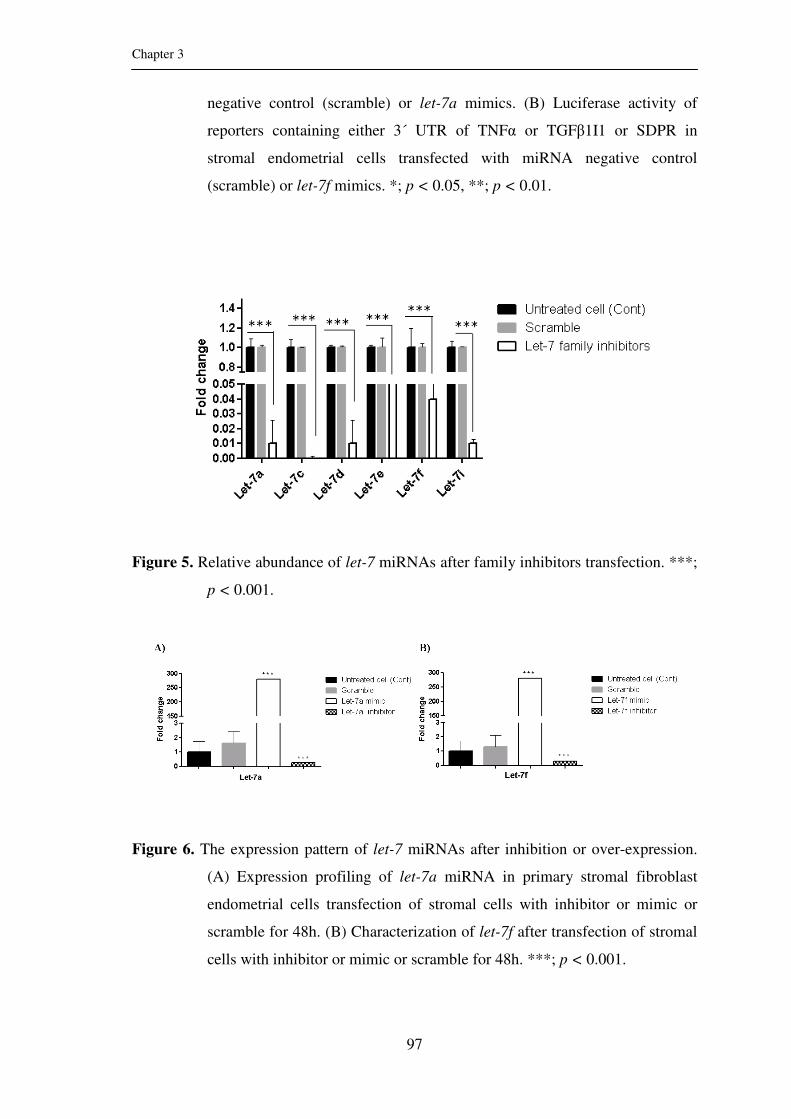
Chapter 3
97
negative control (scramble) or let-7a mimics. (B) Luciferase activity of
reporters containing either 3´ UTR of TNFα or TGFβ1I1 or SDPR in
stromal endometrial cells transfected with miRNA negative control
(scramble) or let-7f mimics. *; p < 0.05, **; p < 0.01.
Figure 5. Relative abundance of let-7 miRNAs after family inhibitors transfection. ***;
p < 0.001.
Figure 6. The expression pattern of let-7 miRNAs after inhibition or over-expression.
(A) Expression profiling of let-7a miRNA in primary stromal fibroblast
endometrial cells transfection of stromal cells with inhibitor or mimic or
scramble for 48h. (B) Characterization of let-7f after transfection of stromal
cells with inhibitor or mimic or scramble for 48h. ***; p < 0.001.

Chapter 3
98
Figure 7. Functional modulation of let-7a revealed clear alterations in selected
inflammatory cytokines and other target genes expression in LPS treated
cells. (A) TNFα, TGFβ1I1 and SDPR expression showed significantly
reciprocal pattern upon transfection with let-7a mimic (M) or inhibitor (I)
or scramble (Scr). (B) The changes in IL6, NFκB and CASP3 expression
profile after transfection with let-7a mimic or inhibitor or scramble. *; p <
0.05, **; p < 0.01, ***; p < 0.001.

Chapter 3
99
Figure 8. Effect of let-7f modulation on expression of candidate inflammatory
cytokines and other target genes in LPS challenged stromal cells. (A)
Alterations of TNFα, TGFβ1I1 and SDPR expression profile upon
transfection with let-7f mimic (M) or inhibitor (I) or scramble (Scr). (B)
Relative abundance of IL6, NFκB and CASP3 mRNAs after transfection
with let-7f mimic or inhibitor or scramble. *; p < 0.05, **; p < 0.01, ***; p
< 0.001.

Chapter 3
100
Figure 9. Direct/indirect effect of let-7a or let-7f transfection on NFκB and TTP
proteins. (A) The indicated proteins were detected by Western blot from cell
lysate of LPS challenged stomal cells after transfection with let-7a mimic
(M) or inhibitor (I) or scramble (Scr). (B) Immunoreactive bands of
indicated protein from cell lysate of LPS treated stomal cells after
transfection with let-7f mimic or inhibitor or scramble, using Western blot.

Chapter 3
101
Supplementary Figure 1. The potential regulatory let-7a and let-7f miRNAs and their
sequence alignment with the binding sites of 3´ UTR (A)
TNFα, (B) TGFβ1I1 and (C) SDPR candidate genes.

Chapter 3
102
Supplementary Figure 2. Protein expression analysis in LPS challenged stromal cells.
(A) ELISA analysis of TNFα and IL6 after transfection with
let-7a mimic or inhibitor or scramble. (B) ELISA analysis
of TNFα and IL6 after transfection with let-7f mimic or
inhibitor or scramble. (C) prostaglandin E2 (PGE2) to
prostaglandin F2α (PGF2α) ratio in cell culture supernatant
upon transfection with let-7a mimic or inhibitor or
scramble. *; p < 0.05, **; p < 0.01, ***; p < 0.001.

Chapter 3
103
Supplementary Figure 3. LPS regulates let-7 miRNAs expression in primary
endometrial stomal cells. (A) Luciferase activity of
reporters containing either 3´ UTR of TNFα or TGFβ1I1
or SDPR in cells transfected with miRNA negative
control (scramble) or let-7a mimics (M) or let-7a
inhibitors (I) or let-7a inhibitors+LPS. (B) Luciferase
activity of reporters containing either 3´ UTR of TNFα
or TGFβ1I1 or SDPR in cells transfected with miRNA
negative control (scramble) or let-7f mimics or let-7f
inhibitors or let-7f inhibitors+LPS. *; p < 0.05, **; p <
0.01, ***; p < 0.001.

Acknowledgements
First of all, I would like to express my sincere thanks to Almighty Allah, for giving
power and helping me to accomplish this work.
Life is a big collaboration and I definitely did not get here on my own. Accordingly, I
would like to acknowledge the contribution of the following people, without whom this
study would not have been possible:
My sincere gratitude and great thanks go to my first supervisor Prof. Dr. Karl
Schellander, for allowing me to join his group and giving me the opportunity to perform
my thesis. I am really grateful for invaluable support, guidance and enthusiastic
supervision during my PhD study. Words alone can not express my sincere gratitude to
Prof. Dr. Schellander for his understanding, encouragement and his thoughtful
feedbacks which always push me forward. I have received extraordinary support from
him in so many ways. His attitude is always that obstacles are there to be overcome, and
challenges to be solved. I would also like to thank Prof. Dr. agr. Brigitte Petersen for
her willingness to be my second supervisor, valuable advice and evaluation of my work.
My deepest thanks also go to PD Dr. Dawit Tesfaye; who selflessly devoted his time
and energy to guiding me through this study on a daily basis. He shared his expertise to
help me develop an understanding and passion for molecular biology, and his
meticulous appraisal of manuscripts, presentations and thesis draft. I am also thankful to
Prof. Dr. Christan Looft and Dr. Tholen for the helpful suggestions and comments
during my presentation sessions.
A number of individuals within the Institute of Animal Science who helped me along
the way deserve special acknowledgement. I want to thank, Dr. Michael Höelker and
Ms. Franka Rings, for providing me valuable experimental samples. I gratefully
acknowledge to Dr. Dessie Salilew Wondim for his help, support and manuscript
revision. My special thanks go to Dr. Christine Große-Brinkhaus, for helping me in
statistical analysis as well as the German version of the abstract (zusammenfassung).

My sincere thanks goes to my colleagues at the Institute of Animal Science for helping
me in one way or another to successfully accomplish this task, especially to Dr. Huitao
Fan, Dr. Dagnachew Hailemariam, Dr. Ulas Mehmet Cinar, Dr. Pritam Bala Sinha, Dr.
Muhammad Jasim Uddin, Dr. Ahmed Yehia Gad, Dr. Simret Weldenegodguad, Dr. Eva
Held, Dr. Luc Frieden, Dr. Hanna Heidt, Dr. Christiane Neuhoff, Dr. Maren Pröll, Dr.
Mohammad Ariful Islam, Dr. Mahmudol Hasan Sohel, Dr. Asep Gunawan, Dr. Sina
Seifi, Dr. Sudeep Sahadevan, Ms. Sarah Bergfelder, Mr. Ahmed Amin, Mr. Sigit
Prastowo, Mr. Samuel Etay, Mr. Aminul Islam, Mr. Eryk Andreas, Ms. Julia
Welzenbach, Mr. Hari Om Pandey and Ms. Sharmin Aqter Rony, not only for their
being my super colleagues but also for many great things that they have shared with me
during the years I lived far away from home.
To my dearest friends and colleagues at the Institute of Animal Science Dr. Walla Abd-
Nabi, Dr. Ijaz Ahmed, Ms. Xueqi Qu, Mr. Rui Zhang and Ms. Qin Yang, Mr.
Mohammed Zidane, I am forever grateful to you for your unyielding support and
encouragement. Above all, thank you for your friendship all through, and making
friendly environment just like home in abroad.
I am sincerely grateful to Ms. Helga Brodeßer for her sincere and dedicated help in the
cell culture lab. Especial thanks go to all technical assistants: Ms. Birgit Koch-Fabritius,
Ms. Stephanie Fuchs and Ms. Nadine Leyer, Mr. Heinz Björnsen for offering me
samples and technical assistance and providing me good working environment
whenever I need. I would like to thank all administrative members of the Institute of
Animal Science, particularly Ms. Bianca Peters for her kind help with all documents.
Thanks also go to Mr. Peter Müller for his really useful help in computer technique and
Mr. Stephan Knauf for his technical assistance.
I would also like to thank Dr. rer. nat. Jochen Reinsberg at
Gynäkologische Endokrinologie und Reproduktionsmedizin Zentrum für Geburtshilfe
und Frauenheilkunde Laborleitung Endokrinologie, Universitäatsklinikum Bonn for his
help and allowing me to use Luminometer, and Dr. Till Schäberle at Institut für
Pharmazeutische Biologie, Bonn University for his help for cloning process. Moreover,

we are thankful to Prof. Dr. Andreas Meyer at INERS ‒ Chemical Signalling institute,
Bonn University for his help and allowing me to use confocal microscope.
I gratefully acknowledge German Academic Exchange Service (DAAD) for providing
me financial support during the PhD study period. Moreover, I would like to thank
National Research Center, for granting me study leave.
Finally, I would like to express my sincere appreciation to my dearest friends and
colleagues for their support throughout my PhD study. Thanks for sharing my highs and
lows, without their encouragement and persistent confidence in me this could not have
been possible. In particular, I would like to thank Ms. Latifa Ait Sghir, Ms. Eman
Kamel, Ms. Azza Tolba, Ms. Rita Wende, Dr. Muna Soltan, Dr. Heba Fawzy, Ms.
Fatima Mohammed, Ms. Ibtisam Fawzy, Ms. Fatima Zahra Khartoum, Ms. Enas
Elshara, Ms. Mona Saad, Mr. Esam Tolba, Mr. Abdelmonem Amer and Mr. Sherif
Elazab for their understanding and moral support.
Last but not least, I send the warmest and most precious thank you to my beloved
mother and my lovely siblings for their endless love, patience, support and endless
encouragement not just with work but all aspects of my life during that always inspire
me to finish my study successfully.

Curriculum vitae
4. Research publications
a) Peer reviewed published articles
1) W.M. Ahmed, M.M. Bashandy, A.K. Ibrahim, S.I.A Shalaby, Sherein I. Abd
El-Moez, Faragalla M. El Moghazy and Sally Ibrahim (2010): Investigations
on Delayed Puberty in Egyptian Buffalo-Heifers with Emphasis on
Clinicopathological Changes and Treatment Using GnRH (Receptal®). Global
Veterinaria 4, 78‒85.
2) D. Hailemariam, S. Ibrahim, M. Hoelker, M. Drillich, W. Heuwieser, C.
Looft, M. U. Cinar, E. Tholen, K. Schellander and D. Tesfaye (2013):
MicroRNA-regulated molecular mechanism underlying bovine subclinical
endometritis. Reproduction, Fertility and Development 26, 898‒913.

b) Article under review
1) Sally Ibrahim, Dessie Salilew-Wondim, Franca Rings, Michael Hoelker,
Christiane Neuhoff, Ernst Tholen, Christian Looft, Karl Schellander, Dawit
Tesfaye (2014): Expression pattern of inflammatory response genes and
their regulatory miRNAs in bovine oviductal cells in response to
lipopolysaccharide: Implication for early embryonic development. Plos One
(PONE-D-14-35766R2 - [EMID:1d643e9e6a99d05b]).
c) Thesis
1) M.V.SC. Thesis: Sally Ibrahim (2008): Some Clinicopathological Studies
on Delayed Puberty Phenomenon in Buffalo-heifers. Faculty of Veterinary
Medicine, Department of Clinical Pathology, Cairo University, Egypt
d) Abstract / posters
1) D Hailemariam, S Ibrahim, M Hoelker, M Drillich, W Heuwieser, C Looft,
M U Cinar, E Tholen, K Schellander, D Tesfaye (2013): Sub-clinical
endometritis induced changes on miRNA expression profile in bovine
endometrium. DGfZ/GfT-Gemeinschaftstagung, 4./5.9.2013, Göttingen,
Deutschland, Tagungsband: B2 (Proc).
2) S Ibrahim, D Salilew-Wondim, F Rings, M Hoelker, C Neuhoff, E Tholen,
C Looft, K Schellander, D Tesfaye (2014): Transcriptomic profile of bovine
oviduct epithelium after lipopolysaccharide (LPS) challenge in vitro and its
implication on embryonic development. Annual Conference of the European
Society for Domestic Animal Reproduction (ESDAR), 10.-13.9.2014,
Helsinki, Finland, Book of Abstracts (Abstr).
3) S Ibrahim, D Salilew-Wondim, F Rings, M Hoelker, C Neuhoff, E Tholen,
C Looft, K Schellander, D Tesfaye (2014): Expression profiling of
inflammatory response mRNAs and their potential regulatory miRNAs in
bovine oviduct after lipopolysaccharide challenge and its implication on
early embryonic development. DGfZ/GfT-Gemeinschaftstagung,
17./18.9.2014, Dummerstorf, Deutschland, Tagungsband: A23 (Proc).
Related Documents