Safety of physical examination alone for managing well-appearing neonates ≥ 35 weeks’ gestation at risk for early-onset sepsis Alberto Berardi, MD, 1 Sara Fornaciari, MD, 1 Cecilia Rossi, MD, 2 Viviana Patianna, MD, 1 Maria Letizia Bacchi Reggiani, MD 3 Filippo Ferrari, MD, 4 Isabella Neri, MD, 5 and Fabrizio Ferrari, MD. 1 1 Unità Operativa di Terapia Intensiva Neonatale, Azienda Ospedaliero-Universitaria Policlinico, Modena, Italy 2 Unità Operativa di Terapia Intensiva Neonatale, Azienda Ospedaliera Santa Maria Nuova, Reggio-Emilia, Italy 3 Dipartimento Cardiovascolare, Università di Bologna, Italy 4 Struttura Complessa di Microbiologia e Virologia, Azienda Ospedaliero-Universitaria Policlinico, Modena, Italy 5 Dipartimento Materno-Infantile, Azienda Ospedaliero-Universitaria Policlinico, Modena, Italy Corresponding author: Alberto Berardi, Unità Operativa di Terapia Intensiva Neonatale, Azienda Ospedaliero- Universitaria Policlinico, Via del Pozzo, 71 - 41100 Modena (MO), Italy. Phone: ++39 059 422 4921; Fax: ++39 059 422 3770. e-mail: [email protected] Short Title: The management of neonates at-risk for early-onset sepsis 1

Welcome message from author
This document is posted to help you gain knowledge. Please leave a comment to let me know what you think about it! Share it to your friends and learn new things together.
Transcript
Safety of physical examination alone for managing well-appearingneonates
≥ 35 weeks’ gestation at risk for early-onset sepsis
Alberto Berardi, MD,1 Sara Fornaciari, MD,1 Cecilia Rossi, MD,2
Viviana Patianna, MD,1 Maria Letizia Bacchi Reggiani, MD3 FilippoFerrari, MD,4 Isabella Neri, MD,5 and Fabrizio Ferrari, MD.1
1 Unità Operativa di Terapia Intensiva Neonatale, AziendaOspedaliero-Universitaria Policlinico, Modena, Italy
2 Unità Operativa di Terapia Intensiva Neonatale, AziendaOspedaliera Santa Maria Nuova, Reggio-Emilia, Italy
3 Dipartimento Cardiovascolare, Università di Bologna, Italy
4 Struttura Complessa di Microbiologia e Virologia, AziendaOspedaliero-Universitaria Policlinico, Modena, Italy
5Dipartimento Materno-Infantile, Azienda Ospedaliero-UniversitariaPoliclinico, Modena, Italy
Corresponding author:
Alberto Berardi, Unità Operativa di Terapia Intensiva Neonatale,
Azienda Ospedaliero-
Universitaria Policlinico, Via del Pozzo, 71 - 41100 Modena (MO),
Italy. Phone: ++39 059 422
4921; Fax: ++39 059 422 3770. e-mail:
Short Title: The management of neonates at-risk for early-onset
sepsis
1

Key words: Group B streptococcus, Neonatal sepsis, Management, Intrapartum antibiotic prophylaxis, Prevention
Abbreviations:
EOS - early-onset sepsis PEA - physical examination alone WAARNs - well-appearing at-risk newborns
2
ABSTRACT
OBJECTIVE: published data to support recommendations for
prevention and management of well-appearing at-risk newborns
(WAARNs) for early-onset sepsis (EOS) are limited.
METHODS: retrospective cohort study comparing 2 different
strategies for managing WAARNs (≥35 weeks’ gestation) during a 6-
year period (Period 1, 2005-2007; Period 2, 2009-2011).
WAARNs were defined as healthy-appearing neonates evaluated
because of risk factors for EOS.
Laboratory evaluation plus simplified physical examination (Period
1) was compared with physical examination alone (PEA, Period 2).
The use of antibiotics, the length of stay, the timeliness of
diagnosis and the risk of falling ill immediately after hospital
discharge in both periods were also compared.
RESULTS: WAARNs receiving empirical antibiotics were 14/500
(Period 1) and 3/500 (Period 2, p=0.01). Median length of stay
was 4 (Period 1) and 3 days (Period 2, p=0.04).
Symptoms of EOS were earlier than laboratory evaluation results in
42/44 neonates. Severe disease was diagnosed within 6 hours of
life in all neonates. No WAARNs presented with EOS following
hospital discharge.
3
CONCLUSIONS: WAARNs managed through PEA received less unnecessary
antibiotics and had a shorter length of stay. They had no
increased risk of severe complications or increased risk of
becoming ill following hospital discharge.
4

Introduction
Early-onset sepsis is typically defined as sepsis occurring within
the first 3 or 7 days after birth. Seven days is typically used
for group B streptococcus, a leading cause of EOS in newborn
infants (1). Prevention guidelines as well as obstetrical and
neonatal care have reduced the incidence of both group B
streptococcus-specific (2) and overall EOS (3,4). In the United
States, overall incidence has recently declined to 0.8-1.0
cases/1000 live births (2-4). However, sepsis remains a major
cause of mortality and morbidity in the newborn. Initial symptoms
of sepsis are often subtle, but the clinical course may be
fulminant (1).
Up to 10-15% of all newborn babies are evaluated to rule out EOS
(5,6). However, the sensitivity of blood cultures following
intrapartum antibiotic prophylaxis is low (7), as is the positive
predictive value of a complete blood count in well-appearing at-
risk newborns (WAARNs) (8). A complete blood count provides more
information about the risk of sepsis after the first 4 hours
following birth (9). C-reactive-protein is one of the standard
parameters in the workup of neonatal sepsis. However, limitations
of C-reactive-protein include low sensitivity during the early
phases of sepsis and raised values in many non-infectious diseases
5

(10). More promising markers are expensive and unavailable in most
centers. Consequently, EOS evaluation remains a subject of
significant controversy.
As clinical signs are a sensitive indicator of neonatal sepsis
(11), the 2010 revised Centers for Disease Control and Prevention
guidelines recommend observation alone instead of laboratory
evaluation plus observation for full-term WAARNs whose mother has
<18 hours of membrane rupture and received inadequate intrapartum
antibiotic prophylaxis. However, the algorithm for neonatal
management includes the option of performing a limited diagnostic
evaluation at age 6–12 hours (2).
There is no consensus for this recommendation. In 2013, the
American Academy of Pediatrics (Committee on the Fetus and
Newborn) recommended observation for 48 hours without further
testing or cultures in WAARNs (≥ 37 weeks’ gestation) in cases of
inadequate intrapartum antibiotic prophylaxis and membrane rupture
>18 hours (12). The discordance in the algorithms has prompted
questions by the paediatric community as to which recommendations
to follow.
We recently reported that infants with ≥35 weeks’ gestation at
risk of group B streptococcus EOS can be safely managed with
physical examination alone (PEA) (13). However, we did not assess
6
the effectiveness of this strategy for all cases of EOS, its
impact on the number of antibiotic treatments or length of stay.
Furthermore, there is a concern that neonates may fall ill after
hospital discharge if not duly evaluated. Current data concerning
PEA in WAARNs are limited and further data supporting this
strategy should be considered.
The purpose of this study was i) to assess whether PEA leads to a
timely diagnosis of proven/suspected EOS in neonates with ≥35
weeks’ gestation and ii) whether PEA affected rates of unnecessary
antibiotics and length of stay or increased the risk of falling
ill after hospital discharge.
7
Methods
Study design: this is a retrospective cohort study of infants
delivered at Modena University Hospital (Italy), a high volume
tertiary care centre with approximately 3500 live births/yr. The
project was approved by the local ethical committee (N°125/11)
The Modena University Hospital advocates a strategy of recto-
vaginal culture screening at 35-37 weeks of gestation. Women with
prenatal group B streptococcus colonization are given intrapartum
antibiotic prophylaxis. Furthermore, intrapartum antibiotic
prophylaxis is administered to women with unknown group B
streptococcus status and risk factors, such as group B
streptococcus bacteriuria identified during the current pregnancy,
a previous group B streptococcus -infected newborn, preterm birth
(<37 weeks’ gestation), rupture of membrane 18 hours, or
intrapartum fever 38° C (a surrogate for chorioamnionitis).(2)
Full maternal data (gestational age, mode of delivery, group B
streptococcus status, risk factors for EOS, duration of
intrapartum antibiotic prophylaxis) is routinely recorded in
neonatal charts.
Management of WAARNs 35 weeks gestation: in Period 1 (2005 to
2007) a laboratory evaluation (blood culture, C-reactive protein
test and complete blood count) was recommended at age 6-12 hours
8
for neonates born to mothers with intrapartum fever or neonates
receiving inadequate intrapartum antibiotic prophylaxis (14).
Empirical antibiotics were given routinely to neonates born to
mothers with intrapartum fever or neonates with abnormal
laboratory evaluation.
In Period 2 (2009 to 2011) PEA became the standard of care (13)
Laboratory evaluation was recommended only in a few WAARNs
(neonates born to mothers with intrapartum fever or neonates with
at least 2 risk factors receiving inadequate intrapartum
antibiotic prophylaxis). PEA is performed by midwives within 2
hours of birth, and then by nurses and clinicians in turn. Each
examiner fills in and signs a standardized form (detailing general
wellbeing, skin colour including perfusion, and the presence of
respiratory signs) at standard intervals (at age 3-6-12-18-36-48
hours). A baby with any symptom of possible sepsis is immediately
referred to a neonatal care specialist.
Comparison of screening practices: compliance with the maternal
prevention strategy, rates of laboratory evaluations and
antibiotic treatments were compared by reviewing medical records
of 1000 neonatal births (35 weeks gestation). In order to ensure
a representative sample of the broad population of neonatal births
9

in the two study periods, we performed a random selection of
neonatal charts, 500 from Period 1 and 500 from Period 2.
Live births occurring during 2008 were excluded, because the
neonatal management approach changed in that year.
Three investigators (S.F, V.P. and. C.R.) abstracted information
from neonatal records. Standardized forms were used to collect
data (antenatal screening, gestational age, ethnicity, mode of
delivery, hours since membrane rupture, intrapartum antibiotics,
Apgar score, sex, birth weight, health status, laboratory
evaluation, length of stay).
Cases of EOS: with the aim of confirming whether PEA leads to a
timely diagnosis, we i) searched the laboratory database for
pathogens yielded in blood or cerebrospinal fluid cultures during
both study periods and ii) we reviewed the medical records of
neonates with International Code of Diagnosis (Version:2010) of
EOS at discharge from hospital (sepsis, 038.0, 038.4, 038.9,
038.42, 995.91, 995.92, 771.81; bacterial meningitis, 320.0-9;
pneumonia and additional infections, 482.0-9, 041.02, 941.19,
041.4, 599.0). Charts of sick neonates readmitted to our Unit
within 7 days of birth with a code diagnosis of sepsis were also
abstracted from our registry and were reviewed.
10
All neonates were analysed by comparing their ages at the first
suspicion of sepsis (based on symptoms and/or abnormal laboratory
evaluation results). Since sepsis presenting after the age of 3
days may be the result of horizontal transmission, and as we
wished to assess the long-term effects of PEA, neonates presenting
with illness from 0 to 72 and from 73 to 168 hours were analyzed
separately.
Definitions: Culture proven early-onset sepsis is defined as isolation of a
pathogen from a normally sterile body site (blood or cerebrospinal
fluid) within 7 days of birth and clinical signs and symptoms
consistent with sepsis.(1)
Suspected early-onset sepsis is defined as the presence of clinical signs
and symptoms consistent with sepsis (see reference 1), plus
abnormal complete blood count and/or an elevated C-reactive-
protein level in the absence of a positive blood culture.
Well-appearing status, refers to cases in which EOS evaluation is
performed only in the presence of risk factors, without clinical
symptoms of sepsis.
At-risk newborn is defined as an infant whose mother is group B
streptococcus colonized or has risk factors for EOS.
11
Severe disease is any of the following: death, meningitis, seizures,
brain lesions at discharge from hospital, need for catecholamine
support or mechanical ventilation.
Statistical analysis: A calculation was made of the sample size
needed to detect a difference between neonatal management
approaches: assuming that at least 4% and 1% of WAARNs underwent
laboratory evaluation in Period 1 and Period 2, respectively, with
alpha = 0.05 and a power of 80%, the required sample was
determined as 489 births in Period 1 and 489 births in Period 2
Statistical analysis was performed using the 2 test and Mann-
Whitney test for independent samples, when appropriate. A p value
<.05 was used as a threshold for statistical significance.
12
Results
During the 6-year study period there were 20,401 live births (9832
live births in Period 1; and 10569 in Period 2), of which 19,504
(95.6%) with ≥35 weeks’ gestation.
Comparison of screening practices: Eleven records were incomplete
and were excluded; 1000 neonatal medical records randomly selected
were abstracted and reviewed. Table I compares the characteristics
of 500 records from Period 1 and 500 records from Period 2.
Antenatal screening and recto-vaginal cultures increased
significantly in Period 2.
Table II compares laboratory evaluations and antibiotic
treatments. With respect to Period 1, in Period 2 the rates of
laboratory evaluations significantly decreased in WAARNs. Fewer
WAARNs received empirical antibiotics and the length of stay was
significantly shorter.
Forty-six neonates in Period 2 underwent PEA; among them 2 had
signs of illness within 6 hours of birth. Sepsis was ruled out in
both neonates.
Neonates with a code diagnosis of EOS. Among the 19,504 babies
with 35 or more weeks’ gestation, 93 were diagnosed with EOS. Two
13
of the 93 neonates (1 in Period 1 and 1 in Period 2) presented
with symptoms at age 73 to 168 hours; neither was classified as
at-risk. Eleven of the 93 neonates (8 in Period 1 and 3 in period
2) had no apparent signs of illness and cultures were sterile.
Their diagnoses of infection were based on abnormal results of
tests performed to rule out sepsis in WAARNs.
Among the remaining 80 neonates with symptoms within 72 hours of
birth, 44 were at risk and 36 had no identifiable risk factor.
Eight out of 36 neonates without risk factors (4 in period 1 and 4
in period 2) had culture-proven sepsis (group B streptococcus,
n=2; Staphylococcus aureus, n=3; Escherichia coli, n=2; Listeria
monocytogenes, n=1).
At-risk neonates with a code diagnosis of EOS: Each at-risk
neonate was analysed in terms of age at the first suspicion of
sepsis (based on symptoms and/or abnormal laboratory evaluation
results). Results were informative before the presentation of
symptoms of EOS only for 2 out of 44 at-risk neonates. Age at the
first suspicion of sepsis did not change between Period 1 (median
2 hours, range 0-36, IQ 0.0-7.0) and Period 2 (median 0, range 0-
60, IQ 0.0-2.0, p 0.40).
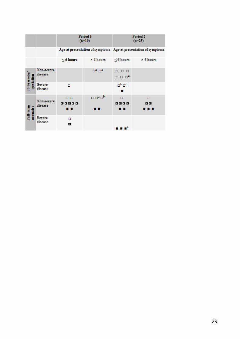
Figure 1 details the 44 at-risk neonates with early presentation
of symptoms (≤72 hours) according to age at presentation and
14
intrapartum antibiotic prophylaxis exposure. Severe disease (n= 9)
presented within 6 hours of life in all cases. Eight neonates (4
in Period 1 and 4 in Period 2) had culture-proven EOS.
Findings that were relevant for the prediction of sepsis through
PEA in Period 2 were: tachypnoea and grunting (n=4), poor
perfusion (n=3) or lethargy (n=2).
Positive blood cultures in WAARNs: During the study period, there
were moreover 26 positive blood cultures in WAARNs (coagulase-
negative Staphylococci, n=23; Brevibacterium spp, n=2;
Flavobacterium/ Chryseobacterium spp, n=1). Among them, 21
occurred in Period 1 and 5 in Period 2. None of the 26 neonates
had symptoms within the first week of life. C-reactive-protein and
complete blood count yielded normal results and isolates were
considered as probable contaminants. These positive cultures were
not included among true cases of EOS.
15
Discussion
Neonatal EOS has become more rare than in the past. However,
pediatricians are frequently faced with the challenge of
evaluating and managing WAARNs born to mothers with inadequate
intrapartum antibiotic prophylaxis. Algorithms based on risk-
factor threshold values may result in large numbers of uninfected
newborns being evaluated and treated with empiric antibiotics,
leading to maternal/infant separation and unnecessary antibiotic
exposure. The recent literature underlines that up to 50% of
neonates evaluated for sepsis are treated with antibiotics, even
if only few are subsequently proven to have had an infection
(5,6). Antibiotic exposure may lead to the selection of resistant
organisms, or may induce changes in the gut flora composition
resulting in long-term effects (15-17).
Given these concerns, data supporting safe alternative strategies
for managing at-risk neonates should be considered. Because
information is limited, the American Academy of Pediatrics urges
that data be provided to support recommendations for prevention
and management of neonates at risk of EOS (12).
Cantoni and co-workers (18) evaluated a cohort of 15239 full-term
neonates, of whom 3092 were at risk. They demonstrated that
16
laboratory tests plus standardized physical examination offered no
advantage over standardized PEA. This latter strategy was also
associated with fewer infants treated with antibiotics. However,
they excluded preterm neonates from their analysis; furthermore,
the only outcome data provided by the authors concerned
respiratory support, which is only a rough outcome measure.
We recently reviewed cases of culture-proven group B streptococcus
-specific EOS in Emilia-Romagna (13), an Italian region with
active group B streptococcus surveillance (19). Infants with ≥35
weeks’ gestation were diagnosed in a timely manner through PEA.
This threshold of gestational age was used because signs of sepsis
are quite similar in preterm (35-36 weeks’ gestation) and full-
term neonates
What is left unanswered in both studies is whether neonates have
an increased risk of falling ill following hospital discharge due
to the failure to perform laboratory evaluation in the first days
of life.
In the current study, the group B streptococcus prevention
strategy had been implemented progressively over time. The rates
of antenatal screening and recto-vaginal cultures increased
significantly in Period 2.
17
Approximately 45% of neonates with a diagnosis of EOS in both
study periods were not at risk, consequently they had received no
intrapartum antibiotic prophylaxis.
In both periods most diagnoses of EOS were initially based on an
evaluation of symptoms, as most neonates with EOS and all neonates
with severe disease became ill within 6 hours of birth. The
clinical yield of laboratory testing performed at age 6-12 hours
was negligible.
There was no evidence that neonates undergoing PEA in period 2 had
an increased risk of falling ill immediately after hospital
discharge. Furthermore, significantly fewer WAARNs had laboratory
evaluations, contaminants in blood cultures, or received empirical
antibiotics. Consequently they had a shorter length of stay.
This study has some limitations. Firstly, our results may not be
generalizable to centres that cannot perform frequent clinical
evaluations of neonates in the first hours of life.
In addition, this is an observational, retrospective study, and
the cases of EOS were checked through the diagnosis code.
Consequently, some cases of suspected EOS in both periods may have
been missed. Furthermore, we could not exclude that some neonates
discharged home were readmitted elsewhere. However, Modena
18
University Hospital is a reference hospital for the entire
province that receives the vast majority of neonates.
Despite these limitations our findings show that WAARNs need to be
observed closely during the first few hours of life for having a
timely diagnosis of EOS.
Declaration of Interest statement
The authors report no declarations of interest.
19
Figure 1 – At-risk neonates (≥ 35 weeks’ gestation) presenting
with sepsis within 72 hours of birth: comparison between Period 1
(n=19) and Period 2 (n=25)
Figure 1 legend - Open squares indicate neonates without
intrapartum antibiotic prophylaxis exposure. Half-solid squares
indicate neonates exposed to inadequate intrapartum antibiotic
prophylaxis. Solid squares indicate neonates with adequate
intrapartum antibiotic prophylaxis.
Letters in superscript next to 8 squares show the infants with
culture-proven sepsis:
a: Group B streptococcus (n=5)
b: Staphylococcus aureus (n=2)
c: Listeria monocytogenes (n=1)
20
References:
1. Palazzi D, Klein J, Baker C. Bacterial sepsis and meningitis.
In Infectious diseases of the fetus and newborn infant. 6th
edition. Edited by Remington J, Klein J, Wilson C, Baker C.
Philadelphia: Elsevier Saunders; 2006: 247-295.
2. Verani JR, McGee L, Schrag SJ. Division of Bacterial Diseases,
National Center for Immunization and Respiratory Diseases,
Centres for Disease Control and Prevention (CDC). Prevention of
perinatal group B streptococcal disease — revised guidelines
from CDC, 2010. MMWR Recomm Rep 2010, 59 (RR-10):1-36.
3. Weston EJ , Pondo T, Lewis MM, Martell-Cleary P, Morin C, Jewell
B, et al: The burden of invasive earlyonset neonatal sepsis in
the United States, 2005-2008. Pediatr Infect Dis J 2011,
30:937-941.
4. Stoll BJ , Hansen NI, Sánchez PJ, Faix RG, Poindexter BB, Van
Meurs KP et al:: Early onset neonatal sepsis: The burden of
group B streptococcal and E. coli disease continues. Pediatrics
2011, 127:817-826.
5. National Institute for Health and Clinical
Excellence. Antibiotics for early-onset neonatal infection:
antibiotics for the prevention and treatment of early-onset
21
neonatal infection. (CG102) London: NICE;
2012. http://guidance.nice.org.uk/CG149/Guidance/pdf/English
(accessed 13 Sep 2013)
6. Mukhopadhyay S, Puopolo KM: Risk assessment in neonatal early
onset sepsis. Semin Perinatol. 2012, 36:408-15.
7. Hsu KK, Pelton SI, Shapiro DS: Detection of group B
streptococcal bacteremia in simulated intrapartum antimicrobial
prophylaxis. Diagn Microbiol Infect Dis 2003;45:23–7.
8. Ottolini MC, Lundgren K, Mirkinson LJ, Cason S, Ottolini MG:
Utility of complete blood count and blood culture screening to
diagnose neonatal sepsis in the asymptomatic at risk newborn.
Pediatr Infect Dis J 2003;22:430–4.
9. Newman TB, Puopolo KM, Draper D, Escobar GJ: Interpreting
complete blood counts soon after birth in newborns at risk for
sepsis. Pediatrics. 2010;126:903-9.
10. Hofer N, Zacharias E, Müller W, Resch B: An update on the use
of C-reactive protein in early-onset neonatal sepsis: current
insights and new tasks. Neonatology. 2012;102:25-36.
11. Escobar GJ , Li DK, Armstrong MA, Gardner MN, Folck BF, Verdi
JE, et al:: Neonatal sepsis workups in infants >/=2000 grams at
birth: A population-based study. Pediatrics 2000;106:256-63.
22
12. Brady MT, Polin RA: Prevention and management of infants with
suspected or proven neonatal sepsis. Pediatrics. 2013;132:166-8.
13. Berardi A, Lugli L, Rossi C, Guidotti I, Lanari M, Creti R, et
al; GBS Prevention Working Group, Emilia-Romagna: Impact of
Perinatal Practices for Early-Onset Group-B Streptococcal
Disease Prevention. Pediatr Infect Dis J. 2013;32:e265-71
14. Berardi A , Lugli L, Baronciani D, Rossi C, Ciccia M, Creti R et
al:; GBS Prevention Working Group of Emilia-Romagna: Group B
Streptococcus early-onset disease in Emilia-Romagna: review
after introduction of a screening-based approach. Pediatr Infect
Dis J 2010;29:115-2.
15. Bedford Russell AR, Murch SH: Could peripartum antibiotics have
delayed health consequences for the infant? BJOG. 2006;113:758-
65
16. Moore MR, Schrag SJ, Schuchat A: Effects of intrapartum
antimicrobialprophylaxis for prevention of group-B-streptococcal
disease on the incidence and ecology of early-onset neonatal
sepsis. Lancet Infect Dis 2003;3:201–13.
17. Alm B, Erdes L, Möllborg P, Pettersson R, Norvenius SG, Aberg
N, et al:: Neonatal antibiotic treatment is a risk factor for
early wheezing. Pediatrics 121:697-702, 2008
23

18. Cantoni L, Ronfani L, Da Riol R, Demarini S: Physical
Examination Instead of Laboratory Tests for Most Infants Born to
Mothers Colonized with Group B Streptococcus: Support for the
Centers for Disease Control and Prevention's 2010
Recommendations. J Pediatr. 2013;163:568-73
19. Berardi A, Lugli L, Baronciani D, Creti R, Rossi K, et al:; GBS
Prevention Working Group of Emilia-Romagna: Group B
streptococcal infections in a Northern region of Italy.
Pediatrics 2007;120:e487-93.
24
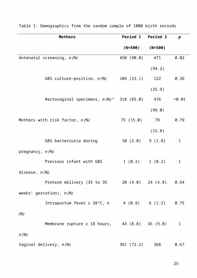
Table I: Demographics from the random sample of 1000 birth records
Mothers Period 1
(N=500)
Period 2
(N=500)
p
Antenatal screening, n (%) 450 (90.0) 471
(94.2)
0.02
GBS culture-positive, n (%) 104 (23.1) 122
(25.9)
0.36
Rectovaginal specimens, n (%) ° 318 (85.0) 476
(99.0)
<0.01
Mothers with risk factor, n (%) 75 (15.0) 79
(15.8)
0.79
GBS bacteriuria during
pregnancy, n (%)
10 (2.0) 9 (1.8) 1
Previous infant with GBS
disease, n (%)
1 (0.2) 1 (0.2) 1
Preterm delivery (35 to 36
weeks’ gestation), n (%)
20 (4.0) 24 (4.8) 0.64
Intrapartum fever ≥ 38°C, n
(%)
4 (0.8) 6 (1.2) 0.75
Membrane rupture ≥ 18 hours,
n (%)
44 (8.8) 45 (9.0) 1
Vaginal delivery, n (%) 361 (72.2) 368 0.67
25

(73.6)
IAP administration, n (%) 132 (26.4) 148
(29.6)
0.29
IAP administration in culture-
positive women, n (%) †
85 (90.4) 103
(94.5)
0.40
Gestational age, weeks, median (IQ) 39 (38-40) 39 (38-
40)
0.84
Birth weight, g, median (IQ) 3350(3030-3650)
3315(3057-3625)
0.55
IAP, Intrapartum antibiotic prophylaxis; GBS, group B streptococcus; IAP, intrapartum antibiotic prophylaxis; IQ, interquartile range 25th -75th
° information on specimen (whether vaginal or recto-vaginal) was not available in 126 and 19 cases (period 1 and period 2 respectively); % rates include only women with known information.
† women with planned caesarean section were excluded from calculation
26
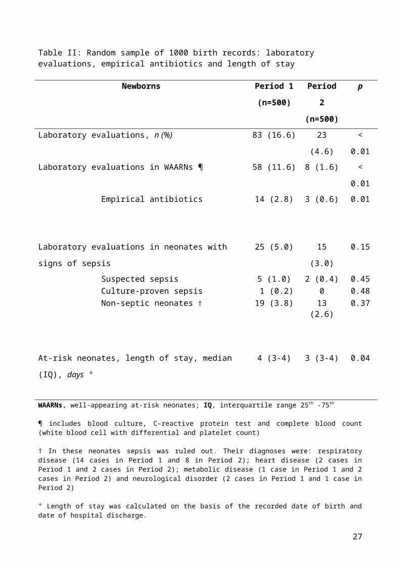
Table II: Random sample of 1000 birth records: laboratory evaluations, empirical antibiotics and length of stay
Newborns Period 1
(n=500)
Period
2
(n=500)
p
Laboratory evaluations, n (%) 83 (16.6) 23
(4.6)
<
0.01 Laboratory evaluations in WAARNs ¶ 58 (11.6) 8 (1.6) <
0.01 Empirical antibiotics 14 (2.8) 3 (0.6) 0.01
Laboratory evaluations in neonates with
signs of sepsis
25 (5.0) 15
(3.0)
0.15
Suspected sepsis 5 (1.0) 2 (0.4) 0.45 Culture-proven sepsis 1 (0.2) 0 0.48 Non-septic neonates † 19 (3.8) 13
(2.6)0.37
At-risk neonates, length of stay, median
(IQ), days °
4 (3-4) 3 (3-4) 0.04
WAARNs, well-appearing at-risk neonates; IQ, interquartile range 25th -75th
¶ includes blood culture, C-reactive protein test and complete blood count(white blood cell with differential and platelet count)
† In these neonates sepsis was ruled out. Their diagnoses were: respiratorydisease (14 cases in Period 1 and 8 in Period 2); heart disease (2 cases inPeriod 1 and 2 cases in Period 2); metabolic disease (1 case in Period 1 and 2cases in Period 2) and neurological disorder (2 cases in Period 1 and 1 case inPeriod 2)
° Length of stay was calculated on the basis of the recorded date of birth anddate of hospital discharge.
27

28
29
Related Documents