根 の 研 究(Root Research) 13(3):117-131(2004) Root cortex: structural and functional variability and responses to environmental stress Alexander Lux*) Dept. of Plant Physiology, Faculty of Natural Sciences, Comenius University in Bratislava, Slovak Republic; Field Production Science Center, Graduate School of Agricultural and Life Sciences, The University of Tokyo, Japan Miroslava Luxova I nstitute of Botany, Slovak Academy of Sciences, Bratislava, Slovak Republic Jun Abe Dept. of Agricultural and Environmental Biology, Graduate School of Agricultural and Life Sciences, The University of Tokyo, Japan Shigenori Morita Field Production Science Center, Graduate School of Agricultural and Life Sciences, The University of Tokyo, Japan Abstract: The cortex is the basic part of the primary root body and represents an important constituent of the root, both structurally and functionally. In monocotyledons, it might persist during the entire life of the root. In dicotyledons, with limited secondary thickening it might persist for a long time and be subject to dilatation growth. In intensively secondary-thickening roots, the cortex gradually deteriorates and is replaced by secondary tissues-the periderm. The functions of cortical tissues are diverse. The endodermis, and to some extent the exodermis, represent apoplastic barriers that control the uptake and radial transport of water and solutes by the root. However, these layers have several additional functions such as mechanically protecting the stele and protection against pathogens and parasites. Although the mid-cortex (or mesodermis) is primarily the site for reserve material deposition it can also have several different functions that depend on the species and growth conditions. These include aeration in hypoxia (aerenchyma formation), and the location for symbiosis and even photosynthesis. The cortex varies widely amongst species and even in various root types of the same species. It might be designated as a root buffer zone, especially under stress conditions. Some aspects of the development, structure and function of cortical tissues are discussed in this report. Keywords: cortex, endodermis, exodermis, hypodermis, mid-cortex 1. Introduction Root cortical tissues have long attracted the attention of researchers because they are a complex of tissues at the site in which radial movement of water and solutes into the roots is regulated. Originally, it was mainly the endodermis that was considered the root apoplastic barrier-it controlled "free" water and ion movement through the apoplast and forced all transported solutes to pass through the plasma membrane and into the symplast. The plasma membranes selective function regulated the passage of ions at this point of the transport pathway. In the early nineties, due to the introduction of new fluorescence staining techniques (Brundrett et al., 1988, 1991), a series of papers appeared that demonstrated the presence of a second apoplastic barrier in the majority of angiosperms-the exodermis (Perumalla et al., 1990; Peterson and Perumalla, 1990). However, this cortical layer is somehow ecologically 2004年8月31日 受付 * 連 絡先 Dept. of Plant Physiology, Faculty of Natural Sciences, Comenius University, Mlynska dolina B-2, SK-842 15,Bratjslava, Slovak Republic Fax: +421-2-65429064 E-mail: [email protected] -117-

Welcome message from author
This document is posted to help you gain knowledge. Please leave a comment to let me know what you think about it! Share it to your friends and learn new things together.
Transcript
根 の 研 究(Root Research) 13(3):117-131(2004)
Root cortex: structural and functional variability and
responses to environmental stress
Alexander Lux*)
Dept. of Plant Physiology, Faculty of Natural Sciences, Comenius University in Bratislava, Slovak
Republic; Field Production Science Center, Graduate School of Agricultural and Life Sciences,
The University of Tokyo, Japan
Miroslava Luxova
I nstitute of Botany, Slovak Academy of Sciences, Bratislava, Slovak Republic
Jun Abe
Dept. of Agricultural and Environmental Biology, Graduate School of Agricultural and Life Sciences, The University of Tokyo, Japan
Shigenori Morita
Field Production Science Center, Graduate School of Agricultural and Life Sciences, The University of Tokyo, Japan
Abstract: The cortex is the basic part of the primary root body and represents an important constituent of the root, both structurally and functionally. In monocotyledons, it might persist during the entire life of the root. In dicotyledons, with limited secondary thickening it might persist for a long time and be subject to dilatation growth. In intensively secondary-thickening roots, the cortex gradually deteriorates and is replaced by secondary tissues-the periderm. The functions of cortical tissues are diverse. The endodermis, and to some extent the exodermis, represent apoplastic barriers that control the uptake and radial transport of water and solutes by the root. However, these layers have several additional functions such as mechanically protecting the stele and protection against pathogens and parasites. Although the mid-cortex (or mesodermis) is primarily the site for reserve material deposition it can also have several different functions that depend on the species and growth conditions. These include aeration in hypoxia (aerenchyma formation), and the location for symbiosis and even photosynthesis. The cortex varies widely amongst species and even in various root types of the same species. It might be designated as a root buffer zone, especially under stress conditions. Some aspects of the development, structure and function of cortical tissues are discussed in this report. Keywords: cortex, endodermis, exodermis, hypodermis, mid-cortex
1. Introduction
Root cortical tissues have long attracted the
attention of researchers because they are a
complex of tissues at the site in which radial
movement of water and solutes into the roots is
regulated. Originally, it was mainly the endodermis
that was considered the root apoplastic barrier-it
controlled "free" water and ion movement through
the apoplast and forced all transported solutes to
pass through the plasma membrane and into the
symplast. The plasma membranes selective function
regulated the passage of ions at this point of the transport pathway. In the early nineties, due to the
introduction of new fluorescence staining techniques
(Brundrett et al., 1988, 1991), a series of papers appeared that demonstrated the presence of a
second apoplastic barrier in the majority of angiosperms-the exodermis (Perumalla et al.,
1990; Peterson and Perumalla, 1990). However, this cortical layer is somehow ecologically
2004年8月31日 受 付
* 連 絡 先 Dept. of Plant Physiology, Faculty of Natural Sciences, Comenius University, Mlynska dolina B-2, SK-842 15, Bratjslava, Slovak Republic
Fax: +421-2-65429064 E-mail: [email protected]
-117-
Lux et al./根 の 研 究(Root Research) 13(3):117-131(2004)
dependent, and unlike the endodermis, does not
usually represent a uniform sheath that covers the
entire root (see Peterson, 1997). A partially
neglected tissue is the mid-cortical layer, which is
also called the medodermis. It is often a multilayered
tissue that plays an important role in the function
of the root as a storage place for reserve materials,
is metabolically active and produces various
organic compounds, facilitates aeration of roots in
anaerobic conditions and sometimes even provides
assimilates and is a site for photosynthesis.
A structural and functional study of the root
cortex currently provides some new information. In
particular, the endo- and exodermis are the subjects of several recent reviews (Enstone et al.,
2003; Ma and Peterson, 2003, and references
therein). Thus, the main purpose of this review is to
point out some of the less-discussed aspects of
peripheral root tissues and to stimulate research in these areas. It is mainly aimed at (1) the structural
(and, thus, also functional) variability of cortical tissues in various plant species, (2) differences
between individual root types of the same plant, (3)
the little studied processes occurring in the root
cortex of plants with secondary thickened roots, (4)
some aspects of various stress factors, and (5)
inter- and intraspecific differences in the root
cortex that are related to stress tolerance.
2. Concept of cortical layers
The root cortex is a well-defined part of the
primary root body (Fig 1A). Its development is the result of periclinal, anticlinal and transversal cell
divisions of the apical initials (see Morita, 2000;
Barlow, 2002; Baum et al., 2002; Chapman et al.,
2003; and references therein). Meristematic cells
derived from initial cells divide: (1) transversally,
which increases the number of cells in columns in a
longitudinal direction, (2) periclinally, which
increases the number of cell layers in a radial
direction, and (3) anticlinally, which increases the
number of cells in individual cell layers. Therefore,
the root pattern, as can be observed in cross
sections, is formed by periclinal and anticlinal
divisions. In the majority of roots, the sequence of
periclinal divisions is centripetal: the first cells that cease periclinal divisions are at the periphery of the
root cortex, while the final periclinally dividing cells
produce the innermost cortical cells, the cells of the endodermis (see Kawata and Lai, 1965).
However, exceptions to this rule exist, and the
internal part of the cortex shows centripetal growth
while the external part shows a division in a
centrifugal direction in roots of some species.
Despite this, the innermost cortical layer is the
endodermis, and it is a mistake to describe the root
structure as being composed of the cortex and
endodermis, simply because the endodermis is part
of the cortex. Unfortunately, this confusion is quite
common in recent literature, mainly due to the
incorrect interpretation of the Arabidopsis root
structure. This excellent model plant has a
beautifully simple and usually highly regular root
structure. Its cortex often consists of only two
cortical layers, the outer and inner, the innermost
of which is the endodermis. It is quite common to
describe this root structure as consisting of one
layer of cortex and one layer of endodermis.
However, this is not correct.
The external part of the root cortex can be of a
very variable composition. In dicotyledonous plants,
it is usually simple, while in monocotyledonous
plants it is more complex. The most complicated structure of peripheral cortical tissues might be
described as the complex hypodermis, which often
consists of one or several (multiseriate) peripheral
layers of exodermis situated below the epidermis
and the centripetally developed internal sclerenchymatous
layer(s) (Fig. 1B). This model is rather common in
grasses (e.g., in rice, see Morita and Nemoto,
1995; Kondo et al., 2000). The term exodermis is
reserved for the peripheral cortical layer(s) that
develop Casparian bands and form the second (or
first, if we start counting from the root surface)
root apoplastic barrier (Peterson and Perumalla,
1990). However, this layer is somehow less
constant, and unlike the endodermis does not
usually form a compact and uninterrupted sheath
covering the whole root body. It might develop in a
patchy form, discontinuously, or even be absent in some roots of plants that possess the ability to form
exodermis (see Zimmerman and Steudle, 1998). In
species in which the exodermis is absent, the term
hypodermis is used when the external (subepidermal) cell
layer differs from the rest of the cortical tissues.
This difference is sometimes only a result of the
shape, but most frequently a result of the
sclerenchymatous character of the hypodermal
layer(s), which form a mechanical protection over
the root surface.
-118-
LUX et al./根 の 研 究(Root Research) 13(3):117-131(2004)
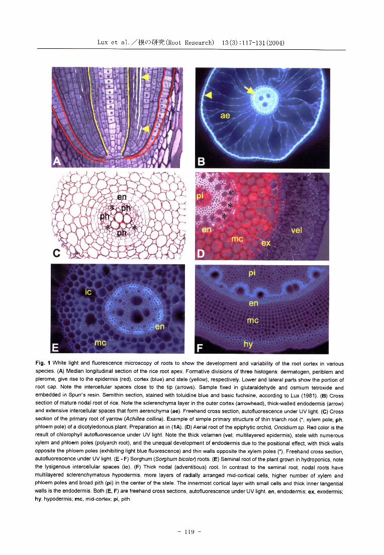
Fig. 1 White light and fluorescence microscopy of roots to show the development and variability of the root cortex in various
species. (A) Median longitudinal section of the rice root apex. Formative divisions of three histogens: dermatogen , periblem and plerome, give rise to the epidermis (red), cortex (blue) and stele (yellow), respectively. Lower and lateral parts show the portion of root cap. Note the intercellular spaces close to the tip (arrows). Sample fixed in glutaraldehyde and osmium tetroxide and
embedded in Spurr's resin. Semithin section, stained with toluidine blue and basic fuchsine, according to Lux (1981) . (B) Cross section of mature nodal root of rice. Note the sclerenchyma layer in the outer cortex (arrowhead), thick-walled endodermis (arrow)
and extensive intercellular spaces that form aerenchyma (ae). Freehand cross section, autofluorescence under UV light. (C) Cross
section of the primary root of yarrow (Achillea collina). Example of simple primary structure of thin triarch root (*, xylem pole; ph , phloem pole) of a dicotyledonous plant. Preparation as in (1A). (D) Aerial root of the epiphytic orchid, Oncidium sp. Red color is the result of chlorophyll autofluorescence under UV light. Note the thick velamen (vel; multilayered epidermis) , stele with numerous xylem and phloem poles (polyarch root), and the unequal development of endodermis due to the positional effect, with thick walls
opposite the phloem poles (exhibiting light blue fluorescence) and thin walls opposite the xylem poles (*). Freehand cross section, autofluorescence under UV light. (E - F) Sorghum (Sorghum bicolor) roots . (E) Seminal root of the plant grown in hydroponics, note the lysigenous intercellular spaces (ic). (F) Thick nodal (adventitious) root . In contrast to the seminal root, nodal roots have
multilayered sclerenchymatous hypodermis, more layers of radially arranged mid-cortical cells, higher number of xylem and
phloem poles and broad pith (pi) in the center of the stele. The innermost cortical layer with small cells and thick inner tangential walls is the endodermis. Both (E, F) are freehand cross sections, autofluorescence under UV light. en, endodermis; ex, exodermis;
hy, hypodermis; mc, mid-cortex; pi, pith.
-119-
Lux et al./根 の 研 究(Root Research) 13(3):117-131(2004)
The mid-cortical layers are sometimes called the
mesodermis. Although this part of the cortex usually remains parenchymatous (Figs. 1C-E), in
some cases secondary thickening of the cell walls might also occur. The older term, mesodermis,
which is somehow neglected in recent literature,
was rather useful, and referred to the middle
position of this cortical part. The arrangement of mesodermal layers, as observed in cross sections, might be in orderly radial rows (radial cell
arrangement, see Fig. 1F) or cells might alternate in successive concentric layers (alternate type, see
Fig1D; Sinnot and Bloch, 1941). In some roots, the
central radial arrangement changes to alternate in
peripheral layers. The presence of schizogenous intercellular spaces is typical for the mesodermis, which usually starts to form close to the apex (Fig.
1A) and might become extensive in some roots. Lacunae are developed in this way. Large
intercellular spaces might also be formed by the breakdown of cells through lysigeny; lysigenous
intercellular spaces are formed in this manner (Fig.
1E). The extensive formation of intercellular spaces or lacunae is typical for aquatic plants and might
also be induced by anaerobic conditions in some non-aquatic species.
Variability of root cortex composition in various
species can be found in Guttenberg (1968) and more recently in the Root Atlas (Kutschera and
Sobotik, 1992). In most cases, these two very extensive works depict the structure of seminal or
adventitious roots. However, the variability of root structure within the given species is accomplished
with lateral roots, which might be of two (or
perhaps even more) types in some species (see Hochholdinger et al., 2004). Lateral roots
represent the majority of the root systems external surface and are probably responsible for the bulk of
nutrient absorption: at least in some species, as has recently become evident (Kirk, 2003). The
structural study of these roots is still very limited, and a detailed comparison of various root types in a
given species is rare. Kawata et al. (1977) found two anatomically distinct lateral root types in rice. Irrespective of the order of branching, the first type
were thick lateral roots that had a similar structure to the nodal root (epidermis, exodermis,
sclerenchyma, aerenchyma, endodermis and the
stele, which included one late metaxylem, although the size of each tissue was smaller). The second
type, thin lateral roots, had the epidermis, exodermis, sclerenchyma and endodermis.
However, the mid-cortical layer was absent (no aerenchyma existed) and the stele had no late
metaxylem. Similarly, Yamauchi et al., (1996)
classified the lateral roots of cereals into two types: L-type (large) and S-type (small).
Generally, lateral roots are thinner, with a smaller diameter and a lower number of cell layers.
The cortical tissues might be reduced to two layers,
even in species with a multilayered cortex in seminal roots. However, adventitious roots, e.g.,
the nodal roots of grasses, are considerably thicker, with a thick multilayered cortex. They are often
composed of a broad hypodermis, in contrast to the simple and narrow cortex of the seminal root (Figs.
1E, F).
3. Ontogenesis of cortical layers 3.1 Development of the endodermis
The endodermis has typical cell wall
modifications: the Casparian band in the first developmental state (Fig. 2A) and suberin lamella
in the second (Fig. 2B) (Guttenberg, 1968; Clarkson and Sanderson, 1974; Sanderson, 1983;
Barnabas and Peterson, 1992). Due to these cell wall modifications, the endodermis becomes an
apoplastic barrier that regulates the radial flow of
water and ions in younger root parts (Peterson et al., 1993; Steudle and Peterson, 1998; Morita,
2000). In the third state, development of thick secondary walls in the endodermis protects the
vascular tissues in older root parts, and the
function of the endodermis is already mostly mechanical (Fig. 2C). Secondary cell walls at this
state are sometimes called tertiary walls because a secondary layer of suberin lamellae has been
deposited on the primary cell wall.
Creation of Casparian bands might be the final stage in some dicotyledons and monocotyledons,
e.g., in aquatic plants of the Nymphaeaceae (Seago, 2002). In other species, mostly dicotyledons, the
development of the endodermis has finished by the second state. In some of these species, the
difference between the second and third states
might not be very clear (Esau, 1965). Published results of developmental studies of
individual ontogenetic states of the endodermis in various species and root types are still scarce.
They indicate that the first state occurs as close as
-120-
Lux et al./根 の 研 究(Root Research) 13(3):117-131(2004)
0.1 mm from the root tip in Libocedrus decurrens
(Wilcox, 1962), 2-3 mm from the root tip in rice
(Kawata and Lai, 1967), and 10 mm in Ranunculus acris and Zea mays (Scott and Peterson, 1979; Perumalla and Peterson, 1986). There is usually a
broad zone between the first-formed Casparian
bands and the distance to all endodermal cells with the bands. Moreover, as growth of the various
species studied was under specific conditions, it is difficult to compare the data. Generally, the faster
a root grows, the longer the distance from the tip
to where Casparian bands are formed (Enstone et al., 2003; Ma and Peterson, 2003). This also means
that any stress that affects root growth will result in a reduced distance of Casparian band formation
to the root tip (Reinhardt and Rost, 1995). In tea, the white adventitious roots of plants grown
hydroponically developed Casparian bands at a
distance of 4 mm from the tip, while in the soil this distance was shorter. In lateral roots of the same
species, it was only 1 mm from the root tip
(Tanimoto et al., 2004, and unpublished results). Variable distances of Casparian band formation were also found between clones of the same species
(for Salix, see Lux et al., 2004b). Precise data about the second and third states of
endodermal development are even scarcer. The
zone of gradual development of suberin lamellae along the root axis is usually very long. In
hydroponically grown maize, the development of suberin lamellae occured from 80 to 240 mm from
the tip to where the first endodermal cells entered the second state and the distance to all endodermal
cells with suberin lamellae (Zeier et al., 1999). In soil-grown gentian roots, these distances ranged
5-30 mm from the root tip (Sottnikova and Lux,
2003). In hydroponically grown pea, the first endodermal cells of primary seminal roots were
found in the second state at a distance of about 100 mm from the tip (Lux and Tanimoto, unpublished
results). In adventitious roots of hydroponically
grown tea this distance was 50 mm from the tip, while in lateral roots of the same species it was only
1.5 mm from the tip (Tanimoto et al., 2004; Lux and Homma, unpublished results). Considerable
intraspecific differences in the second endodermal
state were found in willow adventitious roots (Lux et al., 2004b). Equally, as was the case of the first
state, distances to the second and third states were affected by the speed of root growth and, thus, also
by stress factors. In the third state, a thick cellulose secondary
wall (sometimes classified as the tertiary wall) was
deposited over the suberin lamellae. This wall, together with the original primary wall, might
become lignified. As an addition to organic
substances, impregnation of walls with silicon might occur in some species (see section 4.1). In many
species, this thickening is limited to radial and inner tangential walls, which results in a so-called
U-shaped thickening (Fig. 1C). In other species, the thickening is uniform and O-shaped thickening
occurs.
For additional details about endodermal development and chemistry, see recent reviews by
Enstone et al. (2003) and Ma and Peterson (2003).
3.2 Development of the exodermis The exodermis is defined as a special type of
hypodermis that develops Casparian bands
(Peterson and Perumala, 1990; Fig. 2D). This layer, similarly to the endodermis, can be developed in
three states: (1) Casparian band formation, (2) suberin lamellae deposition, and (3) secondary wall
formation (referred by some authors as tertiary walls). Since there is an excellent recent review of
exodermal development by Ma and Peterson (2003),
apart from a brief summary of this topic, only some additional notes will be mentioned here.
After considerable discussion about the existence of Casparian bands, and thus about the
existence of additional apoplastic barriers in roots, it was finally shown by Peterson and co-workers
that this layer existed in the roots of the majority of
angiosperm species. However, some difficulties make this tissue less accessible to investigation
than other cortical tissues. The second state of
development, in which suberin lamellae are usually deposited immediately after Casparian band
formation, often masks the Casparian bands. Progress in the study of exodermal development
was mostly undertaken as a result of the berberine
staining procedure introduced by Brundrett et al.
(1988), which allowed the amorphous suberin of Casparian bands to be distinguished from the suberin of lamellae. This allowed to identify the
exodermis also in species where it was previously
unknown (e.g. in rice; Morita et al., 1996).
-121-
Lux et al./根 の 研 究(Root Research) 13(3):117-131(2004)
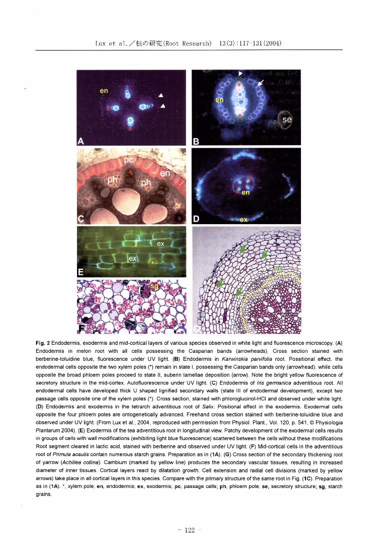
Fig. 2 Endodermis, exodermis and mid-cortical layers of various species observed in white light and fluorescence microscopy. (A)
Endodermis in melon root with all cells possessing the Casparian bands (arrowheads). Cross section stained with
berberine-toluidine blue, fluorescence under UV light. (B) Endodermis in Karwinskia parvifolia root. Possitional effect, the
endodermal cells opposite the two xylem poles (*) remain in state ‡T, possessing the Casparian bands only (arrowhead), while cells
opposite the broad phloem poles proceed to state ‡U, suberin lamellae deposition (arrow). Note the bright yellow fluorescence of
secretory structure in the mid-cortex. Autofluorescence under UV light. (C) Endodermis of Iris germanica adventitious root. All
endodermal cells have developed thick U shaped lignified secondary walls (state ‡V of endodermal development), except two
passage cells opposite one of the xylem poles (*). Cross section, stained with phloroglucinol-HCl and observed under white light.
(D) Endodermis and exodermis in the tetrarch adventitious root of Salix. Positional effect in the exodermis. Exodermal cells
opposite the four phloem poles are ontogenetically advanced. Freehand cross section stained with berberine-toluidine blue and
observed under UV light. (From Lux et al., 2004, reproduced with permission from Physiol. Plant., Vol. 120, p. 541, (c) Physiologia
Plantarum 2004). (E) Exodermis of the tea adventitious root in longitudinal view. Patchy development of the exodermal cells results
in groups of cells with wall modifications (exhibiting light blue fluorescence) scattered between the cells without these modifications
Root segment cleared in lactic acid, stained with berberine and observed under UV light. (F) Mid-cortical cells in the adventitious
root of Primula acaulis contain numerous starch grains. Preparation as in (1A). (G) Cross section of the secondary thickening root
of yarrow (Achillea collina). Cambium (marked by yellow line) produces the secondary vascular tissues, resulting in increased
diameter of inner tissues. Cortical layers react by dilatation growth. Cell extension and radial cell divisions (marked by yellow
arrows) take place in all cortical layers in this species. Compare with the primary structure of the same root in Fig. (1C). Preparation
as in (1A). *, xylem pole; en, endodermis; ex, exodermis; pc, passage cells; ph, phloem pole; se, secretory structure; sg, starch
grains.
-122-
Lux et al./根 の 研 究(Root Research) 13(3):117-131(2004)
The exodermis might develop in a uniform
fashion, composed of similarly sized and formed cells. This type has been most studied in maize.
Another type of exodermis, which has mostly been studied in the onion, is dimorphic, with alternating
long and short cells. Moreover, the exodermis can be uniseriate or multiseriate. The type, presence or
absence of the exodermis appears to be a constant characteristic at the family level (Perumalla et al.,
1990; Peterson and Perumalla, 1990).
Other difficulties in the study of the exodermis are the influence of external conditions on its
development, and its absence under specific conditions (e.g., hydroponics), even in species
where it is normally present. The exodermis usually
forms at a greater distance from the root tip than the endodermis. In maize, cultivated in moist vermiculite, exodermal Casparian bands appear
after four days of cultivation, when roots are 60 mm
long (Enstone and Peterson, 1997). Similarly in onion, they appear after five days and about 50 mm
from the tip. However, a reverse sequence of differentiation was found in some species, with the
exodermis differentiating earlier (closer to the root tip) than the endodermis. This phenomenon was
found in some wetland plants (Seago et al., 1999;
Soukup et al., 2002) and in tea plants (Homma et al., 2000; Tanimoto et al., 2004).
Gradual and patchy development of exodermis along the root axis is another characteristic of this
cortical layer. There is always a long root zone with
non-mature exodermis, and what is most interesting is that development need not be
continuous along the cell files. This results in the formation of "islands" or groups of cells with
developed Casparian bands and suberin lamellae scattered between the cells without these wall
modifications. This can best be seen in longitudinal views (Fig. 2E). Sometimes preferential
development of exodermis against the phloem poles can be observed (see section 3.4)
3.3 Development of mid-cortex (mesodermis) Mesodermal layers attract less attention than
the endo- and hypodermis. Nevertheless, these cortical layers are important as storage places
(mainly depositing starch). They are important metabolically, often depositing or synthesizing various secondary metabolites that are sometimes
accumulated in specialized secretory structures
(see Lux et al., 2004a; Fig. 2B). They also
represent a place for root symbiosis with fungi.
Thickening of mesodermal cell walls, sometimes in a
curious form of phi thickenings (resembling in cross
sections the Greek letter ƒ³), is supposed to
mostly serve for the mechanical strengthening of
the cortex (Esau, 1965; Enstone et al., 2003).
Aerial roots often contain chloroplasts in their
mid-cortical layers, and also in the pith. This
characteristic is typical and well known for aerial
roots of epiphytes (Fig. 1D). However, parenchyma
cells in grass nodal roots exposed to the light are
also full of chloroplasts. This phenomenon requires
a more detailed study.
The majority of recent structural work that deals
with the medodermis has focused on aerenchyma
formation. Aerenchyma is defined as a tissue that
contains enlarged gas spaces, which exceed the
commonly present intercellular spaces. It can be
formed constitutively or induced by abiotic stresses,
usually by hypoxia that results from waterlogging.
Sometimes it is induced by other stresses such as
drought, high temperature or nutrient deficiency.
The main reason for the interest in aerenchyma is
its importance for crop survival under waterlogged
conditions. Aerenchyma is also important, as it is a
major pathway for the release of the global warming
gas, methane, to the atmosphere in flooded soils.
Therefore, an understanding of the regulation of its
development is considered a research priority
(Evans, 2003).
Aerenchyma formation has been intensively
investigated in rice plants because of the vital
importance of internal aeration to roots in paddy
fields. It was found that the main roots (numerous
nodal roots forming the skeleton of the root
system) were aerenchymatous, with continuous gas
channels from the root base to the tip. However,
they were covered by gas-impermeable peripheral
cortical layers (Morita and Nemoto, 1995; Colmer,
2003; Fig. 1B). This structural model conflicts with
the need for efficient water and nutrient absorption,
therefore the main water and solute uptake should
be realized with short, fine lateral roots, which are
considerably less aerenchymatous, and also with
less-thickened peripheral cortical walls (Kawata et
al., 1977; Matsuo and Hoshikawa, 1993). They
account for the majority of the external surface of
the rice root system and are supposed to be
responsible for the bulk of water and nutrient
-123-
Lux et al./根 の 研 究(Root Research) 13(3):117-131(2004)
absorption by the root system (Kirk, 2003).
Aerenchyma development has also been well studied in various wetland plants, with considerable
differences in cortical layer composition noted (for
details, see review by Seago, 2002). The entire topic of aerenchyma formation was recently
published as the Tansley review (Evans, 2003).
3.4 Effect of the radial organization of
the stele, determined by xylem and
phloem poles, on cortex development-positionsl effect
Radial organization of the root vascular bundle with alternating xylem and phloem poles has a
significant impact on the development of the cortex. This effect is clear and well known in the
endodermis, starting from the proendodermal stage, and is particularly evident during the second state
of development. The first state, Casparian band development, sometimes starts in all endodermal
cells at the same distance from the root tip, or at least appears to. A more detailed study usually
shows that, even at this state, the endodermal
sectors opposite the phloem poles are "privileged" and form Casparian bands earlier than the
endodermal cells opposite the xylem poles. The long distance along the root axis with gradually
formed suberin lamellae (second state) is also
subject to this positional effect. In some species
(e.g., the lateral roots of the shrub, Karwinskia spp., family Rhamnaceae; Fig. 2B), endodermal
cells opposite the xylem poles remain permanently
in the first state, while cells opposite the phloem
poles proceed to the second state. If delayed, development of certain cells continues to the stage in which the majority of endodermal cells pass to
the third state. This results in the formation of so-called passage cells (Fig2C). However, in some
species, passage cells might later complete their ontogenesis and proceed to the second or third
state.
The positional effect was found not only in the endodermis, but also in mid-cortical layers during
aerenchyma formation in maize and willow. Living
cortical cells are located opposite the protoxylem, and air channels are formed opposite the phloem
poles as a result of cell autolysis (Scott, 1928; Konings and Verschuren, 1980; Barlow, 2002).
This indicates that the signals radiating from the
phloem and/or xylem poles that regulate the events
of cell death and cell wall synthesis in cortical
layers can extend across several cell layers. Recently, we found clear evidence of the positional
effect in the exodermis of willow adventitious roots
opposite the phloem poles with advanced development of this layer (Lux et al., 2004b). As
these roots are usually tetrarch, four sectors of developed exodermis can be found at some distance
from the root tip (Fig. 2D). Another evidence of signals radiating from the stele up to the exodermis
is the formation of a "window" in the region of exodermis opposite the growing lateral root
primordium. In this window, the exodermis lacks suberin and lignin (Soukup et al., 2002). New findings can be expected in connection with the
positional effect phenomenon in cortical cells.
3.5 Secondary dilatation growth of
cortical tissues There is extensive literature that deals with early
changes of root cortical layers, especially in
regards to the endo- and exodermis. Much less studied are the older root parts. However, the
cortex can persist, even in quite old secondarily
thickening roots. In early stages of cambial activity, extension of peripheral layers that correspond to
secondary thickening of vascular cylinders takes
place in all roots. This enlargement of peripheral tissues is called dilatation growth. Thus, the secondary growth of roots comprises cambial
growth (realized by vascular cambium and cork cambia activity) and dilatation growth (present in the primary cortex, rhizodermis, in axial phloem
parenchyma and in the ray parenchyma of bark; Esau, 1965; Lev-Yadun and Aloni, 1992).
Dilatation growth involves cell enlargement and/or cell renewed division.
Changes that occur in endodermal cells are
probably the most interesting feature of dilatation
growth. These cells develop specific wall modifications (see section 3.1) and represent highly specialized cells with extremely important functions
for the regulation of radial transport in the roots.
However, during dilatation growth, their cell walls must extend. Moreover, in some species these cells
radially divide. The additional division of endodermal cells is a species-specific feature, and
the number of cells along the circumference of the
endodermal sheath increases as a result. The number of additional divisions of an individual
-124-
Lux et al./根 の 研 究(Root Research) 13(3):117-131(2004)
endodermal cell is also variable and
species-specific, ranging from single division (e.g.,
in Primula acaulis, Lux and Luxova, 2003/4) up to
24 new radial walls being formed (Gentiana
asclepiadea; Sottnikova and Lux, 2003).
Previous reports by authors (e.g., Bond, 1930)
indicate that the additional division of cells starts
during endodermal developmental states ‡T or ‡V.
After the division of endodermal cells during state ‡T,
the new radial walls gain a typical endodermal
character and Casparian bands are deposited soon
after the new wall forms. Thus, the endodermal
apoplastic barrier is not interrupted by these new
walls. If the additional division takes place in
endodermal cells during state ‡V, the new radial
walls remain without Casparian bands in the
majority of species. These new walls have the
character of supporting prop walls, and protect
strongly-expanded endodermal cells from collapse.
An exception to this rule was found in Primula, with
Casparian bands developing in new radial walls after
the division of thick-walled endodermal cells (Lux
and Luxova, 2001, 2003/4). The role of these
Casparian bands remains obscure, as no apoplastic
transport is expected across endodermal cell walls
in state ‡V.
Expansion of the cortex in secondary thickening
roots also induces dilatation growth in mid-cortical
and hypodermal layers. In some species, even these
cells might additionally divide. Radial division in
mid-cortical layers is relatively common (Fig. 2G),
while division of the hypodermis is rare. This was
found in species of the genus Gentiana (Luhan,
1954; Sottnikova and Lux, 2003)
The additional division of endodermal cells is
a rather frequently occurring phenomenon in roots
of dicotyledons (for a review, see Lux and Luxova,
2001). However, several cytological and functional
aspects and consequences of this process remain to
be elucidated.
4. Reaction of the cortex to stress
The reaction of the root and the specificity of its
cortical tissues to stress factors is a very broad
subject, and might be the topic of a separate review.
Here, only some aspects of this subject will be
discussed. Special attention would require the
interaction of cortical cells with symbiotic and
pathogenic organisms, which is outside the scope of this review. Despite this, some new data about this
very interesting topic can be found in the review of
arbuscular mycorrhiza (Strack et al., 2003) and in
the review of root endo- and exodermal responses
to the environment (Enstone et al., 2003). It is also
interesting to note that a protective role of the
exodermis against pathogenic fungi was recently
proven in barley roots by Reissinger et al. (2003). The entire cortex can be understood as a kind of
root "buffer zone". Peripheral tissues, at least up
to the endodermal layer, which includes the
epidermis, hypodermis and mesodermis, might be
damaged and even deteriorate in some root parts.
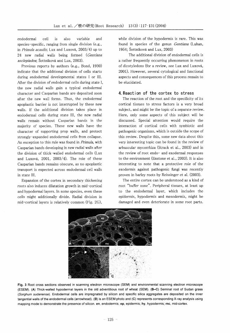
Fig. 3 Root cross sections observed in scanning electron microscope (SEM) and environmental scanning electron microscope
(ESEM). (A) Thick-walled hypodermal layers in the old adventitious root of wheat (SEM). (B-C) Seminal root of Sudan grass
(Sorghum sudanense). Endodermal cells are impregnated by silicon and specific silica aggregates are deposited on the inner tangential walls of the endodermal cells (arrowhead). (B) is an ESEM photo and (C) represents corresponding X-ray analysis using
mapping mode to demonstrate the presence of silicon. en, endodermis; ep, epidermis; hy, hypodermis; mc, mid-cortex.
-125-
Lux et al./根 の 研 究(Root Research) 13(3):117-131(2004)
However, the root can still provide its most
important function - to supply water and ions to
the plant. The first cortical barrier, the hypodermis
(or exodermis), is the first important part of this buffer zone. It usually reacts very sensitively to
various stresses, as was demonstrated by the
development of wall modifications characteristic to
exodermal cells. For example, in maize, the
exodermis is absent in hydroponically grown plants,
but present in plants grown aeroponically and in
the soil (Zimmermann and Steudle, 1998). Cell wall
modifications and impregnation by suberin, lignin or
some other unidentified substances were observed
in hypodermal and deeper cortical layers as a
reaction to mechanical injury. The exodermis
develops up to the root apex in roots that are
subject to stress and have stopped or substantially
slowed their growth (Brundrett et al., 1990;
Tanimoto and Lux, unpublished results from tea
roots).
Specific adaptations of the roots of plants
tolerant to various forms of stress factors require
more attention. For example, in roots of
drought-tolerant dicotyledonous succulents from
the family Cactaceae, the hypodermal layer is
composed of large cells thickened only in their
outer tangential walls (Lux and Inanaga, 1997).
These cells probably function mainly as water
reservoirs. However, the hypodermal layers of
grasses are frequently formed from small cells with thick walls (Fig. 3A), which is an important barrier
that protects inner tissues. In sorghum,
sclerification and wall thickness in a
drought-tolerant cultivar was greater when
compared with a drought-sensitive cultivar (Salih
et al., 1999). Kondo et al. (2000) reported that
some rice varieties (mostly upland varieties) formed
two or three layers of sclerenchyma in the outer
cortex, while most other rice varieties formed only
one layer. Developmental plasticity of cortical
layers in desert plants related to sources of water
was shown by North and Nobel (1998, 2000).
Cell wall thickening of root cortical cells is
associated with high salt and heavy metal stresses
(Reinhardt and Rost, 1995). The endodermis was
demonstrated to be an effective barrier to Na+ in
the alkali grass, Puccinellia tenuiflora (Peng et al.,
2004). The effects of salinity on the development of
Casparian bands in the primary roots of maize were
studied by Karahara et al. (2004). They found that
Casparian bands matured closer to the root tip in
plants grown with an increased concentration of
NaCl compared with the control. Moreover, the
radial width of Casparian bands increased in the
presence of salt. This suggests that the function of
the bands is enhanced under salt stress.
Other types of stress can also induce changes in
the cortical cell wall structure and composition.
For example, a water deficit increased the
lignification of the exodermis in tomato roots
(Nakano et al., 2003). In maize roots, multiple
environmental stresses (cultivation in municipal
solid waste slag with high pH, high salt and heavy
metal content) induced more intensive wall
thickenings in the inner tangential walls of
endodermal cells when compared with the roots of
control plants. Slag-grown plants also had higher
amounts of lignin in the endodermal and
hypodermal layers when compared with control
plants (Degenhardt and Gimmler, 2000). In wheat roots, hypoxia induced cell wall thickening. In this
case, it accounted for an increase in cellulose, but
not in lignin content (Albrecht and Mustroph,
2003). However, cell wall thickening in the
endodermis of rice only occurs in upland cultivars
(Kawata and Lai, 1966). Some stresses result in
specific root structural responses, e.g., aluminum
induces radial cell expansion of cortical cells and
callose formation in epidermal and outer cortical
layers of cereals (Blancaflor et al., 1998; Budikova,
1999; Budikova and Mistrik, 1999; Ciamporova,
2002). However, species-specificity must be taken
into account, even in this case, as it was shown
that the absence of aluminum in the nutrient
solution was a stress factor for hydroponically
grown tea roots (Tanimoto, unpublished results).
4.1 Silicon deposition in the endodermis
and its relation to stresses
In all these cases, the synthesis and deposition of
organic substances represents a high cost to the
plant, as these compounds cannot be reused in
plant metabolism and represent a dead end for the metabolic pathways. In this manner, silicon
deposition as an impregnating substance might be
exceptionally advantageous. Silicon is metabolically "cheap" and is usually freely available to plants
from the soil solution. Exceptions are some specific
soils, and some intensively agriculturally used soils
in which excessive extraction of silicon by plants
-126-
Lux et al./根 の 研 究(Root Research) 13(3):117-131(2004)
without its replacement could result in silicon
deficiency symptoms (Ma and Takahashi, 2002).
The mechanism of impregnation of cell walls by silicon has evolved in some plant species, and
several so-called silicon accumulators deposit
considerable quantities of this element in root endodermal cells (Sangster and Parry, 1976a, b;
Lux et al., 1999, 2002, 2003a, b). In sorghum, and
other species of the tribe Andropogoneae (family Poaceae), deposits of silicon form conspicuous
silica aggregates on the inner tangential walls of the endodermis (Figs. 3B, C). We found that silicon
accumulation in endodermal cell walls can even be higher than the silicification of leaves (Lux et al., 2003a). Silicon deposition was also found to be
intraspecifically variable, and cultivars of both rice
and sorghum with higher tolerance to drought have a higher silicification of endodermal cell walls than
walls of drought-susceptible cultivars (Lux et al.,
1999, 2002). Recently, it was found that sorghum supplied with silicon could extract a greater amount
of water from the soil under drought conditions due to an acceleration of root growth and an
enhancement of the water uptake ability (Hattori et al., in press). Root growth stimulation under dry
conditions might be related to an increase of cell
wall extensibility in an apical part of sorghum roots caused by silicon, as reported by Hattori et al.
(2003). This high extensibility would allow the roots to elongate even when turgor pressure drops due to
soil drying. Although silicon impregnation of endodermal walls acts as a protection against
parasites (Bennett, 1982), this topic requires a more detailed study. Another beneficial effect of
root silicification was to alleviate the effect on some toxic elements, mainly manganese, but also
aluminum in some species (for a recent review of
this subject, see Ma, 2004). Evidence appears to be accumulating about the unknown manner of silicon
induced tolerance to several other toxic metals
such as zinc (Neumann et al., 1997) and cadmium
(Wang et al., 2000). This problem certainly requires more attention because of the importance
of food contamination by toxic metals.
4.2 Intraspecific differences of cortical tissues related with a reaction to
toxic metals In our recent study, we analyzed the structural
differences in roots of Salix clones that
substantially differed in the accumulation and
translocation of cadmium and their sensitivity to
this toxic metal (Lux et al., 2004b). An ontogenetic
study of the endodermis indicated that the
apoplastic movement of cadmium into the stele and
the upward translocation might vary due to the
development of this cortical layer. Development of
Casparian bands in clones characterized with a high
accumulation of cadmium started more distantly
from the root tip than in clones with a low
accumulation. Even more prominent were
differences in the second state of endodermal
development. The suberin lamellae were formed
more distantly from the root tip in clones with a
high translocation of cadmium (5-15 mm from the
root tip) compared with those with a low
translocation (2-5 mm from the tip). Furthermore,
a quantitative comparison of area proportions of
individual cortical layers showed a relationship with
tolerance to cadmium. Clones with a high tolerance
had a higher proportion of endo- and exodermis
than sensitive clones. It is interesting to note that
Rincon et al. (2003) found that the major genotypic
differences in soybean root structure related with
variations in resistance to water movement through
the roots were in the surface area of the stele that
approximates the dimension of the endodermal
layer. Although there are several potential barriers
to water conductance and ion uptake by the root
system, the aforementioned results, together with
the genotypic differences of cortical tissues related
with the cadmium accumulation and sensitivity
found in the willow, point to the importance of
these tissues and the importance of research on
this topic.
Acknowledgement
The work was partially supported by the Field
Production Science Center, the University of
Tokyo, grant 1/0100/03 from the Slovak Grant
Agency VEGA and COST Action 859. We
apologize to all colleagues whose works were not
included due to the lack of space. The authors
appreciate the generosity of the Keyence Company
in allowing us to use their high sensitive CCD color
camera to take some photos for this review.
References
Albrecht, G., Mustroph, A. 2003. Localization of sucrose
synthase in wheat roots: Increased in situ activity of
-127-
Lux et al./根 の 研 究(Root Research) 13(3):117-131(2004)
sucrose synthase correlates with cell wall thickening
by cellulose deposition under hypoxia. Planta 217:
252-260.
Barlow, P.W. 2002. Cellular patterning in root
meristems: its origins and significance. In Waisel, Y.,
Eshel, A., Kafkafi, U. eds., Plant Roots The Hidden
Half. 3rd Edition. pp49-82. (Marcel Dekker, Inc., New
York, Basel)
Baum, S.F., Dubrovsky J.G., Rost, T.L. 2002. Apical
organization and maturation of the cortex and vascular
cylinder in Arabidopsis thaliana (Brassicaceae) roots.
Am. J. Bot. 89: 908-920.
Barnabas, A.D.; Peterson, C.A. 1992. Development of
Casparian bands and suberin lamellae in the
endodermis of onion roots. Can. J. Bot. 70:
2233-2237.
Bennett, D.M. 1982. Silicon deposition in the roots of
Hordeum sativum Jess., Avena sativa L. and Triticum
aestivum L. Ann. Bot. 50: 239-245.
Blancaflor, E.B., Jones, D.L., Gilroy, S. 1998.
Alterations in the cytoskeleton accompany
aluminum-induced growth inhibition and
morphological changes in primary roots of maize. Plant
Physiol. 118: 159-172.
Bond, G. 1930. The occurrence of cell division in the
endodermis. Proc. R. Soc. Edinb. 50: 38-50.
Brundrett, M.C., Enstone, D.E., Peterson, C.A. 1988. A
berberine-aniline blue fluorescent staining procedure
for suberin, lignin, and callose in plant tissue.
Protoplasma 146: 133-142.
Brundrett, M.C., Kendrick, B., Peterson, C.A. 1991.
Efficient lipid staining in plant material with Sudan red
7B or Fluoral yellow 088 in polyethylene
glycol-glycerol. Biotech. Histochem. 66: 111-116. Brundrett, M.C., Murase, G., Kendrick, B. 1990.
Comparative anatomy of roots and mycorrhizae of
common Ontario trees. Can. J. Bot. 68: 551-578.
Budikova, S. 1999 Structural changes and aluminium
distribution in maize root tissues. Biol. Plant. 42:
259-266.
Budikova, S., Mistrik, I. 1999. Cultivar characterisation
of aluminium tolerance of barley seedlings by root
growth, aluminium and callose distribution. Biologia 54: 447-451.
Chapman, K., Groot, E.P., Nichol, S.A,. Rost, T.L.
2003. Primary root growth and the pattern of root
apical meristem organization are coupled J. Plant
Growth Regul. 21: 287-295.
Ciamporova, M. 2002. Morphological and structural
responses of plant roots to aluminium at organ, tissue,
and cellular levels. Biol. Plant. 45: 161-171.
Clarkson, D.T., Sanderson, J. 1974. The endodermis and
its development in barley roots as related to radial
migration of ions and water. In: Kolek J. ed., Structure
and Function of Primary Root Tissue. pp87-100.
(Veda, Bratislava, Czechoslovakia)
Colmer T.D. 2003. Long-distance transport of gases in
plants: a perspective on internal aeration and radial
oxygen loss from roots. Plant Cell Environ. 26: 17-36.
Degenhardt, B., Gimmler, H. 2000. Cell wall adaptations
to multiple environmental stresses in maize roots. J.
Exp. Bot. 51: 595-603.
Esau, K. 1965 Plant Anatomy. 2nd Edition. p767. (John
Wiley & Sons, Inc., New York, London, Sydney)
Evans, D.E. 2003. Aerenchyma formation. New Physol.
161: 35-49.
Enstone, D.E., Peterson, C.A. 1997. Suberin deposition
and band plasmolysis in the corn (Zea mays L.) root
exodermis. Can. J. Bot. 75: 1188-1199.
Enstone, D.E., Peterson, C.A., Ma, F. 2003. Root
endodermis and exodermis: structure, function, and
responses to the environment. J. Plant Growth Regul.
21: 335-351.
Guttenberg von, H. 1968. Der primare Bau der
Angiospermenwurzel ‡[/5. In Linsbauer K. ed.,
Handbuch der Pflanzenanatomie. (Gebruder
Borntraeger Verlachsbuchhandlung, Berlin-Stuttgart,
Germany)
Hattori, T., Inanaga, S., Araki, H., An, P., Morita, S.,
Luxova, M., Lux, A. Application of silicon enhanced
drought tolerance in Sorghum bicolor (L.) Moench.
Physiol. Plant. - in press
Hattori, T., Inanaga, S., Tanimoto, E., Lux, A., Luxova,
M. 2003. Silicon-induced changes in viscoelastic
properties of sorghum root cell walls. Plant Cell
Physiol. 44: 743-749.
Hochholdinger, F., Park, W.J., Sauer, M., Woll, K. 2004.
From weeds to crops: genetic analysis of root
development in cereals. Trends Plant Sci. 9: 42-48.
Homma, T., Miyama, D., Lux, A., Tanimoto, E. 2000.
Histological characteristics and acidic region observed
in tea roots. Bull. Tokai Branch Crop Sci. Soc. Jpn.
129: 39-40. (in Japanese)
Karahara I., Ikeda, A., Kondo, T., Uetake, Y. 2004.
Development of the Casparian strip in primary roots of
maize under salt stress. Planta 219: 41-47.
Kawata, S., Lai, K-L. 1965. On the meristematic state of
the endodermis in the crown roots of rice plants. Proc.
Crop Sci. Soc. Jpn. 34: 210-216. (in Japanese with
English summary)
-128-
Lux et al./根 の 研 究(Root Research) 13(3):117-131(2004)
Kawata, S., Lai, K-L. 1966. On the cell wall thickening
of the endodermis in the crown roots of rice plnats.
Proc. Crop Sci. Soc. Jpn. 34: 440-447. (in Japanese
with English summary)
Kawata, S., Lai, K-L. 1967. On the differentiation of
Casparian dots of the endodermis in the crown roots
of rice plants. Proc. Crop Sci. Soc. Jpn. 36: 75-84. (in
Japanese with English summary)
Kawata, S., Sasaki, O. and Yamazaki, K. 1977. On the
structure of the crown root and the lateral root, and
vessel connection between them, in rice plants. Jpn. J.
Crop Sci. 46: 569-579. (in Japanese with English
summary)
Kirk, G.J.D. 2003. Rice root properties for internal
aeration and efficient nutrient acquisition in
submerged soil. New Phytol. 159: 185-194.
Konings and Verschuren, 1980; Formation of
aerenchyma in roots of Zea mays in aerated solutions,
and its relation to nutrient supply. Physiol. Plant. 49:
265-270
Kondo, M., Aguilar, A., Abe, J. and Morita, S. 2000.
Anatomy of nodal roots in tropical upland and lowland
rice varieties. Plant Prod. Sci. 3: 437-445.
Kutschera, L., Sobotik, M. 1992. Wurzelatlas
mitteleuropaischer Grundlandpflanzen. Band 2, Teil 2
Anatomy. (Gustav Fisher Verlag, Stuttgart, Germany)
Lev-Yadun, S., Aloni, R. 1992. Experimental induction
of dilatation meristemss in Melia azedarach L. Ann.
Bot. 70: 379-386.
Luhan, M. 1954. Zur Wurzelanatomie unserer
Alpenpflanzen: ‡V. Gentianaceae. Sitzungsberichte
der Akademie der Wissenschaften Wien,
Matematisch-Naturwissenschaftliche Klasse,
Abteilung I. 163: 89-107.
Lux, A. 1981. Rapid method for staining of semi-thin
sections from plant material (in Slovak). Biologia 36:
753-757.
Lux, A., Inanaga, S. 1997. Die atypische Struktur der
jungen Wurzeln einer Wuestensukkulente
Echinocactus platyacanthus. Stapfia 50: 289-293.
Lux, A., Liskova, D., Masarovicova, E., Kakoniova, D.,
Hanackova, Z., Argalasova-Sutovska, K., Kollarova,
K., Henselova, M., Ordonez, J.R. 2004a. Biology of
Karwinskia spp., experimental cultivation and
secondary metabolites production. In J.N. Govil, P.A.
Kumar, V.K. Singh, eds., Recent Progress in
Medicinal Plants - Vol. 4. Biotechnology and Genetic
Engineering. pp175-200. (Studium Press LLC,
U.S.A.)
Lux, A., Luxova, M. 2001. Secondary dilatation growth
in the root endodermis. In: Gasparikova, O.,
Ciamporova, M., Mistrik, I., Baluska, F., eds., Recent
Advances of Plant Root Structure and Function.
pp31-37. (Kluwer Academic Publishers, Dordrecht,
The Netherlands)
Lux, A., Luxova, M. 2003/4. Growth and differentiation
of root endodermis in Primula acaulis Jacq. Biol. Plant.
47: 91-97.
Lux, A., Luxova, M., Abe, J., Morita, S., Inanaga, S.
2003a. Silification of bamboo (Phyllostachys
heterocycla Mitf.) root and leaf. Plant Soil 255: 85-91.
Lux, A., Luxova, M., Abe, J., Tanimoto. E., Hattori, T.,
Inanaga, S. 2003b. The dynamics of silicon deposition
in the sorghum root endodermis. New Phytol. 158:
437-441.
Lux. A., Luxova, M., Inanaga, S., Sugimoto, Y. 2002.
Silicification in sorghum (Sorghum bicolor) cultivars
with different drought tolerance. Physiol. Plant. 115:
87-92.
Lux, A., Luxova, M., Morita, S., Abe, J., Inanaga, S.
1999. Endodermal silicification in developing seminal
roots of lowland and upland cultivars of rice (Oryza
sativa L.). Can. J. Bot. 77: 955-960.
Lux, A., Sottnikova, A., Opatrna, J., Greger, M. 2004b.
Differences in structure of adventitious roots in Salix
clones with contrasting characteristics of cadmium
accumulation and sensitivity Physiol. Plant. 120:
537-545.
Ma, F., Peterson, C.A. 2003. Current insights into the
development, structure, and chemistry of the
endodermis and exodermis of roots. Can. J. Bot. 81:
405-421.
Ma, J.F. 2004. Role of silicon in enhancing the resistance
of plants to biotic and abiotic stresses. Soil Sci. Plant
Nutr. 50: 11-18
Ma, J.F., Takahashi, E. 2002. Soil, Fertilizer, and Plant
Silicon Research in Japan. (Elsevier Science,
Amsterdam)
Mager, H. 1932. Beitrage zur Kenntnis der primaren
Wurzelrinde. Planta 16: 666-708.
Matsuo, T., Hoshikawa, K. 1993. Science of the Rice
Plant. I Morphology. (Food and Agriculture Policy
Research Center, Tokyo)
Morita, S. 2000. Root Growth and Root System
Development. (University of Tokyo Press, Tokyo). (in
Japanese)
Morita, S., Lux, A., Enstone, D.E., Peterson, C.A., Abe,
J. 1996. Reexamination of rice seminal root ontogeny
using fluorescence microscopy. Jpn. J. Crop Sci.
65 (Extra issue 2): 37-38.
-129-
Lux et al./根 の 研 究(Root Research) 13(3):117-131(2004)
Morita, S., Nemoto, K. 1995. Morphology and anatomy
of rice roots with special reference to coordination in
organo- and histogenesis. In F. Baluska et al. eds.,
Structure and Function of Roots, pp.75-86. (Kluwer
Academic Publ. Dordrecht, The Netherlands)
Nakano, Y., Nakano, A., Watanabe, S., Okano, K.,
Tatsumi J. 2003. External and internal root structures
of tomato plants grown hydroponically in a humid
atmosphere or in a nutrient solution. J. Jpn. Soc. Hort.
Sci. 72: 148-155.
North, G.B., Nobel, P.S. 1998. Water uptake and
structural plasticity along roots of a desert succulent
during prolonged drought. Plant Cell Environ. 21:
705-713.
North, G.B., Nobel, P.S. 2000. Heterogeneity in water
availability alters cellular development and hydraulic
conductivity along roots of a desert succulent. Ann.
Bot. 85: 247-255.
Neuman, D., Niedel, U.Z., Schwieger, W., Leopold, I.,
Lichtenberger, O. 1997. Heavy metal tolerance of
Minuartia verna. J. Plant Physiol. 151: 101-108.
Peng YH, Zhu, YF, Mao YQ, Wang SM, Su WA, Tang ZC
2004. Alkali grass resists salt stress through high [K+]
and an endodermis barrier to Na+. J. Exp. Bot. 55:
939-949.
Perumala, C.J., Peterson, C.A. 1986. Deposition of
Casparian bands and suberin lamellae in the exodermis
and endodermis of young corn and onion roots. Can. J.
Bot. 64: 1873-1878.
Perumala, C.J., Peterson, C.A., Enstone, D.E. 1990. A
survey of angiosperm species to detect hypodermal
Casparian bands. ‡T. Roots with a uniseriate
hypodermis and epidermis. Bot. J. Linnean Soc. 103:
93-112.
Peterson, C.A. 1997. The exodermis and its interactions
with the environment. In Flores, H.E., ed. Radical
Biology: Advances and Perspectives on the Function
of Plant Roots. pp131-138. (An American Society of
Plant Physiologists Series 18)
Peterson, C.A., Murrmann, M., Steudle, E. 1993.
Location of major barriers to water and ion movement
in young roots of Zea mays L. Planta 190: 127-136.
Peterson, C.A., Perumalla, C.J. 1990. A survey of
angiosperm species to detect hypodermal Casparian
bands. ‡U. Roots with a multiseriate hypodermis or
epidermis. Bot. J. Linnean Soc. 103: 113-125.
Reinhardt D.H., and Rost T.L. 1995. Salinity
accelerates endodermal development and induces an
exodermis in cotton seedling roots. Environ. Exp. Bot.
35: 563-574.
Reissinger A., Winter, S., Steckelbroeck, S., Hartung,
W., Sikora, R.A. 2003. Infection of barley roots by
Chaetomium globosum: evidence for protective role of
the exodermis. Mycol. Res. 107: 1094-1102.
Rincon, C.A., Raper, C.D., Patterson, R.P. 2003.
Genotypic differences in root anatomy affecting water
movement through roots of soybean. Int. J. Plant Sci.
164: 543-551.
Salih, A.A., Ali, I.A., Lux, A., Luxova, M., Cohen, Y.,
Sugimoto, Y., Inanaga, S. 1999. Rooting, water uptake,
and xylem structure adaptation to drough of two
sorghum cultivars. Crop Sci. 39: 168-173.
Sanderson, J. 1983. Water uptake by different regions of
the barley root. Pathways for radial flow in relation to
development of the endodermis. J. Exp. Bot. 34:
240-253.
Sangster A.G., Parry, D.W. 1976a. Endodermal silicon
deposits and their linear distribution in developing
roots of Sorghum bicolor (L.) Moench. Ann. Bot. 40:
361-371.
Sangster A.G., Parry, D.W. 1976b. Endodermal
silicification in mature, nodal roots of Sorghum bicolor
(L.) Moench. Ann. Bot. 40: 373-379.
Scott, L.I. 1928. The root as an absorbing organ. ‡U. The
delimitation of the absorbing zone. New Phytol. 27:
141-174.
Scott, M.G., Peterson, R.L. 1979. The endodermis in
Ranunculus acris. ‡T. Structure and ontogeny. Can. J.
Bot. 57: 1040-1062.
Seago, J.L.Jr. 2002. The root cortex of the
Nymphaeaceae, Cabombaceae and Nelumbonaceae. J.
Torrey Bot. Soc. 129: 1-9.
Seago, J.L.Jr., Peterson, C.A., Enstone, D.E., Scholey,
Ch.A. 1999. Development of the endodermis and
hypodermis of Typha glauca Godr. and Typha
angustifolia L. roots. Can. J. Bot. 77: 122-134.
Sinnnot, E.W., Bloch, R. 1941. The relative position of
cell walls in developing plant tissues. Am. J. Bot. 28:
607-617.
Sottnikova, A., Lux, A. 2003. Development, dilatation
and subdivision of cortical layers of gentian (Gentiana
asclepiadea L.) root. New Phytol. 160: 135-143.
Soukup, A., Votrubova, O., Cizkova, H., 2002.
Development of anatomical structure of roots of
Phragmites australis. New Phytol. 153: 277-287.
Steudle, E., Peterson, C.A. 1998. How does water get
through roots? J. Exp. Bot. 49: 775-788.
Strack D., Fester, T., Hause, B., Schlieman, W., Walter
M.H. 2003. Arbuscular mycorrhiza: Biological,
chemical and molecular aspects. J. Chem. Ecol. 29:
-130-
Lux et al./根 の 研 究(Root Research) 13(3):117-131(2004)
1955-1979.
Tanimoto E, Homma T, Matsuo K, Hoshino T, Lux A and
Luxova M 2004. Root structure and cell-wall
extensibility of adventitious roots of tea (Camellia
sinensis L. cv. Yabukita). Biologia (in press)
Wang L., Wang, Y., Chen, Q., Cao, W., Li, M., Zhang, F.
2000 Silicon induced cadmium tolerance of rice
seedlings. J. Plant Nutr. 23: 1397-1406.
Wilcox, H. 1962. Growth studies of the root of incense
cedar, Libocedrus decurrens. ‡T. The origin and
development of primary tissues. Am. J. Bot. 49:
221-236.
Yamauchi, A., Pardales Jr., J.R. and Kono, Y. 1996.
Root system structure and its relation to stress
tolerance. In Ito, O. et al. eds. Dynamics of roots and
nitrogen in cropping systems of semi-arid tropics. pp.
211-233. (Japan International Research Center for
Agricultural Sciences, Tsukuba)
Zeier, J., Ruel, K., Ryser, U., Schreiber, L. 1999.
Chemical analysis and immunolocalisation of lignin and
suberin in endodermal and hypodermal/rhizodermal
cell walls of developing maize (Zea mays L.) Primary
roots. Planta 209: 1-12.
Zimmerman, H.M., Steudle, E. 1998. Apoplastic
transport across young maize roots: effect of the
exodermis. Planta 206: 7-19.
-131-
Related Documents