doi:10.1182/blood-2006-04-016907 Prepublished online August 31, 2006; 2006 108: 4178-4186 Bavi and Khawla S. Al-Kuraya Moorji, Valerie Atizado, Fouad Al-Dayel, Asim Belgaumi, Hassan El-Solh, Adnan Ezzat, Prashant Shahab Uddin, Azhar R. Hussain, Abdul K. Siraj, Pulicat S. Manogaran, Naif A. Al-Jomah, Azadali B-cell lymphoma survival -kinase/AKT pathway in diffuse large ′ Role of phosphatidylinositol 3 http://bloodjournal.hematologylibrary.org/content/108/13/4178.full.html Updated information and services can be found at: (1930 articles) Signal Transduction (4212 articles) Neoplasia Articles on similar topics can be found in the following Blood collections http://bloodjournal.hematologylibrary.org/site/misc/rights.xhtml#repub_requests Information about reproducing this article in parts or in its entirety may be found online at: http://bloodjournal.hematologylibrary.org/site/misc/rights.xhtml#reprints Information about ordering reprints may be found online at: http://bloodjournal.hematologylibrary.org/site/subscriptions/index.xhtml Information about subscriptions and ASH membership may be found online at: Copyright 2011 by The American Society of Hematology; all rights reserved. Washington DC 20036. by the American Society of Hematology, 2021 L St, NW, Suite 900, Blood (print ISSN 0006-4971, online ISSN 1528-0020), is published weekly For personal use only. by guest on February 1, 2014. bloodjournal.hematologylibrary.org From For personal use only. by guest on February 1, 2014. bloodjournal.hematologylibrary.org From

Welcome message from author
This document is posted to help you gain knowledge. Please leave a comment to let me know what you think about it! Share it to your friends and learn new things together.
Transcript

doi:10.1182/blood-2006-04-016907Prepublished online August 31, 2006;2006 108: 4178-4186
Bavi and Khawla S. Al-KurayaMoorji, Valerie Atizado, Fouad Al-Dayel, Asim Belgaumi, Hassan El-Solh, Adnan Ezzat, Prashant Shahab Uddin, Azhar R. Hussain, Abdul K. Siraj, Pulicat S. Manogaran, Naif A. Al-Jomah, Azadali B-cell lymphoma survival
-kinase/AKT pathway in diffuse large′Role of phosphatidylinositol 3
http://bloodjournal.hematologylibrary.org/content/108/13/4178.full.htmlUpdated information and services can be found at:
(1930 articles)Signal Transduction � (4212 articles)Neoplasia �
Articles on similar topics can be found in the following Blood collections
http://bloodjournal.hematologylibrary.org/site/misc/rights.xhtml#repub_requestsInformation about reproducing this article in parts or in its entirety may be found online at:
http://bloodjournal.hematologylibrary.org/site/misc/rights.xhtml#reprintsInformation about ordering reprints may be found online at:
http://bloodjournal.hematologylibrary.org/site/subscriptions/index.xhtmlInformation about subscriptions and ASH membership may be found online at:
Copyright 2011 by The American Society of Hematology; all rights reserved.Washington DC 20036.by the American Society of Hematology, 2021 L St, NW, Suite 900, Blood (print ISSN 0006-4971, online ISSN 1528-0020), is published weekly
For personal use only. by guest on February 1, 2014. bloodjournal.hematologylibrary.orgFrom For personal use only. by guest on February 1, 2014. bloodjournal.hematologylibrary.orgFrom

NEOPLASIA
Role of phosphatidylinositol 3�-kinase/AKT pathway in diffuselarge B-cell lymphoma survivalShahab Uddin, Azhar R. Hussain, Abdul K. Siraj, Pulicat S. Manogaran, Naif A. Al-Jomah, Azadali Moorji, Valerie Atizado, Fouad Al-Dayel,Asim Belgaumi, Hassan El-Solh, Adnan Ezzat, Prashant Bavi, and Khawla S. Al-Kuraya
Phosphatidylinositol 3�-kinase (PI3K) is akey player in cell-growth signaling in anumber of lymphoid malignancies, but itsrole in diffuse large B-cell lymphoma(DLBCL) has not been fully elucidated.Therefore, we investigated the role of thePI3K/AKT pathway in a panel of 5 DLBCLcell lines and 100 clinical samples. Inhibi-tion of PI3K by a specific inhibitor,LY294002, induced apoptosis in SUDHL4,SUDHL5, and SUDHL10 (LY-sensitive)cells, whereas SUDHL8 and OCI-LY19 (LY-resistant) cells were refractory to
LY294002-induced apoptosis. AKT wasphosphorylated in 5 of 5 DLBCL cell linesand inhibition of PI3K caused dephos-phorylation/inactivation of constitutivelyactive AKT, FOXO transcription factor,and GSK3 in LY-sensitive cell lines. Inaddition, there was a decrease in theexpression level of inhibitory apoptoticprotein, XIAP, in the DLBCL cell linessensitive to LY294002 after treatment.However, no effect was observed in XIAPprotein levels in the resistant DLBCL celllines following LY294002 treatment. Fi-
nally, using immunohistochemistry, p-AKT was detected in 52% of DLBCL tu-mors tested. Furthermore, in univariateanalysis, high p-AKT expression was as-sociated with short survival. In multivari-ate analysis, this correlation was nolonger significant. Altogether, these re-sults suggest that the PI3K/AKT pathwaymay be a potential target for therapeuticintervention in DLBCL. (Blood. 2006;108:4178-4186)
© 2006 by The American Society of Hematology
Introduction
B-cell lymphoma represents the malignant counterpart of normal Bcells arrested at specific maturational stages. Diffuse large B-celllymphoma (DLBCL) is considered to be the most common type oflymphoma in adults, accounting for 30% to 40% of cases ofnon-Hodgkin lymphoma.1 Although patients with DLBCLs arepotentially curable with combination chemotherapy, the diseaseproves fatal in approximately 50% of patients.2 The cause of mostDLBCLs remains unknown; however, dysregulation of apoptosisor defective repair plays a role in lymphogenesis.3
A number of constitutively activated growth signaling pathwayshave frequently been observed in DLBCL including protein kinaseAKT and nuclear factor �B (NF-�B) transcription factor.4-6 Proteinkinases have been implicated as having crucial roles in regulatingcell growth, metabolic responses, cell proliferation, migration, andapoptosis, which altogether contribute to tumorigenesis. Constitu-tive activation of these protein kinases, mainly by phosphorylation,has been implicated as contributing to malignant phenotypes in anumber of human cancers.7-9 AKT is a serine threonine kinase thatgets activated on growth factor and cytokine stimulation. Whenphosphoinositide-3,4,5-triphosphate (PIP3) is generated by phospha-tidylinositol 3�-kinase (PI3K) in response to an intracellular signal,it binds to the PH domain of AKT and translocates to the plasmamembrane resulting in the activation of phosphoinositide-dependent protein kinases (PDK1 and PDK2). Activated PDK1 andPDK2 phosphorylate at the Thr308 and Ser473 residues of the AKTkinase domain, resulting in its activation.10 AKT protein kinaseregulates a variety of cellular processes, including apoptosis, cellsurvival, and proliferation.11-13 AKT-mediated phosphorylation
may alter the activity of proteins such as caspase-9, some Bcl-2family members, and NF-�B and other transcription factors thattrigger or restrain apoptosis. PI3K/Akt deregulation may alsocontribute to tumorigenesis, metastasis, and resistance to chemo-therapy.14,15 For this reason, the PI3K/Akt signaling pathway mightrepresent a promising target for therapeutic intervention. Actually,the PI3K inhibitors LY294002 and wortmannin were observed toexert antitumor activity in animal cell models.14,15 AKT has beenshown to play critical role in the tumorigenesis of many celltypes.16-19 The biologic significance of AKT kinase activity inlymphogenesis has been recently established in a mouse model.20
A number of studies have shown that the cell-death–inducingeffect of chemotherapy depends on induction of apoptosis and thatdisruption of the apoptosis signaling cascade may thus be animportant cause for chemotherapy resistance.21,22 Two majorapoptosis pathways have been described23,24: (1) an intrinsicmitochondrial, stress-induced pathway involving mitochondrialsignaling and caspases and (2) an extrinsic, death receptor–mediated pathway. The stress-induced pathway can be inhibited atmany levels. The most potent inhibitors of this pathway appear tobe Bcl-2 and the X-linked inhibitor of apoptosis (XIAP).25,26
To explore the role of PI3K in the survival, proliferation, andapoptosis of DLBCL, we used a panel of DLBCL cell lines toexamine the involvement of AKT and its substrates, forkheadtranscription factors, and GSK3 in these cells. We next examinedwhether inhibition of PI3K plays any role in the regulation ofproapoptotic and antiapoptotic members of the Bcl-2 family inmaterializing the induction of apoptosis and cell growth inhibition.
From the Research Center, King Fahad National Center for Children’s Cancerand Research, Biological and Medical Research, Department of Pathology,Pediatric Hematology-Oncology, King Faisal Cancer Center, King FaisalSpecialist Hospital and Research Center, Riyadh, Saudi Arabia.
Submitted April 12, 2006; accepted July 24, 2006. Prepublished online asBlood First Edition Paper, 8/31/2006; DOI 10.1182/blood-2006-04-016907.
The online version of this article contains a data supplement.
The publication costs of this article were defrayed in part by page chargepayment. Therefore, and solely to indicate this fact, this article is herebymarked ‘‘advertisement’’ in accordance with 18 USC section 1734.
© 2006 by The American Society of Hematology
4178 BLOOD, 15 DECEMBER 2006 � VOLUME 108, NUMBER 13
For personal use only. by guest on February 1, 2014. bloodjournal.hematologylibrary.orgFrom

We further analyzed the role of inhibition of PI3K on themitochondrial-mediated apoptotic pathway in DLBCL cells via therelease of cytochrome c into cytosol. Finally, we studied the releaseof cytochrome c–propagating death signals to caspase-3, causingits activation and leading to the cleavage of PARP and eventuallycell death after PI3K inhibition. To study the clinical relevance ofthe PI3K/AKT pathway in DLBCL, p-AKT expression was studiedin a group of 100 patients using tissue microarray technology. Ourdata show that overexpression of p-AKT in DLBCL is associatedwith poor outcome.
Patients, materials, and methods
Materials and methods
Cell culture. The human DLBCL cell lines SUDHL4, SUDHL5, SUDHL8,SUDHL10, and OCI-LY19 were obtained from Deutsche Sammlung vonMikroorganismen und Zellkulturen (DSMZ, Braunschweig, Germany). Thecell lines were cultured in RPMI 1640 medium supplemented with 20%(vol/vol) fetal bovine serum (FBS), 100 U/mL penicillin, and 100 U/mLstreptomycin at 37°C in a humidified atmosphere containing 5% CO2. Allthe experiments were performed in RPMI 1640 containing 5% serum.
PI3K/AKT inhibitors. LY294002 and AKT inhibitor (1L-6-hydroxy-methyl-chiro-inositol 2-(R)-2-O-methyl-3-O-octadecylcarbonate) were ob-tained from Calbiochem (San Diego, CA). Wortmannin was obtained fromSigma Chemical (St Louis, MO).
Reagents and antibodies. Anti–cytochrome c, anti–caspase-3, andanti-PARP antibodies were purchased from Santa Cruz Biotechnology(Santa Cruz, CA). The anti–phospho-AKT (Ser473), phospho-AKT (Ser473)blocking peptide, anti–phospho-GSK3�/�, anti–phospho-FKHRL1, anti–cleaved caspase-3, and anti-BID antibodies were purchased from CellSignaling Technologies (Beverly, MA). Anti–�-actin was purchased fromAbcam (Cambridge, United Kingdom). Anti–caspase-8 antibody wasobtained from R&D Systems (Minneapolis, MN). Antibody against Ki-67was purchased from Dako (Carpinteria, CA). The TdT-mediated dUTPnick-end labeling (TUNEL) assay kit was obtained from MBL (Watertown,MA). Annexin V was purchased from Molecular Probes (Eugene, OR). Theapoptotic DNA ladder kit was obtained from Roche (Penzberg, Germany).
Cell death assay
Following indicated treatments, cells were incubated with trypan blue for 5to 10 minutes at room temperature. Trypan blue–positive and total cellswere counted per microscope field for a total of 4 fields per condition. Theproportion of cell death was calculated by dividing the number of dead cellsby total number/field.
TUNEL assay
DLBCL cell lines were treated with LY294002 as described in the figurelegends. Apoptotic cells were measured using the TUNEL assay asdescribed earlier.27 Briefly, after 24 hours of treatment with differentconcentrations of LY294002, 1 � 106 cells were washed twice with PBScontaining 0.2% BSA and fixed with 4% paraformaldehyde at 4°C for 30minutes. This was followed by 2 washes with PBS containing 0.2% BSAand the cells were permeabilized in 70% ethanol at �20°C for 30 minutes.The cells were then washed twice with PBS containing 0.2% BSA andincubated with 30 �L TdT buffer (TdT buffer II, FITC-dUTP, and TdT inthe ratio of 18:1:1) for 1 hour at 37°C. This was followed by 2 washes withPBS and the cells were resuspended in 500 �L PBS. Stained cells wereanalyzed using FACScan flow cytometry equipped with a Cell Quest dataanalysis program (Becton Dickinson, San Diego, CA).
Annexin V staining
DLBCL cell lines were treated with different concentrations of LY294002and AKT inhibitor (1L-6-hydroxymethyl-chiro-inositol 2-(R)-2-O-methyl-
3-O-octadecylcarbonate) as described in the figure legends. Cells wereharvested and the percentage apoptosis was measured by flow cytometryafter staining with fluorescein-conjugated annexin V and propidium iodide(PI; Molecular Probes) as described previously.28,29 We scored viable cellsas those that are negative for annexin V and PI. Percentage of apoptosis wascalculated from the reduction of the number of viable cells between thetreated and untreated samples. The amount of necrotic cells (annexin V�,PI�) was always minimal.
Cell cycle analysis
Cell lines were treated either with or without LY294002 for 24 hours andthe cells were washed once with PBS, resuspended in 500 �L hypotonicstaining buffer (sodium citrate 250 mg, Triton X 0.75 mL, PI 25 �g,ribonuclease A 5 �g, and 250 mL water), and analyzed by flow cytometry asdescribed previously.30
DNA laddering
Cells (2 � 106) were treated with and without 25 �M LY294002 for 24hours. The cells were then harvested and resuspended in 200 �L 1 � PBS.Then 200 �L lysis buffer containing 6 M guanidine HCl, 10 mM urea, 10mM Tris-HCl, and 20% Triton X (vol/vol), pH 4.4, was added to the cellsand incubated for 10 minutes at room temperature. Isopropanol (100 �L)was added and shaken for 30 seconds on a vortex. Then samples werepassed through a filter and spun at 4500 g for 1 minute and the supernatantwas discarded. The pellets were washed 3 times with wash buffercontaining 20 mM NaCl, 2 mM Tris-HCl, and 80% ethanol. The pelletswere then transferred into a new 1.5-mL tube and eluted with 200 �Lprewarmed elution buffer. After measuring the DNA, 2 �g DNA waselectrophoresed on a 1.5% agarose gel containing ethidium bromide at 75 Vfor 2 hours and visualized using a UV light source.
Cell lysis and immunoblotting
Cells were treated with LY294002 as described in the figure legends andlysed as previously described.31,32 Briefly, cell pellets were resuspended inphosphorylation lysis buffer (0.5-1.0% Triton X-100, 150 mM NaCl, 1 mMEDTA, 200 �M sodium orthovanadate, 10 mM sodium pyrophosphate, 100mM sodium fluoride, 1.5 mM magnesium chloride, 1 mM phenylmethylsulfonyl fluoride, 10 �g/mL aprotinin). Protein concentrations were as-sessed by Bradford assay before loading the samples. Equal amounts ofproteins were separated by sodium dodecyl sulfate-polyacrylamide gelelectrophoresis (SDS-PAGE) and transferred to polyvinylidene diflouridemembranes (Immobilon, Millipore). Immunoblotting was performed withdifferent antibodies and visualized by an enhanced chemiluminescence(ECL; Amersham, Arlington Heights, IL) method.
Assay for cytochrome c release
Release of cytochrome c from mitochondria was assayed as describedearlier.33 Briefly, cells were treated with and without LY294002 asdescribed in figure legends and centrifuged at 1000g. Cell pellets wereresuspended in 5 volumes of a hypotonic buffer (20 mM HEPES-KOH,pH 7.5, 10 mM KCl, 1.5 mM MgCl2, 1 mM EDTA, 1 mM EGTA, 1 mMDTT, 20 �g/mL leupeptin, 10 �g/mL aprotinin, 250 mM sucrose) andincubated for 15 minutes on ice. Cells were homogenized by 15 to 20passages through a 22-gauge needle, 1.5 inches long. The lysates werecentrifuged at 1000g for 5 minutes at 4°C to pellet nuclei and unbrokencells. Supernatants were collected and centrifuged at 12 000g for 15minutes. The resulting mitochondrial pellets were resuspended in lysisbuffer. Supernatants were transferred to new tubes and centrifuged againat 12 000g for 15 minutes and resulting supernatants representingcytosolic fractions were separated. Twenty to 25 �g protein from thecytosolic and mitochondrial fraction of each sample was analyzed byimmunoblotting using an anti–cytochrome c antibody.
PI3-KINASE/AKT SIGNALING IN DLBCL 4179BLOOD, 15 DECEMBER 2006 � VOLUME 108, NUMBER 13
For personal use only. by guest on February 1, 2014. bloodjournal.hematologylibrary.orgFrom

Measurement of mitochondrial potential using the JC-1assay kit
Cells (1 � 106) cells were treated with 25 �M LY294002 for 24 hours.Cells were washed twice with PBS and suspended in mitochondrialincubation buffer. JC-1 (5, 5�, 6, 6�-tetrachloro-1, 1�, 3,3�-tetraethylben-zimidazolylcarbocyanine iodide) was added to a final concentration of10 �M and cells were incubated at 37°C in the dark for 30 minutes. Cellswere washed twice with PBS and resuspended in 500 �L mitochondrialincubation buffer and mitochondrial membrane potential (percent ofgreen and red aggregates) was determined by flow cytometry asdescribed previously.34
Patient selection and tissue microarray construction
One hundred patients with DLBCL diagnosed between 1994 and 2004 wereselected from the files of the King Faisal Specialist Hospital and ResearchCentre. All samples were analyzed in a tissue microarray (TMA) format.TMA construction was performed as described earlier.35 Briefly, tissuecylinders with a diameter of 0.6 mm were punched from representativetumor regions of each donor tissue block and brought into recipient paraffinblock using a homemade semiautomatic robotic precision instrument. Anoverview of a DLBCL TMA section is shown in Figure 6A. Three 0.6-mmcores of DLBCL were arrayed from each case.
Patients were reclassified according to the World Health Organization(WHO) classification.36 Age, tumor stage, lactate dehydrogenase (LDH)level, performance status, and the number of extranodal sites of the diseasewere used to determine the International Prognostic Index (IPI).37 Forstatistical analysis, patients were classified as low risk, low-intermediaterisk, high-intermediate risk, and high risk. The number of risk categorieswas condensed from 4 to 2 as follows: a low-risk group encompassingpatients defined in the IPI as low and low-intermediate risk and a high-riskgroup encompassing patients defined in the IPI as high-intermediate andhigh risk. The Institutional Review Board of the King Faisal SpecialistHospital & Research Centre approved the study, in accordance with theDeclaration of Helsinki.
Immunohistochemistry
Tissue microarray sections (3-4 �m thick) were stained with the p-AKT(Ser473) antibody using Survival Marker: Signal Stain Phospho-AKT(Ser473) immunohistochemistry (IHC) detection kit (Cell Signaling Tech-nology, product no. 8100). The IHC protocol mentioned in the kit wasfollowed using all the reagents provided in the kit. Antigen retrieval wasperformed by heating the slides for 10 minutes at 96°C in citrate buffer.Incubating the tissue in blocking solution blocked nonspecific binding.Endogenous peroxidase activity was quenched using peroxidase quenchsupplied along with the kit. Endogenous biotin was blocked and counterstain-ing was carried out with Harris hematoxylin.
Only fresh-cut slides were stained simultaneously to minimize theinfluence of slide aging and maximize repeatability and reproducibilityof the experiment. Two types of negative controls were used. One wasthe negative control in the kit in which the primary antibody wasomitted. A preabsorption experiment using p-AKT Ser473 blockingpeptide (Cell Signaling Technology, product no. 1140) was used as thesecond negative control.
p-AKT was scored depending on an intensity scale ranging from 0 to 2.Scoring was performed as follows: 0, no appreciable staining in tumor cells;1, barely detectable staining in tumor cells similar to p-AKT expression inthe B-cell areas of normal lymphoid tissues; 2, readily appreciable stainingdistinctly marking tumor cell ranging from moderate to intense. Thisscoring system has been reported previously.38,39 For statistical analysis, allcases with score 0 to 1 were grouped as p-AKT� and cases with score 2were grouped as p-AKT�. The DLBCL cases were arrayed in 3 replicablocks and IHC scoring of p-AKT was performed on all blocks. The p-AKTexpression was recorded in all 3 cores and the core showing maximalstaining was taken as the final score, as reported previously.40
Slides of normal lymphoid tissue (4 tonsils and 1 lymph node) werestained with p-AKT antibody simultaneously in the same IHC run with
DLBCL array slides to study the p-AKT expression in the B-cell areas ofthese normal lymphoid tissues. The staining for p-AKT in the B-cell areasof these normal tissues ranged from complete absence of staining to weakstaining. This was scored as 0 or 1. Moderate to high staining was scored as2. All the normal lymphoid tissues showed a p-AKT expression rangingbetween 0 and 1. The slides were all evaluated in 1 day by one pathologist(P.B.) to minimize interobserver and intraobserver variability of the results.
Statistics
The software used for statistical analysis was Statview 5.0 (SASInstitute, Cary, NC). For survival analysis, patients with p-AKT weaklypositive (score 1) and negative (score 0) tumors were grouped togetherto emphasize on p-AKT–overexpressing tumors. Survival curves wereconstructed according to the Kaplan-Meier method. Differences be-tween the curves were analyzed using the log-rank test. The limit ofsignificance for all analyses was defined as a P value of .05; 2-sided testswere used in all calculations.
Results
Inhibition of PI3K/AKT induces apoptosis in DLBCL cells
The PI3K pathway has been implicated in the growth and survivalof a range of cell types,41 but its effects on DLBCL cells have notbeen analyzed in detail. We sought to determine whether theinhibition of PI3K by its specific inhibitor, LY294002, caused celldeath and apoptosis in DLBCL cells. LY294002 is a syntheticflavanoid that acts as a potent, competitive, reversible inhibitor ofthe ATP-binding site of class I PI3K.42 We first sought to determinethe dose-dependent effect of LY294002 on the viability of DLBCLcell lines. SUDHL4, SUDHL8, and OCI-LY19 cells were treatedwith various doses ranging from 5 to 100 �M LY294002 for 24hours and cell death was determined by trypan blue exclusionassay. As shown in Figure 1A, LY294002 treatment caused celldeath in a dose-dependent manner in the SUDHL4 cell line with anIC50 of about 12 �M. On the other hand, SUDHL8 and OCI-LY19required higher doses of LY294002 to induce cell death (IC50 80-90 �M). Because the possibility of nonspecific and toxic effects athigher doses cannot be ruled out, we chose a working concentrationof 10 to 25 �M, which has previously been shown to be specific forinhibition of PI3K.43 We further sought to determine the effect ofLY294002 on cell death in all DLBCL cell lines. LY294002treatment caused a significant (P .001) loss of viability at 10 and25 � M in SUDHL4, SUDHL5, and SUDHL10 cell lines, whereasSUDHL8 and OCI-LY19 were refractory to LY294002-inducedcell death (Figure 1B).
To determine whether cell death induced by PI3K inhibition growthwas attributable to cell-cycle arrest or apoptosis, DLBCL cells weretreated with and without 10 and 25 �M LY294002 for 24 hours.Cell-cycle fractions were determined by flow cytometry. As shown inFigure 1C, the sub-G1 population of SUDHL4 cells was increased from4.96% in the control to 28.07% and 70.60% at 10 and 25 �MLY294002, respectively. Similar results were obtained in SUDHL5,from 2.42% to 23.54 and 42.20%, and SUDHL10, from 4.54% to21.01% and 76.12% increase in the sub-G1 population. This increase insub-G1 populations was accompanied by loss of cells in G0/G1, S andG2/M phases. On the other hand, SUDHL8 and OCI-LY19 cell lines didnot show appreciable apoptotic sub-G1 population fractions afterLY294002 treatment. To evaluate the possibility that early cell-cyclearrest may be occurring prior to the initiation of apoptosis, SUDHL4 andSUDHL5 cell lines were treated with 25 �M LY294002 for various timeperiods and cell-cycle status was evaluated by flow cytometry. LY294002
4180 UDDIN et al BLOOD, 15 DECEMBER 2006 � VOLUME 108, NUMBER 13
For personal use only. by guest on February 1, 2014. bloodjournal.hematologylibrary.orgFrom
treatment caused apoptosis in a time-dependent manner without arrest-ing cells in G0/G1 phase at any time point, suggesting that there is nocell-cycle arrest (Figure S1, available on the Blood website; see theSupplemental Materials link at the top of the online article). It has beenreported that cells with these features are those dying of apoptosis.44
These results indicate that DLBCLcells underwent apoptosis rather thancell-cycle arrest following LY294002 treatment.
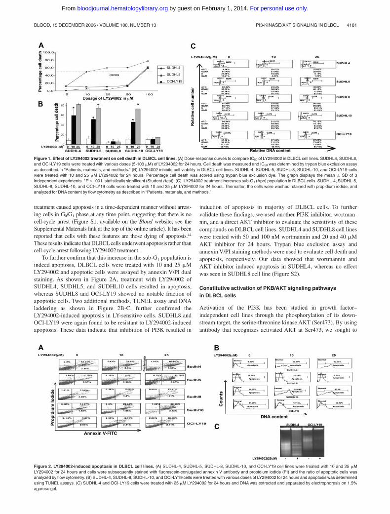
To further confirm that this increase in the sub-G1 population isindeed apoptosis, DLBCL cells were treated with 10 and 25 �MLY294002 and apoptotic cells were assayed by annexin V/PI dualstaining. As shown in Figure 2A, treatment with LY294002 ofSUDHL4, SUDHL5, and SUDHL10 cells resulted in apoptosis,whereas SUDHL8 and OCI-LY19 showed no notable fraction ofapoptotic cells. Two additional methods, TUNEL assay and DNAladdering as shown in Figure 2B-C, further confirmed theLY294002-induced apoptosis in LY-sensitive cells. SUDHL8 andOCI-LY19 were again found to be resistant to LY294002-inducedapoptosis. These data indicate that inhibition of PI3K resulted in
induction of apoptosis in majority of DLBCL cells. To furthervalidate these findings, we used another PI3K inhibitor, wortman-nin, and a direct AKT inhibitor to evaluate the sensitivity of thesecompounds on DLBCL cell lines. SUDHL4 and SUDHL8 cell lineswere treated with 50 and 100 nM wortmannin and 20 and 40 �MAKT inhibitor for 24 hours. Trypan blue exclusion assay andannexin V/PI staining methods were used to evaluate cell death andapoptosis, respectively. Our data showed that wortmannin andAKT inhibitor induced apoptosis in SUDHL4, whereas no effectwas seen in SUDHL8 cell line (Figure S2).
Constitutive activation of PKB/AKT signaling pathwaysin DLBCL cells
Activation of the PI3K has been studied in growth factor–independent cell lines through the phosphorylation of its down-stream target, the serine-threonine kinase AKT (Ser473). By usingantibody that recognizes activated AKT at Ser473, we sought to
Figure 1. Effect of LY294002 treatment on cell death in DLBCL cell lines. (A) Dose-response curves to compare IC50 of LY294002 in DLBCL cell lines. SUDHL4, SUDHL8,and OCI-LY19 cells were treated with various doses (5-100 �M) of LY294002 for 24 hours. Cell death was measured and IC50 was determined by trypan blue exclusion assayas described in “Patients, materials, and methods.” (B) LY294002 inhibits cell viability in DLBCL cell lines. SUDHL-4, SUDHL-5, SUDHL-8, SUDHL-10, and OCI-LY19 cellswere treated with 10 and 25 �M LY294002 for 24 hours. Percentage cell death was scored using trypan blue exclusion dye. The graph displays the mean � SD of 3independent experiments. *P .001, statistically significant (Student t test). (C). LY294002 treatment increases sub-G1 (Apo) population in DLBCL cells. SUDHL-4, SUDHL-5,SUDHL-8, SUDHL-10, and OCI-LY19 cells were treated with 10 and 25 �M LY294002 for 24 hours. Thereafter, the cells were washed, stained with propidium iodide, andanalyzed for DNA content by flow cytometry as described in “Patients, materials, and methods.”
Figure 2. LY294002-induced apoptosis in DLBCL cell lines. (A) SUDHL-4, SUDHL-5, SUDHL-8, SUDHL-10, and OCI-LY19 cell lines were treated with 10 and 25 �MLY294002 for 24 hours and cells were subsequently stained with fluorescein-conjugated annexin V antibody and propidium iodide (PI) and the ratio of apoptotic cells wasanalyzed by flow cytometry. (B) SUDHL-4, SUDHL-8, SUDHL-10, and OCI-LY19 cells were treated with various doses of LY294002 for 24 hours and apoptosis was determinedusing TUNEL assays. (C) SUDHL-4 and OCI-LY19 cells were treated with 25 �M LY294002 for 24 hours and DNA was extracted and separated by electrophoresis on 1.5%agarose gel.
PI3-KINASE/AKT SIGNALING IN DLBCL 4181BLOOD, 15 DECEMBER 2006 � VOLUME 108, NUMBER 13
For personal use only. by guest on February 1, 2014. bloodjournal.hematologylibrary.orgFrom

determine the constitutive activation status of AKT in DLBCL celllines as well as to determine whether inhibition of PI3K byLY294002 abrogates phosphorylation of AKT. DLBCL cell lineswere treated in the presence and absence of LY294002 for 24 hoursas indicated, cells were lysed, and proteins were analyzed byWestern blot. As shown in Figure 3A, AKT was constitutivelyphosphorylated in the LY-sensitive as well as in the LY-resistantcell lines. Treatment with LY294002 dephosphorylated AKT inSUDHL4 and SUDHL5 cells, whereas LY294002 treatment did notproduce any change in AKT phosphorylation level in SUDHL8 andOCI-LY19 cell lines.
The forkhead family of transcription factors has been reportedas a downstream target of AKT, mediating apoptosis in othersystems.45 Active FKHR transcription factors promote transcrip-tion of genes involved in cell-cycle arrest and apoptosis.46 Onemechanism by which AKT promotes cell survival is by phosphory-lating FKHR transcription factors, which inactivates them andprevents apoptosis.47 We thus studied the level of FKHR/FOXO1phosphorylation in LY294002-treated and -untreated DLBCL cellsby Western blotting. As shown in Figure 3B, constitutive phosphor-ylation of FKHR was seen in all DLBCL cell lines, but LY294002treatment resulted in dephosphorylation in LY-sensitive cell linesonly and no effect was seen on the resistance of FKHRLphosphorylation in SUDHL8 and OCI-LY19 cell lines.
We next determined the activation of GSK3 in DLBCL cells,which has been recently reported to be a target of PI3K/AKT and is
involved in promotion of cell survival.48 All DLBCL cell linesshowed constitutive phosphorylation of GSK3 and dephosphoryla-tion in the presence of LY294002 (Figure 3C) was observed only inLY-sensitive cell lines with no significant effects in LY-resistantcells. These results suggest that AKT and its downstream effectorsplay a role in LY294002-induced apoptosis in DLBCL cell lines.
Effects of the inhibition of PI3K/AKT signaling at themitochondrial level in DLBCL cells
The apoptotic signaling cascade starts with activation of caspase-8and truncation of BID that translocates to the mitochondrialmembrane allowing activation of proapoptotic proteins and releaseof cytochrome c. Therefore, we sought to determine whetherinhibition of PI3K signaling involves the mitochondria. Activatedcaspase-8 is capable of cleaving caspase-3 either directly or bydigesting BID to its active form (tBID), which leads to the releaseof cytochrome c from mitochondria.49 LY294002 treatment for 24hours resulted in activation of caspase-8 leading to truncation ofBID in SUDHL4 and SUDHL5 cells (Figure 4A) as inferred by thedecreased intensity of the full-length BID band. On the other hand,SUDHL8 and OCI-LY19 cell lines were resistant to LY294002-induced activation of BID and caspase-8. We then tested the effectof LY294002 on the mitochondrial membrane potentials in thesecells. DLBCL cells were treated with 25 �M LY294002 for 24hours and labeled with JC-1 dye and mitochondrial membranepotential was measured by flow cytometry. As shown in Figure 4B,inhibition of PI3K resulted in loss of mitochondrial membranepotential in SUDHL4, SUDHL5, and SUDHL10 cells as measuredby JC-1–stained green fluorescence depicting apoptotic cells. InSUDHL8 and OCI-LY19 cells, no change in mitochondrial mem-brane was observed. We then studied release of cytochrome c fromthe mitochondria in cells treated for 24 hours with LY294002.Cytosolic-specific, mitochondria-free as well as mitochondrialextracts were prepared as described in “Patients, materials, andmethods.” Cytochrome c was released to the cytosol after LY294002treatment in SUDHL4 and SUDHL5 but not in OCI-LY19 cells(Figure 4C). On the other hand, the level of cytochrome cdecreased in the mitochondrial fraction of LY-sensitive cells only.These results suggest that inhibition of PI3K/AKT pathwaysdisrupts the mitochondrial membrane potential leading to therelease of cytochrome c to the cytosol. We then sought to determinewhether LY294002-induced release of cytochrome c was capableof activation of caspase-3 and PARP. Figure 5A shows thatLY294002 treatment resulted in the activation of caspase-3 andcleavage of PARP in SUDHL4 and SUDHL5 cells but not in
Figure 3. Inhibition of PI3K/AKT signaling pathway during LY294002-inducedapoptosis. SUDHL-4, SUDHL-5, SUDHL8, and OCI-LY19 cells were treated withand without 10 and 25 �M LY294002 for 24 hours. After cell lysis equal amounts ofproteins were separated by SDS-PAGE, transferred to Immobilon membrane, andimmunoblotted with antibodies against phospho-AKT (Ser473), phospho FKHR, andphosphoGSK3�/�-actin as indicated.
Figure 4. LY294002-induced activation of the mitochondrial apoptotic pathway in DLBCL cell lines. (A) LY294002-induced activation of caspase-8 and cleavage of BID.SUDHL-4, SUDHL-5, SUDHL-8, and OCI-LY19 cells were treated with 10 and 25 �M LY294002 for 24 hours. Cells were lysed and equal amounts of proteins were separatedby SDS-PAGE, transferred to Immobilon membrane, and immunoblotted with antibodies against caspase-8, BID, and actin as indicated. (B) Loss of mitochondrial membranepotential by LY294002 treatment in DLBCL cells. SUDHL-4, SUDHL-5, SUDHL-8, SUDHL10, and OCI-LY19 cells were treated with and without 25 �M LY294002 for 24 hours.Live cells with intact mitochondrial membrane potential (f) and dead cells with lost mitochondrial membrane potential (u) were measured by JC-1 staining and analyzed byflow cytometry as described in “Patients, materials, and methods.” (C) LY294002-induced release of cytochrome c. SUDHL-4, SUDHL-5, and OCI-LY19 cell lines were treatedwith and without 25 �M LY294002 for 24 hours. Mitochondrial-free cytoplasmic as well as mitochondrial fractions were isolated as described in “Patients, materials, andmethods.” Cell extracts were separated on SDS-PAGE, transferred to PVDF membrane, and immunoblotted with antibodies against cytochrome c and actin as indicated.
4182 UDDIN et al BLOOD, 15 DECEMBER 2006 � VOLUME 108, NUMBER 13
For personal use only. by guest on February 1, 2014. bloodjournal.hematologylibrary.orgFrom

SUDHL8 and OCI-LY19 cells. These results are consistent with thedata on cytochrome c release and indicate that activation of effectorcaspases participate in LY294002-induced apoptosis in DLBCLcells. In addition, pretreatment of DLBCL cells with 80 �Mz-VAD-fmk, a universal inhibitor of caspases, abrogated apoptosisand prevented cell death and caspase-3 activation induced byLY294002 (Figure 6), clearly indicating that caspases play a criticalrole in LY294002-induced apoptosis in DLBCL cells. XIAP is amember of inhibitors of apoptosis protein family and a physiologicsubstrate of AKT that is stabilized to inhibit programmed cell deathand has a direct effect on caspase-3 and caspase-9.50 To determinewhether XIAP plays a role in protecting DLBCL cells fromLY294002-induced apoptosis, SUDHL4, SUDHL8, and SUDHL10cells were treated with and without LY294002. Expression of XIAP
was significantly decreased in sensitive SUDHL4 and SUDHL10cells after LY294002 treatment, whereas no effect was seen in theresistant SUDHL8 cell line. These data suggest that XIAP is animportant survival molecule that mediates AKT-induced cell sur-vival in DLBCL cells (Figure S3).
Expression of activated AKT in DLBCL tumors
We subsequently stained DLBCL cell lines for p-AKT expressionby immunohistochemistry (Figure 7). Our data showed thatintensity of p-AKT staining was low in LY-sensitive cell lines(SUDHL 4, SUDHL10), whereas the expression was high inLY-resistant DLBCL cell lines (SUDHL8, OCI-LY19; Figure S4),suggesting that overexpression of p-AKT may be the cause ofresistance to apoptosis. As a result of these findings, we sought todetermine the expression pattern of p-AKT in patients withDLBCL. Using immunohistochemistry, p-AKT expression washigh in 50 of 97 (51.54%) patients with interpretable data. Weperformed Kaplan-Meier survival analysis to determine the associa-tion of overall survival with p-AKT expression. Survival analysiswas performed in 91 of the 97 p-AKT interpretable cases (6patients had no follow-up data). A statistical trend toward inferior5-year survival for patients with p-AKT� tumors was observed inthe DLBCL (47 cases versus 44 cases, P .05, not significant,log-rank).
Correlation of p-AKT with clinical and laboratory parameters
The major clinical and laboratory findings of the patients groupedaccording to high or low p-AKT expression are summarized inTable 1. All the parameters required to determine IPI were availablein 64 patients; however, the LDH values were available in only 62patients. Because these patients had a total IPI score of 4, they werecategorized in the high-risk group and included in the analysis. Inthis patient population the IPI score correlated strongly withoutcome (P .002), consistent with previous studies.37
Although there were a higher proportion of patients withp-AKT overexpression in the high-risk group (high-intermediaterisk and high-risk) as compared to the low-risk group (low-intermediate risk and low-risk), this was not statistically significant
Figure 7. Tissue microarray-based p-AKT analysis in DLBCL patients. (A)Overview of the TMA containing 100 DLBCL tissue samples. (B) Kaplan-Meiersurvival curve of DLBCL of p-AKT expression by immunohistochemistry. (C) Immuno-histochemistry of a tissue spot showing high p-AKT expression in DLBCL tumor(objective, 20�/0.70 NA). (D) A tissue spot showing negative p-AKT expression(objective, 20�/0.70 NA). Immunohistochemical staining images were obtained witha BX51 Olympus microscope and an Olympus DP12 camera (Olympus, Melville,NY). Images were viewed through a universal semi-apochromat objective lens(UPlan F1; Olympus America, Woodbury, NY). Magnifcation, 200�.
Figure 5. Activation of caspase-3 and cleavage of PARP induced by LY294002treatment in DLBCL cells. SUDHL-4, SUDHL-5, SUDHL-8, and OCI-LY19 cellswere treated with and without 10 and 25 �M LY294002 for 24 hours. Cells were lysedand equal amounts of proteins were separated by SDS-PAGE, transferred to PVDFmembrane, and immunoblotted with antibodies against procaspase-3, cleavedcaspase-3, PARP, and actin as indicated.
Figure 6. Effect of z-VAD on the LY294002-induced apoptosis. (A) Effect ofz-VAD/fmk on LY294002-induced apoptosis in DLBCL cells. SUDHL-4, SUDHL-5,and SUDHL10 cells were pretreated with 80 �M z-VAD/fmk for 2 hours andsubsequently treated with 10 and 25 �M LY294002 for 24 hours. Apoptosis wasmeasured by annexin V/PI staining. (B) Effect of z-VAD/fmk on LY294002-inducedcell death in DLBCL cells. SUDHL-4, SUDHL-5, and SUDHL-10 cells were pretreatedwith 80 �M z-VAD/fmk for 2 hours and subsequently treated with 10 and 25 �MLY294002 for 24 hours. Live and dead cells were scored using trypan blue exclusiondye. The graph displays the mean � SD of 3 independent experiments. (C)z-VAD/fmk abrogates LY294002-induced activation of caspase-3. SUDHL-4,SUDHL-5, and SUDHL10 cells were pretreated with 80 �M z-VAD/fmk for 2 hoursand subsequently treated with 25 �M for 24 hours. Cells were lysed and equalamounts of proteins were separated by SDS-PAGE, transferred to PVDF membrane,and immunoblotted with antibodies against procaspase-3, cleaved caspase, andactin as indicated.
PI3-KINASE/AKT SIGNALING IN DLBCL 4183BLOOD, 15 DECEMBER 2006 � VOLUME 108, NUMBER 13
For personal use only. by guest on February 1, 2014. bloodjournal.hematologylibrary.orgFrom

(P .114). A more consistent correlative trend was noted in theproportion of p-AKT overexpressing tumors in the low-risk(39.1%), intermediate-risk (56%), and high-risk (75%) IPI groups(P .079; Figure S5). Extranodal disease in more than one sitewas more common in patients with high p-AKT expression in thetumors than in patients with low p-AKT expression (P .01).Presence of p-AKT overexpression was significantly associated(P .003) with more frequent presentation in patients with Bsymptoms. In addition, Ki 67 expression, a proliferation marker,was significantly higher in p-AKT overexpressing DLBCL tumors(P .009; Table 1).
Discussion
In the present study, we provide evidence that constitutive activa-tion of the PI3K-PKB/AKT signaling pathway plays a critical rolein regulating the growth and survival of DLBCL cells. Our datashow that LY294002, a specific inhibitor of PI3K at 10 to 25 �M,causes apoptosis as determined by the increase of both sub-G1
hypodiploid nuclei and annexin V� cell population. We alsoidentified a set of DLBCL cell lines (SUDHL8 and OCI-LY19) thatshowed no response to LY294002-induced apoptosis (Figures 1-2).We found that PI3K is frequently activated, as confirmed by thedetection of constitutive phosphorylation of different substratesdownstream of PI3K, including AKT, FKHR, and GSK3 in allDLBCL cells tested (Figure 3). However, cell lines differed in the
consequent effect on dephosphorylation of AKT, cytochrome crelease from the mitochondria, and activation of caspase cascadesystem after LY294002 treatment. The oncogenic role of thederegulated PI3K pathway is probably related to its simultaneousactions on growth and survival. Different mechanisms of PI3Kderegulation and activation have been reported in different sys-tems. Amplification of the p110 subunit of PI3K is observed inovarian cancer51 and this catalytic subunit found in a chicken tumorvirus mediates its transforming effects through AKT.52 AKT isoverexpressed in ovarian and pancreatic carcinomas.53,54 A recentstudy has shown that growth factor deprivation induces proteolyticcleavage of the proapoptotic Bcl-2 family member BID to yield itsactive truncated form, tBID.55 However, activated AKT inhibitedmitochondrial cytochrome c release and apoptosis following BIDcleavage. In concordance with this, our data show that inhibition ofthe PI3K/AKT pathway induced cleavage of BID, loss of mitochon-drial membrane potential, and release of cytochrome c in thosecells (SUDHL4 and SUDHL10) in which inhibition of PI3Kdephosphorylated AKT. On the other hand, cell lines (SUDHL8and OCI-LY19) that exhibit resistance to AKT, FKHR, and GSK3dephosphorylation in response to LY294002 treatment did not losemitochondrial potential and did not allow the release of cyto-chrome c to the cytosol. It has been shown that AKT inhibitsapoptosis downstream of BID cleavage involving hexokinases.55
AKT has also been shown to accumulate in the mitochondrialmatrix and membrane after activation of PI3K.56
Apoptosis is a multistep process and an increasing number of geneshave been identified that are involved in the control or execution ofapoptosis.57 Caspases play a crucial role in apoptosis. Among the 14known members of IL-1–converting enzyme family of proteases,caspase-3 has been shown to be a key component of the apoptoticmachinery.58 Caspase-3 is activated in apoptotic cells and cleavesseveral cellular proteins, including PARP. The cleavage of PARP is usedas a hallmark of apoptosis by various antitumor agents.59 In this study,we show that inhibition of PI3K activates caspase-3 and cleaves PARPin LY294002-sensitive DLBCL cell lines. This implies that activation ofcaspase-3 is involved in LY294002-induced apoptosis.
The PI3K/AKT pathway is abnormally active in multiple tumortypes, including lymphoid malignancies,60,61 most frequently byinactivating mutations of PTEN but also by amplification andoverexpression of PI3K/AKT.62 Activated AKT has been shown tobe associated with poor disease-free survival in patients with breastcancer and non–small-cell lung cancer.63,64 The data from ourDLBCL cell lines suggest that inhibition of PI3K by LY294002induces an appreciable amount of apoptosis in cell lines with a lowlevel of p-AKT expression as compared to cell lines that haverelatively high expression of p-AKT. We have previously shownthat inhibition of PI3K by LY294002 did not completely block theAKT phosphorylation in a primary effusion lymphoma cell linethat caused resistance to LY294002-induced apoptosis.33 Theresidual activated AKT protects the integrity of the mitochondrialmembrane and does not allow the release of cytochrome c.55
Furthermore XIAP, a physiologic substrate of AKT, is stabilized toinhibit programmed cell death and has a direct effect on caspase-3and caspase-9.50 Our data have shown that LY294002 treatmentdown-regulated XIAP in SUDHL4 and SUDHL10, whereas inSUDHL8 PI3K inhibition has no effect on the status of XIAP(Figure S3), suggesting phosphorylated AKT in SUDHL8 mayprotect XIAP from degradation in response to PI3K inhibition andrender this cell line resistant to apoptosis. Our data suggest that inDLBCL cell lines where constitutive expression of AKT is eitherhigh or residual AKT activity remained after PI3K inhibition,
Table 1. Clinical characteristics and p-AKT Ser473 expressionstatus of patients with DLBCL
Total
Highp-AKT
Lowp-AKT
Pn % n %
No. of patients 64 35 55 29 45
Age, y .279
60 or younger 35 17 49 18 51
Older than 60 29 18 62 11 38
Sex .330
Female 17 11 65 6 35
Male 47 24 51 23 49
Performance status .133
Lower than 2 40 19 48 21 52
2 or higher 24 16 67 8 33
Stage .225
I or II 30 14 47 16 53
III or IV 34 21 62 13 38
Extranodal sites involved .01
1 site 40 17 43 23 57
More than 1 site 24 18 75 6 25
LDH level* .343
Normal 26 12 46 14 54
High 36 21 58 15 42
IPI .111
Low to low-intermediate 35 16 46 19 54
High-intermediate to high 29 19 66 10 34
B symptoms .003
Absent 29 10 35 19 65
Present 35 25 71 10 29
Ki-67 expression (n � 95) .009
Positive 83 46 55 37 45
Negative 12 2 17 10 83
*LDH categorization is based on the range for normal values in the clinicallaboratory at our institution. LDH levels were missing in 2 patients but both patientshad a total IPI score of 4 and were categorized in the high-risk group.
4184 UDDIN et al BLOOD, 15 DECEMBER 2006 � VOLUME 108, NUMBER 13
For personal use only. by guest on February 1, 2014. bloodjournal.hematologylibrary.orgFrom

targeting of additional molecules such as XIAP in addition withPI3K inhibitor may overcome resistance and induce apoptosis.
Our immunohistochemistry data suggest that p-AKT overexpres-sion in the DLBCL tissue array was associated with a pooroutcome. Moreover, p-AKT overexpression was associated with ahigher IPI score. In addition, it was significantly associated withinvolvement of more than one extranodal site and with B symp-toms. A trend was seen with IPI score that did not reach statisticalsignificance. Furthermore, high p-AKT expression was associatedwith relative risk of 1.4 for death in bivariate analysis in combina-tion with IPI and 1.9 in univariate analysis by Cox regressionalanalysis (Table S1). However, bivariate analysis with IPI demon-strated no statistical significance (P .451). This could be attribut-able to the small sample size, which was further reduced due to IPIbeing available only in 64 cases. These associations, we believe,would be significant if studied in a larger cohort of patients.
In summary, our results establish that the PI3K/AKT pathway isconstitutively activated in human DLBCL cell lines. Inhibition ofPI3K leads to apoptosis in most DLBCL cells through release ofcytochrome c from the mitochondria and activation of downstreamcaspases. In addition, patients with high p-AKT expression showeda poor survival. These studies may have important implications forfuture preclinical and clinical studies in DLBCL. Not only couldp-AKT expression be used for prognostication, but also, thesefindings may pave the way for investigations aimed at determiningthe usefulness of a novel strategy for treating DLBCL withinhibitors of the P13K/AKT pathway, either alone or in combina-tion with other agents. Further animal, preclinical, and clinical
studies are needed to validate the data presented here, which nowhave greater impact with recent identification of therapeuticstrategies using inhibitors of small molecules.
Acknowledgment
We thank Dr Shakaib Siddiqui for collecting and reviewingclinical data.
Authorship
Contribution: S.U. designed research, performed experiments,analyzed data, and wrote the paper; A.R.H. designed research,performed experiments, analyzed data, and helped in writing thepaper; A.K.S. analyzed data and did statistical analysis; P.S.M.,N.A.A., A.M., and V.A. performed experiments; F.A., A.B., H.E.,and A.E. provided clinical samples and data for performance ofexperiments and validation of data; P.B. performed experiments,analyzed data, did statistical analysis, and helped in writing thepaper; and K.S.A. designed research, analyzed data, and helped inwriting the paper.
Conflict-of-interest disclosure: The authors declare no compet-ing financial interests.
Correspondence: Shahab Uddin, King Fahad National Centerfor Children’s Cancer and Research, King Faisal Specialist Hospi-tal and Research Center, MBC 98-16, PO Box 3354, Riyadh 11211,Saudi Arabia; e-mail: [email protected].
References
1. The Non-Hodgkin’s Lymphoma ClassificationProject. A clinical evaluation of the InternationalLymphoma Study Group classification of non-Hodgkin’s lymphoma. Blood. 1997;89;3909-3916.
2. Muris JJ, Cillessen SA, Vos W, et al. Immunohis-tochemical profiling of caspase signaling path-ways predicts clinical response to chemotherapyin primary nodal diffuse large B-cell lymphomas.Blood. 2005;105:2916-2923.
3. Hartge P Wang SS, Overview of the etiology andepidemiology of lymphoma. In: Mauch PM, Armit-age JO, Coiffier B, Dalla-Favera R, Harris NL,eds. Non-Hodgkin’s Lymphomas. New York, NY:Lippincott, Williams and Wilkins; 2004:711-727.
4. Smith PG, Wang F, Wilkinson KN, et al. Thephosphodiesterase PDE4B limits cAMP-associ-ated PI3K/AKT-dependent apoptosis in diffuselarge B-cell lymphoma. Blood. 2005;105:308-316.
5. Lam LT, Davis RE, Pierce J, et al. Small moleculeinhibitors of IkappaB kinase are selectively toxicfor subgroups of diffuse large B-cell lymphomadefined by gene expression profiling. Clin CancerRes. 2005;11:28-40.
6. Houldsworth J, Olshen AB, Cattoretti G, et al. Re-lationship between REL amplification, REL func-tion, and clinical and biologic features in diffuselarge B-cell lymphomas. Blood. 2004;103:1862-1868.
7. Pandolfi PP. Breast cancer—loss of PTEN pre-dicts resistance to treatment. N Engl J Med.2004;35:2337-2338.
8. Saal LH, Holm K, Maurer M, et al. PIK3CA muta-tions correlate with hormone receptors, node me-tastasis, and ERBB2, and are mutually exclusivewith PTEN loss in human breast carcinoma. Can-cer Res. 2005;65:2554-2559.
9. Zhang P, Ostrander JH, Faivre EJ, Olsen A,Fitzsimmons D, Lange CA. Regulated associa-tion of protein kinase B/Akt with breast tumor ki-nase. J Biol Chem. 2005;280:1982-1991.
10. Alessi DR, Cohen P. Mechanism of activation andfunction of protein kinase B. Curr Opin GenetDev. 1998;8:55-62.
11. Al-Sakkaf KA, Mooney LM, Dobson PR, BrownBL. Possible role for protein kinase B in the anti-apoptotic effect of prolactin in rat Nb2 lymphomacells. J Endocrinol. 2000;167:85-92.
12. Blanc A, Pandey NR, Srivastava AK. Synchro-nous activation of ERK 1/2, p38mapk and PKB/Akt signaling by H2O2 in vascular smooth musclecells: potential involvement in vascular disease.Int J Mol Med. 2003;11:229-234.
13. Eves EM, Xiong W, Bellacosa A, et al. Akt, a tar-get of phosphatidylinositol 3-kinase, inhibits apo-ptosis in a differentiating neuronal cell line. MolCell Biol. 1998;18:2143-2152.
14. West KA, Castillo SS, Dennis PA. Activation ofthe PI3K/Akt pathway and chemotherapeutic re-sistance. Drug Resist Updates. 2002;5:234-248.
15. Franke TF, Hornik CP, Segev L, Shostak GA,Sugimoto C. PI3K/Akt and apoptosis: size mat-ters. Oncogene. 2003;22:8983-8998.
16. Bacus SS, Altomare DA, Lyass L, et al. AKT2 isfrequently upregulated in HER-2/neu-positivebreast cancers and may contribute to tumor ag-gressiveness by enhancing cell survival. Onco-gene. 2002;21:3532-3540.
17. Luo J, Manning BD, Cantley LC. Targeting thePI3K-Akt pathway in human cancer: rationale andpromise. Cancer Cell. 2003;4:257-262.
18. Nicholson KM, Anderson NG. The protein kinaseB/Akt signalling pathway in human malignancy.Cell Signal. 2002;14:381-395.
19. Testa JR, Bellacosa A. AKT plays a central role intumorigenesis. Proc Natl Acad Sci U S A. 2001;98:10983-10985.
20. Wendel HG, De Stanchina E, Fridman JS, et al. Sur-vival signaling by Akt and eIF4E in oncogenesis andcancer therapy. Nature. 2004;428:332-337.
21. Johnstone RW, Ruefli AA, Lowe SW. Apoptosis: alink between cancer genetics and chemotherapy.Cell. 2002;108:153-164.
22. Schmitt CA, Lowe SW. Bcl-2 mediates chemore-sistance in matched pairs of primary E (mu)-myclymphomas in vivo. Blood Cells Mol Dis. 2001;27:206-216.
23. Rathmell JC, Thompson CB. The central effectorsof cell death in the immune system. Annu RevImmunol. 1999;17:781-828.
24. Kroemer G, Reed JC. Mitochondrial control of celldeath. Nat Med. 2000;6:513-519.
25. Korsmeyer SJ. BCL-2 gene family and the regu-lation of programmed cell death. Cancer Res.1999;59:1693s-1700s.
26. Schimmer AD, Welsh K, Pinilla C, et al. Small-molecule antagonists of apoptosis suppressorXIAP exhibits broad antitumor activity. CancerCell. 2004;5:25-35.
27. Uddin S, Hussain A, Manogaran PS, et al. Cur-cumin suppresses growth and induces apoptosisin primary effusion lymphoma. Oncogene 2005;24:7022-7030.
28. Uddin S. Hussain A, Al-Hussein K, PlataniasLC, Bhatia KG. Inhibition of phosphatidylinosi-tol 3�-kinase induces preferentially killing ofPTEN-null T leukemias through AKT pathway.Biochem Biophys Res Commun. 2004;320:932-938.
29. Hussain A, Doucet JP, Gutierrez M, et al. Tumornecrosis factor-related apoptosis-inducing ligand(TRAIL) and Fas apoptosis in Burkitt’s lympho-mas with loss of multiple pro-apoptotic proteins.Haematologica. 2003;88:167-175.
30. Krishan A. Rapid flow cytofluorometric analysis ofmammalian cell cycle by propidium iodide stain-ing. J Cell Biol. 1975;66:188-193.
31. Uddin S, Ah-Kang J, Ulaszek J, Mahmud D,
PI3-KINASE/AKT SIGNALING IN DLBCL 4185BLOOD, 15 DECEMBER 2006 � VOLUME 108, NUMBER 13
For personal use only. by guest on February 1, 2014. bloodjournal.hematologylibrary.orgFrom

Wickrema A. Differentiation stage-specific activa-tion of p38 mitogen-activated protein kinase iso-forms in primary human erythroid cells. Proc NatlAcad Sci U S A. 2004;101:147-152.
32. Uddin S, Fish EN, Sher D, et al. The IRS-pathwayoperates distinctively from the Stat-pathway inhematopoietic cells and transduces common anddistinct signals during engagement of the insulinor interferon-alpha receptors. Blood 1997;90:2574-2582.
33. Uddin S. Hussain A, Manogaran PS, et al. Inhibi-tion of phosphatidylinositol 3�-kinase/AKT-signal-ing promotes apoptosis of primary effusion lym-phoma cells. Clin Cancer Res. 2005;11:3102-3108.
34. Hussain A, Al-Rasheed, Manogaran PS, et al.Curcumin induced apoptosis in acute T cell leuke-mias. Apoptosis 2005;11:245-254.
35. Kononen J, Bubendorf L, Kallioniemi A, et al. Tis-sue microarrays for high-throughput molecularprofiling of tumor specimens. Nat Med. 1998;4:844-847.
36. Jaffe ES, Harris NL, Stein H, Vardiman JW. Pa-thology and genetics of tumors of hematopoieticand lymphoid tissues: World Health OrganizationClassification of Tumors. Lyon, France; IARCPress: 2001.
37. The International Non-Hodgkin’s LymphomaPrognostic Factors Project. A predictive model foraggressive non-Hodgkin’s lymphoma. N EnglJ Med. 1993;329:987-994.
38. David O, Jett J, LeBeau H, et al. Phospho-Aktoverexpression in non–small cell lung cancerconfers significant stage-independent survivaldisadvantage. Clin Cancer Res. 2004;10:6865-6871.
39. Bose S, Chandran S, Mirocha JM, Bose N. TheAkt pathway in human breast cancer: a tissue-array-based analysis. Mod Pathol. 2006;19:238-245.
40. Tsao AS, McDonnell T, Lam S, et al. Increasedphospho-AKT (Ser473) expression in bronchialdysplasia: implications for lung cancer preventionstudies. Cancer Epidemiol Biomarkers Prev.2003;12:660-664.
41. Franke TF, Kaplan DR, Cantle LC. PI3K: down-stream AKT1 on blocks apoptosis. Cell. 1997;88:435-437,
42. Vlahos CJ, Matter WF, Hui KY, Brown RF. A spe-cific inhibitor of phosphatidylinositol 3-kinase,2-(4-morpholinyl)-8-phenyl-4H-1-benzopyran-4-one (LY294002). J Biol Chem. 1994;269:5241-5248.
43. Vlahos CJ, Matter WF, Brown RF, et al. Investiga-tion of neutrophil signal transduction using a spe-cific inhibitor of phosphatidylinositol 3-kinase.J Immunol. 1995;154:2413-2422.
44. Zhang C, Hazarika P, Ni X, Weidner DA, Duvic M.Induction of apoptosis by bexarotene in cutane-ous T-cell lymphoma cells: relevance to mecha-nism of therapeutic action. Clin Cancer Res.2002;8:1234-1240.
45. Brunet A, Park J, Tran H, Hu LS, Hemmings BA,Greenberg ME. Protein kinase SGK mediatessurvival signals by phosphorylating the forkheadtranscription factor FKHRL1 (FOXO3a). Mol CellBiol. 2001;21:952-965.
46. Alvarez B, Martinez-A C, Burgering BM, CarreraAC. Forkhead transcription factors contribute toexecution of the mitotic programme in mammals.Nature. 2001;413:744-747.
47. Ciechomska I, Pyrzynska B, Kazmierczak P, Ka-minska B. Inhibition of Akt kinase signalling andactivation of forkhead are indispensable for up-regulation of FasL expression in apoptosis ofglioma cells. Oncogene. 2003;23:7617-7627.
48. Cross DA, Alessi DR, Cohen P, Andjelkovich M,Hemmings BA. Inhibition of glycogen synthasekinase-3 by insulin mediated by protein kinase B.Nature. 1378:785-789.
49. Gross A, McDonnell JM, Korsmeyer SJ. BCL-2family members and the mitochondria in apopto-sis. Genes Dev. 1999;13:1899-1911.
50. Dan HC, Sun M, Kaneko S, et al. Akt phosphory-lation and stabilization of X-linked inhibitor of apo-ptosis protein (XIAP). J Biol Chem. 2004;279:5405-5412.
51. Shayesteh L, Lu Y, Kuo WL, et al. PIK3CA is im-plicated as an oncogene in ovarian cancer. NatGenet. 1999;21:99-102.
52. Chang HW, Aoki M, Fruman D, et al. Transforma-tion of chicken cells by the gene encoding thecatalytic subunit of PI 3-kinase. Science 1997;276:1848-1850.
53. Bellacosa A, de Feo D, Godwin AK, et al. Molecu-
lar alterations of the AKT2 oncogene in ovarianand breast carcinomas. Int J Cancer. 1995;64:280-285.
54. Cheng JQ, Ruggeri B, Klein WM, et al. Amplifica-tion of AKT2 in human pancreatic cells and inhibi-tion of AKT2 expression and tumorigenicity byantisense RNA. Proc Natl. Acad Sci U S A. 1996;93:3636-3641.
55. Majewski N, Nogueira V, Robey RB, Hay N. Aktinhibits apoptosis downstream of BID cleavagevia a glucose-dependent mechanism involvingmitochondrial hexokinases. Mol Cell Biol. 2004;24:730-740.
56. Bijur GN, Jope RS. Rapid accumulation of Akt inmitochondria following phosphatidylinositol 3-ki-nase activation. J Neurochem. 2003;87:1427-1435.
57. Gastman BR. Apoptosis and its clinical impact.Head Neck. 2001;23:409-425.
58. Nunez G, Benedict MA, Hu Y, Inohara N. Cas-pases: the proteases of the apoptotic pathway.Oncogene. 1998;24:3237-3245.
59. Duriez PJ, Desnoyers S, Hoflack JC, et al. Char-acterization of anti-peptide antibodies directedtowards the automodification domain and apopto-tic fragment of poly (ADP-ribose) polymerase.Biochim Biophys Acta. 1997;1334:65-72.
60. Sakai A, Thieblemont C, Wellmann A, Jaffe ES,Raffeld M. PTEN gene alterations in lymphoidneoplasms. Blood. 1998;92:3410-3415.
61. Dahia PL, Aguiar RC, Alberta J, et al. PTEN isinversely correlated with the cell survival factorAkt/PKB and is inactivated via multiple mecha-nisms in haematological malignancies. Hum MolGenet. 1999;8:185-193.
62. Vivanco I, Sawyers CL. The phosphatidylinositol3-kinase AKT pathway in human cancer. Nat RevCancer. 2002;2:489-501.
63. Zhou X, Tan M, Stone Hawthorne V, et al. Activa-tion of the Akt/mammalian target of rapamycin/4E-BP1 pathway by ErbB2 overexpression pre-dicts tumor progression in breast cancers. ClinCancer Res. 2004;10:6779-6788.
64. David O, Jett J, LeBeau H, et al. Phospho-Aktoverexpression in non-small cell lung cancer con-fers significant stage-independent survival disad-vantage. Clin Cancer Res. 2004;10:6865-6871.
4186 UDDIN et al BLOOD, 15 DECEMBER 2006 � VOLUME 108, NUMBER 13
For personal use only. by guest on February 1, 2014. bloodjournal.hematologylibrary.orgFrom
Related Documents