The ends of linear chromosomes cannot be replicated completely by the same enzymatic machinery that repli- cates the internal portions. In the absence of a special replication mechanism, chromosomes literally shrink from their ends. This DNA end-replication problem is overcome in most eukaryotes by the enzyme telomerase. Telomerase is an RNP enzyme, and the function of one portion of its RNA subunit is gratifyingly easy to under- stand: It provides the template for extension of telomeric DNA (Greider and Blackburn 1989; Yu et al. 1990). The RNA itself performs other functions, the best-established example being the formation of the “template boundary,” a short intramolecular helix that defines the stopping point for reverse transcription (Tzfati et al. 2000). Finally, the RNA binds a number of proteins, foremost among these being the telomerase reverse transcriptase (TERT, or Est2p in Saccharomyces cerevisiae). The RNA also binds a number of accessory proteins, but these are more species-specific. In yeast, they consist of Est1p, which recruits or activates telomerase at the chromosome end; the Ku heterodimer, the DNA-repair protein that has a special role in recruiting telomerase to broken chromo- some ends for de novo telomere addition; and the Sm pro- teins, which also contribute to biogenesis of the snRNPs. Yet the function, or absence of function, of the majority of the telomerase RNA remains unknown. At an even more fundamental level, we still do not even know how to envision the structure of the telomerase RNP. Is it a more-or-less specific three-dimensional structure formed by a precise series of RNA–RNA, RNA–protein, and protein–protein interactions, along the lines of the ribosome or RNase P, such that it could someday be crystallized? Alternatively, is it possibly a loose collection of proteins held together by RNA “strings” such that no two complexes have an identical overall shape? In the case of yeast telomerase, we describe evidence that supports the latter model: RNA as a flexible scaffold for proteins. Our interrogation of yeast telomerase has stimulated us to think more gener- ally about the structure–function relationships in RNPs, especially those involved in catalysis, and we present those thoughts in the Conclusions. YEAST TELOMERASE RNA SECONDARY STRUCTURE For noncoding RNAs, knowing the nucleotide sequence is of very limited value by itself, but having a good secondary structure model provides a useful framework for investigating structure–function relation- ships. In the case of telomerase RNAs, secondary struc- tures were determined for ciliated protozoan examples and showed a single-stranded template region, a nearby pseudoknot, and a long “handle” (Stem IV) whose terminal loop is essential for function (Romero and Blackburn 1991; ten Dam et al. 1991; Lingner et al. 1994; Sperger and Cech 2001). Vertebrate telomerase RNAs folded into a structure somewhat similar to that of the ciliates, followed by a 3′-terminal pair of stem-loop structures characteristic of box H/ACA snoRNAs (Mitchell et al. 1999; Chen et al. 2000). Budding yeast telomerase RNA structures were diffi- cult to decipher due to their unusual length (typically >1000 nucleotides) and rapid divergence, which frustrates sequence alignments and makes it difficult to apply com- parative sequence analysis. The Blackburn laboratory was able to model several helices of telomerase RNAs from Kluyveromyces species (Tzfati et al. 2003). Furthermore, for S. cerevisiae telomerase RNA (TLC1), several local structures involved in binding of the Ku and Est1 proteins RNA as a Flexible Scaffold for Proteins: Yeast Telomerase and Beyond D.C. ZAPPULLA AND T.R. CECH Howard Hughes Medical Institute, Department of Chemistry and Biochemistry, University of Colorado, Boulder, Colorado 80309 Yeast telomerase, the enzyme that adds a repeated DNA sequence to the ends of the chromosomes, consists of a 1157- nucleotide RNA (TLC1) plus several protein subunits: the telomerase reverse transcriptase Est2p, the regulatory subunit Est1p, the nonhomologous end-joining heterodimer Ku, and the seven Sm proteins involved in ribonucleoprotein (RNP) maturation. The RNA subunit provides the template for telomeric DNA synthesis. In addition, we have reported evidence that it serves as a flexible scaffold to tether the proteins into the complex. More generally, we consider the possibility that RNPs may be considered in three structural categories: (1) those that have specific structures determined in large part by the RNA, including RNase P, other ribozyme–protein complexes, and the ribosome; (2) those that have specific structures determined in large part by proteins, including many small nuclear RNPs (snRNPs) and small nucleolar RNPs (snoRNPs); and (3) flexible scaffolds, with no specific structure of the RNP as a whole, as exemplified by yeast telomerase. Other can- didates for flexible scaffold structures are other telomerases, viral IRES (internal ribosome entry site) elements, tmRNA (transfer-messenger RNA), the SRP (signal recognition particle), and Xist and roX1 RNAs that alter chromatin structure to achieve dosage compensation. Cold Spring Harbor Symposia on Quantitative Biology, Volume LXXI. © 2006 Cold Spring Harbor Laboratory Press 978-087969817-1 217 Cold Spring Harbor Laboratory Press on January 31, 2015 - Published by symposium.cshlp.org Downloaded from

Welcome message from author
This document is posted to help you gain knowledge. Please leave a comment to let me know what you think about it! Share it to your friends and learn new things together.
Transcript
The ends of linear chromosomes cannot be replicatedcompletely by the same enzymatic machinery that repli-cates the internal portions. In the absence of a specialreplication mechanism, chromosomes literally shrinkfrom their ends. This DNA end-replication problem isovercome in most eukaryotes by the enzyme telomerase.
Telomerase is an RNP enzyme, and the function of oneportion of its RNA subunit is gratifyingly easy to under-stand: It provides the template for extension of telomericDNA (Greider and Blackburn 1989; Yu et al. 1990). TheRNA itself performs other functions, the best-establishedexample being the formation of the “template boundary,”a short intramolecular helix that defines the stoppingpoint for reverse transcription (Tzfati et al. 2000).Finally, the RNA binds a number of proteins, foremostamong these being the telomerase reverse transcriptase(TERT, or Est2p in Saccharomyces cerevisiae). TheRNA also binds a number of accessory proteins, but theseare more species-specific. In yeast, they consist of Est1p,which recruits or activates telomerase at the chromosomeend; the Ku heterodimer, the DNA-repair protein that hasa special role in recruiting telomerase to broken chromo-some ends for de novo telomere addition; and the Sm pro-teins, which also contribute to biogenesis of the snRNPs.Yet the function, or absence of function, of the majorityof the telomerase RNA remains unknown.
At an even more fundamental level, we still do not evenknow how to envision the structure of the telomeraseRNP. Is it a more-or-less specific three-dimensionalstructure formed by a precise series of RNA–RNA,RNA–protein, and protein–protein interactions, alongthe lines of the ribosome or RNase P, such that it couldsomeday be crystallized? Alternatively, is it possibly aloose collection of proteins held together by RNA“strings” such that no two complexes have an identical
overall shape? In the case of yeast telomerase, wedescribe evidence that supports the latter model: RNA asa flexible scaffold for proteins. Our interrogation ofyeast telomerase has stimulated us to think more gener-ally about the structure–function relationships in RNPs,especially those involved in catalysis, and we presentthose thoughts in the Conclusions.
YEAST TELOMERASE RNA SECONDARYSTRUCTURE
For noncoding RNAs, knowing the nucleotidesequence is of very limited value by itself, but having agood secondary structure model provides a usefulframework for investigating structure–function relation-ships. In the case of telomerase RNAs, secondary struc-tures were determined for ciliated protozoan examplesand showed a single-stranded template region, a nearbypseudoknot, and a long “handle” (Stem IV) whoseterminal loop is essential for function (Romero andBlackburn 1991; ten Dam et al. 1991; Lingner et al.1994; Sperger and Cech 2001). Vertebrate telomeraseRNAs folded into a structure somewhat similar to that ofthe ciliates, followed by a 3′-terminal pair of stem-loopstructures characteristic of box H/ACA snoRNAs(Mitchell et al. 1999; Chen et al. 2000).
Budding yeast telomerase RNA structures were diffi-cult to decipher due to their unusual length (typically>1000 nucleotides) and rapid divergence, which frustratessequence alignments and makes it difficult to apply com-parative sequence analysis. The Blackburn laboratory wasable to model several helices of telomerase RNAs fromKluyveromyces species (Tzfati et al. 2003). Furthermore,for S. cerevisiae telomerase RNA (TLC1), several localstructures involved in binding of the Ku and Est1 proteins
RNA as a Flexible Scaffold for Proteins:Yeast Telomerase and Beyond
D.C. ZAPPULLA AND T.R. CECHHoward Hughes Medical Institute, Department of Chemistry and Biochemistry,
University of Colorado, Boulder, Colorado 80309
Yeast telomerase, the enzyme that adds a repeated DNA sequence to the ends of the chromosomes, consists of a 1157-nucleotide RNA (TLC1) plus several protein subunits: the telomerase reverse transcriptase Est2p, the regulatory subunitEst1p, the nonhomologous end-joining heterodimer Ku, and the seven Sm proteins involved in ribonucleoprotein (RNP)maturation. The RNA subunit provides the template for telomeric DNA synthesis. In addition, we have reported evidencethat it serves as a flexible scaffold to tether the proteins into the complex. More generally, we consider the possibility thatRNPs may be considered in three structural categories: (1) those that have specific structures determined in large part bythe RNA, including RNase P, other ribozyme–protein complexes, and the ribosome; (2) those that have specific structuresdetermined in large part by proteins, including many small nuclear RNPs (snRNPs) and small nucleolar RNPs (snoRNPs);and (3) flexible scaffolds, with no specific structure of the RNP as a whole, as exemplified by yeast telomerase. Other can-didates for flexible scaffold structures are other telomerases, viral IRES (internal ribosome entry site) elements, tmRNA(transfer-messenger RNA), the SRP (signal recognition particle), and Xist and roX1 RNAs that alter chromatin structure toachieve dosage compensation.
Cold Spring Harbor Symposia on Quantitative Biology, Volume LXXI. © 2006 Cold Spring Harbor Laboratory Press 978-087969817-1 217
217-224_Zappulla_Symp71.qxd 2/7/07 2:23 PM Page 217
Cold Spring Harbor Laboratory Press on January 31, 2015 - Published by symposium.cshlp.orgDownloaded from
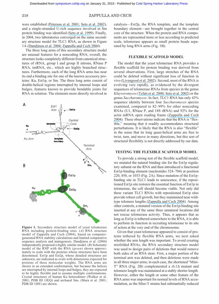
were established (Peterson et al. 2001; Seto et al. 2002),and a single-stranded U-rich sequence involved in Smprotein binding was identified (Seto et al. 1999). Finally,in 2004, two laboratories converged on the same second-ary structure model for TLC1 RNA, as shown in Figure1A (Dandjinou et al. 2004; Zappulla and Cech 2004).
The three long arms of this secondary structure modelare unusual features for a noncoding RNA; overall, thestructure looks completely different from canonical struc-tures of rRNA, group I and group II introns, RNase PRNA, tmRNA, etc., which are highly branched struc-tures. Furthermore, each of the long RNA arms has nearits end a binding site for one of the known accessory pro-teins: Ku, Est1p, or Sm. The three long arms consist ofdouble-helical regions interrupted by internal loops andbulges, features known to provide bendable joints forRNA in solution. The elements more directly involved in
catalysis—Est2p, the RNA template, and the templateboundary element—are brought together in the centralcore of the structure. When the protein and RNA compo-nents are represented more or less according to predictedscale, telomerase appears as small protein beads sepa-rated by long RNA arms (Fig. 1B).
FLEXIBLE SCAFFOLD MODEL
The model that the yeast telomerase RNA provides aflexible scaffold for protein binding was derived fromseveral observations. First, large stretches of the RNAcould be deleted without significant loss of function invivo (Livengood et al. 2002). Second, most of the RNA isevolving very rapidly, as evidenced by the divergentsequences of telomerase RNAs from species in the genusKluyveromyces (Tzfati et al. 2000; Seto et al. 2002) or thegenus Saccharomyces. In fact, TLC1 RNA has only 43%sequence identity between four Saccharomyces speciesexamined, compared to 82–99% for other noncodingRNAs (U1, RNase P, and 18S rRNA) and 92% for theactin mRNA open reading frame (Zappulla and Cech2004). These observations indicate that the RNA is “flex-ible,” meaning that it readily accommodates structuralperturbations. It is likely that the RNA is also “flexible”in the sense that its long quasi-helical arms are free totwist, turn, and move in many directions, but this sort ofstructural flexibility is not directly addressed by our data.
TESTING THE FLEXIBLE SCAFFOLD MODEL
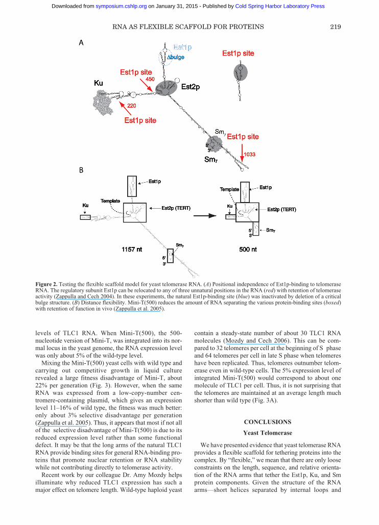
To provide a strong test of the flexible scaffold model,we mutated the natural binding site for the Est1p regula-tory subunit on the RNA and then introduced a functionalEst1p-binding element (nucleotides 524–704) at position220, 450, or 1033 (Fig. 2A). Since mutation of the Est1p-binding site in TLC1 leads to senescence, if the reposi-tioned Est1p site restores the essential function of Est1p intelomerase, the cell should become viable. Not only didthese variant TLC1 RNAs with repositioned Est1p sitesprovide robust cell growth, but they maintained near wild-type telomere lengths (Zappulla and Cech 2004). Amongother controls, a mutated version of the Est1p-binding siteinserted at any of the same three unnatural locations didnot rescue telomerase activity. Thus, it appears that aslong as Est1p is tethered somewhere to the RNA, it is ableto perform its function in recruiting telomerase to its siteof action at the very end of the chromosome.
Given that yeast telomerase appeared to consist of pro-teins tethered by flexible RNA arms, we next askedwhether the arm length was important. To avoid creatingmisfolded RNAs, the RNA secondary structure modelwas used to design pairs of deletions that would removeboth sides of an RNA stem. First, a major portion of theterminal arm was deleted, and then deletions were madein all three major arms; in each case, the shortened “Mini-T” RNA (Fig. 2B) complemented a tlc1Δ mutant andtelomere length was maintained at a stably shorter length.However, either the length or some other feature of theRNA arms was important for normal levels of RNA accu-mulation, as the Mini-T strains had substantially reduced
218 ZAPPULLA AND CECH
Figure 1. Secondary structure model of yeast telomeraseRNA including protein-binding sites. (A) RNA structuremodel of Zappulla and Cech (2004), based on computer-generated RNA stability calculations and limited comparativesequence analysis and mutagenesis. Dandjinou et al. (2004)independently proposed a highly similar model. (B) Schematicmodel of the RNP with the RNA (green) shown approxi-mately to scale with the proteins whose structures have beendetermined. Est1p and Est2p, whose detailed structures areunknown, are indicated as ovals with dimensions expected forproteins of these molecular weights. The RNA arms areshown in an extended conformation, but because the helicesare interrupted by internal loops and bulges, they are expectedto be highly flexible and to assume multiple conformations.Crystal structures of human Ku heterodimer (Walker et al.2001; PDB ID 1JEQ) and archaeal Sm7 (Mura et al. 2001;PDB ID 1I8F) are shown.
217-224_Zappulla_Symp71.qxd 2/7/07 2:23 PM Page 218
Cold Spring Harbor Laboratory Press on January 31, 2015 - Published by symposium.cshlp.orgDownloaded from
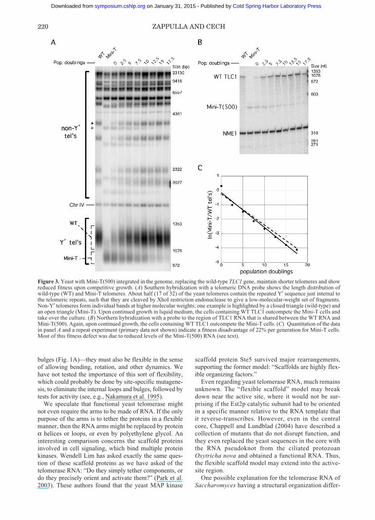
levels of TLC1 RNA. When Mini-T(500), the 500-nucleotide version of Mini-T, was integrated into its nor-mal locus in the yeast genome, the RNA expression levelwas only about 5% of the wild-type level.
Mixing the Mini-T(500) yeast cells with wild type andcarrying out competitive growth in liquid culturerevealed a large fitness disadvantage of Mini-T, about22% per generation (Fig. 3). However, when the sameRNA was expressed from a low-copy-number cen-tromere-containing plasmid, which gives an expressionlevel 11–16% of wild type, the fitness was much better:only about 3% selective disadvantage per generation(Zappulla et al. 2005). Thus, it appears that most if not allof the selective disadvantage of Mini-T(500) is due to itsreduced expression level rather than some functionaldefect. It may be that the long arms of the natural TLC1RNA provide binding sites for general RNA-binding pro-teins that promote nuclear retention or RNA stabilitywhile not contributing directly to telomerase activity.
Recent work by our colleague Dr. Amy Mozdy helpsilluminate why reduced TLC1 expression has such amajor effect on telomere length. Wild-type haploid yeast
contain a steady-state number of about 30 TLC1 RNAmolecules (Mozdy and Cech 2006). This can be com-pared to 32 telomeres per cell at the beginning of S phaseand 64 telomeres per cell in late S phase when telomereshave been replicated. Thus, telomeres outnumber telom-erase even in wild-type cells. The 5% expression level ofintegrated Mini-T(500) would correspond to about onemolecule of TLC1 per cell. Thus, it is not surprising thatthe telomeres are maintained at an average length muchshorter than wild type (Fig. 3A).
CONCLUSIONS
Yeast Telomerase
We have presented evidence that yeast telomerase RNAprovides a flexible scaffold for tethering proteins into thecomplex. By “flexible,” we mean that there are only looseconstraints on the length, sequence, and relative orienta-tion of the RNA arms that tether the Est1p, Ku, and Smprotein components. Given the structure of the RNAarms—short helices separated by internal loops and
RNA AS FLEXIBLE SCAFFOLD FOR PROTEINS 219
Figure 2. Testing the flexible scaffold model for yeast telomerase RNA. (A) Positional independence of Est1p-binding to telomeraseRNA. The regulatory subunit Est1p can be relocated to any of three unnatural positions in the RNA (red) with retention of telomeraseactivity (Zappulla and Cech 2004). In these experiments, the natural Est1p-binding site (blue) was inactivated by deletion of a criticalbulge structure. (B) Distance flexibility. Mini-T(500) reduces the amount of RNA separating the various protein-binding sites (boxed)with retention of function in vivo (Zappulla et al. 2005).
217-224_Zappulla_Symp71.qxd 2/7/07 2:24 PM Page 219
Cold Spring Harbor Laboratory Press on January 31, 2015 - Published by symposium.cshlp.orgDownloaded from
bulges (Fig. 1A)—they must also be flexible in the senseof allowing bending, rotation, and other dynamics. Wehave not tested the importance of this sort of flexibility,which could probably be done by site-specific mutagene-sis, to eliminate the internal loops and bulges, followed bytests for activity (see, e.g., Nakamura et al. 1995).
We speculate that functional yeast telomerase mightnot even require the arms to be made of RNA. If the onlypurpose of the arms is to tether the proteins in a flexiblemanner, then the RNA arms might be replaced by proteinα helices or loops, or even by polyethylene glycol. Aninteresting comparison concerns the scaffold proteinsinvolved in cell signaling, which bind multiple proteinkinases. Wendell Lim has asked exactly the same ques-tion of these scaffold proteins as we have asked of thetelomerase RNA: “Do they simply tether components, ordo they precisely orient and activate them?” (Park et al.2003). These authors found that the yeast MAP kinase
scaffold protein Ste5 survived major rearrangements,supporting the former model: “Scaffolds are highly flex-ible organizing factors.”
Even regarding yeast telomerase RNA, much remainsunknown. The “flexible scaffold” model may breakdown near the active site, where it would not be sur-prising if the Est2p catalytic subunit had to be orientedin a specific manner relative to the RNA template thatit reverse-transcribes. However, even in the centralcore, Chappell and Lundblad (2004) have described acollection of mutants that do not disrupt function, andthey even replaced the yeast sequences in the core withthe RNA pseudoknot from the ciliated protozoanOxytricha nova and obtained a functional RNA. Thus,the flexible scaffold model may extend into the active-site region.
One possible explanation for the telomerase RNA ofSaccharomyces having a structural organization differ-
220 ZAPPULLA AND CECH
Figure 3. Yeast with Mini-T(500) integrated in the genome, replacing the wild-type TLC1 gene, maintain shorter telomeres and showreduced fitness upon competitive growth. (A) Southern hybridization with a telomeric DNA probe shows the length distribution ofwild-type (WT) and Mini-T telomeres. About half (17 of 32) of the yeast telomeres contain the repeated Y′ sequence just internal tothe telomeric repeats, such that they are cleaved by XhoI restriction endonuclease to give a low-molecular-weight set of fragments.Non-Y′ telomeres form individual bands at higher molecular weights; one example is highlighted by a closed triangle (wild-type) andan open triangle (Mini-T). Upon continued growth in liquid medium, the cells containing WT TLC1 outcompete the Mini-T cells andtake over the culture. (B) Northern hybridization with a probe to the region of TLC1 RNA that is shared between the WT RNA andMini-T(500). Again, upon continued growth, the cells containing WT TLC1 outcompete the Mini-T cells. (C) Quantitation of the datain panel A and a repeat experiment (primary data not shown) indicate a fitness disadvantage of 22% per generation for Mini-T cells.Most of this fitness defect was due to reduced levels of the Mini-T(500) RNA (see text).
217-224_Zappulla_Symp71.qxd 2/7/07 2:24 PM Page 220
Cold Spring Harbor Laboratory Press on January 31, 2015 - Published by symposium.cshlp.orgDownloaded from
ent from those of other eukaryotes is the absence ofRNA interference (RNAi) in the budding yeasts. Eventhough the long RNA arms of TLC1 are not perfectdouble helices, they may be close enough to attractdicer or other elements of the RNAi machinery. Thus,budding yeast telomerase RNA might have evolvedwithout the constraint of avoiding large semihelicalextensions. It is noteworthy that both U1 and U2snRNAs from S. cerevisiae are also much larger thantheir mammalian counterparts, and, like TLC1, can besubstantially reduced in size with retention of function(Igel and Ares 1988; Shuster and Guthrie 1988;Siliciano et al. 1991). The RNase P RNAs from theyeast Candida glabrata and the fungus Phanerochaetechrysosporium are also unusually large (Kachouri et al.2005; Piccinelli et al. 2005). This supports the idea thatin some yeast and other fungi, there is either selectiveadvantage to larger size RNAs or else the absence of aselective disadvantage.
Three Classes of RNPs
In considering RNPs more generally, we quickly cometo the realization that the existing data are insufficient toprove or disprove many of them as being flexible scaffoldstructures, in the sense that much of their RNA frame-work can accommodate insertions, deletions, and basechanges without disruption of function. In some cases,the “accommodativeness” or malleability of the RNA canbe inferred from comparative sequence analysis of func-tionally equivalent RNAs from divergent species. Inaddition, in many cases, we know something about thestructural flexibility of the RNP—whether each particlehas more or less the same arrangement of protein andRNA components, albeit with some conformationalswitching, or whether, on the other hand, proteins areloosely tethered into the complex. Here, we make the pro-visional assumption that structural flexibility of the RNPcorrelates with the ability of the RNA to accommodateinsertions, deletions, and base changes in regions thatserve as tethers; but in doing so, we realize that there maybe cases where these features diverge. On the basis ofthese assumptions, we propose that RNPs may fit intothree general structural categories, as follows.
1. RNA-determined RNP structures. Specific structure(or family of structures), determined in large part bythe folded RNA. Examples: Ribosome, RNase P, and other ribo-zyme–protein complexes such as the yeast mitochon-drial group I intron bI5 bound to Cbp2 protein, or anumber of Neurospora group I introns bound to CYT-18 protein. Evidence: The RNP, and in some cases the RNA,can be crystallized. Individual ribosomal proteinsoften have disordered tails or internal loops, andthese become ordered only upon RNP formation, soat least for the ribosome, the protein does not providea pre-ordered template upon which the RNA folds(Brodersen et al. 2002; Klein et al. 2004). Not onlydo the ribosomal RNAs have conserved secondary
structures, but there are bases throughout the struc-tures that are universally conserved among the corre-sponding rRNAs of all three primary kingdoms of life(Noller 1993). In Saccharomyces species, the sequenceconservation of the rRNA far exceeds that of thetelomerase RNA (99% and 43% sequence identity,respectively) (Zappulla and Cech 2004).
2. Protein-determined RNP structures. Specific struc-ture (or family of structures), determined in large partby protein–protein interactions.Examples: U1, U2, U11/U12, and U5 snRNPs, boxC/D, and box H/ACA snoRNPs.Evidence for specific structure: High-resolutioncryo-electron microscopy (cryo-EM) structures of U1and U11/U12 (Stark et al. 2001; Golas et al. 2005).Evidence for structure being protein-based: Themajority of U2-specific proteins form stable het-eromeric complexes in absence of U2 snRNA (Willand Luhrmann 2006), U5 proteins assemble in absenceof snRNA (Achsel et al. 1998), and the crystal structureof Cbf5–Nop10–Gar1 complex has been determined inthe absence of snoRNA (Rashid et al. 2006).
3. Flexible scaffold RNPs. Proteins are tethered into thecomplex by a flexible RNA scaffold, with no specificstructure of the RNP as a whole.Example: Yeast telomerase, other candidates dis-cussed below.Evidence for flexibility: Phylogenetic variation insequence even among closely related Saccharomycesspecies, ability to delete large portions of stems andstill retain function, and ability to transplant Est1p-binding element to new locations with function.Evidence against specific structure: The RNA doesnot have the properties one would expect for a specifictertiary structure, so structure would have to comefrom the protein; but protein–protein interactions arenot strong, unlike snRNPs and snoRNPs above.Prediction: The RNP would not crystallize, andwould be heterogeneous by EM.
Telomerases from humans and from ciliated protozoahave been extensively studied, their RNA secondarystructures are well established (Romero and Blackburn1991; Chen et al. 2000), and a specific tertiary structurehas been proposed for the core of the human RNA(Theimer et al. 2005). We find it attractive to think thatthese much smaller telomerase RNAs may be naturalMini-T RNAs, with the protein-binding arms reduced toa minimal length so that it is now difficult to even recog-nize the arms. If this is the case, it should be possible toadd back flexible RNA arms to separate various func-tional elements of these other telomerase RNAs withoutperturbing activity. On the other hand, these othertelomerases may be assembled with protein–proteininteractions having replaced some of the RNA–proteininteractions seen in yeast telomerase, in which case, theRNP may have a defined three-dimensional structure.
IRES elements that permit 5′-cap-independent initia-tion of protein synthesis of certain viral and cellular
RNA AS FLEXIBLE SCAFFOLD FOR PROTEINS 221
217-224_Zappulla_Symp71.qxd 2/7/07 2:24 PM Page 221
Cold Spring Harbor Laboratory Press on January 31, 2015 - Published by symposium.cshlp.orgDownloaded from
mRNAs provide an interesting case. They bind multipleprotein complexes, including the 12-protein initiationfactor eIF3, as well as the small ribosomal subunit. Thecomplex between the hepatitis C virus IRES RNA andeIF3 has some conformational flexibility, as determinedby cryo-EM reconstructions (Siridechadilok et al. 2005).Both the IRES RNA and the ribosomal subunits undergofurther conformational changes as translation is initiatedand commences (Spahn et al. 2001; Boehringer et al.2005). Thus, the hepatitis C IRES may be a flexible col-lection of proteins that becomes conformationally fixedupon binding to the ribosome. All IRESs are not identi-cal, however. In the case of the cricket paralysis virusIRES, the RNA by itself forms a highly compact structurethat interacts directly with the ribosome to achieve factor-independent translational initiation (Batey 2006).
tmRNA, named because it combines the functions oftRNA and mRNA, rescues stalled ribosomes by switch-ing translation from a damaged mRNA to a sequenceinternal to the tmRNA. The tmRNA open reading frameencodes a protein degradation tag. The RNA binds sev-eral proteins, although not necessarily at the same time.These include the core subunit SmpB, EF-Tu, to recruitthe RNP to the stalled ribosome, a ribonuclease that maycontribute to degradation of the damaged mRNA, andseveral other proteins (Karzai and Sauer 2001). Certainly,it appears that the RNP must be conformationally flexibleto carry out its function (Haebel et al. 2004). It is prema-ture to know whether or not the RNP switches betweendiscrete tertiary structures (Burks et al. 2005) or insteadhas no specific tertiary structure, in which case it wouldqualify as a flexible scaffold.
The SRP (signal recognition particle) is an RNP thatrecognizes the signal sequence on a nascent secretory ormembrane protein as it exits the ribosome, arrests furthertranslation, and then targets the protein to a membrane-associated receptor. The protein is then translocated intothe endoplasmic reticulum or through the bacterial innermembrane. The SRP RNA has an S domain, which bindsproteins that recognize the peptide signal sequence, and anAlu domain, which binds to the ribosome and causestranslational arrest. These two domains are separated by aflexible hinge region comprising double-stranded RNAinterspersed with internal loops and bulges (Egea et al.2005), much like the yeast telomerase RNA “arms.” TheSRP may therefore be considered to be a flexible scaffoldthat tethers the S-domain RNA–protein complex to theAlu RNA or RNP element that binds to the ribosome. TheSRP then undergoes an induced-fit conformational changeand forms a discrete structure when it binds to the trans-lating ribosome (Halic et al. 2004). The SaccharomycesSRP RNA is considerably longer than those of otherspecies because of large quasi-helical extensions of theAlu domain (Van Nues and Brown 2004).
Noncoding RNAs involved in sex chromosome gene-dosage compensation may be thought of as flexible scaf-folds which tether proteins that affect the transcriptionalstate (Wutz 2003). In mammals, the Xist RNA promotesheterochromatization of one of the female X chromo-somes, whereas in Drosophila, the roX1 and roX2 RNAsincrease transcription of the single male X chromosome.
Different domains of Xist RNA are responsible for tran-scriptional repression and association with chromatin, thelatter accomplished by functionally redundant sequencesdispersed through the RNA (Wutz et al. 2002). Deletionanalysis has shown that multiple 10% segments of roX1RNA are dispensable for function, consistent with theRNA providing a flexible scaffold to bind multiple MSL(male-specific lethal) protein complexes (Stuckenholz etal. 2003).
Another intriguing candidate for a flexible scaffold isthe NRON (noncoding repressor of NFAT) recently iden-tified as a repressor of a transcription factor, NFAT(nuclear factor of activated T cells) (Willingham et al.2005). NRON binds a number of proteins, includingmembers of the importin β family, and appears to act as arepressor of the transcription factor NFAT. It will beimportant to understand something of its structure and thespatial arrangement of its protein-binding sites.
It may seem surprising that we have placed yeast telom-erase into a different category from the snoRNPs.Superficially at least, they have similar features: In allcases, the RNA provides a “guide sequence” that base-pairs to the site of a chemical reaction in a nucleic acid sub-strate. The reaction is 2′-O-methylation in the case of thebox C/D snoRNPs (Kiss-Laszlo et al. 1996), conversion ofU to pseudouracil in the case of the box H/ACA snoRNPs(Ganot et al. 1997), and nucleotide addition to single-stranded DNA in the case of telomerase. In all cases, theRNA also binds a protein enzyme that catalyzes the reac-tion: fibrillarin or Nop1p in the case of 2′-O-methylation(Tollervey et al. 1993; Wang et al. 2000; Omer et al.2002), Nap57/dyskerin or Cbf5p in the case of pseudo-uridylation (Zebarjadian et al. 1999), and TERT/Est2p inthe case of telomerase (Lingner et al. 1997). Certainly,there is some flexibility in the box H/ACA snoRNP sys-tem: The protein trimer can accommodate about 100 dif-ferent snoRNAs (Meier 2006). We place the snoRNPs in adifferent category from yeast telomerase because the for-mer form a specific protein structure in the absence of theRNA, whereas stable protein–protein interactions have notbeen observed between Est2p and Est1p, Ku, or Sm inyeast. Time will tell whether this is a useful distinction orwhether the apparent differences arise in large part becauseof the primitive state of our current knowledge.
ACKNOWLEDGMENTS
We thank Karen Goodrich for excellent technical assis-tance; Rob Batey, Quentin Vicens, and Art Zaug for help-ful discussions; and Anne Stellwagen (Boston College)for an ongoing collaboration on the Ku protein. This workwas supported in part by a grant from the NationalInstitutes of Health.
REFERENCES
Achsel T., Ahrens, K., Brahms H., Teigelkamp S., andLuhrmann R. 1998. The human U5-220kD protein (hPrp8)forms a stable RNA-free complex with several U5-specificproteins, including an RNA unwindase, a homologue of ribo-somal elongation factor EF-2, and a novel WD-40 protein.Mol. Cell. Biol. 18: 6756.
222 ZAPPULLA AND CECH
217-224_Zappulla_Symp71.qxd 2/7/07 2:24 PM Page 222
Cold Spring Harbor Laboratory Press on January 31, 2015 - Published by symposium.cshlp.orgDownloaded from
Batey R.T. 2006. Structures of regulatory elements in mRNAs.Curr. Opin. Struct. Biol. 16: 299.
Boehringer D., Thermann R., Ostareck-Lederer A., Lewis J.D.,and Stark H. 2005. Structure of the hepatitis C virus IRESbound to the human 80S ribosome: Remodeling of the HCVIRES. Structure 13: 1695.
Brodersen D.E., Clemons W.M., Jr., Carter A.P., WimberlyB.T., and Ramakrishnan V. 2002. Crystal structure of the 30S ribosomal subunit from Thermus thermophilus: Structureof the proteins and their interactions with 16 S RNA. J. Mol.Biol. 316: 725.
Burks J., Zwieb C., Muller F., Wower I., and Wower J. 2005.Comparative 3-D modeling of tmRNA. BMC Mol. Biol. 6: 14.
Chappell A.S. and Lundblad V. 2004. Structural elementsrequired for association of the Saccharomyces cerevisiaetelomerase RNA with the Est2 reverse transcriptase. Mol.Cell. Biol. 24: 7720.
Chen J.L., Blasco M.A., and Greider C.W. 2000. Secondarystructure of vertebrate telomerase RNA. Cell 100: 503.
Dandjinou A.T., Levesque N., Larose S., Lucier J.F., Abou ElelaS., and Wellinger R.J. 2004. A phylogenetically based sec-ondary structure for the yeast telomerase RNA. Curr. Biol.14: 1148.
Egea P.F., Stroud R.M., and Walter P. 2005. Targeting proteinsto membranes: Structure of the signal recognition particle.Curr. Opin. Struct. Biol. 15: 213.
Ganot P., Bortolin M.L., and Kiss T. 1997. Site-specificpseudouridine formation in preribosomal RNA is guided bysmall nucleolar RNAs. Cell 89: 799.
Golas M.M., Sander B., Will C.L., Luhrmann R., and Stark H.2005. Major conformational change in the complex SF3bupon integration into the spliceosomal U11/U12 di-snRNP asrevealed by electron cryomicroscopy. Mol. Cell 17: 869.
Greider C.W. and Blackburn E.H. 1989. A telomeric sequencein the RNA of Tetrahymena telomerase required for telomererepeat synthesis. Nature 337: 331.
Haebel P.W., Gutmann S., and Ban N. 2004. Dial tm for rescue:tmRNA engages ribosomes stalled on defective mRNAs.Curr. Opin. Struct. Biol. 14: 58.
Halic M., Becker T., Pool M.R., Spahn C.M., Grassucci R.A.,Frank J., and Beckmann R. 2004. Structure of the signalrecognition particle interacting with the elongation-arrestedribosome. Nature 427: 808.
Igel A.H. and Ares M., Jr. 1988. Internal sequences that distin-guish yeast from metazoan U2 snRNA are unnecessary forpre-mRNA splicing. Nature 334: 450.
Kachouri R., Stribinskis V., Zhu Y., Ramos K.S., Westhof E.,and Li Y. 2005. A surprisingly large RNase P RNA inCandida glabrata. RNA 11: 1064.
Karzai A.W. and Sauer R.T. 2001. Protein factors associatedwith the SsrA•SmpB tagging and ribosome rescue complex.Proc. Natl. Acad. Sci. 98: 3040.
Kiss-Laszlo Z., Henry Y., Bachellerie J.P., Caizergues-FerrerM., and Kiss T. 1996. Site-specific ribose methylation of pre-ribosomal RNA: A novel function for small nucleolar RNAs.Cell 85: 1077.
Klein D.J., Moore P.B., and Steitz T.A. 2004. The roles of ribo-somal proteins in the structure assembly, and evolution of thelarge ribosomal subunit. J. Mol. Biol. 340: 141.
Lingner J., Hendrick L.L., and Cech T.R. 1994. TelomeraseRNAs of different ciliates have a common secondary struc-ture and a permuted template. Genes Dev. 8: 1984.
Lingner J., Hughes T.R., Shevchenko A., Mann M., LundbladV., and Cech T.R. 1997. Reverse transcriptase motifs in thecatalytic subunit of telomerase. Science 276: 561.
Livengood A.J., Zaug A.J., and Cech T.R. 2002. Essentialregions of Saccharomyces cerevisiae telomerase RNA:Separate elements for Est1p and Est2p interaction. Mol. Cell.Biol. 22: 2366.
Meier U.T. 2006. How a single protein complex accommodatesmany different H/ACA RNAs. Trends Biochem. Sci. 31: 311.
Mitchell J.R., Cheng J., and Collins K. 1999. A box H/ACAsmall nucleolar RNA-like domain at the human telomeraseRNA 3′ end. Mol. Cell. Biol. 19: 567.
Mozdy A.D. and Cech T.R. 2006. Low abundance of telomerasein yeast: Implications for telomerase haploinsufficiency. RNA12: 1721.
Mura C., Cascio D., Sawaya M.R., and Eisenberg D.S. 2001.The crystal structure of a heptameric archaeal Sm protein:Implications for the eukaryotic snRNP core. Proc. Natl.Acad. Sci. 98: 5532.
Nakamura T.M., Wang Y.H., Zaug A.J., Griffith J.D., and CechT.R. 1995. Relative orientation of RNA helices in a group 1ribozyme determined by helix extension electronmicroscopy. EMBO J. 14: 4849.
Noller H.F. 1993. On the origin of the ribosome: Coevolution ofsubdomains of tRNA and rRNA. In The RNA world (ed. R.F.Gesteland and J.F. Atkins), p. 137. Cold Spring HarborLaboratory Press, Cold Spring Harbor, New York.
Omer A.D., Ziesche S., Ebhardt H., and Dennis P.P. 2002. In vitroreconstitution and activity of a C/D box methylation guideribonucleoprotein complex. Proc. Natl. Acad. Sci. 99: 5289.
Park S.H., Zarrinpar A., and Lim W.A. 2003. Rewiring MAPkinase pathways using alternative scaffold assembly mecha-nisms. Science 299: 1061.
Peterson S.E., Stellwagen A.E., Diede S.J., Singer M. S.,Haimberger Z.W., Johnson C.O., Tzoneva M., andGottschling, D.E. 2001. The function of a stem-loop in telom-erase RNA is linked to the DNA repair protein Ku. Nat.Genet. 27: 64.
Piccinelli P., Rosenblad M.A., and Samuelsson T. 2005.Identification and analysis of ribonuclease P and MRP RNAin a broad range of eukaryotes. Nucleic Acids Res. 33: 4485.
Rashid R., Liang B., Baker D.L., Youssef O.A., He Y., PhippsK., Terns R.M., Terns M.P., and Li H. 2006. Crystal structureof a Cbf5-Nop10-Gar1 complex and implications in RNA-guided pseudouridylation and dyskeratosis congenita. Mol.Cell 21: 249.
Romero D.P. and Blackburn E.H. 1991. A conserved secondarystructure for telomerase RNA. Cell 67: 343.
Seto A.G., Livengood A.J., Tzfati Y., Blackburn E., and CechT.R. 2002. A bulged stem tethers Est1p to telomerase RNA inbudding yeasts. Genes Dev. 16: 2800.
Seto A.G., Zaug A.J., Sobel S.G., Wolin S.L., and Cech T.R.1999. Saccharomyces cerevisiae telomerase is an Sm smallnuclear ribonucleoprotein particle. Nature 401: 177.
Shuster E.O. and Guthrie C. 1988. Two conserved domains ofyeast U2 snRNA are separated by 945 nonessentialnucleotides. Cell 55: 41.
Siliciano P.G., Kivens W.J., and Guthrie C. 1991. More thanhalf of yeast U1 snRNA is dispensable for growth. NucleicAcids Res. 19: 6367.
Siridechadilok B., Fraser C.S., Hall R.J., Doudna J.A., andNogales E. 2005. Structural roles for human translation factoreIF3 in initiation of protein synthesis. Science 310: 1513.
Spahn C.M., Beckmann R., Eswar N., Penczek P.A., Sali A.,Blobel G., and Frank J. 2001. Structure of the 80S ribosomefrom Saccharomyces cerevisiae—tRNA-ribosome andsubunit-subunit interactions. Cell 107: 373.
Sperger J.M. and Cech T.R. 2001. A stem-loop of Tetrahymenatelomerase RNA distant from the template potentiates RNAfolding and telomerase activity. Biochemistry 40: 7005.
Stark H., Dube P., Luhrmann R., and Kastner B. 2001.Arrangement of RNA and proteins in the spliceosomal U1small nuclear ribonucleoprotein particle. Nature 409: 539.
Stuckenholz C., Meller V.H., and Kuroda M.I. 2003. Functionalredundancy within roX1, a noncoding RNA involved indosage compensation in Drosophila melanogaster. Genetics164: 1003.
ten Dam E., van Belkum A., and Pleij K. 1991. A conservedpseudoknot in telomerase RNA. Nucleic Acids Res. 19: 6951.
Theimer C.A., Blois C.A., and Feigon J. 2005. Structure of thehuman telomerase RNA pseudoknot reveals conserved terti-ary interactions essential for function. Mol. Cell 17: 671.
Tollervey D., Lehtonen H., Jansen R., Kern H., and Hurt E.C.1993. Temperature-sensitive mutations demonstrate roles foryeast fibrillarin in pre-rRNA processing, pre-rRNA methyla-tion, and ribosome assembly. Cell 72: 443.
RNA AS FLEXIBLE SCAFFOLD FOR PROTEINS 223
217-224_Zappulla_Symp71.qxd 2/7/07 2:24 PM Page 223
Cold Spring Harbor Laboratory Press on January 31, 2015 - Published by symposium.cshlp.orgDownloaded from
Tzfati Y., Fulton T.B., Roy J., and Blackburn E.H. 2000.Template boundary in a yeast telomerase specified by RNAstructure. Science 288: 863.
Tzfati Y., Knight Z., Roy J., and Blackburn E.H. 2003. A novelpseudoknot element is essential for the action of a yeasttelomerase. Genes Dev. 17: 1779.
Van Nues R.W. and Brown J.D. 2004. Saccharomyces SRPRNA secondary structures: A conserved S-domain andextended Alu-domain. RNA 10: 75.
Walker J.R., Corpina R.A., and Goldberg J. 2001. Structure ofthe Ku heterodimer bound to DNA and its implications fordouble-strand break repair. Nature 412: 607.
Wang H., Boisvert D., Kim K.K., Kim R., and Kim S.H. 2000.Crystal structure of a fibrillarin homologue from Methano-coccus jannaschii, a hyperthermophile, at 1.6 Å resolution.EMBO J. 19: 317.
Will C.L. and Luhrmann R. 2006. Spliceosome structure andfunction. In The RNA world (ed. R.F. Gesteland et al.), p.369.Cold Spring Harbor Laboratory Press, Cold Spring Harbor,New York.
Willingham A.T., Orth A.P., Batalov S., Peters E.C., Wen B.G.,
224 ZAPPULLA AND CECH
Aza-Blanc P., Hogenesch J.B., and Schultz P.G. 2005. Astrategy for probing the function of noncoding RNAs finds arepressor of NFAT. Science 309: 1570.
Wutz A. 2003. RNAs templating chromatin structure for dosagecompensation in animals. Bioessays 25: 434.
Wutz A., Rasmussen T.P., and Jaenisch R. 2002. Chromosomalsilencing and localization are mediated by different domainsof Xist RNA. Nat. Genet. 30: 167.
Yu G.L., Bradley J.D., Attardi L.D., and Blackburn E.H. 1990.In vivo alteration of telomere sequences and senescencecaused by mutated Tetrahymena telomerase RNAs. Nature344: 126.
Zappulla D.C. and Cech T.R. 2004. Yeast telomerase RNA: Aflexible scaffold for protein subunits. Proc. Natl. Acad. Sci.101: 10024.
Zappulla D.C., Goodrich K., and Cech T.R. 2005. A miniatureyeast telomerase RNA functions in vivo and reconstitutesactivity in vitro. Nat. Struct. Mol. Biol. 12: 1072.
Zebarjadian Y., King T., Fournier M.J., Clarke L., and CarbonJ. 1999. Point mutations in yeast CBF5 can abolish in vivopseudouridylation of rRNA. Mol. Cell. Biol. 19: 7461.
217-224_Zappulla_Symp71.qxd 2/7/07 2:24 PM Page 224
Cold Spring Harbor Laboratory Press on January 31, 2015 - Published by symposium.cshlp.orgDownloaded from
10.1101/sqb.2006.71.011Access the most recent version at doi: 2006 71: 217-224Cold Spring Harb Symp Quant Biol
D.C. ZAPPULLA and T.R. CECH Telomerase and BeyondRNA as a Flexible Scaffold for Proteins: Yeast
References
http://symposium.cshlp.org/content/71/217#related-urlsArticle cited in:
http://symposium.cshlp.org/content/71/217.refs.htmlat:This article cites 57 articles, 25 of which can be accessed free
serviceEmail alerting
hereclicksign up in the box at the top right corner of the article or
Receive free email alerts when new articles cite this article -
http://symposium.cshlp.org/subscriptions go to: Cold Spring Harbor Symposia on Quantitative BiologyTo subscribe to
Copyright 2006, Cold Spring Harbor Laboratory Press
Cold Spring Harbor Laboratory Press on January 31, 2015 - Published by symposium.cshlp.orgDownloaded from
Related Documents