Rituximab treatment of patients with severe, corticosteroid- resistant thyroid-associated ophthalmopathy Kelvin K.L. Chong, MD, FCOphth *,1,5,7 , Dinesh Khanna, MD, MS 2,* , Nikoo F. Afifiyan, PhD 5 , Catherine J Hwang, MD 1,5 , Diana K. Lee, BS 1,5 , Helene Chokron Garneau, BA 1 , Robert A. Goldberg, MD 1 , Christine H Darwin, MD 4 , Terry J Smith, MD 2,3,4,5 , and Raymond S. Douglas, MD, PhD 1,3,5,6 1 Jules Stein Eye Institute, Department of Medicine, David Geffen School of Medicine, University of California at Los Angeles (UCLA), Los Angeles, California, CA90095 2 Divisions of Rheumatology, Department of Medicine, David Geffen School of Medicine, University of California at Los Angeles (UCLA), Los Angeles, California, CA90095 3 Molecular Medicine, Department of Medicine, David Geffen School of Medicine, University of California at Los Angeles (UCLA), Los Angeles, California, CA90095 4 Endocrinology, Department of Medicine, David Geffen School of Medicine, University of California at Los Angeles (UCLA), Los Angeles, California, CA90095 5 Los Angeles Biomedical Institute at Harbor-UCLA Medical Center, Torrance CA90502 6 Greater Los Angeles Veterans Administration, Los Angeles CA 90073 7 Department of Ophthalmology and Visual Science, the Chinese University of Hong Kong Abstract Purpose—To study the effectiveness of anti-CD20 (Rituximab, RTX, Rituxan®, Genentech Inc. USA) therapy in patients with severe, corticosteroid (CS)-resistant thyroid-associated ophthalmopathy (TAO). Design—Retrospective interventional case series Participants—Six consecutive subjects with severe, progressive TAO unresponsive to CS. Methods—Electronic medical record review of consecutive patients receiving RTX during the previous 18 months. Responses to therapy were graded using standard clinical assessment and flow- cytometric analysis of peripheral lymphocytes. Main outcome measures—Clinical activity score (CAS), proptosis, strabismus, treatment side- effects, and quantification of regulatory T cells. Results—Six patients were studied. Systemic CS failed to alter clinical activity in all patients (CAS= 5.3 + 1.0 (mean + standard deviation) before vs 5.5 + 0.8 during therapy for 7.5+ 6.4 months, © 2009 American Academy of Ophthalmology, Inc. Published by Elsevier Inc. All rights reserved. Address correspondence to Raymond S. Douglas, M.D., Ph.D. Suite 2-267, 100 Stein Plaza, Jules Stein Eye Institute, Los Angeles CA 90095. 310 206 [email protected]. * Authors contributed equally to manuscript Publisher's Disclaimer: This is a PDF file of an unedited manuscript that has been accepted for publication. As a service to our customers we are providing this early version of the manuscript. The manuscript will undergo copyediting, typesetting, and review of the resulting proof before it is published in its final citable form. Please note that during the production process errors may be discovered which could affect the content, and all legal disclaimers that apply to the journal pertain. NIH Public Access Author Manuscript Ophthalmology. Author manuscript; available in PMC 2011 January 1. Published in final edited form as: Ophthalmology. 2010 January ; 117(1): 133. doi:10.1016/j.ophtha.2009.05.029. NIH-PA Author Manuscript NIH-PA Author Manuscript NIH-PA Author Manuscript

Welcome message from author
This document is posted to help you gain knowledge. Please leave a comment to let me know what you think about it! Share it to your friends and learn new things together.
Transcript

Rituximab treatment of patients with severe, corticosteroid-resistant thyroid-associated ophthalmopathy
Kelvin K.L. Chong, MD, FCOphth*,1,5,7, Dinesh Khanna, MD, MS2,*, Nikoo F. Afifiyan,PhD5, Catherine J Hwang, MD1,5, Diana K. Lee, BS1,5, Helene Chokron Garneau, BA1, RobertA. Goldberg, MD1, Christine H Darwin, MD4, Terry J Smith, MD2,3,4,5, and Raymond S.Douglas, MD, PhD1,3,5,61Jules Stein Eye Institute, Department of Medicine, David Geffen School of Medicine, University ofCalifornia at Los Angeles (UCLA), Los Angeles, California, CA900952Divisions of Rheumatology, Department of Medicine, David Geffen School of Medicine, Universityof California at Los Angeles (UCLA), Los Angeles, California, CA900953Molecular Medicine, Department of Medicine, David Geffen School of Medicine, University ofCalifornia at Los Angeles (UCLA), Los Angeles, California, CA900954Endocrinology, Department of Medicine, David Geffen School of Medicine, University of Californiaat Los Angeles (UCLA), Los Angeles, California, CA900955Los Angeles Biomedical Institute at Harbor-UCLA Medical Center, Torrance CA905026Greater Los Angeles Veterans Administration, Los Angeles CA 900737Department of Ophthalmology and Visual Science, the Chinese University of Hong Kong
AbstractPurpose—To study the effectiveness of anti-CD20 (Rituximab, RTX, Rituxan®, Genentech Inc.USA) therapy in patients with severe, corticosteroid (CS)-resistant thyroid-associatedophthalmopathy (TAO).
Design—Retrospective interventional case series
Participants—Six consecutive subjects with severe, progressive TAO unresponsive to CS.
Methods—Electronic medical record review of consecutive patients receiving RTX during theprevious 18 months. Responses to therapy were graded using standard clinical assessment and flow-cytometric analysis of peripheral lymphocytes.
Main outcome measures—Clinical activity score (CAS), proptosis, strabismus, treatment side-effects, and quantification of regulatory T cells.
Results—Six patients were studied. Systemic CS failed to alter clinical activity in all patients(CAS= 5.3 + 1.0 (mean + standard deviation) before vs 5.5 + 0.8 during therapy for 7.5+ 6.4 months,
© 2009 American Academy of Ophthalmology, Inc. Published by Elsevier Inc. All rights reserved.Address correspondence to Raymond S. Douglas, M.D., Ph.D. Suite 2-267, 100 Stein Plaza, Jules Stein Eye Institute, Los Angeles CA90095. 310 206 [email protected].*Authors contributed equally to manuscriptPublisher's Disclaimer: This is a PDF file of an unedited manuscript that has been accepted for publication. As a service to our customerswe are providing this early version of the manuscript. The manuscript will undergo copyediting, typesetting, and review of the resultingproof before it is published in its final citable form. Please note that during the production process errors may be discovered which couldaffect the content, and all legal disclaimers that apply to the journal pertain.
NIH Public AccessAuthor ManuscriptOphthalmology. Author manuscript; available in PMC 2011 January 1.
Published in final edited form as:Ophthalmology. 2010 January ; 117(1): 133. doi:10.1016/j.ophtha.2009.05.029.
NIH
-PA Author Manuscript
NIH
-PA Author Manuscript
NIH
-PA Author Manuscript

p=1.0). However following RTX treatment, CAS improved from 5.5 + 0.8 to 1.3 + 0.5 at 2 monthspost treatment (p<0.03) and remained quiescent in all patients (CAS=0.7 + 0.8; p≤0.0001) at 6.2 +4.5 month follow-up. Vision improved bilaterally in all 4 patients with dysthyroid optic neuropathy(DON). None of the six patients experienced disease relapse following RTX infusion and proptosisremained stable (Hertel measurement = 24 + 3.7mm before and 23.6 + 3.7mm after therapy, p=0.17).The abundance of T regulatory cells, assessed in one patient, increased within one week of RTX andremained elevated at 18 month follow-up.
Conclusions—In progressive, CS-resistant TAO, rapid and sustained resolution of orbitalinflammation and DON followed treatment with RTX.
Keywordsthyroid eye disease; B cells; anti-CD20; Rituximab
IntroductionGraves’ disease (GD), a common autoimmune thyroid disorder, also targets connective tissueof the orbit and skin.1,2 Hyperthyroidism occurs in a majority of patients, resulting fromactivating auto-antibodies directed against the thyroid stimulating hormone receptor (TSHR).3 Thyroid-associated ophthalmopathy (TAO), affecting 30–50% of patients with GD,manifests as orbital inflammation and expansion of fat and extra-ocular muscles. The etiologyof TAO remains uncertain but lymphocytes and other mononuclear cells infiltrate the orbit andare thought to drive tissue remodeling possibly as a consequence of cytokine production andtheir actions on fibroblasts.4,5 Th1 or Th2 T cells predominate the cellular infiltrate, dependingon the phase of the disease.6 B cells which are plentiful in affected tissues may produce locallythe auto-antibodies against TSHR and insulin-like growth factor-1 receptor (IGF-1R).7 Inaddition, B cells efficiently present antigens and provide cognate and cytokine mediated co-stimulation to T cells.8 Thus both T and B cells deserve consideration as therapeutic targets inGD and TAO.
Treatment of the inflammatory component of TAO has not advanced appreciably over severaldecades.9 High-dose systemic corticosteroids (CS) alone or in combination with orbitalirradiation remain imperfect options since recurrences are common following theirdiscontinuation.10 Furthermore, neither modality alters the natural course of TAO.11 An arrayof newly developed therapies have been utilized in autoimmune diseases allied to TAO. Amongthese biological agents, Rituximab (Rituxan®, RTX) represents a human/murine chimericmonoclonal antibody targeting CD20, a transmembrane protein present on immature andmature B cells, but absent on plasma cells.12 RTX depletes B cells by enhancing apoptosis,promoting antibody-dependent cellular toxicity (ADCC) and complement-dependent cellulartoxicity (CDCC).13 It was used initially to treat non-Hodgkin’s B-cell lymphoma14 and morerecently has benefited individuals with rheumatoid arthritis (RA).15 Previous studies, allpreliminary in nature, have suggested that RTX might marginally improve thyroid dysfunctionin GD.16–8 However, the drug appears to reduce the activity of TAO despite persisting orrelapsing hyperthyroidism.16–20 Recently, in an open-label trial, Salvi and colleaguescompared primary treatment with RTX to intravenous CS in patients with severe TAO.18
Patients receiving RTX improved as was evident by reduced disease activity and severity after12 months when compared to those receiving CS. 18
Here we report using RTX in patients with severe, progressive TAO and refractory dysthyroidoptic neuropathy (DON) who had previously failed CS therapy or orbital decompression. Toour knowledge, this is the first demonstration that B cell depletion might represent an effectivestrategy for salvaging these individuals by non-surgical means. Explicit in our findings is the
Chong et al. Page 2
Ophthalmology. Author manuscript; available in PMC 2011 January 1.
NIH
-PA Author Manuscript
NIH
-PA Author Manuscript
NIH
-PA Author Manuscript

potential for RTX and similar B cell-depleting agents to benefit a wider spectrum of patientswith TAO than those treated with CS.
Materials and MethodsThe study was approved by the Institutional Review Board of the Center for Health Sciencesat University of California at Los Angeles (UCLA) and was conducted according to the tenetsof the Declaration of Helsinki. A retrospective review of electronic medical records (McCannMedical Matrix, Salt Lake City, UT, U.S.A.) identified patients in the database of the Orbitaland Oculoplastic Clinic, Jules Stein Eye Institute, who had received RTX treatment over theperiod extending from October 1, 2007 to February 1, 2009. GD was diagnosed using standardclinical parameters such as signs and symptoms of thyrotoxicosis and diffuse goiter, elevatedfree thyroxine (FT4), low or undetectable serum TSH, and presence of thyroid stimulatingimmunoglobulins.3 TAO was identified by the presence of proptosis, lid retraction, restrictivestrabismus, conjunctival injection, chemosis, periocular erythema, and swelling.21 It wasgraded with the 7-item clinical activity score (CAS),22 using the un-operated orbit to avoidbias imposed by surgery. Patient 6 underwent bilateral decompression surgery 2 months priorto RTX treatment and CAS was measured in the more affected orbit. All patients were analyzedwith orbital computerized topography or magnetic resonance imaging.21
Inclusion CriteriaSubjects were considered for RTX infusion therapy if they met one of the following criteria:
1. poor response to CS as defined by a decrease in the CAS of < 2, or
2. recurrence of TAO with an increase in CAS ≥ 2 following the taper of CS, or
3. development of DON during the course of CS.23 or
4. persistence of DON after medial wall orbital decompression and CS therapy24
CS was administered orally at a dose of 1mg/kg/day for at least four weeks either alone orcombined with intravenous pulse-methylprednisolone (n=1), or with an immunosuppressantsuch as CellCept® (mycophenolate mofetil, Roche, Nutley, NJ, USA; n=1). All patients werereceiving CS at the initiation of RTX treatment. An orbital surgeon (RSD), a rheumatologist(DK), and endocrinologists (CD and TJS) managed all patients throughout the study. Pre-treatment laboratory investigations included complete blood count, assessment of liver andkidney function, serum immunoglobulin levels, and hepatitis B and C serology. Priorimmunizations for tetanus, pneumococcus, and diphtheria were verified and updated ifnecessary. Two infusions of RTX, 1 gram each, were administered two weeks apart; inaccordance with the U.S. Food and Drug Administration protocol approved for the treatmentof RA.15 Pre-medications included 100mg intravenous methylprednisolone, 1 gram oralacetaminophen, and 50mg diphenhydramine, given 30 minutes prior to RTX infusion.15Patients were assessed at 2, 4, 6, 8, and more than 12 weeks following RTX administration.Assessment included laboratory testings and standardized ophthalmic examination of CAS,visual acuity, color vision, pupillary response, fundoscopy, and Goldmann applanationtonometry. External photographic documentation was performed and evaluated by twoindependent observers (RSD and KC).
Immunohistochemistry of orbital tissuesPatient 1 underwent orbital decompression for DON 12 days after RTX treatment and orbitaltissues were processed for immunohistochemical studies. Tissues were fixed in 10% neutralbuffered formalin, embedded in paraffin and cut in thin sections (4(µm). The sections weredeparaffinized with xylene and rehydrated through graded ethanol. Endogenous peroxidase
Chong et al. Page 3
Ophthalmology. Author manuscript; available in PMC 2011 January 1.
NIH
-PA Author Manuscript
NIH
-PA Author Manuscript
NIH
-PA Author Manuscript
acitivity was blocked with 3% hydrogen peroxide in methanol for 10 min. Heat-induced antigenretrieval was carried out for all sections in 0.01M sodium citrate buffer, pH = 6.00 using avegetable steamer at 95oC for 25 minutes. Mouse monoclonal antibody to CD20 (DakoCorporation, Carpinteria, CA) and rabbit polyclonal antibody to human CD3 (DakoCorporation) were applied at a dilution of 1:1000 and 1:100, respectively, for 45 minutes atroom temperature. The signal was detected using the DAKO horseradish peroxidase EnVisionkits (DAKO), and visualized with the diaminobenzidine reaction. The sections werecounterstained with hematoxylin and coverslipped.
Flow cytometryPeripheral blood (approximately 5 ml) was stored in tubes containingethylenediaminetetraacetic acid (EDTA). Staining buffer (SB) was prepared in phosphate-buffered saline containing 4% fetal bovine serum and 0.1% sodium azide (Sigma Aldrich).Cell staining was performed within 24 hours of blood collection, according to themanufacturer’s instructions (BD Biosciences, San Jose). Briefly, 100µ1 whole blood wasplaced in 12 × 75 mm polypropylene tubes and fluorochrome-conjugated monoclonalantibodies were added (1 µg/106 cells). These were then incubated in the dark for 20 minutesat room temperature. FACSlyse solution (2 ml) was added for 10 minutes at room temperatureto lyse red blood cells. Cells were washed twice with SB, re-suspended in Cytofix (BDBiosciences) and kept in the dark at 4ºC until analysis performed on a FACS Calibur flowcytometer (BD Biosciences). CD25+ T cells, B cells, and those lymphocytes displaying IGF-1Rwere identified as those with increased fluorescent intensity compared to isotype controls.FACSlyse buffer, Cytofix, and antibodies to CD3, CD4, CD8, CD19, CD25, IGF-1R α sub-unit (clone 1H7), and control mouse IgG1 were purchased from BD Biosciences (San Jose,CA).
Statistical analysis was performed with commercially available software (Microsoft® OfficeExcel 2007, SPSS version 13.0, SPSS Inc, Chicago, IL and GraphPad Prism 5, GraphPadSoftware). Non-parametric Wilcoxon signed rank and Friedman tests were used to comparebetween treatment groups. Data were expressed as the mean + standard deviation (SD).Differences were considered significant at the level of p ≤ 0.05.
ResultsClinical improvement of TAO after RTX
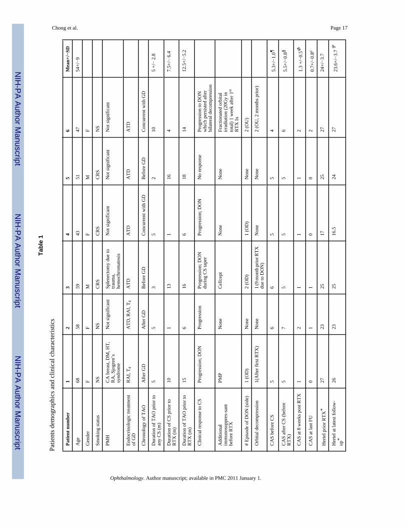
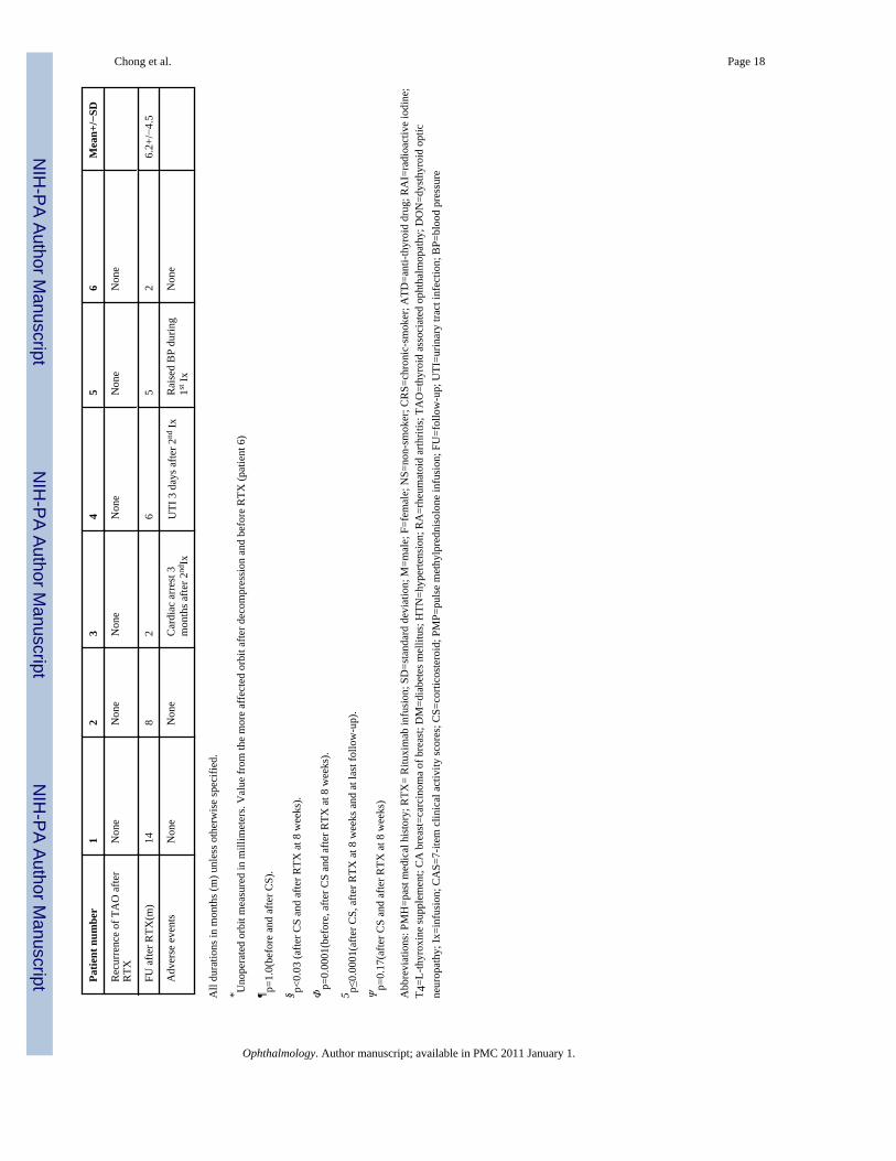
Demographic and clinical data from the 6 patients are shown in Table 1 (available athttp://aaojournal.org). Their age ranged from 43 to 68 years (54.3 + 9.1; mean + SD). Fourpatients were female and 3 smoked tobacco at the time they developed TAO. Two patientsexhibited orbital manifestations prior to hyperthyroidism (patient 3, one month and patient 5,five months). Two patients developed thyroid dysfunction and TAO simultaneously, and 2were hyperthyroid before the onset of TAO. Patients 1 and 2 underwent radioactive iodineablation followed by thyroxine supplementation while patients 3, 4, 5, and 6 were treated withanti- thyroid medications. All patients were euthyroid at the time of RTX infusion and hadpreviously received 1 to 3 courses of CS for active TAO. Duration of TAO prior to CS treatmentwas 5.0 + 2.8 months (range 2 to 10 months). CS was administered for 7.5 + 6.4 months (range1 to 16 months) before RTX and all patients were receiving CS at the time of RTX treatment.Duration of TAO prior to initiation of RTX therapy was 12.5 + 5.2 months (range 6–18 months).
CAS did not improve in any of the 6 patients following treatment with CS (CAS: 5.3 ± 1.0prior to CS vs. 5.5 + 0.8 during CS, p=1.0) (Figure 1A and 1B). Conversely, improvement wasobserved as early as 4 weeks following the first infusion of RTX (CAS 5.3 + 1.0 prior-RTXvs. 1.3 + 0.5, 8 weeks post series of 2 infusions, p<0.03) and this improvement was sustained
Chong et al. Page 4
Ophthalmology. Author manuscript; available in PMC 2011 January 1.
NIH
-PA Author Manuscript
NIH
-PA Author Manuscript
NIH
-PA Author Manuscript
(CAS 0.7 + 0.8 at 6.2 + 4.5 month follow up, p≤0.0001) (Figure 1B and Figure 2). CAS wasmeasured in the unoperated orbit for Patients 1 to 5 and the worse orbit for Patient 6. Figure 2demonstrates clinical photos of Patients 1 and 4. Despite improvement in orbital inflammatorysigns, Patient 4 experienced progressive strabismus after RTX treatment (Figure 2B).
Patients 1, 3, 4, and 6 developed early signs of DON during CS therapy. Patient 1, who hadbilateral asymmetric disease, underwent unilateral, transcaruncular medial wall orbitaldecompression on day 12 after her first RTX infusion due to continued DON.23 Patient 6presented initially with unilateral DON but subsequently developed bilateral involvement andwas sequentially decompressed after failing CS treatment. Despite high dose CSpostoperatively, the DON persisted for 2 months. Alternative causes such as ischemic opticneuropathy were considered unlikely based on clinical evidence and repeated MRI scans ofthe brain and both orbits. RTX infusions were administered 2 months after surgery incombination with 20 Gy fractionated orbital irradiation divided into 10 doses. The patientsubsequently demonstrated improved CAS and visual acuity at 2 weeks post series of 2infusions, which were sustained (Figure 1A). Patient 3 developed unilateral DON, failed torespond to CS therapy and underwent surgical decompression with improvement of visualacuity. Orbital inflammation was exacerbated during steroid taper and he again developedDON in the same eye and underwent RTX treatment 9 months after surgical decompression.DON improved within 4 weeks of RTX treatment (Figure 1A).
All 4 patients who developed DON exhibited improvement in visual acuity within 4 weeks oftheir first RTX infusion and improved to pre-neuropathy vision over 2 months. (Figure 1C).All patients receiving RTX were concurrently receiving CS. Within 4 weeks of the first RTXinfusion, CS was tapered and none of the patients experienced recurrence during the follow upperiod. As shown in Figure 1D, proptosis remained unchanged. Hertel measurements were 24± 3.7 mm before and 23.6± 3.7mm after therapy (p=0.17, n=6). None of the patientsdemonstrated improvement in extraocular motility (data not shown) while patient 4demonstrated progressive strabismus (Figure 2).
Adverse events during RTX therapy included urinary tract infection in Patient 4 which resolvedwith oral antibiotics. Patient 5, having preexisting hypertension, developed further elevationduring the first RTX infusion which was readily controlled. Patient 3 expired from suddencardiac death 3 months after receiving the final infusion.
Absence of orbital B cells after RTXSince the therapeutic efficacy of RTX is associated with depletion of B cells in targettissues25, we assessed the presence of mononuclear cells in orbital tissue obtained duringdecompression surgery for DON in Patient 1. Tissue was obtained 12 days after the initialinfusion of RTX. As demonstrated in Figure 3, adipose cells were heterogeneous in size withprominent fibrovascular septae. There was paucity of mononuclear infiltrates (Figure 3A).Neither B cells (Figure 3B) nor T cells (Figure 3C) were present.
Assessment of B and T cell phenotype before and after RTXTo determine potential biologic mechanism(s) of B cell depletion in TAO, we assessed thefrequency of regulatory T cells (Tregs) and of IGF-1R+ T and IGF-1R+ B cells. Tregs havebeen shown to reflect disease severity and response to treatment in patients with rheumatoidarthritis and diabetes.26 These cells may dampen autoimmune responses when present inadequate number. Their absence is associated with disease progression, while diseaseresolution can be accompanied by increased Treg frequency.27,8 Figure 4 demonstrates thefrequency of CD4+ T cells co-expressing CD25Hi (Tregs) in Patient 1. This cell populationalso expresses Foxp-3, a transcription factor specific to Tregs.29 Prior to RTX, Tregs accounted
Chong et al. Page 5
Ophthalmology. Author manuscript; available in PMC 2011 January 1.
NIH
-PA Author Manuscript
NIH
-PA Author Manuscript
NIH
-PA Author Manuscript

for less than 1% of CD4+ T cells. In contrast, the abundance of Tregs increased within 2 weeksof initiation of RTX (2.3% Tregs at 2 weeks) and was sustained at 18 months follow-up (3.0%Tregs).
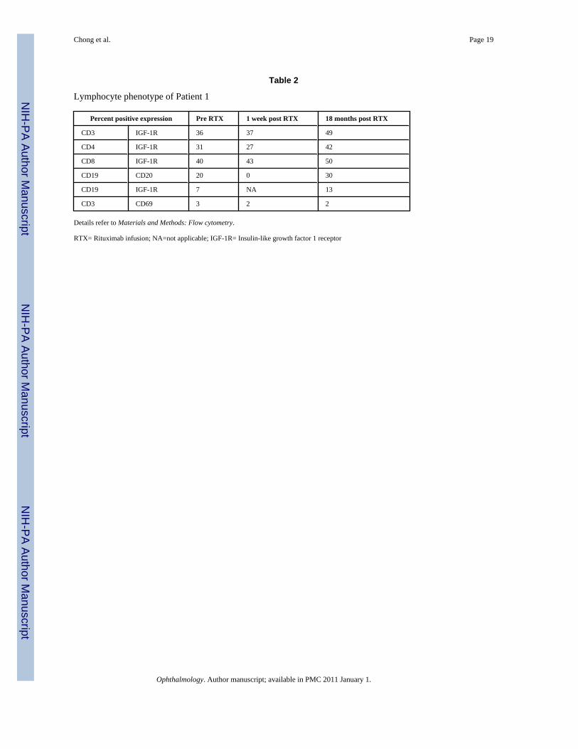
Earlier studies demonstrate increased frequency of IGF-1R+ T and B cells in GD.5,7 Weinvestigated the expression of IGF-1R by T and B cells from Patient 1. Table 2 (available athttp://aaojournal.org) demonstrates increased abundance of IGF-1R+ T cells prior to RTX andthis level was maintained after treatment. CD69, an activation associated marker expressed byT cells was also unaltered after treatment. Thus lymphocyte expression of two surface receptorsassociated with GD, IGF-1R and CD69; do not appear to be altered with RTX.5,7,30
DiscussionIn this retrospective case series we demonstrate that RTX was associated with significantimprovement of the clinical manifestations of severe TAO which were refractory to CS therapyor orbital decompression. Four of the six patients developed DON prior to RTX treatment.Orbital inflammation (elevated CAS scores) and DON improved without recurrence in allpatients but proptosis and strabismus were unimproved by RTX. These results must beinterpreted with caution since the study was uncontrolled regarding concurrent treatmentmodalities including Patient 3 and 6 who underwent decompression surgery prior to RTXinfusion and Patient 6 who received concurrent orbital irradiation. However, as the molecularmechanisms underlying TAO become better understood, more precise drug targets should beidentified. The prominent role of B cells in autoimmune responses, including antibody andcytokine production and antigen presentation, makes them particularly attractive. The successof B cell depletion as a strategy for treating rheumatoid arthritis found unresponsive to tumornecrosis factor- α blocking agents suggests that this approach might also be well-suited forpatients with TAO.31
In earlier studies, RTX appeared to reduce the need for continued thionamide therapy in GD-associated hyperthyroidism. 16–9 Those effects were independent of detectable changes in anti-TSHR antibody levels. 17 However, anti-TSHR antibodies, as measured in that study, compriseboth stimulatory and blocking molecules as well as those not influencing receptor function. Insubsequent studies involving these same patients, levels of stimulatory anti-TSHR antibodieswere found to be reduced by RTX. This finding agrees with those in other autoimmunedisorders where levels of autoreactive antibodies were reduced as a result of preferentialelimination of autoreactive B cells.33 Moreover, reduction of B cells infiltrating affected tissuesappears to be an important indicator of successful RTX therapy.34,6 Here we demonstrate anabsence of B cells infiltrating orbital tissue obtained during decompression surgery 12 daysafter first RTX treatment (Figure 3) which is in agreement with a recent report by Salvi et al.36
The clinical benefit of B cell depletion in TAO may derive in part from the altered abundanceof Tregs, unique T cells which can modulate autoimmune responses.35,7 Their impact onhuman immunity remains uncertain but they appear to act in large part through the transforminggrowth factor (TGF)-β pathway. Their frequency is reduced in autoimmune diseases.26 RAimproves with B cell depletion in patients demonstrating increasing levels of Tregs, a trendpredictive of therapeutic success.27,8 Levels of Tregs following RTX therapy have not beenpreviously examined in GD. Findings in the single patient reported here suggest that they maybecome more frequent following RTX treatment, analogous to the experience in RA27,8(Figure 4). The safety profile of RTX compares favorably to that of oral and IV steroids.18 Allpatients were being treated with CS at the time of RTX treatment and the infusion was welltolerated. In the current study, two well-tolerated infusions of the agent uniformly depleted Bcells. The infusion frequency and dose currently recommended for autoimmune disease are
Chong et al. Page 6
Ophthalmology. Author manuscript; available in PMC 2011 January 1.
NIH
-PA Author Manuscript
NIH
-PA Author Manuscript
NIH
-PA Author Manuscript
substantially lower than those used for lymphoma and are associated with fewer side-effects.38
We report here the first case series of patients with severe, active, and CS resistant TAO treatedwith RTX. Substantial clinical benefit was achieved in all 6 patients receiving the drug. Ourfindings suggest that patients who had already failed to respond to CS represent an expandingindication of RTX as salvage therapy in refractory TAO. Prospective studies are now requiredto establish RTX as a therapeutic option in these clinically challenging patients.
AcknowledgmentsThis work was supported in part by National Institutes of Health grants EY008976, EY011708, DK063121, EY016339,RR00425, K23 AR053858, an unrestricted grant from Research to Prevent Blindness, a Research to Prevent BlindnessCareer Development Award, and the Bell Charitable Foundation. The authors have no proprietary or commercialinterest in any material discussed in this article.
References1. Jacobson DL, Gange SJ, Rose NR, Graham NM. Epidemiology and estimated population burden of
selected autoimmune diseases in the United States. Clin Immunol Immunopathol 1997;84:223–243.[PubMed: 9281381]
2. McGrogan A, Seaman HE, Wright JW, de Vries CS. The incidence of autoimmune thyroid disease: asystematic review of the literature. Clin Endocrin (Oxf) 2008;69:687–696.
3. Weetman AP. Graves' disease. N Engl J Med 2000;343:1236–1248. [PubMed: 11071676]4. Smith TJ, Tsai CC, Shih MJ, et al. Unique attributes of orbital fibroblasts and global alterations in
IGF-1 receptor signaling could explain thyroid-associated ophthalmopathy. Thyroid 2008;18:983–988. [PubMed: 18788919]
5. Douglas RS, Gianoukakis AG, Goldberg RA, et al. Circulating mononuclear cells from euthyroidpatients with thyroid-associated ophthalmopathy exhibit characteristic phenotypes. Clin Exp Immunol2007;148:64–71. [PubMed: 17349012]
6. Aniszewski JP, Valyasevi RW, Bahn RS. Relationship between disease duration and predominantorbital T cell subset in Graves' ophthalmopathy. J Clin Endocrinol Metab 2000;85:776–780. [PubMed:10690890]
7. Douglas RS, Naik V, Hwang CJ, et al. B cells from patients with Graves' disease aberrantly expressthe IGF-1 receptor: implications for disease pathogenesis. J Immunol 2008;181:5768–5774. [PubMed:18832736]
8. Shlomchik MJ, Craft JE, Mamula MJ. From T to B and back again: positive feedback in systemicautoimmune disease. Nat Rev Immunol 2001;1:147–153. [PubMed: 11905822]
9. Bartalena L, Lai A, Compri E, et al. Novel immunomodulating agents for Graves' orbitopathy. OphthalPlast Reconstr Surg 2008;24:251–256.
10. Marcocci C, Bartalena L, Tanda ML, et al. Comparison of the effectiveness and tolerability ofintravenous or oral glucocorticoids associated with orbital radiotherapy in the management of severeGraves' ophthalmopathy: results of a prospective, single-blind, randomized study. J Clin EndocrinolMetab 2001;86:3562–3567. [PubMed: 11502779]
11. Zoumalan CI, Cockerham KP, Turbin RE, et al. Efficacy of corticosteroids and external beamradiation in the management of moderate to severe thyroid eye disease. J Neuroophthalmol2007;27:205–214. [PubMed: 17895822]
12. Reff ME, Carner K, Chambers KS, et al. Depletion of B cells in vivo by a chimeric mouse humanmonoclonal antibody to CD20. Blood 1994;83:435–445. [PubMed: 7506951]
13. Nielsen CH, El Fassi D, Hasselbalch HC, et al. B-cell depletion with rituximab in the treatment ofautoimmune diseases: Graves' ophthalmopathy the latest addition to an expanding family. ExpertOpin Biol Ther 2007;7:1061–1078. [PubMed: 17665994]
14. Boye J, Elter T, Engert A. An overview of the current clinical use of the anti-CD20 monoclonalantibody rituximab. Ann Oncol 2003;14:520–535. [PubMed: 12649096]
Chong et al. Page 7
Ophthalmology. Author manuscript; available in PMC 2011 January 1.
NIH
-PA Author Manuscript
NIH
-PA Author Manuscript
NIH
-PA Author Manuscript
15. Smolen JS, Keystone EC, Emery P, et al. Working Group on the Rituximab Consensus StatementConsensus statement on the use of rituximab in patients with rheumatoid arthritis. Ann Rheum Dis2007;66:143–150. [PubMed: 17068064]
16. El Fassi D, Nielsen CH, Hasselbalch HC, Hegedus L. The rationale for B lymphocyte depletion inGraves' disease: monoclonal anti-CD20 antibody therapy as a novel treatment option. Eur JEndocrinol 2006;154:623–632. [PubMed: 16645007]
17. El Fassi D, Nielsen CH, Bonnema SJ, et al. B lymphocyte depletion with the monoclonal antibodyrituximab in Graves' disease: a controlled pilot study. J Clin Endocrinol Metab 2007;92:1769–1772.[PubMed: 17284622]
18. Salvi M, Vannucchi G, Campi I, et al. Treatment of Graves' disease and associated ophthalmopathywith the anti-CD20 monoclonal antibody rituximab: an open study. Eur J Endocrinol 2007;156:33–40. [PubMed: 17218723]
19. Heemstra KA, Toes RE, Sepers J, et al. Rituximab in relapsing Graves' disease, a phase II study. EurJ Endocrinol 2008;159:609–615. [PubMed: 18628345]
20. Smith TJ. B cell depletion in Graves' disease: the right answer to the wrong question? J Clin EndocrinolMetab 2007;92:1620–1622. [PubMed: 17483377]
21. Bartley GB, Gorman CA. Diagnostic criteria for Graves' ophthalmopathy. Am J Ophthalmol1995;119:792–795. [PubMed: 7785696]
22. Mourits MP, Koornneef L, Wiersinga WM, et al. Clinical criteria for the assessment of disease activityin Graves' ophthalmopathy: a novel approach. Br J Ophthalmol 1989;73:639–644. [PubMed:2765444]
23. Ben Simon GJ, Syed HM, Douglas R, et al. Clinical manifestations and treatment outcome of opticneuropathy in thyroid-related orbitopathy. Ophthalmic Surg Lasers Imaging 2006;37:284–290.[PubMed: 16898388]
24. McCann JD, Goldberg RA, Anderson RL, et al. Medial wall decompression for optic neuropathy butlateral wall decompression with fat removal for non vision-threatening indications. Am J Ophthalmol2006;141:916–917. [PubMed: 16678507]
25. Bonara P, Vannucchi G, Campi I, et al. Rituximab induces distinct intraorbital and intrathyroidaleffects in one patient satisfactorily treated for Graves' ophthalmopathy. Clin Rev Allergy Immunol2008;34:118–123. [PubMed: 18270865]
26. Brusko TM, Putnam AL, Bluestone JA. Human regulatory T cells: role in autoimmune disease andtherapeutic opportunities. Immunol Rev 2008;223:371–390. [PubMed: 18613848]
27. Boissier MC, Assier E, Biton J, et al. Regulatory T cells (Treg) in rheumatoid arthritis. Joint BoneSpine 2009;76:10–14. [PubMed: 19028128]
28. Reis EA, Athanazio DA, Lima I, et al. NK and NKT cell dynamics after rituximab therapy for systemiclupus erythematosus and rheumatoid arthritis. Rheumatol Int 2009;29:469–475. [PubMed:18821073]
29. Brown K, Wong W. Diagnostic value of regulatory T cells: a new facet of a much studied cellpopulation. Transplantation 2008;86:1485–1491. [PubMed: 19077879]
30. Douglas RS, Gianoukakis AG, Kamat S, Smith TJ. Aberrant expression of the insulin-like growthfactor-1 receptor by T cells from patients with Graves' disease may carry functional consequencesfor disease pathogenesis. J Immunol 2007;178:3281–3287. [PubMed: 17312178]
31. Keystone E, Burmester GR, Furie R, et al. Improvement in patient-reported outcomes in a rituximabtrial in patients with severe rheumatoid arthritis refractory to anti-tumor necrosis factor therapy.Arthritis Rheum 2008;59:785–793. [PubMed: 18512710]
32. El Fassi D, Banga JP, Gilbert JA, et al. Treatment of Graves' disease with rituximab specificallyreduces the production of thyroid stimulating autoantibodies. Clin Immunol 2009;130:252–258.[PubMed: 18964302]
33. Ferraro AJ, Drayson MT, Savage CO, MacLennan IC. Levels of autoantibodies, unlike antibodies toall extrinsic antigen groups, fall following B cell depletion with rituximab. Eur J Immunol2008;38:292–298. [PubMed: 18085668]
34. Kavanaugh A, Rosengren S, Lee SJ, et al. Assessment of rituximab’s immunomodulatory synovialeffects (ARISE trial). 1: clinical and synovial biomarker results. Ann Rheum Dis 2008;67:402–408.[PubMed: 17644541]
Chong et al. Page 8
Ophthalmology. Author manuscript; available in PMC 2011 January 1.
NIH
-PA Author Manuscript
NIH
-PA Author Manuscript
NIH
-PA Author Manuscript

35. Vignali DA, Collison LW, Workman CJ. How regulatory T cells work. Nat Rev Immunol 2008;8:523–532. [PubMed: 18566595]
36. Salvi M, Vannucchi G, Campi I, et al. Rituximab treatment in a patient with severe thyroid-associatedophthalmopathy: Effects on orbital lymphocytic infiltrates. Clin Immunol 2009;131:360–365.[PubMed: 19195932]
37. Liossis SN, Sfikakis PP. Rituximab-induced B cell depletion in autoimmune diseases: potential effectson T cells. Clin Immunol 2008;127:280–285. [PubMed: 18337174]
38. Fleischmann RM. Safety of biologic therapy in rheumatoid arthritis and other autoimmune diseases:focus on rituximab. Semin Arthritis Rheum 2009;38:265–280. [PubMed: 18336874]
Chong et al. Page 9
Ophthalmology. Author manuscript; available in PMC 2011 January 1.
NIH
-PA Author Manuscript
NIH
-PA Author Manuscript
NIH
-PA Author Manuscript

Chong et al. Page 10
Ophthalmology. Author manuscript; available in PMC 2011 January 1.
NIH
-PA Author Manuscript
NIH
-PA Author Manuscript
NIH
-PA Author Manuscript

Chong et al. Page 11
Ophthalmology. Author manuscript; available in PMC 2011 January 1.
NIH
-PA Author Manuscript
NIH
-PA Author Manuscript
NIH
-PA Author Manuscript

Chong et al. Page 12
Ophthalmology. Author manuscript; available in PMC 2011 January 1.
NIH
-PA Author Manuscript
NIH
-PA Author Manuscript
NIH
-PA Author Manuscript
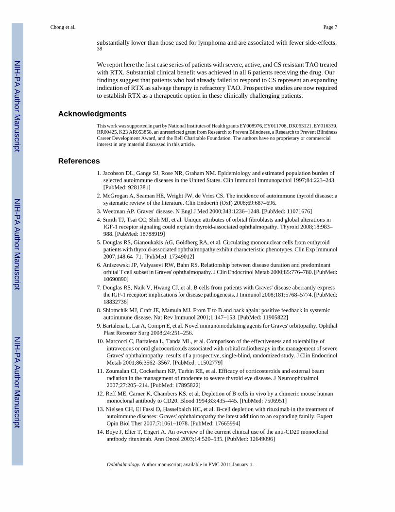
Figure 1. Clinical response after Rituximab (RTX) infusionA) Clinical activity score (CAS) of thyroid associated ophthalmopathy (TAO) evaluated beforeand after infusion of RTX, wk = weeks.* denotes patients who developed dysthyroid optic neuropathy (DON) during corticosteroid(CS) treatment.B) Summary of CAS before and after CS and RTX therapy in all six patients.*p<0.05(after CS and after RTX at 2 months), Wilcoxon signed rank test**p<0.0001(after CS, after RTX at 2 months and at last follow-up), Friedman testC) Change in visual acuity (reciprocal) in subjects with DON before and after RTX infusion.Abscissa represents individual eye, top of arrow indicates initial visual acuity, arrowhead pointsto final visual acuity.(*)Final visual acuity assessed prior to transcaruncular medial wall orbital decompression.(**)Initial visual acuity assessed after transcaruncular medial wall orbital decompression butprior to RTX infusion.D) Assessment of proptosis (mean + standard deviation) before and after RTX.Measure of proptosis was performed before and after RTX in the unoperated orbit for patients1–5 and the worse side for patient 6. P=0.17, Wilcoxon signed rank test
Chong et al. Page 13
Ophthalmology. Author manuscript; available in PMC 2011 January 1.
NIH
-PA Author Manuscript
NIH
-PA Author Manuscript
NIH
-PA Author Manuscript
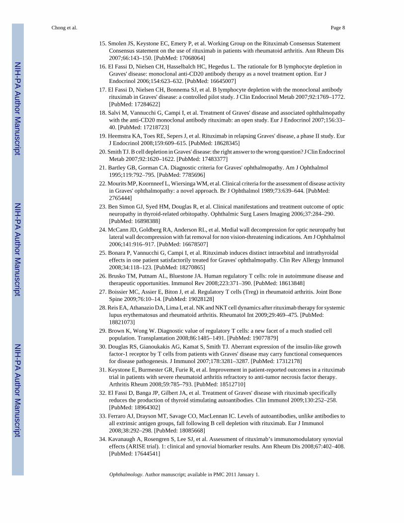
Figure 2.Photographs of two patients demonstrating orbital inflammation before and after Rituximab(RTX) therapy. Patient 4 demonstrated significant periorbital inflammation (A) prior to RTXtreatment which decreased dramatically after therapy (B). Stabismsus and eyelid retractionincreased despite improvement of inflammatory signs and symptoms. Patient 1 prior to RTXtreatment (C) and approximately 18 months after treatment (D).
Chong et al. Page 14
Ophthalmology. Author manuscript; available in PMC 2011 January 1.
NIH
-PA Author Manuscript
NIH
-PA Author Manuscript
NIH
-PA Author Manuscript

Figure 3. Histology and immunohistochemical staining of orbital tissues obtained duringdecompression surgery from patient 1A) High-powered view demonstrating adipocytes with prominent fibrovascular septae andpaucity of mononuclear inflammatory infiltrates.B) High-powered view demonstrating absence of CD20+ B cells and C) High-powered viewdemonstrating absence of CD3+ T cellsA) Hematoxylin and Eosin. B & C) counterstained with Hematoxylin. Magnification, x400,(inset, x40).
Chong et al. Page 15
Ophthalmology. Author manuscript; available in PMC 2011 January 1.
NIH
-PA Author Manuscript
NIH
-PA Author Manuscript
NIH
-PA Author Manuscript

Figure 4.Flow cytometric analysis of regulatory T (Treg) cells in Patient 1 before the initiation oftherapy, 2 weeks and 18 months after Rituximab (RTX) infusion. The data show increasedTreg (CD25Hi) cells among CD4+ T cells.
Chong et al. Page 16
Ophthalmology. Author manuscript; available in PMC 2011 January 1.
NIH
-PA Author Manuscript
NIH
-PA Author Manuscript
NIH
-PA Author Manuscript
NIH
-PA Author Manuscript
NIH
-PA Author Manuscript
NIH
-PA Author Manuscript
Chong et al. Page 17
Tabl
e 1
Patie
nts d
emog
raph
ics a
nd c
linic
al c
hara
cter
istic
s
Patie
nt n
umbe
r1
23
45
6M
ean+
/−SD
Age
6858
5943
5147
54+/−9
Gen
der
FF
MF
MF
Smok
ing
stat
usN
SN
SC
RS
CR
SC
RS
NS
PMH
CA
bre
ast,
DM
, HT,
RA
, Sjo
gren
’ssy
ndro
me
Not
sign
ifica
ntSp
lene
ctom
y du
e to
traum
a,he
moc
hrom
atos
is
Not
sign
ifica
ntN
ot si
gnifi
cant
Not
sign
ifica
nt
Endo
crin
olog
ic tr
eatm
ent
of G
DR
AI,
T 4A
TD, R
AI,
T 4A
TDA
TDA
TDA
TD
Chr
onol
ogy
of T
AO
Afte
r GD
Afte
r GD
Bef
ore
GD
Con
curr
ent w
ith G
DB
efor
e G
DC
oncu
rren
t with
GD
Dur
atio
n of
TA
O p
rior t
oan
y C
S (m
)5
53
52
105
+/−
2.8
Dur
atio
n of
CS
prio
r to
RTX
(m)
101
131
164
7.5+
/− 6
.4
Dur
atio
n of
TA
O p
rior t
oR
TX (m
)15
616
618
1412
.5+/−5
.2
Clin
ical
resp
onse
to C
SPr
ogre
ssio
n; D
ON
Prog
ress
ion
Prog
ress
ion;
DO
Ndu
ring
CS
tape
rPr
ogre
ssio
n; D
ON
No
resp
onse
Prog
ress
ion
to D
ON
whi
ch p
ersi
sted
afte
rbi
late
ral d
ecom
pres
sion
Add
ition
alim
mun
osup
pres
-san
tbe
fore
RTX
PMP
Non
eC
ellc
ept
Non
eN
one
Frac
tiona
ted
orbi
tal
irrad
iatio
n (2
0Gy
into
tal)
1 w
eek
afte
r 1st
RTX
Ix
# Ep
isod
e of
DO
N (s
ide)
1 (O
D)
Non
e2
(OD
)1
(OD
)N
one
2 (O
U)
Orb
ital d
ecom
pres
sion
1(A
fter f
irst R
TX)
Non
e1
(9 m
onth
prio
r RTX
due
to D
ON
)N
one
Non
e2
(OU
, 2 m
onth
s prio
r)
CA
S be
fore
CS
56
65
54
5.3+
/−1.
0¶
CA
S af
ter C
S (b
efor
eR
TX)
57
55
56
5.5+
/−0.
8§
CA
S at
8 w
eeks
pos
t RTX
12
11
12
1.3
+/−0
.5Φ
CA
S at
last
FU
01
10
02
0.7+
/−0.
8ε
Her
tel p
rior R
TX*
2723
2517
2527
24+/−3
.7
Her
tel a
t lat
est f
ollo
w-
up*
2623
2516
.524
2723
.6+/−3
.7 Ψ
Ophthalmology. Author manuscript; available in PMC 2011 January 1.
NIH
-PA Author Manuscript
NIH
-PA Author Manuscript
NIH
-PA Author Manuscript
Chong et al. Page 18
Patie
nt n
umbe
r1
23
45
6M
ean+
/−SD
Rec
urre
nce
of T
AO
afte
rR
TXN
one
Non
eN
one
Non
eN
one
Non
e
FU a
fter R
TX(m
)14
82
65
26.
2+/−
4.5
Adv
erse
eve
nts
Non
eN
one
Car
diac
arr
est 3
mon
ths a
fter 2
ndIx
UTI
3 d
ays a
fter 2
nd Ix
Rai
sed
BP
durin
g1st
IxN
one
All
dura
tions
in m
onth
s (m
) unl
ess o
ther
wis
e sp
ecifi
ed.
* Uno
pera
ted
orbi
t mea
sure
d in
mill
imet
ers.
Val
ue fr
om th
e m
ore
affe
cted
orb
it af
ter d
ecom
pres
sion
and
bef
ore
RTX
(pat
ient
6)
¶ p=1.
0(be
fore
and
afte
r CS)
.
§ p<0.
03 (a
fter C
S an
d af
ter R
TX a
t 8 w
eeks
).
Φp=
0.00
01(b
efor
e, a
fter C
S an
d af
ter R
TX a
t 8 w
eeks
).
5 p≤0.
0001
(afte
r CS,
afte
r RTX
at 8
wee
ks a
nd a
t las
t fol
low
-up)
.
Ψp=
0.17
(afte
r CS
and
afte
r RTX
at 8
wee
ks)
Abb
revi
atio
ns: P
MH
=pas
t med
ical
his
tory
; RTX
= R
ituxi
mab
infu
sion
; SD
=sta
ndar
d de
viat
ion;
M=m
ale;
F=f
emal
e; N
S=no
n-sm
oker
; CR
S=ch
roni
c-sm
oker
; ATD
=ant
i-thy
roid
dru
g; R
AI=
radi
oact
ive
iodi
ne;
T 4=L
-thyr
oxin
e su
pple
men
t; C
A b
reas
t=ca
rcin
oma
of b
reas
t; D
M=d
iabe
tes m
ellit
us; H
TN=h
yper
tens
ion;
RA
=rhe
umat
oid
arth
ritis
; TA
O=t
hyro
id a
ssoc
iate
d op
htha
lmop
athy
; DO
N=d
ysth
yroi
d op
ticne
urop
athy
; Ix=
infu
sion
; CA
S=7-
item
clin
ical
act
ivity
scor
es; C
S=co
rtico
ster
oid;
PM
P=pu
lse
met
hylp
redn
isol
one
infu
sion
; FU
=fol
low
-up;
UTI
=urin
ary
tract
infe
ctio
n; B
P=bl
ood
pres
sure
Ophthalmology. Author manuscript; available in PMC 2011 January 1.
NIH
-PA Author Manuscript
NIH
-PA Author Manuscript
NIH
-PA Author Manuscript
Chong et al. Page 19
Table 2
Lymphocyte phenotype of Patient 1
Percent positive expression Pre RTX 1 week post RTX 18 months post RTX
CD3 IGF-1R 36 37 49
CD4 IGF-1R 31 27 42
CD8 IGF-1R 40 43 50
CD19 CD20 20 0 30
CD19 IGF-1R 7 NA 13
CD3 CD69 3 2 2
Details refer to Materials and Methods: Flow cytometry.
RTX= Rituximab infusion; NA=not applicable; IGF-1R= Insulin-like growth factor 1 receptor
Ophthalmology. Author manuscript; available in PMC 2011 January 1.
Related Documents