Establishment of Mouse Embryonic Stem Cell-Derived Erythroid Progenitor Cell Lines Able to Produce Functional Red Blood Cells Takashi Hiroyama, Kenichi Miharada, Kazuhiro Sudo, Inaho Danjo, Naoko Aoki, Yukio Nakamura * Cell Engineering Division, RIKEN BioResource Center, Tsukuba, Ibaraki, Japan Background. The supply of transfusable red blood cells (RBCs) is not sufficient in many countries. If erythroid cell lines able to produce transfusable RBCs in vitro were established, they would be valuable resources. However, such cell lines have not been established. To evaluate the feasibility of establishing useful erythroid cell lines, we attempted to establish such cell lines from mouse embryonic stem (ES) cells. Methodolog y/Principa l Findings. We developed a robust method to obtain differentiated cell lines foll owing the induc tion of hemat opoi etic differe ntiation of mous e ES cells and esta blish ed five indepen dent hematopoietic cell lines using the method. Three of these lines exhibited characteristics of erythroid cells. Although their precise char acte rist ics varied, each of these lines could diff erentiate in vitr o into more mature erythroid cells, including enu cleate d RBCs. Following transplantat ion of the se er ythroid cel ls int o mic e suf fering fro m acu te anemia, the cel ls proliferated transiently, subsequently differentiated into functional RBCs, and significantly ameliorated the acute anemia. In addition, we did not observe formation of any tumors following transplantation of these cells. Conclusion/Significance. To the best of our knowledge, this is the first report to show the feasibility of establishing erythroid cell lines able to produce mature RBCs. Considering the number of human ES cell lines that have been established so far, the intensive testing of a number of these lines for erythroid potential may allow the establishment of human erythroid cell lines similar to the mouse erythroid cell lines described here. In addition, our results strongly suggest the possibility of establishing useful cell lines committed to specific lineages other than hematopoietic progenitors from human ES cells. Citation: Hiroyama T, Miharada K, Sudo K, Danjo I, Aoki N, et al (2008) Establishment of Mouse Embryonic Stem Cell-De rived Erythroid Progenitor Cell Lines Able to Produce Functional Red Blood Cells. PLoS ONE 3(2): e1544. doi:10.1371/journal.pone.0001544 INTRODUCTION RBC transfusion was the first established transplantation procedure in clinical history, and is a common and indispensable clinical pro- cedure. However, the supply of transfusable RBCs is insufficient in many countries. Thus, there is interest in the developme nt of in vitro proc edur es for the genera tio n of func tio nal RBCs fro m hemato poi eti c stem and/or progenitor cells present in bone marrow or umbilical cord blood [1–3]. Human ES cells possess the potential to produce various differentiated cells able to function in vivo and thus represent another promising resource to produce functional RBCs. Hematopoietic cells including cells of the erythroid lineage have been generated from mouse [4–7], non-human primate [8–10], and human ES cel ls [11–16]. We hav e rec ent ly est abl ish ed a method to culture hematopoietic cells derived from non-human primat e ES cell s long term in vi tro [17] . The ef fi ci ency of generation of erythroid progenitors and/or RBCs varies based on the me thods and ES ce ll li ne s us ed. Even wi th opti ma l experimental procedures and the most appropriate ES cell line, howeve r, the genera tion of abundant RBCs directl y from primate ES cel ls is a time-c ons umi ng proc ess [17]. If huma n ery thr oid progenitor cell li nes we re es tabl is hed that could produ ce transfusable and functional RBCs efficiently, they would represent a much more useful resource to produce RBCs than ES cell lines. Several mous e and human eryt hroi d cell li nes have been established. However, to the best of our knowledge, there is no cell line that can efficiently differentiate into enucleated RBCs. It is generally difficult to establish hematopoietic cell lines from adult hematopoietic stem or progenitor cells, since these somatic cells are quite sensitive to DNA damage and are unable to maintain the length of telomere repeats on serial passage [18]. By contrast, ES cells are quite resistant to DNA damage and maintain telomere length on serial passage [18]. Therefore, we speculated that these cha ra ct eri st ic s of ES ce ll s m ay be adva n ta ge ous for the establishment of cell lines, since differentiated cells derived from ES cells may retain such characteristics. In addition, mouse cells tend to immortalize more readily than human cells, as has been shown to be the case following the induction of pluripotent stem cel l lin es from somati c cel ls [19–22]. Hen ce, we attempted to evaluat e the feasi bilit y of establ ishing hematopoi etic cell lines, erythroid cell lines in particular, from mouse ES cells. RESULTS AND DISCUSSION Establishment of erythroid progenitor cell lines from mouse ES cells To induce differentiation of hematopoietic cells from mouse ES cells, we cultured the latter cells using OP9 cells as feeder cells [5, 6,2 3] in the presen ce of spe cific factors (Ta ble 1). OP9 cells were used not only for induction of hematopoietic differentiation but also for establishment of cell lines in the early phase of long term culture of the induced hematopoietic cells (Table 1). In most Academic Editor: Simon Williams, Texas Tech University Health Sciences Center, United States of America Received November 14, 2007; Accepted January 3, 2008; Published February 6, 2008 Copyright: ß 2008 Hiro yama et al. This is an open -acce ss article distribut ed under the terms of the Creative Commons Attribution License, which permits unre strict ed use, distribu tion, and repr oducti on in any mediu m, prov ided the original author and source are credited. Funding: This work was supported by grants from the Ministry of Education, Culture, Sports, Science, and Technology in Japan. Competing Interests: The authors have declared that no competing interests exist. * To whom correspondence should be addressed. E-mail: [email protected] PLoS ONE | www.plosone.org 1 February 2008 | Issue 2 | e1544

Welcome message from author
This document is posted to help you gain knowledge. Please leave a comment to let me know what you think about it! Share it to your friends and learn new things together.
Transcript

8/6/2019 Riken Mouse Differentiation
http://slidepdf.com/reader/full/riken-mouse-differentiation 1/11
Establishment of Mouse Embryonic Stem Cell-DerivedErythroid Progenitor Cell Lines Able to ProduceFunctional Red Blood CellsTakashi Hiroyama, Kenichi Miharada, Kazuhiro Sudo, Inaho Danjo, Naoko Aoki, Yukio Nakamura *
Cell Engineering Division, RIKEN BioResource Center, Tsukuba, Ibaraki, Japan
Background. The supply of transfusable red blood cells (RBCs) is not sufficient in many countries. If erythroid cell lines able to
produce transfusable RBCs in vitro were established, they would be valuable resources. However, such cell lines have not been
established. To evaluate the feasibility of establishing useful erythroid cell lines, we attempted to establish such cell lines from
mouse embryonic stem (ES) cells. Methodology/Principal Findings. We developed a robust method to obtain differentiated
cell lines following the induction of hematopoietic differentiation of mouse ES cells and established five independent
hematopoietic cell lines using the method. Three of these lines exhibited characteristics of erythroid cells. Although their
precise characteristics varied, each of these lines could differentiate in vitro into more mature erythroid cells, including
enucleated RBCs. Following transplantation of these erythroid cells into mice suffering from acute anemia, the cells
proliferated transiently, subsequently differentiated into functional RBCs, and significantly ameliorated the acute anemia. In
addition, we did not observe formation of any tumors following transplantation of these cells. Conclusion/Significance. To
the best of our knowledge, this is the first report to show the feasibility of establishing erythroid cell lines able to produce
mature RBCs. Considering the number of human ES cell lines that have been established so far, the intensive testing of a
number of these lines for erythroid potential may allow the establishment of human erythroid cell lines similar to the mouse
erythroid cell lines described here. In addition, our results strongly suggest the possibility of establishing useful cell lines
committed to specific lineages other than hematopoietic progenitors from human ES cells.
Citation: Hiroyama T, Miharada K, Sudo K, Danjo I, Aoki N, et al (2008) Establishment of Mouse Embryonic Stem Cell-Derived Erythroid Progenitor CellLines Able to Produce Functional Red Blood Cells. PLoS ONE 3(2): e1544. doi:10.1371/journal.pone.0001544
INTRODUCTIONRBC transfusion was the first established transplantation procedure
in clinical history, and is a common and indispensable clinical pro-
cedure. However, the supply of transfusable RBCs is insufficient in
many countries. Thus, there is interest in the development of in vitro
procedures for the generation of functional RBCs from hematopoietic
stem and/or progenitor cells present in bone marrow or umbilicalcord blood [1–3]. Human ES cells possess the potential to produce
various differentiated cells able to function in vivo and thus represent
another promising resource to produce functional RBCs.
Hematopoietic cells including cells of the erythroid lineage have
been generated from mouse [4–7], non-human primate [8–10],
and human ES cells [11–16]. We have recently established a
method to culture hematopoietic cells derived from non-human
primate ES cells long term in vitro [17]. The efficiency of
generation of erythroid progenitors and/or RBCs varies based on
the methods and ES cell lines used. Even with optimal
experimental procedures and the most appropriate ES cell line,
however, the generation of abundant RBCs directly from primate
ES cells is a time-consuming process [17]. If human erythroid
progenitor cell lines were established that could produce
transfusable and functional RBCs efficiently, they would representa much more useful resource to produce RBCs than ES cell lines.
Several mouse and human erythroid cell lines have been
established. However, to the best of our knowledge, there is no cell
line that can efficiently differentiate into enucleated RBCs. It is
generally difficult to establish hematopoietic cell lines from adult
hematopoietic stem or progenitor cells, since these somatic cells
are quite sensitive to DNA damage and are unable to maintain the
length of telomere repeats on serial passage [18]. By contrast, ES
cells are quite resistant to DNA damage and maintain telomere
length on serial passage [18]. Therefore, we speculated that these
characteristics of ES cells may be advantageous for the
establishment of cell lines, since differentiated cells derived from
ES cells may retain such characteristics. In addition, mouse cells
tend to immortalize more readily than human cells, as has been
shown to be the case following the induction of pluripotent stem
cell lines from somatic cells [19–22]. Hence, we attempted to
evaluate the feasibility of establishing hematopoietic cell lines,
erythroid cell lines in particular, from mouse ES cells.
RESULTS AND DISCUSSION
Establishment of erythroid progenitor cell lines from
mouse ES cellsTo induce differentiation of hematopoietic cells from mouse ES
cells, we cultured the latter cells using OP9 cells as feeder cells
[5,6,23] in the presence of specific factors (Table 1). OP9 cells
were used not only for induction of hematopoietic differentiation
but also for establishment of cell lines in the early phase of long
term culture of the induced hematopoietic cells (Table 1). In most
Academic Editor: Simon Williams, Texas Tech University Health Sciences Center,United States of America
Received November 14, 2007; Accepted January 3, 2008; Published February 6,2008
Copyright: ß 2008 Hiroyama et al. This is an open-access article distributedunder the terms of the Creative Commons Attribution License, which permitsunrestricted use, distribution, and reproduction in any medium, provided theoriginal author and source are credited.
Funding: This work was supported by grants from the Ministry of Education,Culture, Sports, Science, and Technology in Japan.
Competing Interests: The authors have declared that no competing interestsexist.
* To whom correspondence should be addressed. E-mail: [email protected]
PLoS ONE | www.plosone.org 1 February 2008 | Issue 2 | e1544
8/6/2019 Riken Mouse Differentiation
http://slidepdf.com/reader/full/riken-mouse-differentiation 2/11
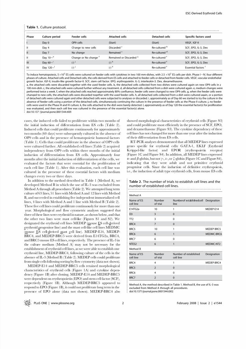
cases, the induced cells failed to proliferate within two months of the initial induction of differentiation from ES cells (Table 2).
Induced cells that could proliferate continuously for approximately
two months (60 days) were subsequently cultured in the absence of
OP9 cells and in the presence of hematopoietic humoral factors
(Table 1). Cells that could proliferate in the absence of OP9 cells
were cultured further. All established cell lines (Table 2) acquired
independency from OP9 cells within three months of the initial
induction of differentiation from ES cells. Approximately four
months after the initial induction of differentiation of the cells, we
evaluated the factors that were essential for the proliferation of
each cell line (Table 1). After this evaluation, each cell line was
cultured in the presence of these essential factors with medium
changes every two or three days.
In addition to the method described in Table 1 (Method A), we
developed Method B in which the use of IL-3 was excluded from
Method A through all procedures (Table 2). We attempted long term
culture of 63 lines, 51 lines with Method A and 12 lines with Method
B, and succeeded in establishing five independent immortalized cell
lines, 4 lines with Method A and 1 line with Method B (Table 2).
These five cell lines could proliferate continuously for more than one
year. Morphological and flow cytometric analyses suggested that
three of these lines were erythroid in nature, as shown below, and that
the other two lines were mast cell-like (Figures S1 and S2). We
designated the erythroid cell lines MEDEP (mouse ES cell-derived
erythroid progenitor line) and the mast cell-like cell lines MEDMC
(mouse ES cell-derived mast cell line). MEDEP-E14, MEDEP-
BRC4, and MEDEP-BRC5 were derived from E14TG2a, BRC4,
and BRC5 mouse ES cell lines, respectively. The presence of IL-3 in
the culture medium (Method A) may not be necessary for theestablishment of erythroid cell lines, as we were able to establish one
erythroid line, MEDEP-BRC4, following culture of the cells in the
absence of IL-3 (Method B) (Table 2). MEDEP cells could proliferate
from single cells following sorting by flow cytometry (data not shown).
MEDEP-E14 and MEDEP-BRC5 cells retained morphological
characteristics of erythroid cells (Figure 1A) and cytokine depen-
dency (Figure 1B) after cloning. MEDEP-E14 and MEDEP-BRC5
were dependent on erythropoietin (EPO) and stem cell factor (SCF),
respectively (Figure 1B). Although MEDEP-BRC5 appeared to
respond to EPO (Figure 1B), it could not proliferate long term in the
presence of EPO alone (data not shown). MEDEP-BRC4 also
showed morphological characteristics of erythroid cells (Figure S3)and could proliferate most efficiently in the presence of SCF, EPO,
and dexamethasone (Figure S3). The cytokine dependency of these
cell lines has not changed for more than one year after the induction
of their differentiation from ES cells.
RT-PCR analyses demonstrated that all MEDEP lines expressed
genes specific for erythroid cells: GATA-1, EKLF (Erythroid
Kruppel-like factor) and EPOR (erythropoietin receptor)
(Figure 1C and Figure S4). In addition, all MEDEP lines expresseda- and b-globin, but not c-, e-,or f-globin (Figure 1C and Figure S4),
indicating that they were adult and not primitive erythroid
progenitor cells. Since the induction of definitive erythropoiesis,
i.e., the induction of adult type erythroid cells, from mouse ES cells
Table 1. Culture protocol.. . . . . . . . . . . . . . . . . . . . . . . . . . . . . . . . . . . . . . . . . . . . . . . . . . . . . . . . . . . . . . . . . . . . . . . . . . . . . . . . . . . . . . . . . . . . . . . . . . . . . . . . . . . . . . . . . . . . . . . . . . . . . . . . . . . . . . . . . . . . . . . . . .
Phase Culture period Feeder cells Attached cells Detached cells Specific factors used
I Day 0 OP9 cells (Start) (Start) VEGF, IGF-II
II Day 4 Change to new cells Discarded a Re-cultured b SCF, EPO, IL-3, Dex
II Day 7 No change Remained c Re-cultured d SCF, EPO, IL-3, Dex
II Day 10,e Change or No change f Remained or Discarded g Re-cultured h SCF, EPO, IL-3, Dex
III Day 60,i (-) j (-) k Re-cultured h SCF, EPO, IL-3, Dex
IV Day 120,l (-) j (-) k Re-cultured h Essential factors l
To induce hematopoiesis, 56105 ES cells were cultured on feeder cells with cytokines in two 100 mm-dishes, with 2.56105 ES cells per dish. Phase I,IV, four differentphases of culture. Attached cells and Detached cells, the cells derived from ES cells and attached to feeder cells or detached from feeder cells. VEGF, vascular endothelialgrowth factor. IGF-II, insulin-like growth factor-II. SCF, stem cell factor. EPO, erythropoietin. IL-3, interleukin-3. Dex, dexamethasone.a, the attached cells were discarded together with the used feeder cells. b, the detached cells collected from two dishes were cultured again on new OP9 cells in a100 mm-dish. c, the attached cells were cultured further without any treatment. d, all detached cells collected from a dish were cultured again. e, medium changes wereperformed twice a week. f, when the attached cells reached approximately 80% confluence, feeder cells were changed to new OP9 cells. g, when the feeder cells werechanged to new cells, the attached cells were discarded together with the used feeder cells. h, all detached cells collected from a dish were cultured again, or a portionof detached cells were cultured again and other detached cells were subjected to analyses or discarded. i, approximately as of Day 60 we started to try the culture in theabsence of feeder cells using a portion of the detached cells, simultaneously continuing the culture in the presence of feeder cells as the Phase II culture. j, no feedercells were used in the Phase III and IV culture. k, the cells attached to the dish were barely detected. l, approximately as of Day 120 the essential factor(s) for proliferationwas evaluated, and then each cell line was cultured in the presence of the essential factor(s) alone.doi:10.1371/journal.pone.0001544.t001 .
.
.
.
.
.
.
.
.
.
.
.
.
.
.
.
.
.
.
.
.
.
.
.
.
.
.
.
.
.
.
.
.
.
.
.
. . .
.
.
.
.
.
.
.
.
.
.
.
.
.
.
.
.
.
.
.
.
.
.
.
.
.
.
.
Table 2. The number of trials to establish cell lines and thenumber of established cell lines.. . . . . . . . . . . . . . . . . . . . . . . . . . . . . . . . . . . . . . . . . . . . . . . . . . . . . . . . . . . . . . . . . . . . . .
Method A
Name of EScell line
Numberof trial
Numberof establishedcellline
Designation
E14TG2a 10 1 MEDEP-E14
D3 3 0
TT2 3 0
BRC4 7 0
BRC5 10 1 MEDEP-BRC5
BRC6 6 1 MEDMC-BRC6
BRC7 6 0
NTES2 6 1 MEDMC-NT2Method B
Name of EScell line
Numberof trial
Number of establishedcell line
Designation
BRC4 4 1 MEDEP-BRC4
BRC5 2 0
BRC6 4 0
BRC7 2 0
Method A, the method described in Table 1. Method B, the use of IL-3 wasexcluded from Method A through all procedures.doi:10.1371/journal.pone.0001544.t002 .
.
.
.
.
.
.
.
.
.
.
.
.
.
.
.
.
.
.
.
.
.
.
.
.
.
.
.
.
.
.
.
.
. . .
.
.
.
.
.
.
.
.
.
.
.
.
.
.
.
.
.
.
.
.
.
.
.
.
.
.
.
.
.
.
.
.
.
.
.
.
.
.
.
.
.
.
. . . .
.
.
.
ESC-Derived Erythroid Cells
PLoS ONE | www.plosone.org 2 February 2008 | Issue 2 | e1544

8/6/2019 Riken Mouse Differentiation
http://slidepdf.com/reader/full/riken-mouse-differentiation 3/11
has previously been reported [6], all MEDEP lines appeared to be
derived from adult type erythroid progenitor cells.
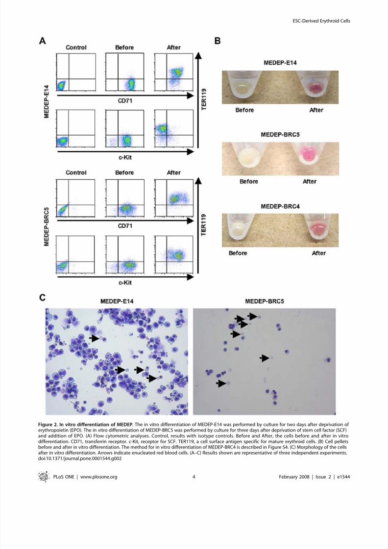
In vitro differentiation of MEDEPNext, we evaluated the potential of MEDEP cells to differentiate into
more mature erythroid cells. We found that all MEDEP lines could
differentiate into more mature erythroid cells by the following treat-
ments: deprivation of EPO for MEDEP-E14 (Figure 2A); deprivation
of SCF and addition of EPO for MEDEP-BRC5 (Figure 2A); and
deprivation of SCF and dexamethasone and addition of EPO for
MEDEP-BRC4 (Figure S4). EPO appeared to be necessary for
MEDEP-BRC5 and MEDEP-BRC4 cells to maintain cell viability
during the differentiation process (data not shown).
Figure 1. Characteristics of erythroid cell lines derived from mouse ES cells, MEDEP. (A) Morphology of two erythroid cell lines, MEDEP-E14 andMEDEP-BRC5. Wright-Giemsa staining. (B) Cytokine dependent proliferation. Cells (16105 cells/ml) were cultured in various conditions for three days.The added factor(s) is shown at the bottom. None, no specific factor. SCF, stem cell factor. EPO, erythropoietin. Broken line, the number of cells at thestart of culture. Values are mean6S.D. Results shown are representative of several independent experiments performed at different time points afterestablishment of the cell lines. (C) RT-PCR analyses. Oct-3/4 and Nanog, transcription factors specific for ES cells. GATA-1 and EKLF (Erythroid Kruppel-
like factor), transcription factors specific for erythroid cells. EPOR, erythropoietin receptor. GAPDH, glyceraldehyde-3-phosphate dehydrogenase. NC,negative control without cDNA. Day 0, E14TG2a cells before differentiation. Day 4, 7, 10, 14 and 21, the cells following induction of differentiation intohematopoietic cells from E14TG2a by the method described in Table 1 (Method A). The cycle numbers performed in each PCR are shown at the right.Results shown are representative of two independent experiments.doi:10.1371/journal.pone.0001544.g001
ESC-Derived Erythroid Cells
PLoS ONE | www.plosone.org 3 February 2008 | Issue 2 | e1544
8/6/2019 Riken Mouse Differentiation
http://slidepdf.com/reader/full/riken-mouse-differentiation 4/11
Figure 2. In vitro differentiation of MEDEP. The in vitro differentiation of MEDEP-E14 was performed by culture for two days after deprivation of erythropoietin (EPO). The in vitro differentiation of MEDEP-BRC5 was performed by culture for three days after deprivation of stem cell factor (SCF)and addition of EPO. (A) Flow cytometric analyses. Control, results with isotype controls. Before and After, the cells before and after in vitrodifferentiation. CD71, transferrin receptor. c-Kit, receptor for SCF. TER119, a cell surface antigen specific for mature erythroid cells. (B) Cell pelletsbefore and after in vitro differentiation. The method for in vitro differentiation of MEDEP-BRC4 is described in Figure S4. (C) Morphology of the cellsafter in vitro differentiation. Arrows indicate enucleated red blood cells. (A–C) Results shown are representative of three independent experiments.doi:10.1371/journal.pone.0001544.g002
ESC-Derived Erythroid Cells
PLoS ONE | www.plosone.org 4 February 2008 | Issue 2 | e1544
8/6/2019 Riken Mouse Differentiation
http://slidepdf.com/reader/full/riken-mouse-differentiation 5/11
The three MEDEP lines exhibited differential expression of TER119 (a cell surface antigen specific for mature erythroid cells)
and CD71 (transferrin receptor) (Figure 2A and Figure S4). Forexample, the expression of CD71 was slightly higher in MEDEP-
E14 cells than in MEDEP-BRC5 cells (Figure 2A).
TER1192CD712 cells differentiate first to TER1192CD71+ cells,
subsequently to TER119++CD71+ cells, and then finally to
TER119+CD712 cells [25]. Consistent with the differences in
their cytokine dependency (Figure 1B), the MEDEPs appeared torepresent different stages of erythroid differentiation. Despite these
differences, following induction of differentiation in vitro by the
methods described above, the expression of TER119 and CD71 in
each of the MEDEP lines exhibited a pattern consistent with a
more mature lineage (Figure 2A and Figure S4), indicating that
each of the three lines was able to differentiate into a more mature
lineage. At present, the cause of the variability between MEDEPs
remains uncertain. However, these results clearly demonstrated
that erythroid progenitor cells could be immortalized at different
stages of their differentiation.
Notably, the vast majority of cells in each MEDEP line could
differentiate into more mature cells, although each MEDEP line
included cells possessing abnormal karyotypes (Figure S5). This
result strongly suggested that the cells possessing abnormal
karyotypes still retained the potential to differentiate into moremature erythroid cells. In general, most immortalized cell lines are
not necessarily homogenous in karyotype and/or characteristic,
even after cloning. The emergence of cells possessing different
karyotypes and/or different characteristics is often observed
following long term utilization of immortalized cell lines. Hence,
periodical recloning and selection of cell lines is recommended to
maintain their characteristics.
Following induction of differentiation in vitro, cell pellets
appeared red while the cell pellets before differentiation appeared
white (Figure 2B). In addition, the appearance of enucleated cells
following differentiation was demonstrated by flow cytometric
analysis using SYTO85 staining (Figure S6). Moreover, it was
confirmed by morphological analysis that enucleated RBCs in
addition to very mature erythroblasts were present following
induction of differentiation (Figure 2C, arrows).
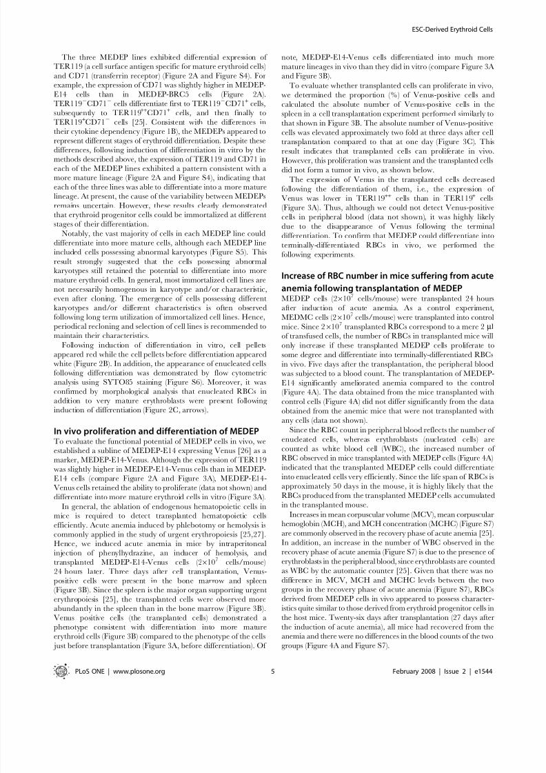
In vivo proliferation and differentiation of MEDEPTo evaluate the functional potential of MEDEP cells in vivo, we
established a subline of MEDEP-E14 expressing Venus [26] as a
marker, MEDEP-E14-Venus. Although the expression of TER119
was slightly higher in MEDEP-E14-Venus cells than in MEDEP-
E14 cells (compare Figure 2A and Figure 3A), MEDEP-E14-
Venus cells retained the ability to proliferate (data not shown) and
differentiate into more mature erythroid cells in vitro (Figure 3A).
In general, the ablation of endogenous hematopoietic cells inmice is required to detect transplanted hematopoietic cells
efficiently. Acute anemia induced by phlebotomy or hemolysis is
commonly applied in the study of urgent erythropoiesis [25,27].
Hence, we induced acute anemia in mice by intraperitonealinjection of phenylhydrazine, an inducer of hemolysis, and
transplanted MEDEP-E14-Venus cells (26107 cells/mouse)
24 hours later. Three days after cell transplantation, Venus-
positive cells were present in the bone marrow and spleen
(Figure 3B). Since the spleen is the major organ supporting urgent
erythropoiesis [25], the transplanted cells were observed more
abundantly in the spleen than in the bone marrow (Figure 3B).
Venus positive cells (the transplanted cells) demonstrated a
phenotype consistent with differentiation into more mature
erythroid cells (Figure 3B) compared to the phenotype of the cells
just before transplantation (Figure 3A, before differentiation). Of
note, MEDEP-E14-Venus cells differentiated into much more
mature lineages in vivo than they did in vitro (compare Figure 3A
and Figure 3B).
To evaluate whether transplanted cells can proliferate in vivo,
we determined the proportion (%) of Venus-positive cells and
calculated the absolute number of Venus-positive cells in the
spleen in a cell transplantation experiment performed similarly to
that shown in Figure 3B. The absolute number of Venus-positive
cells was elevated approximately two fold at three days after celltransplantation compared to that at one day (Figure 3C). This
result indicates that transplanted cells can proliferate in vivo.
However, this proliferation was transient and the transplanted cells
did not form a tumor in vivo, as shown below.
The expression of Venus in the transplanted cells decreased
following the differentiation of them, i.e., the expression of
Venus was lower in TER119++ cells than in TER119+ cells
(Figure 3A). Thus, although we could not detect Venus-positive
cells in peripheral blood (data not shown), it was highly likely
due to the disappearance of Venus following the terminal
differentiation. To confirm that MEDEP could differentiate into
terminally-differentiated RBCs in vivo, we performed the
following experiments.
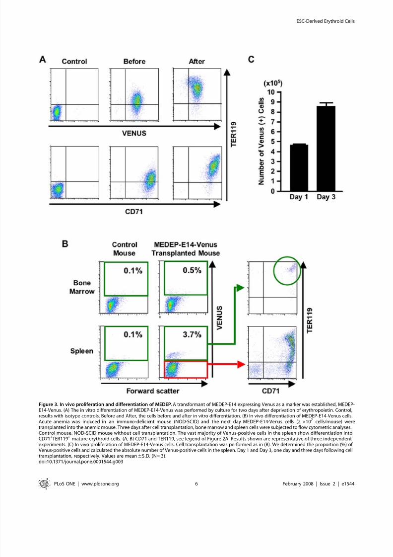
Increase of RBC number in mice suffering from acute
anemia following transplantation of MEDEPMEDEP cells (26107 cells/mouse) were transplanted 24 hours
after induction of acute anemia. As a control experiment,
MEDMC cells (26107 cells/mouse) were transplanted into control
mice. Since 26107
transplanted RBCs correspond to a mere 2 ml
of transfused cells, the number of RBCs in transplanted mice will
only increase if these transplanted MEDEP cells proliferate to
some degree and differentiate into terminally-differentiated RBCs
in vivo. Five days after the transplantation, the peripheral blood
was subjected to a blood count. The transplantation of MEDEP-
E14 significantly ameliorated anemia compared to the control
(Figure 4A). The data obtained from the mice transplanted with
control cells (Figure 4A) did not differ significantly from the data
obtained from the anemic mice that were not transplanted with
any cells (data not shown).
Since the RBC count in peripheral blood reflects the number of
enucleated cells, whereas erythroblasts (nucleated cells) are
counted as white blood cell (WBC), the increased number of
RBC observed in mice transplanted with MEDEP cells (Figure 4A)
indicated that the transplanted MEDEP cells could differentiate
into enucleated cells very efficiently. Since the life span of RBCs is
approximately 50 days in the mouse, it is highly likely that the
RBCs produced from the transplanted MEDEP cells accumulated
in the transplanted mouse.
Increases in mean corpuscular volume (MCV), mean corpuscular
hemoglobin (MCH), and MCH concentration (MCHC) (Figure S7)
are commonly observed in the recovery phase of acute anemia [25].
In addition, an increase in the number of WBC observed in therecovery phase of acute anemia (Figure S7) is due to the presence of
erythroblasts in the peripheral blood, since erythroblasts are counted
as WBC by the automatic counter [25]. Given that there was no
difference in MCV, MCH and MCHC levels between the two
groups in the recovery phase of acute anemia (Figure S7), RBCs
derived from MEDEP cells in vivo appeared to possess character-
istics quite similar to those derived from erythroid progenitor cells in
the host mice. Twenty-six days after transplantation (27 days after
the induction of acute anemia), all mice had recovered from the
anemia and there were no differences in the blood counts of the two
groups (Figure 4A and Figure S7).
ESC-Derived Erythroid Cells
PLoS ONE | www.plosone.org 5 February 2008 | Issue 2 | e1544
8/6/2019 Riken Mouse Differentiation
http://slidepdf.com/reader/full/riken-mouse-differentiation 6/11
Figure 3. In vivo proliferation and differentiation of MEDEP. A transformant of MEDEP-E14 expressing Venus as a marker was established, MEDEP-E14-Venus. (A) The in vitro differentiation of MEDEP-E14-Venus was performed by culture for two days after deprivation of erythropoietin. Control,results with isotype controls. Before and After, the cells before and after in vitro differentiation. (B) In vivo differentiation of MEDEP-E14-Venus cells.Acute anemia was induced in an immuno-deficient mouse (NOD-SCID) and the next day MEDEP-E14-Venus cells (26107 cells/mouse) weretransplanted into the anemic mouse. Three days after cell transplantation, bone marrow and spleen cells were subjected to flow cytometric analyses.Control mouse, NOD-SCID mouse without cell transplantation. The vast majority of Venus-positive cells in the spleen show differentiation intoCD71+TER119+ mature erythroid cells. (A, B) CD71 and TER119, see legend of Figure 2A. Results shown are representative of three independentexperiments. (C) In vivo proliferation of MEDEP-E14-Venus cells. Cell transplantation was performed as in (B). We determined the proportion (%) of Venus-positive cells and calculated the absolute number of Venus-positive cells in the spleen. Day 1 and Day 3, one day and three days following celltransplantation, respectively. Values are mean6S.D. (N= 3).doi:10.1371/journal.pone.0001544.g003
ESC-Derived Erythroid Cells
PLoS ONE | www.plosone.org 6 February 2008 | Issue 2 | e1544
8/6/2019 Riken Mouse Differentiation
http://slidepdf.com/reader/full/riken-mouse-differentiation 7/11
The transplantation of MEDEP-E14-Venus and MEDEP-
BRC5 cells also ameliorated anemia compared to the control
(Table 3). However, the transplantation of MEDEP-BRC5
cells appeared to be less effective for amelioration of anemia
than MEDEP-E14 cells (compare Figure 4A and Table 3).
Given that the in vitro proliferation activity of MEDEP-BRC5
cells was lower than that of MEDEP-E14 cells (Figure 1B), the
in vivo proliferation activity of MEDEP-BRC5 cells mighthave also been lower than that of MEDEP-E14 cells. In
addition, hemoglobin synthesis in MEDEP-BRC5 might have
been less efficient than that in MEDEP-E14 (Figure 2B and
Table 3).
Immunogenicity of human ES cell derivatives is one of the
potential obstacles to the clinical use of them [28,29]. In fact,
transplanted MEDEP cells could not ameliorate acute anemia in
mouse strains other than those from which each individual line
was derived or in immuno-deficient mice, suggesting immunolog-
ical rejection by the heterologous strains. Hence, if human
erythroid cell lines could be established, the clinical application
of such cell lines may require many cell lines expressing the
different major histo-compatibility (MHC) antigens.
Lack of tumorigenicity of MEDEP Approximately three months (82 days) after transplantation, Venus-
positive cells were absent from the bone marrow and spleen of mice
transplanted with MEDEP-E14-Venus cells (Figure S8). In addition,
although we observed all other transplanted mice up to 6 monthsafter transplantation, no tumor was observed in MEDEP-trans-
planted mice or MEDMC-transplanted control mice (data not
shown). Furthermore, subcutaneous transplantation of MEDEP cells
(26107 cells/injection site) did not give rise to any tumors, whereas
subcutaneous transplantation of the same number of parent ES cells
led to the formation of a teratoma (data not shown).
Nevertheless, if human erythroid cell lines were established, the
tumorigenic potential of those cell lines should be thoroughly
analyzed prior to use in the clinic [30,31]. It may be advisable to
engineer such cells so that they could be eliminated should a
malignant phenotype arise for any reason [32].
Figure 4. Amelioration of anemia by transplantation of MEDEP. (A) MEDEP-E14 cells (26107 cells/mouse) were transplanted into an immuno-deficient mouse (NOD-SCID) 24 hours after the induction of hemolysis by phenylhydrazine (60 mg/kg body weight) injection. Day 5 and Day 26, fiveand twenty-six days after cell transplantation. RBC, red blood cell. White bars (n = 10) and black bars (n = 14), the data obtained from the micetransplanted with control cells and MEDEP-E14 cells, respectively. Values are mean6S.D. * p,0.01 (by the Student’s t -test) (B) Increased survival of
mice transplanted with MEDEP cells following induction of severe acute anemia. MEDEP-E14 cells (26
107
cells/mouse) were transplanted into anNOD-SCID mouse 24 hours following the first induction of hemolysis by phenylhydrazine (60 mg/kg body weight) injection. Five days following thecell transplantation, the second induction of hemolysis by phenylhydrazine (80 mg/kg body weight) injection was performed. Statistical analysis wasperformed using the chi-square test. (A, B) Control cell, mast cell line derived from mouse ES cells (MEDMC-NT2) (Figures S1 and S2). MEDMC-NT2cells (26107 cells/mouse) were transplanted similarly as a control experiment.doi:10.1371/journal.pone.0001544.g004
ESC-Derived Erythroid Cells
PLoS ONE | www.plosone.org 7 February 2008 | Issue 2 | e1544
8/6/2019 Riken Mouse Differentiation
http://slidepdf.com/reader/full/riken-mouse-differentiation 8/11
RBCs derived from MEDEP are functional in vivoTo confirm that the RBCs derived from the transplanted MEDEP
cells are functional in vivo, we monitored the response of
transplanted mice to a second induction of hemolysis. Similar to
the experiments shown in Figure 4A and Table 3, a prior
induction of hemolysis and subsequent cell transplantation were
performed. A second induction of hemolysis was performed five
days after the cell transplantation (Figure 4B). Analysis of blood
count was not performed at any time point in this experiment,
because collection of peripheral blood would affect the results. Weobserved that one out of eight mice in the group transplanted with
MEDEP-E14 cells died while seven out of eight mice in the group
transplanted with control cells (MEDMC-NT2) died (Figure 4B).
The mice that did not receive any transplanted cells demonstrated
a mortality similar to that of mice transplanted with control cells
(data not shown). This result was consistent with the increased
RBC number five days after cell transplantation (Figure 4A). In
other words, this result indicated that RBCs derived from MEDEP
cells were functional in vivo and that mice transplanted with
MEDEP cells could survive the induction of severe acute anemia
following a second induction of hemolysis.
Concluding Remarks
At present, we cannot precisely describe the exact mechanismunderlying the establishment of differentiated cell lines from ES cells.
Nevertheless, our results clearly indicate that we can reproducibly
obtain useful erythroid cell lines from mouse ES cells. Given that
differentiation strategies developed for mouse ES cells can differ from
methods applied to human ES cells in many cases [33], the method
we developed here may not be applied to human ES cells directly
and some modifications may be necessary. However, considering the
number of human ES cell lines that have been established so far, we
believe that these human ES cell lines, at least in part, should exhibit
the potential to produce erythroid cell lines. In addition, our results
strongly suggest the possibility of establishing useful cell lines
committed to specific lineages other than hematopoietic progenitors
from human ES cells.
The induction of terminally differentiated cells that no longer
proliferate should enable clinical applications of ES cell derivatives
without an associated risk of tumorigenicity. For example, RBCs lack
nuclei following terminal differentiation, and thus are highly unlikely
to exhibit tumorigenicity in vivo. As such, even if the original ES cells
and/or their derivatives possessed abnormal karyotypes and/or
genetic mutations, they may nonetheless be useful for clinical
applications, provided that they can produce enucleated RBCs. Infact, although our MEDEP lines included many cells possessing
abnormal karyotypes (Figure S5), the vast majority of cells in each
cell line could nevertheless differentiate into mature erythroid cells
(Figure 2A), including enucleated cells (Figure 2C).
We showed a model of transplantation therapy using MEDEPs
in this study as an application of ES cell-derivatives. On the other
hand, methods to produce enucleated RBCs abundantly from
human hematopoietic stem cells in vitro have recently been
reported [2,3]. Therefore, once appropriate erythroid cell lines
have been established, it would be possible to use such methods to
produce enucleated RBCs from such cell lines in vitro. The
establishment of a human erythroid cell line lacking the genes to
produce the A, B, and RhD antigens would be a universal resource
for clinical application, since the cell line produces O/RhD(-)
RBCs which are transfusable into all individuals in theory.
MATERIALS AND METHODS
Mouse ES cell linesMouse ES cell lines, E14TG2a and D3, were obtained from the
American Type Culture Collection (ATCC; Monassas, VA, USA).
E14TG2a and D3 were derived from 129/Ola and 129/Sv+c/+p
mice, respectively. Other mouse ES cell lines were obtained from
the Cell Engineering Division of RIKEN BioResource Center
(Tsukuba, Ibaraki, Japan). TT2 was derived from F1 C57BL/6
and CBA mice. BRC4, BRC5, BRC6, and BRC7 were derived
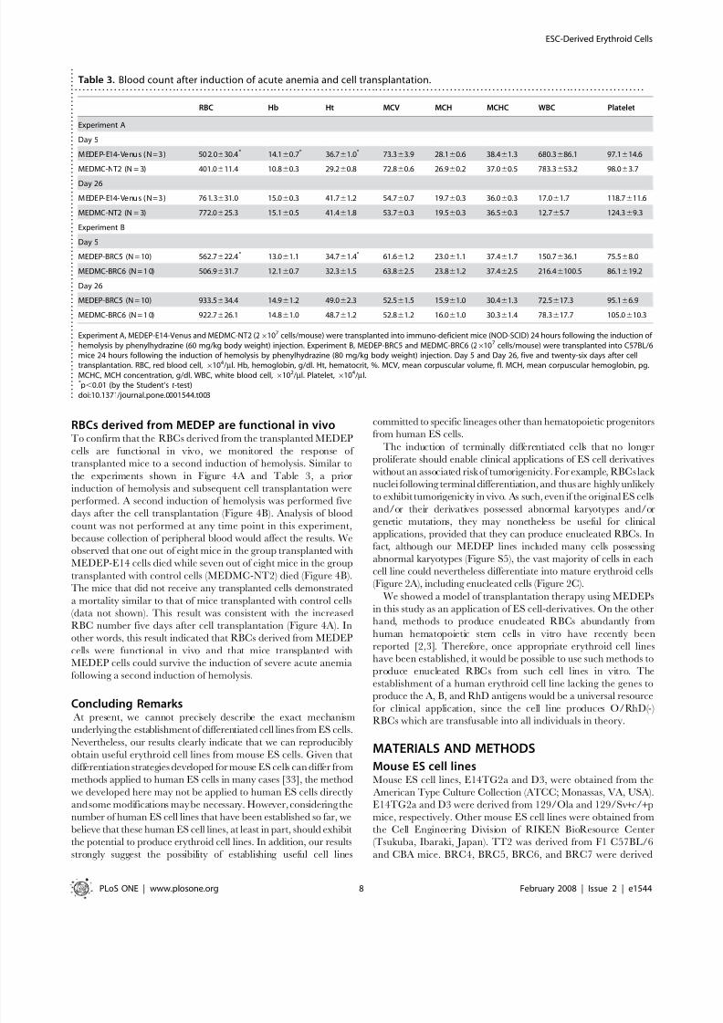
Table 3. Blood count after induction of acute anemia and cell transplantation.. . . . . . . . . . . . . . . . . . . . . . . . . . . . . . . . . . . . . . . . . . . . . . . . . . . . . . . . . . . . . . . . . . . . . . . . . . . . . . . . . . . . . . . . . . . . . . . . . . . . . . . . . . . . . . . . . . . . . . . . . . . . . . . . . . . . . . . . . . . . . . . . . .
RBC Hb Ht MCV MCH MCHC WBC Platelet
Experiment A
Day 5
M EDEP- E14- Venu s ( N=3 ) 50 2. 0630.4* 14.160.7* 36.761.0* 73.363.9 28.160.6 38.461.3 680.3686.1 97.1614.6
MEDMC-NT2 (N = 3) 401.0611.4 10.860.3 29.260.8 72.860.6 26.960.2 37.060.5 783.3653.2 98.063.7
Day 26
M EDEP- E14- Venu s ( N=3 ) 76 1. 3631.0 15.060.3 41.761.2 54.760.7 19.760.3 36.060.3 17.061.7 118.7611.6
MEDMC-NT2 (N = 3) 772.0625.3 15.160.5 41.461.8 53.760.3 19.560.3 36.560.3 12.765.7 124.369.3
Experiment B
Day 5
MEDEP-BRC5 (N = 10) 562.7622.4* 13.061.1 34.761.4* 61.661.2 23.061.1 37.461.7 150.7636.1 75.568.0
MEDMC-BRC6 (N = 1 0) 506.9631.7 12.160.7 32.361.5 63.862.5 23.861.2 37.462.5 216.46100.5 86.1619.2
Day 26
MEDEP-BRC5 (N = 10) 933.5634.4 14.961.2 49.062.3 52.561.5 15.961.0 30.461.3 72.5617.3 95.166.9
MEDMC-BRC6 (N = 1 0) 922.7626.1 14.861.0 48.761.2 52.861.2 16.061.0 30.361.4 78.3617.7 105.0610.3
Experiment A, MEDEP-E14-Venus and MEDMC-NT2 (26107 cells/mouse) were transplanted into immuno-deficient mice (NOD-SCID) 24 hours following the induction of hemolysis by phenylhydrazine (60 mg/kg body weight) injection. Experiment B, MEDEP-BRC5 and MEDMC-BRC6 (26107 cells/mouse) were transplanted into C57BL/6
mice 24 hours following the induction of hemolysis by phenylhydrazine (80 mg/kg body weight) injection. Day 5 and Day 26, five and twenty-six days after celltransplantation. RBC, red blood cell, 6104/ml. Hb, hemoglobin, g/dl. Ht, hematocrit, %. MCV, mean corpuscular volume, fl. MCH, mean corpuscular hemoglobin, pg.MCHC, MCH concentration, g/dl. WBC, white blood cell, 6102/ml. Platelet,6104/ml.* p,0.01 (by the Student’s t -test)doi:10.1371/journal.pone.0001544.t003 .
.
.
.
.
.
.
. . . .
.
.
.
.
.
.
.
.
.
.
.
.
.
.
.
.
.
.
.
.
.
.
.
.
.
.
.
.
.
.
.
.
.
.
.
.
.
.
.
.
.
.
. . .
.
.
.
.
.
.
.
.
.
.
.
.
.
.
.
.
.
.
.
.
.
.
.
.
.
.
.
.
ESC-Derived Erythroid Cells
PLoS ONE | www.plosone.org 8 February 2008 | Issue 2 | e1544

8/6/2019 Riken Mouse Differentiation
http://slidepdf.com/reader/full/riken-mouse-differentiation 9/11
from C57BL/6 mice. NTES-2 was established following transfer
of nuclei of cells derived from 129 mice into oocytes.
ES cells were maintained in an undifferentiated state on mouse
embryonic fibroblasts (MEFs) in a 1:1 mixture of Dulbecco’s
modified Eagle’s medium (DMEM; SIGMA, St Louis, MO, USA)
and Ham’s nutrient mixture F-12 (SIGMA) supplemented with
1,000 U/ml leukemia inhibitory factor (LIF; CHEMICON
International Inc., Temecula, CA, USA), 0.1 mM non-essential
amino acids, 2 mM L-glutamine, 0.1 mM 2-mercaptoethanol, and20% KnockOutTM
Serum Replacement (KSR; Invitrogen,Carlsbad, CA, USA). Before MEFs were used as feeder cells, they
were irradiated with c-rays (50 Gy).
Establishment of hematopoietic cell lines from
mouse ES cell linesThe feeder cell line, OP9 [23], used to induce hematopoietic
differentiation of ES cells [5,6] was obtained from the Cell
Engineering Division of RIKEN BioResource Center and was
cultured in Minimum Essential Medium-a (MEM-a; SIGMA)
containing 20% fetal bovine serum (FBS; SIGMA). Before OP9 cells
were used as feeder cells, they were irradiated with c-rays (50 Gy).
The basal medium used throughout long term culture was
Iscove’s modified Dulbecco’s medium (IMDM; Invitrogen)containing the following materials: 15% FBS (SIGMA); 10 mg/
ml bovine insulin, 5.5 mg/ml human transferrin, and 5 ng/ml
sodium selenite (ITS liquid MEDIA supplement; SIGMA); 50 mg/
ml ascorbic acid (SIGMA); 0.45 mM a-monothioglycerol (SIG-
MA); 100 unit/ml penicillin, 100 mg/ml streptomycin, and 2 mM
L-glutamine (PSQ; Invitrogen).
Specific factors used were as follows: mouse vascular endothelial
growth factor (VEGF, 20 ng/ml; R&D systems, Minneapolis,
MN, USA), mouse insulin-like growth factor-II (IGF-II, 200 ng/
ml; R&D systems), mouse stem cell factor (SCF, 50 ng/ml; R&D
systems), human erythropoietin (EPO, 5 unit/ml; KIRIN Brewery
Company, Tokyo, Japan), mouse interleukin-3 (IL-3, 10 ng/ml;
R&D systems), and Dexamethasone (Dex, 1026 M; SIGMA).
We developed two different methods to establish hematopoietic
cell lines from mouse ES cells, Method A and Method B. Method A is described in Table 1. In Method B, the use of IL-3 was
excluded from Method A through all procedures.
Viable cell number was assessed using an automated cell
counter and an assay based on the trypan blue dye exclusion
method, ViCellTM (BECKMAN COULTER, Fullerton, CA,
USA). Morphology of the established cell lines was analyzed after
Wright-Giemsa (Sysmex International, Kobe, Japan) staining.
Reverse transcription-polymerase chain reaction
(RT-PCR) A semi-quantitative RT-PCR was performed as describedpreviously [24]. PCR was carried out with recombinant Taq
polymerase (TaKaRa Bio Inc., Otsu, Shiga, Japan). Cycling
parameters were as follows: denaturation at 94uC for 30 sec,annealing at 55uC for 30 sec, and extension at 72uC for 30 sec.
PCR products were separated on 1.5% agarose gels and visualized
by ethidium bromide staining. Amplification of the gene encoding
glyceraldehyde-3-phosphate dehydrogenase (GAPDH) was used as
an internal control in the PCR.
The sequences of the PCR primers were as follows: for mouse
Oct-3/4, the sense primer was 59-ACC CAG GCC GAC GTG
GGG CT-39 and the antisense primer was 59-TTC TGG CGC
CGG TTA CAG AAC CA-39 (365-bp PCR product); for mouse
Nanog, the sense primer was 59-TAC CTC AGC CTC CAG
CAG AT-39 and the antisense primer was 59-CCT CCA AAT
CAC TGG CAG-39 (460-bp PCR product); for mouse GATA-1,
the sense primer was 59-ACA GGT CAC TAC CTG TGC AAT
GCC-39 and the antisense primer was 59-CCT GAC AGT ACC
ACA GGT CCT AG-39 (463-bp PCR product); for mouse EKLF
(Erythroid Kruppel-like factor), the sense primer was 59-TAT
GGG CTG CTG TCG GGA TAC CC-39 and the antisense
primer was 59-TCA GAG GTG ACG CTT CAT GTG CAG -3 9
(507-bp PCR product); for mouse EPOR (erythropoietin receptor),
the sense primer was 59-ATC CAT ATC AAT GAA GTA GTGCTC-39 and the antisense primer was 59-CCA CAG CTG GAA
GTT ACC CTT GTG-39 (513-bp PCR product); for mouse a-
globin, the sense primer was 59-CTC TCT GGG GAA GAC
AAA AGC-39 and the antisense primer was 59-GGT GGC TAG
CCA AGG TCA CCA-39 (334-bp PCR product); for mouse b ( b-
major)-globin, the sense primer was 59-GAT GCT GAG AAG
TCT GCT GTC-39 and the antisense primer was 59-CTG GAA
GGC AGC CTG TGC AGC-39 (381-bp PCR product); for
mouse c ( b-H1)-globin, the sense primer was 59-CTC AAG GAG
ACC TTT GCT CA-39 and the antisense primer was 59-AGT
CCC CAT GGA CTC AAA GA-39 (265-bp PCR product); for
mouse e-globin, the sense primer was 59-GGA GAG TCC ATT
AAG AAT CTA-39 and the antisense primer was 59-CTG TGA
ATT CAT TGC CGA AGT-39 (157-bp PCR product); for mouse
f-globin, the sense primer was 59-GCT CAG GCC GAG CCC ATT GG-39 and the antisense primer was 59-TAG CGG TAC
TTC TCA GTC AG-39 (371-bp PCR product); and for mouse
GAPDH (glyceraldehyde-3-phosphate dehydrogenase), the sense
primer was 59-GTC TTC ACC ACC ATG GAG AAG-39 and the
antisense primer was 59-GCC ATC CAC AGT CTT CTG GGT-
39 (270-bp PCR product).
Flow cytometryCells were stained with monoclonal antibodies (MoAbs) and
analyzed by FACS Calibur (BD Biosciences, San Jose, CA, USA)
or sorted by FACSVantage SE (BD Biosciences). The following
MoAbs were purchased from BD Biosciences: a fluorescein
isothiocyanate (FITC)-conjugated MoAb against mouse CD71
(transferrin receptor), a phycoerythrin (PE)-conjugated MoAbagainst TER119 (a cell surface antigen specific for mature
erythroid cells), an allophycocyanin (APC)-conjugated MoAb
against mouse c-Kit (receptor for stem cell factor), a PE-
conjugated MoAb against mouse CD71, and a biotin-conjugated
MoAb against TER119. APC-conjugated streptavidin (BD
Biosciences) was used to detect biotin-conjugated MoAb. To
distinguish nucleated and enucleated cells, we used the SYTO85
nuclear stain (Invitrogen). Cell viability was monitored by
propidium iodide (SIGMA) staining. Flow cytometry data were
analyzed using FlowJo (Tree Star Inc., Ashland, OR, USA)
analysis software. Morphology of the sorted cells was analyzed
following Wright-Giemsa (Sysmex International) staining.
MiceEight-week-old female NOD/shi-scid Jic and C57BL/6NCrj mice
were purchased from CLEA Japan (Tokyo, Japan) and Charles
River Laboratories Japan (Yokohama, Kanagawa, Japan), respec-
tively. Mice were used within a week of delivery in all experiments.
All experimental manipulations of mice were approved by the
Institutional Animal Care and Use Committee of the RIKEN
Tsukuba Institute.
Induction of acute anemia in mice Acute anemia was induced by hemolysis following intraperitoneal
injection of phenylhydrazine (Wako, Osaka, Japan), a chemical
ESC-Derived Erythroid Cells
PLoS ONE | www.plosone.org 9 February 2008 | Issue 2 | e1544
8/6/2019 Riken Mouse Differentiation
http://slidepdf.com/reader/full/riken-mouse-differentiation 10/11
inducer of hemolysis, at doses of 60 mg/kg body weight for NOD/
shi-scid Jic mice and 80 mg/kg body weight for C57BL/6NCrjmice. The second induction of hemolysis shown in Figure 4B was
performed at a dose of 80 mg/kg body weight in NOD/shi-scid Jic mice.
Transplantation of cellsCells (26107 cells/mouse) were injected into the tail vein of an 8-
week-old female mouse.
Blood countPeripheral blood samples were obtained from the retro-orbital
venous plexus. We estimated the number of white blood cells, red
blood cells, and platelets, in addition to the hemoglobin
concentration and hematocrit, using an automated Celltac a
MEK-6358 counter (NIHON-KODEN, Tokyo, Japan).
Statistical analysis All statistical analyses were performed using Statcel (OMS
company, Saitama, Japan) analysis software. As for the two-
sample t -test, the data were analyzed by the F test for variance
followed by the Student’s t -test.
SUPPORTING INFORMATION
Figure S1
Found at: doi:10.1371/journal.pone.0001544.s001 (10.19 MB TIF)
Figure S2
Found at: doi:10.1371/journal.pone.0001544.s002 (10.19 MB TIF)
Figure S3
Found at: doi:10.1371/journal.pone.0001544.s003 (10.19 MB TIF)
Figure S4
Found at: doi:10.1371/journal.pone.0001544.s004 (10.19 MB TIF)
Figure S5
Found at: doi:10.1371/journal.pone.0001544.s005 (10.20 MB TIF)
Figure S6
Found at: doi:10.1371/journal.pone.0001544.s006 (10.19 MB TIF)
Figure S7
Found at: doi:10.1371/journal.pone.0001544.s007 (10.19 MB TIF)
Figure S8
Found at: doi:10.1371/journal.pone.0001544.s008 (10.19 MB TIF)
ACKNOWLEDGMENTSWe thank Dr. A. Miyawaki for Venus cDNA; Dr. H. Miyoshi and Dr. K.
Katayama for virus to express Venus; all members in the Cell Engineering
Division for their help, discussion, and secretarial assistance.
Author Contributions
Conceived and designed the experiments: YN TH. Performed the
experiments: TH KM KS ID NA. Analyzed the data: YN TH KM KS
ID. Wrote the paper: YN TH.
REFERENCES
1. Neildez-Nguyen TM, Wajcman H, Marden MC, Bensidhoum M, Moncollin V,et al. (2002) Human erythroid cells produced ex vivo at large scale differentiateinto red blood cells in vivo. Nat Biotechnol 20: 467–472.
2. Giarratana MC, Kobari L, Lapillonne H, Chalmers D, Kiger L, et al. (2005) Ex vivo generation of fully mature human red blood cells from hematopoietic stemcells. Nat Biotechnol 23: 69–74.
3. Miharada K, Hiroyama T, Sudo K, Nagasawa T, Nakamura Y (2006) Efficientenucleation of erythroblasts differentiated in vitro from hematopoietic stem and
progenitor cells. Nat Biotechnol 24: 1255–1256.4. Keller G, Kennedy M, Papayannopoulou T, Wiles MV (1993) Hematopoieticcommitment during embryonic stem cell differentiation in culture. Mol Cell Biol13: 473–486.
5. Nakano T, Kodama H, Honjo T (1994) Generation of lymphohematopoieticcells from embryonic stem cells in culture. Science 265: 1098–1101.
6. Nakano T, Kodama H, Honjo T (1996) In vitro development of primitive anddefinitive erythrocytes from different precursors. Science 272: 722–724.
7. Carotta S, Pilat S, Mairhofer A, Schmidt U, Dolznig H, et al. (2004) Directeddifferentiation and mass cultivation of pure erythroid progenitors from mouseembryonic stem cells. Blood 104: 1873–1880.
8. Li F, Lu S, Vida L, Thomson JA, Honig GR (2001) Bone morphogenetic protein4 induces efficient hematopoietic differentiation of rhesus monkey embryonicstem cells in vitro. Blood 98: 335–342.
9. Umeda K, Heike T, Yoshimoto M, Shiota M, Suemori H, et al. (2004)Development of primitive and definitive hematopoiesis from nonhuman primateembryonic stem cells in vitro. Development 131: 1869–1879.
10. Kurita R, Sasaki E, Yokoo T, Hiroyama T, Takasugi K, et al. (2006) Tal1/Sclgene transduction using a lentiviral vector stimulates highly efficient hemato-
poietic cell differentiation from common marmoset (Callithrix jacchus)embryonic stem cells. Stem Cells 24: 2014–2022.
11. Kaufman DS, Hanson ET, Lewis RL, Auerbach R, Thomson JA (2001)Hematopoietic colony-forming cells derived from human embryonic stem cells.Proc Natl Acad Sci USA 98: 10716–10721.
12. Chadwick K, Wang L, Li L, Menendez P, Murdoch B, et al. (2003) Cytokinesand BMP-4 promote hematopoietic differentiation of human embryonic stemcells. Blood 102: 906–915.
13. Cerdan C, Rouleau A, Bhatia M (2004) VEGF-A165 augments erythropoieticdevelopment from human embryonic stem cells. Blood 103: 2504–2512.
14. Vodyanik MA, Bork JA, Thomson JA, Slukvin II (2005) Human embryonic stemcell-derived CD34+ cells: efficient production in the coculture with OP9 stromalcells and analysis of lymphohematopoietic potential. Blood 105: 617–626.
15. Wang L, Menendez P, Shojaei F, Li L, Mazurier F, et al. (2005) Generation of hematopoietic repopulating cells from human embryonic stem cells independentof ectopic HOXB4 expression. J Exp Med 201: 1603–1614.
16. Olivier EN, Qiu C, Velho M, Hirsch RE, Bouhassira EE (2006) Large-scale
production of embryonic red blood cells from human embryonic stem cells. Exp
Hematol 34: 1635–1642.
17. Hiroyama T, Miharada K, Aoki N, Fujioka T, Sudo K, et al. (2006) Long-lasting
in vitro hematopoiesis derived from primate embryonic stem cells. Exp Hematol
34: 760–769.
18. Lansdorp PM (2005) Role of telomerase in hematopoietic stem cells. Ann NY
Acad Sci 1044: 220–227.
19. Takahashi K, Yamanaka S (2006) Induction of pluripotent stem cells frommouse embryonic and adult fibroblast cultures by defined factors. Cell 126:
663–676.
20. Okita K, Ichisaka T, Yamanaka S (2007) Generation of germline-competent
induced pluripotent stem cells. Nature 448: 313–317.
21. Wernig M, Meissner A, Foreman R, Brambrink T, Ku M, et al. (2007) In vitro
reprogramming of fibroblasts into a pluripotent ES-cell-like state. Nature 448:
318–324.
22. Maherali N, Sridharan R, Xie W, Utikal J, Eminli S, et al. (2007) Directly
reprogrammed fibroblasts show global epigenetic remodeling and widespread
tissue contribution. Cell Stem Cell 1: 55–70.
23. Kodama H, Nose M, Niida S, Nishikawa S, Nishikawa S (1994) Involvement of
the c-kit receptor in the adhesion of hematopoietic stem cells to stromal cells.
Exp Hematol 22: 979–984.
24. Sudo K, Kanno M, Miharada K, Ogawa S, Hiroyama T, et al. (2007)
Mesenchymal progenitors able to differentiate into osteogenic, chondrogenic,
and/or adipogenic cells in vitro are present in most primary fibroblast-like cell
populations. Stem Cells 25: 1610–1617.
25. Miharada K, Hiroyama T, Sudo K, Nagasawa T, Nakamura Y (2005) Lipocalin
2 functions as a negative regulator of red blood cell production in an autocrinefashion. FASEB J 19: 1881–1883.
26. Nagai T, Ibata K, Park ES, Kubota M, Mikoshiba K, et al. (2002) A variant of
yellow fluorescent protein with fast and efficient maturation for cell-biological
applications. Nat Biotechnol 20: 87–90.
27. Alter BP, Campbell AS, Holland JG, Friend C (1982) Increased mouse minor
hemoglobin during erythroid stress: a model for hemoglobin regulation. Exp
Hematol 10: 754–760.
28. Drukker M, Benvenisty N (2004) The immunogenicity of human embryonic
stem-derived cells. Trends Biotechnol 22: 136–141.
29. Boyd AS, Higashi Y, Wood KJ (2005) Transplanting stem cells: potential targets
for immune attack. Modulating the immune response against embryonic stem
cell transplantation. Adv Drug Deliv Rev 57: 1944–1969.
30. Vogel G (2005) Ready or not? Human ES cells head toward the clinic. Science
308: 1534–1538.
ESC-Derived Erythroid Cells
PLoS ONE | www.plosone.org 10 February 2008 | Issue 2 | e1544

8/6/2019 Riken Mouse Differentiation
http://slidepdf.com/reader/full/riken-mouse-differentiation 11/11
31. Hentze H, Graichen R, Colman A (2007) Cell therapy and the safety of embryonic stem cell-derived grafts. Trends Biotechnol 25: 24–32.
32. Schuldiner M, Itskovitz-Eldor J, Benvenisty N (2003) Selective ablation of human embryonic stem cells expressing a ‘‘suicide’’ gene. Stem cells 21:257–265.
33. Reubinoff BE, Pera MF, Fong CY, Trounson A, Bongso A (2000) Embryonic
stem cell lines from human blastocysts: somatic differentiation in vitro. Nat
Biotechnol 18: 399–404.
ESC-Derived Erythroid Cells
PLoS ONE | www.plosone.org 11 February 2008 | Issue 2 | e1544
Related Documents