Journal of Neurology, Neurosurgery, and Psychiatry, 1974, 37, 1069-1073 Rheumatoid C1-C2 dislocation: pathogenesis and treatment reconsidered CHUN C. KAO, B. MESSERT, S. S. WINKLER, AND J. H. TURNER From the Services of Neurosurgery, Neurology, Radiology, and Pathology, Madison Veterans Administration Hospital, and The University of Wisconsin Center for Health Sciences, Madison, Wisconsin, U.S.A. SYNOPSIS The commonly accepted mechanism of rheumatoid C1-C2 dislocation is the incompe- tence of the transverse ligament of C1 vertebra. The presence of rheumatoid granulation tissue between the anterior arch of C1 vertebra and the odontoid process is an important factor. This might explain why the dislocation becomes irreducible when the disease progresses. In a 55 year old man, transoral removal of the rheumatoid granulation tissue made the previously irreducible C1-C2 dislocation reducible. Stabilization of the reduced C1-C2 articulation was accomplished at a second operation by posterior fusion of the occiput to C1, C2, and C3 vertebrae. Although dislocation of the first or the second cervical vertebra ('C1-C2 dislocation') is a well- recognized complication of rheumatoid arthritis (Conlon et al., 1966), the exact mechanism pro- ducing the dislocation is not yet fully understood. The most common type of C1-C2 dislocation in rheumatoid arthritis is the forward dislocation of C1 on C2 vertebra. The diagnosis is made when, in radiographs of the lateral cervical spine, the distance between the anterior arch of C1 vertebra and the odontoid process is greater than 2-5 mm in females and 3 mm in males (Conlon et al., 1966). During the early stages of the disease, the distance between the anterior arch of C1 vertebra and the odontoid process varies with head position, being greatest with the head flexed and minimal or normal with the head extended. As the disease progresses, the differ- ence between head flexion and extension be- comes less and less obvious, and the distance between the anterior arch of C1 vertebra and the odontoid process eventually becomes fixed and irreducible even under traction. Incompetence of the transverse ligament of C1 vertebra has generally been accepted as the cause of forward dislocation of C1 on C2 vertebra. Martel and Abell (1963) demonstrated that the transverse ligament 'had areas of degeneration 1069 and increased vascularity ... were lax and re- sulted in extreme mobility of the atlanto- odontoid joints'. This was the cause of a fatal dislocation in a 61 year old white female with a 20 year history of rheumatoid arthritis. In a 53 year old female who died of rheumatoid C1-C2 dislocation, Webb et al. (1968) found the trans- verse ligament of C1 vertebra destroyed to such a degree that it could not be identified. In the presence of a lax or ruptured transverse ligament, C1 vertebral body slips forward on C2 vertebra in flexion, but the displacement of the odontoid process remains limited. The odontoid process does not reach the posterior arch of C1. Werne (1957) proposed that the alar ligaments limit the extent of backward dislocation of the odontoid process when the transverse ligament is destroyed. Were this hypothesis correct, the forward dislocation of C1 on C2 vertebra should be reducible with the head placed in extension. However, frequently the reduction remains in- complete and the gap between the anterior arch of C1 vertebra and the odontoid process with the head in extension remains excessive, although less than when the head is flexed. We postulate that this failure to reduce the dislocation might be explained by the presence of rheumatoid granulation tissue between the anterior arch of C1 vertebra and the odontoid process. In a 65 guest. Protected by copyright. on October 3, 2021 by http://jnnp.bmj.com/ J Neurol Neurosurg Psychiatry: first published as 10.1136/jnnp.37.9.1069 on 1 September 1974. Downloaded from

Welcome message from author
This document is posted to help you gain knowledge. Please leave a comment to let me know what you think about it! Share it to your friends and learn new things together.
Transcript

Journal of Neurology, Neurosurgery, and Psychiatry, 1974, 37, 1069-1073
Rheumatoid C1-C2 dislocation: pathogenesis andtreatment reconsidered
CHUN C. KAO, B. MESSERT, S. S. WINKLER, AND J. H. TURNER
From the Services of Neurosurgery, Neurology, Radiology, and Pathology,Madison Veterans Administration Hospital, and The University of Wisconsin Center
for Health Sciences, Madison, Wisconsin, U.S.A.
SYNOPSIS The commonly accepted mechanism of rheumatoid C1-C2 dislocation is the incompe-tence of the transverse ligament of C1 vertebra. The presence of rheumatoid granulation tissuebetween the anterior arch of C1 vertebra and the odontoid process is an important factor. This mightexplain why the dislocation becomes irreducible when the disease progresses. In a 55 year old man,
transoral removal of the rheumatoid granulation tissue made the previously irreducible C1-C2dislocation reducible. Stabilization of the reduced C1-C2 articulation was accomplished at a secondoperation by posterior fusion of the occiput to C1, C2, and C3 vertebrae.
Although dislocation of the first or the secondcervical vertebra ('C1-C2 dislocation') is a well-recognized complication of rheumatoid arthritis(Conlon et al., 1966), the exact mechanism pro-ducing the dislocation is not yet fully understood.The most common type of C1-C2 dislocation
in rheumatoid arthritis is the forward dislocationof C1 on C2 vertebra. The diagnosis is madewhen, in radiographs of the lateral cervicalspine, the distance between the anterior arch ofC1 vertebra and the odontoid process is greaterthan 2-5 mm in females and 3 mm in males(Conlon et al., 1966).During the early stages of the disease, the
distance between the anterior arch of C1vertebra and the odontoid process varies withhead position, being greatest with the headflexed and minimal or normal with the headextended. As the disease progresses, the differ-ence between head flexion and extension be-comes less and less obvious, and the distancebetween the anterior arch of C1 vertebra and theodontoid process eventually becomes fixed andirreducible even under traction.
Incompetence of the transverse ligament of C1vertebra has generally been accepted as the causeof forward dislocation of C1 on C2 vertebra.Martel and Abell (1963) demonstrated that thetransverse ligament 'had areas of degeneration
1069
and increased vascularity ... were lax and re-sulted in extreme mobility of the atlanto-odontoid joints'. This was the cause of a fataldislocation in a 61 year old white female with a20 year history of rheumatoid arthritis. In a 53year old female who died of rheumatoid C1-C2dislocation, Webb et al. (1968) found the trans-verse ligament of C1 vertebra destroyed to such adegree that it could not be identified.
In the presence of a lax or ruptured transverseligament, C1 vertebral body slips forward on C2vertebra in flexion, but the displacement of theodontoid process remains limited. The odontoidprocess does not reach the posterior arch of C1.Werne (1957) proposed that the alar ligamentslimit the extent of backward dislocation of theodontoid process when the transverse ligamentis destroyed. Were this hypothesis correct, theforward dislocation of C1 on C2 vertebra shouldbe reducible with the head placed in extension.However, frequently the reduction remains in-complete and the gap between the anterior archof C1 vertebra and the odontoid process with thehead in extension remains excessive, althoughless than when the head is flexed. We postulatethat this failure to reduce the dislocation mightbe explained by the presence of rheumatoidgranulation tissue between the anterior arch ofC1 vertebra and the odontoid process. In a 65
guest. Protected by copyright.
on October 3, 2021 by
http://jnnp.bmj.com
/J N
eurol Neurosurg P
sychiatry: first published as 10.1136/jnnp.37.9.1069 on 1 Septem
ber 1974. Dow
nloaded from

Chun C. Kao, B. Messert, S. S. Winkler, and J. H. Turner
year old white male who died of rheumatoid C1-C2 dislocation reported by Gleason and Urist(1965), there was a mass of rheumatoid granula-tion tissue resulting in an increased space be-tween the anterior arch of C1 vertebra and theodontoid process. Although the presence of therheumatoid granulation tissue is mentioned inthis (Gleason and Urist, 1965) and one other(Bland, 1967) necropsy report, this informationhas not been applied to solve the clinical prob-lems.
Recently we have seen a quadriparetic patientwith a fixed C1-C2 dislocation. A mass ofgranulation tissue was removed from betweenthe anterior arch of C1 vertebra and the odon-toid process and the dislocation subsequentlybecame reducible.
CASE REPORT
(E.T. 68-64) A 55 year old American Indian malewas admitted to the Madison VA Hospital on 29May 1971 with quadriparesis and urinary retentionof two days' duration.The patient had had rheumatoid arthritis for 15
years and had been confined to a wheelchair forthree years. On the morning of 27 May 1971, after afit of coughing he felt his neck snap and a few hourslater was unable to feed himself at the breakfasttable and pain was noted in the back of his neck. Heremained in bed most of the day and could not void.That evening he was admitted to a local hospital andwas catheterized. The next day the patient was trans-ferred to VAH Madison.At the time of admission he was obtunded. A
cataract was present in the right eye; the left eyeground was normal. An extensive rheumatoid pro-
FIG. 1. (a) Preoperative radiograph ofcervical spine showing dislocated C1 on C2 vertebra. The dislocation wasnot reducible. A halo tong was used to maintain the head position during and after operation. (b) Postoperativecervical spine radiograph showing successful reduction of the C1-C2 dislocation. Halo tong support has beenremoved.
1070
guest. Protected by copyright.
on October 3, 2021 by
http://jnnp.bmj.com
/J N
eurol Neurosurg P
sychiatry: first published as 10.1136/jnnp.37.9.1069 on 1 Septem
ber 1974. Dow
nloaded from
Rheumatoid C1-C2 dislocation: pathogenesis and treatment reconsidered
cess had deformed shoulders, elbows, wrists, knees,and ankles. There was flexion contracture at theknees. The patient was barely able to raise his legsfrom the bed and move his toes. The upper extremi-ties were paralysed and flaccid. Sensory examinationshowed a sensory level with loss of pain and tempera-ture from the neck down with preservation of vibra-tion, position, and light touch sensation in all theextremities and the trunk.He showed evidence of urinary tract infection
caused by Klebsiella and Proteus. The bloodhaemoglobin was 129 g/100 ml and haematocrit37-2%. Serum protein level was 6 6 g and albumin2 8 g/100 ml. The sedimentation rate was 50/36 mmin the first two hours. The Waaler-Rose test waspositive at 1: 1280. No cold agglutinins were detected.The antinuclear antibody test was also negative.The cervical spine radiograph showed a consider-
able amount of C1-C2 dislocation. The odontoidprocess was eroded but not separated from the bodyof C2 vertebra. The distance between the anteriorarch of C1 vertebra and the anterior margin of theodontoid process was 15 mm, and the distancebetween the posterior arch of C1 vertebra and theposterior margin of the odontoid process was only12 mm (Fig. la).The patient was immediately placed on a Stryker
frame with Crutchfield tong skull traction. It thenbecame obvious that the dislocation was not reduc-ible even with neck extension and weight loading.However, the discomfort in the neck disappeared,and the neurological findings were not made worseby the traction.He remained on the Stryker frame and in traction
for the next five months. The dislocation remainedbasically unchanged but the neurological findingsshowed some improvement. He became able to raiseboth arms; limited use of fingers returned.Removal of the posterior arch of C1 vertebra was
considered in the hope of decompressing the spinalcord (Alexander et al., 1953; Dastur et al., 1965).However, this would have resulted in increased in-stability of C1 over C2 vertebra and result in possiblyfatal medullary injury. In view of our hypothesisthat the failure to achieve reduction is due to over-growth of rheumatoid connective tissue between theanterior arch of C1 vertebra and the odontoid pro-cess, it was thought that the treatment of choice toreduce the dislocation without removing any bonewas to remove the tissue between the anterior arch ofC1 vertebra and the odontoid process through atransoral approach.On 10 November 1971, Crutchfield tong traction
was discontinued. A halo tong was used to maintainthe head position. A cervical myelogram showed notissue growth posterior to the odontoid process; the
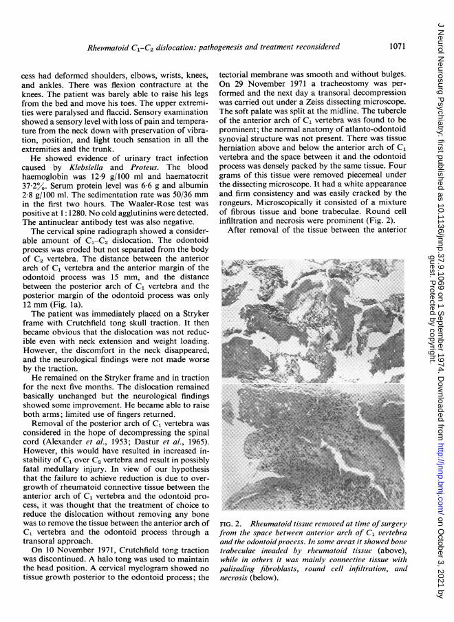
tectorial membrane was smooth and without bulges.On 29 November 1971 a tracheostomy was per-formed and the next day a transoral decompressionwas carried out under a Zeiss dissecting microscope.The soft palate was split at the midline. The tubercleof the anterior arch of C1 vertebra was found to beprominent; the normal anatomy of atlanto-odontoidsynovial structure was not present. There was tissueherniation above and below the anterior arch of C1vertebra and the space between it and the odontoidprocess was densely packed by the same tissue. Fourgrams of this tissue were removed piecemeal underthe dissecting microscope. It had a white appearanceand firm consistency and was easily cracked by therongeurs. Microscopically it consisted of a mixtureof fibrous tissue and bone trabeculae. Round cellinfiltration and necrosis were prominent (Fig. 2).
After removal of the tissue between the anterior
FIG. 2. Rheumatoid tissue removed at time ofsurgeryfrom the space between anterior arch of C1 vertebraand the odontoidprocess. In some areas it showed bonetrabeculae invaded by rheumatoid tissue (above),while in others it was mainly connective tissue withpalisading fibroblasts, round cell infiltration, andnecrosis (below).
1071
guest. Protected by copyright.
on October 3, 2021 by
http://jnnp.bmj.com
/J N
eurol Neurosurg P
sychiatry: first published as 10.1136/jnnp.37.9.1069 on 1 Septem
ber 1974. Dow
nloaded from

Chun C. Kao, B. Messert, S. S. Winkler, and J. H. Turner
arch of C1 vertebra and the odontoid process, andwith the patient still under general anaesthesia andunder direct vision of the dissecting microscope, thepatient's head was readjusted. Movement was seenat both anterior arch of C1 vertebra and the odontoidprocess and the distance between them was reducedto 4 mm. The reduced position was again maintainedby the halo tong.On 6 January 1972 a posterior occipito-Cl-C2-C3
vertebral fusion was carried out using gauge 20wires and acrylic plastic reinforcement. The halotong was removed on the 10th day after fusion. Apostoperative radiograph of the cervical spineshowed the distance between the anterior arch of Civertebra and the odontoid process to be 4 mm andthe distance between the posterior arch of C1vertebra and the odontoid process to be 22 mm (Fig.Ib). The patient was discharged on 27 January 1972.At the time of discharge he was in a wheelchair andable to use his arms and hands for daily activity ofliving. He was able to stand, but could not walkbecause of the flexion contraction of the knees andextensive rheumatoid deformities of the feet.
DISCUSSION
Review of necropsy reports of 12 patients whodied of dislocation of C1 on C2 vertebra due torheumatoid arthritis (Davis and Markley, 1951;Martel and Page, 1960; Martel and Abell, 1963;Gleason and Urist, 1965; Verjaal and Harder,1965; Bland, 1967; Webb et al., 1968) shows thatincompetence of the transverse ligament of C1vertebra was found in only three patients (Marteland Abell, 1963; Webb et al., 1968). Of thesethree patients, two died of upper cervical cordcompression from the displaced odontoid pro-cess (Martel and Abell, 1963). In the thirdpatient (Webb et al., 1968), although the trans-verse ligament was incompetent, the cause ofdeath was due to a sudden collapse of thelateral body of C1 vertebra with bilateralvertebral artery occlusion and the vertical up-ward displacement of the odontoid processwhich penetrated the medulla. Thus in only twopatients can the incompetent transverse ligamentbe related to the patient's death.
In all 12 patients, the fundamental abnor-mality was a chronic synovitis involving almostall the synovial joints of the cervical spine withthe most severe change at the synovial jointsbetween C1 and C2 vertebrae. Significantrheumatoid granulation tissue was found be-
tween the anterior arch of C1 vertebra and theodontoid process on six occasions (Gleason andUrist, 1965; Bland, 1967), in the retro-odontoidspace on two occasions (Martel and Abell, 1963;Gleason and Urist, 1965), and between C4 andC5 vertebrae on one occasion (Bland, 1967). Inone patient backward dislocation of C1 vertebradue to rheumatoid process was associated with alarge intraspinal meningioma attached to thebody of C2 vertebra (Verjaal and Harder, 1965).
These unpredictable findings suggest that acomplete diagnostic evaluation by myelographyis certainly needed to plan the surgical strategyif surgical intervention is elected. A routineposterior decompression or fusion withoutmyelographic study could be disastrous in thesepatients.
There are also three necropsy reports ofimmediate death due to the sudden collapse ofthe lateral body of C1 vertebra or the occipitalcondyle with medullary injury by the odontoidprocess (Davis and Markley, 1951; Martel andPage, 1960; Webb et al., 1968). Extensive boneerosion, fragmentation, and softening involvednot only the odontoid process, the anterior arch,and lateral body of C1 vertebra but the occipitalbone as well.The fact that in rheumatoid arthritis resulting
in C1-C2 intervertebral dislocation the anteriorarch and the lateral body of C1 vertebra mayalso be weakened by the rheumatoid processcreates a serious contraindication to the posteriordecompression by removal of the posterior archof C1 vertebra. The experiences of Alexander etal. (1953) and Dastur et al. (1965) show the highoperative risk that is associated with posteriordecompression even in non-rheumatoid C1-C2dislocation.Of considerable interest is a report by Sukoff
et al. (1972) concerning a patient who had anirreducible rheumatoid C1-C2 dislocation andwas treated initially by posterior decompressionwith removal of the posterior arch of C1 andfusion of C2 vertebra to the occiput. The fusionwas solid but the dislocation remained un-reduced. Six months later quadriparesis re-curred and a transoral removal of part of theanterior arch of C1 vertebra together with therheumatoid granulation tissue and the odontoidprocess was performed to prevent further neuro-logical deterioration. Had the transoral ap-
1072
guest. Protected by copyright.
on October 3, 2021 by
http://jnnp.bmj.com
/J N
eurol Neurosurg P
sychiatry: first published as 10.1136/jnnp.37.9.1069 on 1 Septem
ber 1974. Dow
nloaded from

Rheumatoid C1-C2 dislocation: pathogenesis and treatment reconsidered
proach been performed first as in our case thepathological process would have been correctedand the C1-C2 dislocation reduced. A simplerposterior fusion, rather than decompression,may then be advised for stabilizing the reducedC1-C2 vertebral articulation.
The senior author thanks Dr Manucher J. Javid,Professor and Chairman, Department of Neuro-surgery, University of Wisconsin Center for HealthSciences, for his invaluable advice in management ofthis patient.
REFERENCES
Alexander, E., Jr, Masland, R. L., and Harris, C. (1953).Anterior dislocation of first cervical vertebra simulatingcerebral birth injury in infancy. American Journal ofDiseases of Children, 85, 173-181.
Bland, J. H. (1967). Rheumatoid arthritis of the cervicalspine. Bulletin on the Rheumatic Diseases, 18, 471 476.
Conlon, P. W., Isdale, I. C., and Rose, B. S. (1966). Rheuma-toid arthritis of the cervical spine. An analysis of 333 cases.Annals of the Rheumatic Diseases, 25, 120-126.
Dastur, D. K., Wadia, N. H., Desai, A. D., and Sinh, G.
(1965). Medullospinal compression due to atlanto-axialdislocation and sudden haematomyelia during decompres-sion. Brain, 88, 897-924.
Davis, F. W., Jr, and Markley, H. E. (1951). Rheumatoidarthritis with death from medullary compression. Annals ofInternal Medicine, 35, 451-454.
Gleason, I. O., and Urist, M. R. (1965). Atlanto-axial disloca-tion with odontoid separation in rheumatoid disease.Report of a case. Clinical Orthopaedics and RelatedResearch, 42, 121-129.
Martel, W., and Abell, M. R. (1963). Fatal atlanto-axialluxation in rheumatoid arthritis. Arthritis and Rheumatism,6, 224-231.
Martel, W., and Page, J. W. (1960). Cervical vertebralerosions and subluxations in rheumatoid arthritis andankylosing spondylitis. Arthritis and Rheumatism, 3, 546-556.
Sukoff, M. H., Kadin, M. M., and Moran, T. (1972). Trans-oral decompression for myelopathy caused by rheumatoidarthritis of the cervical spine. Case report. Journal ofNeurosurgery, 37, 493-497.
Verjaal, A., and Harder, N. C. (1965). Backward luxation ofthe atlas. Report of a case. Acta Radiologica. Diagnosis, 3,173-176.
Webb, F. W. S., Hickman, J. A., and Brew, D. St.J. (1968).Death from vertebral artery thrombosis in rheumatoidarthritis. British Medical Journal, 2, 537-538.
Werne, S. (1957). Studies in spontaneous atlas dislocation.Acta Orthopaedica Scandinavica. Suppl. 23, 1-150.
1073
guest. Protected by copyright.
on October 3, 2021 by
http://jnnp.bmj.com
/J N
eurol Neurosurg P
sychiatry: first published as 10.1136/jnnp.37.9.1069 on 1 Septem
ber 1974. Dow
nloaded from
Related Documents