Helena Maria Lourenço Carvalheiro THE ROLE OF CD8 + T CELLS IN THE PATHOGENESIS OF RHEUMATOID ARTHRITIS Tese de Doutoramento em Ciências e Tecnologias da Saúde, especialidade de Biologia Celular e Molecular orientada pela Doutora Maria Margarida Souto Carneiro e pela Professora Doutora Maria Celeste Fernandes Lopes, apresentada à Faculdade de Farmácia da Universidade de Coimbra 2014

Welcome message from author
This document is posted to help you gain knowledge. Please leave a comment to let me know what you think about it! Share it to your friends and learn new things together.
Transcript

Helena Maria Lourenço Carvalheiro
THE ROLE OF CD8+ T CELLS IN THE PATHOGENESIS
OF RHEUMATOID ARTHRITIS
Tese de Doutoramento em Ciências e Tecnologias da Saúde, especialidade de Biologia Celular e Molecular
orientada pela Doutora Maria Margarida Souto Carneiro e pela Professora Doutora Maria Celeste Fernandes Lopes,
apresentada à Faculdade de Farmácia da Universidade de Coimbra
2014
Imagem

i
Helena Maria Lourenço Carvalheiro
CD8+ T cells in the pathogenesis
of Rheumatoid Arthritis
Tese de Doutoramento em Ciências da Saúde, na especialidade de Biologia Celular e
Molecular, apresentada à Faculdade de Farmácia da Universidade de Coimbra para a
obtenção do grau de Doutor.
Orientadores: Doutora Maria Margarida Souto Carneiro e Professora Doutora Maria
Celeste Lopes.
Coimbra, 2014


iii
Front page art:
Reproduction of the painting “My Fear” by painter and RA patient Aleah Denton.
(reproduced with artist’s consent)


v
The research work presented in this thesis was performed at the Center for
Neuroscience and Cell Biology of Coimbra, University of Coimbra and at the Faculty of
Medicine of the University of Coimbra, Portugal, under supervision of Dr. Maria
Margarida Souto Carneiro and Prof. Dr. Maria Celeste Fernandes Lopes.
O trabalho experimental apresentado nesta tese foi elaborado no Centro de Neurociências e
Biologia Celular de Coimbra e na Faculdade de Medicina da Universidade de Coimbra,
Portugal, sob supervisão da Doutora Maria Margarida Souto Carneiro e Professora Doutora
Maria Celeste Fernandes Lopes.
This work was funded by the Portuguese Foundation for Science and Technology, PhD
fellowship SFRH / BD / 60467 / 2009.
Este trabalho foi financiado pela Fundação Portuguesa para a Ciência e Tecnologia, bolsa
de doutoramento SFRH / BD / 60467 / 2009.


vii
Aos meus pais
A todos os doentes com Artrite Reumatóide


ix
The only real mistake is the one from which we learn nothing.
John Powell
Success is not final, failure is not fatal: it is the courage to continue that counts.
Winston Churchill

xi
Agradecimentos/Acknowledgements
Agradeço à Doutora Maria Margarida Souto Carneiro, por me ter acolhido no seu
laboratório no Centro de Neurociências e Biologia Celular em Coimbra. Agradeço toda a
confiança e apoio prestado em todas as etapas do meu doutoramento. Obrigada pela
disponibilidade que sempre demonstrou para discussões científicas, conselhos e sugestões
que permitiram a concretização deste trabalho, e me ajudaram a crescer como cientista.
Agradeço ao Professor Doutor José António Pereira da Silva, que sempre se
mostrou disponível para abrir novos caminhos científicos para este trabalho. Agradeço em
particular as discussões científicas assim como a sua disponibilidade e apoio ao longo
destes últimos anos.
Agradeço também à Professora Doutora Maria Celeste Fernandes Lopes, por ter
sido incansável durante este trabalho de doutoramento, em particular nesta última fase.
Gostaria também de agradecer à Professora Doutora Anabela Mota Pinto, pelo
apoio incondicional prestado em particular nesta última fase do doutoramento.
O meu muito obrigado à Doutora Cátia, pelo empenho nos estudos efectuados em
parceria com a Unidade de Reumatologia dos HUC, e por toda a ajuda prestada, em
particular na análise estatística.
Gostaria de agradecer aos meus colegas de laboratório, Tiago, David, Sandra,
Sandra Íris, Mónica, Geema, Valeria, Guiseppe, Milene, Aline, Mariana, Filipa, Natália,
Ana, Fábio, Gonçalo e Inês entre outros, por toda a ajuda que prestaram, e por tornarem os
dias de trabalho mais agradáveis.
Agradeço aos meus amigos, em particular à Áurea, Susana e Filipe, por todos os
momentos de galhofa, pelas noitadas bem passadas e por todo o apoio nos bons e maus
momentos.
Gostaria de agradecer à minha família, que sempre me apoia em tudo o que faço, e
que estão sempre lá para mim. Um obrigado especial à minha afilhada Sara e à Cristina por

xii
serem umas miúdas à maneira, e que muitas vezes me distraíram dos meus problemas com
o seu bom humor.
Quero agradecer ao meu namorado, por todo o amor, carinho e apoio incondicional,
por me ajudar a ultrapassar as várias etapas deste processo, e por me dar força e acreditar
em mim, por vezes mais do que eu própria. Sem ti isto não seria a mesma coisa.
Por fim, quero agradecer aos meus pais, as pessoas mais importantes da minha vida,
que apesar de se encontrarem a milhares de quilómetros, estão sempre presentes. Agradeço
por tudo o que sempre fizeram por mim, por todo o carinho e compreensão, pelo apoio
incondicional e por me darem sempre força nos bons e maus momentos. A eles devo tudo
o que sou…

xiii
Table of contents
FIGURE INDEX .......................................................................................................XVII
TABLE INDEX .......................................................................................................... XIX
ABBREVIATION LIST ............................................................................................. XXI
RESUMO ................................................................................................................ XXVII
ABSTRACT ............................................................................................................. XXIX
PUBLICATION LIST ............................................................................................. XXXI
1. INTRODUCTION .................................................................................................... 3
1.1. THE IMMUNE SYSTEM ......................................................................................... 3
1.1.1. The innate response .................................................................................... 3
1.1.2. The adaptive response ................................................................................. 4
1.1.3. CD8+ T cells ................................................................................................ 6
1.1.3.1. CD8+ T cell development ........................................................................ 6
1.1.3.2. CD8+ T cell differentiation and subtypes ............................................... 8
1.1.3.3. Cytotoxic immune response ................................................................. 11
1.1.3.4. Suppressor immune response ............................................................... 12
1.2. AUTOIMMUNE DISEASES ................................................................................... 13
1.2.1. Self-tolerance and its loss .......................................................................... 14
1.2.1.1. Peripheral tolerance in CD8+ T cells .................................................... 15
1.2.2. Role of CD8+ T cells in autoimmune diseases ........................................... 16
1.3. RHEUMATOID ARTHRITIS.................................................................................. 18
1.3.1. General perspective of the disease ............................................................. 18
1.3.2. Rheumatoid arthritis classification and clinical features .......................... 19
1.3.3. Clinically relevant autoantibodies in RA ................................................... 22
1.3.4. Treatment of RA ........................................................................................ 23
1.3.5. Environmental and genetic risk factors ..................................................... 25
1.3.6. Pathogenesis of RA ................................................................................... 28
1.3.7. Biological agents currently used in RA ..................................................... 32
1.4. MOUSE MODELS OF ARTHRITIS ......................................................................... 35
1.4.1. Spontaneous arthritis models .................................................................... 35
1.4.1.1. K/BxN model ......................................................................................... 35
1.4.1.2. Other spontaneous arthritis models ..................................................... 38
1.4.2. Induced arthritis models ............................................................................ 38
1.4.2.1. Collagen-induced arthritis.................................................................... 38
1.4.2.2. Other forms of inducing arthritis......................................................... 40
1.5. CD8+ T CELLS IN THE PATHOGENESIS OF RHEUMATOID ARTHRITIS – CURRENT
KNOWLEDGE................................................................................................................ 40
1.5.1. Lessons from animal models of arthritis ................................................... 41
1.5.2. Human studies .......................................................................................... 43
1.5.2.1. Circulating CD8+ T cells in patients and controls. .............................. 43
xiv
1.5.2.2. CD8+ T cells in the synovial fluid ..........................................................44
1.5.2.3. CD8+ T cells in the synovial membrane. ...............................................45
2. DRIVING HYPOTHESES AND OBJECTIVES...................................................49
2.1. DRIVING HYPOTHESES ......................................................................................49
2.2. OBJECTIVES ......................................................................................................49
3. MATERIALS AND METHODS ............................................................................53
3.1. MICE .................................................................................................................53
3.1.1. Common procedures ..................................................................................53
3.1.1.1. Mouse breeding conditions ...................................................................53
3.1.1.2. Blood collection......................................................................................53
3.1.1.3. Routes of administration .......................................................................54
3.1.2. K/BxN poly-arthritis mouse model .............................................................54
3.1.2.1. K/BxN mouse breeding ........................................................................56
3.1.2.2. Arthritis scoring in K/BxN mice ...........................................................57
3.1.2.3. Antibodies and immunization in mice with established arthritis ........57
3.1.2.4. Thymectomy and CD8 depletion ..........................................................58
3.1.2.5. Histochemical analysis ..........................................................................58
3.1.2.6. Enzyme-linked immunosorbent assay (ELISA) for GPI .....................59
3.1.2.7. Flow cytometric analysis .......................................................................59
3.1.2.8. Assessment of intracellular cytokine production by reverse
transcription–polymerase chain reaction (RT-PCR) .........................................60
3.1.2.9. Serum cytokine quantification ..............................................................61
3.1.2.10. Statistical analysis .................................................................................62
3.1.3. B10.Q collagen-induced arthritis mouse model .........................................62
3.1.3.1. Collagen-induced arthritis ....................................................................62
3.1.3.2. Flow cytometric analysis .......................................................................63
3.1.3.3. Serum cytokine quantification ..............................................................64
3.1.3.4. Statistical analysis: ................................................................................64
3.2. HUMAN STUDIES ................................................................................................65
3.2.1. Human subjects and samples .....................................................................65
3.2.2. Flow cytometric analysis ............................................................................66
3.2.3. Statistical analysis ......................................................................................68
4. MONOCLONAL ANTI-CD8 THERAPY INDUCES DISEASE
AMELIORATION IN THE K/BXN MOUSE MODEL OF SPONTANEOUS
CHRONIC POLYARTHRITIS.....................................................................................71
4.1. INTRODUCTION ..................................................................................................71
4.2. RESULTS ............................................................................................................73
4.2.1. Activation of K/BxN mouse CD8+ T cells in the articular infiltrate ...........73
4.2.2. Improvement in macroscopic and microscopic signs of disease by depletion
of CD8+ T cells with mAb .........................................................................................75
4.2.3. Prevention of arthritis relapse by complete thymectomy followed by
depletion of CD8+ T cells ..........................................................................................79

xv
4.2.4. Effect of disease amelioration on anti-GPI antibody titers ........................ 81
4.3. DISCUSSION ...................................................................................................... 83
5. CD8+ T CELLS IN THE COLLAGEN-INDUCED ARTHRITIS MODEL ........ 89
5.1. INTRODUCTION ................................................................................................. 89
5.2. RESULTS ........................................................................................................... 91
5.2.1. Induction of CIA in B10.Q mice – troubleshooting ................................... 91
5.2.2. CD8+ T cells from peripheral blood display an altered phenotype upon CIA
induction ................................................................................................................. 93
5.2.3. Intracellular expression of cytokines and granzyme B in CD8+ T cells..... 96
5.2.4. Serum cytokine profiles on CIA B10.Q mice ............................................. 98
5.3. DISCUSSION .................................................................................................... 100
6. CD8+ T CELL PROFILES IN PATIENTS WITH RHEUMATOID ARTHRITIS
AND THEIR RELATIONSHIP TO DISEASE ACTIVITY ..................................... 107
6.1. INTRODUCTION ............................................................................................... 107
6.2. RESULTS ......................................................................................................... 109
6.2.1. Altered status of peripheral blood CD8+ T cell subsets in RA patients .... 109
6.2.2. Cytokine and cytolytic enzyme expression by CD8+ T cells in RA ........... 111
6.2.3. Functional CD8+ T cell subsets in paired blood and SF samples of RA
patients ................................................................................................................. 112
6.2.4. Correlation of CD8+ T cell subsets in the PB and SF .............................. 113
6.2.5. Correlation of PB CD8+ T cell subsets with DAS28 and influence of
therapies ................................................................................................................ 115
6.3. DISCUSSION .................................................................................................... 117
7. OVERALL PERSPECTIVE AND DISCUSSION .............................................. 123
7.1. CHARACTERIZATION OF CD8+ T CELL PHENOTYPES IN RA ........................... 124
7.2. VIABILITY OF AN ANTI-CD8 THERAPY IN HUMAN RA ..................................... 128
7.3. PROPOSED MODEL FOR THE ROLE OF CD8+ T CELLS IN RA ........................... 130
8. FUTURE DEVELOPMENTS .............................................................................. 141
9. REFERENCES ..................................................................................................... 144

xvii
Figure index
Figure 1 – CD8+ T cell development and differentiation. .............................................. 7
Figure 2 – The main classes of treatment available for RA ......................................... 24
Figure 3 – Progression and development of Rheumatoid Arthritis. ............................ 27
Figure 4 – Pathogenesis of rheumatoid arthritis. Evolution from a healthy to an
arthritic knee joint ................................................................................................. 29
Figure 5 – Disease mechanism – joint destruction. ...................................................... 30
Figure 6 – miRNAs in the regulation of synovial fibroblasts in RA (FLS). ................. 31
Figure 7 – Overview of current and novel therapeutics used in the treatment of RA
and their mechanism of action ............................................................................... 33
Figure 8 – Mechanism of action of abatacept. .............................................................. 34
Figure 9 – Arthritis in K/BxN mice results from the dual specificity of the transgenic
TCR......................................................................................................................... 37
Figure 10 – Intraperitoneal injection. ........................................................................... 54
Figure 11 – K/BxN breeding. ........................................................................................ 55
Figure 12 – Selection for the Vβ6-bearing KRN-C57BL/6 mice for further crossing
with NOD mice. ...................................................................................................... 56
Figure 13 – Thymectomy in the adult mouse. .............................................................. 58
Figure 14 – CD8+ T cells of K/BxN mice present an activated effector memory
phenotype, homing preferentially to the articular tissue where they produce
proinflammatory cytokines. ................................................................................... 74
Figure 15 – Treatment with anti-CD8 monoclonal antibodies (mAb) after
polyarthritis is established ameliorates disease signs in K/BxN mice, and disease
relapse occurs with CD8+ T cell recovery. ............................................................. 76
Figure 16 – Histologic assessment of articular tissue shows clearance of the
inflammatory infiltrate in anti-CD8 monoclonal antibody–treated K/BxN mice. 77
Figure 17 – Treatment with anti-CD8 monoclonal antibodies normalizes the serologic
levels of proinflammatory cytokines in K/BxN mice. ............................................ 78
Figure 18 – Thymectomy followed by CD8+ T cell depletion stops arthritis relapse,
reduces the inflammatory infiltration of the joint, and preserves bone and
articular integrity in K/BxN mice. ......................................................................... 80
xviii
Figure 19 – Blockade of CD8 does not reduce the serologic levels of anti–glucose-6-
phosphate isomerase (anti-GPI) autoantibodies. ...................................................81
Figure 20 – Arthritis scores of B10.Q mice. ..................................................................93
Figure 21 – Phenotypical analysis of circulating CD8+ T cells in non-arthritic (D0),
intermediate (D35) and arthritic (D70) B10.Q mice. .............................................94
Figure 22 – Frequencies of CD8+ T cells with a short-term effector, effector memory
and central memory phenotype. .............................................................................95
Figure 23 – Intracellular cytokine and granzyme B levels. ..........................................97
Figure 24 – MFI of intracellular cytokines and granzyme B........................................98
Figure 25 – Concentration of soluble cytokines from serum of B10.Q mice. ...............99
Figure 26 – Functional phenotyping of peripheral blood CD8+ T cells shows altered
frequencies of subsets expressing activation, homing, memory and effector
molecules in active and remission RA patients when compared to controls. ...... 110
Figure 27 – Functional phenotyping of CD8+ T cells from paired peripheral blood and
synovial fluid from RA patients shows increased frequencies of CD8+T cells
expressing effector, activation and homing molecules in the synovial fluid........ 113
Figure 28 – Values observed in the patients’ PB mirror those in the SF. .................. 114
Figure 29 - The percentage of CD8+ T cells with an inflammatory phenotype increase
with the patients’ DAS28. ..................................................................................... 115
Figure 30 – The loss of circulating total CD8+ T cells, as well as activated (CD69
+) and
effector (CD62Lˉ) CD8+ T cell subsets expressing the CXCR4 homing molecule in
RA patients with active disease when comparing to healthy controls, seems to
derive from their accumulation in the inflamed joints. ....................................... 119
Figure 31 – CD8+ T cells in the RA joint. .................................................................... 131

xix
Table index
Table 1 - CD8+ T cell phenotypes .................................................................................... 9
Table 2 - The 1987 revised classification criteria for Rheumatoid Arthritis ............... 19
Table 3 - The 2010 ACR/EULAR classification criteria for Rheumatoid Arthritis. .. 21
Table 4 - Clinical characteristics of RA patients and healthy donors. ........................ 66
Table 5 - CD8+ T cell phenotypes and surface markers ............................................... 67
Table 6 - Frequency of intracellular cytokines expression and their respective MFI in
peripheral blood CD8+ T cells from RA patients and healthy controls. ............. 111
Table 7 - Frequency of intracellular expression of cytokines and their respective MFI
in CD8+ T cells from PB and SF from RA patients. ........................................... 112
Table 8 - Impact of DAS 28 on intracellular production of pro-inflammatory
cytokines by peripheral blood CD8+ T cells and CD8
+ T cell subsets adjusted for
RA medication doses ............................................................................................ 116


xxi
Abbreviation list
ACPA Anti-citrullinated protein antibodies
ACR American College of Rheumatology
AINR Activation-induced non-responsiveness
AMF Autocrine Motility Factor
APC Allophycocyanin
APCs Antigen-presenting cells
BCR B cell receptor
BiP Binding immunoglobulin protein
Bregs Regulatory B cells
CAIA Collagen-antibody-induced arthritis
CCR7 Chemokine (C-C Motif) Receptor 7
CD11c Integrin alpha X (complement component 3 receptor 4 subunit)
CD122 Interleukin 2 receptor, subunit beta
CD127 Interleukin-7 receptor subunit alpha, i.e. IL7R-α
CD138 Plasma cell marker
CD20 B-lymphocyte antigen
CD25 Interleukin 2 receptor, subunit alpha
CD27 Tumor necrosis factor receptor superfamily, member 7
CD28 T-cell-specific surface glycoprotein CD28
CD3 T-cell co-receptor; part of the T cell receptor complex
CD4 T-cell surface glycoprotein CD4
CD40L CD40 ligand, i.e. CD154 ; T cell activation marker
xxii
CD45RA Protein tyrosine phosphatase, receptor type, C, isoform RA
CD45RO Protein tyrosine phosphatase, receptor type, C, isoform RO
CD56 Neural cell adhesion molecule 1
CD57 Galactosylgalactosylxylosylprotein 3-beta-glucuronosyltransferase 1
CD62L L-selectin
CD69 Early T-cell activation antigen
CD8 T-cell surface glycoprotein CD8
CD80 B cell and monocyte activation marker; works with CD86 to prime T cells
CD86 Protein present on APCs that works with CD80 to prime T cells
CFA Complete Freund’s adjuvant
CIA Collagen-induced arthritis
CNS Central nervous system
CRP C-reactive protein
CSF Cerebrospinal fluid
CTL Cytotoxic T cells
CTLA-4 Cytotoxic T-lymphocyte-associated protein 4
CTLA4 Gene encoding for the cytotoxic T-lymphocyte-associated protein 4
CXCL13 C-X-C motif chemokine 13
CXCR3 Chemokine (C-X-C motif) receptor 3
CXCR4 Chemokine (C-X-C motif) receptor 4
CXCR5 Chemokine (C-X-C motif) receptor 5
DA Dark agouti
DAS28 Disease activity score 28
xxiii
DC Dendritic cells
DMARD Disease-modifying antirheumatic drug
DNA Deoxyribonucleic acid
DP Double positive T cells
EAE Experimental autoimmune encephalomyelitis
EBV Epstein–Barr virus
EULAR European League Against Rheumatism
FDC Follicular dendritic cells
FITC Fluorescein isothiocyanate
FLS Fibroblast-like synoviocytes
FOXP3 Forkhead box protein P3
GPI Glucose-6-phosphate isomerase
GrzB Granzyme B
GZMB Gene encoding for granzyme B
HC Healthy control
HLA Human leukocyte antigen
HP Hematopoietic precursors
HSC Hematopoietic stem cells
IC Immune complex
IDDM Insulin-dependent diabetes mellitus
IFN-γ Interferon gamma
IFNγR Interferon gamma receptor
Ig Immunoglobulin
xxiv
IgD Immunoglobulin D
IgE Immunoglobulin E
IGC Instituto Gulbenkian de Ciência
IgG Immunoglobulin G
IgM Immunoglobulin M
IL-1 Interleukin 1
IL-15 Interleukin 15
IL-2 Interleukin 2
IL-4 Interleukin 4
IL-5 Interleukin 5
IL-6 Interleukin 6
IL-10 Interleukin 10
IL-17 Interleukin 17
KIRs Killer-cell immunoglobulin-like receptor
LP Lymphoid progenitors
mAb Monoclonal antibody
MCP-1 Monocyte chemotactic protein 1
MFI Median fluorescence intensity
MHC Major histocompatibility complex
MMPs Matrix metalloproteinases
MMP1 Matrix metalloproteinase 1
MMP3 Matrix metalloproteinase 3
MRI Magnetic resonance imaging
xxv
MS Multiple sclerosis
MTX Methotrexate
NF-κB Nuclear factor kappa-light-chain - enhancer of activated B cells
NK Natural killer cells
NKT Natural killer T cells
NLK Neuroleukin
NOD Non-obese diabetic
NSAID Nonsteroidal anti-inflammatory drugs
PB Peripheral blood
PBMC Peripheral blood mononuclear cell
PE Phycoerythrin
PerCp Peridinin chlorophyll protein
PRKCQ Gene encoding for the protein kinase C, theta chain
PTPN22 Tyrosine-protein phosphatase non-receptor type 22
RA Rheumatoid Arthritis
RANK Receptor Activator of Nuclear Factor κ B
RANKL Receptor Activator for Nuclear Factor κ B Ligand
REL Gene encoding for the proto-oncogene c-REL
RF Rheumatoid factor
SCID Severe combined immunodeficiency
SD Standard deviation
SF Synovial fluid
SLE Systemic lupus erythematosus

xxvi
SPF Specific Pathogen Free
STAT4 Gene encoding for the signal transducer and activator of transcription 4
StdEr Standard error
Tc CD8+ (cytotoxic) T cells
Tc1 Type 1 CD8+ (cytotoxic) T cells
Tc2 Type 2 CD8+ (cytotoxic) T cells
Tc17 IL-17-secreting CD8+ (cytotoxic) T cells
Tcm Central memory CD8+ T cells
TCR T cell receptor
Tcregs Regulatory CD8+ T cells
Tem Effector memory CD8+ T cells
TGF-β Transforming growth factor beta
Th Helper T cells
Th1 Type 1 helper T cells
Th2 Type 2 helper T cells
Th17 IL-17-secreting helper T cells
TLR Toll-like receptor
TNF-α Tumor necrosis factor
TRAF1 Gene encoding for the TNF receptor-associated factor 1
Tregs Regulatory T cells
Ts Suppressor T cells
Tse Short-lived effector CD8+ T cells
ZAP-70 Zeta-chain-associated protein kinase 70

xxvii
Resumo A artrite reumatóide (AR) é uma doença autoimune crónica caracterizada pela
inflamação do sinóvio, levando à destruição das articulações, complicações sistémicas e
invalidez progressiva. Esta doença afeta 1% da população mundial, sendo mais frequente
em mulheres, com um rácio de 3:1, e uma maior incidência entre os 40 e os 60 anos de
vida.
Aproximadamente 40% das células T que infiltram a membrana sinovial de doentes
de AR são células T CD8+, no entanto, a sua função na patogénese da doença permanece
por esclarecer. Tendo como principal função combater patogéneos intracelulares e
tumores, sendo também referidas como tendo um papel importante nas doenças
autoimunes, quer ao favorecer a resposta imune contra antigénios próprios, quer ao
proteger contra a mesma.
O principal objetivo deste projeto foi estudar a participação das células T CD8+ na
AR. De modo a atingir esse fim, o papel das células T CD8+ foi determinado no modelo de
ratinho K/BxN com poliartrite espontânea. Foi realizada a caracterização fenotípica das
células T CD8+ em circulação e as que infiltram a membrana sinovial. Os ratinhos foram
posteriormente tratados com anticorpos monoclonais capazes de depletar células T CD8+, e
os parâmetros clínicos da doença foram avaliados. As células T CD8+ circulantes e
infiltrantes de ratinhos K/BxN artríticos apresentaram um aumento na frequência do
fenótipo efetor de curta duração e efetor de memória, associado a um aumento da produção
de citocinas pro-inflamatórias. Adicionalmente, foi observada uma melhoria significativa
em ratinhos artríticos quando tratados com anticorpos depletantes de células T CD8+,
principalmente no grupo no qual se efetuou a remoção cirúrgica do timo. Estes resultados
indicam que as células T CD8+ têm um papel preponderante na manutenção da doença, e a
sua remoção leva a uma regressão da doença em ratinhos artríticos K/BxN.
Foram obtidos resultados concordantes num estudo usando o modelo de artrite
induzida por colagénio em ratinhos B10.Q. Observámos que uma maioria significativa das
células T CD8+ circulantes de ratinhos artríticos apresentam um fenótipo efetor de curta
duração, assim como uma produção alterada de citocinas, quando comparados com
ratinhos saudáveis. Estes resultados indicam que em dois modelos distintos de poliartrite as
células T CD8+ apresentam um comportamento semelhante, reforçando a ideia de que estas
têm um papel importante na manutenção da doença.
xxviii
Por último, os fenótipos de células T CD8+ no sangue periférico e líquido sinovial
de doentes com AR foram igualmente avaliados, e correlacionados com a atividade da
doença. Foram observadas frequências aumentadas de células T CD8+ de curta duração no
sangue periférico tanto em doentes com AR activa como em remissão quando comparados
com controlos. As células efectoras de memória estão significativamente diminuídas em
ambos os grupos de doentes quando comparados com controlos. Verifica-se igualmente um
aumento geral de células T CD8+ ativadas, em particular no grupo de doentes em remissão.
As células T CD8+ também apresentam um aumento na produção de citocinas pro-
inflamatórias, assim como de enzimas proteolíticas, principalmente no grupo de doentes
com doença ativa, quando comparados com controlos saudáveis. As células T CD8+
encontradas no líquido sinovial de doentes com AR ativa possuem essencialmente um
fenótipo de memória efectora com uma elevada frequência de fenótipos ativados e de
células expressando o recetor de homing CXCR4, a presença do qual sugere uma
acumulação de células T CD8+ nas articulações inflamadas de doentes com AR. As células
T CD8+ no líquido sinovial mantêm o padrão de produção de citocinas alterado. De
salientar que a produção de citocinas pro-inflamatórias e enzimas proteolíticas se encontra
correlacionada com os níveis observados nas amostras de sangue emparelhadas com o
líquido sinovial. Os fenótipos de células T CD8+ do sangue periférico encontram-se
correlacionados com os níveis da doença, estando a produção de citocinas pro-
inflamatórias fortemente correlacionada com a atividade da AR, e o marcador de homing
CXCR4 apresentando uma correlação fraca negativa.
Em conclusão, os resultados deste trabalho indicam a existência de alterações nos
fenótipos funcionais das células T CD8+ na AR, quer em modelos animais quer em
humanos, podendo contribuir ativamente para a manutenção da doença. Podemos também
concluir que a terapia de depleção de células T CD8+, que se revelou benéfica no modelo
espontâneo de poliartrite K/BxN, apresenta um forte potencial como nova terapia em
doentes com AR.
Palavras-chave: Artrite reumatóide, células T CD8+, modelos de ratinho, líquido sinovial,
fenótipos.
xxix
Abstract
Rheumatoid arthritis (RA) is a chronic autoimmune disorder characterized by
synovial inflammation leading to join destruction, systemic complications and progressive
disability. This disease affects 1% of the population and is more frequent in women than in
men, with a 3:1 ratio, with a higher incidence between 40 and 60 years of age.
CD8+ T cells comprise approximately 40% of the T cells infiltrating the synovial
membrane of RA patients, however, their function in the pathogenesis of the disease is yet
to be fully understood. While the main function of CD8+ T cells is the killing of pathogens,
these cells have also been reported to have an important role in autoimmune disorders,
either by enhancing the immune response against self-antigens or protecting against it.
The main goal of this work is to study the role of CD8+ T cells in RA. In order to
achieve this goal, the role of CD8+ T cells was assessed in the spontaneous polyarthritis
K/BxN mouse strain. A characterization of the circulating as well as the infiltrating
synovial CD8+ T cells was performed. The mice were further treated with a depleting anti-
CD8 therapy, and the disease scores were evaluated. We found that the circulating and
infiltrating CD8+ T cells from arthritic K/BxN mice have short-lived effector and effector
memory phenotypes, associated with an increased production of proinflammatory
cytokines. More importantly, we found that the depletion of CD8+ T cells form arthritic
mice leads to the recovery of arthritic mice, in particular in mice that underwent thymus
removal surgery. These results indicate that CD8+ T cells play a preponderant role in the
maintenance of RA, and their depletion leads to the sustained amelioration of the disease in
K/BxN mice.
Concordant results were found in the study of collagen-induced arthritis in B10.Q
mice. Indeed, it was found that circulating CD8+ T cells in arthritic mice evidenced altered
phenotypes, with increased frequencies of effector phenotypes, and an altered cytokine
production, when compared to healthy controls. These results indicate that CD8+ T cells
have a similar behavior in mouse models of RA, thus reinforcing the idea that they play an
important role in the maintenance of the disease.
Finally, the phenotypes of circulating and infiltrating CD8+ T cells in RA patients
were also evaluated, and correlated with the disease activity. Here an increased frequency
of short-lived effector CD8+ T cells, while memory CD8
+ T cells are decreased in the
xxx
peripheral blood of patients with either active RA or in remission, and a general increase of
activated CD8+ T cells in the periphery of RA patients, with a higher incidence in the
remission group. These cells were also found to have an increased production of
proinflammatory cytokines and proteolytic enzymes, in particular in the activated RA
group, when compared to healthy controls. The CD8+ T cells found in the synovial fluid
from patients with activated RA were mainly effector memory cells with an increased
frequency of the activated phenotypes and of cells harboring the homing receptor CXCR4,
thus indicating that CD8+ T cells accumulate in the inflamed joints of RA patients.
Furthermore, the infiltrated CD8+ T cells maintained altered cytokine production patterns.
Additionally, the synovial production of proinflammatory cytokines and proteolytic
enzymes was correlated to that observed in paired peripheral blood samples. The
phenotypes and cytokine production levels of peripheral blood CD8+ T cells were found to
be correlated with disease activity, with proinflammatory cytokine production showing a
strong positive correlation, and homing marker CXCR4 showing a weak negative
correlation.
In conclusion, the results in this work indicate the existence of alterations in
the CD8+ T cell functional phenotypes in RA, in both animal models and humans, which
can actively contribute to the maintenance of the disease. Furthermore, the CD8+ T cell
depletion therapy, which was found to be beneficial in the K/BxN spontaneous
polyarthritis mouse model, presents a high potential as a new therapy in RA patients.
Key-words: Rheumatoid Arthritis, CD8+ T cells, mouse models, synovial fluid,
phenotypes

xxxi
Publication list
The results presented in this dissertation are partially published or being prepared for
submission for publication in peer-reviewed scientific journals, as follows:
Carvalheiro H, Pereira da Silva JA, Souto-Carneiro MM. Potential roles for CD8+ T cells
in rheumatoid arthritis. Autoimmun Rev. (2013) 12; 401–409.
DOI:10.1016/j.autrev.2012.07.011
Raposo BR, Rodrigues-Santos P*, Carvalheiro H*, Agua-Doce AM, Carvalho L, Pereira
da Silva JA, Graca L, Souto-Carneiro MM. Monoclonal anti-CD8 therapy induces disease
amelioration in the K/BxN mouse model of spontaneous chronic polyarthritis. Arthritis
Rheum. 2010;62(10):2953-62. DOI: 10.1002/art.27729 (* contributed equally)
Carvalheiro H, Silva-Cardoso S, Duarte C, Rodrigues-Sousa T, Antunes D, Pereira da
Silva JA, Souto-Carneiro MM. CD8+ T cell subsets in rheumatoid arthritis, and their
potential in the initiation and maintenance of the disease. Arthritis Rheumatol. 2014 Nov
4. DOI: 10.1002/art.38941.
Other publications in peer-reviewed scientific journals:
Rodrigues-Sousa T, Ladeirinha AF, Santiago R, Carvalheiro H, Raposo B, Alarcão A,
Cabrita A, Holmdahl R, Carvalho L, Souto-Carneiro MM. Deficient production of reactive
oxygen species leads to severe chronic DSS-induced colitis in Ncf1/p47phox-mutant
mice. PLoS One. 2014 May 29;9(5):e97532. DOI: 10.1371/journal.pone.0097532
Abreu MT, Carvalheiro H, Rodrigues-Sousa T, Domingos A, Segorbe-Luis A, Rodrigues-
Santos P, Souto-Carneiro MM. Alterations in the peripheral blood B cell subpopulations of
multidrug-resistant tuberculosis patients. Clin Exp Med. 2013 Sep 26. In Press.


1
CHAPTER 1
INTRODUCTION

3
1. Introduction
1.1. The immune system
The immune system comprises a complex array of molecules, cells and tissues
specialized in the discrimination between self and non-self molecules, leading to the
recognition and elimination of infectious agents, tumor and apoptotic cells among others.
In vertebrates, the immune system uses two different but integrated strategies to defend
itself from foreign elements: the innate and the adaptive immune responses.
1.1.1. The innate response
The innate response provides a first line of defense against pathogens. It is
characterized by a low degree of specificity and is classically defined as unable to generate
memory, however, this assumption has been reconsidered (Quintin et al. 2014). It includes
both physical barriers, such as the skin and mucosae, and chemical barriers, as the
complement system. The cells of the immune system responsible for the innate immune
response include macrophages, neutrophils, basophils, mast cells, eosinophils and a
specific subtype of lymphocytes: the natural killer (NK) cells (Parkin and Cohen 2001). T
lymphocytes are mostly involved in the adaptive immune response and only a small
subgroup of these cells, the NKT cells and γδ T cells (see below) are also members of the
innate response, behaving as a bridge between the two systems (Kabelitz 2011) and
expressing both T and NK cell surface markers (Chen and Freedman 2011). In fact, γδ T
cells are thought to play a role as antigen-presenting cells to adaptive immunity cells,
namely CD8+ T cells (Brandes et al. 2009), but also have a potent cytotoxic potential
(Chen and Freedman 2011). NKT cells, a separate lineage of T lymphocytes that express
surface markers that are typical of regular T and NK cells, can react with self and
microbial ligands and are thought to induce B cell activation (Galli et al. 2003; Van Kaer
2007). The lack of specificity classically attributed to innate immune responses can be
challenged, given that many of the above mentioned cells are equipped with Pattern
4
Recognition Receptors, such as Toll-like receptors (TLRs) or Killer-cell immunoglobulin-
like receptors (KIRs) capable of identifying a restricted variety of ligands. These receptors
include, for example, TLR4 which is capable of identifying gram-negative bacterial
structures, TLR9 which recognizes unmethylated CpG motifs present in bacterial DNA
(Janeway and Medzhitov 2002), and the KIRs, that interact with MHC class I molecules
(Vilches and Parham 2002). These receptors provide some level of specificity although not
as much as the T cell receptor (TCR), the B cell receptor (BCR) and immunoglobulins (Ig).
1.1.2. The adaptive response
The adaptive immune response is specific for a given antigen. It takes longer to
occur but it generates memory, so that a second exposure to the same antigen will trigger a
faster and more efficient response.
The adaptive response can be divided into two subtypes: the humoral and the cell-
based immune responses. The humoral response is characterized by the predominant
involvement of B lymphocytes, which produce specific antibodies against a given antigen.
The cell-based immune response is mediated by T lymphocytes, activated by the
recognition of peptides from foreign antigens presented by antigen-presenting cells
(APCs).
B lymphocytes can be distributed in different subsets according to their origin,
function, and localization. Different clones of B cells, all expressing the B cell receptor
(BCR) have a unique specificity. Each BCR, when in contact with their cognate antigen,
triggers a series of intracellular signals that lead to the activation, differentiation and
generation of plasma and memory B cells (Tobon et al. 2013).
The development of B cells starts in the bone marrow, where lymphoid progenitors,
with the help of stromal cells, further differentiate into pro-B cells, and undergo V(D)J
recombination1 to generate a functional BCR with IgM isotype, and undergo a negative
selection process, in order to eliminate autoreactive cells. After reaching the immature
stage, B cells leave the bone marrow and leave to secondary lymphoid tissues, where they
1 V(D)J recombination: also known as somatic recombination, it is the genetic recombination that occurs in
the primary lymphoid tissues (bone marrow for B cells and thymus for T cells). It leads to the production of
B and T cell receptors by primary B and T cells, by randomly combining genes of the Variable, Diverse and
Joining segments, thus forming proteins that are able to recognize a multitude of antigens.

5
develop into naïve and mature B cells, characterized by the expression of IgD in addition
to IgM (Tobon et al. 2013). Upon arriving in the spleen, B cells give rise to type-1 (T1)
and type-2 (T2) transitional B cells. T1 cells are short-lived and require BCR stimulation to
develop into T2 B cells (Sims et al. 2005). The latter can further differentiate into mature
circulating lymphocytes that will generate germinal centers, or non-circulating
lymphocytes that will settle in the marginal zone (Tobon et al. 2013). Upon encountering
their cognate antigen, activated B cells undergo proliferative expansion and differentiation
in the germinal center, where somatic hypermutation2 and immunoglobulin class switch
3
recombination take place, and further develop into either antibody producing plasmablasts
or memory B cells.
The T cell compartment comprises two major subtypes, which have been identified
for decades, the CD4+, classically designated Thelper/inducer (Th) cells and the CD8
+ also
named cytotoxic/suppressor T cells (Tc or CTLs).
The CD4+ T cell subtype includes Th1, Th2, Th9, Th17, Th22 and T regulatory
(Treg) subsets, which are mainly characterized on the basis of their cytokine production,
reflecting distinct functions in the course of an immune response. Th1 cells produce IFN-γ
and are responsible for phagocyte activation and for inducing the production of opsonizing
and complement-fixing antibodies. Accordingly, they play an important role in protection
against intracellular pathogens, but promote inflammation in autoimmune diseases. Th2
cells produce IL-4, IL-5, IL-9 and IL-13, thus playing a critical role in the immune
response against helminthes, invading cutaneous or mucosal sites, but can also be
responsible for the development of allergic disorders (Annunziato and Romagnani 2009).
Th17 cells produce IL-17, IL-22, and IL-26, and have been strongly implicated in the
pathogenesis of autoimmune diseases, such as rheumatoid arthritis (Lubberts 2010). Recent
studies have indicated that Th17 cells can convert into Th1 cells and acquire the ability to
produce IFN-γ. Both subsets, Th1 and Th17, are believed to exert decisive deleterious
effects in inflammatory disorders (Annunziato and Romagnani 2009). The Th9 and Th22
subsets are recent additions to the Th repertoire. Th9 cells produce high levels of IL-9,
while Th22 cells are potent producers of IL-22 and TNF-α. Both subsets appear to be
2 Somatic hypermutation: process occurring in activated B cells consisting in the introduction of mutations to
the variable region genes, leading to the production of high-affinity antigen receptors. 3 Immunoglobulin class switching: mechanism by which an activated B cell changes the class of antibodies it
produces (IgA, IgD, IgE, IgG or IgM) for another upon encountering their cognate antigen.

6
involved in the pathogenesis of autoimmune diseases (Kaplan 2013). Tregs are a subset of
T cells that facilitate peripheral immune tolerance. The most studied Tregs are the
CD4+CD127
-FoxP3
+CD25
+ population, and their main function is to suppress the immune
response either in a cytokine-independent manner, or through the production of IL-10 and
TGF-β (Anderson and Isaacs 2008).
The cell-based immune response involving CD8+ T cells will be discussed in detail
in the following chapters, as they are the main focus of this work.
1.1.3. CD8+ T cells
CD8+ T cells, or cytotoxic T lymphocytes (CTLs) or Tc, play a major role in the
protection against infectious agents and pathogens, and can also eradicate malignant cells.
An extensive array of molecular and cellular signals drive the development and
differentiation of naïve CD8+ T cells into effector and memory cells. These subsets are
especially known to induce and promote the inflammatory process and secrete
proinflammatory cytokines and proteolytic enzymes. However, CD8+ T cells can also
suppress immune responses through the production of anti-inflammatory cytokines.
Nevertheless, a predominance of proinflammatory over anti-inflammatory signals is
needed for an effective response against pathogens, while a predominance of inhibitory or
suppressive signals are required for the maintenance of tolerance against self-antigens, and
the altered CD8+ T cell response can lead to either the persistence of pathogens or
autoimmune disorders (Andersen et al. 2006).
1.1.3.1. CD8+ T cell development
Lymphocyte precursors arise from hematopoietic stem cells, in the bone marrow.
Their development can take two different pathways. While B cells finish their development
in the bone marrow, a subset of lymphoid progenitors leave the bone marrow and migrate
into the thymus, where they fully develop into the various subtypes of T cells. These cells
comprise the TCRαβ+ T cells which include the CD4
+ and CD8
+ T cells, and the TCRγδ
+ T
cells (Figure 1).
7
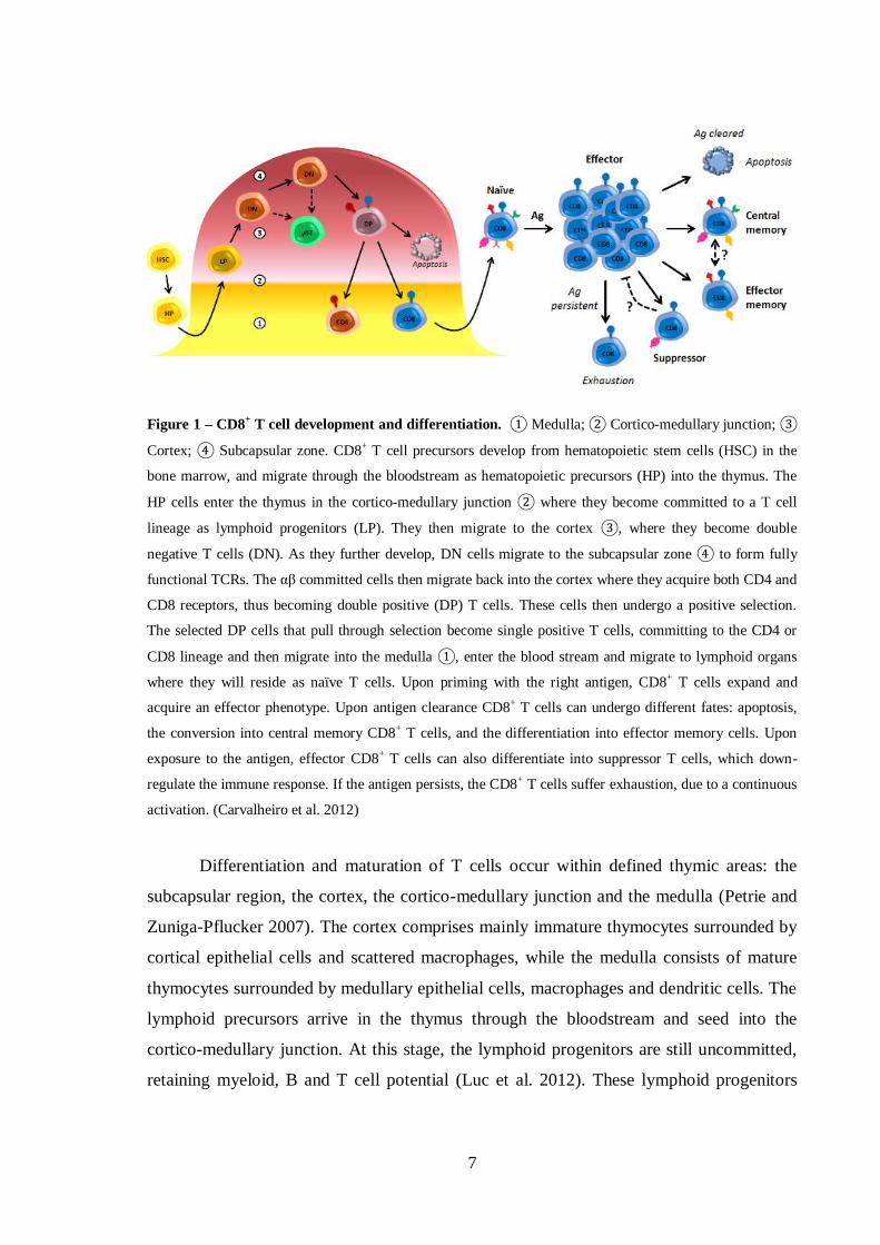
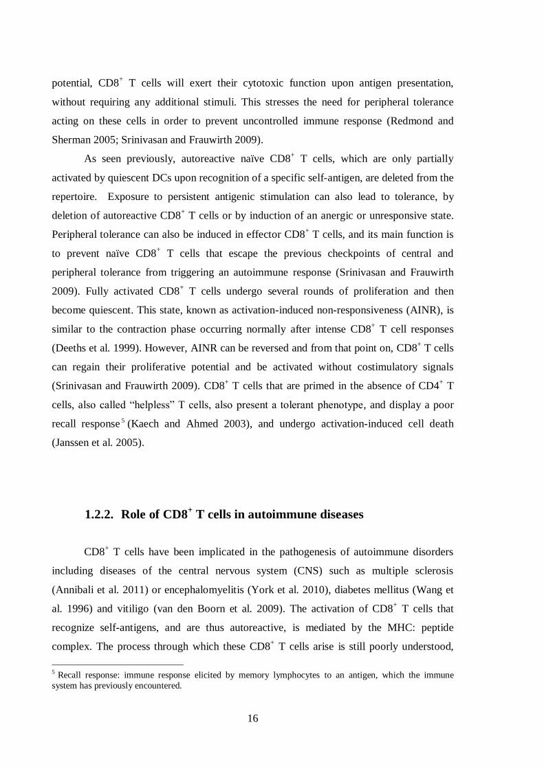
Figure 1 – CD8+ T cell development and differentiation. ① Medulla; ② Cortico-medullary junction; ③
Cortex; ④ Subcapsular zone. CD8+ T cell precursors develop from hematopoietic stem cells (HSC) in the
bone marrow, and migrate through the bloodstream as hematopoietic precursors (HP) into the thymus. The
HP cells enter the thymus in the cortico-medullary junction ② where they become committed to a T cell
lineage as lymphoid progenitors (LP). They then migrate to the cortex ③, where they become double
negative T cells (DN). As they further develop, DN cells migrate to the subcapsular zone ④ to form fully
functional TCRs. The αβ committed cells then migrate back into the cortex where they acquire both CD4 and
CD8 receptors, thus becoming double positive (DP) T cells. These cells then undergo a positive selection.
The selected DP cells that pull through selection become single positive T cells, committing to the CD4 or
CD8 lineage and then migrate into the medulla ①, enter the blood stream and migrate to lymphoid organs
where they will reside as naïve T cells. Upon priming with the right antigen, CD8+ T cells expand and
acquire an effector phenotype. Upon antigen clearance CD8+ T cells can undergo different fates: apoptosis,
the conversion into central memory CD8+ T cells, and the differentiation into effector memory cells. Upon
exposure to the antigen, effector CD8+ T cells can also differentiate into suppressor T cells, which down-
regulate the immune response. If the antigen persists, the CD8+ T cells suffer exhaustion, due to a continuous
activation. (Carvalheiro et al. 2012)
Differentiation and maturation of T cells occur within defined thymic areas: the
subcapsular region, the cortex, the cortico-medullary junction and the medulla (Petrie and
Zuniga-Pflucker 2007). The cortex comprises mainly immature thymocytes surrounded by
cortical epithelial cells and scattered macrophages, while the medulla consists of mature
thymocytes surrounded by medullary epithelial cells, macrophages and dendritic cells. The
lymphoid precursors arrive in the thymus through the bloodstream and seed into the
cortico-medullary junction. At this stage, the lymphoid progenitors are still uncommitted,
retaining myeloid, B and T cell potential (Luc et al. 2012). These lymphoid progenitors

8
then receive signals through the Notch1 receptor which activate specific genes, and induce
T cell lineage determination (Pui et al. 1999). They first evolve into double negative T
cells (CD4-CD8
-), which migrate into the cortical areas where they undergo further
differentiation steps. During their double-negative stage, T cells will also rearrange their β,
γ and δ genes to generate functional TCR chains and thus commit to the major aβ or γδ T
lineages (Burtrum et al. 1996). The main lineage, αβ TCR pathway, leads to the
differentiation into CD4+ or CD8
+ T cells. The γδ lineage leads to the γδ T cells which are
found in mucosae as part of the innate immune response, and may also function as APCs
(Brandes et al. 2009). Differentiation into the αβ or γδ T cells depends on the surface
expression or signaling potential of the γδ TCR complex. A strong signal favors the γδ
lineage development, while a weak γδ signal potentiates the αβ lineage (Hayes et al. 2005).
The αβ-committed lineage of double-negative thymocytes evolves into double positive
CD3+ T cells, as they express both the CD4 and the CD8 surface molecules. These cells are
produced in large numbers, but after positive selection their vast majority undergoes
apoptosis. Cells bearing an αβ TCR complex that recognizes the self-MHC complex with
an intermediate avidity will be positively selected to further differentiate, while their
counterparts will be eliminated (Klein et al. 2009). These selected double-positive
immature T cells then commit to the CD4+ or CD8
+ T cell lineages, and become single-
positive thymocytes. At this point, these semi-mature thymocytes migrate into the medulla
where they undergo negative selection: those harboring TCRs with a high affinity to self-
antigens are eliminated, thus reducing the risk of autoimmune disorders (Klein et al. 2009).
Once in the medulla, the single-positive thymocytes will upregulate the sphingosine-1
phosphate receptor (S1P1) that is required for T cells to leave the thymus (Weinreich and
Hogquist 2008), and further differentiate into other subtypes.
1.1.3.2. CD8+ T cell differentiation and subtypes
CD8+ T cells are currently classified into four subtypes, corresponding to different
levels of differentiation, activation status and cytokine production: Naïve, Effector, Central
memory and Effector memory (Figure 1).

9
Table 1 - CD8+ T cell phenotypes
Naïve Effector Effector
memory
Central
memory
CCR7 +++ - +/- +/-
CD27 +++ - +++ +++
CD28 High Low Low High
CD45RA +++ -/+ - +/-
CD45RO - - +++ +++
CD62L +++ - - +++
Naïve CD8+ T cells still have not encountered their cognate antigen, and thus have
not been primed. They are usually found in the peripheral blood and lymphatic tissues
(Kaech and Ahmed 2001). The central memory subtype is already endowed to a specific
antigen whose presence will induce a strong proliferative response, as well as the
production of a variety of cytokines. Effector CD8+ T cells have proliferative and cytotoxic
properties. They can induce death of infected cells by cytolysis, through the secretion of
cytolytic proteins such as perforin and granzymes. Effector memory CD8+ T cells have
intermediate properties, presenting a lower ability to induce cytotoxic responses than
effector cells, and a much higher capacity to produce cytokines than the memory subtype
(Tomiyama et al. 2002).
Cell surface markers offer an expedite way to distinguish these CD8+ T cell
subtypes. This is based in the presence or absence of co-stimulatory (CD27, CD28,
CD45RA) and adhesion (CD62L) molecules and the chemokine receptor CCR7 (Kaech et
al. 2003). Naïve CD8+ T cells are characterized by the presence of CD27, CD28hi,
CD45RA, CD62L and CCR7. Effector cells express low levels of CD28 and are negative
for all other cell surface markers, while central memory cells can lose the expression of
CD45RA along with CCR7. The effector memory subtype is characterized by the absence
of CD62L and CCR7, the expression of CD28low, while the expression of CD45RA may
vary (Tomiyama et al. 2004) (Figure 1 and Table 1).
Our current understanding indicates that upon antigen encounter, naïve CD8+ T
cells differentiate into effector cells and undergo clonal expansion. Once the antigen is
cleared, 90-95% of all effector cells undergo apoptosis, while the remaining ones
differentiate into central memory CD8+ T cells, thus entering a resting (but vigilant) state.
The effector memory subtype is thought to represent an intermediate state occurring upon
10
the re-encounter of the antigen, when central memory CD8+ T cells gradually differentiate
towards an effector phenotype (Tomiyama et al. 2002).
CD8+ effector T cells are, therefore, characterized by their cytotoxic behavior (thus
the abbreviation Tc) through perforin, granzyme and Fas pathways. Several subtypes have
been identified based on cytokine production, these include the Tc1 subset (characterized
by the production of IFN-γ and not IL-4 and IL-5), and the Tc2 subset (secreting IL-4 and
IL-5 but not IFN-γ) (Mosmann et al. 1997). Both types can induce an inflammatory
response, with Tc1 and Tc2 inducing delayed-type hypersensitivity upon injection of Tc1
and Tc2 allospecific cells into mice bearing the target antigen (Li et al. 1997). Even though
both cell subtypes can induce inflammation, the Tc2-bearing mice had a higher eosinophil
infiltration, thus indicating that these may exert inflammation through a secondary pathway
by recruiting effector cells into the inflammatory site. The study of Tc1 and Tc2 functional
phenotypes also indicates that these cells can induce inflammation by activating CD4+
effector T cells, with Tc1 and Tc2 inducing a Th1 (cellular) and Th2 (humoral) response,
respectively (Vukmanovic-Stejic et al. 2000).
More recently, other functional subtypes have been identified. Special attention has
been devoted to the Tc17, characterized by the production of IL-17 and arising from the
same precursor as other functional subsets of CD8+ T cells (Kondo et al. 2009). Tc17 cells
are typically proinflammatory non-cytotoxic CD8+ T cells that express few or no cytotoxic
granules, and thus typically do not secrete granzyme B and perforin, although some subsets
can produce IFN-γ (Tajima et al. 2011). These cells seem to enhance inflammation in
various diseases, such as SLE (Henriques et al. 2010), immune thrombocytopenia (Hu et
al. 2011) and allergy-induced lung inflammation (Tang et al. 2012). Tc17 cells have also
been shown to promote immunity against infections, by Vaccinia (Yeh et al. 2010) and
Influenza viruses (Hamada et al. 2009), by promoting a proinflammatory response. A
subset of CD8+ T cells, is endowed with suppressor/regulatory capabilities, mediated by
IL-10 and TGF-β (Wang and Alexander 2009). These cells arise upon challenge by their
cognate antigen, and control inflammation by down-regulating the immune response by
effector T cells (Hu et al. 2004). These cells and their role in autoimmunity will be further
discussed.
11
1.1.3.3. Cytotoxic immune response
CD8+ T cells recognize pathogen peptides presented by MHC class I complexes on
the surface of APCs. During the first weeks after an acute infection with a pathogen both
the naïve and the central memory CD8+ T cells undergo activation and proliferation while
acquiring an effector phenotype. This is reflected by a down-regulation of the expression
of CD62L on the cell surface, accompanied by the production of granzymes and perforin,
as well as IFN-γ and TNF-α (Wherry and Ahmed 2004). Effector CD8+ T lymphocytes
cause the death of infected cells either by direct lysis, or by inducing apoptosis through the
activation of the Fas receptor (Barry and Bleackley 2002; Wong and Pamer 2003). After
the clearance of the infected cells, 90–95% of the effector cells undergo apoptosis, while
the surviving portion differentiates into a memory phenotype, regaining the CD62L
expression on their surface. This memory CD8+ T cell pool can later be reactivated,
proliferate and regain effector cytotoxic properties upon a re-encounter with the same
antigen.
Some infectious agents are readily eliminated, corresponding to acute self-limited
clinical manifestations. Chronic or latent infection-causing agents, such as viruses of the
herpes family, remain in the host indefinitely. In such cases, CD8+ T cells are permanently
stimulated and the cytotoxic response remains active, creating a persistent or even
expanding inflammatory response (Wong and Pamer 2003). In some patients, this chronic
state eventually leads to the exhaustion of CD8+ T cells: they gradually lose the ability to
produce cytolytic enzymes and even to proliferate, leading to a decline of the CD8+ T cell
population (Wherry et al. 2003). The exhaustion of CD8+ T cells is accelerated in the
presence of decreased numbers of CD4+ T cells, as they have an important role in
supporting the CD8+ T cell response (Matloubian et al. 1994).
CD8+ T cells exert important functions in the absence of infection: they are key
mediators in the clearance of some target cells, such as graft and tumor cells. In fact, CD8+
T cells have a crucial role in allograft rejection in mouse models (Tomita et al. 1990;
Yoshimura et al. 2000; Halamay et al. 2002), contributing to an accelerated immune
response (Yoshimura et al. 1998). Both Tc1 and Tc2 subsets can induce cardiac allograft
rejection by themselves without CD4+ T cell help. Tc1 cells are important in the early
rejection response, while the Tc2 subtype is involved in the recruitment of other effector
12
cells (Delfs et al. 2001). The cytotoxic behavior of CD8+ T cells is also involved in tumor
immunity, especially through the Tc1 subset (Kemp and Ronchese 2001).
1.1.3.4. Suppressor immune response
The suppressor T cells were initially described in the early 1970s, by Gershon and
colleagues (Gershon et al. 1972), along with classical cytotoxic T cells, as two cell subsets
with opposing roles in disease. Even though interest in CD8+ suppressor T cells faded with
time, they have regained attention in the last decade, in particular due to their possible role
in autoimmune disorders and antitumor activity (Niederkorn 2008).
As we have seen previously, the most widely known type of regulatory T cells is
CD4+CD25
+, commonly addressed as Tregs, and constitutes a distinct lineage of CD4
+ T
cells that arises in the thymus. They function as inflammatory response inhibitors and are
characterized by the production of IL-10 and TGF-β (Huang et al. 2005) or expression of
the transcription factor Foxp3 (Fontenot et al. 2003; Hori et al. 2003), and the loss of their
suppressive function is related to the onset of inflammatory diseases such as SLE (Sawla et
al. 2012). However, Kessel and colleagues have recently demonstrated that Bregs, that are
B cells that express high levels of CD25 on their surface and secrete IL-10 and TGF-β,
induce the production of Foxp3 by Tregs, thus contributing to the inhibition of
inflammatory responses (Kessel et al. 2012).
The CD8+ regulatory or suppressor T cells, commonly called Tcregs or Ts cells, are
less known, but behave in a similar manner to their CD4+CD25
+ counterparts (Cosmi et al.
2003). The most extensively analyzed Ts cells are the murine CD8+ expressing the β chain
of the IL-2/IL-15 receptor (CD122), which have a role in immunity through the production
and release of the anti-inflammatory cytokine IL-10 (Rifa'i et al. 2008). The adoptive
transfer of CD8+CD122
+ Ts cells into mice with established experimental autoimmune
encephalomyelitis (EAE) leads to an amelioration of the disease (Lee et al. 2008). CD122-
deficient mice are a model for autoimmune disease and are characterized by a high number
of abnormally activated T cells. The adoptive transfer of CD8+CD122
+ Ts cells into
CD122-deficient neonates fully prevents the development of these T cells, thus
maintaining T cell homeostasis (Rifa'i et al. 2004). Recently, the CD8+CXCR3
+ Ts cells

13
have been proposed as the human counterpart for the murine CD8+CD122
+ Ts cells, as
they have been shown to have a similar behavior in vivo and in vitro (Shi et al. 2009). CD8
suppressor T cells are thought to be involved in the onset of autoimmune disorders, such as
fibrotic disease, showing a lower suppressive activity (Fenoglio et al. 2012).
1.2. Autoimmune diseases
The immune system consists of an army of cellular and molecular elements whose
core function resides in protecting the body against harm induced by foreign elements. In
normal conditions, the immune system is “self-tolerant”, that is, it is unable to react against
“self” molecules, and thus does not react against endogenous components of the body.
However, when “self-tolerance” is lost, the immune system reacts against the body’s own
constituents, and this process may eventually result in autoimmune disease. Autoimmunity,
which was first described by Paul Ehrlich at the beginning of the 20th century as “horror
autotoxicus” (Murphy 2011), can, therefore, be defined as the result of a sustained immune
response directed against structures of the self, causing tissue damage (Bolon 2012).
Healthy individuals possess circulating, naturally occurring, auto-antibodies which
recognize self-antigens (Elkon and Casali 2008). Their presence indicates that under
normal physiological conditions these natural auto-antibodies act as house-keepers,
removing the debris resulting from natural cellular and tissue breakdown. Only when
autoimmune responses became uncontrolled and lead to exacerbated tissue damage or
symptoms are we in the presence of autoimmune disease.
Autoimmune diseases collectively affect 5% of the population in Western countries
(Jacobson et al. 1997) and they may affect virtually every organ and tissue in the human
body. Their etiology is essentially unknown, although it is believed to reside in the
interplay between both genetic and environmental factors. However, understanding what
triggers immune diseases has proven a difficult challenge, namely when it comes to
understand why so many healthy individuals present autoimmune processes but only a few
will develop clinically significant autoimmune disease (Sener and Afsar 2012).

14
1.2.1. Self-tolerance and its loss
Central tolerance is the process by which T and B cells are rendered unresponsive
to self-peptides during the maturation process in the thymus and bone marrow respectively.
This is the first checkpoint in the acquisition of tolerance to autoantigens.
As explained above, T cell development and maturation (CD4+ and CD8
+ T cells) is
based on a mechanism through which thymocytes are exposed to self-peptides bound to the
MHC complex. This process ultimately leads to the elimination of T cells that react to self-
antigens. However, some autoreactive T cells, with low affinity to these antigens, escape
the negative selection process and enter the blood stream (Klein et al. 2009).
The central tolerance to self-antigens during the maturation of B cells occurs in the
bone marrow. Immature B cells express a BCR molecule on their surface and will undergo
a negative selection process that determines whether the immature B cell will continue its
maturation. This mechanism can lead to the elimination of as much as 50 to 75% of
immature B cells at this stage. Again, some B cells with low autoreactivity levels escape
the negative selection and differentiate into mature B cells (Pelanda and Torres 2012).
In healthy individuals, other mechanisms in the periphery contribute to the active
removal of self-reactive T and B cells. This is done either by directly eliminating the
autoreactive T cells or through regulatory processes that render these cells inactive.
Peripheral tolerance can be obtained by three different processes: clonal ignorance, death
by deletion and induction of functional unresponsiveness (Srinivasan and Frauwirth 2009;
Mueller 2010). Self-reactive cells that escape the negative selection process but are
endowed with low affinity to self-antigens are the most likely to experience clonal
ignorance: because they have an avidity for the self-peptides that is generally lower than
that required to induce peripheral T cell activation, they are “ignored”. Clonal ignorance
may also be achieved when the cognate self-antigen is restricted to an immune privileged4
site. Under normal conditions, naïve T cells are presented their cognate antigen by
dendritic cells (DCs), in lymph nodes. In order to completely activate a naïve T cell, two
signals are required: the activation signal produced by the interaction of MHC-Ag (cognate
antigen within an MHC molecule) with the TCR, and the simultaneous costimulation
4 Immune privilege: Condition in which selected immune responses are suppressed or excluded in certain
organs. Certain sites in the human body, such as the cornea, tolerate the introduction of antigens without
triggering an immune response. The brain, the placenta and the cornea are all immune privileged sites.
15
signal sent by the DC’s molecules to the naïve T cells. Self-antigens are usually presented
by quiescent DCs, which have a reduced number of costimulatory molecules on their
surface, thus failing to produce the second stimulus required for a full T cell activation –
they are, thus, “ignored”. Partially activated naïve T cells are found to be tolerant. These
cells fail to differentiate into fully functional effector T cells, and will ultimately be
rendered unresponsive or eliminated from the T cell repertoire (Redmond and Sherman
2005; Srinivasan and Frauwirth 2009; Mueller 2010).
Functional unresponsiveness and deletion of autoreactive T cells occur upon their
partial activation due to the absence of costimulatory signals from APCs. Both confer
different forms of tolerance, but the mechanisms activating one pathway or the other are
still largely unknown. However, antigenic persistence has been shown to be an important
factor leading to tolerance by deletion (Redmond et al. 2003; Srinivasan and Frauwirth
2009; Nurieva et al. 2011), and is dose-dependent, with high doses of antigen leading to an
incomplete deletion, and low doses leading to complete deletion of the Ag-specific T cells
(Srinivasan and Frauwirth 2009).
Functional unresponsiveness, also called anergy, is a state in which a T cell that has
been exposed to an antigen becomes refractory to any further stimulatory signals. Anergic
cells are characterized by the lack of proliferation and IL-2 production, an irregular
effector function, a defective MAPK signaling pathway, a reduced intracellular calcium
mobilization and a decreased tyrosine phosphorylation. The exposure of T cells to high
doses of antigen can result in the functional unresponsiveness of these cells (Srinivasan
and Frauwirth 2009).
Tolerance breakdown occurs when mechanisms of central and/or peripheral
tolerance do not function properly, thus breaking the cellular homeostasis and triggering an
autoimmune disease.
1.2.1.1. Peripheral tolerance in CD8+ T cells
The establishment of peripheral tolerance in CD8+ T cells is particularly important,
as nearly every cell type can present these cells to their cognate antigen due to the presence
of MHC class I on all nucleated cells. Upon maturation and acquisition of cytotoxic
16
potential, CD8+ T cells will exert their cytotoxic function upon antigen presentation,
without requiring any additional stimuli. This stresses the need for peripheral tolerance
acting on these cells in order to prevent uncontrolled immune response (Redmond and
Sherman 2005; Srinivasan and Frauwirth 2009).
As seen previously, autoreactive naïve CD8+ T cells, which are only partially
activated by quiescent DCs upon recognition of a specific self-antigen, are deleted from the
repertoire. Exposure to persistent antigenic stimulation can also lead to tolerance, by
deletion of autoreactive CD8+ T cells or by induction of an anergic or unresponsive state.
Peripheral tolerance can also be induced in effector CD8+ T cells, and its main function is
to prevent naïve CD8+ T cells that escape the previous checkpoints of central and
peripheral tolerance from triggering an autoimmune response (Srinivasan and Frauwirth
2009). Fully activated CD8+ T cells undergo several rounds of proliferation and then
become quiescent. This state, known as activation-induced non-responsiveness (AINR), is
similar to the contraction phase occurring normally after intense CD8+ T cell responses
(Deeths et al. 1999). However, AINR can be reversed and from that point on, CD8+ T cells
can regain their proliferative potential and be activated without costimulatory signals
(Srinivasan and Frauwirth 2009). CD8+ T cells that are primed in the absence of CD4
+ T
cells, also called “helpless” T cells, also present a tolerant phenotype, and display a poor
recall response 5 (Kaech and Ahmed 2003), and undergo activation-induced cell death
(Janssen et al. 2005).
1.2.2. Role of CD8+ T cells in autoimmune diseases
CD8+ T cells have been implicated in the pathogenesis of autoimmune disorders
including diseases of the central nervous system (CNS) such as multiple sclerosis
(Annibali et al. 2011) or encephalomyelitis (York et al. 2010), diabetes mellitus (Wang et
al. 1996) and vitiligo (van den Boorn et al. 2009). The activation of CD8+ T cells that
recognize self-antigens, and are thus autoreactive, is mediated by the MHC: peptide
complex. The process through which these CD8+ T cells arise is still poorly understood,
5 Recall response: immune response elicited by memory lymphocytes to an antigen, which the immune
system has previously encountered.

17
even though these cells have been shown to have a preponderant role in autoimmune
disorders (Liblau et al. 2002).
In multiple sclerosis (MS) lesions in the brain, infiltrating CD8+ T cells were shown
to outnumber CD4+ T cells and to undergo clonal expansion locally (Babbe et al. 2000).
CD8+ T cells accumulation and clonal expansion has also been described in the
cerebrospinal fluid (CSF) and peripheral blood of these patients (Jacobsen et al. 2002). It
has also been demonstrated that T cells from MS patients frequently displayed resistance to
Fas-induced apoptosis, thus indicating that the cell death mechanism was altered in these
cells, making them prone to accumulation (Comi et al. 2012). These observations suggest
that CD8+ T cells are exposed to their cognate antigen in peripheral blood, CSF and MS
lesions in the brain. Recent data also indicate that MS patients have a higher number of
CNS-reactive CD8+ T cells in circulation than healthy individuals (Zang et al. 2004).
Studies with animal models of EAE have yielded controversial results, with CD8+ deficient
mice presenting a lower mortality but higher incidence of relapses (Jiang et al. 1992; Koh
et al. 1992; Kuchroo et al. 2002; Jiang et al. 2003; Montero et al. 2004; Lee et al. 2008;
York et al. 2010).
In the non-obese diabetic (NOD) mouse, an animal model for type I diabetes
mellitus, autoreactive CD8+ T cells are involved in the destruction of pancreatic β cells,
hence playing a key role in the pathogenesis of insulitis (Pang et al. 2009). Concurringly,
NOD mice treated with anti-CD8 antibody failed to initiate the disease (Wang et al. 1996).
Studies on a skin explant model of vitiligo demonstrated that perilesional CD8+ T
cells were capable of developing an autoimmune reaction against autologous skin explants,
efficiently lysing melanocytes, and inducing keratinocyte apoptosis (van den Boorn et al.
2009).
There is, therefore, a growing body of data suggesting that CD8+ T cells may be
involved in autoimmune diseases. This deleterious influence may be due to an excessive or
autoreactive cytotoxic activity, as suggested in the animal models of type 1 diabetes (Pang
et al. 2009) and EAE (Sun et al. 2001). Conversely, one may hypothesize that the disease
process may be enhanced by a reduced or deficient suppressor role by CD8+ T cells.

18
1.3. Rheumatoid arthritis
1.3.1. General perspective of the disease
Rheumatoid arthritis (RA) is a systemic and chronic autoimmune disease,
associated with a profound negative impact on quality of life, increased mortality and high
socioeconomic costs (McInnes and Schett 2011). RA is biologically mainly characterized
by synovial inflammation leading to chronic persistent pain, joint destruction and
associated deformity, systemic complications and progressive disability. Other organs and
tissues can also be affected by the inflammatory process. It affects around 1% of the
population in industrialized countries, being three times more frequent in women than in
men, with a peak incidence between 40 and 60 years of age (Scott and Steer 2007;
Klareskog et al. 2009).
The cause for RA is still unknown, but several factors (genetic and environmental)
play a role in the onset and course of the disease. A study in a cohort of twins estimated the
contribution of genetic factors to the disease to be about 50%, with the remainder
comprising environmental factors and chance (MacGregor et al. 2000; Klareskog et al.
2009). According to the current paradigm, in individuals that bear disease susceptibility
genes, specific environment factors may potentiate an immune reaction that will ultimately
lead to the production of autoantibodies. Later on in life, other events, such as infection or
trauma can contribute to further development of the disease pathogenesis, eventually
translating into joint inflammation. As the chronicity of the disease settles, patients will
display additional characteristics of the disease, such as joint deformity and systemic
manifestations associated with increased comorbidities (Klareskog et al. 2009).
The chronic inflammatory process is held as directly responsible for the destruction
of cartilage and bone However, the triggers and mechanisms involved in the origin of the
disease process remain vastly elusive (Williams et al. 2000; McInnes and Schett 2011).
Research over the past few decades has elucidated some of the mechanisms responsible for
the maintenance of the inflammatory process and its destructive ability. These efforts have
highlighted the extraordinary complexity of this disease. Although our current
understanding is far from complete, recent research has led to the development of
increasingly effective drugs that have gradually improved the outcome of the disease.

19
Among these new medications, biological agents targeting specific mediators of the
immune response are paramount.
1.3.2. Rheumatoid arthritis classification and clinical features
RA presents a broad spectrum of manifestations. The predominant symptoms are
pain, morning stiffness and swelling preferentially affecting the peripheral joints, in a
strikingly symmetrical fashion. The natural course of the disease is typically composed of
flares and partial remissions. Severity can be quite variable between individual patients,
ranging from mild symptoms without significant disability to a persistently active,
progressively crippling condition.
Table 2 - The 1987 revised classification criteria for Rheumatoid Arthritis (Arnett et al. 1988).
Criterion Definition
1. Morning stiffness Morning stiffness in and around the joints, lasting at least 1 hour before
maximal improvement
2. Arthritis of 3 or more joint areas
At least 3 joint areas simultaneously have had soft tissue swelling or fluid (not bony overgrowth alone) observed by a physician. The 14 possible areas
are right or left PIP, MCP, wrist, elbow, knee, ankle, and MTP joints
3. Arthritis of hand joints At least 1 area swollen (as defined above) in a wrist, MCP, or PIP joint
4. Symmetric arthritis Simultaneous involvement of the same joint areas (as defined in 2) on both
sides of the body (bilateral involvement of PIPs, MCPs, or MTPs is
acceptable without absolute symmetry)
5. Rheumatoid nodules Subcutaneous nodules, over bony prominences, or extensor surfaces, or in
juxtaarticular regions, observed by a physician
6. Serum rheumatoid
factor
Demonstration of abnormal amounts of serum rheumatoid factor by any
method for which the result has been positive in <5% of normal control
subjects
7. Radiographic changes Radiographic changes typical of rheumatoid arthritis on posteroanterior hand
and wrist radiographs, which must include erosions or unequivocal bone
decalcification localized in or most marked adjacent to the involved joints
(osteoarthritis changes alone do not qualify)
* For classification purposes, a patient shall be said to have rheumatoid arthritis if he/she has satisfied at
least 4 of these 7 criteria. Criteria 1 through 4 must have been present for at least 6 weeks. Patients with 2
clinical diagnoses are not excluded. Designation as classic, definite, or probable rheumatoid arthritis is
not to be made.
MCPs = metacarpophalangeal joints; MTPs = metatarsophalangeal joints; PIPs = proximal
interphalangeal joints
20
Joint destruction is common in RA: radiographic evidence of bone erosions in the
periphery of joints, at the site of synovium anchorage in bone, is present in up to 70% of
patients within the first two years of the disease. More refined techniques, such as
magnetic resonance imaging (MRI) may demonstrate the presence of changes in RA joints
as early as 4 months after the onset of the disease, including not only synovial hypertrophy
and bone edema, but also early bone erosive changes (McQueen et al. 1998; McGonagle et
al. 1999). Furthermore, the analysis of apparently unaffected knee joints from untreated
early RA patients indicated that there were significant histological changes, as well as a
subclinical form of synovitis in these joints (Soden et al. 1989), which proves that the lack
of symptoms does not correlate with the clinical progression of the disease.
The analysis of the clinical, biological and radiological course of RA have allowed
the identification of a series of prognostic factors for progressive joint destruction that
generally correspond to a poorer outcome, and are used to support the selection of therapy.
Current standards recommended that effective medication be started as early as possible, to
avoid irreversible joint destruction, and adapted to maintain rigorous remission, i.e. the
absence of any clinical and biological signs of inflammation.
The first criteria for the classification of RA were established in 1958 and revised in
1987 by the American Rheumatism Association (later renamed American College of
Rheumatology - ACR) (Arnett et al. 1988) and are presented in Table 2. To be classified as
having RA according to these criteria, the patients must present at least 4 of the 7 criteria.
In 2010, the ACR and EULAR (European League Against Rheumatism) revised
these criteria (Aletaha et al. 2010) (Table 3), with the stated aim of allowing earlier
diagnosis and thus, more timely and effective therapy. The new classification criteria of
RA are mainly based on clinical features: the presence of synovitis in at least one joint
without a better explanation, and a minimum total score of 6 in the 4 following categories:
number and site of involved joints (range: 0-5), serologic abnormalities (range: 0-3),
elevated acute phase response (range: 0-1) and symptom duration (range: 0-1) (Aletaha et
al. 2010).

21
Table 3 - The 2010 ACR/EULAR classification criteria for Rheumatoid Arthritis. (Aletaha et al. 2010)
Score
Target population (Who should be tested?): Patients who:
1) have at least 1 joint with definite clinical synovitis (swelling)*
2) with the synovitis not better explained by another disease†
Classification criteria for RA (score-based algorithm: add score of categories A-D; a score of ≥
6/10 is needed for classification of a patient as having definite RA) ‡
A. Joint involvement §
1 large joint ¶ 0
2 - 10 large joints 1
1 - 3 small joints (with or without involvement of large joints) # 2
4 - 10 small joints (with or without involvement of large joints) 3
> 10 joints (at least 1 small joint)** 5
B. Serology (at least 1 test result is needed for classification) ††
Negative RF and negative ACPA 0
Low-positive RF or low-positive ACPA 2
High-positive RF or high-positive ACPA 3
C. Acute-phase reactants (at least 1 test result is needed for classification) ‡‡
Normal CRP and normal ESR 0
Abnormal CRP or abnormal ESR 1
D. Duration of symptoms §§
< 6 weeks 0
> 6 weeks 1
* The criteria are aimed at the classification of newly presenting patients. In addition, patients with
erosive disease typical of rheumatoid arthritis (RA) with a history compatible with prior fulfillment of
the 2010 criteria should be classified as having RA. Patients with longstanding disease, including those
whose disease is inactive (with or without treatment) who, based on retrospectively available data, have
previously fulfilled the 2010 criteria should be classified as having RA.
† Differential diagnoses vary among patients with different presentations, but may include conditions
such as systemic lupus erythematosus, psoriatic arthritis, and gout. If it is unclear about the relevant
differential diagnoses to consider, an expert rheumatologist should be consulted.
‡ Although patients with a score < 6/10 are not classifiable as having RA, their status can be reassessed
and the criteria might be fulfilled cumulatively over time.
§ Joint involvement refers to any swollen or tender joint on examination, which may be confirmed by
imaging evidence of synovitis. Distal interphalangeal joints, first carpometacarpal joints, and first
metatarsophalangeal joints are excluded from assessment. Categories of joint distribution are classified
according to the location and number of involved joints, with placement into the highest category
possible based in the pattern of joint involvement.
¶ "Large joints" refers to shoulders, elbows, hips, knees, and ankles.
# "Small joints" refers to the metacarpophalangeal joints, proximal interphalangeal joints, second
through fifth metatarsophalangeal joints, thumb interphalangeal joints, and wrists.
**In this category, at least 1 of the involved joints must be a small joint; the other joints can include any
combination of large and additional small joints, as well as other joints not specifically listed elsewhere
(e.g., temporomandibular, acromioclavicular, sternoclavicular, etc.).
†† Negative refers to IU values that are less than or equal to the upper limit of normal (ULN) for the
laboratory and assay. Where rheumatoid factor (RF) information is only available as positive or negative,
a positive result should be scored as low-positive for RF. ACPA = anti-citrullinated protein antibody.
‡‡ Normal/abnormal is determined by local laboratory standards. CRP = C-reactive protein. ESR =
erythrocyte sedimentation rate.
§§ Duration of symptoms refers to patient serf-report of the duration of signs or symptoms of synovitis
(e.g., pain, swelling, tenderness) of joints that are clinically involved at the time of assessment,
regardless of treatment status.
22
1.3.3. Clinically relevant autoantibodies in RA
RA is consensually subdivided in two groups based on the presence or absence of
anti-citrullinated protein antibodies (ACPAs) and/or the rheumatoid factor (RF) (van der
Helm-van Mil et al. 2007). The frequency of ACPAs in RA patients is around 70-90%,
while RF is generally detected in up to 80% of RA cases (Song and Kang 2010). They
frequently coexist in the same patient, but not always. Both markers are important for
diagnosis and prognosis: their presence, especially in high concentrations, is associated
with more aggressive disease and poorer outcomes. They are routinely tested in RA
patients (Aletaha et al. 2010). However, RFs are not specific of RA patients, as they can
be observed in other autoimmune disorders such as SLE and Sjögren’s syndrome, as well
as in chronic infections and in old age (Song and Kang 2010).
Protein citrullination is a post-translational modification that occurs when the
amino acid arginine is converted to citrulline, thus increasing the morphological and
functional diversity of the proteome. Citrullination of proteins can alter the original
function of the molecule. Furthermore, since citrulline is not a natural amino acid in the
original protein structure, this process can trigger an immune response (Alivernini et al.
2008). In RA, citrullination can occur in the synovium (Chang et al. 2005; Matsuo et al.
2006) but also in extra-articular sites such as the oral cavity, the gut and the lung. It is not
specific to RA, but rather an inflammation-dependent process (Makrygiannakis et al.
2006). Four citrullinated proteins which role in RA is well established are fibrinogen,
collagen II, α-enolase and vimentin (Wegner et al. 2010).
ACPAs develop preferentially in persons baring the genetic susceptibility genes to
RA, namely the so-called shared epitope alleles (Huizinga et al. 2005). ACPAs have been
demonstrated in the circulation long before the onset of the disease (Rantapaa-Dahlqvist et
al. 2003; Nielen et al. 2004). ACPA titers rise progressively until the onset of the disease
(Chibnik et al. 2009), as active citrullination increases in the inflamed rheumatoid
synovium (Kinloch et al. 2008). The presence of ACPA is thus a predictor of the
progression of early undifferentiated arthritis (UA) into RA. Furthermore, ACPA-positive
patients have a higher risk of developing aggressive disease and its extra-articular
manifestations (Luban and Li 2010).

23
The rheumatoid factor (RF), also an autoantibody: it binds to the Fc part of IgG.
However, it can also react with a variety of self-antigens, such as nucleosomes, denatured
DNA and histones. Even though it is commonly referred to as IgM-RF, other Ig subclasses
can display RF activity, such as IgA, IgG, IgD and IgE (Moore and Dorner 1993). RF is
produced in RA by B cells in lymphoid follicles and germinal center-like structures that
develop in the inflamed synovium (Song and Kang 2010). The functions of RFs are to
enhance the clearance of immune complexes6 (Van Snick et al. 1978), to help B cells take
up immune complexes and further present the antigens to T cells (Tighe et al. 1993), and to
facilitate the fixation of the complement to IgG-containing immune complexes (Brown et
al. 1982; Sato et al. 1995; Song and Kang 2010). However, there is no clear evidence of
whether RF production triggers the disease, or is triggered by the disease. Moreover, RF is
not specific to RA, as it is also found in Sjögren’s syndrome and in some types of infection
(Dorner et al. 2004).
1.3.4. Treatment of RA
There are several indicators of a poor prognosis in the early onset of the disease,
such as the early involvement of several joints, high erythrocyte sedimentation rate (ESR)
or C-reactive protein levels (van der Heijde et al. 1988; Scott 2000). The seropositivity for
RF (Bukhari et al. 2002) and ACPAs (De Rycke et al. 2004) are also correlated with a
faster radiographic progression and extra-articular manifestations of the disease.
Interestingly, the presence of specific alleles may also influence the outcome of RA.
Even though RA is an incurable chronic systemic disease, the diagnosis and
effective treatment of RA in its early phases increases the chance of achieving a long-term
remission state with reduced systemic inflammation, leading to an overall increased quality
of life and preservation of structural integrity and function in the long-term. It is of the
utmost importance that diagnosis is made early and immediately followed by effective
treatment, targeted to achieve consistent remission.
There are numerous treatment options available to treat RA patients, and thus
reduce the ongoing inflammation and progression of the disease (Figure 2). There are three
6 Immune Complex: molecular cluster formed by the combination of an antigen and an antibody (mostly
IgG) that tend to accumulate in the body and are associated with various pathological conditions.

24
main types of therapies available to treat RA: disease modifying anti-rheumatic drugs
(DMARDs), which can be synthetic or biological drugs, nonsteroidal anti-inflammatory
drugs (NSAIDs) and corticosteroids (Gaffo et al. 2006; Kumar and Banik 2013).
NSAIDs are analgesics and antipyretics, and are thus used in the management of
pain and inflammation, but have little to no effect on the course of the disease (Cush et al.
1990; Cush et al. 1990). They can also have deleterious side-effects, such as an increased
risk of cardiovascular disease (Atzeni et al. 2010; Lindhardsen et al. 2013).
Corticosteroids are potent immune suppressors that are used in the treatment of RA.
They are currently viewed as disease modifying agents that enhance the effects of
DMARDs without any major adverse effects (Yazici 2012; Caporali et al. 2013). These
drugs have been shown to decrease radiographic progression of the disease (Hickling et al.
1998; van Everdingen et al. 2002; Kirwan et al. 2007; Malysheva and Baerwald 2011;
Yazici 2012).
Figure 2 – The main classes of treatment available for RA (Costenbader and Kountz 2007).
The administration of DMARDs to RA patients leads to the suppression of the
ongoing inflammatory process, and can be particularly beneficial in the early treatment of
RA (Rath and Rubbert 2010). The aggressive treatment of early RA usually involves
25
conventional non-biologic DMARDs, such as methotrexate (MTX), leflunomide,
sulfasalazine and hydroxychloroquine and lead to the decrease of inflammation and joint
erosion (Kumar and Banik 2013). MTX is the most commonly prescribed DMARD for the
treatment of RA (Pincus et al. 2003), and is preferably used alone in the treatment of
DMARD naïve patients (Katchamart et al. 2009), but combinations of other DMARDs
with MTX have also been proven effective (O'Dell et al. 2002; Choy et al. 2005; Dale et al.
2007; Braun 2011; Kumar and Banik 2013).
The treatment with biological DMARDs is only initiated in patients in whom the
MTX-based therapy has been proven ineffective. Biologic DMARDs are monoclonal
antibodies that target a specific protein that contributes to the development of the disease
and block its further action. There are currently four types of biologics used in the
treatment of RA: TNF-α inhibitors (infliximab, etanercept, adalimumab, certolizumab and
golimumab) (Taylor and Feldmann 2009), IL-1 inhibitor (anakinra), IL-6 inhibitor
(tocilizumab), B-cell inhibitor (rituximab) and the inhibitor of the T-cell costimulation
(abatacept) (Scherer and Burmester 2009).
The last decade witnessed progress in the treatment of RA (van Roon et al. 1997;
Pincus et al. 2003; Visser and van der Heijde 2009). This change, which has been named
“The Biologic Revolution” was made possible by the remarkable progress operated in the
understanding of pathogenesis of the disease. This opened the opportunity for the
development of new agents specifically designed to target relevant biological mediators.
Extraordinarily, the optimal use of DMARDs, in particular the anchor DMARD
methotrexate (MTX), and the availability of new biologic agents, have dramatically
enhanced the success of RA management.
1.3.5. Environmental and genetic risk factors
RA is considered a complex disease whose origin and pathogenesis involves an
intricate interaction between genetic susceptibility and environmental factors (Figure 3).
Several genes have been related to the development of the disease, especially upon
exposure to environmental risk factors. The environmental factor that is most correlated to
the development of the disease in genetically susceptible individuals is smoking. This is
26
supported by a study with monozygotic twins who were discordant for RA and smoking.
The twins who smoked developed the disease in 12 cases out of 13 pairs. This indicates
that when the genetic background is kept constant, environmental factors can be pivotal in
triggering the disease (Silman et al. 1996). Also, smoking has been correlated with the
presence of ACPAs, and with a more severe disease progression (Lundstrom et al. 2009;
Morgan et al. 2009; Lundberg et al. 2013; de Rooy et al. 2014). Interestingly, ACPAs can
be present several years before the disease onset (Farid et al. 2013), and were also found in
unaffected first-degree relatives of RA patients. A higher diversity of ACPAs emerges as
the process evolves to arthralgia and overt arthritis (Smolik et al. 2013; Young et al. 2013),
thus indicating that ACPAs play an important role in the development of the disease.
Infectious agents have long been suspected to be potential players in the etiology of
RA (Bennett 1978), as many studies have found antibodies against different pathogens in
RA patients, such as the Epstein-Barr virus, cytomegalovirus, influenza virus and Proteus
mirabilis (Tan et al. 2000; Fazou et al. 2001; Lunemann et al. 2008; Ebringer and Rashid
2009; Hatachi et al. 2010; Arabski et al. 2012; Croia et al. 2013; Ebringer and Rashid
2013). These infectious agents are believed to trigger an autoimmune response in the host
due to molecular mimicry (Sulitzeanu and Anafi 1989; Albani and Carson 1996; Prakken
et al. 2001; Ebringer and Rashid 2009) but the actual mechanisms underlying this
relationship are largely unknown.
As suggested above, genetic predisposition also plays a role in the onset of RA.
Indeed, studies in twin pairs w discordant for RA and smoking have estimated genetic
factors’ contribution to the disease to be about 50%, leaving the remaining part to
environment and chance (Silman et al. 1996). Genome-wide association studies of risk
alleles indicate that the immune system plays the utmost role in the onset of the disease
(Wellcome Trust Case Control Consortium 2007; McInnes and Schett 2011). The most
important genetic association in RA is with the human leukocyte antigen genes (HLA-DR),
which encode for the MHC molecules and function as antigen presenters (Nepom et al.
1987; Wellcome Trust Case Control Consortium 2007). The risk of developing RA is
associated with the presence of specific risk alleles of the MHC class II gene HLA-DRB1,
that encode a sequence of amino acids called “shared epitope” (Gregersen et al. 1987).
This sequence is found in multiple RA-associated DRB1 genes, such as HLA-DR1, HLA-
DR4, HLA-DR14. The structure of MHC class II molecules has long been associated with

27
an increased susceptibility and severity of RA, as is responsible for about 40% of the
genetic influence.
Figure 3 – Progression and development of Rheumatoid Arthritis. The interaction with environmental
factors with genetic predisposition lead to the loss of self-tolerance to proteins containing a citrulline residue.
The anti-citrulline response can be detected in the T and B cell compartments and is likely initiated in the
secondary lymphoid tissues or bone marrow. The mechanisms leading to the settling of the inflammatory
process in the joints id still poorly understood (McInnes and Schett 2011).
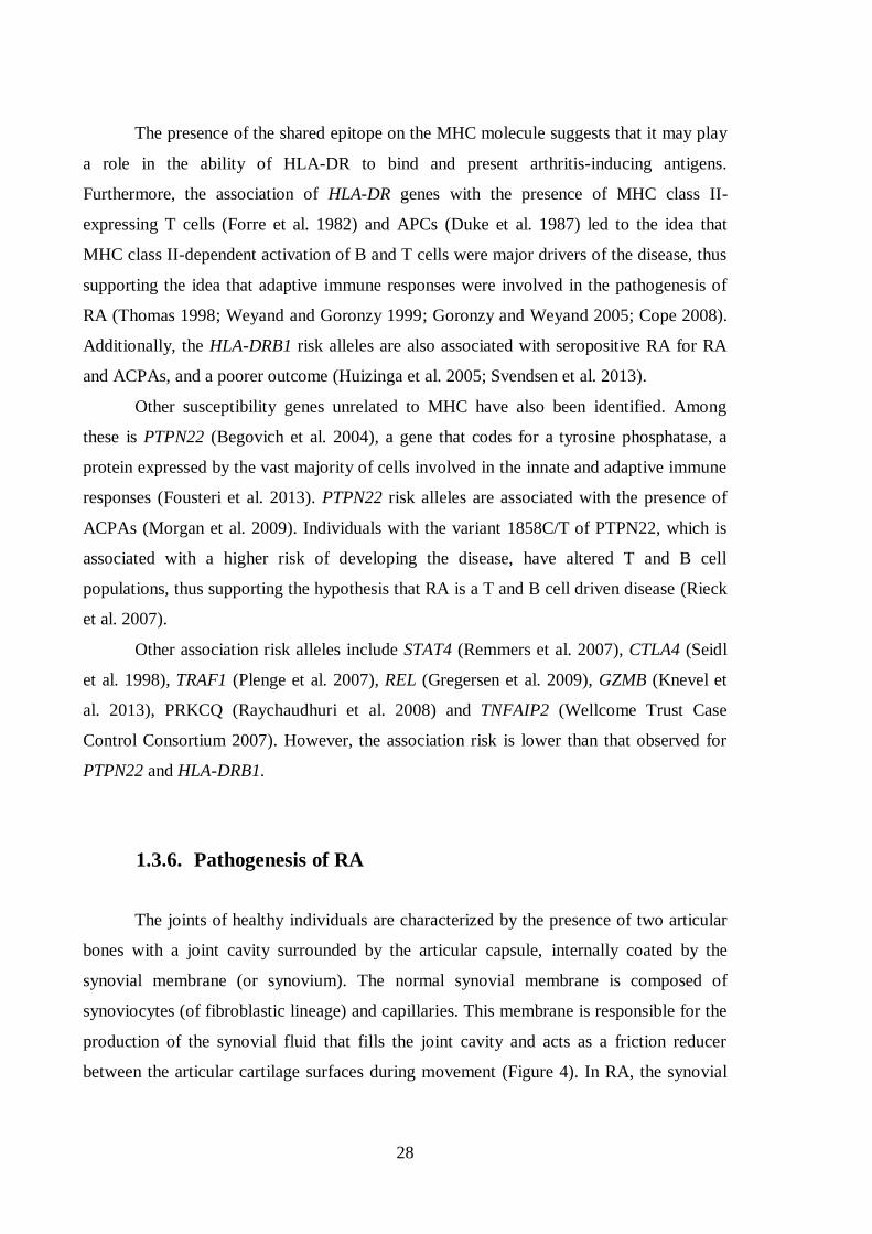
28
The presence of the shared epitope on the MHC molecule suggests that it may play
a role in the ability of HLA-DR to bind and present arthritis-inducing antigens.
Furthermore, the association of HLA-DR genes with the presence of MHC class II-
expressing T cells (Forre et al. 1982) and APCs (Duke et al. 1987) led to the idea that
MHC class II-dependent activation of B and T cells were major drivers of the disease, thus
supporting the idea that adaptive immune responses were involved in the pathogenesis of
RA (Thomas 1998; Weyand and Goronzy 1999; Goronzy and Weyand 2005; Cope 2008).
Additionally, the HLA-DRB1 risk alleles are also associated with seropositive RA for RA
and ACPAs, and a poorer outcome (Huizinga et al. 2005; Svendsen et al. 2013).
Other susceptibility genes unrelated to MHC have also been identified. Among
these is PTPN22 (Begovich et al. 2004), a gene that codes for a tyrosine phosphatase, a
protein expressed by the vast majority of cells involved in the innate and adaptive immune
responses (Fousteri et al. 2013). PTPN22 risk alleles are associated with the presence of
ACPAs (Morgan et al. 2009). Individuals with the variant 1858C/T of PTPN22, which is
associated with a higher risk of developing the disease, have altered T and B cell
populations, thus supporting the hypothesis that RA is a T and B cell driven disease (Rieck
et al. 2007).
Other association risk alleles include STAT4 (Remmers et al. 2007), CTLA4 (Seidl
et al. 1998), TRAF1 (Plenge et al. 2007), REL (Gregersen et al. 2009), GZMB (Knevel et
al. 2013), PRKCQ (Raychaudhuri et al. 2008) and TNFAIP2 (Wellcome Trust Case
Control Consortium 2007). However, the association risk is lower than that observed for
PTPN22 and HLA-DRB1.
1.3.6. Pathogenesis of RA
The joints of healthy individuals are characterized by the presence of two articular
bones with a joint cavity surrounded by the articular capsule, internally coated by the
synovial membrane (or synovium). The normal synovial membrane is composed of
synoviocytes (of fibroblastic lineage) and capillaries. This membrane is responsible for the
production of the synovial fluid that fills the joint cavity and acts as a friction reducer
between the articular cartilage surfaces during movement (Figure 4). In RA, the synovial
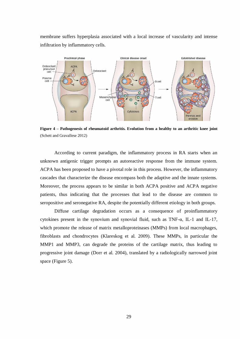
29
membrane suffers hyperplasia associated with a local increase of vascularity and intense
infiltration by inflammatory cells.
Figure 4 – Pathogenesis of rheumatoid arthritis. Evolution from a healthy to an arthritic knee joint
(Schett and Gravallese 2012)
According to current paradigm, the inflammatory process in RA starts when an
unknown antigenic trigger prompts an autoreactive response from the immune system.
ACPA has been proposed to have a pivotal role in this process. However, the inflammatory
cascades that characterize the disease encompass both the adaptive and the innate systems.
Moreover, the process appears to be similar in both ACPA positive and ACPA negative
patients, thus indicating that the processes that lead to the disease are common to
seropositive and seronegative RA, despite the potentially different etiology in both groups.
Diffuse cartilage degradation occurs as a consequence of proinflammatory
cytokines present in the synovium and synovial fluid, such as TNF-α, IL-1 and IL-17,
which promote the release of matrix metalloproteinases (MMPs) from local macrophages,
fibroblasts and chondrocytes (Klareskog et al. 2009). These MMPs, in particular the
MMP1 and MMP3, can degrade the proteins of the cartilage matrix, thus leading to
progressive joint damage (Dorr et al. 2004), translated by a radiologically narrowed joint
space (Figure 5).
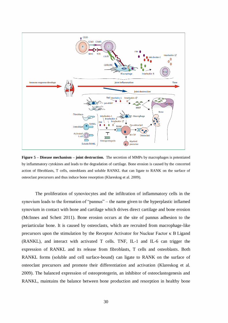
30
Figure 5 – Disease mechanism – joint destruction. The secretion of MMPs by macrophages is potentiated
by inflammatory cytokines and leads to the degradation of cartilage. Bone erosion is caused by the concerted
action of fibroblasts, T cells, osteoblasts and soluble RANKL that can ligate to RANK on the surface of
osteoclast precursors and thus induce bone resorption (Klareskog et al. 2009).
The proliferation of synoviocytes and the infiltration of inflammatory cells in the
synovium leads to the formation of “pannus” – the name given to the hyperplastic inflamed
synovium in contact with bone and cartilage which drives direct cartilage and bone erosion
(McInnes and Schett 2011). Bone erosion occurs at the site of pannus adhesion to the
periarticular bone. It is caused by osteoclasts, which are recruited from macrophage-like
precursors upon the stimulation by the Receptor Activator for Nuclear Factor κ B Ligand
(RANKL), and interact with activated T cells. TNF, IL-1 and IL-6 can trigger the
expression of RANKL and its release from fibroblasts, T cells and osteoblasts. Both
RANKL forms (soluble and cell surface-bound) can ligate to RANK on the surface of
osteoclast precursors and promote their differentiation and activation (Klareskog et al.
2009). The balanced expression of osteoprotegerin, an inhibitor of osteoclastogenesis and
RANKL, maintains the balance between bone production and resorption in healthy bone
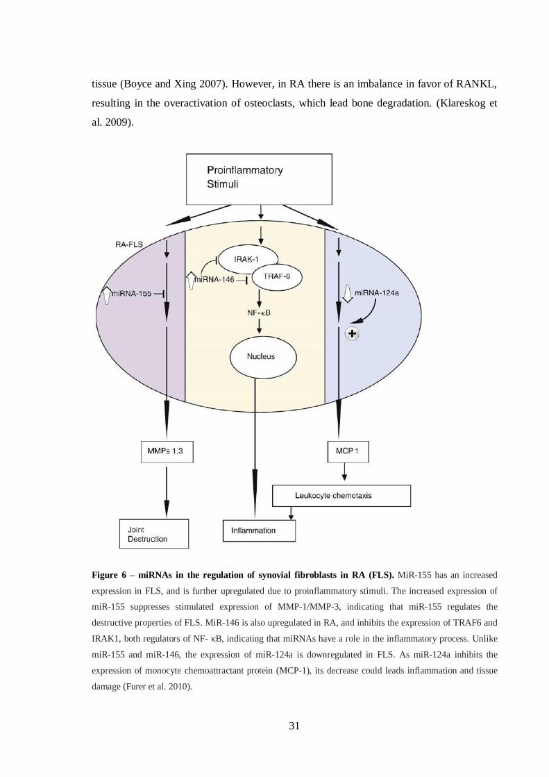
31
tissue (Boyce and Xing 2007). However, in RA there is an imbalance in favor of RANKL,
resulting in the overactivation of osteoclasts, which lead bone degradation. (Klareskog et
al. 2009).
Figure 6 – miRNAs in the regulation of synovial fibroblasts in RA (FLS). MiR-155 has an increased
expression in FLS, and is further upregulated due to proinflammatory stimuli. The increased expression of
miR-155 suppresses stimulated expression of MMP-1/MMP-3, indicating that miR-155 regulates the
destructive properties of FLS. MiR-146 is also upregulated in RA, and inhibits the expression of TRAF6 and
IRAK1, both regulators of NF- κB, indicating that miRNAs have a role in the inflammatory process. Unlike
miR-155 and miR-146, the expression of miR-124a is downregulated in FLS. As miR-124a inhibits the
expression of monocyte chemoattractant protein (MCP-1), its decrease could leads inflammation and tissue
damage (Furer et al. 2010).

32
Recent studies have revealed that the expression of miRNA 7 in RA patients is
impaired, and may contribute to the development of the disease (Nakasa et al. 2011). The
expression profile of various miRNAs was analyzed in RA patients, with special attention
to the fibroblast-like synoviocytes (FLS) (Figure 6). The miRNA miR-124a proved to be
downregulated in FLS from RA patients. Additionally, it was demonstrated that the
overexpression of this miRNA led to the obliteration of FLS proliferation and subsequent
arrest of the cell cycle (Nakamachi et al. 2009). Other miRNAs such as miR-146a and
miR-155 were shown to be overexpressed in synovial tissue (Stanczyk et al. 2008), both
contributing to the local inflammation. MiR-146a is overexpressed in CD4+ T cells from
the SF and is closely correlated with TNF-α levels (Li et al. 2010), while miR-155 is up-
regulated in macrophages form SF and synovial membrane and its inhibition leads to a
decreased production of TNF-α (Kurowska-Stolarska et al. 2011).
1.3.7. Biological agents currently used in RA
The knowledge revised above created the opportunity for the development of the
new biological agents that changed the clinical landscape of RA in this century.
Biologic DMARDs interfere directly with proinflammatory cytokines signaling
pathways, or cell to cell interactions (Figure 7). Biologic therapies currently available in
the clinic target TNF-α, IL-6 or IL-1, inhibit T cell co-stimulation or selectively deplete B
cells expressing CD20 on their surface (Scherer and Burmester 2009).
The first-line biologic therapy administered is TNF-α-inhibitory agents (Taylor and
Feldmann 2009). TNF-α is expressed at high levels in the inflamed joints of RA patients,
where they contribute considerably to the inflammatory process, therefore the use of anti-
TNF-α biologic agents tend to be highly beneficial (Navarro-Millan and Curtis 2013). The
combination of anti-TNF-α therapy with MTX has proven more effective than biologic
monotherapy (Choy et al. 2005; Soliman et al. 2011). However, as anticipated, anti-TNF-α
7 miRNA: Class of small endogenous non-coding RNAs of approximately 22 nucleotides that influence the
stability and translation of mRNA. miRNAs regulate gene expression by binding the 3’-untranslated region
of their target mRNAs leading to translational repression or mRNA degradation.
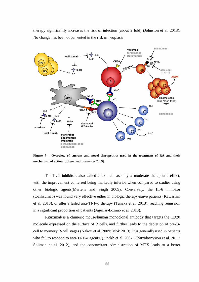
33
therapy significantly increases the risk of infection (about 2 fold) (Johnston et al. 2013).
No change has been documented in the risk of neoplasia.
Figure 7 – Overview of current and novel therapeutics used in the treatment of RA and their
mechanism of action (Scherer and Burmester 2009).
The IL-1 inhibitor, also called anakinra, has only a moderate therapeutic effect,
with the improvement conferred being markedly inferior when compared to studies using
other biologic agents(Mertens and Singh 2009). Conversely, the IL-6 inhibitor
(tocilizumab) was found very effective either in biologic therapy-naïve patients (Kawashiri
et al. 2013), or after a failed anti-TNF-α therapy (Tanaka et al. 2013), reaching remission
in a significant proportion of patients (Aguilar-Lozano et al. 2013).
Rituximab is a chimeric mouse/human monoclonal antibody that targets the CD20
molecule expressed on the surface of B cells, and further leads to the depletion of pre-B-
cell to memory B-cell stages (Nakou et al. 2009; Mok 2013). It is generally used in patients
who fail to respond to anti-TNF-α agents, (Finckh et al. 2007; Chatzidionysiou et al. 2011;
Soliman et al. 2012), and the concomitant administration of MTX leads to a better

34
outcome, with a significantly lower radiological progression of the disease when compared
to patients receiving monotherapy only (Cohen et al. 2006; Mok 2013).
Figure 8 – Mechanism of action of abatacept. Abatacept binds to CD80/86 on the surface of APCs and
blocks its interaction with CD28 on the surface of T cells, resulting in the inhibition of the co-stimulation of
T cells, thus preventing their activation. This mechanism further leads to the downregulation of the
inflammatory cascade and normalization of the levels cytokines and antibodies and inhibition of osteoclast
activity (von Kempis et al. 2012).
Abatacept is the only biologic DMARD currently in use that directly targets not
only CD8+ T cells, but total T cells by preventing their activation. It consists of the
extracellular domain of human cytotoxic T-lymphocyte-associated protein 4 (CTLA-4)
fused with the modified Fc portion of human immunoglobulin G1 (IgG1), and functions by
binding to the CD80 and CD86 molecules on the antigen-presenting cell surface, thus
inhibiting the binding of CD28 (Figure 8). It inhibits the co-stimulation of T cells, as
35
activated T cells have an important role in the pathogenesis of RA. Abatacept reduces T
cell proliferation and inhibits the production of proinflammatory cytokines, such as TNF-α,
IL-6 and IFN-γ, as well as MMPs (Weisman et al. 2006; Buch et al. 2009). The reduction
of proinflammatory cytokines leads to the inhibition of osteoclast activity, and the reduced
production of MMPs leads to a decreased cartilage degradation in the RA joint (von
Kempis et al. 2012). Abatacept is generally used when anti-TNF-α therapy is ineffective
(Gaffo et al. 2006; Nogid and Pham 2006; Buch et al. 2009; von Kempis et al. 2012).
The introduction of these biological therapies, together with new, targeted,
treatment strategies has operated a profound revolution in the treatment of rheumatoid
arthritis: disease remission, once seldom seen, has become the consensual objective of
therapy. It can be achieved in up to 60% of appropriately treated patients. Remission
provides the best assurance that bone erosion, loss of cartilage and functional deterioration
can he halted. This is achieved with manageable but not irrelevant toxicity.
Despite this, many patients still do not respond adequately to any of the
therapeutical agents available and there are no tools to predict response to individual
molecules. Further knowledge is dearly needed.
1.4. Mouse models of arthritis
Animal models have long had an important role in the study of the pathogenesis of
rheumatoid arthritis. These include induced-arthritis models and spontaneous arthritis
strains in rodents. In this section only mouse models of arthritis will be discussed.
1.4.1. Spontaneous arthritis models
1.4.1.1. K/BxN model
The K/BxN mouse model spontaneously develops an aggressive form of arthritis
and shares many features similar to those of human RA, including leukocyte invasion,
synoviocyte proliferation, pannus formation, synovitis, cartilage degradation and bone
erosion (Kouskoff et al. 1996; Korganow et al. 1999). This model also presents other

36
similarities with human RA, such as the polyclonal B cell activation with increased B cell
numbers, hypergammaglobulinemia8 and the production of autoantibodies. However, this
model lacks the production of RF, which is characteristic of RA (Ditzel 2004).
The K/BxN mice are originally originated from the crossing of KRN-C57BL/6
mice bearing a transgenic TCR (truncated Vβ6 TCR) with NOD (non-obese diabetic) mice,
which are known to be prone to autoimmune disorders.
The transgenic TCR Vβ6 from the KRN mice recognizes a bovine ribonuclease
peptide presented by I-Ak MHC class II molecule. Interestingly, the KRN transgenic TCR
in the context of the NOD-derived Ag7
MHC class II molecule also recognizes a peptide
(GPI 282–294) from the ubiquitous cytosolic enzyme glucose-6-phosphate isomerase
(GPI; EC 5.3.1.9), which catalyzes the interconversion of D-glucose 6-phosphate and D-
fructose-6-phosphate, an essential reaction of glycolysis and gluconeogenesis. This dual
specificity is responsible for inducing autoreactive T cells that cause severe arthritis with
an inset within the first 4-5 weeks of age (Ditzel 2004) (Figure 9). The autoreactive T cells
generated in the Vβ6-bearing K/BxN mice in the Ag7
background will help B cells by
presenting the autoantigen, and thus promote the production of anti-GPI autoantibodies.
Even though the arthritis developed in this model is due to the formation of
autoreactive T cells against a specific peptide in GPI, it was proven that the onset of
arthritis is triggered by autoantibodies. This was demonstrated by transferring serum or
purified immunoglobulin from TCR transgenic, I-Ag7
-positive K/BxN mice into wild-type,
B-cell-deficient and lymphocyte-deficient mice led to the rapid onset of arthritis, with
symptoms observed as early as 24 hours after the transfer, but unlike the arthritis
developed in K/BxN mice, this form of arthritis is transient, and is resolved in 15 to 30
days (Korganow et al. 1999).
GPI, which is known for being an isomerase that catalyzes an essential reaction in
gluconeogenesis. Nevertheless, multiple identities have been attributed to the secreted form
of this protein, such as neuroleukin (NLK) or autocrine motility factor (AMF). NLK was
found to be a lymphokine9 produced by activated T cells, and induced the differentiation of
B cells into antibody-secreting B cells (Gurney et al. 1986; Gurney et al. 1986). AMF was
8 Hypergammaglobulinemia: condition in which the patient has an abnormally high level of gamma
globulins, a class of plasma proteins which comprises antibodies. 9 Lymphokine: General term for any soluble protein mediators supposedly released by activated
lymphocytes, mainly T cells, on contact with an antigen. Lymphokines are believed to play a role in
macrophage activation, lymphocyte transformation, and cell-mediated immunity.
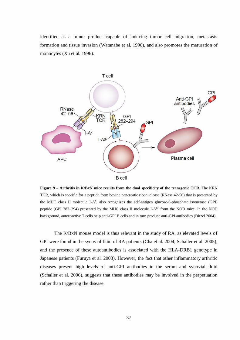
37
identified as a tumor product capable of inducing tumor cell migration, metastasis
formation and tissue invasion (Watanabe et al. 1996), and also promotes the maturation of
monocytes (Xu et al. 1996).
Figure 9 – Arthritis in K/BxN mice results from the dual specificity of the transgenic TCR. The KRN
TCR, which is specific for a peptide form bovine pancreatic ribonuclease (RNase 42-56) that is presented by
the MHC class II molecule I-Ak, also recognizes the self-antigen glucose-6-phosphate isomerase (GPI)
peptide (GPI 282–294) presented by the MHC class II molecule I-Ag7 from the NOD mice. In the NOD
background, autoreactive T cells help anti-GPI B cells and in turn produce anti-GPI antibodies (Ditzel 2004).
The K/BxN mouse model is thus relevant in the study of RA, as elevated levels of
GPI were found in the synovial fluid of RA patients (Cha et al. 2004; Schaller et al. 2005),
and the presence of these autoantibodies is associated with the HLA-DRB1 genotype in
Japanese patients (Furuya et al. 2008). However, the fact that other inflammatory arthritic
diseases present high levels of anti-GPI antibodies in the serum and synovial fluid
(Schaller et al. 2006), suggests that these antibodies may be involved in the perpetuation
rather than triggering the disease.
38
1.4.1.2. Other spontaneous arthritis models
Other transgenic spontaneous arthritis mouse models have been used in the study of
RA, such as the TNF-α transgenic mouse model, the SKG mouse strain or the
human/SCID chimeric mice.
The TNF-α transgenic mouse model was engineered to over-express the human
TNF-α, and was first described by Keffer et. al. (Keffer et al. 1991). This mouse model
develops a chronic inflammatory erosive polyarthritis, and the treatment with TNF-α
depleting antibodies completely prevents the disease (Keffer et al. 1991).
The SKG mouse strain is characterized by the presence of a point mutation in the
Zeta-chain-associated protein kinase 70 (ZAP-70), which is associated with thymic T-cell
selection defects, and leads to the onset of chronic arthritis at about 2 months of age
(Sakaguchi et al. 2003). However, they are influenced by their environment, and only
develop arthritis under conventional conditions, whereas they are healthy under specific
pathogen free (SPF) condition. In that case, arthritis can be induced by zymosan 10
(Kobayashi et al. 2006).
The human/SCID chimeric mice were initially originated by having SCID mice
implanted with human synovial tissue in the renal capsule (Geiler et al. 1994) and knee
joints (Sack et al. 1994), and both experiments indicated that the implants underwent
pannus formation and erosion of cartilage and bone, thus indicating that this model is
useful in studying pathogenetic aspects of joint destruction in RA.
1.4.2. Induced arthritis models
1.4.2.1. Collagen-induced arthritis
Collagen-induced arthritis (CIA) is widely used to study the pathogenesis of RA
and potential therapeutic targets, as it shares many similarities with human RA. It is
induced by immunization with emulsified autologous or heterologous type II collagen and
Freund’s adjuvant (Williams 2004), and develops through the generation of antibodies
10 Zymosan: polysaccharide from the cell wall of yeast, used to induce inflammation.

39
against type II collagen and self-peptides upon the breakdown of self-tolerance. CIA was
first studied in rats (Trentham et al. 1977; Trentham et al. 1978), and was subsequently
found to be also inducible in mouse strains (Courtenay et al. 1980; Wooley et al. 1981;
Stuart et al. 1982).
As in human RA, susceptibility to CIA is strongly associated with MHC class II
genes, developing mainly in strains containing the MHC class II H-2q haplotypes.
However, different strains display different degrees of susceptibility to the induction of
arthritis. The development of polyarthritis is accompanied by a T- and B-cell dependent
response to type II collagen (Holmdahl et al. 1985; Hom et al. 1986; Hom et al. 1986;
Zhang et al. 2002).
DBA/1 are the most frequently used mice in CIA studies. Clinical symptoms of
arthritis first appear 21-25 days after the first immunization, affecting preferentially the
joints of the limbs. Synovial inflammatory infiltration of polymorphonuclear and
mononuclear cells, pannus formation, eventually leading to cartilage degradation, bone
erosion and fibrosis are observed (Boissier et al. 1987). The peak of disease severity is
expected around day 35, after which DBA/1 mice enter remission. Similarly to human RA,
studies using homologous type II collagen have reported the occurrence of chronic
relapsing polyarthritis (Holmdahl et al. 1986; Malfait et al. 2001).
However, the induction of arthritis in DBA/1 mice has a major caveat: since the T
cell population peaks early and is in decline by the time of disease onset, the utility of this
model for studying T cell in the onset of the disease is limited. One alternative to DBA/1
mice are transgenic mice with C57BL/6 background. This strain was regarded as resistant
to CIA (Szeliga et al. 1996; Pan et al. 2004), but a new CIA protocol has successfully
managed to induce arthritis in these mice (Inglis et al. 2008). The C57BL/6 mice typically
develop arthritis 4-7 days later than DBA/1 mice, but with a comparable severity (Inglis et
al. 2007; Inglis et al. 2008). However, the incidence of the disease in the C57BL/6 mice is
lower than that of DBA/1 mice, and varies greatly among the different substrains with
C57BL/6 background.
CIA can also be successfully induced in the C57BL/10 (also called B10) strain.
These mice are very similar to the C57BL/6 strain, having been reported to differ only in 6
loci on chromosome 4 (McClive et al. 1994), and are often considered equivalent. Many
transgenic substrains of B10 mice that are commonly used in the induction of arthritis,

40
especially those bearing CIA susceptibility genes, such as the H-2q haplotype derived from
DBA/1 mice seen in the B10.Q strain (http://jaxmice.jax.org/strain/002024.html). The CIA
model is however known for having a variable incidence, severity and inconsistency
among different groups, which reflects the various strains sensitivity to environment,
maintenance conditions and stress.
1.4.2.2. Other forms of inducing arthritis
Collagen-antibody-induced arthritis (CAIA), an antibody-mediated model of
arthritis, is induced by using IgG antibodies against type II collagen. The disease onset
occurs within 48h of antibody administration, and develops in all strains, regardless of the
MHC class II haplotype. Even though the clinical development of the disease is similar to
that observed in CIA and RA, CAIA is characterized by the presence of macrophages and
polymorphonuclear cells in the inflamed joints (Santos et al. 1997), and is not driven by T-
or B-cells. Interestingly, the transfer of type II collagen reactive T cells was proven to
increase the disease severity (Nandakumar et al. 2004).
Other less known methods of induction of arthritis can also be used in mice, such as
the administration of zymosan and pristane. Zymosan, a polysaccharide found on the cell
wall of Saccharomyces cerevisae, can be injected into the joints of mice, resulting in the
local inflammation of the joint characterized by the infiltration of mononuclear cells,
synovial hypertrophy and pannus formation. Similarly, a single subcutaneous injection of
small amounts of pristane (2,6,10,14-tetramethylpentadecane), leads to a chronic relapsing
arthritis (Olofsson and Holmdahl 2007).
1.5. CD8+ T cells in the pathogenesis of Rheumatoid Arthritis –
Current knowledge
The role of CD8+ T cells in rheumatoid arthritis has attracted relatively little
attention. This is probably due to the remarkably conflicting results obtained with animal

41
models of polyarthritis, rendering researchers unable to discern if the global effect of CD8+
T cells in the disease process is protective or deleterious.
1.5.1. Lessons from animal models of arthritis
Mercuric chloride-induced arthritis in the Brown Norway rat is associated with
increased numbers of circulating CD4+ and CD8
+ T cells, and higher serum levels of IL-4
and IgE. The treatment of these animals with R73 (anti-aβ TCR monoclonal antibody
(mAb)) leads to a marked decrease in IgE and IgG levels as well as in B cell counts,
yielding an amelioration of the disease (Kiely et al. 1995; Prigent et al. 1995). In this
model, the depletion of CD8+ T cells with the OX8 depleting monoclonal antibody led to
reduced severity and incidence of the disease (Kiely et al. 1996). This was paralleled by an
increased production of IFN-γ, thus indicating a possible regulation of the disease through
a type I response (Kiely et al. 1996). These studies suggest an aggressive role for CD8+ T
cells in this disease model, presumably exerted through cytotoxicity. However, the
depletion of these cells with OX8 mAb in oil-induced arthritis in DA rats led to an earlier
onset of the disease, indicating a protective role, presumably mediated by their suppressor
functions (Jansson et al. 2000).
Studies using a depleting anti-CD3 antibody in collagen-induced arthritis in DBA/1
mice also argue for a protective role of CD8+ T cells in experimental arthritis. In the
repopulation of the T cell compartment after CD3-depletion, there was an enrichment of
CD4+ and CD8
+ T cells with regulatory/suppressor phenotype. Regulatory CD8
+ T cells
from treated mice were able to suppress IL-17 production, CD4+ T cell proliferation and
IFN-γ production. This suggests CD8+ T cells as responsible for maintaining the persistent
amelioration observed following anti-CD3 therapy (Notley et al. 2010). Taneja et al.
reported that transgenic CD8+ T cell deficient mice expressing the RA susceptibility gene
HLA-DQ8 have a higher incidence and severity of the disease than in the wild-type
counterparts. Conversely, the CD4+ T cell deficient mice failed to develop the disease.
These observations suggest that CD8+ T cells have a protective effect and CD4
+ T cells
have an initiator function in this model (Taneja et al. 2002). Studies with collagen-induced
arthritis (CIA) on B10.Q also suggest that CD4+ T cells have a globally deleterious

42
influence, mainly due to the IL-4 production, while CD8+ T cells appear to have little
effect on the disease. Moreover, CD8-deficient B10.Q mice show a tendency towards a
later onset of the disease, which might be related to the decreased production of
proinflammatory cytokines such as IFN-γ (Ehinger et al. 2001).
Conversely, CD8-/- DBA/1 mice are less susceptible to develop CIA on a first
collagen boost than their heterozygous counterparts, although the severity of the disease is
not significantly altered, thus indicating that CD8+ T cells may have a promoting role in
the initiation of the disease. After full recovery from the initial CIA, CD8-deficient mice
appear to be more susceptible to develop the disease than their heterozygous littermates,
thus indicating that CD8+ T cells may acquire a predominantly regulatory or suppressive
role (Tada et al. 1996).
The depletion of CD8+ T cells in BALB/c mice with proteoglycan aggrecan-
induced arthritis led to an aggravation of the disease, without affecting the amount of anti-
proteoglycan-antibodies at the peak of the disease (Banerjee et al. 1992).
The transfer of CD8+ T cells from thoracic duct lymph of adjuvant induced arthritic
DA rats into healthy normal syngeneic recipients failed to induce the disease (Spargo et al.
2001). However, the recipients had their normal CD8+ T cell population, which may have
eliminated the transferred CD8+ T cell population thus preventing the transference of the
disease by these cells. On the contrary, the transference of CD8+ T cell clones from SKG
mice, which develop a T cell-mediated autoimmune arthritis, to nude mice led to the
induction of arthritis and also pneumonitis, indicating that CD8+ T cells from this mouse
model are arthritogenic and have the ability to transfer the disease (Wakasa-Morimoto et
al. 2008).
Taken together, these studies suggest that CD8+ T cells have an important impact in
the pathogenesis of a variety of experimental models of arthritis, both in its initiation and
in the course of the disease. Additionally, they indicate that the global effect of eliminating
CD8+ T cells varies according to the disease model and the phase the disease. However, in
all those studies the total CD8+ T cell pool was manipulated, thus abrogating any insight
regarding the role of the different CD8+ T cell subsets. Since such subsets have distinct and
even opposing functions, it is plausible that the contradictions between studies might
derive, at least in part, from the importance of particular CD8+ T cell subsets in different
models and phases of the experimental disease. Hence, in our opinion, further studies

43
targeting particular CD8+ T cell subsets are indispensable to understand their role in
arthritis and explore their therapeutic potential.
1.5.2. Human studies
Several lines of indirect evidence suggest that CD8+ T cells are involved in the
pathogenesis of rheumatoid arthritis.
1.5.2.1. Circulating CD8+ T cells in patients and controls.
Several studies have looked for changes in the number and function of CD8+ T cells
in RA. Martinez-Taboada et al. compared the absolute numbers of circulating CD8+ T cells
in patients with active RA and healthy controls, concluding that RA patients tend to have
decreased numbers of circulating CD8+ T cells, though the differences failed to reach
statistical significance (Martinez-Taboada et al. 2001).
Peripheral blood CD8+ T cells from RA patients tend to have an increased
proportion of central memory phenotype (CD62L+CD45RA
-) while the proportion of the
effector memory subtype (CD62L-CD45RA
+) is decreased, in comparison with healthy
controls (Maldonado et al. 2003). Moreover, the levels of memory CD8+CD45RO
+ T cells
are correlated with the levels of IgM-rheumatoid factor (IgM-RF). It was also observed
that patients shifting from low to high levels of IgM-RF presented a decrease in naïve T
cells and an increase in the transient CD8+CD45RA
+CD45RO
+ T cell subset (Neidhart et
al. 1996).
A study of regulatory T cells in RA patients by Sempere-Ortells and colleagues
shows that increased numbers of regulatory CD8+CD28
- T cells correlated with the activity
of the disease, measured by the DAS28 (Disease Activity Score) (Sempere-Ortells et al.
2009). Little is known about changes in CD8+
T cell subpopulations in relation to disease
activity or effects of medications. Kao et al, reported that the regulatory CD8+CD11c
+
subpopulation, found to be highly expressed in an arthritic mouse model, is not correlated
with disease activity in RA patients (Kao et al. 2007).
44
1.5.2.2. CD8+ T cells in the synovial fluid
CD8+ T cells comprise approximately 40% of all T cells in the synovial fluid
(McInnes 2003). The analysis of serial synovial fluid samples obtained from different
arthritic joints in the same patient indicates that the CD8+ T cell accumulation in inflamed
joints is persistent (Masuko-Hongo et al. 1997). Furthermore, there is evidence that these
cells undergo clonal expansion in the synovial fluid, their TCR repertoire may be skewed,
they are genetically as well as environmentally determined, and can be induced by a
common antigen (DerSimonian et al. 1993; Fitzgerald et al. 1995; Hall et al. 1998).
CD8+T cells from synovial fluid of rheumatoid arthritis patients typically present
higher expression of both short-term and long-term activation markers (i.e. CD69 and
CD25) than observed in the peripheral blood (Afeltra et al. 1997). A study by Marrack and
colleagues has shown that type I interferons have the capability of keeping activated T
cells alive upon infection (Marrack et al. 1999), which can contribute to the high
percentage of persistently activated CD8+ T cells in RA joints. These cells (Tc1) are
characterized by the production of large amounts of IFN-γ, suggesting a potential to induce
local inflammatory responses, but also present an increased production of IL-10, which can
counteract the inflammatory process in the joint (Berner et al. 2000).
Autoreactive CD8+ T cells in rheumatoid inflamed joints have been characterized
as CD57+, oligoclonally expanded and in a terminal differentiation status. They are
functionally active but lack replicative capacity thus representing a state of “clonal
exhaustion” (Strioga et al. 2011). These cells are present in higher numbers in the synovial
fluid of RA patients than in matched peripheral blood (Arai et al. 1998).
The accumulating CD8+ T cells in the synovial fluid from RA patients are also
characterized by an oligoclonal TCR repertoire, i.e. different patients share the same TCR
sequence pattern. This is taken as a strong indicator of a common antigen-driven CD8+ T
cell response (Fitzgerald et al. 1995; Hingorani et al. 1996; Hall et al. 1998). It has been
suggested that the antigen driving this autoreactive CD8+ T cell response in RA may not be
related to the disease. The hypothesis was enunciated by Fazou et al. after observing that
the TCR repertoire of synovial fluid CD8+ T cells in RA patients was specific for several
types of virus, namely Epstein–Barr virus (EBV) (Klatt et al. 2005), cytomegalovirus and
influenza virus (Fazou et al. 2001). Another study reported that up to 15.5% of synovial
45
CD8+ T cells presented specificity for a single EBV epitope in a cohort of 15 EBV-
seropositive patients. These cells presented higher activation levels and increased secretion
of proinflammatory cytokines, suggesting that they could contribute to the maintenance of
the local inflammatory response (Tan et al. 2000). However, another study found little
correlation between disease progression and CD8+ T cell response to EBV in RA patients
(Berthelot et al. 2003).
Antibodies anti-BiP (immunoglobulin binding protein), can be found in the serum
of RA patients and in several mouse models of arthritis. CD8+ T cell clones responding to
BiP autoantigen are producers of IL-10, but also of other cytokines such as IFN-γ, IL-4 and
IL-5 (Bodman-Smith et al. 2003). This has been interpreted as an indication that CD8+ T
cells with a Tc2 phenotype can become regulatory upon BiP stimulation and undergo
clonal expansion locally, thus exerting a regulatory/suppressor function (Bodman-Smith et
al. 2000). In this line of thought, Davila and co-workers (Davila et al. 2005) demonstrated
that suppressor CD8+ T cells can be used as effective cell-based immunosuppressive
therapy. In fact, CD8+CD28
-CD56
+ T cell clones from synovial tissues of RA patients
displayed an anti-inflammatory immunosuppressive activity in NOD-SCID mice engrafted
with synovial tissue from RA patients. This was reflected by a decrease in the production
of proinflammatory cytokines and in the expression of activation markers by the engrafted
tissue. More recently, Cho et al. strengthened the hypothesis that CD8 exert a
predominantly suppressor effect in RA by showing that there is an accumulation of Ts cells
in the synovial fluid (Cho et al. 2012). However, a previous study observed a correlation of
CD8+ T cell numbers and proinflammatory cytokines in the synovial fluid of RA patients,
indicating that CD8+ T cells can produce high amounts of cytokines and thus contribute
actively to the inflammation and joint degradation in RA (Hussein et al. 2008).
1.5.2.3. CD8+ T cells in the synovial membrane.
Follicular structures – reminiscent of those found in secondary lymphoid organs –
can be found in the inflamed synovial membrane of approximately 50% of RA patients,
with a clearly organized ectopic germinal center present in approximately half of these
patients (Takemura et al. 2001). These structures are thought to contribute greatly to the
pathogenesis of RA due to their ability to produce autoantibodies, cytokines and
46
rheumatoid factor, which are known to contribute to tissue damage in this disease. Many
RA patients present T and B cell aggregates in the synovium that lack a typical germinal
center structure and have no follicular dendritic cells (FDCs). Along with these cells, the T
follicular helper cells, a subset of CD4+ T cells, is found in these follicular structures and
are thought to drive the B cell differentiation into plasma cells (Dong et al. 2011). This has
been interpreted as indicating that the formation of ectopic germinal centers in inflamed
joints depends solely on antigen recognition by TCRs and BCRs. The fact that T and B
cells can aggregate without the presence of FDCs can indicate that T and B cells may be
seeding in the synovial membrane prior to the FDCs, and may therefore be responsible for
their recruitment and maintenance in the synovial membrane (Takemura et al. 2001).
Indeed, the formation of ectopic germinal structures is associated with the local expression
of CXCL13, a strong B-cell chemoattractant that guides B cells into the synovium, thus
contributing to the formation of ectopic germinal structures and aggregates (Shi et al.
2001). Even though FDCs secrete large amounts of CXCL13, this chemokine can also be
produced by fibroblasts and endothelial cells (Weyand and Goronzy 2003).
The presence of ectopic germinal centers in the synovial membrane is associated
with a poorer disease prognosis (Wagner et al. 1998). CD8+ T cells are recognized as
essential for the formation of ectopic germinal centers in the synovial membrane of
inflamed RA joints. Indeed, after the engraftment with synovial membranes containing
ectopic germinal centers in NOD-SCID mice, they were treated with a depleting anti-CD8
antibody, which resulted in the disintegration of the synovial follicles, with a significant
decrease in the local production of TNF-α and IFN-γ (Wagner et al. 1998; Kang et al.
2002).
However, cytotoxic CD8+ T cells present in the synovial fluid contribute greatly to
the local increased production of proinflammatory cytokines, and may thus have a
predominantly deleterious effect in arthritis. Several studies have shown that CD8+ T cells
are as responsible as the CD4+ for type I proinflammatory cytokine secretion in the
synovial membrane (Berner et al. 2000).

47
CHAPTER 2
DRIVING HYPOTHESES
OBJECTIVES

49
2. Driving hypotheses and objectives
2.1. Driving Hypotheses
CD8+ T cells, formerly called killer T cells, have earned the reputation of being the
driving force behind proinflammatory processes, as they have the ability to induce cell
death in neighboring cells through the production of proteolytic enzymes, upon recognition
of a specific antigen. Concordantly, CD8+ T cells have been proven to play an important
role in the pathogenesis of several inflammatory disorders, such as multiple sclerosis
(Saxena et al. 2011) or allograft rejection (Halamay et al. 2002).
Research on the immune cells involved in the pathogenesis of rheumatoid arthritis -
regardless of using human samples or animal models- has mainly focused on the role of B
cells, CD4+ T cells and macrophages. Nevertheless, the few existing studies on CD8
+ T
cells present evidence that these cells are equally involved in the inflammatory process
underlying RA.
While it is unequivocal that CD8+ T cells have a role in the pathogenesis of RA, the
nature of that role, being it protective or deleterious, still remains to be elucidated. Indeed,
it is known that 40% of the T cells infiltrating the rheumatoid synovial membrane are
CD8-positive (McInnes 2003), however their importance in the pathogenesis and
maintenance of rheumatoid arthritis (RA) is still scarcely defined. Interestingly, many
studies have pointed towards a proinflammatory role of CD8+ T cells in RA (Fitzgerald et
al. 1995; Kang et al. 2002), while others defend that they have a protective role in RA
(Suzuki et al. 2008).
2.2. Objectives
In order to determine the role of CD8+ T cells in the pathogenesis of RA, the
following objectives were pursued:
50
• Understand the possible role played by the CD8+ T cells infiltrating the synovial
fluid in rheumatoid arthritis and the joint in animal models of experimental chronic
polyarthritis in initiating and maintaining disease chronicity;
• Phenotypic and functional characterization of CD8+ T cells isolated from the
synovial fluid and peripheral blood from RA patients comparing to healthy
controls, and from the articular infiltrate and peripheral blood of arthritic mice or
wild type controls;
• Define the similarities and differences in CD8+ T cell involvement in the
pathogenesis of RA and in the pathogenesis of experimental chronic polyarthritis,
to test the suitability of the animal models for in vivo studies of CD8+ T cell role in
chronic polyarthritis;
• Explore the therapeutic potential of manipulating CD8+ T cell function (through
blockade, or depletion) to ameliorate and/or reverse disease progression and signs
in the mouse model of chronic spontaneous polyarthritis K/BxN.

51
CHAPTER 3
MATERIALS AND METHODS


53
3. Materials and methods
3.1. Mice
3.1.1. Common procedures
3.1.1.1. Mouse breeding conditions
The KRN, NOD, K/BxN and B10.Q mice were group-housed in type III-H cages
(Tecniplast, Italy) and maintained in specific environmental conditions (22-24ºC, 45-65%
humidity, 15 changes/hour ventilation, 12 h artificial light/dark cycle) and free access to
irradiated standard rodent chow (4 RFN/I GLP certificate, Mucedola, Italy) and acidified
water (at pH 3.5 with HCl to avoid bacterial contamination). The research procedures were
carried out in accordance with the European directives (Directive 86/609/EEC and
Directive 2010/63/EU) on the protection of animals used for scientific purposes, and
according to the ethical standards for animal manipulation.
3.1.1.2. Blood collection
Blood collection from K/BxN and B10.Q mice was performed through the section
of the lateral caudal veins. The mice were heated under a heating lamp, and then
anesthetized with the volatile anesthetic isofluorane (IsoFlo®, Esteve Veterinaria,
Portugal). When the mouse reached unconsciousness, the lateral veins were incised with a
sterile surgical blade (Swann-Morton, Sheffield, UK), and the blood drops were collected
into blood collection tubes with either K2EDTA, or clot activator and gel for serum
separation (Microtainer™ Tubes, Becton Dickinson, New Jersey, USA). Blood samples
from K2EDTA were put in a blood tube rotator at room temperature to prevent blood clot
formation until they were processed.
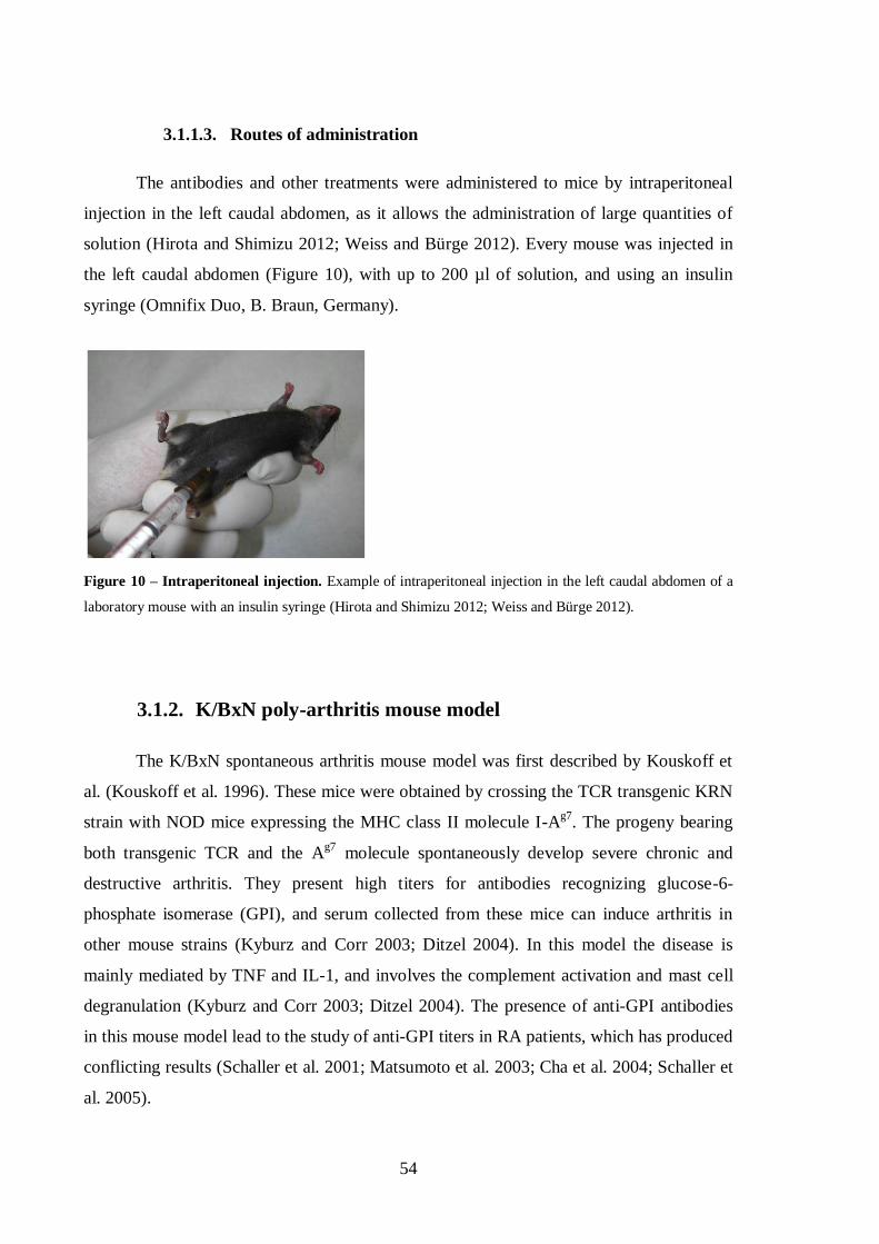
54
3.1.1.3. Routes of administration
The antibodies and other treatments were administered to mice by intraperitoneal
injection in the left caudal abdomen, as it allows the administration of large quantities of
solution (Hirota and Shimizu 2012; Weiss and Bürge 2012). Every mouse was injected in
the left caudal abdomen (Figure 10), with up to 200 µl of solution, and using an insulin
syringe (Omnifix Duo, B. Braun, Germany).
Figure 10 – Intraperitoneal injection. Example of intraperitoneal injection in the left caudal abdomen of a
laboratory mouse with an insulin syringe (Hirota and Shimizu 2012; Weiss and Bürge 2012).
3.1.2. K/BxN poly-arthritis mouse model
The K/BxN spontaneous arthritis mouse model was first described by Kouskoff et
al. (Kouskoff et al. 1996). These mice were obtained by crossing the TCR transgenic KRN
strain with NOD mice expressing the MHC class II molecule I-Ag7
. The progeny bearing
both transgenic TCR and the Ag7
molecule spontaneously develop severe chronic and
destructive arthritis. They present high titers for antibodies recognizing glucose-6-
phosphate isomerase (GPI), and serum collected from these mice can induce arthritis in
other mouse strains (Kyburz and Corr 2003; Ditzel 2004). In this model the disease is
mainly mediated by TNF and IL-1, and involves the complement activation and mast cell
degranulation (Kyburz and Corr 2003; Ditzel 2004). The presence of anti-GPI antibodies
in this mouse model lead to the study of anti-GPI titers in RA patients, which has produced
conflicting results (Schaller et al. 2001; Matsumoto et al. 2003; Cha et al. 2004; Schaller et
al. 2005).
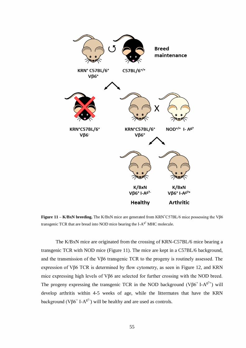
55
Figure 11 – K/BxN breeding. The K/BxN mice are generated from KRN+C57BL/6 mice possessing the Vβ6
transgenic TCR that are bread into NOD mice bearing the I-Ag7 MHC molecule.
The K/BxN mice are originated from the crossing of KRN-C57BL/6 mice bearing a
transgenic TCR with NOD mice (Figure 11). The mice are kept in a C57BL/6 background,
and the transmission of the Vβ6 transgenic TCR to the progeny is routinely assessed. The
expression of Vβ6 TCR is determined by flow cytometry, as seen in Figure 12, and KRN
mice expressing high levels of Vβ6 are selected for further crossing with the NOD breed.
The progeny expressing the transgenic TCR in the NOD background (Vβ6+
I-Ag7+
) will
develop arthritis within 4-5 weeks of age, while the littermates that have the KRN
background (Vβ6+ I-A
g7-) will be healthy and are used as controls.

56
Figure 12 – Selection for the Vβ6-bearing KRN-C57BL/6 mice for further crossing with NOD mice.
The expression of the Vβ6 transgenic TCR is determined in T cells, marked using the anti-CD3 anti-mouse
antibodies. The expression is considered positive in animals presenting a percentage above 20% of Vβ6-
expressing T cells.
3.1.2.1. K/BxN mouse breeding
The TCR-transgenic KRN mice were a kind gift from Dr. C. Benoist (Harvard
University, Boston, MA) and were maintained on a C57BL/6 background (K/B). The
KRN+C57BL/6
+ progeny bearing the Vβ6-transgenic TCR were identified at 3–4 weeks of
age by flow cytometry. The red blood cells were removed from the samples using red
blood cell (RBC) lysis buffer and were washed with phosphate buffered solution (PBS).
The peripheral blood cells were then stained using phycoerythrin (PE)–labeled anti-CD8
(clone YTS169.4; Instituto Gulbenkian de Ciência [IGC] Cell Imaging Unit, Oeiras,
Portugal) and fluorescein isothiocyanate (FITC)–labeled anti-Vβ6 (BD Pharmingen,
Becton Dickinson, Franklin Lakes, NJ, USA) antibodies. The samples were analyzed on a
4-color FACSCalibur system (Becton Dickinson, NJ, USA), and data were analyzed with
FlowJo 7.5.5 software (Tree Star, Ashland, OR, USA). The KRN+C57BL/6
+ mice with
over 20% of the CD8+ T cells expressing the Vβ6-transgenic TCR were selected for further
crossing with NOD mice while the Vβ6-negative mice were euthanized.
57
Arthritic mice (K/BxN) were obtained by crossing KRN+C57BL/6
+ bearing the
Vβ6-transgenic TCR mice with NOD I-Ag7
-bearing mice. C57BL/6 and NOD mice were
provided by the IGC Animal Facility. The K/BxN progeny generated Vβ6+/A
g7+ that
developed arthritis within the first 4-5 weeks, and Vβ6+/A
g7- that did not develop arthritis
and were used as negative controls.
3.1.2.2. Arthritis scoring in K/BxN mice
The scoring system used to monitor arthritis in K/BxN mice was the following:
each swollen fore paw or hind paw was given a score of 1 point, each swollen wrist or
ankle was given a score of 1 point, and each swollen finger or toe was given a score of 0.5
point, resulting in a maximum of 17 points per mouse. Scoring was performed every
second day for the first 3 weeks and then once weekly for the remaining observation
period.
3.1.2.3. Antibodies and immunization in mice with established arthritis
The therapy on arthritic mice was based on the combination of nondepleting
followed by depleting antibody injections. The depleting anti-CD8 (clone YTS169.4),
nondepleting anti-CD8 (clone YTS105), and rat IgG2a isotype control (clone YKIX302)
mAb were a kind donation from Prof. H. Waldmann (Oxford University, Oxford, UK).
One of the main obstacles to the use of monoclonal antibodies as treatment is the
production of anti-antibodies in response to antibody administration (Shawler et al. 1985;
Bruggemann et al. 1989; Isaacs 1990). The aim of combining nondepleting YTS105 mAb
(Qin et al. 1990) and depleting YTS169.4 mAb (Cobbold et al. 1986) was to reduce the
immunogenic potential of the antibodies (and their subsequent neutralization) that could be
created after repeated injections.
Mice with ages between 8–10 weeks old with an arthritis score above 8 were
injected intraperitoneally with either 150 µg of nondepleting anti-CD8 (n = 20) or anti-dog
IgG isotype control (n = 19) on day 0. A second and third dose of 150 µg of depleting anti-
CD8 or anti-dog IgG isotype control antibodies were injected intraperitoneally on days 7
and 16 after the first injection.
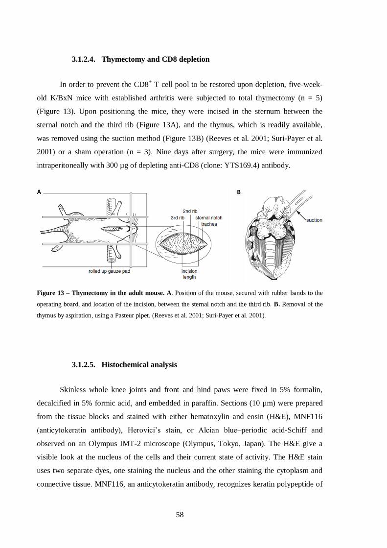
58
3.1.2.4. Thymectomy and CD8 depletion
In order to prevent the CD8+ T cell pool to be restored upon depletion, five-week-
old K/BxN mice with established arthritis were subjected to total thymectomy (n = 5)
(Figure 13). Upon positioning the mice, they were incised in the sternum between the
sternal notch and the third rib (Figure 13A), and the thymus, which is readily available,
was removed using the suction method (Figure 13B) (Reeves et al. 2001; Suri-Payer et al.
2001) or a sham operation (n = 3). Nine days after surgery, the mice were immunized
intraperitoneally with 300 µg of depleting anti-CD8 (clone: YTS169.4) antibody.
Figure 13 – Thymectomy in the adult mouse. A. Position of the mouse, secured with rubber bands to the
operating board, and location of the incision, between the sternal notch and the third rib. B. Removal of the
thymus by aspiration, using a Pasteur pipet. (Reeves et al. 2001; Suri-Payer et al. 2001).
3.1.2.5. Histochemical analysis
Skinless whole knee joints and front and hind paws were fixed in 5% formalin,
decalcified in 5% formic acid, and embedded in paraffin. Sections (10 µm) were prepared
from the tissue blocks and stained with either hematoxylin and eosin (H&E), MNF116
(anticytokeratin antibody), Herovici’s stain, or Alcian blue–periodic acid-Schiff and
observed on an Olympus IMT-2 microscope (Olympus, Tokyo, Japan). The H&E give a
visible look at the nucleus of the cells and their current state of activity. The H&E stain
uses two separate dyes, one staining the nucleus and the other staining the cytoplasm and
connective tissue. MNF116, an anticytokeratin antibody, recognizes keratin polypeptide of
59
45, 46 and 56.5 kDa, and has a broad pattern of reactivity with human epithelial tissues.
The Herovici’s stain is used to differentiate young and mature collagen , and the Alcian
blue–periodic acid-Schiff stain was used to mark glycoproteins (Yamabayashi 1987).
Images were analyzed with ImageJ 1.38x software (National Institutes of Health, Bethesda,
MD, USA).
3.1.2.6. Enzyme-linked immunosorbent assay (ELISA) for GPI
High-affinity Maxisorb 96-well ELISA plates (Nunc, Thermo Scientific, Waltham,
MA, USA) were coated with 10 nM Saccharomyces cerevisiae GPI (Sigma-Aldrich, St.
Louis, MO, USA) in potassium phosphate buffer. Plates were blocked with phosphate
buffered saline/Tween/1% gelatin. Anti-GPI antibodies in sera were detected with
horseradish peroxidase-labeled goat anti-mouse IgG (Southern Biotechnology,
Birmingham, AL, USA) followed by incubation with tetramethylbenzidine solution
(Sigma-Aldrich, St. Louis, MO, USA). Absorption was measured at an optical density of
450 nm.
3.1.2.7. Flow cytometric analysis
Peripheral blood samples were collected from the base of the tails of arthritic
K/BxN mice on days 0, 7, 14, 21, and 35 after the first treatment with either anti-CD8 or
control mAb. Mononuclear cells were isolated through a Ficoll gradient (Amersham, GE
Healthcare, Pittsburg, PA, USA) and stained with antimouse mAb as follows: fluorescein
isothiocyanate (FITC)–labeled anti-CD3 (clone 145.2C11), PE-labeled anti-CD8 (clone
YTS169.4), and allophycocyanin (APC)–labeled anti-CD4 (clone GK1.5-8) (all from the
IGC Cell Imaging Unit, Oeiras, Portugal). To determine the differences in CXCR4 and
CXCR5 expression and the frequency of CD8+ T cell subsets, peripheral blood samples
were collected from the tails of 8-10-week-old untreated arthritic K/BxN mice (n = 9) and
their non-arthritic littermates (n = 10). Mononuclear cells were isolated through a Ficoll
gradient (Amersham, GE Healthcare, Pittsburg, PA, USA) and stained with anti-mouse
mAb, as follows: FITC-labeled anti-CD19 (clone 1D3; IGC Cell Imaging Unit), PE-

60
labeled anti-CXCR4 (eBioscience, San Diego, CA, USA), peridinin chlorophyll A protein
(PerCP)–labeled anti-CD4 (eBioscience, San Diego, CA, USA), APC-labeled anti-CD8
(clone YTS169.4; IGC Cell Imaging Unit, Oeiras, Portugal), FITC-labeled anti-CD3 (clone
145.2C11; IGC Cell Imaging Unit, Oeiras, Portugal), PE-labeled anti-CD8 (clone
YTS169.4; IGC Cell Imaging Unit, Oeiras, Portugal), biotinylated anti-CD62 ligand plus
streptavidin–PerCP, and APC-labeled anti-CD27 (all from eBioscience, San Diego, CA,
USA). For determination of intracellular cytokine production, saponin-permeabilized
peripheral blood mononuclear cells (PBMCs) were stained with APC-labeled anti-CD8
(clone YTS169.4; IGC Cell Imaging Unit, Oeiras, Portugal), PerCP-Cy5.5–labeled anti-
CD3, FITC-labeled anti-TNF-α, and PE-labeled anti–IL-6 (all from eBioscience, San
Diego, CA, USA). All samples were analyzed on a 4-color FACSCalibur system (Becton
Dickinson, Franklin Lakes, NJ, USA), and data were analyzed with FlowJo 7.5.5 software
(Tree Star, Ashland, OR, USA).
3.1.2.8. Assessment of intracellular cytokine production by reverse
transcription–polymerase chain reaction (RT-PCR)
The real-time, fluorescence-based reverse transcription polymerase chain reaction
(RT-PCR) has come to be the go to technique to make the detection, quantification and
evaluation of a target mRNA (Bustin et al. 2005). Here the intracellular production of
cytokines was determined by the assessing their respective mRNAs in CD8+ T cells.
PBMCs and mononuclear cells from articular tissue were collected from untreated
arthritic mice (n = 4), and PBMCs were collected from their healthy control littermates (n
= 4). CD8+ T cells were isolated by magnetic cell separation using the CD8a
+ T Cell
Isolation kit (Miltenyi Biotech, Bergisch Gladbach, Germany).
Total RNA from sorted CD8+ T cells was isolated using an RNeasy Micro kit
(Qiagen, Venlo, Netherlands). RNA integrity and quantification were analyzed using a
6000 Nano Chip kit in an Agilent 2100 Bioanalyzer. RNA was reverse transcribed with a
SuperScript III First-Strand Synthesis System for RT-PCR (Invitrogen, Carlsbad, CA,
USA) using oligo(dT) plus random hexamers according to the manufacturer’s instructions.
Relative quantification of gene expression by real-time PCR was performed using a
thermocycler LightCycler 480 II (Roche, Basel, Switzerland). Normalization for gene

61
expression quantification was performed with geNorm Housekeeping Gene Selection
Mouse kit (PrimerDesign, outhampton, UK) and geNorm software (Ghent University
Hospital, Center for Medical Genetics, Belgium) to select optimal reference genes for this
study (Vandesompele et al. 2002).
Real-time PCRs used specific Mus musculus Quanti-Tect Primer Assays (Qiagen,
Venlo, Netherlands) with optimized primers for the genes of interest, Gzmb (QT00114590)
coding for granzyme B, Ifn (QT01038821) coding for IFN-γ, Il10(QT00106169) coding
for IL-10, Il17a (QT00103278) coding for IL-17, Il2 (QT00112315) coding for IL-2, Il4
(QT00160678) coding for IL-4, Tnf (QT00104006) coding for TNF-α, and the reference
genes Ywhaz (QT00105350) coding for 14-3-3 protein zeta/delta and Rn18s
(QT01036875) coding for 18S ribosomal RNA, together with a QuantiTect SYBR Green
PCR Gene Expression kit (Qiagen, Venlo, Netherlands), according to the manufacturer’s
instructions. Reactions were performed with the following thermal profile: 10 minutes at
95°C plus 40 cycles of 15 seconds at 95°C, 30 seconds at 60°C, and 30 seconds at 72°C.
Quantitative real-time PCR results were analyzed using LightCycler 480 software (Roche,
Basel, Switzerland) and quantified using the qBasePlus software package (Biogazelle,
Zwijnaarde, Belgium).
3.1.2.9. Serum cytokine quantification
The cytokine concentration in the serum was determined by cytometric bead array
(CBA). Different-sized beads with antibodies on their surface will attach to a specific
cytokine, and the medium fluorescence intensity measured by flow cytometry and the
concentration is calculated from a standard curve (Castillo and MacCallum 2012).
Serum samples from arthritic K/BxN mice before (n = 11) and after (n = 11) anti-
CD8 treatment and from their non-arthritic control littermates (n = 7) were obtained from
whole blood after centrifugation. TNF-α, IFN-γ, IL-2, IL-4, IL-5, IL-6, IL-10, IL-12p70,
and monocyte chemoattractant protein 1 (MCP-1) titers in the sera were quantified using
the cytometric bead arrays, a Mouse Th1/Th2 Cytokine kit and a Mouse Inflammation kit
(Becton Dickinson) according to the manufacturer’s instructions, and analyzed with BD
Cytometric Bead Array Software (Becton Dickinson, Franklin Lakes, NJ, USA). IL-17a
titers were determined using a Mouse IL-17A ELISA kit (Invitrogen, Carlsbad, CA, USA).
62
Test sensitivity thresholds for the different cytokines were as follows: for TNF-α, 6.3
pg/ml; for IFN-γ, 2.5 pg/ml; for IL-2, 5.0 pg/ml; for IL-4, 5.0 pg/ml; for IL-5, 5.0 pg/ml;
for IL-6, 5.0 pg/ml; for IL10, 17.5 pg/ml; for IL-12p70, 10.7 pg/ml; for IL-17a, 5.0 pg/ml;
and for MCP-1, 52.7 pg/ml. Mean titers below those thresholds were considered
undetectable.
3.1.2.10. Statistical analysis
Data were checked for normality, in order to decide whether to use the parametric
one-way analysis of variance and post hoc Tukey’s test or the nonparametric Kolmogorov-
Smirnov test. Data were analyzed using StatView 5.0 software (Abacus Concepts). P
values less than 0.05 were considered significant.
3.1.3. B10.Q collagen-induced arthritis mouse model
B10.Q bear the (H-2q) haplotype, that leads to the production of the I-Aq molecule,
and confers susceptibility to collagen-induced arthritis in mice (Nabozny et al. 1994;
Kjellen et al. 1998). H-2, homologous to the human HLA, is a complex of loci on
chromosome 17 that is responsible for defining the MHC in mice.
The homozygous B10.Q mice were a kind gift from Dr. R. Holmdahl (Karolinska
Institutet, Stockholm, Sweden). These mice were kept under normal breeding conditions in
a specific pathogen-free (SPF) facility with a climate-controlled environment and having a
light/dark cycle of 12h. Animals were fed standard rodent chow and water ad libitum in
individually ventilated cages containing corn cob bedding. The animals in use were 2 to 6
months old. For this study only male mice were used.
3.1.3.1. Collagen-induced arthritis

63
Collagen-induced arthritis (CIA) was achieved by immunizing all mice on day 0
with 100 µg of rat collagen type II (rCII) (Chondrex, Redmond, WA, USA) emulsified in
50µl of Freund’s Complete Adjuvant (Sigma-Aldrich, St. Louis, MO, USA). The emulsion
was performed on ice using a syringe-syringe procedure. The immunization was performed
by injecting the emulsion intradermally at the base of the tail, and 35 days later the mice
received a second boost at the same location with an emulsion of 50 µg of rCII and
Freund’s Incomplete Adjuvant (Sigma-Aldrich, St. Louis, MO, USA). The mice were
monitored three times a week and scored for arthritis. Each swollen joint from the fore and
hid paws were given a score of 1 point, each wrist or ankle were given a score of 5 points,
resulting in a maximum of 15 points per paw, and 60 points in total. Scoring was
performed every three days.
3.1.3.2. Flow cytometric analysis
Peripheral blood samples were collected from the base of the tails of B10.Q mice
on days 0, 35 and 70. Peripheral blood mononuclear cells (PBMCs) were obtained after a
red blood cell lysis with a buffer containing 0.84% NH4Cl, and stained with anti-mouse
mAbs: PerCp/Cy5.5-labelled anti-CD3 (clone: 145-2C11), FITC-labeled anti-CD27 (clone:
LG.7F9), PE-labeled anti-CD95 (clone: 15A7), PE-labeled anti-CXCR4 (clone: 2B11),
PE-labeled anti-CCR7 (clone: 4B12), PE-labeled anti-CD40L (clone: MR1), (all from
eBioscience, San Diego, CA, USA), and FITC-labeled anti-CD4 (clone: GK1.5),
PerCp/Cy5.5-labelled anti-CD8 (clone: 53-6.7), APC-labeled anti-CD8 (clone: 53-6.7),
PerCp/Cy5.5-labelled anti-CD62L (clone: MEL-14), APC-labeled anti-CD62L (clone:
MEL-14) and PE-labeled anti-CD69 (clone: H1.2F3) (all from BioLegend San Diego, CA,
USA).
For intracellular cytokine quantitation, after staining for the cell surface antigens,
the samples were formalin-fixed and permeabilized using a saponin-based buffer prior to
the incubation with fluorescence-conjugated mouse anti-human monoclonal antibodies
against: PE-labeled anti-IFN-γ (clone: XMG1.2), FITC-labeled anti-Granzyme B (clone:
16G6), FITC-labeled anti-TNF-α (clone: MP6-XT22), PE-labeled anti-IL-10 (clone: JES5-
16E3), (all from eBioscience, San Diego, CA, USA), and PE-labeled anti-IL-17a (clone:
64
TC11-18H10.1), APC-labeled anti-IL-21 (clone: BL25168) (both from BioLegend San
Diego, CA, USA).
All samples were analyzed on a FACSCalibur flow cytometer (Becton Dickinson,
Franklin Lakes, NJ, USA) and resulting data were quantified using FlowJo Software
(Treestar, Ashland, OR, USA). Analysis of CD8+ T cell subsets was performed on total
CD8+ cells in the lymphocyte gate.
3.1.3.3. Serum cytokine quantification
Serum samples from B10.Q mice were collected at three different times in the
induction of CIA: before the induction (n = 8), before the second collagen boost at day 35
(n = 8) and at the peak of the disease at day 70 (n = 3). The serum was obtained from
whole blood collected into blood collection tubes with clot activator and gel for serum
separation (Microtainer™ Tubes, Becton Dickinson, New Jersey, USA). TNF-α, IFN-γ,
IL-1β, IL-2, IL-4, IL-5, IL-6, IL-10, IL-13, IL-17, IL-22 and IL-27 titers were measured by
cytometric bead arrays, the mouse kit Th1/Th2/Th17 kit FlowCytomix (eBioscience, San
Diego, CA, USA) according to the manufacturer’s instructions.
3.1.3.4. Statistical analysis:
Statistical differences were determined with non-parametric Kruskal-Wallis and
Dunn’s post test to compare the different groups. Data were analyzed using GraphPad
Prism 5 (GraphPad, San Diego, CA, USA). Differences were considered statistically
significant for P values less than 0.05.
65
3.2. Human studies
3.2.1. Human subjects and samples
96 RA patients from Rheumatology Department of Centro Hospitalar Universitário
de Coimbra were enrolled for this study (Table 1). RA disease activity was assessed at the
time of blood collection through tender and swollen joint counts, Erythrocyte
Sedimentation Rate and C-reactive protein) levels. Disease activity groups were defined
according to the DAS28-CRP (3 variables) score: < 2.6 = remission; ≥ 2.6 < 3.2 = low; >
3.2 = moderate to highly active disease (Shammas et al. 2010). SF was collected from
patients with active disease whenever possible (n=10). The use of different medications
was very similar in the three disease-activity groups, with the exception of anti-TNF
agents, used by six patients, all with active disease. A total of 64 gender and age-matched
healthy individuals (HC) were recruited among family members of patients in the same
Department. Exclusion criteria: known or suspected ongoing infections, or, for HC, any
history of autoimmune disease or immunosuppressive therapy.
The study was approved by the institutional ethics committee and performed
according to the Helsinki declaration on studies with human subjects. All subjects signed
an informed written consent prior to any study procedure. Table I summarizes the
demographic, clinical and therapeutic data of all subjects.

66
Table 4 - Clinical characteristics of RA patients and healthy donors.
Therapy
Controls Total RA Active Low Remission
N (SF donors) 64 96 (10) 34 (10) 18 (0) 44 (0)
Gender (F:M) 55:19 77:19 27:7 14:4 36:8
Medication
N (Avg. Dose)
MTX - 82
(17.4 mg/wk)
27
(19.5 mg/wk)
15
(16.3 mg/wk)
39
(16.3 mg/wk)
Hydroxychloroquine - 21
(366.7 mg/day)
9
(344.5 mg/day)
2
(400 mg/day)
10
(380 mg/day)
Sulfasalazine - 20
(1800 mg/day) 6
(1916.7 mg/day) 3
(1833.3 mg/day) 11
(1727.3 mg/day)
Prednisolone - 55
(5.3 mg/day)
21
(6 mg/day)
10
(5.8 mg/day)
25
(4.4 mg/day)
Leflunomide - 2
(15 mg/day)
1
(20 mg/day)
1
(10 mg/day) 0
Azathioprine - 1
(20 mg/day) 0 0
1
(20 mg/day)
Folic Acid - 56
(7,95 mg/wk)
18
(8.9 mg/wk)
8
(6.3 mg/wk)
29
(7.9 mg/wk)
NSAIDs - 50 19 9 22
TNF inhibitors - 5 5 0 0
3.2.2. Flow cytometric analysis
After red blood cell lysis using a hypotonic solution, the peripheral blood
mononuclear cells were stained for cell surface markers using fluorescence conjugated
mouse anti-human monoclonal antibodies against: FITC-labeled anti-CD8 (clone: SK1),
PerCp/Cy5.5-labelled anti-CD3 (clone: UCHT1), APC-labeled anti-CD4 (clone: OKT4),
APC-labeled anti-CD8 (clone: SK1), FITC-labeled anti-CD25 (clone: BC96), FITC-
labeled anti-CD27 (clone: O323), PE-labeled anti-CD27 (clone: M-T271), PerCp/Cy5.5

67
anti-CD62L (clone: DREG-56), PE-labeled anti-CD69 (clone: FN50), PE-labeled anti-
CCR7 (clone: 3D12), APC-labeled anti-CXCR4 (clone: 12G5) (all from BioLegend, San
Diego, CA, USA). For intracellular cytokine quantitation, after staining for the cell surface
antigens, the samples were formalin-fixed and permeabilized using a saponin-based buffer
prior to the incubation with fluorescence conjugated mouse anti-human monoclonal
antibodies against: PE-labeled anti-IFN-γ (clone: 4S.B3), FITC-labeled anti-Granzyme B
(clone: GB11), PE-labeled anti-IL-17a (clone: BL168), Alexa Fluor 488-labelled anti-
TNF-α (clone: MAb11), PE-labeled anti-IL-6 (clone: MQ2-13A5), (all from Biolegend,
San Diego, CA, USA) and PE-labeled anti-IL-10 (clone: JES3-19F1 ) (BD Biosciences,
Becton Dickinson, New Jersey, USA) and FITC-labeled anti-Perforin (clone: delta G9)
(Immunotools, Friesoythe, Germany). Irrelevant, directly conjugated, murine IgG1 or IgG2
(Biolegend, San Diego, CA, USA) were used to ascertain background staining. All samples
were analyzed on a FACScalibur cytometer (Becton Dickinson, Franklin Lakes, NJ, USA),
with 50000 events collected within the lymphocyte gate. After calibration with CST beads
single-fluorochrome stained cells were used for instrument compensation and PMT-setup.
Resulting data were quantified using FlowJo Software (Treestar, Ashland, OR, USA).
Analysis of CD8+ T cell subsets was performed on total CD8+ T cells in the lymphocyte
gate. Table 5 summarizes the markers profile for each subset.
Table 5 - CD8+ T cell phenotypes and surface markers
CD8+ T cell phenotypes Name
CD25+ Activated cells (late activation marker)
CD69+ Activated cells (early activation marker)
CD27+CD62L
+ Central memory cells
CD27+CD62L
- Effector memory cells
CD27-CD62L
- (CCR7
-) Short-term effector cells
CXCR4+ "Homing" chemokine receptor
CD62L-CD69
+ Activated effector cells

68
3.2.3. Statistical analysis
SPSS v.20 (IBM, Armonk, New York, USA) was used to analyze the results. We
elected to compare cells obtained from people with active RA (DAS28 > 3.2), vs. cells
from RA patients in remission (DAS28 < 2.6) vs. cells obtained from age and gender-
matched HC. Differences between independent samples were assessed through one-way
ANOVA followed by LSD post-hoc test. Paired PB and SF samples were compared
through the Wilcoxon rank sum test. Correlation between PB and SF was analyzed using
Spearman correlation coefficient. Correlation between DAS28 and PB CD8+
T cells was
analyzed using the Pearson correlation including all RA patients. PB CD8+
T cells were
also correlated with MTX and glucocorticoid’s doses through Pearson Correlation.
Correlation coefficients were considered weak for R above 0.1, moderate for R values
above 0.3, strong above 0.5 and very strong above 0.75.
In order to explore whether the influence of therapy upon the changes in biological
parameters significantly correlated with DAS in univariate analysis, we performed a
multivariate linear regression analysis of these measures, including the doses of
medications (methotrexate, antimalarials, glucocorticoids and sulfasalazine) and DAS28 as
covariates.
Statistical significance was considered for p < 0.05 in all analyses.
69
CHAPTER 4
MONOCLONAL ANTI-CD8 THERAPY INDUCES DISEASE
AMELIORATION IN THE K/BXN MOUSE MODEL OF SPONTANEOUS
CHRONIC POLYARTHRITIS


71
4. Monoclonal Anti-CD8 Therapy Induces Disease
Amelioration in the K/BxN Mouse Model of Spontaneous
Chronic Polyarthritis
4.1. Introduction
Approximately 40% of the T cells infiltrating the rheumatoid synovial membrane
are CD8+ T cells (McInnes 2003). However, their importance in the pathogenesis of
rheumatoid arthritis (RA) remains to be fully elucidated.
The primary function of CD8+ T cells is the killing of virus- or cytosolic bacteria–
infected cells. Moreover, they seem to play several important roles in autoimmune
diseases, either protecting against or enhancing the disease. In experimental autoimmune
encephalomyelitis (EAE), an animal model of multiple sclerosis, CD8+ T cells have been
shown to be crucial for resistance to a second induction of the disease (Jiang et al. 1992).
Recently, a particular subset of CD8+ T cells (CD8
+CD122
+) was shown to accelerate the
recovery of animals with EAE after CD8+ T cells were transferred (Lee et al. 2008). In
contrast, insulitis failed to develop in NOD mice treated with anti-CD8 monoclonal
antibodies (mAb) (Wang et al. 1996). This experimental treatment also inhibited the
transfer of insulin-dependent diabetes mellitus (IDDM) and the development of
spontaneous IDDM (Parish et al. 1998).
In RA, some patients show CD8+ T cell clonal expansions with a memory
phenotype that are correlated with rheumatoid factor (RF) levels (al-Azem et al. 1992;
Fitzgerald et al. 1995; Neidhart et al. 1996; Neidhart et al. 1996; Masuko-Hongo et al.
1997). This is most likely attributable to the important role of CD8+ T cells in maintaining
ectopic germinal center structures in RA synovium (Wagner et al. 1998; Kang et al. 2002).
In different animal models of collagen-induced arthritis (CIA), the absence of CD8+
T cells resulted in a reduced incidence (Larsson et al. 1989; Tada et al. 1996) and severity
(Kiely et al. 1996) of the disease. However, higher susceptibility was observed when
animals were rechallenged (Kiely et al. 1996; Tada et al. 1996). Additionally, in CD8+/-
and CD8-/-
mice, a trend toward a delayed onset of CIA was observed, without a significant
impact on disease susceptibility (Ehinger et al. 2001). More recently, CD8+ T cell clones
72
generated from the arthritic joints of SKG mice transferred to histocompatible athymic
nude mice led to joint swelling and synovitis with destruction of cartilage and bone
(Wakasa-Morimoto et al. 2008).
In order to assess the role of CD8+ T cells in experimental chronic polyarthritis, the
clinical phenotype and cytokine production of articular and peripheral blood CD8+ T cells
from K/BxN mice were studied. Arthritis in these mice results from the simultaneous
expression of the class II major histocompatibility complex Ag7
molecule and a transgenic
T cell receptor (TCR), followed by the production of autoantibodies against glucose-6-
phosphate isomerase (GPI) (Kouskoff et al. 1996; Korganow et al. 1999; Matsumoto et al.
1999). Subsequently, we assessed whether treatment with specific anti-CD8 mAb, with and
without thymectomy, improved the course of established arthritis in K/BxN mice. Our
results showed, for the first time, that K/BxN mouse activated and effector memory CD8+
T cells are present in the peripheral blood and joints and that they play an important role in
arthritis maintenance, because treatment with specific anti-CD8 mAb significantly
improved the disease signs. These results document that CD8+ T cells should be regarded
as major players in the K/BxN mouse model of experimental arthritis, along with CD4+ T
cells and B cells.
73
4.2. Results
4.2.1. Activation of K/BxN mouse CD8+ T cells in the articular
infiltrate
In an effort to characterize the CD8+ T cells in K/BxN mice, mononuclear cells
were isolated from the peripheral blood and from the articular inflammatory infiltrate and
analyzed for the expression of surface markers and cytokine production. In contrast to the
circulating CD4+ T cell pool, a significantly higher (P< 0.05) percentage of circulating
CD8+ T cells from K/BxN mice expressed the Vβ6-transgenic TCR (mean ± SD 32 ± 10%
and 84 ± 7% for CD4 and CD8, respectively) at 3 weeks after birth, before any external
clinical signs of arthritis could be detected.
The frequencies of CD8+ T cell subsets defined by the expression of CD27 and
CD62L in the peripheral blood and articular infiltrate from arthritic K/BxN mice were
compared with those in the peripheral blood of healthy mice (Figure 14A). The frequency
of CD27 -CD62L-short-lived effector CD8+ T cells (Tse) was similar in both K/BxN
mouse tissue and peripheral blood from healthy mice. However, the peripheral blood of
arthritic K/BxN mice presented a significantly (P = 0.019) higher frequency of
CD27+CD62L
-effector memory CD8
+ T cells (Tem) than the peripheral blood of healthy
mice. Moreover, the frequency of this Tem subset was higher, although not reaching
statistical significance (P= 0.0791), in the articular infiltrate than in the peripheral blood of
K/BxN mice. The frequency of CD27+CD62L
+ central memory CD8
+ T cells (Tcm) was
comparable in the peripheral blood of K/BxN mice and that of healthy mice. However, the
frequency of Tcm was significantly lower (P = 0.008) in the articular infiltrate of K/BxN
mice than in the peripheral blood of K/BxN mice.
In contrast to what was observed in healthy mice, the majority of CD8+ T cells
circulating in K/BxN mouse peripheral blood expressed the early activation marker CD69,
and this increased expression of CD69 was also observed on the surface of CD8+ T cells
infiltrating the joints.
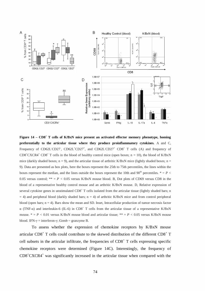
74
Figure 14 – CD8+ T cells of K/BxN mice present an activated effector memory phenotype, homing
preferentially to the articular tissue where they produce proinflammatory cytokines. A and C,
Frequency of CD62L-CD27-, CD62L+CD27+, and CD62L-CD27+ CD8+ T cells (A) and frequency of
CD8+CXCR4+ CD8+ T cells in the blood of healthy control mice (open boxes; n = 10), the blood of K/BxN
mice (darkly shaded boxes; n = 9), and the articular tissue of arthritic K/BxN mice (lightly shaded boxes; n =
9). Data are presented as box plots, here the boxes represent the 25th to 75th percentiles, the lines within the
boxes represent the median, and the lines outside the boxes represent the 10th and 90th percentiles. * = P <
0.05 versus control; ** = P < 0.05 versus K/BxN mouse blood. B, Dot plots of CD69 versus CD8 in the
blood of a representative healthy control mouse and an arthritic K/BxN mouse. D, Relative expression of
several cytokine genes in unstimulated CD8+ T cells isolated from the articular tissue (lightly shaded bars; n
= 4) and peripheral blood (darkly shaded bars; n = 4) of arthritic K/BxN mice and from control peripheral
blood (open bars; n = 4). Bars show the mean and SD. Inset, Intracellular production of tumor necrosis factor
α (TNF-α) and interleukin-6 (IL-6) in CD8+ T cells from the articular tissue of a representative K/BxN
mouse. * = P < 0.01 versus K/BxN mouse blood and articular tissue; ** = P < 0.05 versus K/BxN mouse
blood. IFN-γ = interferon-γ; Gzmb = granzyme B.
To assess whether the expression of chemokine receptors by K/BxN mouse
articular CD8+ T cells could contribute to the skewed distribution of the different CD8
+ T
cell subsets in the articular infiltrate, the frequencies of CD8+ T cells expressing specific
chemokine receptors were determined (Figure 14C). Interestingly, the frequency of
CD8+CXCR4
+ was significantly increased in the articular tissue when compared with the
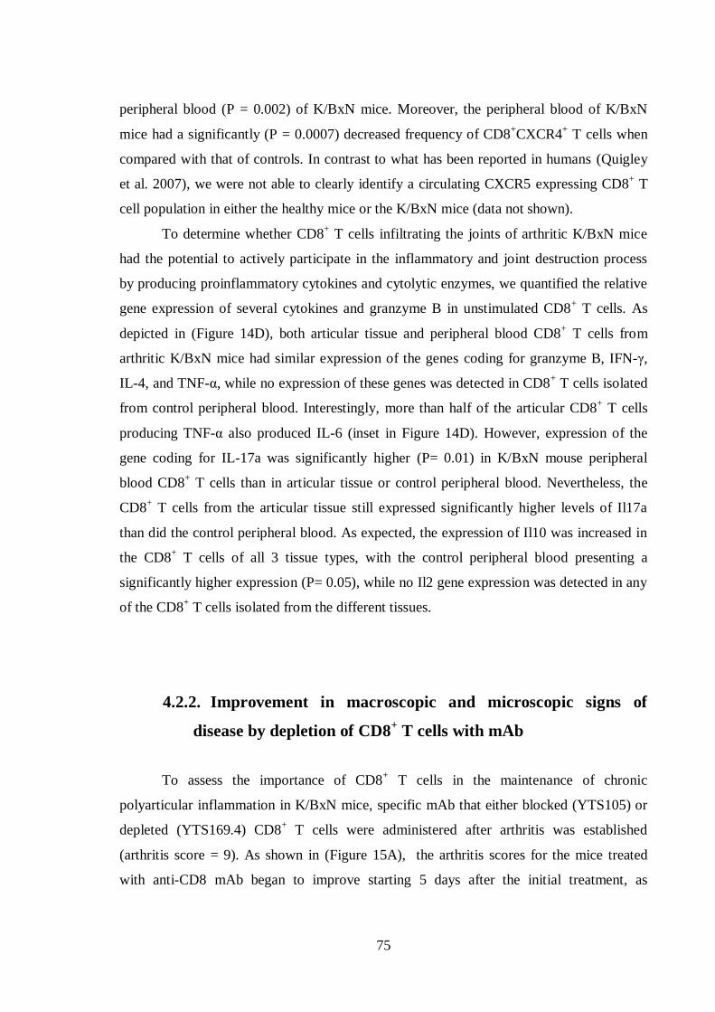
75
peripheral blood (P = 0.002) of K/BxN mice. Moreover, the peripheral blood of K/BxN
mice had a significantly (P = 0.0007) decreased frequency of CD8+CXCR4
+ T cells when
compared with that of controls. In contrast to what has been reported in humans (Quigley
et al. 2007), we were not able to clearly identify a circulating CXCR5 expressing CD8+ T
cell population in either the healthy mice or the K/BxN mice (data not shown).
To determine whether CD8+ T cells infiltrating the joints of arthritic K/BxN mice
had the potential to actively participate in the inflammatory and joint destruction process
by producing proinflammatory cytokines and cytolytic enzymes, we quantified the relative
gene expression of several cytokines and granzyme B in unstimulated CD8+ T cells. As
depicted in (Figure 14D), both articular tissue and peripheral blood CD8+ T cells from
arthritic K/BxN mice had similar expression of the genes coding for granzyme B, IFN-γ,
IL-4, and TNF-α, while no expression of these genes was detected in CD8+ T cells isolated
from control peripheral blood. Interestingly, more than half of the articular CD8+ T cells
producing TNF-α also produced IL-6 (inset in Figure 14D). However, expression of the
gene coding for IL-17a was significantly higher (P= 0.01) in K/BxN mouse peripheral
blood CD8+ T cells than in articular tissue or control peripheral blood. Nevertheless, the
CD8+ T cells from the articular tissue still expressed significantly higher levels of Il17a
than did the control peripheral blood. As expected, the expression of Il10 was increased in
the CD8+ T cells of all 3 tissue types, with the control peripheral blood presenting a
significantly higher expression (P= 0.05), while no Il2 gene expression was detected in any
of the CD8+ T cells isolated from the different tissues.
4.2.2. Improvement in macroscopic and microscopic signs of
disease by depletion of CD8+ T cells with mAb
To assess the importance of CD8+ T cells in the maintenance of chronic
polyarticular inflammation in K/BxN mice, specific mAb that either blocked (YTS105) or
depleted (YTS169.4) CD8+ T cells were administered after arthritis was established
(arthritis score = 9). As shown in (Figure 15A), the arthritis scores for the mice treated
with anti-CD8 mAb began to improve starting 5 days after the initial treatment, as
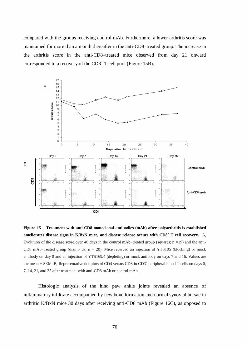
76
compared with the groups receiving control mAb. Furthermore, a lower arthritis score was
maintained for more than a month thereafter in the anti-CD8–treated group. The increase in
the arthritis score in the anti-CD8–treated mice observed from day 21 onward
corresponded to a recovery of the CD8+ T cell pool (Figure 15B).
Figure 15 – Treatment with anti-CD8 monoclonal antibodies (mAb) after polyarthritis is established
ameliorates disease signs in K/BxN mice, and disease relapse occurs with CD8+ T cell recovery. A,
Evolution of the disease score over 40 days in the control mAb–treated group (squares; n =19) and the anti-
CD8 mAb–treated group (diamonds; n = 20). Mice received an injection of YTS105 (blocking) or mock
antibody on day 0 and an injection of YTS169.4 (depleting) or mock antibody on days 7 and 16. Values are
the mean ± SEM. B, Representative dot plots of CD4 versus CD8 in CD3+ peripheral blood T cells on days 0,
7, 14, 21, and 35 after treatment with anti-CD8 mAb or control mAb.
Histologic analysis of the hind paw ankle joints revealed an absence of
inflammatory infiltrate accompanied by new bone formation and normal synovial bursae in
arthritic K/BxN mice 30 days after receiving anti-CD8 mAb (Figure 16C), as opposed to
77
mice receiving control mAb, which presented an inflamed hyperplastic synovium and
articular erosions (Figure 16D).
Figure 16 – Histologic assessment of articular tissue shows clearance of the inflammatory infiltrate in
anti-CD8 monoclonal antibody–treated K/BxN mice. A, Joint section from a healthy control mouse (H&E
stained; original magnification × 10). Inset, Normal synovial bursae (Herovici stained; original magnification
× 200). B, Joint section from a K/BxN mouse before treatment, showing massive inflammation and
cartilage/bone destruction (H&E stained; original magnification × 10). Upper inset, inflamed hyperplastic
synovium (Herovici stained; original magnification × 100). Lower inset, Chondral sclerosis and fibrous
ankylosis (Alcian blue–periodic acid_Schiff stained; original magnification × 100). C, Joint section from an
anti-CD8 monoclonal antibody–treated K/BxN mouse 30 days after treatment, showing complete clearance
of the inflammatory infiltrate and normalization of the articular architecture (H&E stained; original
magnification × 10). Inset, Normal synovial bursae (Herovici stained; original magnification × 100). D, Joint
section from a control monoclonal antibody– treated K/BxN mouse 30 days after treatment, showing
complete destruction of the joint structure by massive infiltration of inflammatory cells and fibrosis (H&E
stained; original magnification × 10). Inset, Proliferative synovitis with destruction of articular cartilage
(Alcian blue–periodic acid–Schiff stained; original magnification × 100).
To investigate whether the arthritis improvement after anti-CD8 therapy was
associated with changes in the levels of circulating cytokines, the concentrations of IL-2,
IL-4, IL-5, IL-6, IL-10, IL12p70, IL-17a, MCP-1, TNF-α, and IFN-γ were measured in the
serum of healthy control and K/BxN mice at baseline and 20 days after the initial treatment

78
(Figure 17 A-D). Arthritic K/BxN mice that were assessed before treatment had
significantly higher (P= 0.02) serologic titers of IL-5, IL-6, and TNF-α than healthy
controls. Twenty days after anti-CD8 therapy, the serologic levels of all 3 cytokines and
IFN-γ had significantly dropped (P = 0.04) from their baseline values and were comparable
with the ones present in healthy control mice. The serologic levels of all other cytokines
did not pass the minimum test threshold.
Figure 17 – Treatment with anti-CD8 monoclonal antibodies normalizes the serologic levels of
proinflammatory cytokines in K/BxN mice. Titers of tumor necrosis factor α (TNF-α) (A), interferon-γ
(IFN-γ) (B), interleukin-6 (IL-6) (C), and IL-5 (D) in the blood of untreated K/BxN mice (lightly shaded
boxes; n = 11), the blood of healthy control mice (open boxes; n = 7), and the blood of anti-CD8–treated
K/BxN mice (darkly shaded boxes; n = 11) are shown. Data are presented as box plots, where the boxes
represent the 25th to 75th percentiles, the lines within the boxes represent the median, and the lines outside
the boxes represent the 10th and 90th percentiles.
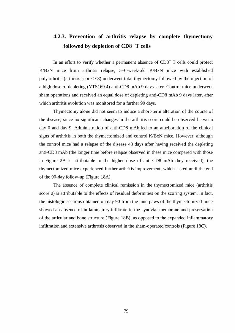
79
4.2.3. Prevention of arthritis relapse by complete thymectomy
followed by depletion of CD8+ T cells
In an effort to verify whether a permanent absence of CD8+ T cells could protect
K/BxN mice from arthritis relapse, 5–6-week-old K/BxN mice with established
polyarthritis (arthritis score > 8) underwent total thymectomy followed by the injection of
a high dose of depleting (YTS169.4) anti-CD8 mAb 9 days later. Control mice underwent
sham operations and received an equal dose of depleting anti-CD8 mAb 9 days later, after
which arthritis evolution was monitored for a further 90 days.
Thymectomy alone did not seem to induce a short-term alteration of the course of
the disease, since no significant changes in the arthritis score could be observed between
day 0 and day 9. Administration of anti-CD8 mAb led to an amelioration of the clinical
signs of arthritis in both the thymectomized and control K/BxN mice. However, although
the control mice had a relapse of the disease 43 days after having received the depleting
anti-CD8 mAb (the longer time before relapse observed in these mice compared with those
in Figure 2A is attributable to the higher dose of anti-CD8 mAb they received), the
thymectomized mice experienced further arthritis improvement, which lasted until the end
of the 90-day follow-up (Figure 18A).
The absence of complete clinical remission in the thymectomized mice (arthritis
score 0) is attributable to the effects of residual deformities on the scoring system. In fact,
the histologic sections obtained on day 90 from the hind paws of the thymectomized mice
showed an absence of inflammatory infiltrate in the synovial membrane and preservation
of the articular and bone structure (Figure 18B), as opposed to the expanded inflammatory
infiltration and extensive arthrosis observed in the sham-operated controls (Figure 18C).

80
Figure 18 – Thymectomy followed by CD8+ T cell depletion stops arthritis relapse, reduces the
inflammatory infiltration of the joint, and preserves bone and articular integrity in K/BxN mice. A,
Evolution of the arthritis score for 90 days after CD8+ T cell depletion in sham-operated control mice
(squares; n = 3) and thymectomized mice (diamonds; n = 5). Surgery was performed on day 9, and CD8
depletion was performed on day 0. Values are the mean ± SEM. B, Section from the hind paw of a K/BxN
mouse 90 days after thymectomy and CD8+ T cell depletion (H&E stained; original magnification × 10).
Inset, Preserved joint and synovial proliferation without inflammation (MNF116 stained; original
magnification × 100). C, Section from the hind paw of a K/BxN mouse 90 days after sham operation and
CD8+ T cell depletion (H&E stained; original magnification × 10). Inset, Arthrosis of the joint (Herovici
stained; original magnification × 40).
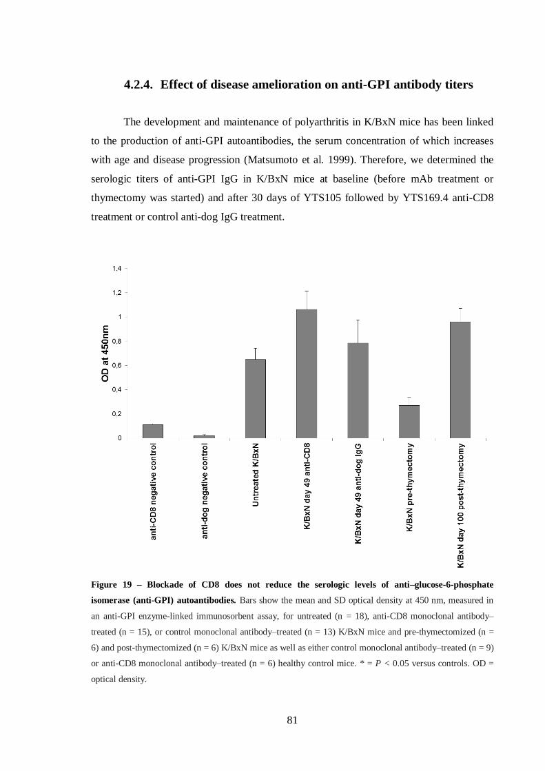
81
4.2.4. Effect of disease amelioration on anti-GPI antibody titers
The development and maintenance of polyarthritis in K/BxN mice has been linked
to the production of anti-GPI autoantibodies, the serum concentration of which increases
with age and disease progression (Matsumoto et al. 1999). Therefore, we determined the
serologic titers of anti-GPI IgG in K/BxN mice at baseline (before mAb treatment or
thymectomy was started) and after 30 days of YTS105 followed by YTS169.4 anti-CD8
treatment or control anti-dog IgG treatment.
Figure 19 – Blockade of CD8 does not reduce the serologic levels of anti–glucose-6-phosphate
isomerase (anti-GPI) autoantibodies. Bars show the mean and SD optical density at 450 nm, measured in
an anti-GPI enzyme-linked immunosorbent assay, for untreated (n = 18), anti-CD8 monoclonal antibody–
treated (n = 15), or control monoclonal antibody–treated (n = 13) K/BxN mice and pre-thymectomized (n =
6) and post-thymectomized (n = 6) K/BxN mice as well as either control monoclonal antibody–treated (n = 9)
or anti-CD8 monoclonal antibody–treated (n = 6) healthy control mice. * = P < 0.05 versus controls. OD =
optical density.
82
Similar assessments were performed 90 days after thymectomy or sham operation
and YTS169.4 treatment in K/BxN mice and age-matched nonarthritic control littermates
(treated either with YTS105 followed by YTS169.4 anti-CD8 or control anti-dog IgG), but
no significant effects of treatment on the anti-GPI IgG titers were observed (Figure 19). No
significant effects of treatment on anti-GPI IgG titers were observed (Figure 19). Actually,
the titers of anti-GPI antibodies increased in the thymectomized mice even though
inflammation of the joints subsided. The frequency of circulating CD138+ plasma cells was
also not affected by any of the treatments (data not shown).
83
4.3. Discussion
The role of CD8+ T cells in the pathogenesis of RA remains unclear. Nevertheless,
several studies in patients with RA that associated the effector functions and the memory
CD45RO+ and activated “false” memory CD29
+CD45RA
+CD45RO
- phenotypes of CD8
+
T cells with RF production and disease activity point out that the contribution of CD8+ T
cells to RA should be reevaluated (al-Azem et al. 1992; Fitzgerald et al. 1995; Neidhart et
al. 1996; Neidhart et al. 1996). This is also the case in animal models, because data from
the literature on experimental arthritis are few and contradictory. Several studies on the
CIA model of experimental polyarthritis focusing on the involvement of CD8+ T cells in
initiating arthritis (Larsson et al. 1989; Williams et al. 1989; Tada et al. 1996) revealed that
anti-CD8 treatment rendered experimental animals less susceptible (Tada et al. 1996) or
fully resistant (Larsson et al. 1989) to the disease. Another study involving NOD/SCID
mice engrafted with human rheumatoid synovium stressed the importance of CD8+ T cells
for the maintenance of synovial follicular-like structures (Kang et al. 2002).
Most studies were carried out in the CIA model and manipulated the CD8+ T cell
response before arthritis induction, thus focusing on the potential role of CD8+ cells in the
initiation of the disease (Larsson et al. 1989; Williams et al. 1989; Tada et al. 1996;
Ehinger et al. 2001). This is consistent with current paradigms regarding the role of CD8+
cells but was also favored because of the transient nature of CIA, which would render it
difficult to distinguish between the ameliorating effect of CD8 blockade and natural
disease remission. Therefore, the contribution of CD8+ T cells to the chronicity of
polyarthritis has not been addressed.
The recent development of murine models of persistent chronic polyarthritis - the
SKG (Sakaguchi et al. 2003), the K/BxN (Kouskoff et al. 1996), and the B10.Q/Ncf1*
(Gelderman et al. 2006) models - provide new tools for studying CD8+ T cell involvement
in arthritis maintenance.
In the present study, we used the K/BxN mouse model of chronic polyarthritis to
show that CD8+ T cells circulating in the peripheral blood and infiltrating the joints are
responsible for maintaining chronic articular inflammation. An evident decrease in
articular swelling and redness a few days after the initial anti-CD8 treatment was
confirmed by the absence of histologic signs of inflammatory infiltrates and evidence of de
84
novo ossification. Moreover, normalization of the serologic levels of proinflammatory
cytokines, such as TNF-α, IFN-γ, and IL-6, in the anti-CD8 mAb–treated mice represents
evidence that the role of CD8+ T cells in arthritis maintenance is at least partially mediated
through self-production of these cytokines or by (co)stimulation of production in other
cells. Additionally, normalization of the serologic levels of IL-5, a cytokine involved in
growth and differentiation of both B cells and eosinophils (Yokota et al. 1987), after anti-
CD8 treatment accompanied a reduction in joint inflammation. Further evidence for the
involvement of CD8+ T cells in K/BxN mouse polyarthritis was provided by the disease
relapse observed in treated mice as soon as the numbers of circulating CD8+ T cells were
normalized.
Nevertheless, it was important to establish whether the permanent absence of CD8+
T cells prevented arthritis relapse. Therefore, 5-week-old K/BxN mice with established
polyarthritis were thymectomized and subsequently inoculated with a high dose of CD8+ T
cell–depleting mAb. Amelioration of the clinical signs of arthritis was evident after 2
weeks, and no relapses were observed in the 90-day follow-up period. In fact, after those
90 days, normal levels of TCR-transgenic CD4+ T cells were still present in the circulation,
and the levels of B cells and plasma cells did not change when compared with those in
sham-operated mice. Such observations strengthen the hypothesis that CD8+ T cells, and
not only CD4+ T cells and B cells (Kouskoff et al. 1996), are essential to the maintenance
and even the initiation of chronic polyarthritis in K/BxN mice. In contrast to findings with
therapies involving CD40 blockade (Kyburz et al. 2000), no changes were observed in the
serologic levels of anti-GPI autoantibodies after any of the anti-CD8 therapies, suggesting
that CD8 blockade stops/reverses arthritis progression without influencing the autoreactive
B cell and plasma cell pools.
Even though the CD8+ T cells of K/BxN mice express the transgenic Vβ6 TCR,
thus rendering them potentially autoreactive (as extensively described for the K/BxN
mouse TCR-transgenic CD4+ T cells (Kouskoff et al. 1996)), they have been poorly
studied. A functional and phenotypic characterization of the transgenic CD8+ T cells in this
mouse model is especially important in view of its larger and earlier expansion in
comparison with CD4+ T cells: the CD8
+ T cell pool comprised up to 85% of TCR-
transgenic cells 21 days after birth and 2 weeks before arthritis was established.
85
CD8+ T cells are usually subdivided into particular phenotypes with characteristic
effector functions, homing properties, and proliferative capacity. The expansion phase after
antigen presentation is dominated by short-lived effector CD8+CD27
-CD62L
- T cells (Tse)
capable of producing proinflammatory cytokines (IFN-γ, TNF-α, IL-2, IL-17) and
cytotoxic molecules (perforin, granzyme B). These cells heavily migrate into the peripheral
organs (Baars et al. 2005; Stemberger et al. 2007; Tajima et al. 2008). Upon interaction
with CD154 expressed on helper CD4+ T cells (Tanchot and Rocha 2003; Huster et al.
2004) (33,34), subsets of effector CD8+ T cells and antigen-primed naive CD8
+ T cells,
respectively, develop into CD8+CD27
+CD62L
- effector memory cells (Tem) or
CD8+CD27
+CD62L
+ central memory cells (Tcm) (Kaech et al. 2003; Jabbari and Harty
2006; Hikono et al. 2007; Stemberger et al. 2007). While Tem accumulate in the peripheral
organs and rapidly become effector cells upon reencounter with antigen but have poor
expansion and self-renewal capacity, Tcm accumulate in the lymphoid organs and are
capable of large expansion upon antigen reencounter and frequent self-renewal (Kaech et
al. 2003; Lefrancois and Marzo 2006; Stemberger et al. 2007). Considering these
functional and homing differences, it is not surprising to observe that the K/BxN mouse
articular tissue showed an accumulation of the 2 effector subsets, particularly the Tem
subset, which are more likely to participate in the local autoantigen-driven tissue
destruction.
The presence of TNF-α-, IL-6-, IFN-γ-, IL-17-, and granzyme B-producing CD8+ T
cells in the articular infiltrate and the elevated frequency of CD8+ T cells expressing the
homing chemokine CXCR4 suggest that the joint-infiltrating effector CD8+ T cells might
be subdivided into 2 main groups. A first group might be actively participating in joint
destruction through granzyme B secretion. A second group may be involved in the
recruitment and priming of other immune cells into the joint, which are the IL-17a-
producing CD8+ T cells that have been described as proinflammatory but with reduced
cytotoxic potential (Huber et al. 2009). Additionally, the elevated presence of both CD69-
expressing CD8+ T cells, which are markers of early activation, and Tem in the peripheral
blood of arthritic K/BxN mice suggests that there is continuous systemic activation, and
eventually recruitment, of (pathogenic) CD8+ T cells in the K/BxN mouse model of
spontaneous chronic polyarthritis.


87
CHAPTER 5
CD8+ T CELLS IN THE COLLAGEN-INDUCED ARTHRITIS MODEL

89
5. CD8+ T cells in the collagen-induced arthritis model
5.1. Introduction
Collagen-induced arthritis is an animal model of RA that is commonly used, and
extensively studied, as it shares various similarities with human RA. CIA is an
inflammatory disease that develops in the joints as a result of an experimentally induced
immune response of B and T cells against collagen type II (CII). It is clinically
characterized by the development of chronic and destructive inflammation in the paws.
Pathology reveals hyperplasia and inflammatory infiltration of the synovial membrane
associated with bone erosion and cartilage degradation. CIA is induced by immunizing
mice from susceptible strains with heterologous type II collagen (CII) in Complete
Freund’s Adjuvant (CFA). The arthritis develops within 3 weeks after immunization. CIA
has been intensively studied in rats (Trentham et al. 1977) as well as in susceptible mouse
strains (Boissier et al. 1987).
While the requirement for T cells in the development of CIA is undeniable, the
underlying mechanisms are not fully understood, and the role of CD8+ T cells in CIA
remains unclear. In fact, some several studies have yielded contrasting results regarding the
role of CD8+ T cells in CIA (Chiocchia et al. 1993; Gao and McMichael 1996). However,
the depletion of CD8+ T cells in CIA has been reported to not have a significant effect on
the disease in the rat (Larsson et al. 1989), but appears to suppress the disease in mice
(Arai et al. 1996). A study with CIA in DBA/1 CD8-knock-out mice has reported a lower
incidence of the disease, even though the severity of CIA is maintained, suggesting that
CD8+ T cells have a regulatory function in arthritis (Tada et al. 1996). Another study in
B10.Q CD8-knock-out mice reported that the lack of CD8+ T cells had no significant
impact on the disease (Ehinger et al. 2001).
We set out to determine the role of CD8+ T cells in the pathogenesis of CIA. To this
purpose, we induced the disease in the susceptible B10.Q mouse strain and assessed the
phenotype of peripheral CD8+ T cells and their intracellular cytokine levels at 3 different
stages of the induction of the disease: before the induction, intermediate state and peak of
the disease. We found string suggestions that CD8+ T cells display altered phenotypes and
their intracellular cytokine production is altered upon the induction of CIA. However,

90
definite conclusions have been hampered by difficulties encountered in the reproducibility
of the model, which could not be totally overcome before the closure of this thesis. This
work presented herein not as a conclusive piece of research but rather as a report of the
learning experience derived from the work, together with limited data collected from the
experiments and its possible interpretations.

91
5.2. Results
5.2.1. Induction of CIA in B10.Q mice – troubleshooting
As explained before, CIA in B10.Q mice is characterized by the onset of arthritis
between days 20 – 30 after induction. Upon a second boost with type II collagen, the mice
display overt clinical features of arthritis, with the peak of the disease severity being
observed at days 60 - 70. The incidence is of about 60 - 80% in male mice. However, in the
course of this study, mice consistently failed to develop arthritis at the incidence and
severity expected for this strain. Preliminary results were actually very encouraging, with
an incidence of 80% in 2 month-old mice and 100% in 6 month-old mice with average
severity scores of up to 40 in the 6 month-old group, and 24 in the 2-month-old group.
However, the incidence dropped to about 0% in subsequent experiments, with mice
reaching arthritis scores significantly lower than expected (average ranging from 9 to 14).
Several reasons were considered to explain these results, with emphasis on housing
conditions and environmental stress. Housing conditions, namely, the amount and variety
of pathogens that the mice are exposed to can have a dramatic effect in the development of
experimental arthritis. For example, the SKG spontaneous poly-arthritis mouse strain only
develops arthritis when bred in SPF conditions, but in open cages. In SPF conditions, but
within constrained cages with filtered ventilation system (venti-rack cages), these mice are
healthy and only develop arthritis when injected with zymosan (Kobayashi et al. 2006).
This hypothesis is particularly viable in our case, since the mice used in the preliminary
results, where an incidence of 80% for 2 month-old mice was achieved, had been bred in
conventional conditions, while the other mice used in subsequent experiments were
maintained in SPF conditions. In fact, the team that provided us these mice had a similar
experience, observing lower incidence and severity of arthritis in SPF conditions
(Batsalova et al. 2012; Forster et al. 2012) when compared to conventional housing
practices (Geng et al. 2008).
One of the reasons for a reduced ability of mice reared in SPF conditions using
venti-rack cages to develop severe arthritis may be related to the fact that these mice lack
several commensal microbiota, which have long been proven to have an influence on the
development of the immune system, including its maturation and development of the B cell

92
repertoire (Coates 1975; Rhee et al. 2004; Lanning et al. 2005; Mazmanian et al. 2005). In
fact, even alterations in the strains’ nutrition (Nagura et al. 2005), along with other
husbandry practices, can result in an altered microflora and is one of the most overlooked
variables that can potentially alter mouse physiology and experimental outcomes (Ma et al.
2012).
Changes in collagen origin and quality were also considered as potential causes for
low incidence of arthritis in our hands. In fact, collagen molecules present differences in
their sequence depending on the species they are isolated from, as the sequence recognized
by the immune system of the mouse is strain specific. For example, mice expressing the I-
Aq molecule, such as the B10.Q and DBA/1 strains, are responsive to rat, bovine, chick,
and human, but not to porcine type II collagen. Conversely, mice expressing I-Ar, such as
the B10.RIII strain, develop arthritis when immunized with bovine or porcine, but not with
chick or human type II collagen (Wooley et al. 1985; Brand et al. 2003). The initial
immunizations on B10.Q mice had been made with bovine type II collagen. However,
taking the above facts into consideration we admitted that the collagen could have been
degraded or denatured, and therefore could not successfully induce arthritis in mice
(denatured collagen does not induce arthritis in susceptible breeds (Stuart et al. 1982)).
This prompted us to switch to rat type II collagen. Two different forms of rat type II
collagen were tested. The first batch was purchased from Dr. Rikard Holmdahl’s lab
(Karolinska Institute, Sweden), which also yielded unsatisfactory arthritis incidence levels.
The second batch, purchased from Chondrex, Inc ((Chondrex, Redmond, WA, USA),
allowed for somewhat better, though not fully satisfactory, results. Experiments performed
with this collagen are described and discussed below.
CIA was induced in 2 month old B10.Q mice, through immunization with an
emulsion composed of rat type II collagen (Chondrex) and complete Freund’s adjuvant at
the beginning of the experiment, and with incomplete Freund’s adjuvant at day 35 for the
second boost. The incidence of arthritis is shown in Figure 20. Mice started to develop
arthritis about 24 days after the first injection, reaching the peak of the disease around day
70. The incidence, however, was only 37%, with only 3 out of 8 mice displaying overt
clinical features of arthritis. The highest arthritis score, observed in only one animal,
reached 31 on day 70, while the other two exhibited milder symptoms, with very low
numbers of inflamed joints.
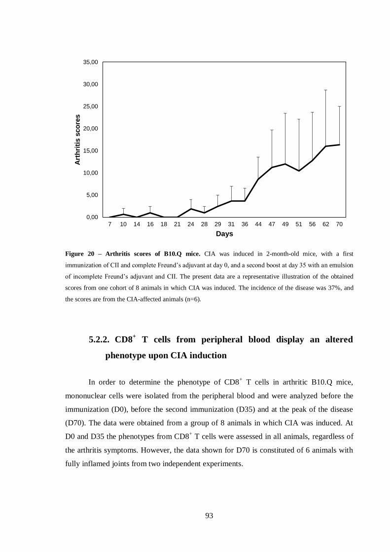
93
Figure 20 – Arthritis scores of B10.Q mice. CIA was induced in 2-month-old mice, with a first
immunization of CII and complete Freund’s adjuvant at day 0, and a second boost at day 35 with an emulsion
of incomplete Freund’s adjuvant and CII. The present data are a representative illustration of the obtained
scores from one cohort of 8 animals in which CIA was induced. The incidence of the disease was 37%, and
the scores are from the CIA-affected animals (n=6).
5.2.2. CD8+ T cells from peripheral blood display an altered
phenotype upon CIA induction
In order to determine the phenotype of CD8+ T cells in arthritic B10.Q mice,
mononuclear cells were isolated from the peripheral blood and were analyzed before the
immunization (D0), before the second immunization (D35) and at the peak of the disease
(D70). The data were obtained from a group of 8 animals in which CIA was induced. At
D0 and D35 the phenotypes from CD8+ T cells were assessed in all animals, regardless of
the arthritis symptoms. However, the data shown for D70 is constituted of 6 animals with
fully inflamed joints from two independent experiments.
0,00
5,00
10,00
15,00
20,00
25,00
30,00
35,00
7 10 14 16 18 21 24 28 29 31 36 44 47 49 51 56 62 70
Art
hri
tis s
co
res
Days
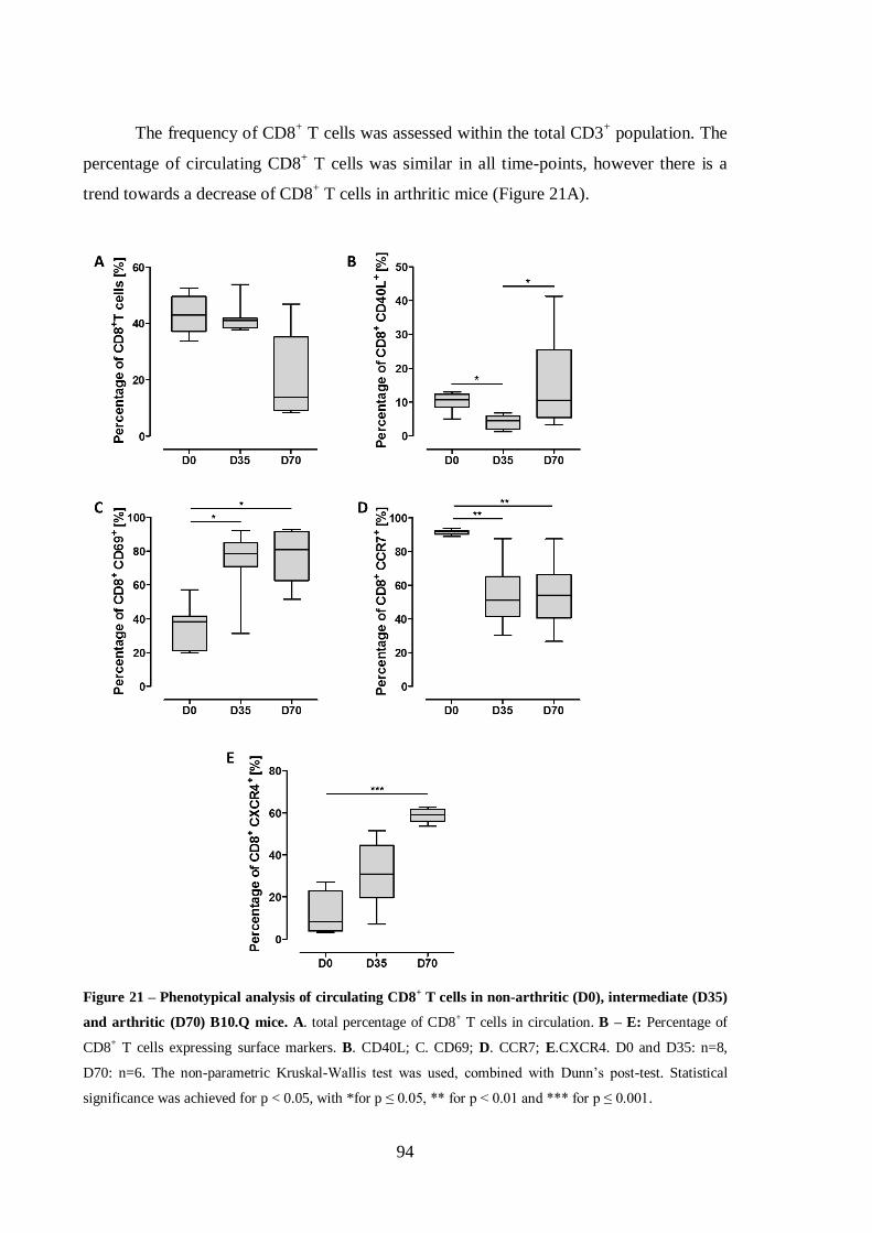
94
The frequency of CD8+ T cells was assessed within the total CD3
+ population. The
percentage of circulating CD8+ T cells was similar in all time-points, however there is a
trend towards a decrease of CD8+ T cells in arthritic mice (Figure 21A).
Figure 21 – Phenotypical analysis of circulating CD8+ T cells in non-arthritic (D0), intermediate (D35)
and arthritic (D70) B10.Q mice. A. total percentage of CD8+ T cells in circulation. B – E: Percentage of
CD8+ T cells expressing surface markers. B. CD40L; C. CD69; D. CCR7; E.CXCR4. D0 and D35: n=8,
D70: n=6. The non-parametric Kruskal-Wallis test was used, combined with Dunn’s post-test. Statistical
significance was achieved for p < 0.05, with *for p ≤ 0.05, ** for p < 0.01 and *** for p ≤ 0.001.
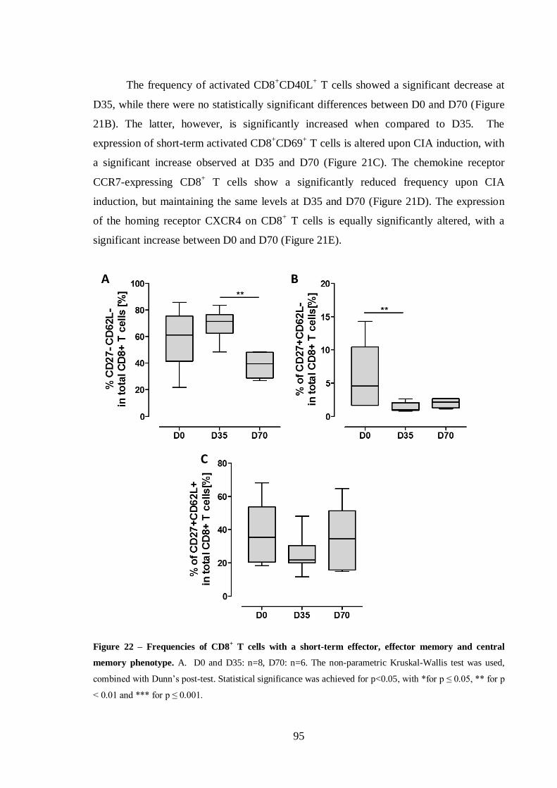
95
The frequency of activated CD8+CD40L
+ T cells showed a significant decrease at
D35, while there were no statistically significant differences between D0 and D70 (Figure
21B). The latter, however, is significantly increased when compared to D35. The
expression of short-term activated CD8+CD69
+ T cells is altered upon CIA induction, with
a significant increase observed at D35 and D70 (Figure 21C). The chemokine receptor
CCR7-expressing CD8+ T cells show a significantly reduced frequency upon CIA
induction, but maintaining the same levels at D35 and D70 (Figure 21D). The expression
of the homing receptor CXCR4 on CD8+ T cells is equally significantly altered, with a
significant increase between D0 and D70 (Figure 21E).
Figure 22 – Frequencies of CD8+ T cells with a short-term effector, effector memory and central
memory phenotype. A. D0 and D35: n=8, D70: n=6. The non-parametric Kruskal-Wallis test was used,
combined with Dunn’s post-test. Statistical significance was achieved for p<0.05, with *for p ≤ 0.05, ** for p
< 0.01 and *** for p ≤ 0.001.

96
Next, the CD8+ T cell in PB from pre-induction, intermediate and arthritic B10.Q
mice were further characterized. The frequency of CD8+ T cells with a CD27
-CD62L
-
short-term effector phenotype is altered, showing a significant decrease at D70 when
compared with D35 (Figure 22A). Concomitantly, there was an accentuated decrease in the
percentage of effector memory CD27+CD62L
- CD8
+ T cells (Figure 22B). The
CD8+CD27
+CD62L
+ central memory T cells remained unaltered (Figure 22C).
5.2.3. Intracellular expression of cytokines and granzyme B in
CD8+ T cells
To further determine the CD8+ T cells contribution to the onset of the disease in the
PB, the intracellular cytokine and granzyme B levels were determined in the same animals.
The percentage of unstimulated CD8+ T cells positive for intracellular proinflammatory
cytokines TNF-α and IL-17A, the anti-inflammatory cytokine IL-10 and granzyme B was
significantly altered during the induction of CIA (Figure 23).
Significantly increased levels of intracellular cytokines were observed for TNF-α
(Figure 23A), which is increased at D35 but returns to baseline levels at D70, and IL-17A
(Figure 23C), which upon the induction of CIA appears significantly decreased at D35
when compared to D0 levels, and significantly increased at D70 when compared to D35.
The changes for IFN-γ failed to reach significance. Nevertheless a trend for increased
frequency of IFN-γ-expressing CD8+ T cells can be observed at D70 (Figure 23B). The
percentage of IL-10-expressing CD8+ T cells appears significantly increased at D70, when
compared to D35 (Figure 23D). As for the frequency of CD8+ T cells positive for
intracellular granzyme B, a gradual increase is observed upon the induction of CIA, with
granzyme B levels at D70 significantly being significantly increased in comparison to
baseline (Figure 23E).
The median fluorescence intensity (MFI), which is correlated to the amount of
cytokines present in CD8+ T cells was determined. For intracellular cytokines measured in
unstimulated CD8+ T cells before and after induction of CIA failed to produce significant

97
results, and thus remained unaltered. An altered MFI was observed for granzyme B, which
is significantly increased at D70 when compared to D35.
Figure 23 – Intracellular cytokine and granzyme B levels. A. TNF-α; B. IFN-γ; C. IL-17; D. IL-10; E.
Granzyme B. D0 and D35: n=8, D70: n=6. The non-parametric Kruskal-Wallis test was used, combined with
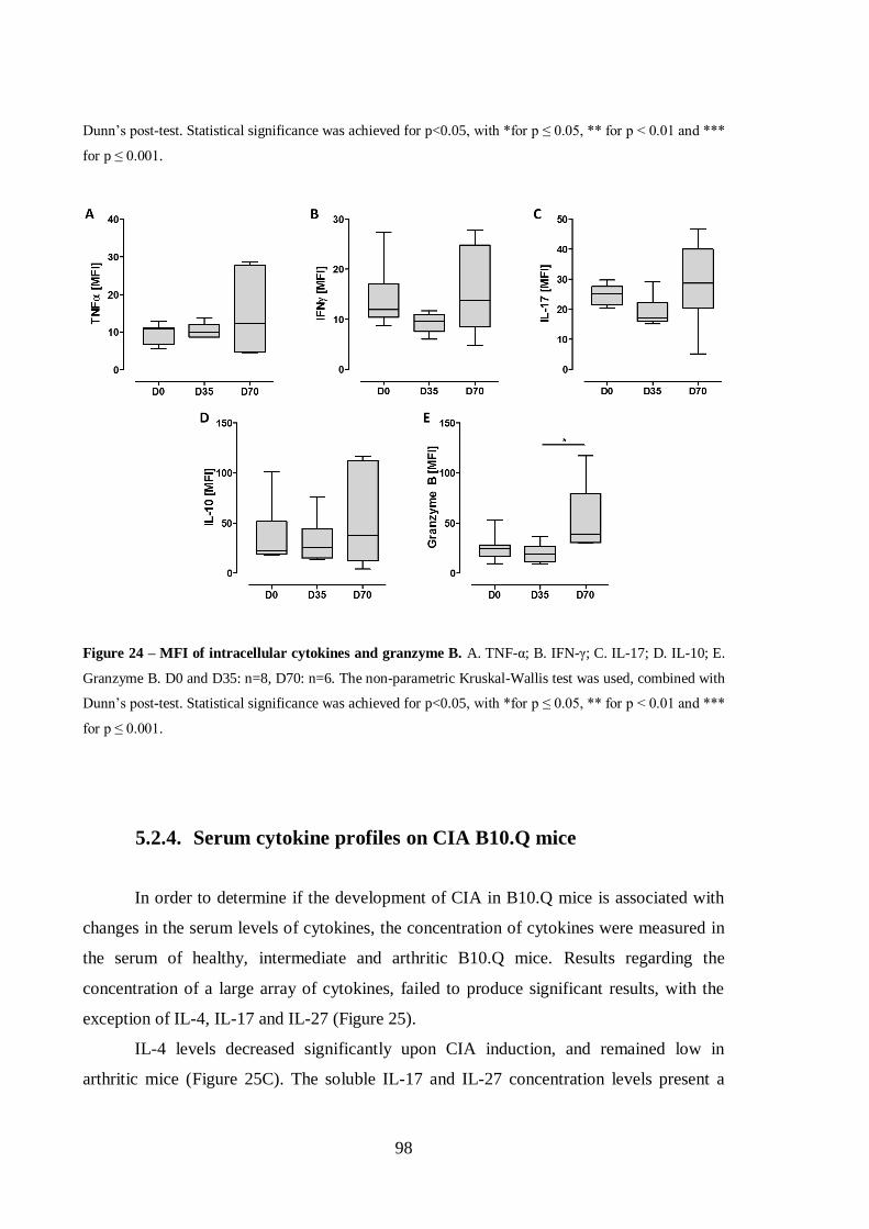
98
Dunn’s post-test. Statistical significance was achieved for p<0.05, with *for p ≤ 0.05, ** for p < 0.01 and ***
for p ≤ 0.001.
Figure 24 – MFI of intracellular cytokines and granzyme B. A. TNF-α; B. IFN-γ; C. IL-17; D. IL-10; E.
Granzyme B. D0 and D35: n=8, D70: n=6. The non-parametric Kruskal-Wallis test was used, combined with
Dunn’s post-test. Statistical significance was achieved for p<0.05, with *for p ≤ 0.05, ** for p < 0.01 and ***
for p ≤ 0.001.
5.2.4. Serum cytokine profiles on CIA B10.Q mice
In order to determine if the development of CIA in B10.Q mice is associated with
changes in the serum levels of cytokines, the concentration of cytokines were measured in
the serum of healthy, intermediate and arthritic B10.Q mice. Results regarding the
concentration of a large array of cytokines, failed to produce significant results, with the
exception of IL-4, IL-17 and IL-27 (Figure 25).
IL-4 levels decreased significantly upon CIA induction, and remained low in
arthritic mice (Figure 25C). The soluble IL-17 and IL-27 concentration levels present a

99
similar evolution, with a significant decrease in serum levels upon CIA induction, and
maintenance of these levels in arthritic mice (Figure 25H, J). Despite not reaching
significance, the proinflammatory cytokine IL-1α and anti-inflammatory cytokine IL-10
show a trend for increased levels in arthritic mice at D70 (Figure 25 A,F). Interestingly,
TNF-α, also measured in this experiment, remained largely undetectable (data not shown).
Figure 25 – Concentration of soluble cytokines from serum of B10.Q mice. A. IL-1α; B. IL-2; C. IL-4; D.
IL-5; E. IL-6; F. IL-10; G. IL-13; H. IL-17; I. IL-22; J. IL-27; K. IFN-γ. D0 and D35: n=8, D70: n=6. The
non-parametric Kruskal-Wallis test was used, combined with Dunn’s post-test. Statistical significance was
achieved for p<0.05, with *for p ≤ 0.05, ** for p < 0.01 and *** for p ≤ 0.001.
100
5.3. Discussion
The role of CD8+ T cells in RA has yet to be fully determined. However, the
importance of CD8+ T cells is being gradually attested in humans (Cho et al. 2012).
Studies of CD8+ T cells in animal models of arthritis have yielded conflicting results. The
depletion of CD8+ T cells results in the amelioration of the disease in the mercuric
chloride-induced arthritis model and in the K/BxN model of spontaneous arthritis,
indicating a role for CD8+ T cells in the development of this condition (Kiely et al. 1996;
Raposo et al. 2010). In CD8-deficient mice, the incidence of CIA was significantly
decreased (Tada et al. 1996), while CD8+ T cell knock-out B10.Q mice showed no
alteration in the impact of collagen-induced arthritis (Ehinger et al. 2001), thus pointing in
the opposite direction.
In the present study we aimed at determine the characteristics of circulating CD8+ T
cells in the B10.Q mouse model of collagen-induced arthritis, as well as their potential role
in the maintenance of arthritis in this model.
We found that the frequency of CD8+ T cells in the peripheral blood tends to
decrease with the onset of the disease. Upon CIA induction, CD8+ T cells acquire an
activated phenotype, showing increased relative frequencies of CD40L- and CD69-
expressing CD8+ T cells. The relative percentage of short-term effector decrease
significantly at the peak of the disease, while effector memory CD8+ T cells decrease
significantly upon CIA induction and maintain a low frequency at the peak of the disease.
Along with these phenotypical alterations, CD8+ T cells decrease their expression of
CCR7, which is typically found in structures similar to germinal centers (Bruhl et al.
2008), while the relative frequency of CXCR4-expressing CD8+ T cells increase
dramatically.
CD8+ T cells from the PB from B10.Q mice also show an increased intracellular
expression of proinflammatory cytokines (TNF-α and IL-17), anti-inflammatory cytokine
IL-10 and granzyme B. However, when comparing with their MFI levels it can be seen that
the amount of cytokines produced remain unchanged after the induction of CIA, except for
granzyme B, which clearly shows an increased production in arthritic mice. From the
general cytokine concentrations seen in the serum we can notice that the cytokines with
101
altered concentration at the different stages of CIA induction are IL-4, IL-17 and IL-27,
and all show diminished concentrations in arthritic mice’s serum.
As seen in the K/BxN mouse model of polyarthritis (Raposo et al. 2010), the B10.Q
arthritic mice are also characterized by the reduced relative frequencies of short-term
effector and effector memory CD8+ T cells subsets, and increased percentages of central
memory CD8+ T cells. These results are also corroborated by those found in RA patients,
which also present a decrease in CD62L-CD27
- and CD62L
-CD27
+ cells in the periphery.
Conversely, the opposite result was found in the articular tissue from arthritic K/BxN mice,
which was corroborated with our results in RA patients, which present an enrichment of
effector memory and short-term effector CD8+ T cells in the synovial fluid. Our data
therefore indicates that CD8+ T cells of B10.Q arthritic mice with effector and therefore
cytotoxic potential are decreased in the periphery and may be being recruited to the
inflamed joints.
Additionally, CD8+ T cells from arthritic B10.Q mice present an accentuated
activated CD69+ phenotype in arthritic mice, which was also observed in the K/BxN
mouse model. These results were also supported by RA patients’ data, and are thus
indicators of the ongoing systemic activation.
Interestingly, the increased expression of the homing chemokine receptor CXCR4
on the surface of CD8+ T cells is not corroborated by the results found on K/BxN mice,
which present an accentuated decrease in CXCR4-expressing CD8+ T cells in arthritic
mice, when compared to healthy littermates, while a significant increase of the frequency
of these cells can be observed in the inflamed joints of arthritic mice. Similarly, results
obtained in RA patients indicate a reduction of the frequency of peripheral CD8+CXCR4
+
cells, coupled to a significant enrichment of these cells in the synovial fluid from RA
patients. Indeed, CXCR4 plays an important role in the recruitment of leukocytes to
inflammatory sites, and has been proven to be crucial in the recruitment of activated T cells
in both RA patients (Bryant et al. 2012) and in mice with CIA (Chung et al. 2010), as a
high frequency of CXCR4-expressing T cells were found in inflamed joints in both
humans and animal models. In order to assess if a similar behavior of CXCR4-expressing
CD8+ T cells is occurring in this arthritis model, it would have been beneficial to determine
the frequency of these cells in the inflamed joints of CIA-affected B10.Q mice, but the

102
peripheral increase of these cells alone suggests an important role of CXCR4 in the
development of CIA in B10.Q mice.
Here we found that the chemokine receptor CCR7 has a reduced frequency in the
peripheral blood of B10.Q arthritic mice at the peak of the disease. CCR7 is known for
driving lymphoid neogenesis11
in CIA as well as RA (Wengner et al. 2007), and is
implicated in the recruitment of memory T cells to the synovial fluid in juvenile idiopathic
arthritis (Gattorno et al. 2005). Remarkably, the loss of the CCR7 expression is
characteristic of the acquisition of an effector function (Sallusto et al. 1999). The decrease
of memory T cell subsets expressing CCR7 is also seen in RA patients, which is coincident
with our findings (Matsuki et al. 2013). Therefore our data suggest that CD8+CCR7
+ T
cells are being recruited to the inflamed joints, where they have the potential to induce the
formation of ectopic germinal centers (Kang et al. 2002). Once they are established in
inflamed joints, ectopic germinal centers become autonomous lymphoid structures, leading
to chronic inflammation locally.
CD40L is expressed by activated T cells and binds to the CD40 molecule on the
surface of B cells, contributing to T cell-dependent B cell activation (Chatzigeorgiou et al.
2009). We found that CD8+CD40L
+ T cells are increased in the peripheral blood of B10.Q
arthritic mice. Similar results have been described in CD4+ T cells from RA patients,
showing an increased percentage of CD40L-expressing CD4+ T cells (Berner et al. 2000).
Since CD8+CD40L
+ T cells are involved in the formation of ectopic germinal centers in the
inflamed joints of RA patients (Wagner et al. 1998), our data suggest that the increased
relative percentage CD8+CD40L
+ T cells from arthritic B10.Q mice may be an indicator of
recruitment of these cells into the inflamed joints to promote the formation of an ectopic
germinal center.
The relative percentages of intracellular cytokines in CD8+ T cells from arthritic
B10.Q mice shows and increased frequency of CD8+TNF-α
+ at an intermediate state of the
induction of CIA (D35), but this increase is not maintained in fully arthritic mice, which is
not concurrent with previous studies (Marinova-Mutafchieva et al. 1997). These findings
are concurrent with the observation that CD8+ T cells from RA patients have higher
percentages of CD8+TNF-α
+ T cells in the PB than healthy individuals. Equally
significantly increased in arthritic B10.Q mice is the frequency of CD8+IL-17
+ T cells,
11 Lymphoid neogenesis: ectopic de novo formation of organized lymphoid tissue at an inflammatory site,
leading to chronic inflammation.
103
which supports the idea that IL-17 contributes to the synovial inflammation and joint
erosion in CIA (Lubberts et al. 2001), and that IL-17 knock-out mice fail to develop CIA
(Nakae et al. 2003). These findings are in line with our studies in RA showing higher
percentages of CD8+IL-17
+ T cells in the PB of RA patients than in healthy individuals.
Also increased in the arthritic B10.Q mice was the frequency of CD8+IL-10
+ cells. IL-10 is
known for being an anti-inflammatory cytokine with protective effects in CIA
(Henningsson et al. 2012), and in IL-10 knock-out mice the induction of CIA results in an
aggravated disease. IL-10 is also known for having an anti-inflammatory function in
human RA, and its frequency also is also increased in the PB of arthritic individuals. The
percentage of circulating CD8+Granzyme B
+ T cells are gradually increased in the PB upon
induction of CIA in B10.Q mice and peaking in fully arthritic mice. These results are
concordant with previous results from human RA which show an increased frequency of
Granzyme-B-expressing CD8+ T cells. Additionally, CD8
+Granzyme B
+ T cells also
present an increased MFI for arthritic B10.Q mice, indicating that not only is the relative
percentage of CD8+ T cells expressing granzyme B in the intracellular compartment
increased, but also is the amount of granzyme B produced by these cells. This suggests that
CD8+ T cells from arthritic B10.Q mice possess large amounts of granzyme B in their
cytosol, and thus have an increased cytotoxic potential when compared to non-arthritic
mice.
Interestingly, the concentrations obtained for the serologic cytokines have yielded
some unexpected results, such as the failure to detect TNF-α in the sera, or the reduction of
the cytokines IL-4, IL-17 and IL-27 in arthritic B10.Q mice. IL-17 is described to be
increased in the serum of mice with CIA (Sarkar et al. 2009) and RA patients (Hussein et
al. 2008; Rosu et al. 2012), while IL-4 and IL-27 have a regulatory function, in which they
can modulate the Th17 response (Sarkar et al. 2009; Pickens et al. 2011). These results
suggest that the CD8+ T cells’ response to the induction of CIA in B10.Q mice is not
associated with serologic levels of circulating cytokines.
Taken together, these data reinforce the importance that CD8+ T cells have in the
development of CIA, as in the arthritic mice they present a phenotype that is activated,
secrete proinflammatory cytokines and granzyme B, thus being capable of exerting an
inflammatory response. Also, the fact that these cells express high levels of homing
receptors indicates that they may be actively recruited to the sites of inflammation.

104
105
CHAPTER 6
CD8+ T CELL PROFILES IN PATIENTS WITH RHEUMATOID ARTHRITIS
AND THEIR RELATIONSHIP TO DISEASE ACTIVITY


107
6. CD8+ T cell profiles in patients with rheumatoid arthritis and
their relationship to disease activity
6.1. Introduction
Genome-wide association studies and long standing phenotypic and relevant
murine model data strongly implicate T cells in the pathogenesis of rheumatoid arthritis
(RA). CD8+ T cells comprise approximately 40% of all T cells infiltrating the rheumatoid
synovial compartment (McInnes 2003), and they are detected in the pre-clinical stages of
disease development (de Hair et al. 2013). CD8+ T cells can be subdivided into different
functional subsets that include a short-lived effector subset (with high migratory capacity
and intense production of pro-inflammatory cytokines and cytotoxic molecules); an
effector-memory subset (which accumulates in the peripheral organs, is apoptosis-resistant
and becomes effector upon reencounter with antigen), a central memory subset (which
offers rapid proliferation and cytokine production but little cytotoxicity upon reencounter
with cognate antigen), and a suppressor subset (IL-10-producing cells which down-
modulate the inflammatory response) (Gupta and Gollapudi 2007; Marzo et al. 2007;
Carvalheiro et al. 2012).
One prior study found that peripheral blood (PB) central memory CD8+ T cells
were more frequent in RA patients when compared to healthy controls (HC) whereas the
opposite profile was seen with effector memory CD8+ T cells. (Maldonado et al. 2003).
Recently, the frequency of effector memory but not central memory CD8+ T cells was
reported to be elevated in the PB and synovial fluid (SF) of RA patients when compared to
PB samples from HC (Cho et al. 2012). An accumulation of autoreactive, clonally-related
memory CD8+ T cells was found in RA SF (Sottini et al. 1993; Behar et al. 1995;
Fitzgerald et al. 1995; Morley et al. 1995) and their frequency correlated with serum
rheumatoid factor (RF) levels (al-Azem et al. 1992). RA patients with DAS28 > 3.2 appear
to have a slight increase in the frequency of circulating IL-17A-producing CD8+ T cells
(Henriques et al. 2010). CD8+ T cells are crucial in maintaining synovial ectopic germinal
108
centers, which are associated in turn with more aggressive disease (Wagner et al. 1998;
Kang et al. 2002; Croia et al. 2013). However, some studies indicate that a suppressor
subset of CD8+ T cells associates with disease amelioration (Davila et al. 2005; Suzuki et
al. 2008). Key outstanding questions remain including the identity of an overarching
phenotype and the production of cytokines and cytotoxic molecules by CD8+ T cells in
peripheral blood and the synovial compartment and their relationship with RA disease
activity. As SF is becoming harder to obtain, it must be established whether studies in
blood samples provide a reliable representation of the biological events taking place at the
inflammatory site, reflected by the SF. Herein we address these critical issues.
109
6.2. Results
6.2.1. Altered status of peripheral blood CD8+ T cell subsets in RA
patients
The relative frequency of circulating CD8+ T cells within the total lymphocyte
population was similar in all groups (Figure 26A). The absolute number of circulating
CD8+ T cells was similar in RA patients with active disease and in controls but was
significantly lower (p < 0.05) in patients in remission (HC: 394.2 ± 1.6 cells/µl; Active
RA: 400.0 ± 3.7 cells/µl; Remission RA: 351.7 ± 1.6 cells/µl). This apparently arises from
generalized lymphopenia in RA patients in remission (HC: 2478.3 ± 156.4 cells/µl; Active:
2185.7 ± 266.8 cells/µl; Remission: 1825.0 ± 159.6 cells/µl) and suggests that the latter
status is not commensurate with normal immunologic homeostasis.
The relative frequencies of CD27+CD62L
+CCR7
+ central memory CD8
+ T cells
was lower in active RA than in HC (Figure 26B). Remission was associated with
accentuation of this difference (Figure 26C). The frequency of CD27+CD62L
- effector
memory CD8+ T cells was similar in all three groups (data not shown). The frequency of
the short-term effector CD27-CD62L
-CD8
+ T cell subset was significantly higher in the
active disease group when compared to controls (Figure 26D). This difference persisted in
patients in remission.
Both RA groups had lower relative frequencies of CD25+CD8
+ T cells compared
with HC, although was significant only for those patients in remission (Figure 26E). The
frequency of PB CD69+CD8
+ T cells in active disease was similar to that in HC. The
remission group had significantly more circulating CD69+CD8
+ T cells than the active
disease and the HC (Figure 26F). There was an accumulation of CD69-expressing CD8+ T
cells within the total CD62L- effector compartment of both patient groups when compared
to HC, the difference being more pronounced in remission (Figure 26G). The frequency of
PB CD8+ T cells expressing CXCR4 was significantly lower in both patient groups than in
controls (Figure 26H). When focusing the analysis on the activated total effector CD8+ T
cell population, the significant reduction of the proportion of cells expressing CXCR4 was
maintained in both patient groups when compared to HC (Figure 26I).

110
Figure 26 – Functional phenotyping of peripheral blood CD8+ T cells shows altered frequencies of
subsets expressing activation, homing, memory and effector molecules in active and remission RA
patients when compared to controls. A. Dotplots gated on CD8+ T cells of CD62L vs. CD27 for
representative control, active RA and remission RA individuals. B. Boxplot representing the 90%, 75%,
median, 25% and 10% ranges of the frequency of total circulating CD8+ T cells within the whole T cell pool
for the three groups. Boxplots representing the 90%, 75%, median, 25% and 10% ranges of the frequency of
circulating CD8+ T cell subsets within the total CD8+ T cell pool: C: CD27+CD62L+CCR7+, D. CD27-
CD62L-, E. CD25+, F. CD69+, G. CD69+CD62L-, H. CXCR4+, I. CXCR4+CD69+CD62L-. P values
calculated by one-way ANOVA followed by LSD post-hoc test. Control: N = 64; Active RA: N = 34;
Remission RA: N = 44.
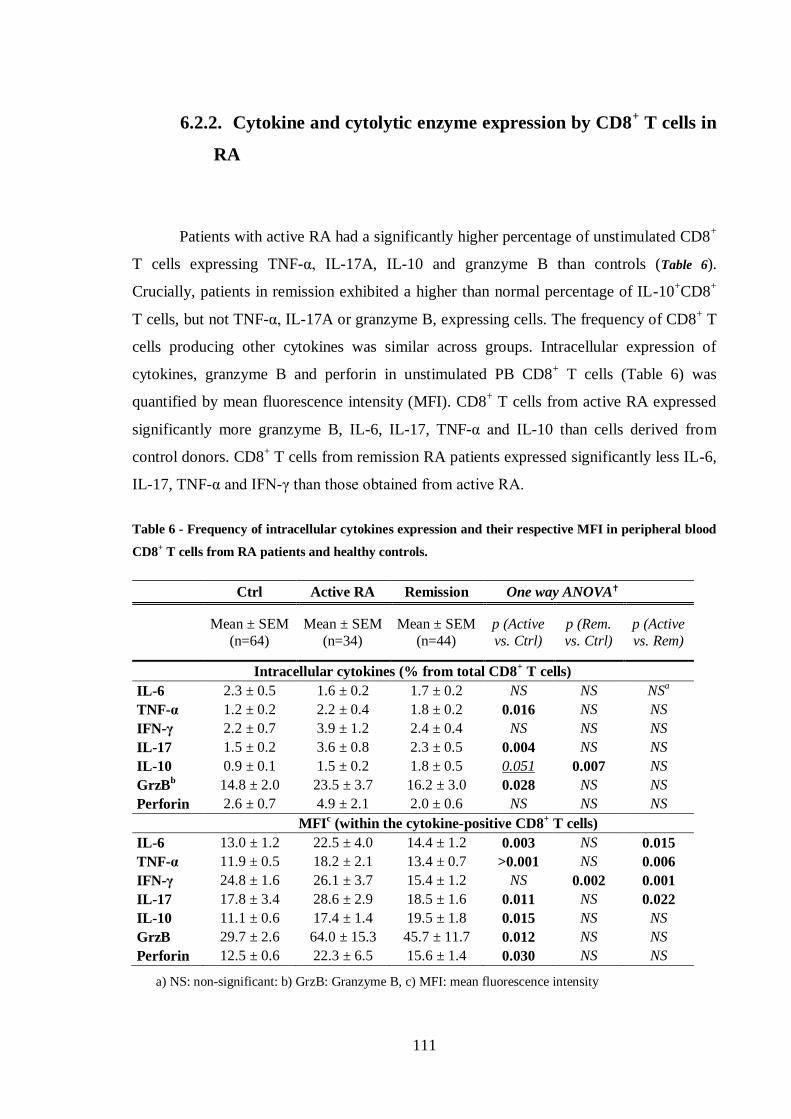
111
6.2.2. Cytokine and cytolytic enzyme expression by CD8+ T cells in
RA
Patients with active RA had a significantly higher percentage of unstimulated CD8+
T cells expressing TNF-α, IL-17A, IL-10 and granzyme B than controls (Table 6).
Crucially, patients in remission exhibited a higher than normal percentage of IL-10+CD8
+
T cells, but not TNF-α, IL-17A or granzyme B, expressing cells. The frequency of CD8+ T
cells producing other cytokines was similar across groups. Intracellular expression of
cytokines, granzyme B and perforin in unstimulated PB CD8+ T cells (Table 6) was
quantified by mean fluorescence intensity (MFI). CD8+ T cells from active RA expressed
significantly more granzyme B, IL-6, IL-17, TNF-α and IL-10 than cells derived from
control donors. CD8+ T cells from remission RA patients expressed significantly less IL-6,
IL-17, TNF-α and IFN-γ than those obtained from active RA.
Table 6 - Frequency of intracellular cytokines expression and their respective MFI in peripheral blood
CD8+ T cells from RA patients and healthy controls.
Ctrl Active RA Remission One way ANOVA
Mean ± SEM
(n=64)
Mean ± SEM
(n=34)
Mean ± SEM
(n=44)
p (Active
vs. Ctrl)
p (Rem.
vs. Ctrl)
p (Active
vs. Rem)
Intracellular cytokines (% from total CD8+ T cells)
IL-6 2.3 ± 0.5 1.6 ± 0.2 1.7 ± 0.2 NS NS NSa
TNF-α 1.2 ± 0.2 2.2 ± 0.4 1.8 ± 0.2 0.016 NS NS
IFN-γ 2.2 ± 0.7 3.9 ± 1.2 2.4 ± 0.4 NS NS NS
IL-17 1.5 ± 0.2 3.6 ± 0.8 2.3 ± 0.5 0.004 NS NS
IL-10 0.9 ± 0.1 1.5 ± 0.2 1.8 ± 0.5 0.051 0.007 NS
GrzBb 14.8 ± 2.0 23.5 ± 3.7 16.2 ± 3.0 0.028 NS NS
Perforin 2.6 ± 0.7 4.9 ± 2.1 2.0 ± 0.6 NS NS NS
MFI
c (within the cytokine-positive CD8
+ T cells)
IL-6 13.0 ± 1.2 22.5 ± 4.0 14.4 ± 1.2 0.003 NS 0.015
TNF-α 11.9 ± 0.5 18.2 ± 2.1 13.4 ± 0.7 >0.001 NS 0.006
IFN-γ 24.8 ± 1.6 26.1 ± 3.7 15.4 ± 1.2 NS 0.002 0.001
IL-17 17.8 ± 3.4 28.6 ± 2.9 18.5 ± 1.6 0.011 NS 0.022
IL-10 11.1 ± 0.6 17.4 ± 1.4 19.5 ± 1.8 0.015 NS NS
GrzB 29.7 ± 2.6 64.0 ± 15.3 45.7 ± 11.7 0.012 NS NS
Perforin 12.5 ± 0.6 22.3 ± 6.5 15.6 ± 1.4 0.030 NS NS
a) NS: non-significant: b) GrzB: Granzyme B, c) MFI: mean fluorescence intensity
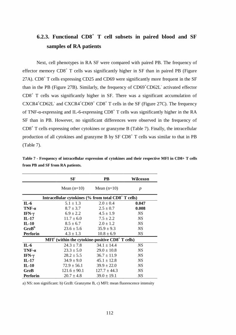
112
6.2.3. Functional CD8+ T cell subsets in paired blood and SF
samples of RA patients
Next, cell phenotypes in RA SF were compared with paired PB. The frequency of
effector memory CD8+ T cells was significantly higher in SF than in paired PB (Figure
27A). CD8+ T cells expressing CD25 and CD69 were significantly more frequent in the SF
than in the PB (Figure 27B). Similarly, the frequency of CD69+CD62L
- activated effector
CD8+ T cells was significantly higher in SF. There was a significant accumulation of
CXCR4+CD62L
- and CXCR4
+CD69
+ CD8
+ T cells in the SF (Figure 27C). The frequency
of TNF-α-expressing and IL-6-expressing CD8+ T cells was significantly higher in the RA
SF than in PB. However, no significant differences were observed in the frequency of
CD8+ T cells expressing other cytokines or granzyme B (Table 7). Finally, the intracellular
production of all cytokines and granzyme B by SF CD8+ T cells was similar to that in PB
(Table 7).
Table 7 - Frequency of intracellular expression of cytokines and their respective MFI in CD8+ T cells
from PB and SF from RA patients.
SF PB Wilcoxon
Mean (n=10) Mean (n=10) p
Intracellular cytokines (% from total CD8+ T cells)
IL-6 5.1 ± 1.3 2.0 ± 0.4 0.047
TNF-α 8.7 ± 3.7 2.5 ± 0.7 0.008
IFN-γ 6.9 ± 2.2 4.5 ± 1.9 NS
IL-17 11.7 ± 6.0 7.5 ± 2.2 NS
IL-10 8.5 ± 6.7 2.0 ± 1.2 NS
GrzBb 23.6 ± 5.6 35.9 ± 9.3 NS
Perforin 4.3 ± 1.3 10.8 ± 6.9 NS
MFIc (within the cytokine-positive CD8
+ T cells)
IL-6 24.3 ± 7.8 34.1 ± 14.4 NS
TNF-α 23.3 ± 5.0 29.0 ± 10.8 NS
IFN-γ 28.2 ± 5.5 36.7 ± 11.9 NS
IL-17 34.9 ± 9.0 45.1 ± 12.8 NS
IL-10 72.9 ± 56.1 39.9 ± 22.0 NS
GrzB 121.6 ± 90.1 127.7 ± 44.3 NS
Perforin 20.7 ± 4.8 39.0 ± 19.1 NS
a) NS: non significant: b) GrzB: Granzyme B, c) MFI: mean fluorescence intensity

113
Figure 27 – Functional phenotyping of CD8+ T cells from paired peripheral blood and synovial fluid
from RA patients shows increased frequencies of CD8+T cells expressing effector, activation and
homing molecules in the synovial fluid. Boxplots representing the 90%, 75%, median, 25% and 10% ranges
of the frequency of circulating CD8+ T cell subsets within the total CD8+ T cell pool: A: CD27+CD62L- and
CD27-CD62L-CCR7-, B. CD25+, CD69+ and CD69+CD62L-, C. CXCR4+, CXCR4+ CD62L- and
CXCR4+CD69+. P values calculated by Wilcoxon non parametric test. Synovial fluid (SF) and peripheral
blood (PB): N = 10.
6.2.4. Correlation of CD8+ T cell subsets in the PB and SF
The frequencies of total CD8+ T cells in SF and PB were strongly correlated
(Figure 28A). Total activated CD25+CD8
+ T cells and the CD25
+CD62L
+ memory subset
in PB were strongly positively correlated to the expression of the same subsets in the SF
(Figure 28B-C). Strong correlations were found between intracellular production of
granzyme B, IFN-γ, IL-6, IL-17A by CD8+ T cells from PB and SF (Figure 28G). The

114
expression of CXCR4 in SF was weakly correlated (R = -0.188) with expression in PB, but
failed to reach significance (data not shown).
Figure 28 – Values observed in the patients’ PB mirror those in the SF. A-G: Correlation plots between
CD8+ T cell subsets in the PB and SF of RA patients (N = 10). Correlations considered weak for r>0.2,
moderate for r>0.3, strong for r >0.5 and very strong for r >0.75. Significance achieved for p < 0.05. Values
obtained using the Spearman correlation.
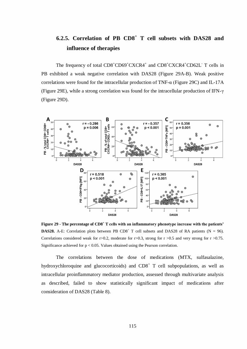
115
6.2.5. Correlation of PB CD8+ T cell subsets with DAS28 and
influence of therapies
The frequency of total CD8+CD69
+CXCR4
+ and CD8
+CXCR4
+CD62L
- T cells in
PB exhibited a weak negative correlation with DAS28 (Figure 29A-B). Weak positive
correlations were found for the intracellular production of TNF-α (Figure 29C) and IL-17A
(Figure 29E), while a strong correlation was found for the intracellular production of IFN-γ
(Figure 29D).
Figure 29 - The percentage of CD8+ T cells with an inflammatory phenotype increase with the patients’
DAS28. A-E: Correlation plots between PB CD8+ T cell subsets and DAS28 of RA patients (N = 96).
Correlations considered weak for r>0.2, moderate for r>0.3, strong for r >0.5 and very strong for r >0.75.
Significance achieved for p < 0.05. Values obtained using the Pearson correlation.
The correlations between the dose of medications (MTX, sulfasalazine,
hydroxychloroquine and glucocorticoids) and CD8+ T cell subpopulations, as well as
intracellular proinflammatory mediator production, assessed through multivariate analysis
as described, failed to show statistically significant impact of medications after
consideration of DAS28 (Table 8).
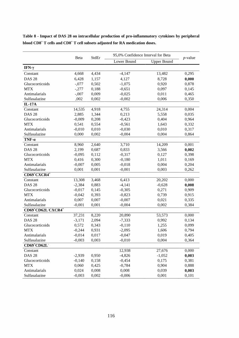
116
Table 8 - Impact of DAS 28 on intracellular production of pro-inflammatory cytokines by peripheral
blood CD8+ T cells and CD8
+ T cell subsets adjusted for RA medication doses.
Beta StdEr
95,0% Confidence Interval for Beta p-value
Lower Bound Upper Bound
IFN-γ
Constant 4,668 4,434 -4,147 13,482 0,295
DAS 28 6,428 1,157 4,127 8,728 0,000
Glucocorticoids -,077 0,502 -1,075 0,920 0,878
MTX -,277 0,188 -0,651 0,097 0,145
Antimalarials -,007 0,009 -0,025 0,011 0,465
Sulfasalazine ,002 0,002 -0,002 0,006 0,350
IL-17A
Constant 14,535 4,918 4,755 24,314 0,004
DAS 28 2,885 1,344 0,213 5,558 0,035
Glucocorticoids -0,009 0,208 -0,423 0,404 0,964
MTX 0,541 0,554 -0,561 1,643 0,332
Antimalarials -0,010 0,010 -0,030 0,010 0,317
Sulfasalazine 0,000 0,002 -0,004 0,004 0,864
TNF-α
Constant 8,960 2,640 3,710 14,209 0,001
DAS 28 2,199 0,687 0,833 3,566 0,002
Glucocorticoids -0,095 0,112 -0,317 0,127 0,398
MTX 0,416 0,300 -0,180 1,011 0,169
Antimalarials -0,007 0,005 -0,018 0,004 0,204
Sulfasalazine 0,001 0,001 -0,001 0,003 0,262
CD69+CXCR4
+
Constant 13,308 3,468 6,413 20,202 0,000
DAS 28 -2,384 0,883 -4,141 -0,628 0,008
Glucocorticoids -0,017 0,145 -0,305 0,271 0,909
MTX -0,042 0,393 -0,823 0,739 0,915
Antimalarials 0,007 0,007 -0,007 0,021 0,335
Sulfasalazine -0,001 0,001 -0,004 0,002 0,384
CD69+CD62L
-CXCR4
+
Constant 37,231 8,220 20,890 53,573 0,000
DAS 28 -3,171 2,094 -7,333 0,992 0,134
Glucocorticoids 0,572 0,343 -0,110 1,255 0,099
MTX -0,244 0,931 -2,095 1,606 0,794
Antimalarials -0,014 0,017 -0,047 0,019 0,405
Sulfasalazine -0,003 0,003 -0,010 0,004 0,364
CD69+CD62L
-
Constant 12,938 27,676 0,000
DAS 28 -2,939 0,950 -4,826 -1,052 0,003
Glucocorticoids -0,140 0,158 -0,454 0,175 0,381
MTX 0,060 0,425 -0,784 0,904 0,888
Antimalarials 0,024 0,008 0,008 0,039 0,003
Sulfasalazine -0,003 0,002 -0,006 0,001 0,101
117
6.3. Discussion
Herein we report that PB CD8+ T cells from active and remission RA present an
activated phenotype with a marked pro-inflammatory profile. We show that the expression
of pro-inflammatory cytokines by circulating CD8+ T cells is directly correlated with the
DAS28 score. CD8+ T cells from the SF of active RA exhibit an exacerbated effector and
activated phenotype compared to those in paired PB. Finally, we observed that the
production of cytokines by SF CD8+ T cells is correlated with that in paired PB derived
cells.
Contrasting to a previous report (Cho et al. 2012), we did not find any differences
in the frequency of total CD8+ T cells in PB and SF of RA patients. We suggest that these
contradictions arise from the fact that they compared SF data to blood data of the whole
RA cohort regardless of disease activity, whereas we performed a paired analysis restricted
to patients with active disease. The circulating CD8+ T cell compartment of RA patients,
regardless of disease activity, had a skewed distribution of central memory and short-term
effector CD8+ T cell subsets, with enrichment of the latter. Accumulation of effector
memory CD8+ T cells in the SF compared to the paired blood was equally present. RA
patients accumulate effector CD8+ T cells both in the blood and in the SF - and at the same
time present a reduction in the central memory CD8+ T cell subset. These results partially
mimic our previously reported observations in K/BxN mice (Raposo et al. 2010).
Our data confirm previous observations that CD8+ T cells in RA frequently express
the early activation marker CD69 (Afeltra et al. 1993; Fernandez-Gutierrez et al. 1995;
Iannone et al. 1996; Afeltra et al. 1997). The increased frequency of effector CD8+ T cells
expressing CD69 in RA patients’ blood – independent of disease activity – and SF,
suggests that these cells might be constantly stimulated by the presence of their cognate
antigen(s). Also in the K/BxN model of arthritis, the expression of CD69 in CD8+ T cells is
increased in both the PB and articular tissue of arthritic mice (Raposo et al. 2010). These
data, together with previous studies, indicate that activated CD8+ T cells are enriched in
RA PB cells (Laffon et al. 1991). Even though, the peripheral blood only represents a small
fraction of the total T cell pool of an individual, we speculate that the accumulation of
activated CD8+ T cells in the PB during remission rather than active disease suggests that

118
these cells remain in circulation and might be recruited into the joint when the disease
increases its activity. The surge in CD8+ T cells expressing CD69 as well as
CD69+CXCR4
+ in the SF of active RA patients when compared to parallel PB samples,
also indicates that these cells are enriched in the joints during disease flares. This
interpretation is supported by our finding of a weak negative correlation between the
frequency of PB effector and activated CXCR4+CD8
+ T cells and the DAS28 score, since
CXCR4 is responsible for cytotoxic T cell-homing into inflammatory sites. Clearly, the
correlations are too weak to establish this functional link but they mirror our previous
results in the K/BxN mouse (Raposo et al. 2010).
We measured ex vivo cytokine, perforin and granzyme B production by PB and SF
CD8+ T cells without in vitro stimulation, in order to assess whether these cells actively
contribute to the pro-inflammatory environment in RA and consequent joint destruction.
Our data show that regardless of the similar numbers of circulating effector CD8+ T cells,
remission and active disease are associated with distinct production of cytokines and
cytotoxic molecules, and that the production of pro-inflammatory cytokines in PB was
directly correlated with the DAS28 score.
We show that the expression of granzyme B by CD8+ T cells from PB and SF from
active RA patients is higher than in PB from controls. The difference between remission
and control is not significant. We confirm previous observations that granzyme B+CD8
+ T
cells are commonly found in the synovium of RA patients (Kummer et al. 1994; Croia et
al. 2013). Given that we excluded patients with known ongoing infections, we suggest that
the increased production of granzyme B and perforin by CD8+ T cells in active RA is
stimulated by the presence of autologous antigens and the pro-inflammatory environment.
It also shows that CD8+ T cells are actively involved in maintaining the chronic
inflammatory process and that after medication-induced remission granzyme B and
perforin production by CD8+ T cells returns to normal levels.
The positive correlations obtained between the intracellular production of
Granzyme B, IL-17A, IL-6 and IFN-γ by CD8+ T cells in the PB and those in the SF
indicate that variations observed in the patients’ PB mirror those in the SF. Hence, we
demonstrate, for the first time, that variations in the production of these cytokines on
peripheral CD8+ T cells provide a good representation of similar processes taking place at
the joint level. This is an important finding, given that synovial fluid is now rarely
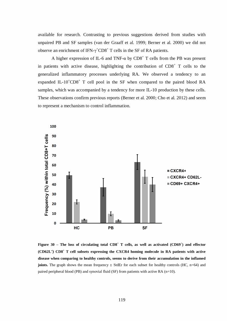
119
available for research. Contrasting to previous suggestions derived from studies with
unpaired PB and SF samples (van der Graaff et al. 1999; Berner et al. 2000) we did not
observe an enrichment of IFN-γ+CD8
+ T cells in the SF of RA patients.
A higher expression of IL-6 and TNF-α by CD8+ T cells from the PB was present
in patients with active disease, highlighting the contribution of CD8+ T cells to the
generalized inflammatory processes underlying RA. We observed a tendency to an
expanded IL-10+CD8
+ T cell pool in the SF when compared to the paired blood RA
samples, which was accompanied by a tendency for more IL-10 production by these cells.
These observations confirm previous reports (Berner et al. 2000; Cho et al. 2012) and seem
to represent a mechanism to control inflammation.
Figure 30 – The loss of circulating total CD8+ T cells, as well as activated (CD69
+) and effector
(CD62Lˉ) CD8+ T cell subsets expressing the CXCR4 homing molecule in RA patients with active
disease when comparing to healthy controls, seems to derive from their accumulation in the inflamed
joints. The graph shows the mean frequency ± StdEr for each subset for healthy controls (HC, n=64) and
paired peripheral blood (PB) and synovial fluid (SF) from patients with active RA (n=10).

120
Our results show that increased IFN-γ-production by PB CD8+ T cells is directly
correlated with DAS28. This directly implies these activated T cells in the autoimmune
reaction. We have carefully scrutinized the potential relationship between medications and
this observation, through multivariate analysis. No influence of any of the medications
upon this parameter persisted significant after considering DAS28.
Overall, our observations support the following model: active RA disease is
characterized by a marked enhancement of CD8+ T cells’ effector properties, and homing
of those subsets into the joints (Figure 30). The expression of pro-inflammatory cytokines
by CD8+ T cells in the PB (and SF) is strongly correlated with disease activity, suggesting
that these cells have a relevant contribution to the systemic inflammatory milieu. After
therapy-induced remission, CD8+ T cells recover some characteristics typical of healthy
individuals, with significant reduction of cytokine production. However, some significant
alterations, such as increased effector and activated phenotype, still persist and may be
capable of maintaining the disease in a new biological equilibrium, with the potential to
relapse. Through multivariate analysis we could not find a significant impact of any of the
medications used, upon the frequency of CD8+ T cell subpopulations and intracellular
production of effector molecules, after considering DAS28. Despite this, we believe that
the influence of medication cannot be securely ruled-out by our data, given the limited
sample size and the multiple combinations of therapies used.
Our results suggest that CD8+ T cells play a bigger role in RA than recognized in
current paradigms of the disease pathogenesis and maintenance, according to which
pathogenic T cells are HLA class II-restricted, i.e. CD4+. Further investigation is warranted
to clarify their involvement in disease onset and course, joint destruction and response to
therapy.

121
CHAPTER 7
OVERALL PERSPECTIVE AND DISCUSSION

123
7. Overall perspective and discussion
Rheumatoid arthritis is a chronic autoimmune inflammatory disease that is mainly
characterized by leukocyte infiltration in the synovium of affected joints, pannus
formation, cartilage degradation and ultimately bone erosion (Klareskog et al. 2009). The
etiology of the disease is still largely unknown, and the mechanisms underlying the
pathogenesis of RA remain unclear. It is known however, that lymphocytes play the utmost
role in the disease, and their function is greatly influenced by their interaction with
cytokines (McInnes and Schett 2007; Brennan and McInnes 2008; Youinou et al. 2009;
Lubberts 2010; Tian et al. 2013).
Significant work has been carried out in the last decades to identify the mechanism
by which the disease is triggered, and maintained. It is now known that environmental
factors such as smoking, in association with genetic predisposition, are key factors that
lead to the onset of the disease (Silman et al. 1996; Morgan et al. 2009; Scott et al. 2013;
de Rooy et al. 2014). In fact, the combination of these two factors lead to a breach in self-
tolerance which leads to the production of self-reactive immune cells, as well as the
production of autoantibodies (McInnes and Schett 2011). The loss of tolerance to citrulline,
a residue added to self-proteins by a post-translational modification, results in the
production of anti-citrullinated antibodies (ACPAs) that recognize self-proteins that bear
citrulline residues. Even though not all RA patients express these autoantibodies, their
presence are synonym of a poor prognosis (De Rycke et al. 2004; Nishimura et al. 2007).
B lymphocytes have long been associated to the pathogenesis of RA, as they can
produce autoantibodies, such as RF and ACPAs that can form immune complexes that
deposit in the joints causing inflammation, release cytokines, present antigens which can
lead to the activation of T cells, and also participate in ectopic germinal center formation in
inflamed joints (Moura et al. 2012). The important role of these cells in the development of
RA lead to the discovery of the anti-CD20 B-cell-depleting biologic treatment called
Rituximab.
T lymphocytes are equally important in the pathogenesis of RA. Understanding the
roles of CD4+ and CD8
+ T cells in the disease is therefore critical. Even though alterations
in CD4+ T cell subsets have been associated with RA (Morimoto et al. 1988; Maurer et al.
1992; Beacock-Sharp et al. 1998; Matsuki et al. 2013), their relevance as therapeutic
124
targets is yet to be proven (Mason et al. 2002; Scheerens et al. 2011). Contrastingly, CD8+
T cells have long been implicated in the pathogenesis of RA. Indeed, CD8+ T cells undergo
clonal expansions in the synovial fluid (DerSimonian et al. 1991; DerSimonian et al. 1993)
as well as the peripheral blood of RA patients (Hall et al. 1998), which indicates that CD8+
T cells proliferate upon being primed with a local antigen. Furthermore, several clonally
expanded CD8+ T cells from the synovial fluid of RA patients were found to be
autoreactive (Behar et al. 1998), thus indicating that not only do CD8+ T cells exist in high
numbers in the synovial fluid, but the fact that they are autoreactive indicates that they can
actively contribute to local tissue damage. Interestingly, several studies demonstrated that
clonally expanded CD8+ T cells from the synovial fluid were specific for various virus,
such as the Epstein-Barr virus (EBV), cytomegalovirus, and influenza virus (Tan et al.
2000; Fazou et al. 2001; Klatt et al. 2005; Lunemann et al. 2008). The fact that these
clonally expanded virus-specific CD8+ T cells may also be autoreactive is in concordance
with the molecular mimicry theory in RA. The gp110 EBV-encoded protein possesses
sequences identical to the shared epitope of the human HLA-DR4. Additionally, antibodies
against the major epitope of the EBV-encoded EBNA-1 antigen, recognize and bind to
denatured collagen and keratin. These results support the theory that molecular mimicry,
either by influencing TCR recognition of the HLA shared epitope or through the
production of autoantibodies against joint proteins, is involved in the pathogenesis of RA
(Costenbader and Karlson 2006).
Interestingly, various studies have shown that CD8+ T cells can also have a
regulatory function in RA (Bodman-Smith et al. 2003; Davila et al. 2005; Ceeraz et al.
2013), secreting anti-inflammatory cytokines (Berner et al. 2000; Baek et al. 2008).
With the present work we envisioned to characterize the pools of circulating and
articular CD8+ T cells in the spectrum of rheumatoid arthritis and in animal models of
polyarthritis, and establish their putative function in arthritis development and
maintenance.
7.1. Characterization of CD8+ T cell phenotypes in RA
CD8+ T cells can be subdivided in three main subsets depending on their expression
of surface markers CD27 and CD62L: CD27-CD62L
- constitute the short-term effector

125
subset, CD27+CD62L
- are effector memory cells and CD27
+CD62L
+ represent the central
memory subset.
We found that short-term effector CD8+CD27
-CD62L
- T cells were increased in the
peripheral blood of RA patients when compared to healthy controls. The same results were
observed in the peripheral blood of K/BxN arthritic mice and B10.Q mice with CIA, when
compared to healthy individuals.
According to our results, the effector memory CD8+CD27
+CD62L
- T cells appear
to be an important mediator of the immune response in the synovial fluid, as they
accumulate in high numbers in the synovial fluid of RA patients as well as in the inflamed
articular tissue of K/BxN arthritic mice, thus indicating that the vast majority of CD8+ T
cells homed in the synovial fluid present an effector memory phenotype. Also, these cells
have cytotoxic characteristics, and have the ability to secrete proteolytic enzymes into the
synovial fluid, which strongly indicates that these cells may actively contribute to cartilage
degradation, through the secretion of granzyme B. A study by Marzo et. al. indicates that
CD8+ T cells acquire an effector memory phenotype when entering non-lymphoid tissues
and become capable of exerting lytic activity by producing granzyme B (Marzo et al.
2007). Also, another study indicates that effector memory CD8+ T cells are resistant to the
induction of apoptosis in vitro (Gupta and Gollapudi 2007). These results indicate that this
subset of CD8+ T cells can be a major player in mediating inflammation in the RA joints,
as it possesses lytic capability and the concomitant resistance to apoptosis, which allows
the sustained damage due to continuous cytotoxic activity in the RA joints.
The frequency of central memory CD8+CD27
+CD62L
+ T cells is increased in the
peripheral blood, when compared to the effector memory CD8+CD27
+CD62L
- and short-
term effector CD8+CD27
-CD62L
- T cells, either in RA patients or in arthritic K/BxN and
B10.Q with collagen-induced arthritis mice. These results are corroborated by a previous
study (Maldonado et al. 2003). The CD8+ T cells present in the inflamed joints express the
central memory phenotype less frequently than the short-term effector or the effector
memory phenotypes. However, the central memory CD8+ T cells found in the RA joints
may have the potential to differentiate in to effector cells and exert a cytotoxic activity.
One of the most striking features of CD8+ T cells in the synovial fluid is that the
majority of these cells are activated and therefore express the short-term activation marker
CD69 on their surface, which parallels to previously published work (Afeltra et al. 1993;

126
Fernandez-Gutierrez et al. 1995; Hernandez-Garcia et al. 1996; Afeltra et al. 1997; Ortiz et
al. 2002). Meanwhile, CD8+ T cells found in the peripheral blood do not express such high
levels of CD69 on their surface, even though the frequency of activated CD8+ T cells in the
peripheral blood is higher than those of healthy individuals. These findings were
corroborated by the K/BxN mouse model studies as well as the induction of arthritis with
type II collagen in B10.Q mice. These observations indicate that the expression of CD69
by CD8+ T cells from arthritic individuals, both in human RA and mouse models,
constitutes a hallmark of the disease. However, the function of CD69-expressing CD8+ T
cell function is still unclear. In fact, several studies point towards a regulatory function of
CD69 in inflammatory arthritis, with CD69-knockout mice showing a higher incidence and
severity of the disease (Sancho et al. 2003; Sancho et al. 2006), which may account for the
increased frequency of CD69-expressing CD8+ T cells in the PB form RA patients in
remission.
The CD8+ T cells expressing chemokine receptors CXCR4 and CCR7 also play a
role in the disease in human RA as well as in mouse models in directing lymphocytes to
inflammatory sites (Bryant et al. 2012).
CXCR4 is responsible for the homing of leukocytes to inflammatory sites (Kucia et
al. 2004; Calandra et al. 2010; Bryant et al. 2012). CXCR4-expressing CD8+ T cells are
highly enriched in the synovial fluid of inflamed joints of RA patients and the inflamed
articular tissue of arthritic K/BxN mice. Simultaneously, they are significantly decreased in
the peripheral blood, reflecting the recruitment of CXCR4-expressing CD8+ T cells from
the periphery into the inflamed joints. These results are concurrent with other previously
published studies with RA patients and animal models (Buckley et al. 2000; Nanki et al.
2000; Booth et al. 2008; Chung et al. 2010; Bryant et al. 2012). It was also observed an
enrichment in activated CD69+CXCR4
+CD8
+ T cells in the synovial fluid from RA
patients with active disease, which may indicate that these activated CD8+ T cells relocate
to the inflamed joints only when the disease flares up, while in remission the CD69+CD8
+
T cells remain in the PB, therefore accounting for the high levels of this marker in the PB
of these patients.
CCR7 is known for being a mediator of angiogenesis (Bruhl et al. 2008; Pickens et
al. 2012), and contributor to the formation of ectopic germinal centers. These are
lymphoid-like structures that develop in about 25% of RA patients, and are composed of B
127
cells, T cells and follicular dendritic cells. In inflammatory diseases such as RA or multiple
sclerosis, ectopic germinal centers form at the inflammatory sites, and develop functions
similar to those observed in regular germinal centers, such as the priming of B cells
(Hjelmström 2001; Timmer et al. 2007). The formation of germinal centers in the RA joint
is associated with a poor prognosis, and therefore, the CCR7 chemokine receptor is thus
considered to be an enhancer of inflammation in the joints. Simultaneously, the CCR7-
expressing CD8+ T cells tend to be decreased in the peripheral blood of RA patients when
compared healthy controls. These results are corroborated with those found in B10.Q
arthritic mice, which also show a reduced frequency of CCR7-expressing CD8+ T cells in
the peripheral blood. We can therefore suggest that the lower frequency of CCR7-
expressing CD8+ T cells in the peripheral blood is due to the fact that these cells are being
directed to the inflamed joints.
Cytokines regulate a wide array of inflammatory processes that are involved in the
pathogenesis of rheumatoid arthritis. In inflamed RA joints, the disproportion between pro-
and anti-inflammatory cytokines facilitates the induction of autoimmunity, leading to
chronic inflammation and thus joint degradation (McInnes and Schett 2007).
The relative frequency of intracellular cytokine-expressing CD8+ T cells was found
to be relatively similar in our studies in RA, CIA and the K/BxN polyarthritis model.
Indeed, in the peripheral blood of RA patients with active disease we observe an increased
percentage of CD8+ T cells expressing the intracellular proinflammatory cytokines of IL-
17 and TNF-α, and their intracellular production was equally increased, with the exception
of IFN-γ that showed similar results in both RA patients and healthy individuals.
Interestingly, a significant increase in the frequency of intracellular expression IL-10 was
observed in RA patients in remission, which is concurrent with the anti-inflammatory
function exerted by this cytokine (Fiorentino et al. 1991; Yao et al. 2013). In K/BxN mice
only the expression the specific cytokine genes was determined in CD8+ T cells. Even so,
it was determined that IL-17 presented a higher gene expression in the peripheral blood,
along with the anti-inflammatory cytokine IL-10. In CIA-affected B10.Q mice, a higher
frequency of CD8+ T cells expressing the intracellular cytokines TNF-α, IL-17, but also
IL-10 was increased. The higher frequencies of proinflammatory cytokines expressed by
CD8+ T cells strongly indicates that they actively contribute to the systemic inflammation
in RA, and have thus a deleterious effect in RA.
128
Similar results were obtained from the CD8+ T cells found in the synovial fluid of
RA patients, which have an increased frequency of CD8+ T cells expressing the
intracellular proinflammatory cytokines IL-6 and TNF-α. These data are of the utmost
importance, since IL-6 is known for activating leukocytes, but also osteoclasts (McInnes
and Schett 2011), and thus IL-6-expressing CD8+ T cells actively contributes to the bone
erosion in the RA joints. Similarly, TNF-α’s functions include leukocyte and endothelial
cells and synovial fibroblasts activation, induction of production of other cytokines and
chemokines, suppression of the Treg function, activation of osteoclasts, cartilage and bone
degradation (McInnes and Schett 2011). The increased intracellular levels of TNF-α
observed in synovial fluid CD8+ T cells indicates that they are actively participating in the
inflammation and degradation of the joints. CD8+ T cells expressing high intracellular
levels of other cytokines were also observed, despite not reaching significant differences
between SF and PB levels, such as IL-17, IFN-γ and IL-10. IL-17 is known to have a role
in RA, with IL-17 and IFN-γ being involved in bone erosion mechanisms by inducing
osteoclastogenesis (Kotake et al. 1999; Chabaud et al. 2000; Yago et al. 2009) while IL-10
is known for inhibiting osteoclastogenesis and therefore bone erosion (Ivashkiv et al.
2011), thus indicating that IL-10 production in the SF alone is insufficient to influence the
disease activity level.
Even though the relative percentage of CD8+ T cells’ granzyme B expression in the
arthritic joints is not statistically different from that observed in in the peripheral blood,
this proteolytic enzyme promotes inflammation in the synovial fluid. Therefore, one can
conclude that the granzyme-B-producing CD8+ T cells contribute significantly to the
degradation of the arthritic joints.
7.2. Viability of an anti-CD8 therapy in human RA
The results obtained in this study with the K/BxN polyarthritis mouse model, where
the treatment with anti-CD8 depleting antibody leads to an improvement of the disease,
with a permanent recovery in thymectomized mice, indicates that CD8+ T cells have the
potential to become a successful target in the treatment of RA. Indeed, the treatment with
the depleting antibody lead to the normalization of cytokine levels, indicating that the
inflammatory process ongoing in these mice was controlled by CD8+ T cells. More
129
importantly, the permanent recovery observed in thymectomized mice is believed to be due
to the fact that the CD8+ T cell pool is no longer replenished in these mice upon the
treatment with depleting antibodies.
Presently, the only biologic DMARD available to target CD8+ T cells is abatacept,
which has the inconvenient of targeting all T cells by inhibiting their activation by
preventing the CD28 from binding to the CD80 and CD86 molecules present on the
surface of APCs, and therefore inhibiting the co-stimulation signal. It leads to a decreased
T cell proliferation and a reduced production of proinflammatory cytokines (Buch et al.
2009). This therapy is effective in 70% of the cases, and in 39% of patients who do not
respond to TNF-α blockade (Goldzweig and Hashkes 2011), and is generally used in
patients that did not respond to TNF-α therapy (Gaffo et al. 2006; Nogid and Pham 2006;
von Kempis et al. 2012). Curiously, contradictory results have been published regarding its
safety. Indeed, one meta-analysis indicates that the treatment with abatacept is not
correlated with increased serious infections in treated RA patients (Salliot et al. 2009),
while another indicates a higher rate of infections in patients, when compared to placebo
(Reynolds et al. 2007).
The positive effect of the depletion of CD8+ T cells in arthritic mice indicates that
CD8+ T cell depletion in humans may be a therapy to consider, as it has such a dramatic
effect in the disease outcome in mice. However, totally removing CD8+ T cells from the
circulation can be problematic due to other functions of CD8+ T cells, which are involved
in immunosurveillance and the protection against pathogens. The depletion of total CD8+ T
cells can therefore lead to the development of tumors and the appearance of opportunistic
as well as chronic infections (Harty et al. 2000; Mueller et al. 2009; Gorantla et al. 2010;
Yoshida et al. 2013).
Nevertheless, a targeted depletion of a specific marker on CD8+ T cells leading to
the depletion of a specific subset would be more profitable, as the immunosurveillance and
protection against pathogens would be maintained. Indeed, as indicated above, there is an
enrichment in short-term effector and effector memory CD8+ T cells in the inflamed joints
of RA patients, as well as in the periphery. Targeting these subsets, both in the PB and SF,
thus specifically depleting these subsets from RA patients should be beneficial, since it
would lead to a reduction of the CD8+ T cells that produce effector cytokines and

130
proteolytic enzymes (Sallusto et al. 1999; Bannard et al. 2009), and therefore preserving
CD8+ memory T cells and thus maintaining the defense against pathogens.
Another alternative would be using the “targeted drug delivery” system (Tarner and
Muller-Ladner 2008), which consists in using nanocarriers, such as liposomes, that carry
the therapy specifically into the inflamed joint. This system has been tested in animals with
encouraging results (Avnir et al. 2008; Martinez-Lostao et al. 2010; Komano et al. 2012),
thus indicating that this model can potentially be useful in the treatment of RA with anti-
CD8 depleting antibodies being directly delivered in the arthritic joints, and therefore
bypassing the deleterious side effects such a therapy can have when administrated
systemically.
Even though a lot still needs to be investigated, anti-CD8 depleting therapy has the
potential to become another successful tool against RA.
7.3. Proposed model for the role of CD8+ T cells in RA
This study contributed with new knowledge about the role of CD8+ T cells in the
pathogenesis of RA. Taking these findings in consideration, along with the previous
knowledge on this topic, we hereby propose an integrative model for the role of CD8+ T
cells in RA (Figure 31).
It was previously known that all types of immune system cells are found in RA
inflamed joints, and the overall result of their presence and interactions is the biological
process and symptoms known as RA. Immune cells are attracted to the joint by homing
chemokines that can be secreted by synoviocytes (synovial fibroblasts) as well as
endothelial cells upon an original and still unknown insult. These chemokines such as
CCL19 or CCL21 and CXCL13 guide cells from the peripheral blood into the
inflammatory site by binding to the homing receptor CCR7 and CXCR5 respectively and
have an important role in the lymphoid neogenesis observed in RA (Corsiero et al. 2012).
This is exemplified by the presence of ectopic germinal centers in the synovial membrane
of RA patients with long-standing active disease. These structures lead to the local
maturation of B cells and concomitant production of autoantibodies that are secreted into
the synovial fluid and lead to the consequent degradation of the joint by continuously
fueling the inflammatory response. The infection and persistence of Epstein-Barr virus, has

131
been described as to cause autoreactive B cells to be formed and to persist in the inflamed
joints (Tracy et al. 2012; Croia et al. 2013). Simultaneously, the local CD8+ T cells
undergo clonal expansion upon being primed against EBV residues by the follicular
dendritic cells present in the ectopic germinal center.
Figure 31 – CD8+ T cells in the RA joint. The inflammatory response in the RA joint involves several
immune cell types. These cells are attracted to the joint by the secretion of homing chemokines. The homing
of B and T cells in the synovial membrane may lead to the formation of ectopic germinal centers in 50% of
all RA patients by establishing B and T cell aggregates. Proinflammatory cytokines produced by the CD8+ T
cells in the synovial membrane, such as IL-6, IL-17 and TNF-α are secreted into the synovial fluid, where
they can potentiate bone degradation by stimulating osteoclasts. CD8+ T cells present in the synovial fluid
can have two opposing roles in the overall immune response in the joint: they can have a cytotoxic function,
secreting high levels of proinflammatory cytokines and lytic enzymes, and thus contributing to the
maintenance of the inflammatory process, or they can have a suppressor effect on the inflammatory response
in the arthritic joints by secreting IL-10, which inhibits the inflammatory response by effector cells
(Carvalheiro et al. 2012).
In the following model, this knowledge has been deepened, with the finding that the
chronic inflammatory process depends on the homing of inflammatory cells to the joint,
with activated CD8+ T cells being recruited to the inflamed joints by expressing CXCR4
on their surface, where they are enriched. Upon entering the inflamed joints, these cells are
mainly effector memory or short-term memory cells. Interestingly, both subsets can
132
produce proinflammatory cytokines and proteolytic enzymes, especially the short-term
effector subset, and both subsets can therefore contribute to the inflammatory environment
that is characteristic of the RA joint. Additionally, the proteolytic enzymes can directly
contribute to the degradation of the joint by directly attacking the collagen matrix.
CD8+ T cells that produce high levels of proinflammatory cytokines in the synovial
membrane and synovial fluid, such as IL-6, IL-17, IFN-γ and TNF-α have a deleterious
effect in the RA joint. Indeed, and as shown in previous studies, these proinflammatory
cytokines directly contribute to the degradation of the RA joints, as they are involved in the
activation of osteoclasts, which are responsible for the excessive bone resorption observed
in RA patients, but also in the activation of macrophages which produce MMPs that
degrade the collagen matrix from the joints. However, these cells can also produce high
levels of IL-10, which is an anti-inflammatory cytokine. The IL-10-producing CD8+ T
cells are increased in the PB of patients in remission, thus indicating that these cells can
have a protective effect on the disease. Nevertheless, when found in the SF, the IL-10
production in the inflamed joint is not sufficient to hinder the ongoing inflammatory
process, and thus proinflammatory cytokines continue to stimulate other immune cells in
the joint in a vicious circle.
Interestingly, and for reasons still unknown, the RA joint appears to function as a
semipermeable compartment, with cells going in when the disease is active, and leaving
the joints when the patients enter in remission. This was suggested by the increased
frequency of activated CD8+ T cells present in the SF from activated patients and the
occurrence of CD8+ T cells with the same phenotype was decreased in the PB, while in
patients in remission a high frequency of activated CD8+ T cells was observed in the
periphery. Also in favor of this theory is the fact that the cytokine production by CD8+ T
cells in the periphery appears to mirror that observed in the SF, indicating that there is a
dynamic flow of CD8+ T cells entering and leaving the arthritic joints. Also concurrent
with this idea is the fact that the peripheral production of proinflammatory cytokines TNF-
α, IFN-γ and IL-17 is positively and strongly correlated to the RA activity score DAS28,
suggesting that inflammatory CD8+ T cells produce proinflammatory cytokines in the
inflamed joints and in the periphery. This indicates that a disease with a higher activity is
characterized by a higher production of inflammatory cytokines, leading to a generalized
state of inflammation.

133
CHAPTER 8
FUTURE DEVELOPMENTS

135
8. Future developments
CD8+ T cells present in RA patients contribute to the disease. However, there is
little knowledge on how CD8+ T cells enter the joints in order to contribute to the
degradation of the joint structures as cartilage and bone. Future studies will try to uncover
the mechanisms by which CD8+ T cells communicate with other cell types and lead to joint
destruction, and which subsets cause the most damage in the joint. For example, the studies
of the interaction of CD8+ T cells from the synovial fluid from RA patients, with other
cells important in the degradation of the joint, such as macrophages and B lymphocytes. It
would also be interesting to investigate if reactive CD8+ T cells can lead to collagen
degradation in vivo and in vitro, due to their contribution of proinflammatory cytokines and
proteolytic enzymes to the synovial fluid.
Also, it would be of great interest to investigate the variation of CD8+ T cells in RA
patients treated with various biologic treatments, such as anti-IL-6 or anti-CD20
antibodies, and determine their role in the cases where the treatments are found to be
inadequate or non-responsive.
As for the anti-CD8 depleting therapy, it would be of great use to determine if one
can engineer an antibody that targets specifically CD8+ effector T cells, as they are great
contributors to the inflammatory environment observed in RA. In the same line of thought,
it would also be interesting to test the delivery of anti-CD8 depleting antibodies in
liposomes in arthritic K/BxN mice, and assess if only the arthritic CD8+ T cells are
depleted and whether this leads to an improvement of the disease.


137
REFERENCES
138
9. References
Afeltra, A., M. Galeazzi, et al. (1993). "Expression of CD69 antigen on synovial fluid T
cells in patients with rheumatoid arthritis and other chronic synovitis." Ann Rheum
Dis 52(6): 457-460.
Afeltra, A., M. Galeazzi, et al. (1997). "Coexpression of CD69 and HLADR activation
markers on synovial fluid T lymphocytes of patients affected by rheumatoid
arthritis: a three-colour cytometric analysis." Int J Exp Pathol 78(5): 331-336.
Aguilar-Lozano, L., J. D. Castillo-Ortiz, et al. (2013). "Sustained clinical remission and
rate of relapse after tocilizumab withdrawal in patients with rheumatoid arthritis." J
Rheumatol 40(7): 1069-1073.
al-Azem, I., F. Fernandez-Madrid, et al. (1992). "The immunoregulation of rheumatoid
factor by the CD8 T lymphocyte subset in rheumatoid arthritis." J Rheumatol 19(9):
1337-1341.
Albani, S. and D. A. Carson (1996). "A multistep molecular mimicry hypothesis for the
pathogenesis of rheumatoid arthritis." Immunol Today 17(10): 466-470.
Aletaha, D., T. Neogi, et al. (2010). "2010 Rheumatoid arthritis classification criteria: an
American College of Rheumatology/European League Against Rheumatism
collaborative initiative." Arthritis Rheum 62(9): 2569-2581.
Alivernini, S., A. L. Fedele, et al. (2008). "Citrullination: the loss of tolerance and
development of autoimmunity in rheumatoid arthritis." Reumatismo 60(2): 85-94.
Andersen, M. H., D. Schrama, et al. (2006). "Cytotoxic T cells." J Invest Dermatol 126(1):
32-41.
Anderson, A. E. and J. D. Isaacs (2008). "Tregs and rheumatoid arthritis." Acta Reumatol
Port 33(1): 17-33.
Annibali, V., G. Ristori, et al. (2011). "CD161(high)CD8+T cells bear pathogenetic
potential in multiple sclerosis." Brain 134(Pt 2): 542-554.
Annunziato, F. and S. Romagnani (2009). "Heterogeneity of human effector CD4+ T
cells." Arthritis Res Ther 11(6): 9.
Arabski, M., R. Fudala, et al. (2012). "The presence of anti-LPS antibodies and human
serum activity against Proteus mirabilis S/R forms in correlation with TLR4
(Thr399Ile) gene polymorphism in rheumatoid arthritis." Clin Biochem 45(16-17):
1374-1382.
Arai, K., S. Yamamura, et al. (1996). "Extrathymic differentiation of resident T cells in the
joints of mice with collagen-induced arthritis." J Immunol 157(11): 5170-5177.
139
Arai, K., S. Yamamura, et al. (1998). "Increase of CD57+ T cells in knee joints and
adjacent bone marrow of rheumatoid arthritis (RA) patients: implication for an anti-
inflammatory role." Clin Exp Immunol 111(2): 345-352.
Arnett, F. C., S. M. Edworthy, et al. (1988). "The American Rheumatism Association 1987
revised criteria for the classification of rheumatoid arthritis." Arthritis Rheum
31(3): 315-324.
Atzeni, F., M. Turiel, et al. (2010). "The effect of pharmacological therapy on the
cardiovascular system of patients with systemic rheumatic diseases." Autoimmun
Rev 9(12): 835-839.
Avnir, Y., R. Ulmansky, et al. (2008). "Amphipathic weak acid glucocorticoid prodrugs
remote-loaded into sterically stabilized nanoliposomes evaluated in arthritic rats
and in a Beagle dog: a novel approach to treating autoimmune arthritis." Arthritis
Rheum 58(1): 119-129.
Baars, P. A., S. Sierro, et al. (2005). "Properties of murine (CD8+)CD27- T cells." Eur J
Immunol 35(11): 3131-3141.
Babbe, H., A. Roers, et al. (2000). "Clonal expansions of CD8(+) T cells dominate the T
cell infiltrate in active multiple sclerosis lesions as shown by micromanipulation
and single cell polymerase chain reaction." J Exp Med 192(3): 393-404.
Baek, H. J., L. Zhang, et al. (2008). "Increased IL-4+ CD8+ T cells in peripheral blood and
autoreactive CD8+ T cell lines of patients with inflammatory arthritis."
Rheumatology (Oxford) 47(6): 795-803.
Banerjee, S., C. Webber, et al. (1992). "The induction of arthritis in mice by the cartilage
proteoglycan aggrecan: roles of CD4+ and CD8+ T cells." Cell Immunol 144(2):
347-357.
Bannard, O., M. Kraman, et al. (2009). "Pathways of memory CD8+ T-cell development."
Eur J Immunol 39(8): 2083-2087.
Barry, M. and R. C. Bleackley (2002). "Cytotoxic T lymphocytes: all roads lead to death."
Nat Rev Immunol 2(6): 401-409.
Batsalova, T., I. Lindh, et al. (2012). "Comparative analysis of collagen type II-specific
immune responses during development of collagen-induced arthritis in two B10
mouse strains." Arthritis Res Ther 14(6).
Beacock-Sharp, H., J. L. Young, et al. (1998). "Analysis of T cell subsets present in the
peripheral blood and synovial fluid of reactive arthritis patients." Ann Rheum Dis
57(2): 100-106.
Begovich, A. B., V. E. Carlton, et al. (2004). "A missense single-nucleotide polymorphism
in a gene encoding a protein tyrosine phosphatase (PTPN22) is associated with
rheumatoid arthritis." Am J Hum Genet 75(2): 330-337.

140
Behar, S. M., C. Roy, et al. (1995). "Expansions of V alpha 12 CD8+ T-cells in rheumatoid
arthritis." Ann N Y Acad Sci 756: 130-137.
Behar, S. M., C. Roy, et al. (1998). "Clonally expanded Valpha12+ (AV12S1),CD8+ T
cells from a patient with rheumatoid arthritis are autoreactive." Arthritis Rheum
41(3): 498-506.
Bennett, J. C. (1978). "The infectious etiology of rheumatoid arthritis. New
considerations." Arthritis Rheum 21(5): 531-538.
Berner, B., D. Akca, et al. (2000). "Analysis of Th1 and Th2 cytokines expressing CD4+
and CD8+ T cells in rheumatoid arthritis by flow cytometry." J Rheumatol 27(5):
1128-1135.
Berner, B., G. Wolf, et al. (2000). "Increased expression of CD40 ligand (CD154) on
CD4+ T cells as a marker of disease activity in rheumatoid arthritis." Ann Rheum
Dis 59(3): 190-195.
Berthelot, J. M., X. Saulquin, et al. (2003). "Search for correlation of CD8 T cell response
to Epstein-Barr virus with clinical status in rheumatoid arthritis: a 15 month
followup pilot study." J Rheumatol 30(8): 1673-1679.
Bodman-Smith, M. D., A. Anand, et al. (2000). "Decreased expression of FcgammaRIII
(CD16) by gammadelta T cells in patients with rheumatoid arthritis." Immunology
99(4): 498-503.
Bodman-Smith, M. D., V. M. Corrigall, et al. (2003). "BiP, a putative autoantigen in
rheumatoid arthritis, stimulates IL-10-producing CD8-positive T cells from normal
individuals." Rheumatology (Oxford) 42(5): 637-644.
Boissier, M. C., X. Z. Feng, et al. (1987). "Experimental autoimmune arthritis in mice. I.
Homologous type II collagen is responsible for self-perpetuating chronic
polyarthritis." Ann Rheum Dis 46(9): 691-700.
Bolon, B. (2012). "Cellular and molecular mechanisms of autoimmune disease." Toxicol
Pathol 40(2): 216-229.
Booth, G., P. Newham, et al. (2008). "Gene expression profiles at different stages of
collagen-induced arthritis." Autoimmunity 41(7): 512-521.
Boyce, B. F. and L. Xing (2007). "Biology of RANK, RANKL, and osteoprotegerin."
Arthritis Res Ther 9 Suppl 1: S1.
Brand, D. D., A. H. Kang, et al. (2003). "Immunopathogenesis of collagen arthritis."
Springer Semin Immunopathol 25(1): 3-18.
Brandes, M., K. Willimann, et al. (2009). "Cross-presenting human gammadelta T cells
induce robust CD8+ alphabeta T cell responses." Proc Natl Acad Sci U S A 106(7):
2307-2312.

141
Braun, J. (2011). "Methotrexate: optimizing the efficacy in rheumatoid arthritis." Ther Adv
Musculoskelet Dis 3(3): 151-158.
Brennan, F. M. and I. B. McInnes (2008). "Evidence that cytokines play a role in
rheumatoid arthritis." J Clin Invest 118(11): 3537-3545.
Brown, P. B., F. A. Nardella, et al. (1982). "Human complement activation by self-
associated IgG rheumatoid factors." Arthritis Rheum 25(9): 1101-1107.
Bruggemann, M., G. Winter, et al. (1989). "The immunogenicity of chimeric antibodies." J
Exp Med 170(6): 2153-2157.
Bruhl, H., M. Mack, et al. (2008). "Functional expression of the chemokine receptor CCR7
on fibroblast-like synoviocytes." Rheumatology 47(12): 1771-1774.
Bryant, J., D. J. Ahern, et al. (2012). "CXCR4 and vascular cell adhesion molecule 1 are
key chemokine/adhesion receptors in the migration of cytokine-activated T cells."
Arthritis Rheum 64(7): 2137-2146.
Buch, M. H., D. L. Boyle, et al. (2009). "Mode of action of abatacept in rheumatoid
arthritis patients having failed tumour necrosis factor blockade: a histological, gene
expression and dynamic magnetic resonance imaging pilot study." Ann Rheum Dis
68(7): 1220-1227.
Buckley, C. D., N. Amft, et al. (2000). "Persistent induction of the chemokine receptor
CXCR4 by TGF-beta 1 on synovial T cells contributes to their accumulation within
the rheumatoid synovium." J Immunol 165(6): 3423-3429.
Bukhari, M., M. Lunt, et al. (2002). "Rheumatoid factor is the major predictor of
increasing severity of radiographic erosions in rheumatoid arthritis: results from the
Norfolk Arthritis Register Study, a large inception cohort." Arthritis Rheum 46(4):
906-912.
Burtrum, D. B., S. Kim, et al. (1996). "TCR gene recombination and alpha beta-gamma
delta lineage divergence: productive TCR-beta rearrangement is neither exclusive
nor preclusive of gamma delta cell development." J Immunol 157(10): 4293-4296.
Bustin, S. A., V. Benes, et al. (2005). "Quantitative real-time RT-PCR--a perspective." J
Mol Endocrinol 34(3): 597-601.
Calandra, G., G. Bridger, et al. (2010). "CXCR4 in clinical hematology." Current topics in
microbiology and immunology 341: 173-191.
Caporali, R., C. A. Scire, et al. (2013). "The role of low-dose glucocorticoids for
rheumatoid arthritis in the biologic era." Clin Exp Rheumatol 31(4 Suppl 78): 3.
Carvalheiro, H., J. A. da Silva, et al. (2012). "Potential roles for CD8(+) T cells in
rheumatoid arthritis." Autoimmun Rev.

142
Castillo, L. and D. M. MacCallum (2012). "Cytokine measurement using cytometric bead
arrays." Methods Mol Biol 845: 425-434.
Ceeraz, S., C. Hall, et al. (2013). "Defective CD8+CD28+ regulatory T cell suppressor
function in rheumatoid arthritis is restored by tumour necrosis factor inhibitor
therapy." Clin Exp Immunol 174(1): 18-26.
Cha, H. S., T. J. Kim, et al. (2004). "Autoantibodies to glucose-6-phosphate isomerase are
elevated in the synovial fluid of rheumatoid arthritis patients." Scand J Rheumatol
33(3): 179-184.
Chabaud, M., P. Garnero, et al. (2000). "Contribution of interleukin 17 to synovium matrix
destruction in rheumatoid arthritis." Cytokine 12(7): 1092-1099.
Chang, X., R. Yamada, et al. (2005). "Citrullination of fibronectin in rheumatoid arthritis
synovial tissue." Rheumatology (Oxford) 44(11): 1374-1382.
Chatzidionysiou, K., E. Lie, et al. (2011). "Highest clinical effectiveness of rituximab in
autoantibody-positive patients with rheumatoid arthritis and in those for whom no
more than one previous TNF antagonist has failed: pooled data from 10 European
registries." Ann Rheum Dis 70(9): 1575-1580.
Chatzigeorgiou, A., M. Lyberi, et al. (2009). "CD40/CD40L signaling and its implication
in health and disease." Biofactors 35(6): 474-483.
Chen, Z. and M. S. Freedman (2011). "gammadelta T cells and multiple sclerosis: Friends,
foes, or both?" Autoimmun Rev 10(6): 364-367.
Chibnik, L. B., L. A. Mandl, et al. (2009). "Comparison of threshold cutpoints and
continuous measures of anti-cyclic citrullinated peptide antibodies in predicting
future rheumatoid arthritis." J Rheumatol 36(4): 706-711.
Chiocchia, G., M. C. Boissier, et al. (1993). "T cell regulation of collagen-induced arthritis
in mice. II. Immunomodulation of arthritis by cytotoxic T cell hybridomas specific
for type II collagen." Eur J Immunol 23(2): 327-332.
Cho, B. A., J. H. Sim, et al. (2012). "Characterization of Effector Memory CD8(+) T Cells
in the Synovial Fluid of Rheumatoid Arthritis." J Clin Immunol.
Choy, E. H., C. Smith, et al. (2005). "A meta-analysis of the efficacy and toxicity of
combining disease-modifying anti-rheumatic drugs in rheumatoid arthritis based on
patient withdrawal." Rheumatology 44(11): 1414-1421.
Chung, S. H., K. Seki, et al. (2010). "CXC chemokine receptor 4 expressed in T cells plays
an important role in the development of collagen-induced arthritis." Arthritis Res
Ther 12(5): 12.
Coates, M. E. (1975). "Gnotobiotic animals in research: their uses and limitations." Lab
Anim 9(4): 275-282.
143
Cobbold, S. P., G. Martin, et al. (1986). "Monoclonal antibodies to promote marrow
engraftment and tissue graft tolerance." Nature 323(6084): 164-166.
Cohen, S. B., P. Emery, et al. (2006). "Rituximab for rheumatoid arthritis refractory to
anti-tumor necrosis factor therapy: Results of a multicenter, randomized, double-
blind, placebo-controlled, phase III trial evaluating primary efficacy and safety at
twenty-four weeks." Arthritis Rheum 54(9): 2793-2806.
Comi, C., T. Fleetwood, et al. (2012). "The role of T cell apoptosis in nervous system
autoimmunity." Autoimmun Rev.
Cope, A. P. (2008). "T cells in rheumatoid arthritis." Arthritis Res Ther 10(1): 15.
Corsiero, E., M. Bombardieri, et al. (2012). "Role of lymphoid chemokines in the
development of functional ectopic lymphoid structures in rheumatic autoimmune
diseases." Immunol Lett 145(1-2): 62-67.
Cosmi, L., F. Liotta, et al. (2003). "Human CD8+CD25+ thymocytes share phenotypic and
functional features with CD4+CD25+ regulatory thymocytes." Blood 102(12):
4107-4114.
Costenbader, K. H. and E. W. Karlson (2006). "Epstein-Barr virus and rheumatoid
arthritis: is there a link?" Arthritis Res Ther 8(1): 16.
Costenbader, K. H. and D. S. Kountz (2007). "Treatment and management of early RA: A
primary care primer." J Fam Pract 56(7 Suppl): S1-7; quiz S8.
Courtenay, J. S., M. J. Dallman, et al. (1980). "Immunisation against heterologous type II
collagen induces arthritis in mice." Nature 283(5748): 666-668.
Croia, C., B. Serafini, et al. (2013). "Epstein-Barr virus persistence and infection of
autoreactive plasma cells in synovial lymphoid structures in rheumatoid arthritis."
Ann Rheum Dis 72(9): 1559-1568.
Cush, J. J., H. E. Jasin, et al. (1990). "Relationship between clinical efficacy and laboratory
correlates of inflammatory and immunologic activity in rheumatoid arthritis
patients treated with nonsteroidal antiinflammatory drugs." Arthritis Rheum 33(5):
623-633.
Cush, J. J., P. E. Lipsky, et al. (1990). "Correlation of serologic indicators of inflammation
with effectiveness of nonsteroidal antiinflammatory drug therapy in rheumatoid
arthritis." Arthritis Rheum 33(1): 19-28.
Dale, J., N. Alcorn, et al. (2007). "Combination therapy for rheumatoid arthritis:
methotrexate and sulfasalazine together or with other DMARDs." Nature clinical
practice. Rheumatology 3(8): 450-458.
Davila, E., Y. M. Kang, et al. (2005). "Cell-based immunotherapy with suppressor CD8+ T
cells in rheumatoid arthritis." J Immunol 174(11): 7292-7301.

144
de Hair, M. J., M. G. van de Sande, et al. (2013). "Features of the synovium of individuals
at risk of developing rheumatoid arthritis: Implications for understanding
preclinical rheumatoid arthritis." Arthritis Rheum 18(10): 38273.
de Rooy, D. P., J. A. van Nies, et al. (2014). "Smoking as a risk factor for the radiological
severity of rheumatoid arthritis: a study on six cohorts." Ann Rheum Dis 3(10):
2013-203940.
De Rycke, L., I. Peene, et al. (2004). "Rheumatoid factor and anticitrullinated protein
antibodies in rheumatoid arthritis: diagnostic value, associations with radiological
progression rate, and extra-articular manifestations." Ann Rheum Dis 63(12): 1587-
1593.
Deeths, M. J., R. M. Kedl, et al. (1999). "CD8+ T cells become nonresponsive (anergic)
following activation in the presence of costimulation." J Immunol 163(1): 102-110.
Delfs, M. W., Y. Furukawa, et al. (2001). "CD8+ T cell subsets TC1 and TC2 cause
different histopathologic forms of murine cardiac allograft rejection."
Transplantation 71(5): 606-610.
DerSimonian, H., H. Band, et al. (1991). "Increased frequency of T cell receptor V alpha
12.1 expression on CD8+ T cells: evidence that V alpha participates in shaping the
peripheral T cell repertoire." J Exp Med 174(3): 639-648.
DerSimonian, H., M. Sugita, et al. (1993). "Clonal V alpha 12.1+ T cell expansions in the
peripheral blood of rheumatoid arthritis patients." J Exp Med 177(6): 1623-1631.
Ditzel, H. J. (2004). "The K/BxN mouse: a model of human inflammatory arthritis."
Trends Mol Med 10(1): 40-45.
Dong, W., P. Zhu, et al. (2011). "Follicular helper T cells in systemic lupus erythematosus:
a potential therapeutic target." Autoimmun Rev 10(6): 299-304.
Dorner, T., K. Egerer, et al. (2004). "Rheumatoid factor revisited." Curr Opin Rheumatol
16(3): 246-253.
Dorr, S., N. Lechtenbohmer, et al. (2004). "Association of a specific haplotype across the
genes MMP1 and MMP3 with radiographic joint destruction in rheumatoid
arthritis." Arthritis Res Ther 6(3): R199-207.
Duke, O., Y. Gordon, et al. (1987). "Synovial fluid mononuclear cells exhibit a
spontaneous HLA-DR driven proliferative response." Clin Exp Immunol 70(1): 10-
17.
Ebringer, A. and T. Rashid (2009). "Rheumatoid arthritis is caused by Proteus: the
molecular mimicry theory and Karl Popper." Front Biosci 1: 577-586.
Ebringer, A. and T. Rashid (2013). "Rheumatoid arthritis is caused by a Proteus urinary
tract infection." APMIS 29(10): 12154.
145
Ehinger, M., M. Vestberg, et al. (2001). "Influence of CD4 or CD8 deficiency on collagen-
induced arthritis." Immunology 103(3): 291-300.
Elkon, K. and P. Casali (2008). "Nature and functions of autoantibodies." Nature clinical
practice. Rheumatology 4(9): 491-498.
Farid, S., G. Azizi, et al. (2013). "Anti-citrullinated protein antibodies and their clinical
utility in rheumatoid arthritis." Int J Rheum Dis 16(4): 379-386.
Fazou, C., H. Yang, et al. (2001). "Epitope specificity of clonally expanded populations of
CD8+ T cells found within the joints of patients with inflammatory arthritis."
Arthritis Rheum 44(9): 2038-2045.
Fenoglio, D., F. Bernuzzi, et al. (2012). "Th17 and regulatory T lymphocytes in primary
biliary cirrhosis and systemic sclerosis as models of autoimmune fibrotic diseases."
Autoimmun Rev.
Fernandez-Gutierrez, B., C. Hernandez-Garcia, et al. (1995). "Characterization and
regulation of CD69 expression on rheumatoid arthritis synovial fluid T cells." J
Rheumatol 22(3): 413-420.
Finckh, A., A. Ciurea, et al. (2007). "B cell depletion may be more effective than switching
to an alternative anti-tumor necrosis factor agent in rheumatoid arthritis patients
with inadequate response to anti-tumor necrosis factor agents." Arthritis Rheum
56(5): 1417-1423.
Fiorentino, D. F., A. Zlotnik, et al. (1991). "IL-10 inhibits cytokine production by activated
macrophages." J Immunol 147(11): 3815-3822.
Fitzgerald, J. E., N. S. Ricalton, et al. (1995). "Analysis of clonal CD8+ T cell expansions
in normal individuals and patients with rheumatoid arthritis." Journal of
immunology 154(7): 3538-3547.
Fontenot, J. D., M. A. Gavin, et al. (2003). "Foxp3 programs the development and function
of CD4+CD25+ regulatory T cells." Nat Immunol 4(4): 330-336.
Forre, O., J. H. Dobloug, et al. (1982). "Augmented numbers of HLA-DR-positive T
lymphocytes in the synovial fluid and synovial tissue of patients with rheumatoid
arthritis and juvenile rheumatoid arthritis: in vivo-activated T lymphocytes are
potent stimulators in the mixed lymphocyte reaction." Scand J Immunol 15(2): 227-
231.
Forster, M., B. Raposo, et al. (2012). "Genetic control of antibody production during
collagen-induced arthritis development in heterogeneous stock mice." Arthritis
Rheum 64(11): 3594-3603.
Fousteri, G., S. N. Liossis, et al. (2013). "Roles of the protein tyrosine phosphatase
PTPN22 in immunity and autoimmunity." Clin Immunol 149(3): 556-565.
146
Furer, V., J. D. Greenberg, et al. (2010). "The role of microRNA in rheumatoid arthritis
and other autoimmune diseases." Clin Immunol 136(1): 1-15.
Furuya, T., I. Matsumoto, et al. (2008). "Anti-glucose-6-phosphate isomerase, anti-cyclic
citrullinated peptide antibodies and HLA-DRB1 genotypes in Japanese patients
with early rheumatoid arthritis." Clin Exp Rheumatol 26(5): 918-921.
Gaffo, A., K. G. Saag, et al. (2006). "Treatment of rheumatoid arthritis." Am J Health Syst
Pharm 63(24): 2451-2465.
Galli, G., S. Nuti, et al. (2003). "Innate immune responses support adaptive immunity:
NKT cells induce B cell activation." Vaccine 21 Suppl 2: S48-54.
Gao, X. M. and A. J. McMichael (1996). "Cytotoxic T lymphocytes specific for murine
type II collagen do not trigger arthritis in B10 mice." Clin Exp Immunol 103(1):
89-93.
Gattorno, M., I. Prigione, et al. (2005). "Phenotypic and functional characterisation of
CCR7+ and CCR7- CD4+ memory T cells homing to the joints in juvenile
idiopathic arthritis." Arthritis Res Ther 7(2): 12.
Geiler, T., J. Kriegsmann, et al. (1994). "A new model for rheumatoid arthritis generated
by engraftment of rheumatoid synovial tissue and normal human cartilage into
SCID mice." Arthritis Rheum 37(11): 1664-1671.
Gelderman, K. A., M. Hultqvist, et al. (2006). "T cell surface redox levels determine T cell
reactivity and arthritis susceptibility." Proc Natl Acad Sci U S A 103(34): 12831-
12836.
Geng, H., S. Carlsen, et al. (2008). "Cartilage oligomeric matrix protein deficiency
promotes early onset and the chronic development of collagen-induced arthritis."
Arthritis Res Ther 10(6): 14.
Gershon, R. K., P. Cohen, et al. (1972). "Suppressor T cells." J Immunol 108(3): 586-590.
Goldzweig, O. and P. J. Hashkes (2011). "Abatacept in the treatment of polyarticular JIA:
development, clinical utility, and place in therapy." Drug Des Devel Ther 5: 61-70.
Gorantla, S., E. Makarov, et al. (2010). "CD8+ cell depletion accelerates HIV-1
immunopathology in humanized mice." J Immunol 184(12): 7082-7091.
Goronzy, J. J. and C. M. Weyand (2005). "Rheumatoid arthritis." Immunol Rev 204: 55-
73.
Gregersen, P. K., C. I. Amos, et al. (2009). "REL, encoding a member of the NF-kappaB
family of transcription factors, is a newly defined risk locus for rheumatoid
arthritis." Nat Genet 41(7): 820-823.
147
Gregersen, P. K., J. Silver, et al. (1987). "The shared epitope hypothesis. An approach to
understanding the molecular genetics of susceptibility to rheumatoid arthritis."
Arthritis Rheum 30(11): 1205-1213.
Gupta, S. and S. Gollapudi (2007). "Effector memory CD8+ T cells are resistant to
apoptosis." Ann N Y Acad Sci: 145-150.
Gurney, M. E., B. R. Apatoff, et al. (1986). "Neuroleukin: a lymphokine product of lectin-
stimulated T cells." Science 234(4776): 574-581.
Gurney, M. E., S. P. Heinrich, et al. (1986). "Molecular cloning and expression of
neuroleukin, a neurotrophic factor for spinal and sensory neurons." Science
234(4776): 566-574.
Halamay, K. E., R. L. Kirkman, et al. (2002). "CD8 T cells are sufficient to mediate
allorecognition and allograft rejection." Cell Immunol 216(1-2): 6-14.
Hall, F. C., K. Thomson, et al. (1998). "TCR beta spectratyping in RA: evidence of clonal
expansions in peripheral blood lymphocytes." Ann Rheum Dis 57(5): 319-322.
Hamada, H., L. Garcia-Hernandez Mde, et al. (2009). "Tc17, a unique subset of CD8 T
cells that can protect against lethal influenza challenge." J Immunol 182(6): 3469-
3481.
Harty, J. T., A. R. Tvinnereim, et al. (2000). "CD8+ T cell effector mechanisms in
resistance to infection." Annu Rev Immunol 18: 275-308.
Hatachi, S., A. Kunitomi, et al. (2010). "CD8(+) T-cell lymphoproliferative disorder
associated with Epstein-Barr virus in a patient with rheumatoid arthritis during
methotrexate therapy." Mod Rheumatol 20(5): 500-505.
Hayes, S. M., L. Li, et al. (2005). "TCR signal strength influences alphabeta/gammadelta
lineage fate." Immunity 22(5): 583-593.
Henningsson, L., T. Eneljung, et al. (2012). "Disease-dependent local IL-10 production
ameliorates collagen induced arthritis in mice." PLoS One 7(11): 16.
Henriques, A., L. Ines, et al. (2010). "Frequency and functional activity of Th17, Tc17 and
other T-cell subsets in Systemic Lupus Erythematosus." Cell Immunol 264(1): 97-
103.
Hernandez-Garcia, C., B. Fernandez-Gutierrez, et al. (1996). "The CD69 activation
pathway in rheumatoid arthritis synovial fluid T cells." Arthritis Rheum 39(8):
1277-1286.
Hickling, P., R. K. Jacoby, et al. (1998). "Joint destruction after glucocorticoids are
withdrawn in early rheumatoid arthritis. Arthritis and Rheumatism Council Low
Dose Glucocorticoid Study Group." Br J Rheumatol 37(9): 930-936.
148
Hikono, H., J. E. Kohlmeier, et al. (2007). "Activation phenotype, rather than central- or
effector-memory phenotype, predicts the recall efficacy of memory CD8+ T cells."
J Exp Med 204(7): 1625-1636.
Hingorani, R., J. Monteiro, et al. (1996). "Oligoclonality of V beta 3 TCR chains in the
CD8+ T cell population of rheumatoid arthritis patients." J Immunol 156(2): 852-
858.
Hirota, J. and S. Shimizu (2012). Chapter 5.2 - Routes of Administration. The Laboratory
Mouse (Second Edition). Boston, Academic Press: 709-725.
Hjelmström, P. (2001). "Lymphoid neogenesis: de novo formation of lymphoid tissue in
chronic inflammation through expression of homing chemokines." Journal of
Leukocyte Biology 69(3): 331-339.
Holmdahl, R., L. Jansson, et al. (1986). "Homologous type II collagen induces chronic and
progressive arthritis in mice." Arthritis Rheum 29(1): 106-113.
Holmdahl, R., L. Klareskog, et al. (1985). "T lymphocytes in collagen II-induced arthritis
in mice. Characterization of arthritogenic collagen II-specific T-cell lines and
clones." Scand J Immunol 22(3): 295-306.
Hom, J. T., J. M. Stuart, et al. (1986). "Murine T cells reactive to type II collagen. I.
Isolation of lines and clones and characterization of their antigen-induced
proliferative responses." J Immunol 136(3): 769-775.
Hom, J. T., J. M. Stuart, et al. (1986). "Murine T cells reactive to type II collagen. II.
Functional characterization." J Immunol 136(3): 776-782.
Hori, S., T. Nomura, et al. (2003). "Control of regulatory T cell development by the
transcription factor Foxp3." Science 299(5609): 1057-1061.
Hu, D., K. Ikizawa, et al. (2004). "Analysis of regulatory CD8 T cells in Qa-1-deficient
mice." Nat Immunol 5(5): 516-523.
Hu, Y., D. X. Ma, et al. (2011). "Increased number of Tc17 and correlation with Th17 cells
in patients with immune thrombocytopenia." PLoS One 6(10): e26522.
Huang, X., J. Zhu, et al. (2005). "Protection against autoimmunity in nonlymphopenic
hosts by CD4+ CD25+ regulatory T cells is antigen-specific and requires IL-10 and
TGF-beta." J Immunol 175(7): 4283-4291.
Huber, M., S. Heink, et al. (2009). "A Th17-like developmental process leads to CD8(+)
Tc17 cells with reduced cytotoxic activity." Eur J Immunol 39(7): 1716-1725.
Huizinga, T. W., C. I. Amos, et al. (2005). "Refining the complex rheumatoid arthritis
phenotype based on specificity of the HLA-DRB1 shared epitope for antibodies to
citrullinated proteins." Arthritis Rheum 52(11): 3433-3438.

149
Hussein, M. R., N. A. Fathi, et al. (2008). "Alterations of the CD4(+), CD8 (+) T cell
subsets, interleukins-1beta, IL-10, IL-17, tumor necrosis factor-alpha and soluble
intercellular adhesion molecule-1 in rheumatoid arthritis and osteoarthritis:
preliminary observations." Pathol Oncol Res 14(3): 321-328.
Huster, K. M., V. Busch, et al. (2004). "Selective expression of IL-7 receptor on memory T
cells identifies early CD40L-dependent generation of distinct CD8+ memory T cell
subsets." Proc Natl Acad Sci U S A 101(15): 5610-5615.
Iannone, F., V. M. Corrigal, et al. (1996). "CD69 on synovial T cells in rheumatoid
arthritis correlates with disease activity." Br J Rheumatol 35(4): 397.
Inglis, J. J., G. Criado, et al. (2007). "Collagen-induced arthritis in C57BL/6 mice is
associated with a robust and sustained T-cell response to type II collagen." Arthritis
Res Ther 9(5): R113.
Inglis, J. J., E. Simelyte, et al. (2008). "Protocol for the induction of arthritis in C57BL/6
mice." Nat Protoc 3(4): 612-618.
Isaacs, J. D. (1990). "The antiglobulin response to therapeutic antibodies." Semin Immunol
2(6): 449-456.
Ivashkiv, L. B., B. Zhao, et al. (2011). "Feedback inhibition of osteoclastogenesis during
inflammation by IL-10, M-CSF receptor shedding, and induction of IRF8." Ann N
Y Acad Sci.
Jabbari, A. and J. T. Harty (2006). "Simultaneous assessment of antigen-stimulated
cytokine production and memory subset composition of memory CD8 T cells." J
Immunol Methods 313(1-2): 161-168.
Jacobsen, M., S. Cepok, et al. (2002). "Oligoclonal expansion of memory CD8+ T cells in
cerebrospinal fluid from multiple sclerosis patients." Brain 125(Pt 3): 538-550.
Jacobson, D. L., S. J. Gange, et al. (1997). "Epidemiology and Estimated Population
Burden of Selected Autoimmune Diseases in the United States." Clinical
Immunology and Immunopathology 84(3): 223-243.
Janeway, C. A., Jr. and R. Medzhitov (2002). "Innate immune recognition." Annu Rev
Immunol 20: 197-216.
Janssen, E. M., N. M. Droin, et al. (2005). "CD4+ T-cell help controls CD8+ T-cell
memory via TRAIL-mediated activation-induced cell death." Nature 434(7029):
88-93.
Jansson, A. M., J. C. Lorentzen, et al. (2000). "CD8+ cells suppress oil-induced arthritis."
Clin Exp Immunol 120(3): 532-536.
Jiang, H., S. Curran, et al. (2003). "Regulatory CD8+ T cells fine-tune the myelin basic
protein-reactive T cell receptor V beta repertoire during experimental autoimmune
encephalomyelitis." Proc Natl Acad Sci U S A 100(14): 8378-8383.
150
Jiang, H., S. I. Zhang, et al. (1992). "Role of CD8+ T cells in murine experimental allergic
encephalomyelitis." Science 256(5060): 1213-1215.
Johnston, S. S., A. Turpcu, et al. (2013). "Risk of infections in rheumatoid arthritis patients
switching from anti-TNF agents to rituximab, abatacept, or another anti-TNF agent,
a retrospective administrative claims analysis." Semin Arthritis Rheum 43(1): 39-
47.
Kabelitz, D. (2011). "gammadelta T-cells: cross-talk between innate and adaptive
immunity." Cell Mol Life Sci 68(14): 2331-2333.
Kaech, S. M. and R. Ahmed (2001). "Memory CD8+ T cell differentiation: initial antigen
encounter triggers a developmental program in naive cells." Nat Immunol 2(5):
415-422.
Kaech, S. M. and R. Ahmed (2003). "CD8 T cells remember with a little help." Science
300(5617): 263-265.
Kaech, S. M., J. T. Tan, et al. (2003). "Selective expression of the interleukin 7 receptor
identifies effector CD8 T cells that give rise to long-lived memory cells." Nat
Immunol 4(12): 1191-1198.
Kang, Y. M., X. Zhang, et al. (2002). "CD8 T cells are required for the formation of
ectopic germinal centers in rheumatoid synovitis." J Exp Med 195(10): 1325-1336.
Kao, J. K., Y. T. Hsue, et al. (2007). "Role of new population of peripheral
CD11c(+)CD8(+) T cells and CD4(+)CD25(+) regulatory T cells during acute and
remission stages in rheumatoid arthritis patients." J Microbiol Immunol Infect
40(5): 419-427.
Kaplan, M. H. (2013). "Th9 cells: differentiation and disease." Immunological Reviews
252(1): 104-115.
Katchamart, W., J. Trudeau, et al. (2009). "Efficacy and toxicity of methotrexate (MTX)
monotherapy versus MTX combination therapy with non-biological disease-
modifying antirheumatic drugs in rheumatoid arthritis: a systematic review and
meta-analysis." Ann Rheum Dis 68(7): 1105-1112.
Kawashiri, S. Y., T. Suzuki, et al. (2013). "Confirmation of effectiveness of tocilizumab by
ultrasonography and magnetic resonance imaging in biologic agent-naive early-
stage rheumatoid arthritis patients." Mod Rheumatol 13: 13.
Keffer, J., L. Probert, et al. (1991). "Transgenic mice expressing human tumour necrosis
factor: a predictive genetic model of arthritis." EMBO J 10(13): 4025-4031.
Kemp, R. A. and F. Ronchese (2001). "Tumor-specific Tc1, but not Tc2, cells deliver
protective antitumor immunity." J Immunol 167(11): 6497-6502.
151
Kessel, A., T. Haj, et al. (2012). "Human CD19(+)CD25(high) B regulatory cells suppress
proliferation of CD4(+) T cells and enhance Foxp3 and CTLA-4 expression in T-
regulatory cells." Autoimmun Rev 11(9): 670-677.
Kiely, P. D., D. O'Brien, et al. (1996). "Anti-CD8 treatment reduces the severity of
inflammatory arthritis, but not vasculitis, in mercuric chloride-induced
autoimmunity." Clin Exp Immunol 106(2): 280-285.
Kiely, P. D., S. Thiru, et al. (1995). "Inflammatory polyarthritis induced by mercuric
chloride in the Brown Norway rat." Lab Invest 73(2): 284-293.
Kinloch, A., K. Lundberg, et al. (2008). "Synovial fluid is a site of citrullination of
autoantigens in inflammatory arthritis." Arthritis Rheum 58(8): 2287-2295.
Kirwan, J. R., J. W. Bijlsma, et al. (2007). "Effects of glucocorticoids on radiological
progression in rheumatoid arthritis." Cochrane Database Syst Rev 24(1).
Kjellen, P., U. Brunsberg, et al. (1998). "The structural basis of MHC control of collagen-
induced arthritis; binding of the immunodominant type II collagen 256-270
glycopeptide to H-2Aq and H-2Ap molecules." Eur J Immunol 28(2): 755-767.
Klareskog, L., A. I. Catrina, et al. (2009). "Rheumatoid arthritis." Lancet 373(9664): 659-
672.
Klatt, T., Q. Ouyang, et al. (2005). "Expansion of peripheral CD8+ CD28- T cells in
response to Epstein-Barr virus in patients with rheumatoid arthritis." J Rheumatol
32(2): 239-251.
Klein, L., M. Hinterberger, et al. (2009). "Antigen presentation in the thymus for positive
selection and central tolerance induction." Nat Rev Immunol 9(12): 833-844.
Knevel, R., A. Krabben, et al. (2013). "A genetic variant in granzyme B is associated with
progression of joint destruction in rheumatoid arthritis." Arthritis Rheum 65(3):
582-589.
Kobayashi, K., T. Suda, et al. (2006). "Cytokine production profile of splenocytes derived
from zymosan A-treated SKG mice developing arthritis." Inflamm Res 55(8): 335-
341.
Koh, D. R., W. P. Fung-Leung, et al. (1992). "Less mortality but more relapses in
experimental allergic encephalomyelitis in CD8-/- mice." Science 256(5060): 1210-
1213.
Komano, Y., N. Yagi, et al. (2012). "Arthritic joint-targeting small interfering RNA-
encapsulated liposome: implication for treatment strategy for rheumatoid arthritis."
The Journal of pharmacology and experimental therapeutics 340(1): 109-113.
Kondo, T., H. Takata, et al. (2009). "Cutting edge: Phenotypic characterization and
differentiation of human CD8+ T cells producing IL-17." J Immunol 182(4): 1794-
1798.
152
Korganow, A. S., H. Ji, et al. (1999). "From systemic T cell self-reactivity to organ-
specific autoimmune disease via immunoglobulins." Immunity 10(4): 451-461.
Kotake, S., N. Udagawa, et al. (1999). "IL-17 in synovial fluids from patients with
rheumatoid arthritis is a potent stimulator of osteoclastogenesis." J Clin Invest
103(9): 1345-1352.
Kouskoff, V., A. S. Korganow, et al. (1996). "Organ-specific disease provoked by
systemic autoimmunity." Cell 87(5): 811-822.
Kuchroo, V. K., A. C. Anderson, et al. (2002). "T cell response in experimental
autoimmune encephalomyelitis (EAE): role of self and cross-reactive antigens in
shaping, tuning, and regulating the autopathogenic T cell repertoire." Annu Rev
Immunol 20: 101-123.
Kucia, M., K. Jankowski, et al. (2004). "CXCR4-SDF-1 signalling, locomotion,
chemotaxis and adhesion." J Mol Histol 35(3): 233-245.
Kumar, P. and S. Banik (2013). "Pharmacotherapy options in rheumatoid arthritis." Clin
Med Insights Arthritis Musculoskelet Disord 6: 35-43.
Kummer, J. A., P. P. Tak, et al. (1994). "Expression of granzymes A and B in synovial
tissue from patients with rheumatoid arthritis and osteoarthritis." Clin Immunol
Immunopathol 73(1): 88-95.
Kurowska-Stolarska, M., S. Alivernini, et al. (2011). "MicroRNA-155 as a
proinflammatory regulator in clinical and experimental arthritis." Proc Natl Acad
Sci U S A 108(27): 11193-11198.
Kyburz, D., D. A. Carson, et al. (2000). "The role of CD40 ligand and tumor necrosis
factor alpha signaling in the transgenic K/BxN mouse model of rheumatoid
arthritis." Arthritis Rheum 43(11): 2571-2577.
Kyburz, D. and M. Corr (2003). "The KRN mouse model of inflammatory arthritis."
Springer Semin Immunopathol 25(1): 79-90.
Laffon, A., R. Garcia-Vicuna, et al. (1991). "Upregulated expression and function of VLA-
4 fibronectin receptors on human activated T cells in rheumatoid arthritis." J Clin
Invest 88(2): 546-552.
Lanning, D. K., K.-J. Rhee, et al. (2005). "Intestinal bacteria and development of the B-
lymphocyte repertoire." Trends in immunology 26(8): 419-425.
Larsson, P., T. J. Goldschmidt, et al. (1989). "Oestrogen-mediated suppression of collagen-
induced arthritis in rats. Studies on the role of the thymus and of peripheral CD8+ T
lymphocytes." Scand J Immunol 30(6): 741-747.
Lee, Y. H., Y. Ishida, et al. (2008). "Essential role of CD8+CD122+ regulatory T cells in
the recovery from experimental autoimmune encephalomyelitis." J Immunol
180(2): 825-832.

153
Lefrancois, L. and A. L. Marzo (2006). "The descent of memory T-cell subsets." Nat Rev
Immunol 6(8): 618-623.
Li, J., Y. Wan, et al. (2010). "Altered microRNA expression profile with miR-146a
upregulation in CD4+ T cells from patients with rheumatoid arthritis." Arthritis Res
Ther 12(3): R81.
Li, L., S. Sad, et al. (1997). "CD8Tc1 and Tc2 cells secrete distinct cytokine patterns in
vitro and in vivo but induce similar inflammatory reactions." J Immunol 158(9):
4152-4161.
Liblau, R. S., F. S. Wong, et al. (2002). "Autoreactive CD8 T cells in organ-specific
autoimmunity: emerging targets for therapeutic intervention." Immunity 17(1): 1-6.
Lindhardsen, J., G. H. Gislason, et al. (2013). "Non-steroidal anti-inflammatory drugs and
risk of cardiovascular disease in patients with rheumatoid arthritis: a nationwide
cohort study." Ann Rheum Dis 8: 8.
Luban, S. and Z. G. Li (2010). "Citrullinated peptide and its relevance to rheumatoid
arthritis: an update." Int J Rheum Dis 13(4): 284-287.
Lubberts, E. (2010). "Th17 cytokines and arthritis." Semin Immunopathol 32(1): 43-53.
Lubberts, E., L. A. Joosten, et al. (2001). "IL-1-independent role of IL-17 in synovial
inflammation and joint destruction during collagen-induced arthritis." J Immunol
167(2): 1004-1013.
Luc, S., T. C. Luis, et al. (2012). "The earliest thymic T cell progenitors sustain B cell and
myeloid lineage potential." Nat Immunol.
Lundberg, K., C. Bengtsson, et al. (2013). "Genetic and environmental determinants for
disease risk in subsets of rheumatoid arthritis defined by the anticitrullinated
protein/peptide antibody fine specificity profile." Ann Rheum Dis 72(5): 652-658.
Lundstrom, E., H. Kallberg, et al. (2009). "Gene-environment interaction between the
DRB1 shared epitope and smoking in the risk of anti-citrullinated protein antibody-
positive rheumatoid arthritis: all alleles are important." Arthritis Rheum 60(6):
1597-1603.
Lunemann, J. D., O. Frey, et al. (2008). "Increased frequency of EBV-specific effector
memory CD8+ T cells correlates with higher viral load in rheumatoid arthritis." J
Immunol 181(2): 991-1000.
Ma, B. W., N. A. Bokulich, et al. (2012). "Routine habitat change: a source of
unrecognized transient alteration of intestinal microbiota in laboratory mice." PLoS
One 7(10): 17.
MacGregor, A. J., H. Snieder, et al. (2000). "Characterizing the quantitative genetic
contribution to rheumatoid arthritis using data from twins." Arthritis Rheum 43(1):
30-37.

154
Makrygiannakis, D., E. af Klint, et al. (2006). "Citrullination is an inflammation-dependent
process." Ann Rheum Dis 65(9): 1219-1222.
Maldonado, A., Y. M. Mueller, et al. (2003). "Decreased effector memory CD45RA+
CD62L- CD8+ T cells and increased central memory CD45RA- CD62L+ CD8+ T
cells in peripheral blood of rheumatoid arthritis patients." Arthritis Res Ther 5(2):
R91-96.
Malfait, A. M., R. O. Williams, et al. (2001). "Chronic relapsing homologous collagen-
induced arthritis in DBA/1 mice as a model for testing disease-modifying and
remission-inducing therapies." Arthritis Rheum 44(5): 1215-1224.
Malysheva, O. and C. G. Baerwald (2011). "Low-dose corticosteroids and disease
modifying drugs in patients with rheumatoid arthritis." Clin Exp Rheumatol 29(5
Suppl 68): 22.
Marinova-Mutafchieva, L., R. O. Williams, et al. (1997). "Dynamics of proinflammatory
cytokine expression in the joints of mice with collagen-induced arthritis (CIA)."
Clin Exp Immunol 107(3): 507-512.
Marrack, P., J. Kappler, et al. (1999). "Type I interferons keep activated T cells alive." J
Exp Med 189(3): 521-530.
Martinez-Lostao, L., F. Garcia-Alvarez, et al. (2010). "Liposome-bound APO2L/TRAIL is
an effective treatment in a rabbit model of rheumatoid arthritis." Arthritis Rheum
62(8): 2272-2282.
Martinez-Taboada, V. M., R. Blanco, et al. (2001). "Circulating CD8+ T cells in
polymyalgia rheumatica and giant cell arteritis: a review." Semin Arthritis Rheum
30(4): 257-271.
Marzo, A. L., H. Yagita, et al. (2007). "Cutting edge: migration to nonlymphoid tissues
results in functional conversion of central to effector memory CD8 T cells." J
Immunol 179(1): 36-40.
Mason, U., J. Aldrich, et al. (2002). "CD4 coating, but not CD4 depletion, is a predictor of
efficacy with primatized monoclonal anti-CD4 treatment of active rheumatoid
arthritis." J Rheumatol 29(2): 220-229.
Masuko-Hongo, K., T. Sekine, et al. (1997). "Long-term persistent accumulation of CD8+
T cells in synovial fluid of rheumatoid arthritis." Ann Rheum Dis 56(10): 613-621.
Matloubian, M., R. J. Concepcion, et al. (1994). "CD4+ T cells are required to sustain
CD8+ cytotoxic T-cell responses during chronic viral infection." J Virol 68(12):
8056-8063.
Matsuki, F., J. Saegusa, et al. (2013). "CD45RA-Foxp3(high) activated/effector regulatory
T cells in the CCR7+CD45RA-CD27+CD28+central memory subset are decreased
in peripheral blood from patients with rheumatoid arthritis." Biochem Biophys Res
Commun 438(4): 778-783.
155
Matsumoto, I., D. M. Lee, et al. (2003). "Low prevalence of antibodies to glucose-6-
phosphate isomerase in patients with rheumatoid arthritis and a spectrum of other
chronic autoimmune disorders." Arthritis Rheum 48(4): 944-954.
Matsumoto, I., A. Staub, et al. (1999). "Arthritis provoked by linked T and B cell
recognition of a glycolytic enzyme." Science 286(5445): 1732-1735.
Matsuo, K., Y. Xiang, et al. (2006). "Identification of novel citrullinated autoantigens of
synovium in rheumatoid arthritis using a proteomic approach." Arthritis Res Ther
8(6): R175.
Maurer, D., T. Felzmann, et al. (1992). "Evidence for the presence of activated CD4 T
cells with naive phenotype in the peripheral blood of patients with rheumatoid
arthritis." Clin Exp Immunol 87(3): 429-434.
Mazmanian, S. K., C. H. Liu, et al. (2005). "An Immunomodulatory Molecule of
Symbiotic Bacteria Directs Maturation of the Host Immune System." Cell 122(1):
107-118.
McClive, P. J., D. Huang, et al. (1994). "C57BL/6 and C57BL/10 inbred mouse strains
differ at multiple loci on chromosome 4." Immunogenetics 39(4): 286-288.
McGonagle, D., P. G. Conaghan, et al. (1999). "The relationship between synovitis and
bone changes in early untreated rheumatoid arthritis: a controlled magnetic
resonance imaging study." Arthritis Rheum 42(8): 1706-1711.
McInnes, I. B. (2003). "Leukotrienes, mast cells, and T cells." Arthritis Res Ther 5(6):
288-289.
McInnes, I. B. and G. Schett (2007). "Cytokines in the pathogenesis of rheumatoid
arthritis." Nat Rev Immunol 7(6): 429-442.
McInnes, I. B. and G. Schett (2011). "The Pathogenesis of Rheumatoid Arthritis." New
England Journal of Medicine 365(23): 2205-2219.
McQueen, F. M., N. Stewart, et al. (1998). "Magnetic resonance imaging of the wrist in
early rheumatoid arthritis reveals a high prevalence of erosions at four months after
symptom onset." Ann Rheum Dis 57(6): 350-356.
Mertens, M. and J. A. Singh (2009). "Anakinra for rheumatoid arthritis." Cochrane
Database Syst Rev 21(1).
Mok, C. C. (2013). "Rituximab for the treatment of rheumatoid arthritis: an update." Drug
Des Devel Ther. 8: 87-100.
Montero, E., G. Nussbaum, et al. (2004). "Regulation of experimental autoimmune
encephalomyelitis by CD4+, CD25+ and CD8+ T cells: analysis using depleting
antibodies." J Autoimmun 23(1): 1-7.
Moore, T. L. and R. W. Dorner (1993). "Rheumatoid factors." Clin Biochem 26(2): 75-84.
156
Morgan, A. W., W. Thomson, et al. (2009). "Reevaluation of the interaction between
HLA-DRB1 shared epitope alleles, PTPN22, and smoking in determining
susceptibility to autoantibody-positive and autoantibody-negative rheumatoid
arthritis in a large UK Caucasian population." Arthritis Rheum 60(9): 2565-2576.
Morimoto, C., P. L. Romain, et al. (1988). "Abnormalities in CD4+ T-lymphocyte subsets
in inflammatory rheumatic diseases." Am J Med 84(5): 817-825.
Morley, J. K., F. M. Batliwalla, et al. (1995). "Oligoclonal CD8+ T cells are preferentially
expanded in the CD57+ subset." J Immunol 154(11): 6182-6190.
Mosmann, T. R., L. Li, et al. (1997). "Functions of CD8 T-cell subsets secreting different
cytokine patterns." Semin Immunol 9(2): 87-92.
Moura, R. A., L. Graca, et al. (2012). "To B or not to B the conductor of rheumatoid
arthritis orchestra." Clin Rev Allergy Immunol 43(3): 281-291.
Mueller, D. L. (2010). "Mechanisms maintaining peripheral tolerance." Nat Immunol
11(1): 21-27.
Mueller, Y. M., D. H. Do, et al. (2009). "CD8+ cell depletion of SHIV89.6P-infected
macaques induces CD4+ T cell proliferation that contributes to increased viral
loads." J Immunol 183(8): 5006-5012.
Murphy, K. (2011). Janeway's Immunobiology, Garland Science.
Nabozny, G. H., M. J. Bull, et al. (1994). "Collagen-induced arthritis in T cell receptor V
beta congenic B10.Q mice." J Exp Med 180(2): 517-524.
Nagura, T., S. Hachimura, et al. (2005). "Characteristic intestinal microflora of specific
pathogen-free mice bred in two different colonies and their influence on postnatal
murine immunocyte profiles." Exp Anim 54(2): 143-148.
Nakae, S., A. Nambu, et al. (2003). "Suppression of immune induction of collagen-induced
arthritis in IL-17-deficient mice." J Immunol 171(11): 6173-6177.
Nakamachi, Y., S. Kawano, et al. (2009). "MicroRNA-124a is a key regulator of
proliferation and monocyte chemoattractant protein 1 secretion in fibroblast-like
synoviocytes from patients with rheumatoid arthritis." Arthritis Rheum 60(5):
1294-1304.
Nakasa, T., Y. Nagata, et al. (2011). "A mini-review: microRNA in arthritis." Physiol
Genomics 43(10): 566-570.
Nakou, M., G. Katsikas, et al. (2009). "Rituximab therapy reduces activated B cells in both
the peripheral blood and bone marrow of patients with rheumatoid arthritis:
depletion of memory B cells correlates with clinical response." Arthritis Res Ther
11(4): 28.
157
Nandakumar, K. S., J. Backlund, et al. (2004). "Collagen type II (CII)-specific antibodies
induce arthritis in the absence of T or B cells but the arthritis progression is
enhanced by CII-reactive T cells." Arthritis Res Ther 6(6): 23.
Nanki, T., K. Hayashida, et al. (2000). "Stromal cell-derived factor-1-CXC chemokine
receptor 4 interactions play a central role in CD4+ T cell accumulation in
rheumatoid arthritis synovium." J Immunol 165(11): 6590-6598.
Navarro-Millan, I. and J. R. Curtis (2013). "Newest clinical trial results with antitumor
necrosis factor and nonantitumor necrosis factor biologics for rheumatoid arthritis."
Curr Opin Rheumatol 25(3): 384-390.
Neidhart, M., K. Fehr, et al. (1996). "The levels of memory (CD45RA-, RO+) CD4+ and
CD8+ peripheral blood T-lymphocytes correlate with IgM rheumatoid factors in
rheumatoid arthritis." Rheumatol Int 15(5): 201-209.
Neidhart, M., F. Pataki, et al. (1996). "Flow cytometric characterisation of the "false naive"
(CD45RA+, CD45RO-, CD29 bright+) peripheral blood T-lymphocytes in health
and in rheumatoid arthritis." Rheumatol Int 16(2): 77-87.
Nepom, G. T., J. A. Hansen, et al. (1987). "The molecular basis for HLA class II
associations with rheumatoid arthritis." J Clin Immunol 7(1): 1-7.
Niederkorn, J. Y. (2008). "Emerging concepts in CD8(+) T regulatory cells." Curr Opin
Immunol 20(3): 327-331.
Nielen, M. M., D. van Schaardenburg, et al. (2004). "Specific autoantibodies precede the
symptoms of rheumatoid arthritis: a study of serial measurements in blood donors."
Arthritis Rheum 50(2): 380-386.
Nishimura, K., D. Sugiyama, et al. (2007). "Meta-analysis: diagnostic accuracy of anti-
cyclic citrullinated peptide antibody and rheumatoid factor for rheumatoid
arthritis." Ann Intern Med 146(11): 797-808.
Nogid, A. and D. Q. Pham (2006). "Role of abatacept in the management of rheumatoid
arthritis." Clin Ther 28(11): 1764-1778.
Notley, C. A., F. E. McCann, et al. (2010). "ANTI-CD3 therapy expands the numbers of
CD4+ and CD8+ Treg cells and induces sustained amelioration of collagen-induced
arthritis." Arthritis Rheum 62(1): 171-178.
Nurieva, R. I., X. Liu, et al. (2011). "Molecular mechanisms of T-cell tolerance." Immunol
Rev 241(1): 133-144.
O'Dell, J. R., R. Leff, et al. (2002). "Treatment of rheumatoid arthritis with methotrexate
and hydroxychloroquine, methotrexate and sulfasalazine, or a combination of the
three medications: results of a two-year, randomized, double-blind, placebo-
controlled trial." Arthritis Rheum 46(5): 1164-1170.

158
Olofsson, P. and R. Holmdahl (2007). "Pristane-induced arthritis in the rat." Methods in
molecular medicine 136: 255-268.
Ortiz, A. M., A. Laffon, et al. (2002). "CD69 expression on lymphocytes and interleukin-
15 levels in synovial fluids from different inflammatory arthropathies." Rheumatol
Int 21(5): 182-188.
Pan, M., I. Kang, et al. (2004). "Resistance to development of collagen-induced arthritis in
C57BL/6 mice is due to a defect in secondary, but not in primary, immune
response." J Clin Immunol 24(5): 481-491.
Pang, S., L. Zhang, et al. (2009). "CD8(+) T cells specific for beta cells encounter their
cognate antigens in the islets of NOD mice." Eur J Immunol 39(10): 2716-2724.
Parish, N. M., L. Bowie, et al. (1998). "Thymus-dependent monoclonal antibody-induced
protection from transferred diabetes." Eur J Immunol 28(12): 4362-4373.
Parkin, J. and B. Cohen (2001). "An overview of the immune system." Lancet 357(9270):
1777-1789.
Pelanda, R. and R. M. Torres (2012). "Central B-cell tolerance: where selection begins."
Cold Spring Harb Perspect Biol 4(4).
Petrie, H. T. and J. C. Zuniga-Pflucker (2007). "Zoned out: functional mapping of stromal
signaling microenvironments in the thymus." Annu Rev Immunol 25: 649-679.
Pickens, S. R., N. D. Chamberlain, et al. (2011). "Local expression of interleukin-27
ameliorates collagen-induced arthritis." Arthritis Rheum 63(8): 2289-2298.
Pickens, S. R., N. D. Chamberlain, et al. (2012). "Role of the CCL21 and CCR7 pathways
in rheumatoid arthritis angiogenesis." Arthritis Rheum 64(8): 2471-2481.
Pincus, T., Y. Yazici, et al. (2003). "Methotrexate as the "anchor drug" for the treatment of
early rheumatoid arthritis." Clin Exp Rheumatol 21(5 Suppl 31): S179-185.
Plenge, R. M., M. Seielstad, et al. (2007). "TRAF1-C5 as a risk locus for rheumatoid
arthritis--a genomewide study." N Engl J Med 357(12): 1199-1209.
Prakken, B. J., D. A. Carson, et al. (2001). "T cell repertoire formation and molecular
mimicry in rheumatoid arthritis." Current directions in autoimmunity 3: 51-63.
Prigent, P., A. Saoudi, et al. (1995). "Mercuric chloride, a chemical responsible for T
helper cell (Th)2-mediated autoimmunity in brown Norway rats, directly triggers T
cells to produce interleukin-4." J Clin Invest 96(3): 1484-1489.
Pui, J. C., D. Allman, et al. (1999). "Notch1 expression in early lymphopoiesis influences
B versus T lineage determination." Immunity 11(3): 299-308.
Qin, S. X., M. Wise, et al. (1990). "Induction of tolerance in peripheral T cells with
monoclonal antibodies." Eur J Immunol 20(12): 2737-2745.
159
Quigley, M. F., V. D. Gonzalez, et al. (2007). "CXCR5+ CCR7- CD8 T cells are early
effector memory cells that infiltrate tonsil B cell follicles." Eur J Immunol 37(12):
3352-3362.
Quintin, J., S. C. Cheng, et al. (2014). "Innate immune memory: towards a better
understanding of host defense mechanisms." Curr Opin Immunol 29C: 1-7.
Rantapaa-Dahlqvist, S., B. A. de Jong, et al. (2003). "Antibodies against cyclic
citrullinated peptide and IgA rheumatoid factor predict the development of
rheumatoid arthritis." Arthritis Rheum 48(10): 2741-2749.
Raposo, B. R., P. Rodrigues-Santos, et al. (2010). "Monoclonal anti-CD8 therapy induces
disease amelioration in the K/BxN mouse model of spontaneous chronic
polyarthritis." Arthritis Rheum 62(10): 2953-2962.
Rath, T. and A. Rubbert (2010). "Drug combinations with methotrexate to treat rheumatoid
arthritis." Clin Exp Rheumatol 28(5 Suppl 61): 28.
Raychaudhuri, S., E. F. Remmers, et al. (2008). "Common variants at CD40 and other loci
confer risk of rheumatoid arthritis." Nat Genet 40(10): 1216-1223.
Redmond, W. L., J. Hernandez, et al. (2003). "Deletion of naive CD8 T cells requires
persistent antigen and is not programmed by an initial signal from the tolerogenic
APC." J Immunol 171(12): 6349-6354.
Redmond, W. L. and L. A. Sherman (2005). "Peripheral tolerance of CD8 T lymphocytes."
Immunity 22(3): 275-284.
Reeves, J. P., P. A. Reeves, et al. (2001). Survival Surgery: Removal of the Spleen or
Thymus. Current Protocols in Immunology, John Wiley & Sons, Inc.
Remmers, E. F., R. M. Plenge, et al. (2007). "STAT4 and the risk of rheumatoid arthritis
and systemic lupus erythematosus." N Engl J Med 357(10): 977-986.
Reynolds, J., K. Shojania, et al. (2007). "Abatacept: a novel treatment for moderate-to-
severe rheumatoid arthritis." Pharmacotherapy 27(12): 1693-1701.
Rhee, K.-J., P. Sethupathi, et al. (2004). "Role of Commensal Bacteria in Development of
Gut-Associated Lymphoid Tissues and Preimmune Antibody Repertoire." The
Journal of Immunology 172(2): 1118-1124.
Rieck, M., A. Arechiga, et al. (2007). "Genetic variation in PTPN22 corresponds to altered
function of T and B lymphocytes." J Immunol 179(7): 4704-4710.
Rifa'i, M., Y. Kawamoto, et al. (2004). "Essential roles of CD8+CD122+ regulatory T cells
in the maintenance of T cell homeostasis." J Exp Med 200(9): 1123-1134.
Rifa'i, M., Z. Shi, et al. (2008). "CD8+CD122+ regulatory T cells recognize activated T
cells via conventional MHC class I-alphabetaTCR interaction and become IL-10-
producing active regulatory cells." Int Immunol 20(7): 937-947.
160
Rosu, A., C. Margaritescu, et al. (2012). "IL-17 patterns in synovium, serum and synovial
fluid from treatment-naive, early rheumatoid arthritis patients." Rom J Morphol
Embryol 53(1): 73-80.
Sack, U., H. Kuhn, et al. (1994). "Synovial tissue implants from patients with rheumatoid
arthritis cause cartilage destruction in knee joints of SCID.bg mice." J Rheumatol
21(1): 10-16.
Sakaguchi, N., T. Takahashi, et al. (2003). "Altered thymic T-cell selection due to a
mutation of the ZAP-70 gene causes autoimmune arthritis in mice." Nature
426(6965): 454-460.
Salliot, C., M. Dougados, et al. (2009). "Risk of serious infections during rituximab,
abatacept and anakinra treatments for rheumatoid arthritis: meta-analyses of
randomised placebo-controlled trials." Ann Rheum Dis 68(1): 25-32.
Sallusto, F., D. Lenig, et al. (1999). "Two subsets of memory T lymphocytes with distinct
homing potentials and effector functions." Nature 401(6754): 708-712.
Sancho, D., M. Gomez, et al. (2006). "CD69 targeting differentially affects the course of
collagen-induced arthritis." J Leukoc Biol 80(6): 1233-1241.
Sancho, D., M. Gomez, et al. (2003). "CD69 downregulates autoimmune reactivity through
active transforming growth factor-beta production in collagen-induced arthritis." J
Clin Invest 112(6): 872-882.
Santos, L. L., E. F. Morand, et al. (1997). "Anti-neutrophil monoclonal antibody therapy
inhibits the development of adjuvant arthritis." Clin Exp Immunol 107(2): 248-253.
Sarkar, S., L. A. Cooney, et al. (2009). "Regulation of pathogenic IL-17 responses in
collagen-induced arthritis: roles of endogenous interferon-gamma and IL-4."
Arthritis Res Ther 11(5): 26.
Sato, Y., H. Watanabe, et al. (1995). "Complement-activating properties of IgM
rheumatoid factors reacting with IgG subclasses." Clin Rheumatol 14(4): 425-428.
Sawla, P., A. Hossain, et al. (2012). "Regulatory T cells in systemic lupus erythematosus
(SLE); Role of peptide tolerance." Autoimmun Rev 11(9): 611-614.
Saxena, A., G. Martin-Blondel, et al. (2011). "Role of CD8 T cell subsets in the
pathogenesis of multiple sclerosis." FEBS Letters 585(23): 3758-3763.
Schaller, M., D. R. Burton, et al. (2001). "Autoantibodies to GPI in rheumatoid arthritis:
linkage between an animal model and human disease." Nat Immunol 2(8): 746-753.
Schaller, M., W. Stohl, et al. (2006). "Patients with inflammatory arthritic diseases harbor
elevated serum and synovial fluid levels of free and immune-complexed glucose-6-
phosphate isomerase (G6PI)." Biochem Biophys Res Commun 349(2): 838-845.
161
Schaller, M., W. Stohl, et al. (2005). "Raised levels of anti-glucose-6-phosphate isomerase
IgG in serum and synovial fluid from patients with inflammatory arthritis." Ann
Rheum Dis 64(5): 743-749.
Scheerens, H., Z. Su, et al. (2011). "MTRX1011A, a humanized anti-CD4 monoclonal
antibody, in the treatment of patients with rheumatoid arthritis: a phase I
randomized, double-blind, placebo-controlled study incorporating
pharmacodynamic biomarker assessments." Arthritis Res Ther 13(5): 26.
Scherer, H. U. and G. R. Burmester (2009). "A clinical perspective of rheumatoid
arthritis." Eur J Immunol 39(8): 2044-2048.
Schett, G. and E. Gravallese (2012). "Bone erosion in rheumatoid arthritis: mechanisms,
diagnosis and treatment." Nature reviews. Rheumatology 8(11): 656-664.
Scott, D. L. (2000). "Prognostic factors in early rheumatoid arthritis." Rheumatology 1: 24-
29.
Scott, D. L. and S. Steer (2007). "The course of established rheumatoid arthritis." Best
Pract Res Clin Rheumatol 21(5): 943-967.
Scott, I. C., S. D. Seegobin, et al. (2013). "Predicting the risk of rheumatoid arthritis and its
age of onset through modelling genetic risk variants with smoking." PLoS genetics
9(9): 19.
Seidl, C., H. Donner, et al. (1998). "CTLA4 codon 17 dimorphism in patients with
rheumatoid arthritis." Tissue Antigens 51(1): 62-66.
Sempere-Ortells, J. M., V. Perez-Garcia, et al. (2009). "Quantification and phenotype of
regulatory T cells in rheumatoid arthritis according to disease activity score-28."
Autoimmunity 42(8): 636-645.
Sener, A. G. and I. Afsar (2012). "Infection and autoimmune disease." Rheumatol Int
32(11): 3331-3338.
Shammas, R. M., V. K. Ranganath, et al. (2010). "Remission in rheumatoid arthritis." Curr
Rheumatol Rep 12(5): 355-362.
Shawler, D. L., R. M. Bartholomew, et al. (1985). "Human immune response to multiple
injections of murine monoclonal IgG." J Immunol 135(2): 1530-1535.
Shi, K., K. Hayashida, et al. (2001). "Lymphoid chemokine B cell-attracting chemokine-1
(CXCL13) is expressed in germinal center of ectopic lymphoid follicles within the
synovium of chronic arthritis patients." J Immunol 166(1): 650-655.
Shi, Z., Y. Okuno, et al. (2009). "Human CD8+CXCR3+ T cells have the same function as
murine CD8+CD122+ Treg." Eur J Immunol 39(8): 2106-2119.
162
Silman, A. J., J. Newman, et al. (1996). "Cigarette smoking increases the risk of
rheumatoid arthritis. Results from a nationwide study of disease-discordant twins."
Arthritis Rheum 39(5): 732-735.
Sims, G. P., R. Ettinger, et al. (2005). "Identification and characterization of circulating
human transitional B cells." Blood 105(11): 4390-4398.
Smolik, I., D. B. Robinson, et al. (2013). "First-degree relatives of patients with
rheumatoid arthritis exhibit high prevalence of joint symptoms." J Rheumatol
40(6): 818-824.
Soden, M., M. Rooney, et al. (1989). "Immunohistological features in the synovium
obtained from clinically uninvolved knee joints of patients with rheumatoid
arthritis." Br J Rheumatol 28(4): 287-292.
Soliman, M. M., D. M. Ashcroft, et al. (2011). "Impact of concomitant use of DMARDs on
the persistence with anti-TNF therapies in patients with rheumatoid arthritis: results
from the British Society for Rheumatology Biologics Register." Ann Rheum Dis
70(4): 583-589.
Soliman, M. M., K. L. Hyrich, et al. (2012). "Rituximab or a second anti-tumor necrosis
factor therapy for rheumatoid arthritis patients who have failed their first anti-tumor
necrosis factor therapy? Comparative analysis from the British Society for
Rheumatology Biologics Register." Arthritis Care Res 64(8): 1108-1115.
Song, Y. W. and E. H. Kang (2010). "Autoantibodies in rheumatoid arthritis: rheumatoid
factors and anticitrullinated protein antibodies." QJM 103(3): 139-146.
Sottini, A., L. Imberti, et al. (1993). "Selection of T lymphocytes in two rheumatoid
arthritis patients defines different T-cell receptor V beta repertoires in CD4+ and
CD8+ T-cell subsets." J Autoimmun 6(5): 621-637.
Spargo, L. D., L. G. Cleland, et al. (2001). "Characterization of thoracic duct cells that
transfer polyarthritis." Clin Exp Immunol 126(3): 560-569.
Srinivasan, M. and K. A. Frauwirth (2009). "Peripheral tolerance in CD8+ T cells."
Cytokine 46(2): 147-159.
Stanczyk, J., D. M. Pedrioli, et al. (2008). "Altered expression of MicroRNA in synovial
fibroblasts and synovial tissue in rheumatoid arthritis." Arthritis Rheum 58(4):
1001-1009.
Stemberger, C., M. Neuenhahn, et al. (2007). "Origin of CD8+ effector and memory T cell
subsets." Cell Mol Immunol 4(6): 399-405.
Strioga, M., V. Pasukoniene, et al. (2011). "CD8+ CD28- and CD8+ CD57+ T cells and
their role in health and disease." Immunology 134(1): 17-32.
163
Stuart, J. M., A. S. Townes, et al. (1982). "Nature and specificity of the immune response
to collagen in type II collagen-induced arthritis in mice." J Clin Invest 69(3): 673-
683.
Sulitzeanu, D. and M. Anafi (1989). "EBV, molecular mimicry and rheumatoid arthritis: a
hypothesis." Immunol Lett 20(2): 89-91.
Sun, D., J. N. Whitaker, et al. (2001). "Myelin antigen-specific CD8+ T cells are
encephalitogenic and produce severe disease in C57BL/6 mice." J Immunol
166(12): 7579-7587.
Suri-Payer, E., K. Wei, et al. (2001). "The day-3 thymectomy model for induction of
multiple organ-specific autoimmune diseases." Curr Protoc Immunol 15(15).
Suzuki, M., C. Konya, et al. (2008). "Inhibitory CD8+ T cells in autoimmune disease."
Hum Immunol 69(11): 781-789.
Svendsen, A. J., J. V. Hjelmborg, et al. (2013). "The impact of genes on the occurrence of
autoantibodies in rheumatoid arthritis. A study on disease discordant twin pairs." J
Autoimmun 41: 120-125.
Szeliga, J., H. Hess, et al. (1996). "IL-12 promotes cellular but not humoral type II
collagen-specific Th 1-type responses in C57BL/6 and B10.Q mice and fails to
induce arthritis." Int Immunol 8(8): 1221-1227.
Tada, Y., A. Ho, et al. (1996). "Collagen-induced arthritis in CD4- or CD8-deficient mice:
CD8+ T cells play a role in initiation and regulate recovery phase of collagen-
induced arthritis." J Immunol 156(11): 4520-4526.
Tajima, M., D. Wakita, et al. (2008). "IL-6-dependent spontaneous proliferation is required
for the induction of colitogenic IL-17-producing CD8+ T cells." J Exp Med 205(5):
1019-1027.
Tajima, M., D. Wakita, et al. (2011). "IL-17/IFN-gamma double producing CD8+ T
(Tc17/IFN-gamma) cells: a novel cytotoxic T-cell subset converted from Tc17 cells
by IL-12." Int Immunol 23(12): 751-759.
Takemura, S., A. Braun, et al. (2001). "Lymphoid neogenesis in rheumatoid synovitis." J
Immunol 167(2): 1072-1080.
Tan, L. C., A. G. Mowat, et al. (2000). "Specificity of T cells in synovial fluid: high
frequencies of CD8(+) T cells that are specific for certain viral epitopes." Arthritis
Res 2(2): 154-164.
Tanaka, Y., T. Takeuchi, et al. (2013). "Effect of interleukin-6 receptor inhibitor,
tocilizumab, in preventing joint destruction in patients with rheumatoid arthritis
showing inadequate response to TNF inhibitors." Mod Rheumatol 5: 5.
Tanchot, C. and B. Rocha (2003). "CD8 and B cell memory: same strategy, same signals."
Nat Immunol 4(5): 431-432.

164
Taneja, V., N. Taneja, et al. (2002). "CD4 and CD8 T cells in susceptibility/protection to
collagen-induced arthritis in HLA-DQ8-transgenic mice: implications for
rheumatoid arthritis." J Immunol 168(11): 5867-5875.
Tang, Y., S. P. Guan, et al. (2012). "Antigen-specific effector CD8 T cells regulate allergic
responses via IFN-gamma and dendritic cell function." J Allergy Clin Immunol.
Tarner, I. H. and U. Muller-Ladner (2008). "Drug delivery systems for the treatment of
rheumatoid arthritis." Expert Opin Drug Deliv 5(9): 1027-1037.
Taylor, P. C. and M. Feldmann (2009). "Anti-TNF biologic agents: still the therapy of
choice for rheumatoid arthritis." Nature reviews. Rheumatology 5(10): 578-582.
Thomas, R. (1998). "Antigen-presenting cells in rheumatoid arthritis." Springer Semin
Immunopathol 20(1-2): 53-72.
Tian, T., S. Yu, et al. (2013). "Th22 and related cytokines in inflammatory and
autoimmune diseases." Expert Opin Ther Targets 17(2): 113-125.
Tighe, H., P. P. Chen, et al. (1993). "Function of B cells expressing a human
immunoglobulin M rheumatoid factor autoantibody in transgenic mice." J Exp Med
177(1): 109-118.
Timmer, T. C., B. Baltus, et al. (2007). "Inflammation and ectopic lymphoid structures in
rheumatoid arthritis synovial tissues dissected by genomics technology:
identification of the interleukin-7 signaling pathway in tissues with lymphoid
neogenesis." Arthritis Rheum 56(8): 2492-2502.
Tobon, G. J., J. H. Izquierdo, et al. (2013). "B Lymphocytes: Development, Tolerance, and
Their Role in Autoimmunity-Focus on Systemic Lupus Erythematosus."
Autoimmune Dis. 2013.
Tomita, Y., H. Mayumi, et al. (1990). "Tumor allograft rejection is mainly mediated by
CD8+ cytotoxic T lymphocytes stimulated with class I alloantigens in cooperation
with CD4+ helper T cells recognizing class II alloantigens." J Immunol 144(6):
2425-2435.
Tomiyama, H., T. Matsuda, et al. (2002). "Differentiation of human CD8(+) T cells from a
memory to memory/effector phenotype." J Immunol 168(11): 5538-5550.
Tomiyama, H., H. Takata, et al. (2004). "Phenotypic classification of human CD8+ T cells
reflecting their function: inverse correlation between quantitative expression of
CD27 and cytotoxic effector function." Eur J Immunol 34(4): 999-1010.
Tracy, S. I., K. Kakalacheva, et al. (2012). "Persistence of Epstein-Barr virus in self-
reactive memory B cells." Journal of virology 86(22): 12330-12340.
Trentham, D. E., A. S. Townes, et al. (1977). "Autoimmunity to type II collagen an
experimental model of arthritis." J Exp Med 146(3): 857-868.
165
Trentham, D. E., A. S. Townes, et al. (1978). "Humoral and cellular sensitivity to collagen
in type II collagen-induced arthritis in rats." J Clin Invest 61(1): 89-96.
van den Boorn, J. G., D. Konijnenberg, et al. (2009). "Autoimmune destruction of skin
melanocytes by perilesional T cells from vitiligo patients." J Invest Dermatol
129(9): 2220-2232.
van der Graaff, W. L., A. P. Prins, et al. (1999). "Quantitation of interferon gamma- and
interleukin-4-producing T cells in synovial fluid and peripheral blood of arthritis
patients." Rheumatology (Oxford) 38(3): 214-220.
van der Heijde, D. M., P. L. van Riel, et al. (1988). "Influence of prognostic features on the
final outcome in rheumatoid arthritis: a review of the literature." Semin Arthritis
Rheum 17(4): 284-292.
van der Helm-van Mil, A. H., T. W. Huizinga, et al. (2007). "Emerging patterns of risk
factor make-up enable subclassification of rheumatoid arthritis." Arthritis Rheum
56(6): 1728-1735.
van Everdingen, A. A., J. W. Jacobs, et al. (2002). "Low-dose prednisone therapy for
patients with early active rheumatoid arthritis: clinical efficacy, disease-modifying
properties, and side effects: a randomized, double-blind, placebo-controlled clinical
trial." Ann Intern Med 136(1): 1-12.
Van Kaer, L. (2007). "NKT cells: T lymphocytes with innate effector functions." Curr
Opin Immunol 19(3): 354-364.
van Roon, J. A., C. M. Verhoef, et al. (1997). "Decrease in peripheral type 1 over type 2 T
cell cytokine production in patients with rheumatoid arthritis correlates with an
increase in severity of disease." Ann Rheum Dis 56(11): 656-660.
Van Snick, J. L., E. Van Roost, et al. (1978). "Enhancement by IgM rheumatoid factor of
in vitro ingestion by macrophages and in vivo clearance of aggregated IgG or
antigen-antibody complexes." European Journal of Immunology 8(4): 279-285.
Vandesompele, J., K. De Preter, et al. (2002). "Accurate normalization of real-time
quantitative RT-PCR data by geometric averaging of multiple internal control
genes." Genome Biol 3(7): RESEARCH0034.
Vilches, C. and P. Parham (2002). "KIR: diverse, rapidly evolving receptors of innate and
adaptive immunity." Annu Rev Immunol 20: 217-251.
Visser, K. and D. van der Heijde (2009). "Optimal dosage and route of administration of
methotrexate in rheumatoid arthritis: a systematic review of the literature." Ann
Rheum Dis 68(7): 1094-1099.
von Kempis, J., J. Dudler, et al. (2012). "Use of abatacept in rheumatoid arthritis." Swiss
Med Wkly 142: w13581.
166
Vukmanovic-Stejic, M., B. Vyas, et al. (2000). "Human Tc1 and Tc2/Tc0 CD8 T-cell
clones display distinct cell surface and functional phenotypes." Blood 95(1): 231-
240.
Wagner, U. G., P. J. Kurtin, et al. (1998). "The role of CD8+ CD40L+ T cells in the
formation of germinal centers in rheumatoid synovitis." J Immunol 161(11): 6390-
6397.
Wakasa-Morimoto, C., T. Toyosaki-Maeda, et al. (2008). "Arthritis and pneumonitis
produced by the same T cell clones from mice with spontaneous autoimmune
arthritis." International immunology 20(10): 1331-1342.
Wang, B., A. Gonzalez, et al. (1996). "The role of CD8+ T cells in the initiation of insulin-
dependent diabetes mellitus." Eur J Immunol 26(8): 1762-1769.
Wang, Y. M. and S. I. Alexander (2009). "CD8 regulatory T cells: what's old is now new."
Immunol Cell Biol 87(3): 192-193.
Watanabe, H., K. Takehana, et al. (1996). "Tumor cell autocrine motility factor is the
neuroleukin/phosphohexose isomerase polypeptide." Cancer research 56(13): 2960-
2963.
Wegner, N., K. Lundberg, et al. (2010). "Autoimmunity to specific citrullinated proteins
gives the first clues to the etiology of rheumatoid arthritis." Immunol Rev 233(1):
34-54.
Weinreich, M. A. and K. A. Hogquist (2008). "Thymic emigration: when and how T cells
leave home." J Immunol 181(4): 2265-2270.
Weisman, M. H., P. Durez, et al. (2006). "Reduction of inflammatory biomarker response
by abatacept in treatment of rheumatoid arthritis." J Rheumatol 33(11): 2162-2166.
Weiss, T. and T. Bürge (2012). Chapter 5.1 - Handling and Restraint. The Laboratory
Mouse (Second Edition). Boston, Academic Press: 697-708.
Wellcome Trust Case Control Consortium (2007). "Genome-wide association study of
14,000 cases of seven common diseases and 3,000 shared controls." Nature
447(7145): 661-678.
Wengner, A. M., U. E. Hopken, et al. (2007). "CXCR5- and CCR7-dependent lymphoid
neogenesis in a murine model of chronic antigen-induced arthritis." Arthritis
Rheum 56(10): 3271-3283.
Weyand, C. M. and J. J. Goronzy (1999). "HLA polymorphisms and T cells in rheumatoid
arthritis." Int Rev Immunol 18(1-2): 37-59.
Weyand, C. M. and J. J. Goronzy (2003). "Ectopic germinal center formation in
rheumatoid synovitis." Ann N Y Acad Sci 987: 140-149.

167
Wherry, E. J. and R. Ahmed (2004). "Memory CD8 T-cell differentiation during viral
infection." J Virol 78(11): 5535-5545.
Wherry, E. J., J. N. Blattman, et al. (2003). "Viral persistence alters CD8 T-cell
immunodominance and tissue distribution and results in distinct stages of
functional impairment." J Virol 77(8): 4911-4927.
Williams, R. O. (2004). "Collagen-induced arthritis as a model for rheumatoid arthritis."
Methods in molecular medicine 98: 207-216.
Williams, R. O., M. Feldmann, et al. (2000). "Cartilage destruction and bone erosion in
arthritis: the role of tumour necrosis factor alpha." Ann Rheum Dis 59 Suppl 1:
i75-80.
Williams, R. O., A. Whyte, et al. (1989). "Resistance to collagen-induced arthritis in
DBA/1 mice by intraperitoneal administration of soluble type II collagen involves
both CD4+ and CD8+ T lymphocytes." Autoimmunity 4(4): 237-245.
Wong, P. and E. G. Pamer (2003). "CD8 T cell responses to infectious pathogens." Annu
Rev Immunol 21: 29-70.
Wooley, P. H., H. S. Luthra, et al. (1985). "Type II collagen-induced arthritis in mice. IV.
Variations in immunogenetic regulation provide evidence for multiple arthritogenic
epitopes on the collagen molecule." J Immunol 135(4): 2443-2451.
Wooley, P. H., H. S. Luthra, et al. (1981). "Type II collagen-induced arthritis in mice. I.
Major histocompatibility complex (I region) linkage and antibody correlates." J
Exp Med 154(3): 688-700.
Xu, W., K. Seiter, et al. (1996). "The differentiation and maturation mediator for human
myeloid leukemia cells shares homology with neuroleukin or phosphoglucose
isomerase." Blood 87(11): 4502-4506.
Yago, T., Y. Nanke, et al. (2009). "IL-17 induces osteoclastogenesis from human
monocytes alone in the absence of osteoblasts, which is potently inhibited by anti-
TNF-alpha antibody: a novel mechanism of osteoclastogenesis by IL-17." J Cell
Biochem 108(4): 947-955.
Yamabayashi, S. (1987). "Periodic acid-Schiff-alcian blue: a method for the differential
staining of glycoproteins." Histochem J 19(10-11): 565-571.
Yao, Y., A. R. Simard, et al. (2013). "IL-10-producing lymphocytes in inflammatory
disease." Int Rev Immunol 32(3): 324-336.
Yazici, Y. (2012). "Corticosteroids as disease modifying drugs in rheumatoid arthritis
treatment." Bull NYU Hosp Jt Dis 1: 11-13.
Yeh, N., N. L. Glosson, et al. (2010). "Tc17 cells are capable of mediating immunity to
vaccinia virus by acquisition of a cytotoxic phenotype." J Immunol 185(4): 2089-
2098.

168
Yokota, T., R. L. Coffman, et al. (1987). "Isolation and characterization of lymphokine
cDNA clones encoding mouse and human IgA-enhancing factor and eosinophil
colony-stimulating factor activities: relationship to interleukin 5." Proc Natl Acad
Sci U S A 84(21): 7388-7392.
York, N. R., J. P. Mendoza, et al. (2010). "Immune regulatory CNS-reactive CD8+T cells
in experimental autoimmune encephalomyelitis." J Autoimmun 35(1): 33-44.
Yoshida, T., S. Suzuki, et al. (2013). "Efficient in vivo depletion of CD8(+) T lymphocytes
in common marmosets by novel CD8 monoclonal antibody administration."
Immunol Lett 154(1-2): 12-17.
Yoshimura, R., J. Chargui, et al. (2000). "Induction of hyperacute rejection of skin
allografts by CD8+ lymphocytes." Transplantation 69(7): 1452-1457.
Yoshimura, R., S. Wada, et al. (1998). "CD8+ T lymphocytes account for acute accelerated
allograft rejection in the rag mouse model." Transplant Proc 30(7): 2952.
Youinou, P., T. E. Taher, et al. (2009). "B lymphocyte cytokines and rheumatic
autoimmune disease." Arthritis Rheum 60(7): 1873-1880.
Young, K. A., K. D. Deane, et al. (2013). "Relatives without rheumatoid arthritis show
reactivity to anti-citrullinated protein/peptide antibodies that are associated with
arthritis-related traits: studies of the etiology of rheumatoid arthritis." Arthritis
Rheum 65(8): 1995-2004.
Zang, Y. C., S. Li, et al. (2004). "Increased CD8+ cytotoxic T cell responses to myelin
basic protein in multiple sclerosis." J Immunol 172(8): 5120-5127.
Zhang, H. G., P. Yang, et al. (2002). "Depletion of collagen II-reactive T cells and
blocking of B cell activation prevents collagen II-induced arthritis in DBA/1j
mice." J Immunol 168(8): 4164-4172.
Related Documents