1 Review of Parathyroid Disease Review of Parathyroid Disease Laura E. Ryan, MD Assistant Director for Special Programs Center for Women’s Health Clinical Assistant Professor of Medicine Division of Endocrinology, Diabetes and Metabolism The Ohio State University Wexner Medical Center Objectives Objectives • Review the normal physiology of pathways influencing parathyroid hormone response • Distinguish between the clinical scenario and causes of primary vs. secondary hyperparathyroidism • Discuss indications for both conservative management and parathyroid surgery • Develop an algorithm for maximizing the success of parathyroid surgery

Welcome message from author
This document is posted to help you gain knowledge. Please leave a comment to let me know what you think about it! Share it to your friends and learn new things together.
Transcript
1
Review of Parathyroid DiseaseReview of Parathyroid Disease
Laura E. Ryan, MDAssistant Director for Special Programs
Center for Women’s HealthClinical Assistant Professor of Medicine
Division of Endocrinology, Diabetes and MetabolismThe Ohio State University Wexner Medical Center
ObjectivesObjectives• Review the normal physiology of pathways
influencing parathyroid hormone responseg p y p
• Distinguish between the clinical scenario and causes of primary vs. secondary hyperparathyroidism
• Discuss indications for both conservative management and parathyroid surgery
• Develop an algorithm for maximizing the success of parathyroid surgery

2
Calcium BalanceCalcium BalanceDiet1000 mg
BoneGut
900 mg
300 mg
125 mg175 mg
9 825
500 mg
500 mg
10 000
ECF Ca++
Gut
Feces825 mg
Urine175 mg
9,825 mg 10,000 mg
Parathyroid HormoneParathyroid Hormone• Maintains normal extracellular serum calcium
• Peptide hormone: p
– Stored and secreted as PTH 1-84 (intact PTH)
– Plasma half life 10 min (bioactive)
• Cleaved in liver and kidney to:
– N-terminal PTH (short-lived, active)– COOH-terminal PTH (long-lived, inactive)COOH terminal PTH (long lived, inactive)
• C-terminal PTH cleared by kidney
– Increased with renal dysfunction

3
PTH ActionsPTH Actions↑ Bone resorptionLiberates Ca & PO4↓Ca
↑ PTH ↑ Ca reabsorption↓ PO4 reabsorption↑ 1 25(OH) D
↑ Ca+
↓Ca
** ↑ 1,25(OH)2 D
↑ Ca absorption↑ PO4 absorption
* Indirect effectVia ↑ 1,25 (OH)2D
**
UVB
Cutaneous Formation ofVitamin D
77--DHC, DHC, provitaminprovitamin DD33
Vitamin D3
KIDNEY
SKIN
LIVERVitD-25-hydroxylase
1,25(OH)2 Vitamin D
25(OH)VitD
1-α hydroxylase

4
Monday morning, 8:15Monday morning, 8:15• 58yo WF presents with worsening bone loss• She has lost 2” of height from her youth, but denies
a history of fractures• She does not smoke, does not require chronic
steroid therapy and she has no strong familysteroid therapy, and she has no strong family history of hip fracture
• She has a long-standing history of intermittent diarrhea, nausea, constipation and has been told that she has “irritable bowel syndrome”
DXA Results:LS T-score -2.8, -6.2% since 2012
Labs reveal:Calcium 9.2 (8.6 – 10.4)LS T score 2.8, 6.2% since 2012
TH T-score -1.8, -5.5%FN T-score -2.1
Ca c u 9 (8 6 0 )Albumin 4.0 (3.5 – 5.0)Phos 2.2 (2.3 – 4.7)Vitamin D 11.1 (30-50)PTH 89 (10 – 72)
Diagnosis?
Vitamin D Deficiency StateVitamin D Deficiency State• Malabsorption is leading cause of stores
• Lack of proper metabolism of 25(OH)D to 1,25(OH)2D in renal disease
– This includes the elderly, with low efficiency
• Phenobarbital 25(OH)D degradation
• Dilantin antagonizes at gut
• Ca, PO4 and PTH

5
Vitamin D DeficiencyVitamin D DeficiencyPrevalencePrevalence
Hospitalized patients: Thomas et al f d 25(OH) itD <25 i 57% f 290found 25(OH)vitD <25 in 57% of 290 pts on a medical service
Holick et al studied 1536 community dwelling women, mean age 71: 52% with 25(OH)vitD <3052% with 25(OH)vitD 30
79% of 80+yo women living in retirement homes in Netherlands
Not everyone needs to have their vitamin D levels checked. Consider in:
• Elderly (age >65-70yo)
I tit ti li d/NH
• Patients with osteoporosis• Institutionalized/NH
• Dark skinned individuals
• Obese individual
• Hospitalized on
osteoporosis
• Fragility fractures
• Meds that increase vitamin D metabolism
• Pregnant women
general medicine service
• Malabsorption
• s/p bariatric surgery

6
How much Vitamin D is How much Vitamin D is enough?enough?
Physiologic response to vitamin D: PTH Physiologic response to vitamin D: PTH Expected inverse relationship between
serum 25(OH)vitamin D and PTH PTH reaches suppressed plateau at
vitamin D concentrations of 28-44 ng/mL
Elevated PTH associated with high bone remodeling rate and osteoporotic bone fragility
Intestinal Calcium AbsorptionIntestinal Calcium Absorption
29.634
50.430
40
orp
tio
n
11.219.6
29.6
0
10
20
30
% F
rac
tio
na
l Ab
s
11.2 19.6 29.6 34 50.4
Serum 25(OH)D (ng/mL)
%
Compiled from Bischoff et al, Heaney et al, Barger-Lux et al

7
Optimal serum level of 25(OH)vitamin D remains debated:Optimal serum level of 25(OH)vitamin D remains debated:
-445 healthy volunteers-Age > 65yo-Normal kidney/hepatic function-National Institute on Ageing-STOP/IT trial
Durazo-Arvizu R A et al. J. Nutr. 2010;140:595-599
Secondary Hyperparathyroidism
Secondary Hyperparathyroidism
• Vitamin D deficiency• Malabsorption
• Renal disease• Poor 1-α hydroxylasep
• Gastric bypass patients• Inadequate sunlight• Elderly• Ethnicity
• Calcium deficiency• Poor intake,
• Phosphorous and magnesium abnormalities
• Loop diuretics• Also causes
hypercalciuriaAnti epilepticsPoor intake,
malabsorption• Hypercalciuria
• Idiopathic• Bartter syndrome• High sodium diet
• Anti-epileptics

8
Tuesday morning, 9amTuesday morning, 9am• 65yo AAM presents for routine physical exam• Labs reveal essentially normal chemistries, except for:• Calcium 10.9mg/dL (normal range 8.5 – 10.5mg/dL)
• You have him return for further discussion• Never had a kidney stone• Does not smoke• Has never had a fracture or height loss• Generally feels well – good cognition, good energy
Repeat labs:Calcium: 10.6 Phos: 3.2Vitamin D: 22 PTH: 126 pg/mL (normal 10-72)creatinine 0.92
Etiology of Etiology of HypercalcemiaHypercalcemia
Primary hyperparathyroidism and M li l i >90% f llMalignancy explain >90% of all cases
Primary hyperparathyroidism explains 90% of outpatient referrals
Malignancy may cause 50% of ill or hospitalized patients
Patients with known malignancy may also have primary hyperparathyroidism

9
Other CausesOther Causes
Vitamin D intoxication Thiazides & lithium Vitamin D intoxication
Milk-alkali syndrome
Thyrotoxicosis
Immobilization
Familial hypocalciuric
Thiazides & lithium
Granulomatous disease
Paget’s
Rhabdo
Pheochromocytomahypercalcemia
HypercalcemiaHypercalcemiaUsually AsymptomaticUsually Asymptomatic
Neurologic symptoms Gastrointestinal Neurologic symptoms predominate Depression
Mental sluggishness
Confusion
Coma
Gastrointestinal Constipation
Anorexia
Nausea/vomiting
PUD, pancreatitis
Muscle Weakness

10
HypercalcemiaHypercalcemia SxSx
Renal Cardiovascular Renal Polyuria – interferes
with action of ADH on distal tubule –nephrogenic DI
Nephrolithiasis
Cardiovascular Hypertension
Shortened QT
Bradycardia
1st Degree AV block Nephrolithiasis
Nephrocalcinosis Severe: ST seg
depression and sinus arrest
Primary HyperparathyroidismPrimary Hyperparathyroidism
• 80% Unifocal adenoma; 20% 2-4 gland adenomas or hyperplasia; <1% carcinoma
• Cause – unclear– Increased “setpoint” of serum calcium– Increased # of functional PTH cells
• Genetic predisposition is rare – can be seen in cases of MEN especially MEN1seen in cases of MEN, especially MEN1
• Risk factors – possibly prior radiation to the neck

11
Natural History of Primary HyperparathyroidismNatural History of Primary Hyperparathyroidism
Silverberg SJ et al. N Silverberg SJ et al. N EnglEngl J Med 1999;341:1249J Med 1999;341:1249--1255.1255.
Improvement in BMD after ParathyroidectomyImprovement in BMD after Parathyroidectomy
Mean (± SE) Change in Bone Mineral Density at Three Sites in Patients with Primary Hyperparathyroidism, According to Treatment.
22
Silverberg SJ et al. N Engl J Med 1999;341:1249-1255.

12
Parathyroidectomy CriteriaParathyroidectomy Criteria• Serum calcium > 1 mg/dL over upper limit of
normal or > 12 mg/dL• Nephrocalcinosis, nephrolithiasis or decreased p , p
creatinine clearance (from any cause)• Reduced cortical bone density or fragility
fracture• Look at forearms and femoral neck BMD
• Age < 50 yo• Symptoms
• Especially mental sluggishness, vague abdominal complaints
Guidelines for the management of asymptomatic primary hyperparathyroidism: summary statement from the third intl workshop.Bilezikian JP, Khan AA, et al. J Clin Endocrinol Metab. 2009;94(2)335.
Diagnosis: Biochemical/LabworkDiagnosis: Biochemical/Labwork
• Hypercalcemia + inappropriately suppressed PTH• Best localization tool:
“L li b t th id !”• “Localize your best parathyroid surgeon!”• 80-85% are single adenomas• 2-5% are double adenomata• 10-15% are diffuse, 4-gland adenomata or
hyperplasiaF l d th id l ti h• Four-gland parathyroid exploration has traditionally been the gold standard
• Localizing studies can reduce the surgical extent and complications of surgery, but continue to be poorly sensitive and specific

13
Imaging for Primary HyperparathyroidismImaging for Primary
Hyperparathyroidism• Sestamibi scanning
• Technitium-99m-methoxyisobutylisonitrile
Ultrasound ofNormal left thyroid bed
• Combined with SPECT has highest PPV
• Negative sestamibi occurs in 12-25% of patients with disease
• Often unrevealing in 4-glandOften unrevealing in 4 gland hyperplasia and in those with coexisting thyroid disease
• SPECT successfully detects 96% single adenomas, but only 45% of multiglandular disease
Ultrasound for localization
Ultrasound for localization
• Noninvasive, inexpensive, reproducible in the OR
Small adenoma in rightInferior thyroid bed
• Particularly helpful in re-operative cases
• Can help identify thyroid pathology to facilitate operative planning
• Moderate sensitivity for single-gland adenoma (70-82%)
• Operator dependent

14
Current ApproachCurrent
Approach• Diagnosis: Labs
• Sestamibi +/- SPECT
Large but faint adenomaIn right posterior lobe
• Along with ultrasound
• If a concordant adenoma, then a unilateral approach
• Use intra-operative PTH
• Evaluate the ipsilateral pgland – if normal, then stop; if abnormal, consider 4-gland exploration
• If imaging studies are not clearly concordant, then 4-gland exploration
Surgical Management of Surgical Management of HyperparathyroidismHyperparathyroidism
Garth F. Essig, Jr., MD, FACSAssistant Professor - Clinical
Department of Otolaryngology – Head & Neck SurgeryThe Ohio State University Wexner Medical Center

15
OutlineOutline
• AnatomyAnatomy
• Localization studies
• Surgical options
• Recurrent laryngeal nerve monitoring
• complications• complications
GoalsGoals
1 Find and treat ALL abnormal parathyroids1. Find and treat ALL abnormal parathyroids
2. Minimize dissection
3. Attain durable cure

16
HistoryHistory
• 1925 – Mandl, first successful h idparathyroidectomy
• 1963 – Berson et al., measured human PTH
• 1988 – Nussbaum et al., reported use of rapid PTH
1990 I i IOPTH• 1990s – Irvin, IOPTH
Anatomy and EmbryologyAnatomy and Embryology
• Usually 4 glands
– Designated by location right/left, superior/inferior
• Inferior parathyroid gland
– 3rd pharyngeal pouch (cephalad to superior glands)
– Migrate with thymusMigrate with thymus
• Superior parathyroid gland
– 4th pharyngeal pouch
– More consistent

17
Creative Commons Attribution-Share Alike 3.0 Unported
Author: Jkwchui

18
HyperparathyroidismHyperparathyroidism• Primary
– Unregulated overproduction of parathyroid hormone (PTH)hormone (PTH)
• Secondary– Overproduction of PTH secondary to a chronic
abnormal stimulus for its production– Chronic kidney disease, overproduction of PTH
occurs in response to hyperphosphatemia, p yp p p ,hypocalcemia, and Vitamin D deficiency
• Tertiary– Excessive secretion PTH after longstanding
secondary hyperparathyroidism and resulting in hypercalcemia.
Primary HyperparathyroidismPrimary Hyperparathyroidism
• DiagnosisElevated serum calcium– Elevated serum calcium
– Elevated PTH– Etiology
– Solitary Adenoma (80-85%)– Double Adenomas (2-4%)– Multiple Gland Hyperplasia (10-30%)– Parathyroid Carcinoma (0.5%)– MEN syndromes

19
Clinical PresentationClinical Presentation"Bones, stones, abdominal groans, and psychic moans.”
•Physical exam
– Usually non contributory
– Laryngeal y g
•Laboratory analysis
•Imaging studies
Surgical IndicationsSurgical Indications• Surgery is advised in patients with overt manifestation of
primary hyperparathyroidism or inability to comply with annual surveillance.
National Institutes of Health (NIH) consensus panel forNational Institutes of Health (NIH) consensus panel forsurgery in asymptomatic primary hyperparathyroidism
Serum calcium (above normal) 1.0mg/dL
Creatinine Clearance <60 cc/min
Bone mineral density T-score <−2.5 at any site and/or previous fracture fragility
Age <50
Bilezikian JP, et al. Guidelines for the Management of Asymptomatic Primary Hyperparathyroidism: Summary Statement from the Third International Workshop. J Clin Endocrinol Metab. Feb 2009; 94(2): 335–339

20
ObservationObservation
• For those who do not undergo surgery
– Calcium every 6 months
– Annual serum creatinine
– Annual bone density (lumbar spine, hip, distal third of radius)
Localization StudiesLocalization Studies
Localization of diseased parathyroid is the• Localization of diseased parathyroid is the major limitation of a minimally invasive approach (MIP)
• Preoperative vs operative techniques

21
Preoperative imaging studiesPreoperative imaging studies
• Ultrasound
– Most frequently used
– Low cost, no ionizing radiation
– User dependent, NO mediastinal visualization
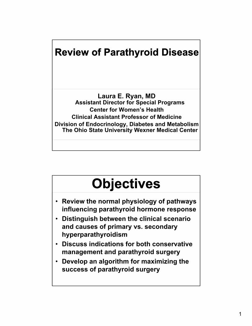
Preoperative imaging studiesPreoperative imaging studies
• Computed tomography (CT)
– May be effective as an adjunct to first li i i t h iline imaging techniques
– 4D-CT
• 3D imaging + contrast perfusion over time
Newer modality appears to be• Newer modality, appears to be effective in localizing hyper-functioning glands
• Magnetic resonance imaging (MRI)
22

23
Preoperative imaging studiesPreoperative imaging studies
• Sestamibi scan– Relies on differential washout times (thyroid
b l th id)vs abnormal parathyroid)
– Thyroid pathology can result in false positives
– Frequently used in conjunction with other anatomic imaging modality
• Single photon emission computed tomography (SPECT)– Superposition of the two radiomarkers results
in more accurate localization

24
Intraoperative AdjunctsIntraoperative Adjuncts
• Radioguided
• Methylene blue
• Rapid IOPTH

25
Intraoperative PTHIntraoperative PTH• Extensively described in patients with
primary HPT
– Confirms complete excision of hyperfunctioning gland prior to leaving the OR
– Alerts surgeon to additional h ti l dhypersecreting glands
Intraoperative PTHIntraoperative PTH
• When peripheral PTH drop >50% from their p p phighest (either preincision or preexcision) value in 10 minutes after removal of abnormal parathyroid then postoperative normal or low calcium levels are predicted with excellent accuracywith excellent accuracy

26
Intraoperative PTHIntraoperative PTH
85%
Surgical TechniqueSurgical Technique
• Bilateral parathyroid exploration
• Minimally invasive parathyroidectomy (MIP)

27
Bilateral Neck ExplorationBilateral Neck Exploration
• Traditional / comprehensive approachp pp
– Less dependent on localization studies
• Examination of all parathyroid glands with appropriate resection of diseased glands
• Caution for multigland disease
– First gland is mildly enlarged (75-200mg)
– Despite IOPTH drop and/or imaging
Bilateral Neck ExplorationBilateral Neck Exploration
• When do you explore?
Known secondary / tertiary HPT– Known secondary / tertiary HPT
– IOPTH fails to drop
– Negative or discordant imaging studies
• What is the downside?
Larger incision– Larger incision
– Hospitalization
– Increased risk to RLN

28
Parathyroidectomy OptionsParathyroidectomy Options
• Single gland excision BNE or MIP• Single gland excision BNE or MIP (unilateral vs focused exploration)
• Subtotal (3.5 gland excision)• Total (4 gland excision) with
autotransplantation
BNE

29
Minimally Invasive ParathyroidectomyMinimally Invasive Parathyroidectomy
• Depends upon preoperative ability to localize adenoma
• US and sestamibi
– Both positive and concordant = focused exploration
– 1 positive, 1 negative = focused exploration +1 positive, 1 negative focused exploration IOPTH or unilateral exploration ( 2 gland)
– Discordant = bilateral exploration + IOPTH
– Both negative = bilateral exploration + IOPTH
What makes a parathyroid operation minimally-invasive?
What makes a parathyroid operation minimally-invasive?
Size of the scar• Size of the scar
• Type of anesthesia
• Number of sides explored
• Number of glands explored• Number of glands explored

30
Minimally Invasive ParathyroidectomyMinimally Invasive Parathyroidectomy
Various approachesVarious approaches
• Small incision parathyroidectomy
– Outpatient
– Local anesthesia
• Endoscopic / video assistedEndoscopic / video assisted parathyroidectomy
Minimally Invasive ParathyroidectomyMinimally Invasive Parathyroidectomy
• Advantagesg
– Less pain
– Smaller incision
– Lower morbidity
– Decreased length of hospital stayDecreased length of hospital stay
31
ComplicationsComplications
• Recurrent laryngeal nerve injury
Fail re to locali e parath roid gland• Failure to localize parathyroid gland
• Persistent hyperparathyroidism
• Hypocalcemia
• Bleeding
S f ti• Scar formation
• infection
Laryngeal examLaryngeal exam• Note vocal quality
– Michel et al., 1988,
– 26% with voice change 3% VCP
• Preoperative endoscopic exam
– 3.5% with VCP
– 2-3% related to benign disease2 3% related to benign disease
Shin JJ, et al. The Surgical Management of Goiter: Part I. Preoperative Evaluation. Laryngoscope. 2011; 121:60-67

32
Nerve MonitoringNerve Monitoring“Garbage in, garbage out…”
Set up
• Flexible nasolaryngoscopy (in clinic)
• ETT position / patient position
• Neural mapping for identificationNeural mapping for identification
• Monitoring
• Flexible nasolaryngoscopy (in clinic)

33
Courtesy of Medtronic
Courtesy of Medtronic

34
Courtesy of Medtronic
Clinical BenefitsClinical Benefits• Reduction in RLN injury
I t ti di i f RLN i j• Intra-operative diagnosis of RLN injury
• Aiding identification / dissection
• Alternative approaches
Dionigi G, et al. Why monitor the recurrent laryngeal nerve in thyroid surgery? J Endocriol Invest. 2010, 33: 819-822.

35
Nerve InjuryNerve Injury• Infrequent but detrimental complication
Unilateral versus bilateral– Unilateral versus bilateral
– Temporary (9.8%) versus permanent (2.3%)
• Genther DJ, et al., 2014
• Direct visualization of nerve has been• Direct visualization of nerve has been shown to be safer than neural avoidance
• Use of the nerve monitor can be prognostic
Treatment FailureTreatment Failure• Persistence
– < 6 months
– Elevated calcium
– Avoidable versus unavoidable
– Suspect multi-gland disease
• Recurrence
– > 6 months
– Elevated calcium/PTH

36
ConclusionConclusion• Important to develop a working
relationship with an endocrinologistrelationship with an endocrinologist
• Gold standard remains BNE
• MIP / focused exploration are approaching cure rates for BNE in most series
Related Documents