Biomed Environ Sci, 2015; 28(1): 57-71 57 doi: 10.3967/bes2015.006 1. Comparative Pathology, Department of Pathology, School of Veterinary Medicine, Shiraz University, Shiraz 71345, Iran; 2. Department of Pathology, School of Veterinary Medicine, Shiraz University, Shiraz 71345, Iran; 3. Veterinary Surgery, Department of Veterinary Surgery and Radiology, Faculty of Veterinary Medicine, Shahrekord University, Shahrekord 8818634141, Iran Review Bone Injury and Fracture Healing Biology Ahmad Oryan 1 , Somayeh Monazzah 2 , and Amin Bigham-Sadegh 3,# Bones are organs of the skeletal system, providing shape, mechanical support, and protection to the body and facilitating the movement. In addition, bones contribute to the mineral homeostasis of the body and have recently been found to participate in endocrine regulation of energy metabolism [1-2] . The well-known limitations associated with clinical use of autografts and allografts continue to drive efforts to develop bone graft substitutes, using the principles of biomaterials and tissue engineering [3] . Under some stressful and continuous compressive conditions, the ability of the bone tissue to tolerate strength decreases. Whenever these forces overcome the toleration of the bone tissue, bone fracture occurs [4] . The highly complex process of fracture healing is still not fully understood; however, research in the recent years have identified associations between various factors that affect the repair process and healing outcome [5] . During skeletal growth or fracture healing, a temporary structure with a matrix of irregularly arranged collagen fibers and randomly dispersed crystals known as woven bone precedes the development of lamellar cortical bone. The osteocytes of the cancellous bone move into the sinusoids in the marrow via the canaliculi as the cancellous bone does not contain Haversian systems [6] . Bone Structure The main function of the bony skeleton is to provide a strong supportive and mechanically optimal structure for the soft tissues and muscles. The skeletal system protects the thoraco-abdominal viscera [7] and serves as a home for the marrow [8] . Bones play a key role in hematopoiesis and calcium metabolism [7] . Bone crystals are the main reservoir for calcium, phosphate, and essential ions for metabolic and physiological processes [9] . Bone is a composite structure and includes cells, extracellular matrix (ECM), and lipids [8] . About 20% of bone is water and the dry weight consists of 30%-35% organic and 65%-70% inorganic substances [10] . The cellular components of the bone include osteoblasts, osteocytes, osteoclasts, and osteogenic precursor cells (mesenchymal osteoprogenitor cells) [6-7] . Osteoblasts and osteocytes are differentiated from the mesenchymal stem cells. Osteocytes are the mature trapped osteoblasts in the lacunae [6] . Scientists use specific terminology for defining osteoblasts such as ‘mesenchymal osteoblasts’ and ‘surface osteoblasts’ [11] . Osteoblasts produce collagen [6] . Other functions of these cells are synthesis, regulation, deposition, and mineralization of the ECM [12] . In addition, these cells have a role in blood-calcium homeostasis and act as a mechanosensor for bones [12] . Multinucleated osteoclasts are derived from the macrophage-monocyte line, produce proteolytic enzymes, and have an important role in bone resorption, calcium and phosphate excretion, bone healing, and remodeling [6] . The osteogenic precursor cells are a kind of stem cell, which are derived from the mesenchymal cells and are able to differentiate into mature osteoblasts and then into bone lining cells and osteocytes [10] . The organic phase of bone matrix includes type I collagen fibers [7] , other types of collagen (such as collagen type III, V, etc.), noncollagenous proteins such as proteoglycans, glycoproteins, phosphoproteins [8] , byglican, decorin, osteonectin, thrombospondin, fibronectin, osteopontin, bone sialoprotein, osteocalcin, [12] and phospholipids [8] . The inorganic phase of bone matrix is mainly crystalline mineral salts and calcium in the form of hydroxyapatite [13] . Besides this, the inorganic substances also include 85% tricalcium phosphate, 10% calcium carbonate, and 5% fluoride derivatives such as calcium fluoride and magnesium fluoride [10] .

Welcome message from author
This document is posted to help you gain knowledge. Please leave a comment to let me know what you think about it! Share it to your friends and learn new things together.
Transcript

Biomed Environ Sci, 2015; 28(1): 57-71 57
doi: 10.3967/bes2015.006 1. Comparative Pathology, Department of Pathology, School of Veterinary Medicine, Shiraz University, Shiraz 71345,
Iran; 2. Department of Pathology, School of Veterinary Medicine, Shiraz University, Shiraz 71345, Iran; 3. Veterinary Surgery, Department of Veterinary Surgery and Radiology, Faculty of Veterinary Medicine, Shahrekord University, Shahrekord 8818634141, Iran
Review
Bone Injury and Fracture Healing Biology
Ahmad Oryan1, Somayeh Monazzah2, and Amin Bigham-Sadegh3,#
Bones are organs of the skeletal system, providing shape, mechanical support, and protection to the body and facilitating the movement. In addition, bones contribute to the mineral homeostasis of the body and have recently been found to participate in endocrine regulation of energy metabolism[1-2]. The well-known limitations associated with clinical use of autografts and allografts continue to drive efforts to develop bone graft substitutes, using the principles of biomaterials and tissue engineering[3]. Under some stressful and continuous compressive conditions, the ability of the bone tissue to tolerate strength decreases. Whenever these forces overcome the toleration of the bone tissue, bone fracture occurs[4]. The highly complex process of fracture healing is still not fully understood; however, research in the recent years have identified associations between various factors that affect the repair process and healing outcome[5]. During skeletal growth or fracture healing, a temporary structure with a matrix of irregularly arranged collagen fibers and randomly dispersed crystals known as woven bone precedes the development of lamellar cortical bone. The osteocytes of the cancellous bone move into the sinusoids in the marrow via the canaliculi as the cancellous bone does not contain Haversian systems[6].
Bone Structure
The main function of the bony skeleton is to provide a strong supportive and mechanically optimal structure for the soft tissues and muscles. The skeletal system protects the thoraco-abdominal viscera[7] and serves as a home for the marrow[8]. Bones play a key role in hematopoiesis and calcium metabolism[7]. Bone crystals are the main reservoir for calcium, phosphate, and essential ions for metabolic and physiological processes[9]. Bone is a composite structure and includes cells, extracellular
matrix (ECM), and lipids[8]. About 20% of bone is water and the dry weight consists of 30%-35% organic and 65%-70% inorganic substances[10].
The cellular components of the bone include osteoblasts, osteocytes, osteoclasts, and osteogenic precursor cells (mesenchymal osteoprogenitor cells)[6-7]. Osteoblasts and osteocytes are differentiated from the mesenchymal stem cells. Osteocytes are the mature trapped osteoblasts in the lacunae[6]. Scientists use specific terminology for defining osteoblasts such as ‘mesenchymal osteoblasts’ and ‘surface osteoblasts’[11]. Osteoblasts produce collagen[6]. Other functions of these cells are synthesis, regulation, deposition, and mineralization of the ECM[12]. In addition, these cells have a role in blood-calcium homeostasis and act as a mechanosensor for bones[12].
Multinucleated osteoclasts are derived from the macrophage-monocyte line, produce proteolytic enzymes, and have an important role in bone resorption, calcium and phosphate excretion, bone healing, and remodeling[6]. The osteogenic precursor cells are a kind of stem cell, which are derived from the mesenchymal cells and are able to differentiate into mature osteoblasts and then into bone lining cells and osteocytes [10].
The organic phase of bone matrix includes type I collagen fibers[7], other types of collagen (such as collagen type III, V, etc.), noncollagenous proteins such as proteoglycans, glycoproteins, phosphoproteins[8], byglican, decorin, osteonectin, thrombospondin, fibronectin, osteopontin, bone sialoprotein, osteocalcin,[12] and phospholipids[8]. The inorganic phase of bone matrix is mainly crystalline mineral salts and calcium in the form of hydroxyapatite[13]. Besides this, the inorganic substances also include 85% tricalcium phosphate, 10% calcium carbonate, and 5% fluoride derivatives such as calcium fluoride and magnesium fluoride[10].

58 Biomed Environ Sci, 2015; 28(1): 57-71
There are two types of bone tissue: 1) cancellous (trabecular) bone, which is present in the flat and cuboidal bones and in the extremities of long bones that are formed by plates and struts called trabeculae and 2) cortical (compact) bone in the outer layers of the long bones. The fundamental functional unit of the cortical bone are cylindrical structures known as osteons or Haversian systems,[14] and there are several lamellae surrounding the Haversian canal (with Volkmanns’s canals). The long bones have dense structure and have the important role of being weight-bearing structures; therefore, their role is to provide the stability for physical function[9]. Lamellar bone is the mature form in the cortical bone, woven in the immature form, and is not normally present in the cortical bone region[7]. During skeletal growth or fracture healing, a temporary structure with a matrix of irregularly arranged collagen fibers and randomly dispersed crystals known as woven bone precedes the development of lamellar cortical bone[9]. The osteocytes of the cancellous bone move into the sinusoids in the marrow via the canaliculi as the cancellous bone does not contain Haversian systems[6].
Bone is a well-vascularized tissue and the endothelium of the blood vessels has a critical role in the homeostasis of bone integrity[15]. Intracortical anastomoses exist between the medullary vessels and the periosteal vessels. The periosteal circulation supplies the periosteum and the upper part of the cortex. The medullary circulation supplies and nourishes the bone marrow and the lower part of the cortex and its terminal ramifications form metaphyseal vessels in the marrow. The metaphyseal vessels supply the lower part beside the osteoprogenitor cells for bone formation. The epiphyseal vessels supply the upper part of the proliferating and hypertrophic tissues[11].
Types of Bone Fracture and Their Mechanisms
Several types of bone fractures have been extensively described in the literature. Here we discuss some of the most important fracture types. While bone fractures in most instances are caused as result of traumas or specific bone diseases, macro-fractures could also occur as a result of accumulation of micro-fractures in the healthy bone, which is called ‘stress fracture’. These micro-fractures normally occur after continuous loading[2,6].
Inducing stresses by accidental inordinate load on a bone, because of an external impact or
intensive contraction of the muscles, causes traumatic fracture. Bones with inferior mechanical properties, which could be due to the development of bone tumors, are susceptible to traumatic fracture. The inability of the soft tissues to absorb the high energy forces also increases the risk of traumatic fracture. Some bone diseases cause bone destruction or weakening to such a degree that even a trivial trauma may produce a fracture (e.g., bone neoplasms, nutritional disturbances affecting bone), which is called pathologic fracture. Production of micro damages by cyclic loads and the inability of repairing and remodeling them can cause micro-cracks, which may further result in macro-cracks. A complete fracture will occur in individuals who have increased repetitive-type physical activities[14].
Bone fractures can be classified based on various characteristics. Based on the shape or pattern of the fractured fragments, fractures are divided into transverse, oblique, spiral, and comminuted. Other types include compression or crush fracture, gunshot fracture, as well as greenstick fracture and avulsion fracture. Based on the etiology, there are three types of fractures including traumatic, fatigue, and pathological. Finally, according to the nature of the fracture, there are closed and open fractures[4].
Mechanisms of Bone Formation
Bone development occurs through two mechanisms, namely, intramembranous and endochondral bone formation. In the intramembranous form, bone formation occurs without mediation of the cartilaginous phase, and the sources of the cells that contribute to this way are present in the inner periosteal osteogenic layer. In the endochondral bone formation, the initial synthesis of cartilage is followed by the endochondral sequence of bone formation[11].
Bone may be synthesized by intramembranous ossification, endochronal ossification, or a combination of both. The essential difference between these processes is the presence or absence of cartilaginous phase. Intramembranous ossification occurs when the mesenchymal precursor cells proliferate and subsequently differentiate directly into osteoblasts; but in the endochondral ossification, the mesenchymal cells at first step differentiate into chondrocytes and secrete the cartilaginous matrix. The woven bone will then be made up of this cartilage. The bone formed from
Bone injury and healing biology 59
endochondral ossification has better biomechanical properties than the bone formed from intramembranous ossification because in the endochondral ossification, a steady matrix from cartilage is made and then calcification is started, but in the intramembranous ossification, only the trabecules of bone are made. Investigation into fracture healing continues along many avenues and usually uses standardized validated animal fracture models, whose biology is assumed to differ from that of humans on a temporal basis[7]. The biochemical factors are locally expressed during distraction osteogenesis and some of them can even be identified systemically. In vivo studies in which serum levels were investigated showed a significant increase in, and correlation between, the osteoblastic marker bone-specific alkaline phosphatase, transforming growth factor-β1 (TGF-β1), and basic fibroblast growth factor (bFGF). This implies that strain-activated osteoblastic cells are a major source of systemically increased bone growth factors during callus distraction[16].
Intramembranous bone healing forms bone without first forming the cartilage. This process is performed by intermediation of the osteoprogenitor and undifferentiated mesenchymal cells and results in the formation of hard callus. In the early phase of bone healing, the endothelial cells change into polymorphic cells and then transform to the osteoblastic phenotype[17]. Endochondral bone formation includes recruitment, proliferation, and differentiation of the undifferentiated mesenchymal cells into cartilage, which is followed by calcification and replacement with bone. The stages of endochondral bone formation include hematoma formation, inflammation, angiogenesis, cartilage formation, cartilage calcification, cartilage removal, bone formation, and finally bone remodeling. The external soft tissues and the periosteum of the fracture region supply the bridging or soft callus that stabilizes the fracture fragments[18]. In the remodeling phase, the young woven bone is gradually replaced by a lamellar bone to restore the mechanical integrity of the healing site[19].
Fracture Healing
Fracture healing is an important biological process that is necessary for the survival of the injured animal. Bone is a unique tissue and its repair process is of great biological importance, as it aims to fully restore the lamellar bone to its original condition, thereby regaining the initial bone
strength[20]. There are three main phases following a fracture in the bone repair process: 1) The early inflammatory stage, 2) The proliferative or fibroplasia stage, and 3) The remodeling stage[10]. In normal bone development, bone remodeling conventionally refers to the removal of calcified bone tissue by osteoclasts. However, in the context of bone repair, there are two phases of tissue catabolism: the removal of the initial cartilaginous soft callus, followed by the eventual remodeling of the bony hard callus[2,21].
Classification of Fracture Healing
Bone is one of the few tissues that can heal without fibrous scar formation[22]. In the classic histological terms, fracture healing has been divided into two types including primary (direct) and secondary (indirect) fracture healing models[23].
Direct or Primary Fracture Healing
Primary fracture healing is a faster healing process than the secondary healing[24]. Direct healing does not commonly occur in the natural process of fracture healing[22]. This kind of healing involves intramembranous bone formation and direct cortical remodeling without any external tissue (callus) formation[25]. It occurs only when rigid internal fixation anatomically reduces the mobility of the fracture fragments, thereby, reducing inter-fragmentary strain[17]. Osteons (Haversian system) traveling along the length of the bone are able to cross the fracture site and bridge the gap, laying down cylinders of bone and progressively, the fracture is healed by the formation of numerous osteons[25]. It usually takes from a few months to a few years, before complete healing is achieved[22].
Primary healing, or primary cortical healing, involves a direct attempt by the cortex to reestablish itself once it has become interrupted. A fracture becomes united when the bone on one side of the cortex is united with the bone on the other side to reestablish mechanical continuity. This process occurs only when there is anatomic restoration of the fracture fragments and when the stability of fracture reduction is ensured with a substantial decrease in the inter-fragmentary strain. Under these conditions, bone resorbing cells on one side of the fracture undergo a tunneling resorptive response whereby they reestablish new Haversian systems by providing pathways for penetration by blood vessels. These new blood vessels are accompanied by

60 Biomed Environ Sci, 2015; 28(1): 57-71
endothelial cells and perivascular mesenchymal cells, which become the osteoprogenitor cells for osteoblasts. These events result in the formation of discrete remodeling units known as cutting cones[23]. In primary fracture healing, if the fracture is anatomically reduced, at the micrometric level, osteonal healing occurs. Osteoclasts create ‘cutting cones’ and primarily cross the fracture site. This requires very high stability and in practice is the rarest type[26]. Osteonal activity increases near the injury and this phenomenon is referred to as ‘regional acceleratory phenomenon’ (RAP) and probably plays an important role in direct fracture healing. The mechanism of RAP is unknown, but the phenomenon may be mediated by the same signaling molecules as seen in other types of tissue repair[19]. With time, extensive remodeling obliterates the osteotomy defect. Also, this type of reparative process is normally known as primary fracture healing[14].
Indirect or Secondary Fracture Healing
The other names of indirect fracture healing are endochondral ossification, secondary healing, and callus healing. Indirect bone healing is an ordered process of bone repair and reorganization[27]. The stages of indirect healing include impaction, inflammation, primary soft callus formation, callus mineralization, and callus remodeling[28]. It typically occurs when some micro-motions might exist between the fracture ends and this commonly happens after intramedullary nailing and external fixation techniques[22]. This type of fracture healing is generally enhanced by motion and inhibited by rigid fixation[18]. Both intramembranous and endochondral bone healing occur in the indirect model of fracture healing[22].
Complications of Fracture Healing
The complications of fracture healing can be classified into three groups:
Immediate complications: Hypovolemic shock (systemic) and injury to major vessels, injury to muscles and tendons, injury to joints, and injury to viscera (local).
Early complications: Hypovolemic shock, Adult respiratory distress syndrome, fat embolism syndrome, deep vein thrombosis, pulmonary syndrome, aseptic traumatic fever, septicemia in open fracture, crush syndrome (systemic), infection, and compartment syndrome (local).
Late complications: Delayed union, nonunion, malunion, and cross union (imperfect union of the fracture) and avascular necrosis, shortening, joint stiffness, Sudeck's dystrophy, osteomyelitis, ischemic contracture, myositis ossificans, and osteoarthritis.
Complications include pain, nerve damage, vascular injury, wound problems, infection, need for further surgery, instability, and hematoma[29]. Low-energy and some open injuries are rarely associated with serious complications, but most open fractures caused by high-energy trauma often pose major problems, which require more detailed analysis. The soft-tissue condition, energy level of the trauma, fracture comminution, initial fracture displacement, treatment method, contamination, and associated injuries can influence fracture healing[30].
Three Phases of Fracture Healing
Inflammatory Phase The first phase occurring immediately following a fracture is the formation of a hematoma in the injured bone[31-32]. This hematoma is caused as a result of bleeding from the ruptured bone and the periosteal vessels that are formed within the medullary canal and beneath the periosteum. The activated coagulation system releases potent vasoactive mediators from the degranulated platelets present in the hematoma[33]. The levels of several inflammatory mediators, including cytokines such as interleukin-1 (IL-1), IL-6, IL-11, IL-18, and tumor necrosis factor-α (TNF-α), are significantly elevated within the first few days after the injury[31]. These proinflammatory mediators have chemotactic effects on other inflammatory cells. Then, further aggregation of platelets and angiogenesis take place[18]. After vascular trauma, the fracture site becomes hypoxic and the osteocytes at the ends of the fracture sites become deprived of their nutrition and undergo degenerative and/or necrotic changes[34]. Macrophages phagocytize the necrotic areas and facilitate the regeneration stage by releasing signaling factors importantly, the growth factors such as bone morphogenic proteins (e.g., BMP-2, -5, -7), bFGF, transforming growth factor-β (TGF-β), platelet-derived growth factor (PDGF), and insulin-like growth factor (IGF). These growth factors are responsible for migration, recruitment, and proliferation of mesenchymal stem cells and their differentiation to angioblasts, chondroblasts, fibroblasts, and osteoblasts[27]. The endothelial cells, fibroblasts, and osteoblasts participate in filling the
Bone injury and healing biology 61
fracture gap by the formation of granulation tissue[35]. During the inflammatory phase, a primitive callus develops and reduces the uncontrolled mobility at the fracture site. Under a normal condition, the inflammatory stage is fast and lasts up to a week after the fracture[20].
Lymphocytes are not required for the initiation of wound healing, but an intact cellular immune response is essential for a normal outcome of tissue repair. Injury affects lymphocyte immune mechanisms leading to generalized immunosupp- ression, which, in turn, increases host susceptibility to infection and sepsis. Although the exact origin of posttraumatic immune suppression remains unknown, stress hormones and immunosuppressive factors, such as inflammatory cytokines, prostaglandin E2, and nitric oxide, affect the lymphocyte function adversely. Posttraumatic impairment of T-lymphocyte immune function is reflected by the decreased lymphocyte numbers, as well as the altered T-cell phenotype and activity. Antibody-producing B lymphocytes are variably affected by injury, probably secondary to alterations of T-lymphocyte function, as a result of their close interaction with helper T cells. Therapeutic modulation of the host immune response may include nonspecific and specific interventions to improve the overall defense mechanisms[36].
To examine the potential roles of neutrophils in bone repair, a neutrophil-neutralizing antiserum or control normal serum has been administered systemically in rats with growth plate injury. The inflammatory response has been found to be temporally associated with increased expression of neutrophil-chemotactic chemokine cytokine-induced chemoattractant-1 and cytokines TNF-α and IL-1β. Following the inflammatory response, mesenchymal infiltration, chondrogenic and osteogenic responses, and bony repair have been observed at the injury site. Neutrophil reduction did not significantly affect the infiltration of other inflammatory cells and the expression of TNF-α, IL-1β, growth factors, PDGFβ, and TGF-β1 at the injured growth plate on day 1 and no effects on mesenchymal infiltration were observed on day 4. However, by day 10, there was a significant reduction in the proportion of mesenchymal repair tissue but an increase (although statistically insignificant) in the bony trabeculae and a decrease in the cartilaginous tissue within the injury site. Consistently, in antiserum-treated rats, there was an increase in the expression of osteoblastic differentiation transcription factor cbf-α1 and bone matrix protein osteocalcin and a
decrease in chondrogenic transcription factor Sox-9 and cartilage matrix collagen-II in the injured growth plate. These results suggest that injury-induced, neutrophil-mediated inflammatory response appears to suppress mesenchymal cell osteoblastic differentiation but enhance chondrogenic differentiation; thus, it may be involved in regulating downstream chondrogenic and osteogenic events for growth plate bony repair[37].
Following the inflammatory response also, macrophages are observed in fibrous callus tissues and in a portion of the newly formed bone. Macrophages regulate the early phases of fracture healing, possibly by directing the differentiation of chondrocytes and regulating vascularization. Macrophages may stimulate the initial differentiation of progenitor cells, which leads to enhanced maturation at later time points[38]. The remodeling process of mostly collagenous molecules is largely attributable to matrix metalloproteinases (MMPs). A variety of members of this protease family and its respective inhibitors-termed tissue inhibitors of matrix metalloproteinases (TIMP)-have been found to be closely related to the fracture healing process. Delays in bone healing or even nonunion could be related to the concentrations of these enzymes or their behavior over time. Supernatants from human fractured tibial bone fragments promote osteogenesis and migration of muscle-derived stromal cells (MDSC) in vitro. The main factor responsible for this is TNF-α, which promotes first MDSC migration and then osteogenic differentiation at low concentrations. However, TNF-α is inhibitory at high concentrations. These data indicate that manipulating the local inflammatory environment to recruit, and then differentiate adjacent MDSC, may be a simple yet effective way to enhance bone formation and accelerate fracture repair. This concluding remark is based on a combination of human specimens and an in vivo murine model; therefore, may translate to clinical care[37]. Proliferative Phase Basically, the proliferative or fibroplasia process has been described in terms of the organization of the fracture hematoma. As fibroplasia phase begins, necrotic bone resorption is carried out by osteoclasts that are derived from the circulating monocytes in the blood and by monoblastic precursor cells originating from the local bone marrow[33]. The fibroplasia phase is characterized by the formation of callus and begins with continued vascular ingrowth, secretion of
62 Biomed Environ Sci, 2015; 28(1): 57-71
osteoid, and the presence of collagen fibers[10]. This phase involves a periosteal response with angiogenesis and formation of connective tissue and soft callus, which is gradually replaced by the immature woven bone formed via intramembranous or endochondral bone formation[35]. The mesenchymal stem cells differentiate into chondrocytes (cartilage-forming cells) in the hypoxic central fracture area where the soft callus will gradually take on the appearance of cartilage and mechanically stabilize the fracture zone[34]. Proliferation and differentiation of the chondrocytes are stimulated by the expression of growth factors including TGF-α2, PDGF, IGF-1, and some BMPs such as BMP-2, -4, -5, and -6[20]. The osteoblasts begin to synthesize intramembranous (woven) bone tissue distal to the fracture site[27]. Endochondral bone formation occurs in the region, which is mechanically less stable. TGF-β2 and -β3, BMPs, and other molecular signals induce endochondral bone ossification in the cartilaginous callus[31]. The woven bone gradually replaces the cartilage through endochondral ossification resulting in the formation of hard callus that increases the stability of the fracture or the osteotomy site[33].
Application of growth factors showed strong stimulating effects on fracture healing[39]. Full vascularization is necessary for bone formation. Therefore, it is not surprising that the principal action of many growth factors is both mitogenic and angiogenic[40]. In addition, substitutes in combination with growth factors are all designed to act as supports for the recruitment, proliferation, and differentiation of bone progenitor cells[41]. Future research based on clinical studies would provide the evidence required in terms of efficacy and safety before the growth factors could be used in the clinical setting as agents for bone regeneration procedures[42]. We must understand how the growth factors interact with each other and with cells, what their effect is, which intracellular pathways are triggered by them, and how they can be activated/inactivated[12]. Remodeling phase The third phase involves the formation and mineralization of the callus and replacement of the mineralized callus with mineralized bone and sculpting of the bone back to its original shape, size, and biomechanical competency via modeling and remodeling[32]. This phase can also be referred to as secondary bone formation and involves converting the irregular woven bone callus into the lamellar bone[21].
In this phase, osteoclasts resorb the newly woven bone and osteoblasts replace this matrix with the lamellar bone[43]. The important functional outcome of the remodeling phase of fracture healing during homeostatic remodeling is the restoration of mechanical strength and stability[43]. Osteoclasts become polarized and adhere to the mineralized surface and continue remodeling of bone. They form a ruffled border, which is sealed off and acid and proteinases are pumped into the resorption domain, and bone resorption by osteoclasts creates erosive pits on the bone surface known as ‘Howship’s lacuna.’ Once completed, osteoblasts are able to lay down new bone on the eroded surface[21].
The process of replacement and repair is a continuous ongoing in the normal skeleton, and the mechanisms involved in fracture healing have major similarities to the mechanism of otherwise healthy skeleton; however, there are some differences in the process depending on whether it is occurring in compact or cancellous bone. In the case of cancellous bone, the cells are never very far away from the blood vessels and so the whole process of bone apposition or replacement can take place on the surface of the trabeculae, a phenomenon often referred to as ‘creeping substitution’[44]. This remodeling phase is regulated by several proinflammatory signals such as IL-1, IL-6, and IL-11, and elevated levels of TNF-α, IL-12, and interferon-γ (IFN-γ)[31]. In addition, growth hormone and parathyroid hormone also play key roles in this phase, speeding up the healing and strengthening of the fractured callus[31]. Electrical fields also influence bone remodeling. When stress is applied to the bone, electropositivity occurs on the convex surface and is associated with osteoclast activity, and electronegativity on the concave surface is associated with osteoblast activity[33].
To enhance the stability and strength at the fracture site, the size of the callus must be sufficiently large to compensate for the relatively poor strength of primitive bone[27]. Lamellae are aligned in a direction parallel to the longitudinal axis of the greatest force and adequate loading is required to enhance osteogenesis and direct the optimal geometric configuration of osteons[27]. Adequate strength develops by 6 months and remodeling phase may occur over months to years[10]. Mechanical bone strains created by muscular forces present during physical activity stimulate the remodeling[33].
Bone injury and healing biology 63
Molecular Events of Fracture Healing
Research in the fields of cellular and molecular biology of fracture healing, using immune-histochemical and DNA/RNA hybridization techniques, have helped to increase our understanding of the subject[7]. For hybridization histochemistry, sample cells and tissues were obtained from bone fracture healing site and treated to fix the target transcripts in place and to increase access of the probe. The probe is either a labeled complementary DNA or, now most commonly, a complementary RNA (riboprobe). The probe hybridizes to the target sequence at elevated temperature, and then the excess probe is washed away (after prior hydrolysis using RNase in the case of unhybridized, excess RNA probe). Solution parameters such as temperature, salt, and/or detergent concentration can be manipulated to remove any nonidentical interactions (i.e., only exact sequence matches will remain bound). Then, the probe that is labeled with either radio-, fluorescent-, or antigen-labeled bases (e.g., digoxigenin) is localized and quantified in the tissue using either autoradiography, fluorescence microscopy, or immunohistochemistry, respectively. In situ hybridization can also use two or more probes, labeled with radioactivity or the other non-radioactive labels, to simultaneously detect two or more transcripts[45]. As an example, in situ hybridization techniques have been used to study the removal of cells during fracture healing and it has been shown that the chondrocytes are removed by undergoing apoptosis, and metaplastic differentiation of chondrocytes to osteoblast does not occur[7]. Several factors regulate the cascades of molecular events in fracture healing, such as migration, proliferation, chemotaxis, differentiation, inhibition, and extracellular protein synthesis, by affecting different points in the osteoblast and chondroblast lineage through various processes[5]. Genomic and proteomic approaches aiming to identify key markers for the related transcriptional and translational shifts involved in cell differentiation, cell proliferation, and skeletal development would be quite useful[46].
In the early phases after bone injury, there is an upregulation of genes related to cell cycle (cell division) and cell-to-cell signaling (cell communication)[47]. There is a peak expression of IL-1 and IL-6 1 day after fracture, followed by a rapid decline to near undetectable levels by day 3[17]. In addition, expression of IGF-1 and IGF-2, PDGF, FGF
receptor, fibronectin, MMPs, glypican, byglican, osteomodulin, osteonectin, tenascin C, cartilage, and bone collagen increases until the immature osteoid synthesis by osteoblast progenitors is histologically detectable[47]. At least 34 members have been identified in the human genomes that are activated by proteolytic enzymes[18]. Many of the genes controlling cell growth and survival are constantly upregulated; whereas those functionally associated with the differentiation of osteogenic precursors and bone matrix formation undergo temporary modulation over time[47]. They act on serine/threonine kinase membrane receptor on target cells. This ligand-receptor interaction activates an intracellular signaling pathway, which ultimately affects gene expression in the nucleus[18].
Using microarray analysis, it was shown that selective gene induction by BMP-2, TGF-β, and activin-A controls and regulates the differentiation of mesenchymal precursor cells into osteoblastic cells[46]. It occurs in osteoprogenitors, mesenchymal cells, osteoblasts, and chondrocytes. BMPs induce a sequential cascade of events for chondro- osteogenesis, including chemotaxis, proliferation, and differentiation of mesenchymal and osteoprogenitor cells and angiogenesis. It also controls ECM synthesis[18]. BMP-2 has an important role in this recruitment and is essential for bone repair, but other BMPs such as BMP-7 may also play a more important role in the recruitment of progenitor cells[22].
Genomic and proteomic approaches are useful analytical tools for monitoring the changes in gene and protein expression[46]. Understanding each of the signaling events in the bone healing pathway extends our ability to intervene in the fracture healing process to rectify inadequate or failed healing[32]. The vascular ingrowth into the developing callus is regulated by FGF, vascular endothelial growth factor (VEGF), and angiopoietins 1 and 2. Angiopoietin 1 has been suggested to be produced and activated during the initial periods of fracture healing, whereas VEGF is expressed, released, and activated later, mainly during endochondral bone formation[17]. Recent studies have also demonstrated an important role for hypoxia inducible factor-1a (HIF-1a) in bone repair and its induction role for the VEGF activity in the revascularization process shows that hypoxic gradients regulate mesenchymal stem cell progenitor cell trafficking by HIF-1[22]. Platelets that have been activated by thrombin and subendothelial collagen release PDGF and TGF-β, which play a role in initiating fracture repair and inducing mesenchymal cell migration, activation and

64 Biomed Environ Sci, 2015; 28(1): 57-71
proliferation, angiogenesis, chemotaxis of acute inflammatory cells, and further aggregation of platelets[17].
Evaluation of Fracture Healing
There are various ways to evaluate the fracture healing. Basically, the evaluation methods could be divided into two major categories including invasive and noninvasive methods. None of the invasive methods are more clinically applicable and pleasant than the noninvasive methods because there is no need for tissue biopsy in the latter approach. Noninvasive methods could be divided into two major groups. In the first group, the assessments are mostly based on physical examinations and activity of the patient. The quality of bone healing is assessed based on the physical activity of the patients and the weight-bearing forces are indirectly measured by gross inspection or directly measured through force plating device. The quality, quantity, and duration of weight bearing and physical activity together with other physical characteristics such as pain degree are indices of bone fracture healing. In the second group of noninvasive methods, the assessments are based on the imaging technologies. Several imaging devices and methods have been invented and introduced to date, which include plain radiography, contrast radiography, magnetic resonance imaging, computed tomography (CT), and finally plain and color Doppler ultrasonography. Each of these methods has its own advantages and disadvantages. Normally, plain radiography and CT scans are more reliable techniques than the other methods and are discussed in this review. In the invasive techniques, there are several methodologies and approaches with the aim to assess bone healing and quality of the reformed tissue in the fractured site. These methods could be basically divided into four major categories including macroscopic and microscopic techniques, tensile testing, biochemistry, and molecular methods. In microscopic techniques, several methods have been introduced with the aim to describe some important characteristics of the healing bone and to measure the bone density. Light microscopy is the basic method and is discussed in this review. Other microscopic techniques include transmission electron microscopy, scanning electron microscopy, phase contrast microscopy, laser microscopy, and immunofluorescence microscopy. Tensile testing is another invasive method in which it is possible to test the resistance of the healing tissue against
various forces including bending, torsion, compression, and strain. In general, for assessments of a long bone fracture healing, bending is the test of choice, but other tests may also be used. Biochemistry is another technique in which we can directly measure the bone compositions, and in molecular methods, it is possible to directly and indirectly assess bone healing by measuring the expression of several proteins such as growth factors, and MMPs. Here, we discuss the most reliable techniques that are used in fracture healing assessments[4]. On the whole, microscopic techniques such as histopathology, immunohisto- chemistry, transmission electron microscopy, scanning electron microscopy, phase contrast microscopy, laser microscopy, and immunofluore- scence microscopy including biomechanical tests need sampling from the healing site and all of them are invasive techniques and used for ex vivo assessment of fracture repair. However, radiological or ultrasonographical techniques are non-invasive techniques and are used for in-vivo assessment of fracture repair applicable in clinical.
Gross Evaluation
Different scoring systems are applied for the gross evaluation of fracture healing to get comparable figures for statistical analysis[48]. Disabilities of the arm, shoulder, and hand (DASH) scoring system and the medical outcomes study short form-36 (SF-36) scores are two suitable scoring systems in the clinical evaluation of bone healing[49].
Radiological Evaluation
Some studies have attempted to use quantitative radiology to measure the changes in fracture healing in both experimental and clinical fractures, but the relationship between these changes and the mechanical properties of the healing fracture are not always clear, unless there is a consistent fracture gap, which is often not the case in clinical fractures. Even though a definition of an endpoint for fracture healing might be difficult, it would be very helpful if a time point could be defined at which healing is complete, as this is important in guiding clinical decisions that have to be made during the treatment of the patients[50]. We can see the fracture line up to 2-3 weeks, during which the soft callus may form and bone union can also be assessed by the treating physician. About 25% of bone formation may take place up to the 14th postoperative day as observed in a rabbit model.
Bone injury and healing biology 65
Usually, there is no evidence of remodeling and union[50-55].
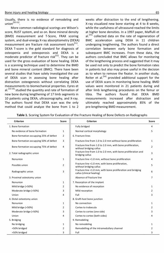
More common radiological scorings are Wilson’s score, RUST system, and so on. Bone mineral density (BMD) measurement and T-Score, FRAX scoring system, and dual-energy X-ray absorptiometry (DEXA) measurement are fracture risk assessment tools[51]. DEXA T-score is the gold standard for diagnosis of osteopenia and osteoporosis and DEXA is a moderate predictor of fracture risk[52]. They can be used for the gross evaluation of bone healing. DEXA is a scanning technique used to determine the BMD and bone mineral content (BMC). There have been several studies that have solely investigated the use of DEXA scan in assessing bone healing after distraction osteogenesis without correlating DEXA measurements to biomechanical properties. Eyres et al.[53-54] studied the quantity and rate of formation of new bone during lengthening of 17 limb segments in 10 patients using DEXA, ultrasonography, and X-ray. The authors found that DEXA scan was the only method that could analyze the bone from 1 to 2
weeks after distraction to the end of lengthening. X-ray visualized new bone starting at 4 to 8 weeks, while the usefulness of ultrasound reached the limits at higher bone densities. In a 1997 paper, Maffulli et al.[55] collected data on the rate of regeneration of BMC acceleration using DEXA in 11 children undergoing lengthening. The authors found a direct correlation between early bone formation and subsequent BMC increases. From these data, the authors concluded that BMC allows for monitoring of the lengthening process and suggested that it may be used not only to predict the bone formation rates in patients but also may prove useful in the decision as to when to remove the fixator. In another study, Reiter et al.[56] provided additional support for the use of DEXA scans in monitoring bone healing. BMD values were monitored in 21 patients during and after limb lengthening procedures on the femur or tibia. The authors found that DEXA BMD measurements increased after distraction and ultimately reached approximately 85% of the pre-lengthening BMD measurement.
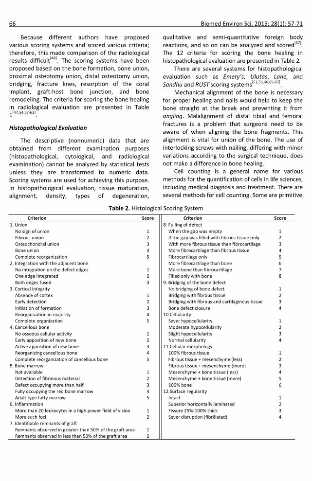
Table 1. Scoring System for Evaluation of the Fracture Healing of Bone Defects on Radiographs
Criterion Score Criterion Score
1. Bone formation Fully bridged 4
No evidence of bone formation 1 Normal cortical morphology 5
Bone formation occupying 25% of defect 2 6. Fracture lines
Bone formation occupying 50% of defect 3 Fracture line from 1.0 to 2.0 mm without bone proliferation 1
Bone formation occupying 75% of defect 4 Fracture line from 1.0 to 2.0 mm, with bone proliferation, without bridging callus
2
2. Total radiographic union Fracture line from 1.0 to 2.0 mm, with bone proliferation and bridging callus
3
Nonunion 1 Fracture line <1.0 mm, without bone proliferation 4
Possible union 2 Fracture line <1.0 mm, with bone proliferation, without bridging callus
5
Radiographic union 3 Fracture line <1.0 mm, with bone proliferation and bridging callus (clinical healing)
6
3. Proximal osteotomy union Absence of fracture line 7
Nonunion 1 7. Resorption of the implant
Mild bridge (<50%) 2 No evidence of resorption 1
Moderate bridge (>50%) 3 Mild resorption 2
Union 4 Full 3
4. Distal osteotomy union 8. Graft-host bone junction
Nonunion 1 No connection 1
Mild bridge (<50%) 2 Cortex to trabecula 2
Moderate bridge (>50%) 3 Cortex to cortex (one side) 3
Union 4 Cortex to cortex (both sides) 4
5. Bridging 9. Remodeling
No bridging 1 No remodeling 1
<50% bridged 2 Remodelling of the intramedullary channel 2
>50% bridged 3 Full 3
66 Biomed Environ Sci, 2015; 28(1): 57-71
Because different authors have proposed various scoring systems and scored various criteria; therefore, this made comparison of the radiological results difficult[48]. The scoring systems have been proposed based on the bone formation, bone union, proximal osteotomy union, distal osteotomy union, bridging, fracture lines, resorption of the coral implant, graft-host bone junction, and bone remodeling. The criteria for scoring the bone healing in radiological evaluation are presented in Table 1[47,54,57-63].
Histopathological Evaluation
The descriptive (nonnumeric) data that are obtained from different examination purposes (histopathological, cytological, and radiological examination) cannot be analyzed by statistical tests unless they are transformed to numeric data. Scoring systems are used for achieving this purpose. In histopathological evaluation, tissue maturation, alignment, density, types of degeneration,
qualitative and semi-quantitative foreign body reactions, and so on can be analyzed and scored[57]. The 12 criteria for scoring the bone healing in histopathological evaluation are presented in Table 2.
There are several systems for histopathological evaluation such as Emery’s, Ulutas, Lane, and Sandhu and RUST scoring systems[51,55,60,65-67].
Mechanical alignment of the bone is necessary for proper healing and nails would help to keep the bone straight at the break and preventing it from angling. Malalignment of distal tibial and femoral fractures is a problem that surgeons need to be aware of when aligning the bone fragments. This alignment is vital for union of the bone. The use of interlocking screws with nailing, differing with minor variations according to the surgical technique, does not make a difference in bone healing.
Cell counting is a general name for various methods for the quantification of cells in life sciences, including medical diagnosis and treatment. There are several methods for cell counting. Some are primitive
Table 2. Histological Scoring System
Criterion Score Criterion Score 1. Union 8. Fulling of defect
No sign of union 1 When the gap was empty 1 Fibrous union 2 If the gap was filled with fibrous tissue only 2 Osteochondral union 3 With more fibrous tissue than fibrocartilage 3 Bone union 4 More fibrocartilage than fibrous tissue 4 Complete reorganization 5 Fibrocartilage only 5
2. Integration with the adjacent bone More fibrocartilage than bone 6 No integration on the defect edges 1 More bone than fibrocartilage 7 One edge integrated 2 Filled only with bone 8 Both edges fused 3 9. Bridging of the bone defect
3. Cortical integrity No bridging of bone defect 1 Absence of cortex 1 Bridging with fibrous tissue 2 Early detection 2 Bridging with fibrous and cartilaginous tissue 3 Initiation of formation 3 Bone defect closure 4 Reorganization in majority 4 10.Cellularity Complete organization 5 Sever hypocellularity 1
4. Cancellous bone Moderate hypocellularity 2 No osseous cellular activity 1 Slight hypocellularity 3 Early apposition of new bone 2 Normal cellularity 4 Active apposition of new bone 3 11.Cellular morphology Reorganizing cancellous bone 4 100% fibrous tissue 1 Complete reorganization of cancellous bone 5 Fibrous tissue + mesenchyme (less) 2
5. Bone marrow Fibrous tissue + mesenchyme (more) 3 Not available 1 Mesenchyme + bone tissue (less) 4 Detection of fibrinous material 2 Mesenchyme + bone tissue (more) 5 Defect occupying more than half 3 100% bone 6 Fully occupying the red bone marrow 4 12.Surface regularity Adult type fatty marrow 5 Intact 1
6. Inflammation Superior horizontally laminated 2 More than 20 leukocytes in a high power field of vision 1 Fissure 25%-100% thick 3 More such foci 2 Sever disruption (fibrillated) 4
7. Identifiable remnants of graft Remnants observed in greater than 50% of the graft area 1 Remnants observed in less than 50% of the graft area 2
Bone injury and healing biology 67
and do not require special equipment; thus, can be done in any biological laboratory, whereas others rely on sophisticated electronic appliances. The methods of cell counting include counting chamber, plating, and counting colony-forming units, spectrophotometry, electrical resistance, flow cytometry, and image analysis. Recent approaches consider the use of high-quality microscopy images over which a statistical classification algorithm is used to perform automated cell detection and counting as an image analysis task.
Biomechanical Evaluation
Monitoring the progression of fracture healing by measuring the biomechanical performance of the healing bone is possible. It can be performed by applying either a direct (from 6 weeks after fracture) or an indirect technique (from the first day after fracture). There are large number of variables to consider when establishing mechanical testing procedures, because there are no established standards for bone biomechanical testing and there are many varieties of bone shapes and sizes[58].
Physical and mechanical properties of new bone formation are important factors for evaluation of bone healing. Monitoring the progression of fracture healing by measuring the biomechanical performance of the healing bone is possible and the outcome information of these measurements is stress, strain, load, deflection, force, displacement, ultimate strength, fracture stiffness, and so onas a function of the healing time. These biomechanical criteria must be measured for bones and be compared with normal bones[50]. Biomechanical properties are obtained by preparation of symmetric specimens allowing for normalization of the specimen properties with respect to their size[58]. However, it is really not practically possible to accurately measure stress and strain acting on the callus due to the irregularity of the callus structure and the tissue types.
The three-point bending test has been performed to determine the mechanical properties of bones[59]. For the mechanical test, the bone ends are placed between the two jaws in the testing machine and the load is exerted at the healing injured area until the failure and the forces, which are needed to break the bone, are recorded[50-51,53,55]. The biomechanical tests are conducted using a universal testing machine. The bone samples are horizontally placed on two rounded supporting bars and are loaded at its central point by lowering the third bar
so that the fracture line is in the center. The samples are loaded and any changes in the toleration of ultimate load and length are detected from the graph sketched by the machine[60]. In obtaining material properties, the specimen size and geometry are neglected and structural properties (force and displacement) are expressed per unit size as material properties (stress and strain)[58]. More common parameters for the biomechanical evaluation of bone are load, deflection, force, displacement, the specimen’s extension at the ultimate strength region, stress[59], cortical area, maximum normalized shear stress, polar moment of inertia, torsional moment of inertia[61], stress (ultimate strength proportion to cross-sectional area), and tan-α (the coefficient of inclination for the linear portion of the load-deformation curve)[59].
The three-point bending test is simple and straightforward, but it has the disadvantage of creating a high shear stress near the middle section of the specimen. Four-point bending yields pure bending at the middle portion of the specimen between the two loading points, without transverse shear stresses being present. However, it requires that the force at each loading point be equal, and specimen length be sufficient to accommodate the two loads. These requirements are simple to achieve for regularly shaped specimens, but somewhat difficult for testing whole bone. Thus, the three-point bending test is used more often to measure the biomechanical properties of whole bones[58].
Stress, strain, and Young’s modulus can be calculated from the force and displacement by testing regularly machined specimens. For three-point bending tests, the values of these parameters can be calculated.
The torsion test is another popular test, which can be used to measure the biomechanical properties of bone in shear. When a specimen is loaded in torsion (twisting moment), shear stress varies from zero at the center of the specimen to the maximum at the surface. For any cross section, the maximum shear stress in torsion can be calculated. Like tensile test specimens, the central portion of a torsion specimen should be reduced to ensure that the failure occurs in the middle of the specimen. Torsion tests yield intrinsic shear properties (e.g., shear strength, shear modulus, and shear toughness) when using regularly machined specimens. In testing of whole bones; however, only structural strength (ultimate load), stiffness (slope of torque versus

68 Biomed Environ Sci, 2015; 28(1): 57-71
twisting angle), and energy to fracture (area under torque-twist curve) can be obtained. It should be borne in mind that these parameters are structural properties of the whole bone, which are influenced by its shape and material quality[58].
Compression tests: Compression tests focus the fracture into a limited volume. Most triaxial tests are done under one of two conditions; firstly, where the confining stress is kept constant or secondly, where it is a constant fraction of the axial stress. Furthermore, the confining pressure is usually applied by a fluid through an impermeable membrane of negligible stiffness. Thus, lateral expansion of the specimen, especially past the peak of the stress-strain curve in the second case, is neither resisted locally nor on average by increasing confinement, as would be expected in a practical situation[62].
The Future
Bones provide shape, physical support, and protection to the soft tissue and expedite the movement. Bones cause the mineral homeostasis of the calcium and essential ions. During development, bones form by two different processes: 1) intramembranous and 2) endochondral ossification. In the first process, cells of the compacted mesenchymal tissue differentiate into osteoblasts and form bone tissue directly, but in the second process, bone formation involves the formation of cartilaginous primordium, which then endures calcification and invasion by vessel buds, resulting in the formation of new bone by MSCs[1]. Fracture healing is a complex process that involves different length and time scales, cellular and biophysical phenomena, and mechanical requirements[14].
The highly complex process of fracture healing is still not fully understood; however, research in the recent years have identified associations between various factors that affect bone repair process and healing outcome[5]. Cellular activity, angiogenesis, proliferation, and differentiation during the fracture healing must be spotted in tentative models. Studies on the factors that initiate and control the responses, the cells that participate in these responses, and the molecules that are synthesized by these cells will lead to new insights and direct further efforts in bone regeneration research[23]. With a comprehensive understaing of the fracture healing process, specially the molecular events, we can accelerate the rate of healing[14]. Future work must be related to define in detail the molecular and
cellular events that regulate fracture healing. These studies might discover more molecules, such as angiopoietins, small-molecule mimetics, or inhibitors, to treat the complications associated with skeletal injuries. It must be determined how to use the protein messages that are embedded within the bone as seeds for bone regeneration[63].
Control of bone regeneration with strategies that mimic the normal cascade of bone formation will offer successful management of conditions requiring enhancement of bone regeneration, and reduce their morbidity and cost in the long term[18]. Addition of stem cell-based therapies to fracture site could provide vascular and osteogenic precursors that will enhance the development of the tissue-engineered constructs[15]. It is unclear how many of the stem cells differentiate into osteoblasts once implanted, and how many stem cells are required to induce bone formation; future studies will be needed to use bone tissue engineering widely in clinical practice[64]. As an ideal bone graft substitute for all situations does not exist and depending on the clinical problem, different types of substitutes or combinations are necessary. Bioactive implants could be used in fracture healing in order to prevent delayed union or nonunion[65]. Combining cell culture with recently developed biomaterials allows the performance of various types of osteo-regenerative therapy. Cell culturing bone growth of these devices relate to the efficiency of these to be used as future bone implants. Solid-free form fabrication where a mould can be built up layer by layer, providing shape and internal vascularization, may provide a suitable method of creating composite structures[66].
Scientists are trying to exert different methods for stimulation of fracture healing such as applying growth factors and osteoconductive materials[5]. Application of growth factors has strong stimulating effects on fracture healing in this model[39]. Full vascularization is necessary for bone formation. Therefore, it is not surprising that the principal action of many growth factors is both mitogenic and angiogenic[40]. In addition, substitutes in combination with growth factors are all designed to act as supports for the recruitment, proliferation, and differentiation of bone progenitor cells[41]. Future research based on clinical studies would provide the evidence required in terms of efficacy and safety before growth factors could be used in the clinical setting as agents for bone regeneration procedures[42]. We must understand how the growth

Bone injury and healing biology 69
factors interact with each other and with cells, what their effect is, which intracellular pathways are triggered by them, and how they can be activated/inactivated[12]. The well-known limitations associated with clinical use of autografts and allografts continue to drive efforts to develop bone graft substitutes, using the principles of biomaterials and tissue engineering[3]. They are bioactive and resorbable, gradually degradable, and replaced by host tissues, thereby, facilitating repair in situ. The use of natural materials is more appealing than synthetic materials[67]. In this context, tissue engineering requires appropriate cell sources, optimal culture conditions, and biodegradable scaffolds as the basic elements. Bone tissue engineering has been heralded as the alternative strategy to regenerate bone. In essence, this discipline aims to combine progenitor or mature cells with biocompatible materials or scaffolds, with or without appropriate growth factors, to initiate repair and regeneration[68]. Currently, as the molecular and cellular events during the fracture healing cascade are becoming gradually more understood, new strategies are being investigated in order to promote or facilitate the healing process[18].
Natural materials applied to bone tissue engineering include biological polymers (such as collagen and hyaluronic acid), as well as inorganic materials (such as hydroxyapatite and tricalcium phosphate). Intuitively, naturally occurring materials in native bone, such as collagen, are favored as they possess the innate biological cues that favor cell attachment and promote chemotactic response when being implanted in vivo[69]. When used as grafts implanted in vivo, these polymers are readily remodeled by the resident cells to the internal environment. Besides, the fibrous property of polymers allows manipulation during scaffold fabrication, so that the scaffold’s structure and porosity can be easily controlled[70]. However, the telopeptide within these polymers may be immunogenic, and some of the polymers’ features (poor inherent rigidity and high degradation rate) limit their application in bone repair. The main minerals in bone matrix, hydroxyapatite and tricalcium phosphate, are other candidates for bone scaffolds. Their mechanical properties are able to provide the mechanical support at the defect area after transplantation. However, these minerals are inherently brittle, and may perform poorly in response to impact. Currently, they are usually combined with polymer materials with higher
fracture toughness to achieve optimized performance in bone tissue engineering application[71].
As compared to natural materials, synthetic materials may be designed and customized for highly specified chemical and physical properties. These properties contribute to controllable mechanical properties of the scaffolds, including tensile strength, resiliency, and degradation rate and to tailor desirable biological outcomes, such as reducing risks of toxicity, immunogenicity, and infection. Synthetic materials; however, lack bioactive properties such as biocompatibility, osteoinductivity, and osteoconductivity, necessitating further modification prior to use. The most often used synthetic materials for three-dimensional (3D) scaffolds are saturated poly-α-hydroxy esters, including polylactic acid, polyglycolic acid, poly lactic-co-glycolic acid, and polycaprolactone. They can be processed by techniques such as gas forming, phase separation, fused deposition, and 3D printing[72-73]. The choice of polymers and fabrication techniques for 3D scaffolds used in tissue engineering is a major aspect in material science, and much progress in this field has been made in the last few decades[73]. As most of these materials individually showed some form of limitations, now researchers mostly design and fabricate composite materials that combine polymers and inorganic minerals to let the different characteristics of materials to complement each other and attain optimal and controllable degradation rate and mechanical properties. The combination can be varied, and the fabrication methods are diverse[74].
Although a great advance in the knowledge of bone biology has been achieved until now, further steps need to be taken in order to better understand what is needed to develop a commercial tissue-engineered bone[12]. Cooperation of two or multiple pathways to promote bone formation in a tissue-engineering application has not yet been fully explored and is a fertile ground for future investigation[75]. The major advances in fracture management are likely to involve recombinant DNA technology, with the development of osteogenic agents or receptor agonists that can be reliably delivered to the fracture site[7].
Conclusions
Fracture healing is a complex physiological process that involves a well-orchestrated series of biological events. A bone fracture can be diagnosed
70 Biomed Environ Sci, 2015; 28(1): 57-71
clinically based on the history given and the physical examination performed. Imaging by X-ray is often performed to view the bone suspected of being fractured. In situations where an X-ray alone is insufficient, a CT scan or an MRI may be performed. The present review provides more recent basic information about bone fracture and healing cascades. This information is necessary for researchers for designing new studies on enhancing the bone healing and regeneration. Knowledge of bone biology has vastly expanded with the increased understanding at the molecular level, resulting in the development of many new treatment methods, with many others (or improvements to current ones) anticipated in the years to come. Research is ongoing among all the relevant fields, and it is hoped that many bone disease processes secondary to trauma, bone resection due to ablative surgery, aging, and metabolic or genetic skeletal disorders will be successfully treated with novel bone regeneration protocols that may address both local and systemic enhancement to optimize the outcome.
Declaration of Interests
All authors declare that there are no conflict of interests.
#Correspondence should be evaluation to Amin Bigham-Sadegh, DVM, D.V.Sc., E-mail: dr.bigham@ gmail.com
Biographical note of first author: Ahmad Oryan, male, 1954, DVM, PhD, in Comparative Pathology, Tissue engineering and orthopedic pathologist.
Received: May 24, 2014; Accepted: October 29, 2014
REFERENCES
1. Marolt D, Knezevic M, Novakovic GV. Bone tissue engineering with human stem cells. Stem Cell Res Ther, 2010; 1, 2063-7059.
2. Bigham-Sadegh A, Oryan A. Basic concepts regarding fracture healing and the current options and future directions in managing bone fractures. International wound journal, 2014; doi: 10.1111/iwj.12231.
3. Healy KE, Guldberg RE. Bone tissue engineering. J Musculoskelet Neuronal Interact, 2007; 7, 328.
4. Oryan A, Alidadi S, Moshiri A. Current concerns regarding healing of bone defects. Hard tissue, 2013; 2, 13.
5. Giannoudis P, Tzioupis C, Almalki T, et al. Fracture healing in osteoporotic fractures: is it really different? A basic science perspective. Injury, 2007; 38, 90-9.
6. Ulstrup AK. Biomechanical concepts of fracture healing in weight-bearing long bones. Acta Orthopaedica Belgica, 2008; 74, 291.
7. Webb JCJ, Tricker J. Bone Biology a review of fracture healing. J Curr Orthopaed, 2000; 14, 457-63.
8. Boskey AL, Coleman R. Aging and bone. Journal of dental research, 2010; 89, 1333-48.
9. Feng X, McDonald JM. Disorders of bone remodeling. Annual review of pathology, 2011; 6, 121.
10. Pilitsis JG, Lucas DR, Rengachary SR. Bone healing and spinal fusion. Neurosurgical focus, 2002; 13, 1-6.
11.Shapiro F. Bone development and its relation to fracture repair. The role of mesenchymal osteoblasts and surface osteoblasts. Eur Cell Mater, 2008; 15, 53-76.
12. Salgado AnJ, Coutinho OP, Reis RL. Bone tissue engineering: state of the art and future trends. Macromolecular bioscience, 2004; 4, 743-65.
13. Shegarfi H, Reikeras O. Review article: Bone transplantation and immune response. Journal of Orthopaedic Surgery, 2009; 17, 20-35.
14. Doblare M, Garcia JM, Gomez MJ. Modelling bone tissue fracture and healing: a review. Engineering Fracture Mechanics, 2004; 71, 1809-40.
15. Kanczler JM, Oreffo RO. Osteogenesis and angiogenesis: the potential for engineering bone. Eur Cell Mater, 2008; 15, 100-14.
16. Augat P, Simon U, Liedert A, et al. Mechanics and mechano-biology of fracture healing in normal and osteoporotic bone. Osteoporosis international, 2005; 16, S36-S43.
17. Tsiridis E, Upadhyay N, Giannoudis P. Molecular aspects of fracture healing: which are the important molecules? Injury, 2007; 38, 11-25.
18. Dimitriou R, Tsiridis E, Giannoudis PV. Current concepts of molecular aspects of bone healing. Injury, 2005; 36, 1392-404.
19. Barry S. Non-steroidal anti-inflammatory drugs inhibit bone healing: A review. Vet Comp Orthopaed, 2010; 23, 385.
20. Brandi ML. How innovations are changing our management of osteoporosis. Medicographia, 2010; 32, 1-6.
21. Schindeler A, McDonald MM, Bokko P, et al. Bone remodeling during fracture repair: the cellular picture. Semin Cell Dev Biol, 2008; 19, 459-66.
22. Marsell R, Einhorn TA. The biology of fracture healing. Injury, 2011; 42, 551-5.
23. Einhorn TA. The cell and molecular biology of fracture healing. Clinical orthopedics and related research, 1998; 355, S7-S21.
24. Aydin A, Memisoglu K, Cengiz A, et al. Effects of botulinum toxin A on fracture healing in rats: an experimental study. J Orthop Sci, 2012; 17, 796-801.
25. Isaksson H, Comas O, van Donkelaar CC, et al. Bone regenera-tion during distraction osteogenesis: mechano-regulation by shear strain and fluid velocity. J Biomech, 2007; 40, 2002-11.
26. Phillips AM. Overview of the fracture healing cascade. Injury, 2005; 36, 5-7.
27. LaStayo PC, Winters KM, Hardy M. Fracture healing: bone healing, fracture management, and current concepts related to the hand. Journal of Hand Therapy, 2003; 16, 81-93.
28. Greenbaum MA, Kanat IO. Current concepts in bone healing. Review of the literature. J Am Podiat Med Assn, 1993; 83, 123-29.
29. Allison DC, Lindberg AW, Samimi B, et al. A comparison of mineral bone graft substitutes for bone defects. J Usoncology & hematology, 2011; 7, 38-49.
30. Karladani AH, Granhed H, Kärrholm J, et al. The influence of fracture etiology and type on fracture healing: a review of 104 consecutive tibial shaft fractures. Archives of orthopedic and trauma surgery, 2001; 121, 325-8.
31. Mountziaris PM, Mikos AG. Modulation of the inflammatory response for enhanced bone tissue regeneration. Tissue Engineering Part B: Reviews, 2008; 14, 179-86.
32. Thompson DD. Introduction-Mechanisms of fracture healing and pharmacologic control. J Musculoskel Neuron Interact, 2003; 3, 295-6.
Bone injury and healing biology 71
33. Haverstock BD, Mandracchia VJ. Cigarette smoking and bone healing: implications in foot and ankle surgery. J Foot Ankle Surg, 1998; 37, 69-74.
34. Geris L, Gerisch A, Sloten JV, et al. Angiogenesis in bone fracture healing: a bioregulatory model. J Theor Biol, 2008; 251, 137-58.
35. Goldhahn J, Fron JM, Kanis J, et al. Implications for fracture healing of current and new osteoporosis treatments: an ESCEO consensus paper. Calcified tissue international, 2012; 90, 343-53.
36. Schaffer M, Barbul A. Lymphocyte function in wound healing and following injury. British journal of surgery, 1998; 85, 444-60.
37. Glass GE, Chan JK, Freidin A, et al. TNF-α promotes fracture repair by augmenting the recruitment and differentiation of muscle-derived stromal cells. Proceedings of the National Academy of Sciences, 2011; 108, 1585-90.
38. Cho-Chung YS. Autoantibody biomarkers in the detection of cancer. Biochimica et Biophysica Acta (BBA)-Molecular Basis of Disease, 2006; 1762, 587-91.
39. Schmidmaier G, Wildemann B, Heeger J, et al. Improvement of fracture healing by systemic administration of growth hormone and local application of insulin-like growth factor-1 and transforming growth factor-b1. Bone, 2002; 31, 165-72.
40. Albrektsson T, Johansson C. Osteoinduction, osteoconduction and osseointegration. European Spine Journal, 2001; 10, S96-S101.
41. Lauzon MA, Bergeron E, Marcos B, et al. Bone repair: new developments in growth factor delivery systems and their mathematical modeling. J Control Release, 2012; 162, 502-20.
42. Keramaris NC, Calori GM, Nikolaou VS, et al. Fracture vascularity and bone healing: a systematic review of the role of VEGF. Injury, 2008; 39, 45-57.
43. Puzas JE, O Keefe RJ, Schwarz EM, et al. Pharmacologic modulators of fracture healing: the role of cyclooxygenase inhibition. J Musculoskelet Neuronal Interact, 2003; 3, 308-12.
44. Kumar G, Narayan B. The Biology of Fracture Healing in Long Bones. Classic Papers in Orthopaedics, 2014; 531-3.
45. Jin L, Lloyd RV. In situ hybridization: methods and applications. Journal of clinical laboratory analysis, 1997; 11, 2-9.
46. Luginbuehl V, Meinel L, Merkle HP, et al. Localized delivery of growth factors for bone repair. Eur J Pharm Biopharm, 2004; 58, 197-208.
47. Arvidson K, Abdallah BM, Applegate LA, et al. Bone regeneration and stem cells. J Cell Mol Med, 2011; 15, 718-46.
48. Tuominen T, Jims T, Tuukkanen J, et al. Bovine bone implant with bovine bone morphogenetic protein in healing a canine ulnar defect. International orthopedics, 2001; 25, 5-8.
49. Vos D, Verhofstad M, Hanson B, et al. Clinical outcome of implant removal after fracture healing. Design of a prospective multicentre clinical cohort study. BMC musculoskeletal disorders, 2012; 13, 147.
50. Claes LE, Cunningham JL. Monitoring the mechanical properties of healing bone. Clin Orthop Relat R, 2009; 467, 1964-71.
51. Verhaar HJJ, Lems WF. PTH analogues and osteoporotic fractures. Expert Opin Biol Th, 2010; 10, 1387-94.
52. Morris MD, Mandair GS. Raman assessment of bone quality. Clin Orthop Relat R, 2011; 469, 2160-9.
53. Eyres KS, Bell MJ, Kanis JA. New bone formation during leg lengthening: evaluated by dual energy X-ray absorptiometry. J Bone Jt Surg Br, 1993; 75, 96-106.
54. Eyres KS, Bell MJ, Kanis JA. Methods of assessing new bone formation during limb lengthening. Ultrasonography, dual energy X-ray absorptiometry and radiography compared. J Bone Jt Surg Br, 1993; 75, 358-64.
55. Maffulli N, Cheng JC, Sher A, et al. Dual-energy X-ray absorptiometry predicts bone formation in lower limb callotasis lengthening. Ann R Coll Surg Engl, 1997; 79, 250-6.
56. Reiter A, Sabo D, Pfeil J, et al. Quantitative assessment of callus distraction using dual energy X-ray absorptiometry. Int Orthop, 1997; 21, 35-40.
57. Oryan A, Moshiri A, Meimandiparizi AH. Effects of sodium-hyaluronate and glucosamine-chondroitin sulfate on remodeling stage of tenotomized superficial digital flexor tendon in rabbits: a clinical, histopathological, ultrastructural, and biomechanical study. Connective Tissue Research, 2011; 52, 329-39.
58. Liebschner MAK. Biomechanical considerations of animal models used in tissue engineering of bone. Biomaterials, 2004; 25, 1697-714.
59. Oryan A, Parizi AM, Shafiei-Sarvestani Z, et al. Effects of combined hydroxyapatite and human platelet rich plasma on bone healing in rabbit model: radiological, macroscopical, hidtopathological and biomechanical evaluation. Cell and tissue banking, 2012; 13, 639-51.
60. Parizi AM, Oryan A, Shafiei-Sarvestani Z, et al. Human platelet rich plasma plus Persian Gulf coral effects on experimental bone healing in rabbit model: radiological, histological, macroscopical and biomechanical evaluation. J Mater Sci Mater Med, 2012; 23, 473-83.
61. Funk JR, Hale JE, Carmines D, et al. Biomechanical evaluation of early fracture healing in normal and diabetic rats. J Orthop Res, 2000; 18, 126-32.
62. Hallbauer DK, Wagner H, Cook NGW. Some observations concerning the microscopic and mechanical behaviour of quartzite specimens in stiff, triaxial compression tests. In International Journal of Rock Mechanics and Mining Sciences & Geomechanics Abstracts, 1973; 10, 713-26.
63. Carano RAD, Filvaroff EH. Angiogenesis and bone repair. Drug Discovery Today, 2003; 8, 980-9.
64. Lee CW, Shin SJ. Prognostic factors for unstable proximal humeral fractures treated with locking-plate fixation. Journal of Shoulder and Elbow Surgery, 2009; 18, 83-8.
65. Janicki P, Schmidmaier G. What should be the characteristics of the ideal bone graft substitute? Combining scaffolds with growth factors and/or stem cells. Injury, 2013; 42, 77-81.
66. Wahl DA, Czernuszka JT. Collagen-hydroxyapatite composites for hard tissue repair. Eur Cell Mater, 2006; 11, 43-56.
67. Brydone AS, Meek D, Maclaine S. Bone grafting, orthopaedic biomaterials, and the clinical need for bone engineering. Proceedings of the Institution of Mechanical Engineers, Part H: Journal of Engineering in Medicine, 2010; 224, 1329-43.
68. Rose FRAJ, Oreffo ROC. Bone tissue engineering: hope vs hype. Biochemical and biophysical research communications, 2002; 292, 1-7.
69. Stevens MM. Biomaterials for bone tissue engineering. Materials Today, 2008; 11, 18-25.
70. Glowacki J, Mizuno S. Collagen scaffolds for tissue engineering. Biopolymers, 2008; 89, 338-44.
71. Ramay HRR, Zhang M. Biphasic calcium phosphate nanocomposite porous scaffolds for load-bearing bone tissue engineering. Biomaterials, 2004; 25, 5171-80.
72. Chen VJ, Ma PX. Nano-fibrous poly(l-lactic acid) scaffolds with interconnected spherical macropores. Biomaterials, 2004; 25, 2065-73.
73. Hutmacher DW. Scaffolds in tissue engineering bone and cartilage. Biomaterials, 2000; 21, 2529-43.
74. Hutmacher DW. Scaffold design and fabrication technologies for engineering tissues- state of the art and future perspectives. Journal of Biomaterials Science-Polymer, 2001; 12, 107-24.
75. Zhang X, Awad HA, O'Keefe RJ, et al. A perspective: engineering periosteum for structural bone graft healing. Clin Orthop Relat Res, 2008; 466, 1777-87.
Related Documents