MAXILLOFACIAL REGION BONE BIOLOGY & HEALING MODERATOR – DR. RAJASEKHAR G. PRESENTED BY- DR. SHEETAL KAPSE

Welcome message from author
This document is posted to help you gain knowledge. Please leave a comment to let me know what you think about it! Share it to your friends and learn new things together.
Transcript

M A X I L LO FAC I A L R E G I O N
BONE BIOLOGY & HEALING
MODERATOR – DR . RA JASEKHAR G .
PRESENTED BY-DR . SHEETAL KAPSE

CONTENTS
• Introduction • Embryology and development • Structure • Chemical composition• Mechanical properties• Biomechanics of craniomaxillofacial skeleton• Fracture and role of blood supply• Biological reaction and healing of bone• Complications of bone healing• Metals, surfaces and tissue interactions
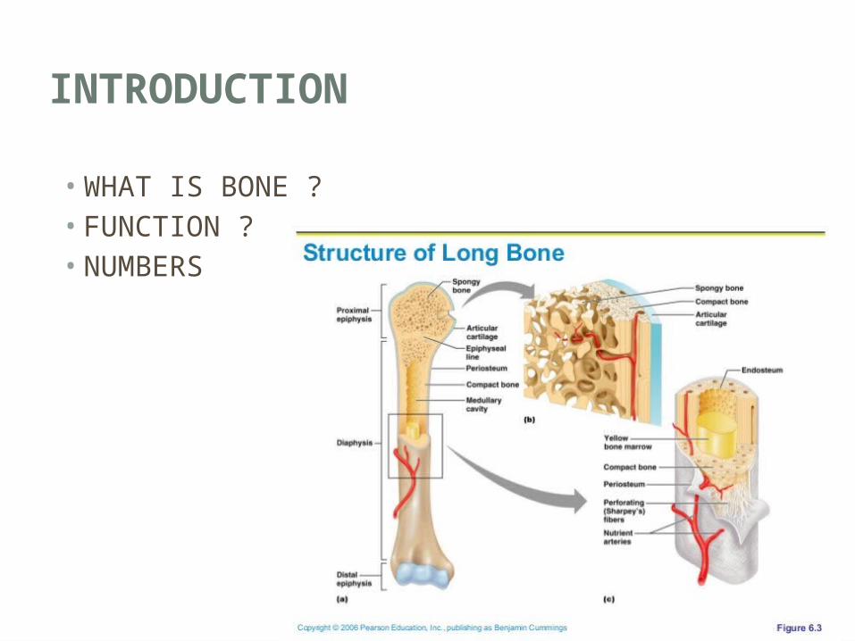
INTRODUCTION
• WHAT IS BONE ?• FUNCTION ?• NUMBERS

EMBRYOLOGY AND DEVELOPMENT
MEMBRANOUS OSSIFICATION
Frontal Parietal
Nasal bones Maxilla Zygoma Mandible
ENDOCHONDRAL OSSIFICATION
Skull base Occipital bone Nasal septum
Internalcomponents of the nose
Skeletal_System__3A_Bone_Formation__28_Intramembranous_Ossification__26_Endochondral_Ossification_29_22.mp4

STRUCTURE
Microscopic_Bone_Structure.mp4

CHEMICAL COMPOSITION
INORGANIC
1. Hydroxyapatite
[Ca10(PO4)6(OH)2]
2. Magnesium
3. Potassium
4. Chlorine
5. Iron
6. Carbonate
ORGANIC
1. 90% collagen, primarily type I
2. 10% Non-collagenous proteins and
lipids
a. 23% osteonectin
b. 15% osteocalcin
c. 9% sialoprotein,
d. 9% phosphoproteins
e. 5% α2-HS-glycoproteins
f. 4% proteoglycans
g. 3% albumin

MECHANICAL PROPERTIESCollagen fibers Mineral phase
Specific orientation
Specific length
Shear forces
Tensile forces Compressive forces
• Elongation of 2%
• Strength about 1Mpa
• Tensile strength = 2/3rd
compressive strength

BIOMECHANICS OF CRANIOMAXILLOFACIAL SKELETON

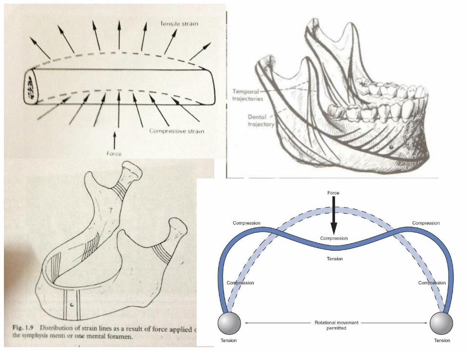

Maximum bite forces in an average population
200 to 300 N - incisor area
300 to 500 N - premolar region
500 to 700 - molar area

R. C. W. Wong, H. Tideman, L. Kin, M. A. W. Merkx: Biomechanics of mandibular reconstruction: a review. Int. J. Oral Maxillofac. Surg. 2010; 39: 313–319.

FRACTURE AND ROLE OF BLOOD SUPPLY
INJURY
INTRAVASCULAR CLOTTING CONGESTION
DECREASED BLOOD SUPPLY
NECROSIS
OSTEOBLASTIC ACTIVITY
OSTEOCLASTIC ACTIVITY
BONY BRIDGING
VASCULAR INVASION

BIOLOGICAL REACTION AND HEALING OF BONE
• Dependent on the biological and biomechanical environment, three basic scenarios can be differentiated:
1. Primary bone healing (contact or gap healing)2. Secondary bone healing via callus formation
Sufficient blood supply
Presence of specific cells
Adequate mechanical conditions
Undisturbed fracture healing

1. PRIMARY BONE HEALING (CONTACT OR GAP HEALING)
• In cases where inter-fragmentary motion can be completely avoided, a
healing pattern results which is characterized by an increased amount of
intracortical remodelling, inside and in between the fragment ends.
• As long as there is no destruction of bone in the contact areas, the motion in
the gap is small enough to keep inter-fragmentary strain below 2%.
• The pattern of direct healing per se is not a goal to strive for, but the absence
of this pattern, ie, the formation of periosteal callus under conditions of plate
fixation is an indicator that complete immobilization was not achieved.

a Functionally stable fixation of a mandibular fracture with excellent repositioning as a precondition for primary bone healing.
b Enlarged section of (a): primary bone healing contact area, direct bony bridging showing osteons crossing the fracture area.
a Stable fixation, load sharing with contact area superiorly and gap area inferiorly.
b Enlarged section of (a): primary healing gap area: complete filling of the fracture gap with lamellar bone in a direction parallel to the fracture surface.

2. SECONDARY BONE HEALING VIA CALLUS FORMATION
• In cases when no fracture fixation or just loose adaptation fixation is done,
macromotion between the fragment ends occurs.
• The strain in between the fragments exceeds what bone can tolerate, and
new bone developing between the fracture ends would be destroyed before
it is formed.
Endosteal callus
Periosteal callus

In between the fracture ends a tissue differentiation cascade takes place, during which stiffness and strength increases and strain tolerance gradually decreases.
Hematoma
Granulation tissue
Connective tissue
Fibrocartilage
Mineralized cartilage
Woven bone
Compact bone

Secondary bone healing,
phase 1: hematoma filling the fracture gap.

Secondary bone healing,
phase 2: granulation tissue and connective tissue replacing the
hematoma in the fracture gap.
• The elongation to rupture is found to be between 5% and 17%.
• Fibrous tissue is found in areas where tensile forces act,
• Cartilage is formed in zones of hydrostatic pressure

Secondary bone healing,
phase 3: fibrocartilage replacing the connective tissue in the
fracture gap.

Secondary bone healing,
phase 4: woven bone replaced by lamellar bone through Haversian
remodelling.

COMPLICATIONS OF BONE HEALING
1. Non-union2. Delayed union3. Malunion
FACTORSPATIENT ASSOCIATED
OPERATOR ASSOCIATED
HARDWARE ASSOCIATED
LOCAL
SYSTEMIC

METALS, SURFACES AND TISSUE INTERACTIONS
62.5% iron
18% chromium
14% nickel
2.5% molybdenum
minor elemental
316 L iron-base alloy
Allergic reactions to nickel 3–15%
Titanium alloys
Ti grades 1–4
Ti-6Al-7Nb alloy
Ti-15Mo alloy
(α & β)
Cell-to-Cell_Communication_-_Osseointegration.mp4
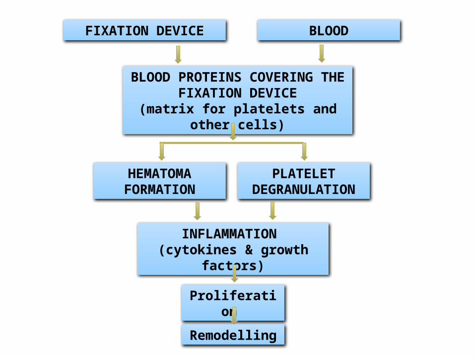
FIXATION DEVICE BLOOD
BLOOD PROTEINS COVERING THE FIXATION DEVICE
(matrix for platelets and other cells)
PLATELET DEGRANULATION
INFLAMMATION (cytokines & growth factors)
HEMATOMA FORMATION
Proliferation
Remodelling

BIODEGRADABLE MATERIALS
Water and CO2
In the future, maxillofacial fracture fixation may utilize biodegradable bone adhesives and composites in lieu of the traditional titanium plate/screw systems. The adhesives currently under study are in the cyanoacrylate polymer family, namely, butyl-2-cyanoacrylate.

REFERENCE
1. Fonseca Raymond J, Walker Robert V, Barber H Dexter, Powers, Michael P, Frost David E. oral and maxillofacial trauma. China: Saunders; 2013.
2. Hom, Hebda, Gosain, Friedman. Essential tissue healing of the face and neck. India. Peoples medical publishing house.
3. AOCMF principles of internal fixations of craniomaxillofacial skeleton, trauma & orthognathic surgery.
4. Rowe NL, William JL. Maxillofacial injuries. 1st ed. India ISBN 978-81-312-1840—2 2009.

THANK YOU
Related Documents