1997, 179(7):2202. J. Bacteriol. J H Vossen, W H Müller, P N Lipke and F M Klis proteins. causes aberrant biogenesis of cell wall anchor synthesis in cwh6/gpi3 yeast cells Restrictive glycosylphosphatidylinositol http://jb.asm.org/content/179/7/2202 Updated information and services can be found at: These include: CONTENT ALERTS more» cite this article), Receive: RSS Feeds, eTOCs, free email alerts (when new articles http://journals.asm.org/site/misc/reprints.xhtml Information about commercial reprint orders: http://journals.asm.org/site/subscriptions/ To subscribe to to another ASM Journal go to: on November 23, 2013 by guest http://jb.asm.org/ Downloaded from on November 23, 2013 by guest http://jb.asm.org/ Downloaded from

Welcome message from author
This document is posted to help you gain knowledge. Please leave a comment to let me know what you think about it! Share it to your friends and learn new things together.
Transcript

1997, 179(7):2202. J. Bacteriol.
J H Vossen, W H Müller, P N Lipke and F M Klis proteins.causes aberrant biogenesis of cell wallanchor synthesis in cwh6/gpi3 yeast cells Restrictive glycosylphosphatidylinositol
http://jb.asm.org/content/179/7/2202Updated information and services can be found at:
These include:
CONTENT ALERTS more»cite this article),
Receive: RSS Feeds, eTOCs, free email alerts (when new articles
http://journals.asm.org/site/misc/reprints.xhtmlInformation about commercial reprint orders: http://journals.asm.org/site/subscriptions/To subscribe to to another ASM Journal go to:
on Novem
ber 23, 2013 by guesthttp://jb.asm
.org/D
ownloaded from
on N
ovember 23, 2013 by guest
http://jb.asm.org/
Dow
nloaded from
JOURNAL OF BACTERIOLOGY,0021-9193/97/$04.0010
Apr. 1997, p. 2202–2209 Vol. 179, No. 7
Copyright q 1997, American Society for Microbiology
Restrictive Glycosylphosphatidylinositol Anchor Synthesis in cwh6/gpi3Yeast Cells Causes Aberrant Biogenesis of Cell Wall Proteins
JACK H. VOSSEN,1* WALLY H. MULLER,2 PETER N. LIPKE,3 AND FRANS M. KLIS1
Fungal Cell Wall Group, Institute for Molecular Cell Biology, University of Amsterdam, 1098 SM Amsterdam,1
and Department of Molecular Cell Biology, University of Utrecht, 3584 CH Utrecht,2 The Netherlands,and Department of Biological Sciences, Hunter College of City University of
New York, New York, New York 100213
Received 16 October 1996/Accepted 31 January 1997
We previously reported that the defects in the Saccharomyces cerevisiae cwh6 Calcofluor white-hypersensitivecell wall mutant are caused by a mutation in SPT14/GPI3, a gene involved in glycosylphosphatidylinositol(GPI) anchor biosynthesis. Here we describe the effect of cwh6/spt14/gpi3 on the biogenesis of cell wall proteins.It was found that the release of precursors of cell wall proteins from the endoplasmic reticulum (ER) wasretarded. This was accompanied by proliferation of ER structures. The majority of the cell wall proteinprecursors that eventually left the ER were not covalently incorporated into the cell wall but were secreted intothe growth medium. Despite the inefficient incorporation of cell wall proteins, there was no net effect on theprotein level in the cell wall. It is postulated that the availability of GPI-dependent cell wall proteinsdetermines the rate of cell wall construction and limits growth rate.
Glycosylphosphatidylinositol (GPI) anchors are commonlyused to attach proteins to cell membranes. GPI modification ofproteins is found in many eukaryotes, and in prokaryotes, adistant relative of a GPI anchor-synthesizing protein has beenidentified (47). The core structure of the glycosyl moiety isconserved from yeast cells to humans and consists of a linearsequence of one inositol, one glucosamine, and three mannoseresidues. Addition of a GPI anchor to protein occurs in thelumen of the endoplasmic reticulum (ER) (46). For importinto the ER, the preproprotein uses an N-terminal signal se-quence, which is removed upon import. This results in theformation of the proprotein. The signal for GPI anchor addi-tion to the proprotein consists of a C-terminal sequence ofhydrophobic amino acids, preceded by suitable amino acids forGPI anchor addition (29, 40). This C-terminal stretch of hy-drophobic amino acids presumably serves as a transient mem-brane anchor until it is cleaved off and replaced by a preformedGPI anchor. The biosynthesis of GPI anchors and their ex-change with the hydrophobic C terminus of the protein arecomplex processes and require multiple enzymes. Several mu-tant human cell lines which are defective in GPI anchor bio-synthesis have been isolated (39). Recently, several yeast mu-tants and corresponding genes involved in GPI anchorbiosynthesis (3, 16–18, 36, 47) and GPI anchor addition (3, 8)have also been isolated. All yeast genes known to be involvedin GPI anchor biosynthesis are essential for viability (8, 18, 47).In higher eukaryotes and also in yeasts, a role for GPI anchorsin targeting proteins to the plasma membrane is generallyaccepted (22). But there is also evidence for additional func-tions of GPI anchors. Attachment of a GPI anchor seems to berequired for efficient packaging of Gas1p into COP II vesicles,which are responsible for the transport from the ER to theGolgi compartment (7). Besides this additional role for GPIanchors, there is evidence that they are involved in cross-linking proteins to the cell wall glucan. For example, the mat-
ing type a-specific sexual agglutinin, Aga1p, which is known tohave a GPI anchor, is released from the plasma membrane andcross-linked to glucan. Simultaneously with the glucan modi-fication, inositol is removed from the protein (20). The GPIanchor apparently plays an important role in this cross-linkingstep. Deletion of the GPI anchor addition signal prevents thecross-linking of Aga1p to the cell wall and results in secretionof an active molecule into the medium (49). On the otherhand, the 30 C-terminal amino acids of Aga1p, which includethe GPI anchor addition signal, are sufficient to target a re-porter enzyme to the cell wall (42). Once a-agglutinin is linkedto glucan, it can be released by treatment with aqueous hy-drofluoric acid (HF), which is known to specifically break phos-phodiester bonds. These phosphodiester bonds probably orig-inate from the GPI anchor (10). In addition to a-agglutinin,other proteins that contain a GPI anchor addition signal arecovalently associated with the cell wall (26, 38, 44, 45). How-ever, as mentioned above, not all proteins containing a GPIanchor are destined for the cell wall. Gas1p is localized in theplasma membrane, and Kre1p is also not covalently linked tothe cell wall (38). The molecular basis of this distinction is notknown.In this study, we have investigated further the importance of
GPI anchors in cross-linking cell wall proteins to glucan. Forthis purpose, the temperature-sensitive cwh6/spt14/gpi3mutantin which GPI anchor biosynthesis is restricted was used (32, 36,47). This resulted in accumulation of ER precursor forms ofcell surface proteins and proliferation of the ER. Evidence thatthe supply of GPI anchors available to cell surface proteins isthe limiting factor in cell wall construction and growth rate ispresented.
MATERIALS AND METHODS
Yeast strains and media. The yeast strains used in this work are listed in Table1. All strains used were isogenic to S288C. Strain JV31 was isolated as thetemperature-sensitive and leucine auxotrophic haploid offspring from the diploidthat arose from crossing AR51 and the cwh6 strain. Strain JV97 was constructedby transforming FY833 strains to leucine prototrophy with a LEU2 disruptionconstruct, described by van der Vaart et al. (44). The result of the single-stepgene disruption (35) was confirmed by a Southern blot. The cwp1 cwh6 doublemutant strain JV79 was constructed by selecting leucine prototrophic and tem-
* Corresponding author. Mailing address: Fungal Cell Wall Group,Institute for Molecular Cell Biology, University of Amsterdam, Kruis-laan 318, 1098 SM Amsterdam, The Netherlands. Phone: (31) 20.5257850. Fax: (31) 20.525 7934. E-mail: [email protected].
2202
on Novem
ber 23, 2013 by guesthttp://jb.asm
.org/D
ownloaded from

perature-sensitive offspring from the diploid arising from crossing strains JV31and JV97. The gas1 cwh6 double mutant JV177 was constructed by selectingleucine prototrophic and temperature-sensitive offspring from the diploid arisingfrom crossing strains JV51 and AR104.The strains were grown in synthetic minimal medium (0.17% yeast nitrogen
base, 1% MES [morpholinoethanesulfonic acid] [Sigma] buffered at pH 6.0 withNaOH, 0.5% NH4SO4, 2% glucose) supplied with necessary amino acids at 28 or378C. When cells were labeled with [35S]methionine, low-sulfate medium (100mMNH4SO4) and labeling medium (20 mMNH4SO4) were used as described byRothblatt and Schekman (34). Yeast genetics and sporulation followed estab-lished procedures (37). Yeast extract, Bacto Peptone, yeast nitrogen base, andBacto Agar were from Difco.[35S]methionine labeling of a-factor-induced cells. Labeling was performed
essentially as described by Lu et al. (19). Cells were precultured in low-sulfatemedium at 288C to an optical density at 600 nm of 0.5 (equivalent to 0.5 3 107
cells/ml). The cells were collected, and 2 3 108 cells were transferred to 25 ml oflabeling medium and incubated at 288C. After 5 min, synthetic a factor (a gift ofFred Naider, College of Staten Island, Staten Island, N.Y.) was added to 50ng/ml. Five minutes later, 0.5 mCi of TRAN35S-LABEL (ICN Biochemicals) wasadded. After a further incubation of 40 min, 0.25 ml of chase mixture (1 MNH4SO4, 100 mM DL-cysteine, 100 mM DL-methionine) was added. Beforeharvesting, NaN3 was added to 10 mM. Cells were harvested after a 0-min anda 40-min chase, and different protein fractions were isolated as described below.Protein determination by labeling with 35S. Total protein contents of different
fractions were determined by counting radioactivity in these fractions after 35Slabeling. For this experiment, the labeling medium as described by Rothblatt andSchekman (34) was slightly modified. Instead of 20 mM NH4SO4 as cold sulfatesource, we used 50 mM DL-cysteine and 50 mM DL-methionine. The cells wereprecultured in this medium, and at t 5 0, cells were diluted in fresh medium toan optical density at 600 nm of 0.25 (equivalent to 0.25 3 107 cells/ml). At t 55 min, 0.5 mCi of TRAN35S-LABEL was added. Cultures were grown for twodoubling times (see Table 2) at 288C. Cells were harvested and fractionated asdescribed below. Protein contents of the different fractions were calculated aspercentages of total label incorporated.Cell harvesting and storage. Cells were grown on synthetic media comple-
mented with the necessary amino acids as described above. Cells were harvestedin the early logarithmic phase by centrifuging at 1,000 3 g for 5 min. Harvestedcells (2 3 108) were washed twice in wash buffer (30 mM Tris-HCl [pH 7.4], 1mM EDTA, 1 mM phenylmethylsulfonyl fluoride [PMSF; Sigma]) and stored at2708C.Isolation of medium proteins. Remaining cells were removed from the me-
dium by an additional centrifugation step at 4,500 3 g for 10 min. Proteins wereprecipitated from the medium with 0.2 mg of sodium desoxycholate per ml and6% trichloroacetic acid, during overnight incubation at 48C (30). To exclude thatcell lysis had occurred, these isolated medium proteins, equivalent to 5 3 107
cells, were examined in a Western blot with anti-HDEL antibody. This antibodyis directed against the peptide with the sequence HDEL and is known to spe-cifically recognize ER lumen proteins.Isolation of sodium dodecyl sulfate (SDS) extracts and SDS-extracted cell
walls. Frozen cells were resuspended in 250 ml of SDS lysis buffer (50 mMTris-HCl [pH 7.4], 150 mM NaCl, 5 mM EDTA, 2% SDS, 1 mM PMSF, 0.7 mgof pepstatin [Boehringer] per ml, 0.5 mg of leupeptin [Sigma] per ml) and werebroken with 0.3 g of glass beads. Two hundred microliters of the total lysate wascollected, and the SDS extract was separated from the cell walls by centrifugationfor 5 min at 14,000 3 g. The supernatants representing the SDS extracts werestored at 2708C. The cell wall pellets were extracted twice more by being boiledfor 5 min in SDS lysis buffer and being washed five times in inhibitor mixture (5mM EDTA, 2% SDS, 1 mM PMSF, 0.7 mg of pepstatin per ml, 0.5 mg ofleupeptin per ml).Preparation of laminarinase extracts. The SDS-extracted cell walls were
washed once in laminarinase buffer (100 mM NaAc [pH 5.5], 1 mM EDTA, 1mM PMSF, 0.7 mg of pepstatin per ml, 0.5 mg of leupeptin per ml). The pellet
was resuspended in 10 ml of laminarinase buffer. Ten microliters of laminarinasesolution (0.25 U of mollusc laminarinase/ml [Sigma], 1 mM PMSF, 0.7 mg ofpepstatin per ml, 0.5 mg of leupeptin per ml, 50 mM NaAc) was added at t 5 0and t 5 2 h, and the mixture was incubated at 378C for 4 h in total. The mixturewas adjusted to 2% SDS for immunoprecipitations, or 15 ml of 33 sample buffer(6% SDS, 2.25% Tris, 30% glycerol, 15% b-mercaptoethanol, 0.003% bromo-phenol blue, adjusted to pH 6.8 with HCl) was added for Western analyses. Themixture was boiled for 5 min. Laminarinase-released proteins were in the super-natant after centrifugation for 5 min at 14,000 3 g.Preparation of HF extracts. SDS-extracted cell walls were dried in a Speed-
Vac, and 100 ml of ice-cold 48% HF was added. The mixture was incubated onice for 48 h. HF was subsequently evaporated by a constant nitrogen flow. Theresidue was washed three times with ice-cold 90% methanol. The pellet wasresuspended in sample buffer and boiled for 5 min. After centrifugation for 5 minat 14,000 3 g, the HF-extractable cell wall proteins were in the supernatant.Isolation of membrane-bound and membrane-free fractions. Frozen cells (23
108) were resuspended in 250 ml of lysis buffer (50 mM Tris-HCl [pH 7.4], 150mM NaCl, 5 mM EDTA, 1 mM PMSF, 0.7 mg of pepstatin per ml, 0.5 mg ofleupeptin per ml) and were broken with 0.3 g of glass beads. Two hundredmicroliters of the total lysate was collected and centrifuged for 5 min at 14,0003g. The resulting supernatant was centrifuged again for 5 min at 14,000 3 g. Thesupernatant was centrifuged for 30 min at 207 kPa (100,0003 g) with a 188A-100rotor in a Beckman Airfuge. The final supernatant, representing the solubleprotein fraction, was collected and stored at 2708C. The membrane pellet wasincubated in 100 mMNa2CO3 for 30 min on ice and recentrifuged for 10 minutesat 207 kPa in a Beckman Airfuge. The pellet was washed twice in membranewash buffer (50 mM Tris-HCl [pH 7.4], 5 mM EDTA, 1 mM PMSF, 0.7 mg ofpepstatin per ml, 0.5 mg of leupeptin per ml). The washed membrane pellet wastaken up in a small volume of 2% SDS and stored at 2708C.Silver staining, immunoblotting, and immunoprecipitation. SDS-polyacryl-
amide gel electrophoresis (14) was performed on gradient gels from 2 to 20%.For silver staining, the gels were subsequently treated as described by De Nobelet al. (6). Western analysis was performed essentially as described by Montijn etal. (24). Blots were kept for 1 h in periodate buffer (100 mM NaAc, 50 mMH5IO6) prior to the blocking step in Western analysis, except when b1-6-glucanantibodies were used. Binding of the peroxidase-labeled secondary antibodieswas visualized with the Enhanced Chemo-Luminescence kit from Amersham.The antisera used in this study were anti-Suc2p (27), which was diluted 1:10,000;anti-Gas1p (kindly provided by L. Popolo [41]), which was diluted 1:2,000;anti-b1,6-glucan (kindly provided by R. Montijn [24]), which was diluted 1:5,000;anti-HDEL (kindly provided by N. Dean [9]), which was diluted 1:500; andanti-Cwp1p (described below), which was diluted 1:1,000. a-Agglutinin was im-munoprecipitated with the AG3 antibody (49). The immunoprecipitation proce-dure was as described by Lu et al. (19). Immunoprecipitates were separated ona 6% separating gel with a 4% stacking gel, unless otherwise stated. The gelswere fixed, treated with En3Hance (Du Pont), dried, and exposed to KodakX-Omat AR film.Raising of antibodies against Cwp1p. The sequence DDGKLKFDDDKYAV
from the central part of Cwp1p was used to make a synthetic peptide. Thispeptide was coupled through an additional C-terminal cysteine to the carrierkeyhole limpet hemocyanin. Rabbits were immunized with this neoprotein, andserum was isolated. The above-described procedure was performed by Eurogen-tec (Seraing, Belgium).Electron microscopy. Cells were grown overnight in YPD (1% yeast extract,
1% Bacto Peptone, 3% glucose) at 308C to early logarithmic phase. Cells werewashed three times in 0.9% NaCl. The washed cells were fixed chemically innonbuffered 1.5% potassium permanganate for 20 min at room temperature.Fixed cells were washed three times in phosphate-buffered saline, cryoprotectedwith 30% dimethyl formamide for 30 min, frozen in liquid propane, and freezesubstituted in anhydrous methanol at2908C for 16 h. The freeze substituted cellswere infiltrated with Spurr’s resin, and polymerization took place at 658C. Afterultramicrotomy, 80-nm sections were viewed in a transmission electron micro-scope, EM 420, at 80 kV.
RESULTS
cwh6 cells contain a temperature-sensitive mutation. cwh6/gpi3 cells contain a temperature-sensitive allele (47) that allowsonly limited growth at permissive temperatures (Table 2). Theexperiments described in this paper were performed at 288C,unless otherwise stated. At the semipermissive temperature,growth rate and, presumably, also the Cwh6 activity are se-verely impaired. As CWH6 is involved in the first biosyntheticstep of GPI synthesis, that is, the addition of N-acetylglu-cosamine to phosphatidylinositol, it is expected that in cwh6cells grown at the semipermissive temperature only a limitedamount of mature GPI anchors will be available for addition toproteins in the ER. As GPI-anchored proteins are destined for
TABLE 1. Yeast strains used in this study
Strain Genotype Reference
AR27 a ura3-52 32cwh6 a ura3-52 cwh6ts 32FY833 a his3D200 ura3-52 leu2D1 lys2D202 trp1D63 48AR 104 a gas1::LEU2 his3D200 ura3-52 leu2D1 31
lys2D202 trp1 D63JV177 a gas1::LEU2 cwh6ts lys2D202 leu2 This paperAR51 a leu2 32JV31 a leu2 cwh6ts This paperJV97 a cwp1::LEU2 his3D200 ura3-52 leu2D1 This paper
lys2D202 trp1D63JV79 a cwp1::LEU2 cwh6ts leu2 lys2 This paper
VOL. 179, 1997 RESTRICTIVE GPI ANCHOR SYNTHESIS IN YEAST CELLS 2203
on Novem
ber 23, 2013 by guesthttp://jb.asm
.org/D
ownloaded from
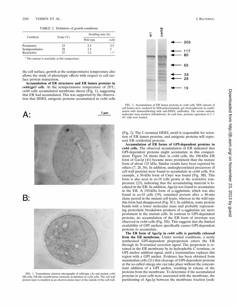
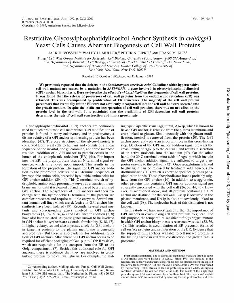
the cell surface, growth at the semipermissive temperature alsoallows the study of phenotypic effects with respect to cell sur-face protein maturation.Accumulation of ER structures and ER lumen proteins in
cwh6/gpi3 cells. At the semipermissive temperature of 288C,cwh6 cells accumulated membrane sheets (Fig. 1), suggestingthat ER had accumulated. This was supported by the observa-tion that HDEL antigenic proteins accumulated in cwh6 cells
(Fig. 2). The C-terminal HDEL motif is responsible for reten-tion of ER lumen proteins, and antigenic proteins will repre-sent ER residential proteins.Accumulation of ER forms of GPI-dependent proteins in
cwh6 cells. The observed accumulation of ER indicated thatGPI-dependent proteins might accumulate in this compart-ment. Figure 3A shows that, in cwh6 cells, the 100-kDa ERform of Gas1p (41) became more prominent than the matureform of about 125 kDa. Similar results have been reported byothers (7, 28, 36). In addition, underglycosylated precursors ofcell wall proteins were found to accumulate in cwh6 cells. Forexample, a 50-kDa form of Cwp1 was found (Fig. 3B). Thisform is also seen in sec18 cells grown at the restrictive tem-perature (23), indicating that the accumulating material is lo-calized in the ER. In addition, Aga1p was found to accumulatein the ER. A 150-kDa form of a-agglutinin, which was alsofound in sec18 cells (19), remained present after a 40-minchase period in the mutant cell lysate, whereas in the wild typethis form had disappeared (Fig. 3C). In addition, some proteinbands with a lower molecular mass and probably represent-ing proteolytic breakdown products of a-agglutinin are moreprominent in the mutant cells. In contrast to GPI-dependentproteins, no accumulation of the ER form of invertase wasobserved in cwh6 cells (Fig. 3D). This suggests that the limitedavailability of GPI anchors specifically causes GPI-dependentproteins to accumulate.The ER form of Aga1p in cwh6 cells is partially released
from the ER membrane. Under normal conditions, a newlysynthesized GPI-dependent preproprotein enters the ERthrough its N-terminal secretion signal. The proprotein is re-tained in the ER membrane by its hydrophobic C terminus, orGPI anchor addition signal, until a transamidase replaces thisregion with a GPI anchor. Evidence has been obtained frommammalian cells (21) that cleavage of GPI-dependent proteinsat the so-called omega site can take place without the concom-itant addition of a GPI anchor, resulting in release of theproteins from the membrane. To determine if the accumulatedproteins in yeast cells were associated with the membrane, thepartitioning of Aga1p between the membrane fraction (sodi-
FIG. 1. Transmission electron micrographs of wild-type (A) and mutant cwh6(B) cells. ER-like membranous elements accumulate in cwh6 cells. The cell wallprotein layer is manifest as an electron-dense layer at the outside of the cell wall.
FIG. 2. Accumulation of ER lumen proteins in cwh6 cells. SDS extracts ofcell lysates were analyzed by SDS-polyacrylamide gel electrophoresis in combi-nation with immunoblotting with anti-HDEL antibodies. The arrows indicatemolecular mass markers (kilodaltons). In each lane, proteins equivalent to 5 3107 cells were loaded.
TABLE 2. Definition of growth conditions
Condition Temp (8C)Doubling time (h)
Wild type cwh6
Permissive 23 2.5 3.5Semipermissive 28 1.5 5Restrictive 37 1.5 —a
a The mutant is nonviable at this temperature.
2204 VOSSEN ET AL. J. BACTERIOL.
on Novem
ber 23, 2013 by guesthttp://jb.asm
.org/D
ownloaded from

um carbonate resistant) and the soluble fraction (100,000 3 gsupernatant) was studied. Figure 4 shows that the low-molec-ular-weight ER form was present only in the membrane frac-tion of wild-type cells, whereas in cwh6 cells the ER form wasfound also in the soluble fraction together with proteolyticbreakdown products. During a 40-min chase period, the ERform of wild-type a-agglutinin was completely converted into a.300-kDa soluble form, which has previously been identifiedas a soluble periplasmic intermediate (19). In cwh6 cells, theER form of a-agglutinin remained present in both the mem-brane-bound and the soluble fractions. From these results, itcan be concluded that Aga1p is partially released from the ERmembrane and that transport of both the membrane and thesoluble form is strongly retarded in cwh6 cells.cwh6 cells release more proteins into the medium. Proteins
that partition into the soluble or luminal fraction are expectedto follow the default pathway and to end up in the vacuole orin the periplasmic region and the growth medium. No HDELantigenic proteins were found in the medium, indicating thatcell lysis was negligible (data not shown). On the other hand,Cwp1p (58 kDa) and Gas1p (125 kDa) were present in themedium of cwh6 cells (Fig. 5A and B). Also, bands of highermolecular weight were observed. These bands most likely rep-resent protein complexes, since they were absent from themedium of the respective double mutants (JV79 and JV177;data not shown). Cwp1p and Gas1p were not detectable in themedium of wild-type cells. In addition to Cwp1p and Gas1p,cwh6 cell medium also contained an additional and smallerform of a-agglutinin that was absent in wild-type cell medium(Fig. 5C). This suggests that mistargeting to the medium ofGPI-dependent proteins is a general phenomenon in cwh6
cells. Silver staining of the medium proteins (Fig. 6) showedthat the medium of cwh6 cells contained several proteins thatwere not seen in the wild-type cell medium. In the medium ofthe cwh6 cwp1 double mutant, one band was specifically miss-ing, indicating that the protein migrating at this position incwh6 cell medium was Cwp1p. This suggests that the otheradditional proteins in cwh6 cell medium are also GPI-depen-
FIG. 3. Intracellular accumulation of precursors of GPI-dependent proteins in cwh6 cells. (A, B, and D) Western blots of SDS extracts are shown with Gas1p,Cwp1p, and Suc2p antibodies, respectively. (C) a-Agglutinin was immunoprecipitated from SDS extracts of 35S-labeled, a-factor-induced cells. The immunoprecipitateswere separated on a 6% separating gel with a 4% stacking gel. The autoradiograms are shown. The arrows indicate molecular mass markers (kilodaltons). In each lane,proteins equivalent to 5 3 107 cells were loaded.
FIG. 4. Membrane association of a-agglutinin precursors. Membrane-bound(A) and membrane-free (B) fractions were prepared as described in Materialsand Methods. a-Agglutinin was immunoprecipitated from these fractions, andthe precipitates were loaded on 2 to 20% gels. The autoradiograms are shown.The arrows indicate molecular mass markers (kilodaltons). In each lane, proteinsequivalent to 5 3 107 cells were loaded.
VOL. 179, 1997 RESTRICTIVE GPI ANCHOR SYNTHESIS IN YEAST CELLS 2205
on Novem
ber 23, 2013 by guesthttp://jb.asm
.org/D
ownloaded from
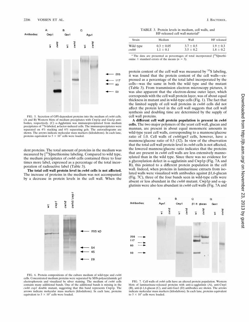
dent proteins. The total amount of proteins in the medium wasmeasured by [35S]methionine labeling. Compared to wild type,the medium precipitates of cwh6 cells contained three to fourtimes more label, expressed as a percentage of the total incor-poration of radioactive label (Table 3).The total cell wall protein level in cwh6 cells is not affected.
The increase of proteins in the medium was not accompaniedby a decrease in protein levels in the cell wall. When the
protein content of the cell wall was measured by 35S labeling,it was found that the protein content of the cell walls—ex-pressed as a percentage of the total label incorporated by thecells—was the same in both the wild type and the mutant(Table 3). From transmission electron microscopy pictures, itwas also apparent that the electron-dense outer layer, whichcorresponds with the cell wall protein layer, was of about equalthickness in mutant and in wild-type cells (Fig. 1). The fact thatthe limited supply of cell wall proteins in cwh6 cells did notaffect the protein level in the cell wall suggests that cell wallsynthesis and doubling time are determined by the supply ofcell wall proteins.A different cell wall protein population is present in cwh6
cells. The two major polymers of the yeast cell wall, glucan andmannan, are present in about equal monomeric amounts inwild-type yeast cell walls, corresponding to a mannose/glucoseratio of 1.0. Cell walls of cwh6/gpi3 cells, however, have amannose/glucose ratio of 0.5 (32). In view of the observationthat the total cell wall protein level in cwh6 cells is not affected,the lowered mannose/glucose ratio indicates that the proteinsthat are present in cwh6 cell walls are less extensively manno-sylated than in the wild type. Since there was no evidence fora glycosylation defect in a-agglutinin and Cwp1p (Fig. 7A andB), this pointed to a different protein population in the cellwall. Indeed, when proteins in laminarinase extracts from iso-lated walls were visualized with antibodies against b1,6-glucan(Fig. 7C), three of the four bands seen in wild-type cells wereabsent or less abundant in the cwh6 mutant. Cwp1p and a-ag-glutinin were also less abundant in cwh6 cell walls (Fig. 7A and
FIG. 5. Secretion of GPI-dependent proteins into the medium of cwh6 cells.(A and B) Western blots of medium precipitates with Cwp1p and Gas1p anti-bodies, respectively. (C) a-Agglutinin was immunoprecipitated from mediumprecipitates of 35S-labeled, a-factor-induced cells. The immunoprecipitates wereseparated on 4% stacking and 6% separating gels. The autoradiograms areshown. The arrows indicate molecular mass markers (kilodaltons). In each lane,proteins equivalent to 5 3 107 cells were loaded.
FIG. 6. Protein compositions of the culture medium of wild-type and cwh6cells. Concentrated medium proteins were separated by SDS-polyacrylamide gelelectrophoresis and visualized by silver staining. The medium of cwh6 cellscontains many additional bands. One of the additional bands is missing in thecwh6 cwp1 double mutant, suggesting that this band represents Cwp1p. Thearrows indicate molecular mass markers (kilodaltons). In each lane, proteinsequivalent to 5 3 107 cells were loaded.
FIG. 7. Cell walls of cwh6 cells have an altered protein population. Westernblots of laminarinase-released proteins with anti-a-agglutinin (A), anti-Cwp1(B), anti-b-1,6-glucan (C), and anti-Gas1 (D) antibodies are shown. The arrowsindicate molecular mass markers (kilodaltons). In each lane, proteins equivalentto 5 3 107 cells were loaded.
TABLE 3. Protein levels in medium, cell walls, andHF-released cell wall materiala
Strain Medium Wall HF released
Wild type 0.3 6 0.05 3.7 6 0.5 1.9 6 0.3cwh6 1.1 6 0.1 3.5 6 0.2 1.8 6 0.2
a The data are presented as percentages of total incorporated [35S]methi-onine 6 standard errors of the means (n 5 3).
2206 VOSSEN ET AL. J. BACTERIOL.
on Novem
ber 23, 2013 by guesthttp://jb.asm
.org/D
ownloaded from
B). In contrast, a new b1,6-glucan-containing band appearedand another band became more abundant than in the wildtype.The observed change in the cell wall protein population
could be explained by assuming that newly acquired cell wallproteins in cwh6 cells were not incorporated through a GPIanchor. This idea was tested by making use of the observationby Kapteyn et al. (11) that cell wall proteins can be releasedwith aqueous HF. This treatment specifically cleaves phos-phodiester bridges which, among others, are present in theGPI anchor between ethanolamine and mannose. It was, how-ever, observed that HF treatment released equal amounts oflabel from [35S]methionine-labeled cell walls in both the wildtype and the mutant (Table 3), suggesting that equal amountsof proteins in the cell walls of both the wild type and themutant were linked through a GPI anchor-derived structure.Mistargeting of Gas1p in cwh6 cells to the cell wall. In the
above paragraph, it was shown that the shift in cell wall proteinpopulation was probably not due to a GPI-independent incor-poration mechanism of cell wall proteins. This indicates that adifferent set of GPI-dependent proteins is incorporated intocwh6 cell walls. As it is known that stress conditions can causea shift in the expression pattern of b-glucosylated cell wallproteins (13), one might argue that in cwh6 cells a limitedsupply of GPI anchors acts as a stress condition, triggering atranscriptional shift in the cell wall protein expression pattern.An alternative or additional explanation for the shift in cellwall protein population might be that proteins that are GPIanchored but that are normally not incorporated into the cellwall, such as Gas1p, are now incorporated in the wall. Inwild-type cells, Gas1p localizes—as expected—only to theplasma membrane, but in cwh6 cells it is also found in thecell wall (Fig. 7D). The absence of any signal in Dgas1 andcwh6tsDgas1 strains indicated that the antibodies used to detectGas1p in cwh6 cell walls were specific (data not shown).
DISCUSSION
All known covalently linked cell wall proteins contain aputative GPI anchor addition signal, and in some cases, thishas been biochemically confirmed (25, 38, 44, 49). Removal ofthe GPI anchor addition signal results in the release of a-ag-glutinin into the medium (49). In addition, the C-terminal 30amino acids of a-agglutinin, which include the GPI anchoraddition signal, are sufficient to target a reporter protein to thecell wall (42). These observations indicate that GPI anchorsplay an important role in the incorporation of proteins in thecell wall. In this study, we wanted to investigate the significanceof GPI anchor modification for the biogenesis of cell wallproteins. For this purpose, the temperature-sensitive cwh6/gpi3cell wall mutant was used. CWH6/GPI3 is involved in the ad-dition of N-acetylglucosamine to phosphatidylinositol, which isthe first step in the formation of a GPI anchor (36). By growingcwh6 cells just below the restrictive temperature, the amountof GPI anchor that is available for addition to GPI-dependentproteins is severely reduced but is enough for survival. In thisway, the effects of a limited supply of GPI anchors on cellsurface growth can be studied. However, formally one cannotexclude the possibility that by growing the cells at a semirestric-tive temperature some phenotypic effects may be the result ofa more general process of adaptation of the cells.Abnormal maturation of GPI-dependent proteins in cwh6
cells. In this study, it is shown that GPI limitation led to theaccumulation of GPI-dependent proteins in the ER, with aconcomitant increase of ER structures. Under conditions ofGPI limitation, export of Gas1p from the ER occurs with
reduced efficiency (7). It is shown here that this is also the casefor Aga1p and Cwp1p, demonstrating that the GPI modifica-tion is a requirement for export from the ER for all GPI-dependent proteins. Since invertase was matured correctly, thisis evidence that this was a specific effect on GPI-dependentproteins. This is consistent with the observation that, in cellsstarved for inositol, which is an essential component of the GPIanchor, secretion of luminal proteins such as invertase and acidphosphatase can continue normally for some time after cellsurface growth has stopped (2).The GPI-dependent proteins that accumulate in the ER are
only partially bound to the membrane, suggesting that theaccumulated proproteins are gradually converted to a non-membrane-associated form. Maxwell et al. (21) have shown inmammalian cells that, under conditions of impaired processingof GPI-dependent proteins, these proteins can dissociate fromthe ER membrane. It is likely that a similar reaction alsooccurs in yeast cells (Fig. 4B) and that, as a result, GPI-dependent proteins are partially mistargeted to the medium(Fig. 6).Decreased levels of GPI-dependent proteins in the plasma
membrane of cwh6 cells. An additional explanation for theappearance of GPI-dependent cell wall proteins in the mediumof cwh6 cells is based on the observation that in cwh6 cells thelevels of the plasma membrane-localized forms of GPI-depen-dent proteins are lower. In lysates of cwh6 cells (Fig. 3), the
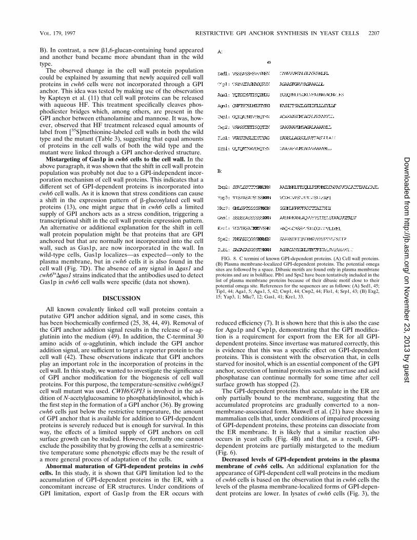
FIG. 8. C termini of known GPI-dependent proteins. (A) Cell wall proteins.(B) Plasma membrane-localized GPI-dependent proteins. The potential omegasites are followed by a space. Dibasic motifs are found only in plasma membraneproteins and are in boldface. Plb1 and Sps2 have been tentatively included in thelist of plasma membrane proteins because of their dibasic motif close to theirpotential omega site. References for the sequences are as follows: (A) Sed1, 45;Tip1, 44; Aga1, 5; Aga1, 5, 42; Cwp1, 44; Cwp2, 44; Flo1, 4; Srp1, 43; (B) Exg2,15; Yap3, 1; Mkc7, 12; Gas1, 41; Kre1, 33.
VOL. 179, 1997 RESTRICTIVE GPI ANCHOR SYNTHESIS IN YEAST CELLS 2207
on Novem
ber 23, 2013 by guesthttp://jb.asm
.org/D
ownloaded from
125-kDa form of Gas1p, the 54-kDa form of Cwp1p, and the.300-kDa form of Aga1, which represent the plasma mem-brane-bound forms of these proteins (19, 23, 41), are dimin-ished. This indicates that this might also be the case for otherplasma membrane-bound forms of GPI-dependent proteinsincluding Kre1p. As Gas1p and Kre1p are believed to be in-volved in cross-linking cell wall proteins to the cell wall matrix(31, 33), the appearance of GPI-dependent cell wall proteins inthe medium of cwh6 cells can then be explained by the lack ofplasma membrane-localized Gas1p (Fig. 3A) and Kre1p.Mistargeting of Gas1p to the cell wall of cwh6 cells. In
wild-type cells, Gas1p is directed exclusively to the plasmamembrane and not found in the cell wall. In cwh6/gpi3 cells,however, it is detected in the walls even after extraction withhot SDS, indicating that it is tightly associated with the cell wallframework. Mistargeting of Gas1p in cwh6 cells to the cell wallcan be explained in several ways. For example, it is possiblethat the protein or protein complex at the plasma membranethat cross-links precursors of GPI-containing cell wall proteinsto the cell wall uses the suboptimal substrate Gas1p because ofa shortage of true cell wall proteins. Alternatively, one mightimagine that plasma membrane-bound GPI-dependent pro-teins, which are expected to be present at a lower level in cwh6cells, are themselves involved in sorting or targeting GPI-de-pendent proteins to the plasma membrane or the cell wall.Interestingly, all putative GPI-dependent plasma membraneproteins including Gas1p possess a dibasic motif, which is lo-cated just N terminal from the omega site and is not seen inknown cell wall proteins (Fig. 8). This suggests that this motifmight be involved in targeting GPI-dependent proteins to theplasma membrane.Cell wall protein levels in cwh6 cells are not affected. De-
spite intracellular accumulation and mistargeting of GPI-de-pendent proteins to the medium and in contrast to the lowerlevels of GPI-dependent proteins in the plasma membrane ofcwh6 cells, the cell wall of cwh6 cells contains similar amountsof proteins as the wild type. As at the semipermissive temper-ature, the doubling time of cwh6 cells has increased more thanthreefold; this indicates that the growth rate of cwh6 cells isdetermined by the supply of cell wall proteins. This suggeststhat GPI-dependent cell wall proteins are essential for cell wallconstruction. It is also consistent with the notion that the cellwall complex GPI-dependent cell wall protein–b1,6-glucan–b1,3-glucan fraction as described by Kapteyn et al. (11), inwhich GPI-dependent cell wall proteins function as receptorsfor the b-glucan fraction, represents a major building block ofthe yeast cell wall.
ACKNOWLEDGMENTS
We thank H. Van Den Ende and J. Chapman for critically readingthe manuscript, F. Naider for synthetic a factor, R. Montijn for sharingunpublished results and b1,6-glucan antiserum, A. Ram for the cwh6mutant and the GAS1 deletion strain, M. van der Vaart for the CWP1deletion construct and for sharing unpublished results, L. Popolo forGas1p antiserum, N. Dean for anti-HDEL antiserum, and A. Verkleyfor stimulating discussions on the electron microscopy work.
REFERENCES1. Ash, J., M. Dominguez, J. J. Bergeron, D. Y. Thomas, and Y. Bourbonnais.1995. The yeast proprotein convertase encoded by YAP3 is a glycophos-phatidylinositol-anchored protein that localizes to the plasma membrane.J. Biol. Chem. 270:20847–20854.
2. Atkinson, K. D., and R. M. Ramirez. 1984. Secretion can proceed uncoupledfrom net plasma membrane expansion in inositol-starved Saccharomycescerevisiae. J. Bacteriol. 160:80–86.
3. Benghezal, M., P. N. Lipke, and A. Conzelmann. 1995. Identification of sixcomplementation classes involved in the biosynthesis of glycosylphosphati-dylinositol anchors in Saccharomyces cerevisiae. J. Cell Biol. 130:1333–1344.
4. Bidard, F., M. Bony, B. Blondin, S. Dequin, and P. Barre. 1995. The Sac-
charomyces cerevisiae FLO1 flocculation gene encodes for a cell surfaceprotein. Yeast 11:809–822.
5. Cappellaro, C., C. Baldermann, R. Rachel, and W. Tanner. 1994. Matingtype-specific cell-cell recognition of Saccharomyces cerevisiae: cell wall at-tachment and active sites of a- and alpha-agglutinin. EMBO J. 13:4737–4744.
6. De Nobel, J. G., C. Dijkers, E. Hooijberg, and F. M. Klis. 1989. Increased cellwall porosity in Saccharomyces cerevisiae after treatment with DTT orEDTA. J. Gen. Microbiol. 135:2077–2084.
7. Doering, T. L., and R. Schekman. 1996. GPI anchor attachment is requiredfor Gas1p transport from the endoplasmic reticulum in COP II vesicles.EMBO J. 15:182–192.
8. Hamburger, D., M. Egerton, and H. Riezman. 1995. Yeast Gaa1p is requiredfor attachment of a completed GPI anchor onto proteins. J. Cell Biol. 129:629–639.
9. Hardwick, K. G., M. J. Lewis, J. Semanza, N. Dean, and R. B. Pelham. 1990.ERD1, a yeast gene required for the retention of luminal endoplasmic re-ticulum proteins, affects glycoprotein processing in the Golgi apparatus.EMBO J. 9:623–630.
10. Kapteyn, J. C., R. C. Montijn, G. J. Dijkgraaf, H. Van den Ende, and F. M.Klis. 1995. Covalent association of beta-1,3-glucan with beta-1,6-glucosy-lated mannoproteins in cell walls of Candida albicans. J. Bacteriol. 177:3788–3792.
11. Kapteyn, J. C., R. C. Montijn, E. Vink, J. de la Cruz, A. Llobell, J. E. Douwes,H. Shimoi, P. N. Lipke, and F. M. Klis. 1996. Retention of Saccharomycescerevisiae cell wall proteins through a phosphodiester-linked b-1,3-/b-1,6-glucan heteropolymer. Glycobiology 6:337–345.
12. Komano, H., and R. S. Fuller. 1995. Shared functions in vivo of a glycosyl-phosphatidylinositol-linked aspartyl protease, Mkc7, and the proprotein pro-cessing protease Kex2 in yeast. Proc. Natl. Acad. Sci. USA 92:10752–10756.
13. Kowalski, L. R., K. Kondo, and M. Inouye. 1995. Cold-shock induction of afamily of Tip-related proteins associated with the membrane in Saccharo-myces cerevisiae. Mol. Microbiol. 15:341–353.
14. Laemmli, U. K. 1970. Cleavage of structural proteins during the assembly ofthe head of bacteriophage T4. Nature (London) 227:680–685.
15. Larriba, G., E. Andaluz, R. Cueva, and R. D. Basco. 1995. Molecular biologyof yeast exoglucanases. FEMS Microbiol. Lett. 125:121–126.
16. Leidich, S. D., D. A. Drapp, and P. Orlean. 1994. A conditionally lethal yeastmutant blocked at the first step in glycosyl phosphatidylinositol anchor syn-thesis. J. Biol. Chem. 269:10193–10196.
17. Leidich, S. D., D. A. Drapp, and P. Orlean. 1995. Isolation and character-ization of yeast glycosylphosphatidylinositol anchoring mutants. MethodsEnzymol. 250:560–571.
18. Leidich, S. D., Z. Kostova, R. R. Latek, L. C. Costello, D. A. Drapp, W. Gray,J. S. Fassler, and P. Orlean. 1995. Temperature-sensitive yeast GPI anchor-ing mutants gpi2 and gpi3 are defective in the synthesis of N-acetylglucosami-nyl phosphatidylinositol. Cloning of the GPI2 gene. J. Biol. Chem. 270:13029–13035.
19. Lu, C. F., J. Kurjan, and P. N. Lipke. 1994. A pathway for cell wall anchorageof Saccharomyces cerevisiae alpha-agglutinin. Mol. Cell. Biol. 14:4825–4833.
20. Lu, C. F., R. C. Montijn, J. L. Brown, F. M. Klis, J. Kurjan, H. Bussey, andP. N. Lipke. 1995. Glycosylphosphatidylinositol-dependent cross-linking ofalpha-agglutinin and beta 1,6-glucan in the Saccharomyces cerevisiae cellwall. J. Cell Biol. 128:333–340.
21. Maxwell, S. E., S. Ramalingam, L. D. Gerber, and S. Udenfriend. 1995.Cleavage without anchor addition accompanies the processing of a nascentprotein to its glycosylphosphatidylinositol-anchored form. Proc. Natl. Acad.Sci. USA 92:1550–1554.
22. McConville, M. J., and M. A. Ferguson. 1993. The structure, biosynthesis andfunction of glycosylated phosphatidylinositols in the parasitic protozoa andhigher eukaryotes. Biochem. J. 294:305–324.
23. Montijn, R. C. Personal communication.24. Montijn, R. C., J. van Rinsum, F. A. van Schagen, and F. M. Klis. 1994.
Glucomannoproteins in the cell wall of Saccharomyces cerevisiae contain anovel type of carbohydrate side chain. J. Biol. Chem. 269:19338–19342.
25. Muller, G., K. Schubert, F. Fiedler, and W. Bandlow. 1992. The cAMP-binding ectoprotein from Saccharomyces cerevisiae is membrane-anchoredby glycosyl-phosphatidylinositol. J. Biol. Chem. 267:25337–25346.
26. Muller, G., E. Gross, S. Wied, and W. Bandlow. 1996. Glucose-inducedsequential processing of a glycosyl-phosphatidylinositol-anchored ectopro-tein in Saccharomyces cerevisiae. Mol. Cell. Biol. 16:442–456.
27. Nguyen, T. H., D. T. Law, and D. B. Williams. 1991. Binding protein BiP isrequired for translocation of secretory proteins into the endoplasmic retic-ulum in Saccharomyces cerevisiae. Proc. Natl. Acad. Sci. USA 88:1565–1569.
28. Nuoffer, C., P. Jeno, A. Conzelmann, and H. Riezman. 1991. Determinantsfor glycophospholipid anchoring of the Saccharomyces cerevisiae Gas1 pro-tein to the plasma membrane. Mol. Cell. Biol. 11:27–37.
29. Nuoffer, C., A. Horvath, and H. Riezman. 1993. Analysis of the sequencerequirements for glycosylphosphatidylinositol anchoring of Saccharomycescerevisiae Gas1 protein. J. Biol. Chem. 268:10558–10563.
30. Ozols, J. 1990. Amino acid analysis. Methods Enzymol. 182:587–601.31. Ram, A. F. J. 1996. Isolation and characterisation of calcofluor white hyper-
sensitive mutants involved in the synthesis of b-glucan in Saccharomyces
2208 VOSSEN ET AL. J. BACTERIOL.
on Novem
ber 23, 2013 by guesthttp://jb.asm
.org/D
ownloaded from
cerevisiae. Ph.D. thesis. University of Amsterdam, Amsterdam, The Nether-lands.
32. Ram, A. F. J., A. Wolters, R. ten Hoopen, and F. M. Klis. 1994. A newapproach for isolating cell wall mutants in Saccharomyces cerevisiae byscreening for hypersensitivity to calcofluor white. Yeast 10:1019–1030.
33. Roemer, T., and H. Bussey. 1995. Yeast Kre1p is a cell surface O-glycopro-tein. Mol. Gen. Genet. 249:209–216.
34. Rothblatt, J., and R. Schekman. 1989. A hitchhiker’s guide to the analysis ofthe secretory pathway in yeast. Methods Cell Biol. 32:1–36.
35. Rothstein, R. 1983. One step gene disruption in yeast. Methods Enzymol.101:202–209.
36. Schonbachler, M., A. Horvath, J. Fassler, and H. Riezman. 1995. The yeastSPT14 gene is homologous to the human PIG-A gene and is required forGPI anchor synthesis. EMBO J. 14:1637–1645.
37. Sherman, F., and J. Hicks. 1991. Guide to yeast genetics and molecularbiology. Methods Enzymol. 194:3–21.
38. Shimoi, H., Y. Iimura, and T. Obata. 1995. Molecular cloning of CWP1: agene encoding a Saccharomyces cerevisiae cell wall protein solubilized withRarobacter faecitabidus protease I. J. Biochem. (Tokyo) 118:302–311.
39. Thomas, L. J., R. DeGasperi, E. Sugiyama, H. M. Chang, P. J. Beck, P.Orlean, M. Urakaze, T. Kamitani, J. F. Sambrook, C. D. Warren, andE. T. H. Yeh. 1991. Functional analysis of T-cell mutants defective in thebiosynthesis of glycosylphosphatidylinositol anchor. Relative importance ofglycosylphosphatidylinositol anchor versus N-linked glycosylation in T-cellactivation. J. Biol. Chem. 266:23175–23184.
40. Udenfriend, S., and K. Kodukula. 1995. How glycosyl phosphatidyl inositolanchored membrane proteins are made. Annu. Rev. Biochem. 64:563–591.
41. Vai, M., L. Popolo, R. Grandori, E. Lacana, and L. Alberghina. 1990. The
cell cycle modulated glycoprotein GP115 is one of the major yeast proteinscontaining glycosylphosphatidylinositol. Biochim. Biophys. Acta 1038:277–285.
42. van Berkel, M. A., L. H. Caro, R. C. Montijn, and F. M. Klis. 1994. Glu-cosylation of chimeric proteins in the cell wall of Saccharomyces cerevisiae.FEBS Lett. 349:135–138.
43. van der Vaart, J. M. Personal communication.44. van der Vaart, J. M., L. H. Caro, J. W. Chapman, F. M. Klis, and C. T.
Verrips. 1995. Identification of three mannoproteins in the cell wall ofSaccharomyces cerevisiae. J. Bacteriol. 177:3104–3110.
45. van der Vaart, J. M., F. S. van Schagen, A. T. A. Mooren, J. W. Chapman,F. M. Klis, and C. T. Verrips. The retention mechanism of cell wall proteinsin Saccharomyces cerevisiae. Wall-bound Cwp2p is b1,6-glucosylated. Bio-chim. Biophys. Acta 1291:206–214.
46. Vidugiriene, J., and A. K. Menon. 1994. The GPI anchor of cell surfaceproteins is synthesized on the cytoplasmic face of the endoplasmic reticulum.J. Cell Biol. 127:333–341.
47. Vossen, J. H., A. F. J. Ram, and F. M. Klis. 1995. Identification of SPT14/CWH6 as the yeast homologue of hPIG-A, a gene involved in the biosyn-thesis of GPI anchors. Biochim. Biophys. Acta 1243:549–551.
48. Winston, F., C. Dollard, and S. L. Ricupero-Hovasse. 1995. Construction ofa set of convenient Saccharomyces cerevisiae strains that are isogenic toS288C. Yeast 11:53–55.
49. Wojciechowicz, D., C. F. Lu, J. Kurjan, and P. N. Lipke. 1993. Cell surfaceanchorage and ligand-binding domains of the Saccharomyces cerevisiae celladhesion protein alpha-agglutinin, a member of the immunoglobulin super-family. Mol. Cell. Biol. 13:2554–2563.
VOL. 179, 1997 RESTRICTIVE GPI ANCHOR SYNTHESIS IN YEAST CELLS 2209
on Novem
ber 23, 2013 by guesthttp://jb.asm
.org/D
ownloaded from
Related Documents