ORIGINAL PAPER Response of the calcifying coccolithophore Emiliania huxleyi to low pH/high pCO 2 : from physiology to molecular level Sophie Richier • Sarah Fiorini • Marie-Emmanuelle Kerros • Peter von Dassow • Jean-Pierre Gattuso Received: 29 May 2010 / Accepted: 3 November 2010 / Published online: 20 November 2010 Ó Springer-Verlag 2010 Abstract The emergence of ocean acidification as a significant threat to calcifying organisms in marine eco- systems creates a pressing need to understand the physio- logical and molecular mechanisms by which calcification is affected by environmental parameters. We report here, for the first time, changes in gene expression induced by variations in pH/pCO 2 in the widespread and abundant coccolithophore Emiliania huxleyi. Batch cultures were subjected to increased partial pressure of CO 2 (pCO 2 ; i.e. decreased pH), and the changes in expression of four functional gene classes directly or indirectly related to calcification were investigated. Increased pCO 2 did not affect the calcification rate and only carbonic anhydrase transcripts exhibited a significant down-regulation. Our observation that elevated pCO 2 induces only limited changes in the transcription of several transporters of cal- cium and bicarbonate gives new significant elements to understand cellular mechanisms underlying the early response of E. huxleyi to CO 2 -driven ocean acidification. Introduction The oceans are the largest active sinks of carbon on Earth, with an estimated 30% of anthropogenic carbon emissions produced since 1800 taken up by oceans (Sabine et al. 2004). This leads to profound changes in the carbonate chemistry of seawater with an increase in pCO 2 , dissolved inorganic carbon (DIC) and bicarbonate ions (HCO 3 - ) concentration, and a decrease in the concentration of car- bonate ions (CO 3 2- ) and pH. These changes are collec- tively referred to as ocean acidification, an anthropogenic perturbation that has been identified as a great threat to marine ecosystems (Halpern et al. 2008) and particularly to calcifying organisms (Orr et al. 2005). A decreased avail- ability of carbonate ions could thus affect the ability of calcifying organisms to precipitate CaCO 3 . This will directly impact marine ecosystems by weakening CaCO 3 skeletons and it will impact the ocean carbon pump as CaCO 3 is thought to enhance the export of organic carbon in the deep ocean (‘‘carbon ballasting’’; Engel et al. 2009). Coccolithophores are the dominant planktonic calcifiers in the present ocean and are estimated to be responsible for about half of all modern precipitation of CaCO 3 (Milliman 1993). Thus it is crucial to understand how these organisms will be affected by ocean acidification in order to effec- tively predict the response of the ocean to this large-scale perturbation and its future ability to absorb anthropogenic CO 2 . Communicated by U. Sommer. S. Richier S. Fiorini M.-E. Kerros J.-P. Gattuso INSU-CNRS, Laboratoire d’Oce ´anographie de Villefranche, B.P. 28, 06234 Villefranche-sur-mer Cedex, France S. Richier S. Fiorini M.-E. Kerros J.-P. Gattuso UPMC University of Paris 06, Observatoire Oce ´anologique de Villefranche, 06230 Villefranche-sur-mer, France S. Fiorini Netherlands Institute of Ecology (NIOO-KNAW), P.O. Box 140, 4400 AC Yerseke, The Netherlands P. von Dassow Departamento de Ecologı ´a, Facultad de Ciencias Biolo ´gicas, Pontificia Universidad Catolica de Chile, Alameda #340, Santiago, Chile S. Richier (&) National Oceanography Centre, Southampton, University of Southampton Waterfront Campus, European Way, Southampton SO14 3ZH, UK e-mail: [email protected] 123 Mar Biol (2011) 158:551–560 DOI 10.1007/s00227-010-1580-8

Welcome message from author
This document is posted to help you gain knowledge. Please leave a comment to let me know what you think about it! Share it to your friends and learn new things together.
Transcript

ORIGINAL PAPER
Response of the calcifying coccolithophore Emiliania huxleyito low pH/high pCO2: from physiology to molecular level
Sophie Richier • Sarah Fiorini •
Marie-Emmanuelle Kerros • Peter von Dassow •
Jean-Pierre Gattuso
Received: 29 May 2010 / Accepted: 3 November 2010 / Published online: 20 November 2010
� Springer-Verlag 2010
Abstract The emergence of ocean acidification as a
significant threat to calcifying organisms in marine eco-
systems creates a pressing need to understand the physio-
logical and molecular mechanisms by which calcification is
affected by environmental parameters. We report here, for
the first time, changes in gene expression induced by
variations in pH/pCO2 in the widespread and abundant
coccolithophore Emiliania huxleyi. Batch cultures were
subjected to increased partial pressure of CO2 (pCO2; i.e.
decreased pH), and the changes in expression of four
functional gene classes directly or indirectly related to
calcification were investigated. Increased pCO2 did not
affect the calcification rate and only carbonic anhydrase
transcripts exhibited a significant down-regulation. Our
observation that elevated pCO2 induces only limited
changes in the transcription of several transporters of cal-
cium and bicarbonate gives new significant elements to
understand cellular mechanisms underlying the early
response of E. huxleyi to CO2-driven ocean acidification.
Introduction
The oceans are the largest active sinks of carbon on Earth,
with an estimated 30% of anthropogenic carbon emissions
produced since 1800 taken up by oceans (Sabine et al.
2004). This leads to profound changes in the carbonate
chemistry of seawater with an increase in pCO2, dissolved
inorganic carbon (DIC) and bicarbonate ions (HCO3-)
concentration, and a decrease in the concentration of car-
bonate ions (CO32-) and pH. These changes are collec-
tively referred to as ocean acidification, an anthropogenic
perturbation that has been identified as a great threat to
marine ecosystems (Halpern et al. 2008) and particularly to
calcifying organisms (Orr et al. 2005). A decreased avail-
ability of carbonate ions could thus affect the ability of
calcifying organisms to precipitate CaCO3. This will
directly impact marine ecosystems by weakening CaCO3
skeletons and it will impact the ocean carbon pump as
CaCO3 is thought to enhance the export of organic carbon
in the deep ocean (‘‘carbon ballasting’’; Engel et al. 2009).
Coccolithophores are the dominant planktonic calcifiers in
the present ocean and are estimated to be responsible for
about half of all modern precipitation of CaCO3 (Milliman
1993). Thus it is crucial to understand how these organisms
will be affected by ocean acidification in order to effec-
tively predict the response of the ocean to this large-scale
perturbation and its future ability to absorb anthropogenic
CO2.
Communicated by U. Sommer.
S. Richier � S. Fiorini � M.-E. Kerros � J.-P. Gattuso
INSU-CNRS, Laboratoire d’Oceanographie de Villefranche,
B.P. 28, 06234 Villefranche-sur-mer Cedex, France
S. Richier � S. Fiorini � M.-E. Kerros � J.-P. Gattuso
UPMC University of Paris 06, Observatoire Oceanologique de
Villefranche, 06230 Villefranche-sur-mer, France
S. Fiorini
Netherlands Institute of Ecology (NIOO-KNAW), P.O. Box 140,
4400 AC Yerseke, The Netherlands
P. von Dassow
Departamento de Ecologıa, Facultad de Ciencias Biologicas,
Pontificia Universidad Catolica de Chile, Alameda #340,
Santiago, Chile
S. Richier (&)
National Oceanography Centre, Southampton,
University of Southampton Waterfront Campus,
European Way, Southampton SO14 3ZH, UK
e-mail: [email protected]
123
Mar Biol (2011) 158:551–560
DOI 10.1007/s00227-010-1580-8
A large range of coccolithophores responses to elevated
pCO2 have been observed in laboratory cultures (Riebesell
et al. 2000; Zondervan et al. 2001; Langer et al. 2006,
2009; Iglesias-Rodriguez et al. 2008; Ridgwell et al. 2009;
Shi et al. 2009; Muller et al. 2010). Resolving this diversity
in responses requires a better understanding of the cellular
and biochemical mechanisms and pathways involved in
calcification and how they are affected by changes in pCO2
and other environmental parameters. The molecular
mechanisms involved in coccolithophore biomineralization
are still poorly understood despite extensive physiological
investigation (reviewed by de Vrind-de Jong and de Vrind
1997; Young et al. 1999; Marsh 2000; Gonzalez 2000;
Paasche 2002; Baeuerlein 2003), and the molecules
responsible for the acquisition and intracellular transport of
Ca2?, HCO3- and CO3
2-, and in the precipitation of
CaCO3 remain to be identified.
However, a whole genome assembly for E. huxleyi
(strain CCMP1516) has been publicly released by the Joint
Genome Institute (available at www.doe.jgi.gov), a grow-
ing number of expressed sequence tags (EST) resources for
this species are now available (Wahlund et al. 2004; Quinn
et al. 2006; von Dassow et al. 2009), and candidate genes
likely to be important for biomineralization can now be
identified by homology to known eukaryotic proteins
involved in the processing of Ca2? and CO2/HCO3-/
CO32-.
In the present study, we chose E. huxleyi (Lohmann)
Hay and Mohler, the most abundant calcifying phyto-
plankton on Earth (Westbroek et al. 1993) to investigate
the effect of atmospheric CO2 emission scenarios expected
by the end of this century (IPCC 2007) on calcification
process and underlying cellular mechanisms. We assessed
the growth and calcification rate of a calcifying strain of
this species in response to pCO2/pH variations. In parallel,
molecular targets were followed for their gene expression
using quantitative PCR.
We focused on two classes of proteins tightly involved
in cellular pH and/or carbonate chemistry regulation (e.g.
carbonic anhydrase and Cl-/HCO3- anion exchanger
family). We studied two classes of carbonic anhydrase
(CA) out of five known (a, b, c, d, and f) and their role in
E. huxleyi cells subjected to lower pH. Carbonic anhyd-
rases are ubiquitous metalloenzymes that catalyze the
reversible hydration of carbon dioxide into bicarbonate and
play different roles in physiological processes such as
photosynthesis, respiration, pH homeostasis and ion
transport.
We also investigated the homologs of Cl-/bicarbonate
exchanger solute carrier family 4 proteins (SLC4), well
known for their roles in intracellular pH regulation in
animal cell (Romero et al. 2004) and recently described as
highly specific to calcifying cells of E. huxleyi (von
Dassow et al. 2009). According to von Dassow et al. (2009)
study, one of the SLC4 Cl-/bicarbonate transcript (cluster
GS05051) was represented by 7/0 reads for calcifying
(2 N) cells compared to non-calcifying (N) cells.
Based on the decrease in calcification (e.g. decrease in
PIC) observed in some coccolithophore cultures subjected
to pCO2 increase (Riebesell et al. 2000; Zondervan et al.
2001; Sciandra et al. 2003; Langer et al. 2006, 2009; Feng
et al. 2008; Muller et al. 2010; Ridgwell et al. 2009),
representative genes of two more protein classes were then
investigated. The protein GPA was chosen since it was
previously found associated with coccolith polysaccharides
and displays Ca2?-binding activity (Corstjens et al. 1998).
We then chose to specifically examine a Ca2?-trans-
porter-related gene. Ca2? ion is not only a regulatory agent
in physiological processes but also the primary cation used
in biomineralized structures. While Ca2? transporters and
specifically the voltage-gated ion channel proteins are
described in detail for vertebrates (Dolphin 2009), little is
known about such transporters in the protist E. huxleyi.
However, as in all biomineralization processes, either
intracellular or extracellular, the primary event is the entry
of Ca2? ions at the cell membrane level. Thus, we
hypothesized that those genes might be involved in calci-
fication of E. huxleyi as it has been previously shown in the
scleractinian coral Stylophora pistillata (Zoccola et al.
1999) and in calcification process in general.
In the present study, the hypothetical roles of the genes
of interest in calcification in relation to the expression
response to pH/pCO2 variations and perspectives for the
future of coccolithophores in a high CO2 world are
discussed.
Materials and methods
Culture condition and sampling
Diploid (2 N) cells of Emiliania huxleyi strain RCC1216
(Tasman sea; 42�180S–169�500W) were provided by the
Algobank culture collection, Caen, France (http://www.
sb-roscoff.fr/Phyto/RCC). Many E. huxleyi strains lose the
capacity to calcify in culture, and cultures often contain a
mix of non-calcified and calcified cells complicating
interpretations. Haploid and diploid life stages of the
studied strain (RCC1217/RCC1216) were first character-
ized on a flow cytometer. Two distinct groups were iden-
tified in cytograms according to their nucleic acid
fluorescence and side scatter. The composition of the
experimental culture was then confirmed to be mainly
diploid. RCC1216 was chosen because a wealth of ESTs
is available from this strain and it exhibits high calcifica-
tion under standard culture conditions. Cultures were
552 Mar Biol (2011) 158:551–560
123

maintained in K/2 (-Si, -Tris) medium prepared from filter-
sterilized seawater (Keller et al. 1987) at 17�C under a 14 h
light: 10 h dark photoperiod with cool white fluorescent light
at 150 lmol photons m-2 s-1, with a salinity of 38 0/00.
Experimental setup
Two 10-l glass bottles (control and experimental treatments)
were filled with sterile culture medium and maintained at
17�C using a thermostated water bath. They were bubbled for
2 h with ambient air (control, ambient pCO2) or a mixture
CO2-free air (generated by the use of soda lime) and pure
CO2 stabilized at the desired partial pressure of 760 ppm
(experimental treatment, high pCO2) by a mass flow con-
troller (GFC, Aalborg) coupled with an infrared gas analyzer
(LICOR Li-6252), respectively. pH, salinity and total alka-
linity (TA) were measured to check the pCO2 in both treat-
ments. The final pCO2 values were 440 and 770 ppm in the
control and the experimental treatments, respectively. Once
the desired pCO2 was reached, triplicate 2-l Nalgene bottles
were filled up with each medium without headspace. An
inoculum of 50 cells ml-1 (calculated from the stock culture)
was added, and the 6 bottles were sealed with Teflon tape to
avoid gas exchange between the medium and the atmosphere
(Langer et al. 2006). Replicates were transferred to an
incubation chamber and kept under the conditions described
earlier (see Culture condition and sampling section) during
all the experimental period. The cells were harvested at
around 50,000 ± 10,000 cells ml-1 in order to work with
low cell densities ensuring well-controlled experimental
conditions and the biomass necessary for a reliable analysis.
The sampling was performed after an 8-day incubation
period at 0900 h (90 min after the beginning of the light
period) for all 6 bottles.
Cell density and growth rate
Cell density was checked daily (10.00 a.m.) from day 3,
using 500 ll of sample on a flow cytometer (FACSCalibur,
BD Biosciences). Coccolithophores were detected by their
red autofluorescence in the FL3 channel.
For determination of the growth rate (l), samples for
cell density were taken at the beginning and at the end of
experiment. Growth rate (l) was calculated as: l = (lnC1-
lnC0)Dt-1 where C0 and C1 are the cell concentrations at
the beginning (inoculation time) and at the end of experi-
ment (harvesting time), respectively, and Dt is the duration
of incubation in days.
Carbonate chemistry measurements
The carbonate system of the experiment was monitored by
measuring total alkalinity (TA), pHT, temperature and
salinity in the cultures. Triplicate 25 ml samples were col-
lected for total alkalinity at the beginning, prior to inocu-
lation, and at the end of the experiments (harvesting time).
They were immediately filtered onto 0.2-lm filters and
analyzed potentiometrically by a custom-made titrator built
with a Metrohm pH electrode and a 665 Dosimat titrator.
TA was calculated using a Gran function applied to the pH
values ranging from 3.5 to 3.0 as described by Dickson et al.
(2007). Titrations of an alkalinity standard, provided by
A. G. Dickson (batch 80), were within 0.7 lmol kg-1 of the
nominal value (SD = 2.6 lmol kg-1; N = 8). According
to Brewer and Goldman (1976), 1 lM EDTA added to a
phytoplankton culture to maintain Fe in solution contributes
about 2 leq to the alkalinity. In our case, the 125 nM EDTA
should contribute about 0.2 leq to the alkalinity in the
medium and can thus be considered as negligible.
pHT was measured on 20 ml samples using a pH meter
(Metrohm, 826 pH mobile) with a glass electrode (Ecotrode,
6.0262.100 Metrohm) calibrated on the total scale using Tris/
HCl and 2-aminopyridine/HCl buffer solutions with a salinity
of 38 at a temperature of 17�C. pCO2, Xcalcite and other
parameters of the carbonate system were calculated from
given TA and pH using the R package seacarb (Lavigne et al.
2008). The carbonate system, at the beginning and at the end
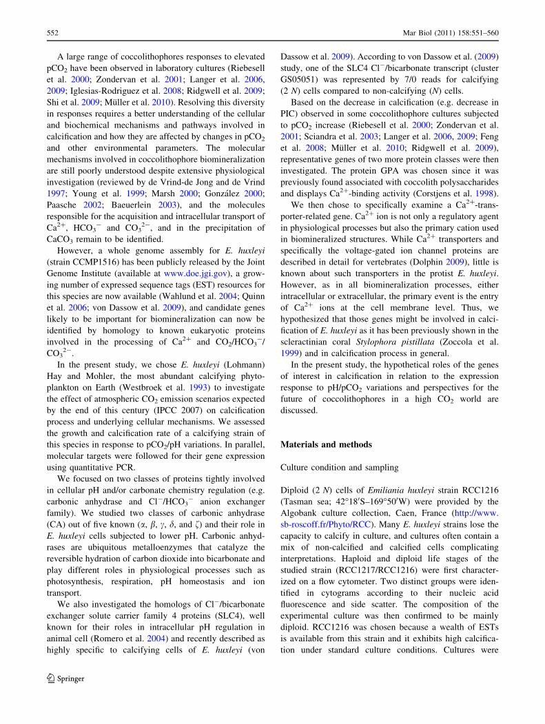
of the incubation period (8 days), is described in Table 1.
Particulate inorganic (PIC) and organic (POC) carbon
measurements
Triplicate samples (*150 lg C per filter) were filtered onto
pre-combusted (4 h, 400�C) glass fiber filters (Whatman GF/
F), dried at 60�C overnight and stored in a desiccator pending
analysis. For POC measurements, the inorganic carbon was
removed from the filters before the analysis by adding 25%
HCl (Nieuwenhuize et al. 1994). Cell content for total par-
ticulate carbon (TPC) and for particulate organic carbon
(POC) (pg cell-1) was subsequently measured on a Thermo
Electron Flash EA 1112 Analyzer as described by Nie-
uwenhuize et al. (1994). Particulate inorganic carbon (PIC)
(pg cell-1) was calculated as the difference between TPC
and POC. Particulate inorganic carbon production, i.e. cal-
cification rate (PPIC, pg PIC cell-1 d-1) was calculated
according to: PPIC = l 9 (cellular inorganic carbon con-
tent in pg PIC per cell). Particulate organic carbon produc-
tion (PPOC, pg POC cell-1 d-1) was calculated according
to: PPOC = l 9 (cellular organic carbon content in pg POC
per cell) (Riebesell et al. 2000).
Quantitative reverse transcriptase-polymerase chain
reaction (q-RT–PCR)
RNA extraction—Total RNA was isolated from coccolitho-
phores with Trizol reagent (Invitrogen, La Jolla, CA)
Mar Biol (2011) 158:551–560 553
123
according to the suggested protocol. Five hundred milliliter
of medium from each bottle was collected by gentle filtra-
tion on polycarbonate filter of 1 lm (Whatman) and resus-
pended in 1 ml of Trizol. Two successive chloroform
(C99%) steps in 200 ll were carried out to precipitate pro-
teins and DNA. RNA was finally precipitated in 500 ll
isopropanol (C99%). The pellets were washed in 75% eth-
anol and resuspended in RNase-free water. The RNA quality
was checked on 1% agarose (w:v) non-denaturing gels and
the purity determined using a Nanodrop spectrophotometer
(Nanodrop 3300, Thermo scientific). All samples presented
ribosomal RNA bands with no sign of degradation. RNA
samples were treated with DNase (1U ll-1, Fermentas) and
quantified using a RiboGreen RNA Quantification Kit
(Molecular Probes). Total RNA concentration was adjusted
to a final concentration of 100 ng ll-1 in all samples, and
the reverse transcription was carried out using the Affinity
Script qPCR cDNA kit (Stratagene). Negative controls
(same reagents mix without reverse transcriptase) were
prepared simultaneously and run on each plate for each
primer pairs to ascertain that no DNA contamination
occurred (Ct values were [40 cycles). No template controls
were also run in parallel on each plate.
Transcript levels were derived from the accumulation of
SYBR green fluorescence measured with a Light Cycler
480 (Roche). The PCR conditions were as follows: 19
SYBR green mix (Roche, Cat. nb: 04707516001), 500 nM
primers and 1 ll (100 ng) of cDNA in a total volume of
20 ll. Each sample was run in triplicate (mean ±
SD \ 0.2). The dissociation curves showed a single
amplification product and no primer dimer. For each primer
pairs, the amplification efficiency (E) was determined on a
5 points 10-time dilution series of 100 ng cDNA extracted
from the two tested conditions (control and experimental
pCO2) to check for primer specificity. The reaction effi-
ciencies had values between 80 and 100% with a corre-
sponding amplification factor between 1.8 and 2.0,
respectively, for all primer combinations. This value allows
for a transformation of the observed changes in cycle
threshold (CT).
RNA transcription levels were determined by the
method of direct comparison of CT values between target
genes and a reference gene. Several genes from E. huxleyi
strain CCMP1516 (JGI, USA) commonly used as house-
keeping genes (HKG) (e.g. actin (JGI, ID 226687),
b-tubulin (JGI, ID 451245) and RPLP0 (JGI, ID 456254))
were tested for their expression stability in experimental
samples using the program geNorm (Vandesompele et al.
2002). While none of them was stable enough to normalize
the data, calmodulin (JGI, ID 442625) was identified as the
most stable gene and used further to normalize the data by
the DDCt method (Livak and Schmittgen 2001). Data were
then transformed into linear form by: 2-DDCT where
-DDCT = (CtTarget-CtHKG)Tx-(CtTarget-CtHKG)T0. Data
were analyzed using one-way analyses of variance
(ANOVA). Since all the steps from RNA extraction to RT
qPCR efficiency have been checked for accuracy, high
standards deviations (SD) reported in Fig. 3 were mainly
attributed to biological variability in experimental batch
cultures.
Genes of interest and primer design
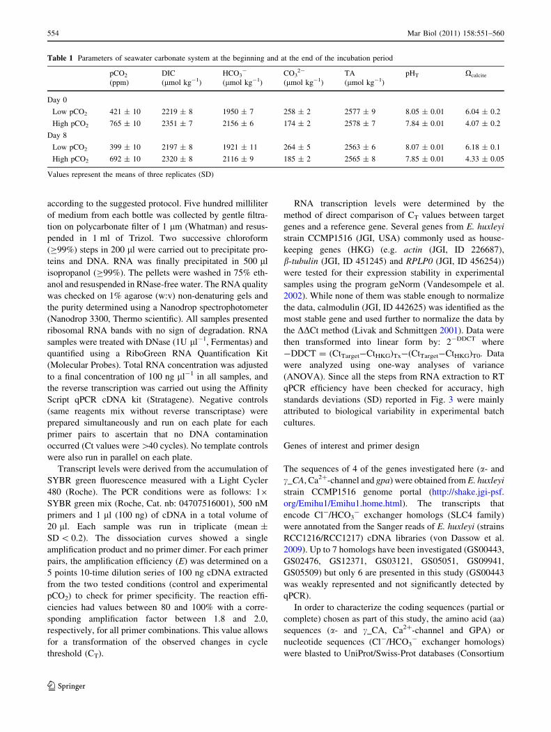
The sequences of 4 of the genes investigated here (a- and
c_CA, Ca2?-channel and gpa) were obtained from E. huxleyi
strain CCMP1516 genome portal (http://shake.jgi-psf.
org/Emihu1/Emihu1.home.html). The transcripts that
encode Cl-/HCO3- exchanger homologs (SLC4 family)
were annotated from the Sanger reads of E. huxleyi (strains
RCC1216/RCC1217) cDNA libraries (von Dassow et al.
2009). Up to 7 homologs have been investigated (GS00443,
GS02476, GS12371, GS03121, GS05051, GS09941,
GS05509) but only 6 are presented in this study (GS00443
was weakly represented and not significantly detected by
qPCR).
In order to characterize the coding sequences (partial or
complete) chosen as part of this study, the amino acid (aa)
sequences (a- and c_CA, Ca2?-channel and GPA) or
nucleotide sequences (Cl-/HCO3- exchanger homologs)
were blasted to UniProt/Swiss-Prot databases (Consortium
Table 1 Parameters of seawater carbonate system at the beginning and at the end of the incubation period
pCO2
(ppm)
DIC
(lmol kg-1)
HCO3-
(lmol kg-1)
CO32-
(lmol kg-1)
TA
(lmol kg-1)
pHT Xcalcite
Day 0
Low pCO2 421 ± 10 2219 ± 8 1950 ± 7 258 ± 2 2577 ± 9 8.05 ± 0.01 6.04 ± 0.2
High pCO2 765 ± 10 2351 ± 7 2156 ± 6 174 ± 2 2578 ± 7 7.84 ± 0.01 4.07 ± 0.2
Day 8
Low pCO2 399 ± 10 2197 ± 8 1921 ± 11 264 ± 5 2563 ± 6 8.07 ± 0.01 6.18 ± 0.1
High pCO2 692 ± 10 2320 ± 8 2116 ± 9 185 ± 2 2565 ± 8 7.85 ± 0.01 4.33 ± 0.05
Values represent the means of three replicates (SD)
554 Mar Biol (2011) 158:551–560
123
U 2009) and NCBI/CDD (Conserved Domains database)
(Marchler-Bauer et al. 2009). The characteristics of the
given sequences are detailed in Tables 2, 3.
qPCR primer sequences were designed using the Primer3
software to have a G ? C content ranging from 50 to 60%
and C’s [ G’s 3 identical dNTPs in a row at the 30 ends to
avoid self complementarities of the primer sequence. Prim-
ers were chosen to generate equivalent amplicon lengths (see
Table 4). The melting temperature of the primers was set at
58�C. The qPCR products were sequenced (MWG, Ger-
many) and all matched the anticipated product. For PCR
products obtained with primers designed from E. huxleyi
strain CCMP1516 (e.g. a- and c_CA, Ca2?-channel and
GPA), sequences from both strains (CCMP1516 and
RCC1216) were aligned and showed 100% identity.
Results and discussion
While previous molecular studies on E. huxleyi dealt with
identification of genes that are associated with the
calcification mechanism (Quinn et al. 2006; Wahlund et al.
2004; Nguyen et al. 2005; von Dassow et al. 2009), our
experiment is the first to investigate gene expression in
response to CO2-driven ocean acidification. Our approach
provides new elements on the molecular and physiological
role of genes of interest in calcification and helps under-
stand the diverse response of coccolithophores to projected
ocean acidification.
Physiological and biochemical response to decreasing
pH
The experimental setup was designed following recom-
mendations of best practices (Riebesell et al. 2010), and
batch cultures were used as many other previous studies
(Riebesell et al. 2000; Zondervan et al. 2001, 2002;
Langer et al. 2006, 2009; Iglesias-Rodriguez et al. 2008),
in order to ensure that data comparison between studies
is possible. The manipulation of the carbonate system
was achieved by bubbling the culture medium with CO2
and/or air before the inoculation, and the experiment was
Table 2 Genes targeted in E. huxleyi strain RCC1216 and related characteristics
Name Suggested
protein
EMBL
[acc. nb.]
Prot ID
(JGI)
Gene
scaffold
(JGI)
KOG
class
KOG ID UniProt
[acc. nb.]
Best hit E-value
a-CA a-carbonic
anhydrase
na 456048 scaffold_166 (1) KOG0382 B6BNC3 Carbonic anhydrase
[Campylobacterales bacteriumGD.1]
1.00E-27
c-CA c-carbonic
anhydrase
na 432493 scaffold_5 (2) KOG0382 Q0ZB85 Gamma carbonic anhydrase
[Emiliania huxleyi]1.00E-130
CAC Ca2? ion
channel
na na scaffold_11 (3) KOG2301 C1FH96 Voltage-gated ion channel superfamily
[Micromonas sp. RCC299]
1.00E-138
gpa calcium-
binding
protein
FP217524 na scaffold_1 (3) KOG2643 Q0MYW8 Putative calcium-binding protein
[Emiliania huxleyi]–
GS00443 Cl-/HCO3-
exchangers
FP221416 na nomap (3) KOG1172 B5Y5V6 Predicted protein [Phaeodactylumtricornutum CCAP 1055/1]
3.00E-45
GS02476 Cl-/HCO3-
exchangers
FP180858 na nomap (3) KOG1172 Q7T1P6 Anion exchanger 1 [Raja erinacea] 2.00E-32
GS12371 Cl-/HCO3-
exchangers
FP187041 na scaffold_18 (3) KOG1172 B3RRA7 Putative uncharacterized protein
[Trichoplax adhaerens]
7.00E-09
GS03121 Cl-/HCO3-
exchangers
FP180021 na scaffold_51 (3) KOG1172 Q4WXW0 Anion exchange family protein
[Aspergillus fumigatus strain
CEA10]
1.00E-38
GS05051 Cl-/HCO3-
exchangers
FP185544 na scaffold_21 (3) KOG1172 C1E0U4 Anion exchanger family
[Micromonas sp. RCC299]
8.00E-18
GS09941 Cl-/HCO3-
exchangers
FP163914 450694 scaffold_31 (3) KOG1172 B7FQY4 Predicted protein [Phaeodactylumtricornutum CCAP 1055/1]
1.00E-08
GS05509 Cl-/HCO3-
exchangers
FP183003 196760 scaffold_4 (3) KOG1172 B7FQY4 Predicted protein [Phaeodactylumtricornutum CCAP 1055/1]
2.00E-36
EMBL accession numbers have been provided for the clusters annotated as part of von Dassow et al. (2009) (see also von Dassow et al. 2009
Additional file 2). KOG (NCBI eukaryote orthologous group) class [(1) general function, (2) cytoskeleton and (3) inorganic ion transport and
metabolism] is also mentioned
na Not available
Mar Biol (2011) 158:551–560 555
123
consequently performed in a closed system avoiding gas
exchanges with the atmosphere. As in the natural envi-
ronment, this method involves changes in pCO2, DIC
and pH, while TA remains constant (Gattuso and
Lavigne 2009). The stress caused to the cultures by the
air bubbling and consequent variability of the response
to tested parameters are eliminated, and the shift in
carbonate parameters due to cell activity is negligible.
Consequently, any change during the experiment can
exclusively be attributed to physiological changes in
response to the CO2 perturbation (Fiorini 2010).
In the past few years, parameters such as growth rate
and organic and inorganic carbon production have been
widely investigated in calcifiers in order to predict the
impact of ocean acidification (Buitenhuis et al. 1999; Clark
and Flynn, 2000; Riebesell et al. 2000; Rost et al. 2002;
Sciandra et al. 2003; Iglesias-Rodriguez et al. 2008: Shi
et al. 2009; Barcelos e Ramos et al. 2010; Muller et al.
2010; Langer et al. 2009). In this study, the response
regarding those parameters is in agreement with the diverse
responses already described for E. huxleyi strains in the
literature. We found a minor effect of elevated pCO2 on the
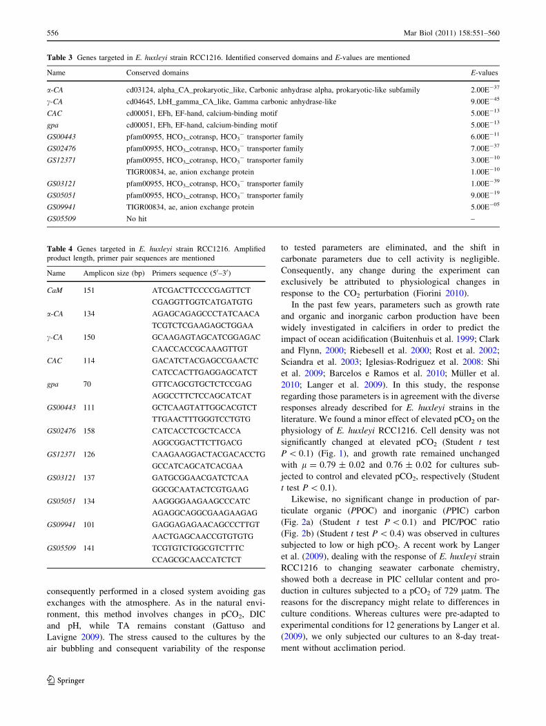
physiology of E. huxleyi RCC1216. Cell density was not
significantly changed at elevated pCO2 (Student t test
P \ 0.1) (Fig. 1), and growth rate remained unchanged
with l = 0.79 ± 0.02 and 0.76 ± 0.02 for cultures sub-
jected to control and elevated pCO2, respectively (Student
t test P \ 0.1).
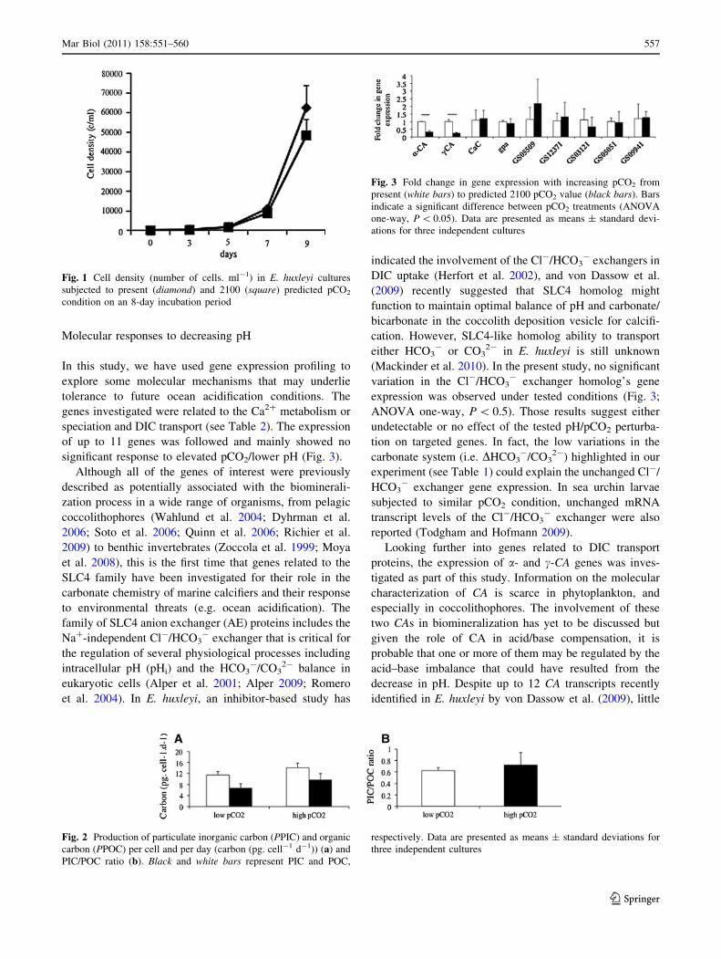
Likewise, no significant change in production of par-
ticulate organic (PPOC) and inorganic (PPIC) carbon
(Fig. 2a) (Student t test P \ 0.1) and PIC/POC ratio
(Fig. 2b) (Student t test P \ 0.4) was observed in cultures
subjected to low or high pCO2. A recent work by Langer
et al. (2009), dealing with the response of E. huxleyi strain
RCC1216 to changing seawater carbonate chemistry,
showed both a decrease in PIC cellular content and pro-
duction in cultures subjected to a pCO2 of 729 latm. The
reasons for the discrepancy might relate to differences in
culture conditions. Whereas cultures were pre-adapted to
experimental conditions for 12 generations by Langer et al.
(2009), we only subjected our cultures to an 8-day treat-
ment without acclimation period.
Table 3 Genes targeted in E. huxleyi strain RCC1216. Identified conserved domains and E-values are mentioned
Name Conserved domains E-values
a-CA cd03124, alpha_CA_prokaryotic_like, Carbonic anhydrase alpha, prokaryotic-like subfamily 2.00E-37
c-CA cd04645, LbH_gamma_CA_like, Gamma carbonic anhydrase-like 9.00E-45
CAC cd00051, EFh, EF-hand, calcium-binding motif 5.00E-13
gpa cd00051, EFh, EF-hand, calcium-binding motif 5.00E-13
GS00443 pfam00955, HCO3_cotransp, HCO3- transporter family 6.00E-11
GS02476 pfam00955, HCO3_cotransp, HCO3- transporter family 7.00E-37
GS12371 pfam00955, HCO3_cotransp, HCO3- transporter family
TIGR00834, ae, anion exchange protein
3.00E-10
1.00E-10
GS03121 pfam00955, HCO3_cotransp, HCO3- transporter family 1.00E-39
GS05051 pfam00955, HCO3_cotransp, HCO3- transporter family 9.00E-19
GS09941 TIGR00834, ae, anion exchange protein 5.00E-05
GS05509 No hit –
Table 4 Genes targeted in E. huxleyi strain RCC1216. Amplified
product length, primer pair sequences are mentioned
Name Amplicon size (bp) Primers sequence (50–30)
CaM 151 ATCGACTTCCCCGAGTTCT
CGAGGTTGGTCATGATGTG
a-CA 134 AGAGCAGAGCCCTATCAACA
TCGTCTCGAAGAGCTGGAA
c-CA 150 GCAAGAGTAGCATCGGAGAC
CAACCACCGCAAAGTTGT
CAC 114 GACATCTACGAGCCGAACTC
CATCCACTTGAGGAGCATCT
gpa 70 GTTCAGCGTGCTCTCCGAG
AGGCCTTCTCCAGCATCAT
GS00443 111 GCTCAAGTATTGGCACGTCT
TTGAACTTTGGGTCCTGTG
GS02476 158 CATCACCTCGCTCACCA
AGGCGGACTTCTTGACG
GS12371 126 CAAGAAGGACTACGACACCTG
GCCATCAGCATCACGAA
GS03121 137 GATGCGGAACGATCTCAA
GGCGCAATACTCGTGAAG
GS05051 134 AAGGGGAAGAAGCCCATC
AGAGGCAGGCGAAGAAGAG
GS09941 101 GAGGAGAGAACAGCCCTTGT
AACTGAGCAACCGTGTGTG
GS05509 141 TCGTGTCTGGCGTCTTTC
CCAGCGCAACCATCTCT
556 Mar Biol (2011) 158:551–560
123
Molecular responses to decreasing pH
In this study, we have used gene expression profiling to
explore some molecular mechanisms that may underlie
tolerance to future ocean acidification conditions. The
genes investigated were related to the Ca2? metabolism or
speciation and DIC transport (see Table 2). The expression
of up to 11 genes was followed and mainly showed no
significant response to elevated pCO2/lower pH (Fig. 3).
Although all of the genes of interest were previously
described as potentially associated with the biominerali-
zation process in a wide range of organisms, from pelagic
coccolithophores (Wahlund et al. 2004; Dyhrman et al.
2006; Soto et al. 2006; Quinn et al. 2006; Richier et al.
2009) to benthic invertebrates (Zoccola et al. 1999; Moya
et al. 2008), this is the first time that genes related to the
SLC4 family have been investigated for their role in the
carbonate chemistry of marine calcifiers and their response
to environmental threats (e.g. ocean acidification). The
family of SLC4 anion exchanger (AE) proteins includes the
Na?-independent Cl-/HCO3- exchanger that is critical for
the regulation of several physiological processes including
intracellular pH (pHi) and the HCO3-/CO3
2- balance in
eukaryotic cells (Alper et al. 2001; Alper 2009; Romero
et al. 2004). In E. huxleyi, an inhibitor-based study has
indicated the involvement of the Cl-/HCO3- exchangers in
DIC uptake (Herfort et al. 2002), and von Dassow et al.
(2009) recently suggested that SLC4 homolog might
function to maintain optimal balance of pH and carbonate/
bicarbonate in the coccolith deposition vesicle for calcifi-
cation. However, SLC4-like homolog ability to transport
either HCO3- or CO3
2- in E. huxleyi is still unknown
(Mackinder et al. 2010). In the present study, no significant
variation in the Cl-/HCO3- exchanger homolog’s gene
expression was observed under tested conditions (Fig. 3;
ANOVA one-way, P \ 0.5). Those results suggest either
undetectable or no effect of the tested pH/pCO2 perturba-
tion on targeted genes. In fact, the low variations in the
carbonate system (i.e. DHCO3-/CO3
2-) highlighted in our
experiment (see Table 1) could explain the unchanged Cl-/
HCO3- exchanger gene expression. In sea urchin larvae
subjected to similar pCO2 condition, unchanged mRNA
transcript levels of the Cl-/HCO3- exchanger were also
reported (Todgham and Hofmann 2009).
Looking further into genes related to DIC transport
proteins, the expression of a- and c-CA genes was inves-
tigated as part of this study. Information on the molecular
characterization of CA is scarce in phytoplankton, and
especially in coccolithophores. The involvement of these
two CAs in biomineralization has yet to be discussed but
given the role of CA in acid/base compensation, it is
probable that one or more of them may be regulated by the
acid–base imbalance that could have resulted from the
decrease in pH. Despite up to 12 CA transcripts recently
identified in E. huxleyi by von Dassow et al. (2009), little
Fig. 1 Cell density (number of cells. ml-1) in E. huxleyi cultures
subjected to present (diamond) and 2100 (square) predicted pCO2
condition on an 8-day incubation period
Fig. 2 Production of particulate inorganic carbon (PPIC) and organic
carbon (PPOC) per cell and per day (carbon (pg. cell-1 d-1)) (a) and
PIC/POC ratio (b). Black and white bars represent PIC and POC,
respectively. Data are presented as means ± standard deviations for
three independent cultures
Fig. 3 Fold change in gene expression with increasing pCO2 from
present (white bars) to predicted 2100 pCO2 value (black bars). Bars
indicate a significant difference between pCO2 treatments (ANOVA
one-way, P \ 0.05). Data are presented as means ± standard devi-
ations for three independent cultures
Mar Biol (2011) 158:551–560 557
123

information is available about the localization and role of
these genes in coccolithophores. A first attempt to char-
acterize CA isoforms in E. huxleyi was performed by Soto
et al. (2006) who speculated on a location for c-EhCA2
protein in the coccolith vesicle with a 25-fold up-regulation
in c-EhCA2 transcripts under calcifying versus non-calci-
fying condition. The role of c-CA isoform in calcification
was also supported by previous studies with up-regulated
transcripts in cultures where calcification was stimulated
by phosphate-depletion (Quinn et al. 2006) and signifi-
cantly higher during the light period in calcifying cells
(RCC1216 strain) compared to non-calcifying ones
(RCC1217) (Richier et al. 2009).
In the present study, the CA sequences were searched
against databases for their conserved domains (see
Table 2). A conserved domain homolog to an ‘‘alpha_
CA_procaryotic like’’ carbonic anhydrase was detected in
a-CA. In this sub-family, the enzyme has been reported to
be part of the organic matrix layer in shells. Other members
of this family may be involved in maintaining pH balance,
in facilitating transport of CO2 or H2CO3, or in sensing
carbon dioxide levels in the environment. We thus delib-
erately chose here to analyze c-CA isoform, for the reasons
outlined earlier, and a-CA isoform for its widespread dis-
tribution in several kingdoms of life (vertebrates, inverte-
brates, bacteria, and some chlorophytes) and its role in
biomineralization of benthic organisms (Moya et al. 2008).
We showed that a- and c-CA genes were down-regulated
when exposed to decreasing pH resulting in a fold change of
2.3 and 3.8, respectively (ANOVA one-way, P \ 0.05)
(Fig. 3). A previous study on E. huxleyi intracellular CA
activity showed no clear trend with increasing pCO2 (from
36 ppmv up to 1,800 ppmv) (Rost et al. 2003). However,
the measurements in that study did not discriminate
between CA isoform classes and it might be that the regu-
lation of CA genes is class specific.
Additionally, it has been previously suggested that the CA
enzymes and SLC4 anion exchangers may interact (Vince
and Reithmeier, 2000; Sterling et al. 2001, 2002; Morgan
et al. 2007). In mammalian cell lines, the cytoplasmic car-
boxy terminal of AE1 has a carbonic anhydrase II (CAII)
binding site that upon inhibition reduces AE1-mediated Cl-/
HCO3- exchange by 50–60% (Sterling et al. 2001). Car-
bonic anhydrase IV (CAIV) interaction sites have also been
identified on the extracellular surface of AE1 isoform.
According to the authors, CAII and IV would increase
HCO3- transport by altering localized HCO3
- levels
enhancing the HCO3- concentration gradient (McMurtrie
et al. 2003). A similar function may occur in coccolitho-
phores with CA interacting with the Cl-/HCO3- exchanger
facilitating the conversion of HCO3- into CO2 at the cyto-
solic face of the plasma membrane decreasing the local
concentration of HCO3- at the cytosolic transport site
(Mackinder et al. 2010). In our study, we could speculate that
increasing pCO2 inhibits both a- and c-CA genes transcrip-
tion and consequently the activity of their relative proteins.
Thus, no interactions with SLC4 homologs would occur,
which is reflected by unchanged Cl-/HCO3- exchanger
transcript level under experimental condition. In the same
way, the unchanged Ca2?-channel (CAC) and gpa transcript
level, in response to tested conditions, would suggest no
reduced capacity of the protein to transport or bind Ca2? to
the sites of calcification and supports the unchanged calci-
fication rate observed in the tested cultures. However, the
regulation of gene of interest related proteins was not
investigated as part of this study. Simultaneous analyses of
both transcripts and corresponding proteins are required to
conclude on any proteins regulation and function.
In conclusion, all the results shown by our study con-
stitute new elements in molecular exploration of genes
involved in E. huxleyi early response to an acidifying
world. No major physiological changes were observed in
the chosen strain in response to ocean acidification and
only CA isoforms, among the tested genes, appeared sig-
nificantly regulated under the experimental condition.
However, no significant variation in expression of most of
the genes might either suggest (1) no major effect of the
near future pCO2 condition in the ocean on the tested strain
or (2) no direct role of the targeted genes in early response
to high pCO2/low pH. An exhaustive investigation into
E. huxleyi transcriptome would be required to identify all the
genes/cellular mechanisms involved in response to pCO2/
pH variation.
Nonetheless, the fact high pCO2-treatment did not
induce major molecular and physiological changes in this
calcified phytoplankton suggests that it may have the
capacity to adapt to future ocean acidification.
AcknowledgmentsWe thank Cornelia Maier and JinWen Liu for
providing access to mass flow controllers and assistance to set up the
high pCO2 experiment. We also thank Steeve Comeau for technical
support with measurements of pH and total alkalinity. We are also
grateful to Anna Macey for her help with the English. This is a
contribution to the ‘‘European Project on Ocean Acidification’’
(EPOCA) which receives funding from the European Community’s
Seventh Framework Programme (FP7/2007-2013) under grant
agreement 211384. We are also grateful to several anonymous
reviewers that significantly improved the manuscript.
References
Alper SL (2009) Molecular physiology and genetics of Na?-
Independent SLC4 anion exchangers. J Exp Biol 212:1672–1683
Alper SL, Chernova MN, Stewart AK (2001) Regulation of Na?-
Independent Cl-/HCO3- exchangers by pH. JOP J Pancreas
2:171–175
Baeuerlein E (2003) Biomineralization of unicellular organisms: an
unusual membrane biochemistry for the production of inorganic
nano- and microstructures. Angew Chem Int Ed 42:614–641
558 Mar Biol (2011) 158:551–560
123

Barcelos e Ramos J, Muller MN, Riebesell U (2010) Short-term
response of the coccolithophore Emiliania huxleyi to abrupt
changes in seawater carbon dioxide concentrations. Biogeo-
sciences 7:177–186
Brewer PG, Goldman JC (1976) Alkalinity changes generated by
phytoplankton growth. Limnol Oceanogr 21:108
Buitenhuis ET, De Baar HJW, Veldhuis MJW (1999) Photosynthesis
and calcification by Emiliania huxleyi (Prymnesiophyceae) as a
function of inorganic carbon species. J Phycol 35:949–959
Clark DR, Flynn KJ (2000) The relationship between the dissolved
inorganic carbon concentration and growth rate in marine
phytoplankton. Proc R Soc Lond 267:953–959
Consortium U (2009) The Universal Protein Resource (UniProt).
Nucleic Acids Res 37:D169–D174
Corstjens PLAM, van der Kooij A, Linschooten C, Brouwers GJ,
Westbroek P, de Vrind-de Jong EW (1998) GPA, a calcium-
binding protein in the coccolithophorid Emiliania huxleyi(Prymnesiophyceae). J Phycol 34:622–630
de Vrind-de Jong EW, de Vrind JPM (1997) Algal deposition of
carbonates and silicates. In: Banfield JF, Nealson KH (eds)
Geomicrobiology: interactions between microbes and minerals.
Mineralogical Society of America, Washington, DC, pp 267–307
Dickson AG, Sabine CL, Christian JR (eds) (2007) Guide to best
practices for ocean CO2 measurements, PICES Special Publica-
tion 3, p 191
Dolphin AC (2009) Calcium channel diversity: multiple roles of
calcium channel subunits. Curr Opin Neurobiol 19:237–244
Dyhrman ST, Haley ST, Birkeland SR, Wurch LL, Cipriano MJ,
McArthur AG (2006) Long serial analysis of gene expression for
gene discovery and transcriptome profiling in the widespread
marine coccolithophore Emiliania huxleyi. Appl Environ Micro-
biol 72:252–260
Engel A, Szlosek J, Abramson L, Liu Z, Lee C (2009) Investigating
the effect of ballasting by CaCO3 in Emiliania huxlei: I.
Formation, settling velocities and physical properties of aggre-
gates. Deep Sea Res II 56:1396–1407
Feng Y, Warner ME, Zhang Y, Sun J, Fu FX, Rose JM, Hutchins DA
(2008) Interactive effects of increased pCO2, temperature and
irradiance on the marine coccolithophore Emiliania huxleyi(Prymnesiophyceae). Eur J Phycol 43:87–98
Fiorini S (2010) Effect of elevated CO2 partial pressure and
temperature on calcifying phytoplankton (coccolithophores).
Ph.D. dissertation, Pierre and Marie Curie University, Paris,
France
Gattuso J-P, Lavigne H (2009) Technical note: approaches and
software tools to investigate the impact of ocean acidification.
Biogeosciences 6:2121–2133
Gonzalez EL (2000) The calcifying vesicle membrane of the
coccolithophore. In: Baeuerlein E (ed) Biomineralization: from
biology to biotechnology and medical application. Wiley-VCH,
Weinheim, pp 269–283
Halpern BS, Walbridge S, Selkoe KA, Kappel CV, Micheli F,
D’Agrosa C et al (2008) A global map of human impact on
marine ecosystems. Science 319:948–952
Herfort L, Thake B, Roberts J (2002) Acquisition and use of
bicarbonate by Emiliania huxleyi. New Phytol 156:427–436
Iglesias-Rodriguez MD, Halloran PR, Rickaby RE, Hall IR, Colmen-
ero-Hidalgo E, Gittins JR et al (2008) Phytoplankton calcifica-
tion in a high-CO2 world. Science 320:336–340
Keller MD, Selvin RC, Claus W, Guillard RRL (1987) Media for the
culture of oceanic ultraphytoplankton. J Phycol 23:633–638
Langer G, Geisen M, Baumann KH, Klas J, Riebesell U, Thoms S,
Young JR (2006) Species-specific responses of calcifying algae
to changing seawater carbonate chemistry. Geochem Geophys
Geosyst 7:Q09006
Langer G, Nehrke G, Probert I, Ly J, Ziveri P (2009) Strain-specific
responses of Emiliania huxleyi to changing seawater carbonate
chemistry. Biogeosciences 6:2637–2646
Lavigne H, Proye A, Gattuso J-P (2008) seacarb 2.0, an R package to
calculate parameters of the seawater carbonate system. Available
at http://cran.rproject.org/web/packages/seacarb/index.html
Livak KJ, Schmittgen TD (2001) Analysis of relative gene expression
data using Real-Time quantitative PCR and the 2-DDCT method.
Methods 25:402–408
Mackinder L, Wheeler G, Schroeder D, Riebesell U, Brownlee C
(2010) Molecular mechanisms underlying calcification in coc-
colithophores. Geomicrobiology 27:585–595
Marchler-Bauer A et al (2009) CDD: specific functional annotation
with the conserved domain database. Nucleic Acids Res
37:205–210
Marsh ME (2000) Polyanions in the CaCO3 mineralization of
coccolithophores. In: Baeuerlein E (ed) Biomineralization: from
biology to biotechnology and medical application. Wiley-VCH,
Weinheim, pp 251–268
McMurtrie HL, Cleary HJ, Alvarez BV, Loiselle FB, Sterling D,
Morgan PE, Johnson DE, Casey JR (2003) The bicarbonate
transport metabolon. In: 6th international conference on carbonic
anhydrases, Taylor & Francis Ltd, Bratislava, Slovakia,
pp 231–236
Milliman JD (1993) Production and accumulation of calcium
carbonate in the ocean—budget of a nonsteady state. Global
Biogeochem Cycles 7:927–957
Morgan PE, Pastorekova S, Stuart-Tilley AK, Alper SL, Casey JR
(2007) Interactions of transmembrane carbonic anhydrase,
CAIX, with bicarbonate transporters. Am J Physiol Cell Physiol
293:738–748
Moya A, Tambutte S, Bertucci A, Tambutte E, Lotto S, Vullo D et al
(2008) Carbonic anhydrase in the scleractinian coral Stylophorapistillata: characterization, localization, and role in biomineral-
ization. J Biol Chem 283:25475–25484
Muller MN, Schulz KG, Riebesell U (2010) Effects of long-term high
CO2 exposure on two species of coccolithophores. Biogeo-
sciences 7:1109–1116
Nguyen B, Bowers RM, Wahlund TM, Read BA (2005) Suppressive
subtractive hybridization of and differences in gene expression
content of calcifying and noncalcifying cultures of Emilianiahuxleyi strain 1516. Appl Environ Microbiol 71:2564–2575
Nieuwenhuize J, Maas YEM, Middelburg JJ (1994) Rapid analysis of
organic carbon and nitrogen in particulate materials. Mar Chem
44:217–224
Orr JC, Fabry VJ, Aumont O, Bopp L, Doney SC, Feely RA et al
(2005) Anthropogenic ocean acidification over the twenty-first
century and its impact on calcifying organisms. Nature
437:681–686
Paasche E (2002) A review of the coccolithophore Emiliania huxleyi(Prymnesiophyceae), with particular reference to growth, coc-
colith formation, and calcification–photosynthesis interactions.
Phycologia 40:503–529
Quinn P, Bowers RM, Zhang X, Wahlund TM, Fanelli MA, Olszova
D, Read BA (2006) cDNA microarrays as a tool for identifica-
tion of biomineralization proteins in the coccolithophore
Emiliania huxleyi (Haptophyta). Appl Environ Microbiol
72:5512–5526
Richier S, Kerros ME, de Vargas C, Hamaraty L, Falkowski PG,
Gattuso J-P (2009) Light-dependent transcriptional regulation of
genes of biogeochemical interest in the diploid and haploid life
cycle stages of Emiliania huxleyi. Appl Environ Microbiol
75:3366–3369
Ridgwell A, Schmidt DN, Turley C, Brownlee C, Maldonado MT,
Tortell P, Young JR (2009) From laboratory manipulations to
Mar Biol (2011) 158:551–560 559
123
earth system models: scaling calcification impacts of ocean
acidification. Biogeosciences 6:2611–2623
Riebesell U, Zondervan I, Rost B, Tortell PD, Morel FFM (2000)
Reduced calcification of marine plankton in response to
increased atmospheric CO2. Nature 407:364–367
Riebesell U, Fabry VJ, Hansson L, Gattuso J-P (eds) (2010) Guide to
best practices for ocean acidification research and data reporting.
Publications Office of the European Union, Luxembourg, p 260
Romero MF, Fulton CM, Boron WF (2004) The SLC4 family of
HCO3- transporters. Eur J Physiol 447:495–509
Rost B, Zondervan I, Riebesell U (2002) Light-dependent carbon
isotope fractionation in the coccolithophore Emiliania huxleyi.Limnol Oceanogr 47:120–128
Rost B, Riebesell U, Burkhardt S (2003) Carbon acquisition of
blooming marine phytoplankton. Limnol Oceanogr 48:55–67
Sabine CL, Feely RA, Gruber N, Key RM, Lee K, Bullister JL et al
(2004) The oceanic sink for anthropogenic CO2. Science
305:367–371
Sciandra A, Harlay J, Lefevre D, Lemee R, Rimmelin P, Denis M,
Gattuso J-P (2003) Response of coccolithophorid Emilianiahuxleyi to elevated partial pressure of CO2 under nitrogen
limitation. Mar Ecol Prog Ser 161:111–122
Shi D, Xu Y, Morel FMM (2009) Effects of the pH/pCO2 control
method on medium chemistry and phytoplankton growth.
Biogeosciences 6:1199–1207
Soto AR, Zheng H, Shoemaker D, Rodriguez J, Read BA, Wahlund
TM (2006) Identification and preliminary characterization of two
cDNAs encoding unique carbonic anhydrases from the marine
alga Emiliania huxleyi. Appl Environ Microbiol 72:5500–5511
Sterling D, Reithmeier RA, Casey JR (2001) A transport metabolon:
functional interaction of carbonic anhydrase II and chloride/
bicarbonate exchangers. J Biol Chem 276:47886–47894
Sterling D, Alvarez BV, Casey JR (2002) The extracellular compo-
nent of a transport metabolon: extracellular loop 4 of the human
AE1 Cl-/HCO3- exchanger binds carbonic anhydrase IV. J Biol
Chem 277:25239–25246
Todgham AE, Hofmann GE (2009) Transcriptomic response of sea
urchin larvae Strogylocentrotus purpuratus to CO2-driven sea-
water acidification. J Exp Biol 212:2579–2594
Vandesompele J, De Preter K, Pattyn F, Poppe B, Van Roy N, De
Paepe A, Speleman F (2002) Accurate normalization of real-time
quantitative RT-PCR data by geometric averaging of multiple
internal control genes. Genome Biol 3:34.1–34.11
von Dassow P, Ogata H, Probert I, Wincker P, Da Silva C, Audic, S,
Claverie J-M, de Vargas C (2009) Transcriptome analysis of
functional differentiation between haploid and diploid cells of
Emiliania huxleyi, a globally significant photosynthetic calcify-
ing cell. Genome Biol. doi:10.1186/gb-2009-10-10-r114
Vince JW, Reithmeier RA (2000) Identification of the carbonic
anhydrase II binding site in the Cl_/HCO3_ anion exchanger AE1.
Biochemistry 39:5527–5533
Wahlund TM, Hadaegh AR, Clark R, Nguyen B, Fanelli M, Read BA
(2004) Analysis of expressed sequence tags from calcifying cells
of marine coccolithophorid (Emiliania huxleyi). Mar. Biotechnol
(NY) 6:278–290
Westbroek P, Brown CW, Vanbleijswijk J, Brownlee C, Brummer GJ,
Conte M, Egge J, Fernandez E, Jordan R, Knappertsbusch M
(1993) A model system approach to biological climate forcing -
the example of Emiliania huxleyi. Glob Planet Change 8:27–46
Young JR, Davis SA, Bown PR, Mann S (1999) Coccolith structure
and biomineralization. J Struct Biol 126:195–215
Zoccola D, Tambutte E, Senegas-Balas F, Michiels JF, Failla JP,
Jaubert J, Allemand D (1999) Cloning of a calcium channel a1
subunit from the reef-building coral, Stylophora pistillata. Gene
227:157–167
Zondervan I, Zeebe RE, Rost B, Riebesell U (2001) Decreasing
marine biogenic calcification: a negative feedback on rising
atmospheric pCO2. Glob. Biogeochem Cycles 15:507–551
Zondervan I, Rost B, Riebesell U (2002) Effect of CO2 concentration
on the PIC/POC ratio in the coccolithophore Emiliania huxleyigrown under light-limiting conditions and different day lengths.
J Exp Mar Biol Ecol 272:55–70
560 Mar Biol (2011) 158:551–560
123
Related Documents