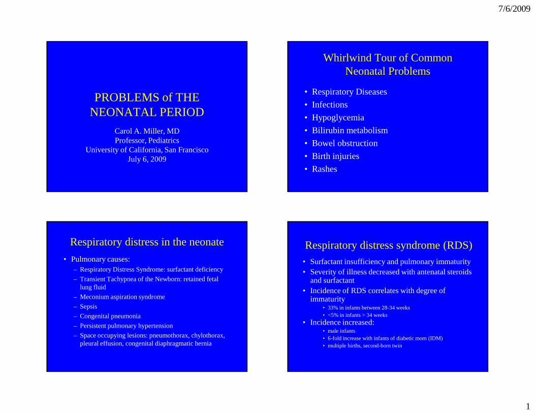
7/6/2009 1 PROBLEMS of THE NEONATAL PERIOD Carol A. Miller, MD Professor, Pediatrics University of California, San Francisco July 6, 2009 Whirlwind Tour of Common Neonatal Problems • Respiratory Diseases • Infections • Hypoglycemia • Bilirubin metabolism • Bowel obstruction • Birth injuries • Rashes Respiratory distress in the neonate • Pulmonary causes: – Respiratory Distress Syndrome: surfactant deficiency – Transient Tachypnea of the Newborn: retained fetal lung fluid – Meconium aspiration syndrome – Sepsis – Congenital pneumonia – Persistent pulmonary hypertension – Space occupying lesions: pneumothorax, chylothorax, pleural effusion, congenital diaphragmatic hernia Respiratory distress syndrome (RDS) • Surfactant insufficiency and pulmonary immaturity • Severity of illness decreased with antenatal steroids and surfactant • Incidence of RDS correlates with degree of immaturity • 33% in infants between 28-34 weeks • <5% in infants > 34 weeks • Incidence increased: • male infants • 6-fold increase with infants of diabetic mom (IDM) • multiple births, second-born twin

Welcome message from author
This document is posted to help you gain knowledge. Please leave a comment to let me know what you think about it! Share it to your friends and learn new things together.
Transcript
7/6/2009
1
PROBLEMS of THE NEONATAL PERIOD
Carol A. Miller, MDProfessor, Pediatrics
University of California, San FranciscoJuly 6, 2009
Whirlwind Tour of Common Neonatal Problems
• Respiratory Diseases
• Infections
• Hypoglycemia
• Bilirubin metabolism
• Bowel obstruction
• Birth injuries
• Rashes
Respiratory distress in the neonate
• Pulmonary causes:– Respiratory Distress Syndrome: surfactant deficiency
– Transient Tachypnea of the Newborn: retained fetal lung fluid
– Meconium aspiration syndrome
– Sepsis
– Congenital pneumonia
– Persistent pulmonary hypertension
– Space occupying lesions: pneumothorax, chylothorax, pleural effusion, congenital diaphragmatic hernia
Respiratory distress syndrome (RDS)• Surfactant insufficiency and pulmonary immaturity • Severity of illness decreased with antenatal steroids
and surfactant• Incidence of RDS correlates with degree of
immaturity• 33% in infants between 28-34 weeks• <5% in infants > 34 weeks
• Incidence increased:• male infants• 6-fold increase with infants of diabetic mom (IDM)• multiple births, second-born twin
7/6/2009
2
Respiratory distress syndrome
Strategies for prevention of RDS
• Prevention of premature delivery– Tocolytics, antibiotics
• Decrease antenatal inflammation/infectionChorioamnionitis, maternal infections
• increases risk for preterm labor
• Antenatal glucocorticoids– Does not prevent all RDS or Bronchopulmonary
dysplasia
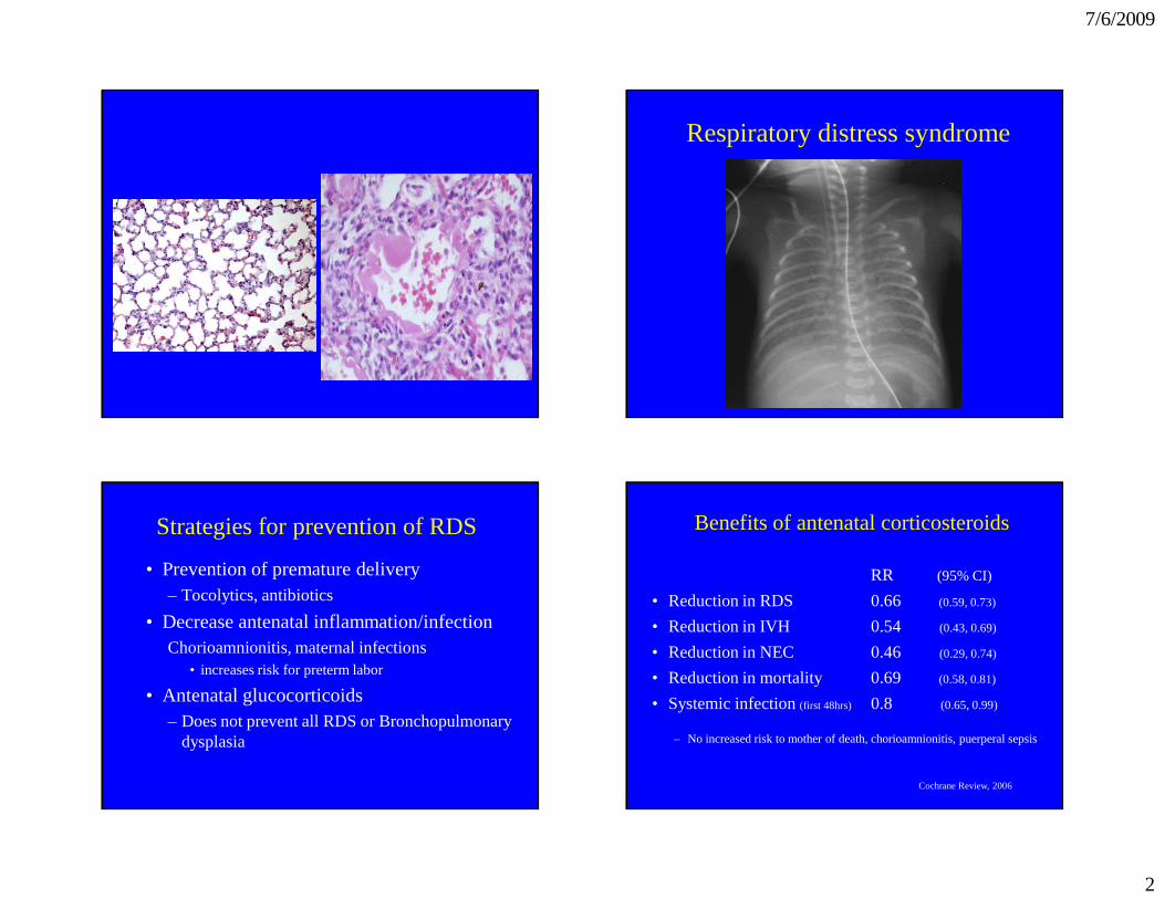
Benefits of antenatal corticosteroids
RR (95% CI)
• Reduction in RDS 0.66 (0.59, 0.73)
• Reduction in IVH 0.54 (0.43, 0.69)
• Reduction in NEC 0.46 (0.29, 0.74)
• Reduction in mortality 0.69 (0.58, 0.81)
• Systemic infection (first 48hrs) 0.8 (0.65, 0.99)
– No increased risk to mother of death, chorioamnionitis, puerperal sepsis
Cochrane Review, 2006
7/6/2009
3
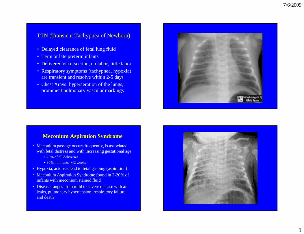
TTN (Transient Tachypnea of Newborn)
• Delayed clearance of fetal lung fluid
• Term or late preterm infants
• Delivered via c-section, no labor, little labor
• Respiratory symptoms (tachypnea, hypoxia) are transient and resolve within 2-5 days
• Chest Xrays: hyperaeration of the lungs, prominent pulmonary vascular markings
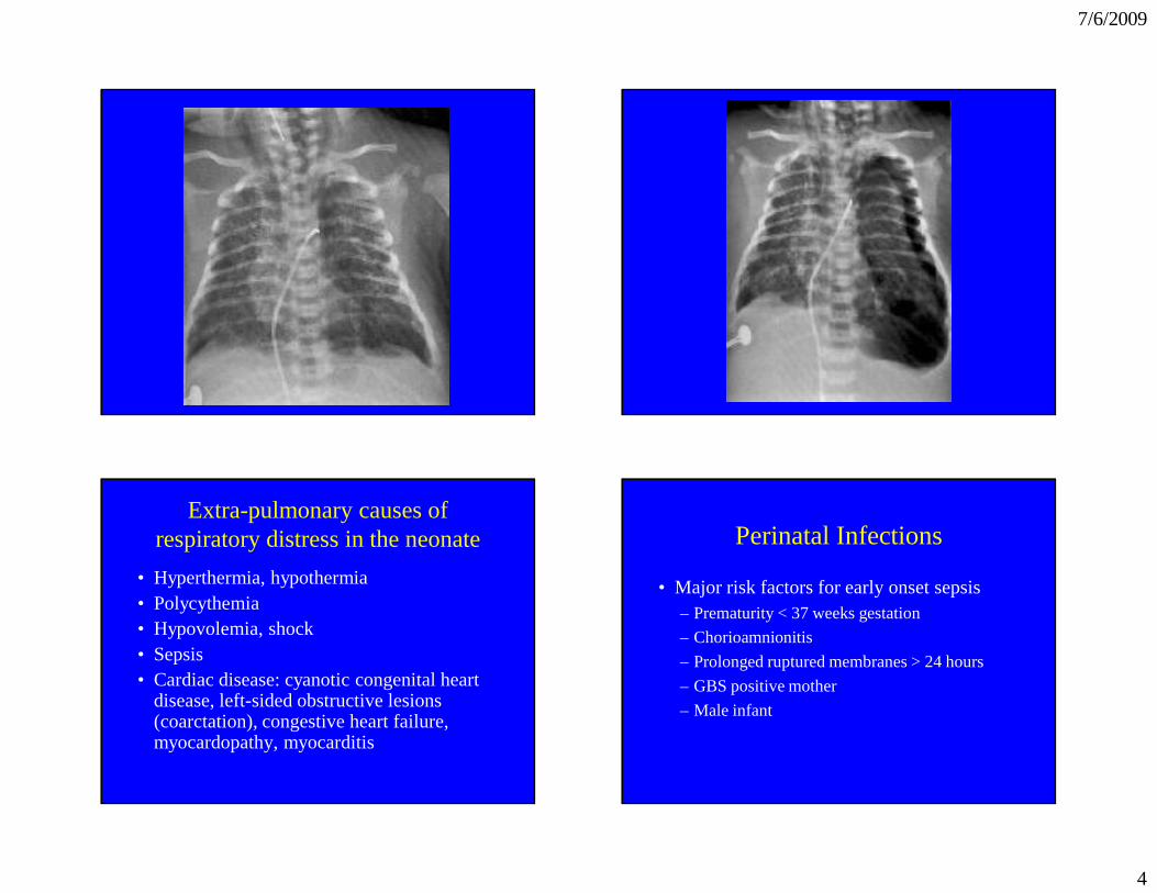
Meconium Aspiration Syndrome
• Meconium passage occurs frequently, is associated with fetal distress and with increasing gestational age
• 20% of all deliveries
• 30% in infants >42 weeks
• Hypoxia, acidosis lead to fetal gasping (aspiration)
• Meconium Aspiration Syndrome found in 2-20% of infants with meconium stained fluid
• Disease ranges from mild to severe disease with air leaks, pulmonary hypertension, respiratory failure, and death
7/6/2009
4
Extra-pulmonary causes of respiratory distress in the neonate
• Hyperthermia, hypothermia• Polycythemia• Hypovolemia, shock• Sepsis• Cardiac disease: cyanotic congenital heart
disease, left-sided obstructive lesions (coarctation), congestive heart failure, myocardopathy, myocarditis
Perinatal Infections
• Major risk factors for early onset sepsis– Prematurity < 37 weeks gestation
– Chorioamnionitis
– Prolonged ruptured membranes > 24 hours
– GBS positive mother
– Male infant
7/6/2009
5
Perinatal Infections• Bacterial infections:
Group B Streptococcus E. coli Listeria monocytogenes
• Viral infectionsHerpes simplex Hepatitis B and C
• TORCH infections: Incidence is 0.5-2.5%; many infants are asymptomatic at delivery– Toxoplasmosa gondii, treponema pallidum– “Other”: syphilis – Rubella– Cytomegalovirus– Herpes
Neonatal Group B Streptococcus
Prevention of GBS neonatal sepsis• Routine antenatal cultures at 35-36 weeks• Treat women:
– with positive cultures with onset of labor– with previously infected infants – with GBS UTI
Strategy misses women who deliver prematurely, women with no prenatal care, and women with false negative test results.
• Work-up for neonatal infections
– CBC with differential including bands and platelets
– Blood culture
– +/- Lumbar Puncture
– +/- C-reactive Protein, specific workup for viral infection
• Management
– Symptomatic: treat with ampicillin and gentamycin (or ampicillin and 2nd/3rd generation cephalosporin for bacterial meningitis). Acyclovir if concerned for herpes.
– Length of treatment depends on clinical findings, CBC, LP, and culture results.
– Asymptomatic (a non-reassuring CBC): treat for 48-72 hours until bacterial cultures negative
Management of neonatal infections Perinatal Hepatitis B
Prevention of transmission:
– Hepatitis B vaccine prior to hospital discharge for all infants (<12 hr if Mom HBsAg positive)
– HBIG (hepatitis B immunoglobulin) plus vaccine for infants born to HBsAg positive mother <12 hours of life
– All infants should receive routine Hepatitis B vaccine during infancy, titers at 9 months
– Breastfeeding safe with HBsAg positive mom with vaccine plus HBIG treatment for the infant
7/6/2009
6
Hepatitis CHigh-risk mothers screened during pregnancy
– Vertical transmission rate is 5-10%
– Hepatitis C antibody titers obtained on infant at 6 and 12 months, or Hepatitis C PCR at 4 mos
What about breastfeeding with Hepatitis C+?
– Studies show variable amounts of virus in milk
– Studies have not shown increase risk of transmission of Hepatitis C with breastfeeding
Perinatal TORCH Infections• Non-specific findings in infants
– SGA, IUGR, postnatal growth failure– Microcephaly, hydrocephalus, intracranial
calcifications– Hepatosplenomegaly, hepatitis, jaundice (elevated
direct component)– Anemia (hemolytic), thrombocytopenia– Skin rashes, petechiae– Abnormalities of long bones– Chorioretinitis, cataracts, glaucoma– Developmental and learning disabilities– Nonimmune hydrops
Perinatal (TORCH) InfectionsSpecific findings:
• Syphilis: osteochondritis, periosteal new bone formation, rash, snuffles
• Cytomegalovirus: microcephaly, periventricular calcifications, hydrocephalus, chorioretinitis, petichiae, thrombocytopenia, hearing loss (progressive)
• Toxoplasmosis: hydrocephalus, chorioretinitis, generalized intracranial calcifications (random distribution)
• Rubella: cataracts, “blueberry muffin rash”, patent ductus arteriosus, pulmonary stenosis
7/6/2009
7
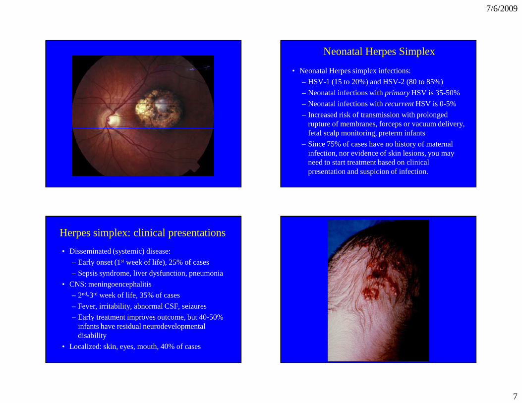
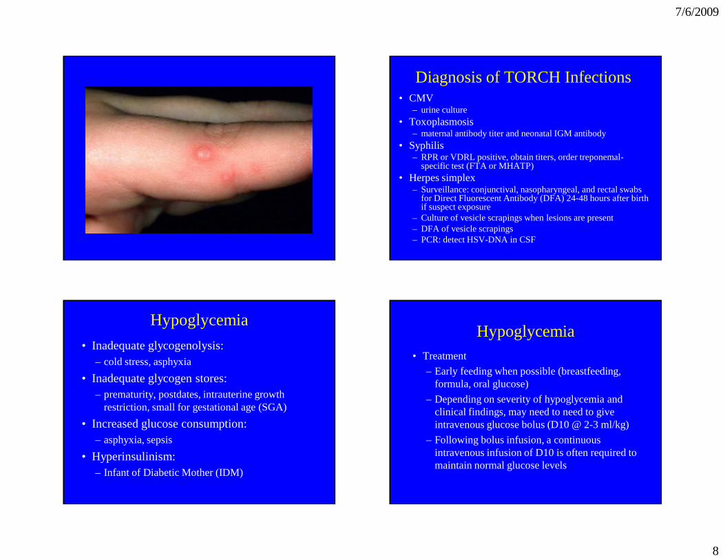
Neonatal Herpes Simplex
• Neonatal Herpes simplex infections:
– HSV-1 (15 to 20%) and HSV-2 (80 to 85%)
– Neonatal infections with primary HSV is 35-50%
– Neonatal infections with recurrent HSV is 0-5%
– Increased risk of transmission with prolonged rupture of membranes, forceps or vacuum delivery, fetal scalp monitoring, preterm infants
– Since 75% of cases have no history of maternal infection, nor evidence of skin lesions, you may need to start treatment based on clinical presentation and suspicion of infection.
Herpes simplex: clinical presentations
• Disseminated (systemic) disease:
– Early onset (1st week of life), 25% of cases
– Sepsis syndrome, liver dysfunction, pneumonia
• CNS: meningoencephalitis
– 2nd-3rd week of life, 35% of cases
– Fever, irritability, abnormal CSF, seizures
– Early treatment improves outcome, but 40-50% infants have residual neurodevelopmental disability
• Localized: skin, eyes, mouth, 40% of cases
7/6/2009
8
Diagnosis of TORCH Infections• CMV
– urine culture • Toxoplasmosis
– maternal antibody titer and neonatal IGM antibody• Syphilis
– RPR or VDRL positive, obtain titers, order treponemal-specific test (FTA or MHATP)
• Herpes simplex– Surveillance: conjunctival, nasopharyngeal, and rectal swabs
for Direct Fluorescent Antibody (DFA) 24-48 hours after birth if suspect exposure
– Culture of vesicle scrapings when lesions are present– DFA of vesicle scrapings – PCR: detect HSV-DNA in CSF
Hypoglycemia
• Inadequate glycogenolysis: – cold stress, asphyxia
• Inadequate glycogen stores: – prematurity, postdates, intrauterine growth
restriction, small for gestational age (SGA)
• Increased glucose consumption: – asphyxia, sepsis
• Hyperinsulinism: – Infant of Diabetic Mother (IDM)
Hypoglycemia• Treatment
– Early feeding when possible (breastfeeding, formula, oral glucose)
– Depending on severity of hypoglycemia and clinical findings, may need to need to give intravenous glucose bolus (D10 @ 2-3 ml/kg)
– Following bolus infusion, a continuous intravenous infusion of D10 is often required to maintain normal glucose levels
7/6/2009
9
Hyperbilirubinemia
• Increased red cell mass and breakdown
• Increased enterohepatic circulation
• Delayed/abnormal conjugation
• Abnormal excretion
Increased bilirubin load• Elevated hemoglobin level, RBC mass
– Polycythemia
• Increased rate of RBC degradation with shorter half-life of RBC – 70 days in preterm infants, 70-90 days in term infants, 120
days in adults
• Effects of delivery: cephalohematoma, caput, bruises, swallowed blood, intracranial bleed
• Effects of plasma albumin-bilirubin binding– Newborns with lower albumin levels than adults, have less
bilirubin-binding capacity
Unconjugated hyperbilirubinemia: increased breakdown
• Hemolysis
– Incompatibility: Rh, ABO, minor blood groups (Kell, Duffy)
– Enzyme defects: G-6-PD, pyruvate kinase
– Sepsis
– RBC membrane defects: Hereditary spherocytosis
– Extravascular blood
Enterohepatic circulation
• Conjugated bilirubin is unconjugated and reabsorbed
• Enterohepatic circulation and reabsorption is enhanced by:
– Gut sterility (urobilin and stercobilin)
– Bowel dysmotility (preterm infants, effects of magnesium, morphine)
– Ileus
– Obstruction: atresia, pyloric stenosis, meconium plugs, cystic fibrosis
– Delayed feeding
7/6/2009
10
Unconjugated hyperbilirubinemia: impaired conjugation
• Delayed/abnormal conjugation – Neonatal hepatitis
– Sepsis
– Prematurity
– Breast milk jaundice
– Hypothyroidism
– Sepsis
– Congenital enzyme deficiency eg Crigler-Najjar
– Metabolic disease eg galactosemia
Conjugated (direct) hyperbilirubinemia
• Obstruction to biliary flow: biliary atresia, choledocal cyst, cystic fibrosis, stones
– dark urine (urine + for bilirubin), light colored stools, persistent jaundice (> 3weeks)
• Hepatic cell injury : syphilis, TORCH infections
• Hepatic dysfunction: E. coli (UTI)
• Toxic effects: hyperalimentation
• Metabolic errors: galactosemia
• Chronic “overload”: erythroblastosis fetalis, G-6PD, spherocytosis
Management of hyperbilrubinemia• Increased susceptibility to neurotoxicity seen with
asphyxia, sepsis, acidosis, prematurity, and hemolysis. Consider treatment at lower levels of unconjugated bilirubin in these cases.
• When to worry– Visible jaundice in the first 24 hours of life– Serum bilirubin rising rapidly > 5 mg/dl/24 hrs– Prolonged hyperbilirubinemia > 1 week term
infant and > 2 weeks in the preterm– Direct bilirubin > 2mg/dl
• Clinical findings suggesting hemolysis– Onset of jaundice in 1st 24 hours
– Rapid rate of rise of bili (>0.5mg/dL per hour)
– Hepatosplenomegaly, pallor
– Family history (G6PD, spherocytosis)
– “set-up” with incompatability, coombs (+), elevated reticulocytes, abnormal smear
• Findings suggesting sepsis or inborn error– Emesis, lethargy, poor feeding
– Hepatosplenomegaly, tachypnea, temperature instability
7/6/2009
11
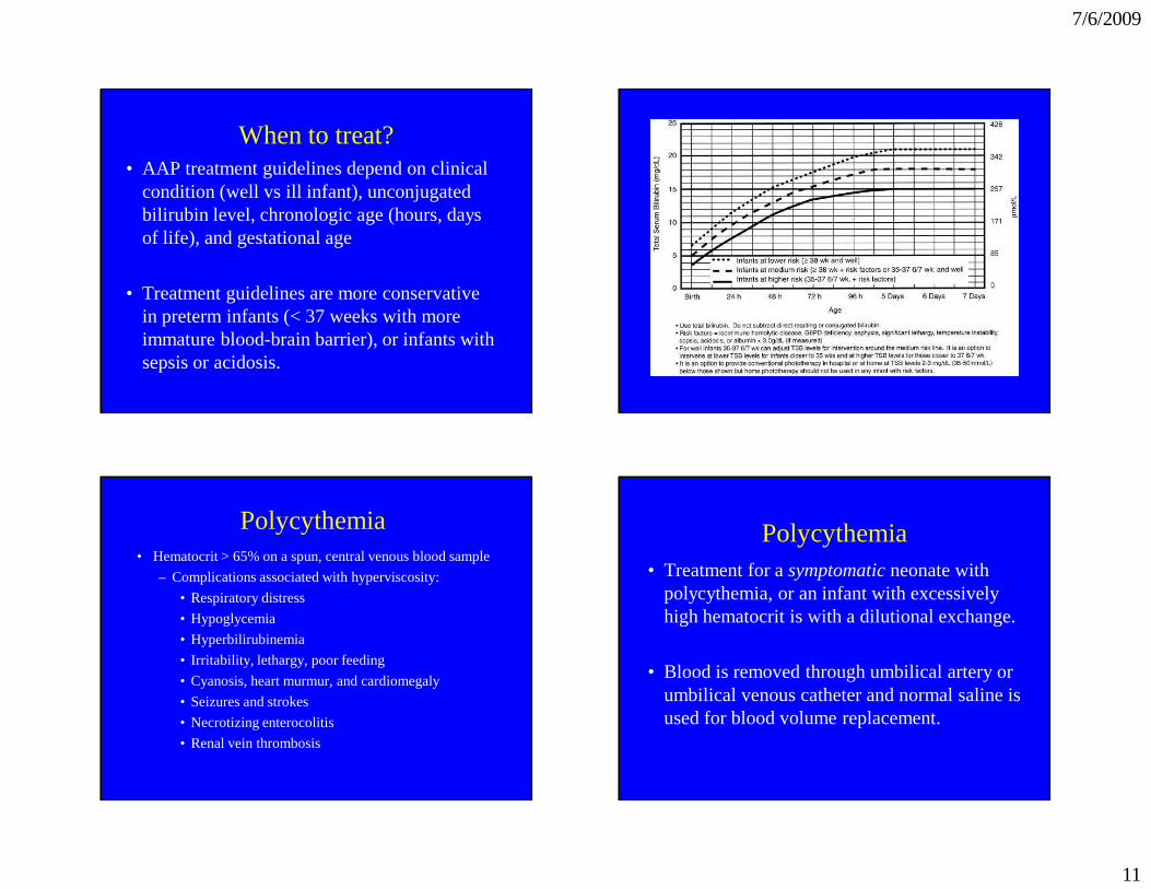
When to treat?• AAP treatment guidelines depend on clinical
condition (well vs ill infant), unconjugated bilirubin level, chronologic age (hours, days of life), and gestational age
• Treatment guidelines are more conservative in preterm infants (< 37 weeks with more immature blood-brain barrier), or infants with sepsis or acidosis.
Polycythemia• Hematocrit > 65% on a spun, central venous blood sample
– Complications associated with hyperviscosity:
• Respiratory distress
• Hypoglycemia
• Hyperbilirubinemia
• Irritability, lethargy, poor feeding
• Cyanosis, heart murmur, and cardiomegaly
• Seizures and strokes
• Necrotizing enterocolitis
• Renal vein thrombosis
Polycythemia• Treatment for a symptomatic neonate with
polycythemia, or an infant with excessively high hematocrit is with a dilutional exchange.
• Blood is removed through umbilical artery or umbilical venous catheter and normal saline is used for blood volume replacement.
7/6/2009
12
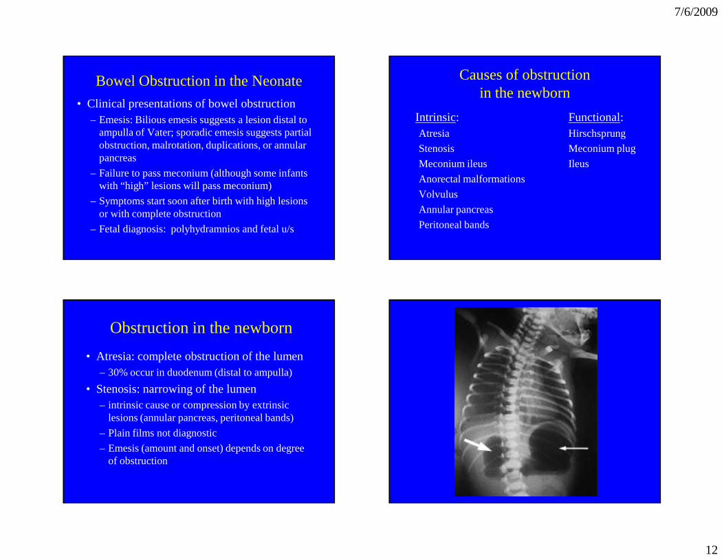
Bowel Obstruction in the Neonate
• Clinical presentations of bowel obstruction– Emesis: Bilious emesis suggests a lesion distal to
ampulla of Vater; sporadic emesis suggests partial obstruction, malrotation, duplications, or annular pancreas
– Failure to pass meconium (although some infants with “high” lesions will pass meconium)
– Symptoms start soon after birth with high lesions or with complete obstruction
– Fetal diagnosis: polyhydramnios and fetal u/s
Causes of obstruction in the newborn
Intrinsic: Functional: Atresia Hirschsprung
Stenosis Meconium plug
Meconium ileus Ileus
Anorectal malformations
Volvulus
Annular pancreas
Peritoneal bands
Obstruction in the newborn
• Atresia: complete obstruction of the lumen– 30% occur in duodenum (distal to ampulla)
• Stenosis: narrowing of the lumen– intrinsic cause or compression by extrinsic
lesions (annular pancreas, peritoneal bands)
– Plain films not diagnostic
– Emesis (amount and onset) depends on degree of obstruction
7/6/2009
13
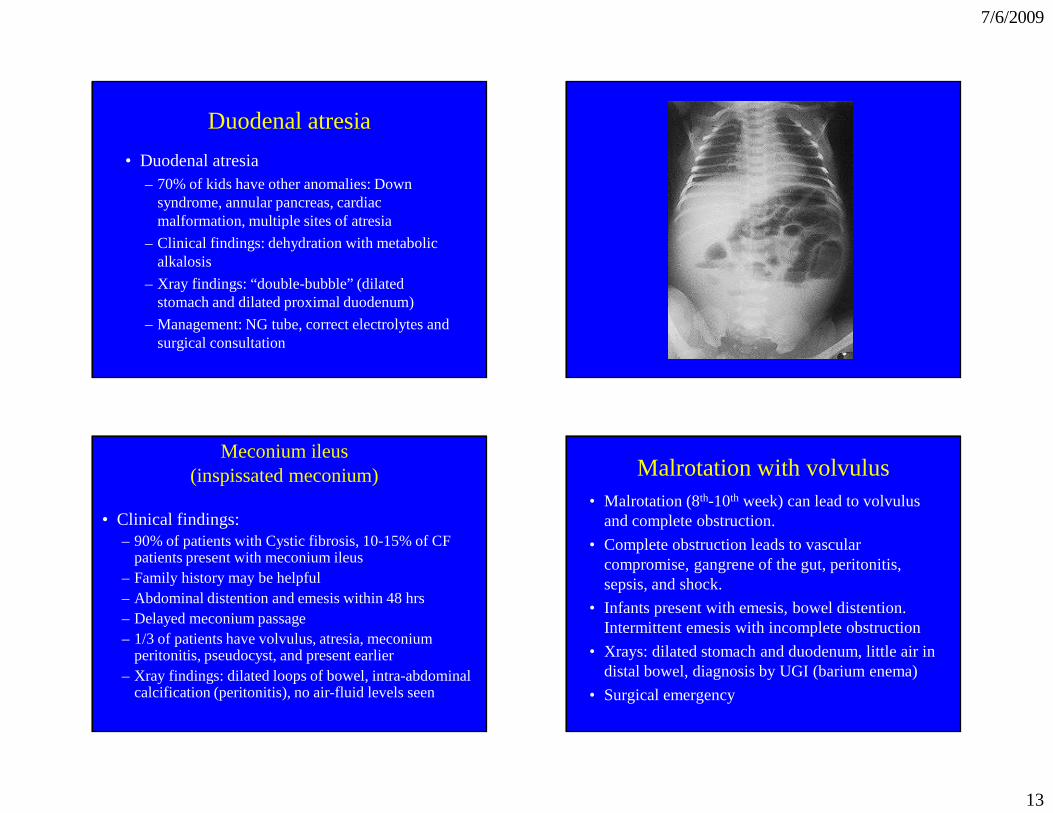
Duodenal atresia
• Duodenal atresia– 70% of kids have other anomalies: Down
syndrome, annular pancreas, cardiac malformation, multiple sites of atresia
– Clinical findings: dehydration with metabolic alkalosis
– Xray findings: “double-bubble” (dilated stomach and dilated proximal duodenum)
– Management: NG tube, correct electrolytes and surgical consultation
Meconium ileus(inspissated meconium)
• Clinical findings:– 90% of patients with Cystic fibrosis, 10-15% of CF
patients present with meconium ileus– Family history may be helpful– Abdominal distention and emesis within 48 hrs– Delayed meconium passage– 1/3 of patients have volvulus, atresia, meconium
peritonitis, pseudocyst, and present earlier– Xray findings: dilated loops of bowel, intra-abdominal
calcification (peritonitis), no air-fluid levels seen
Malrotation with volvulus• Malrotation (8th-10th week) can lead to volvulus
and complete obstruction.
• Complete obstruction leads to vascular compromise, gangrene of the gut, peritonitis, sepsis, and shock.
• Infants present with emesis, bowel distention. Intermittent emesis with incomplete obstruction
• Xrays: dilated stomach and duodenum, little air in distal bowel, diagnosis by UGI (barium enema)
• Surgical emergency
7/6/2009
14
Hirschprung Disease• Lower bowel obstruction: agenesis of ganglion cells
(Auerbach and Meissner plexuses)– Lesion originates in rectum and extends in varying degree;
most patients (80-90%) have lesions that do not extend beyond sigmoid colon
– Associated w/Downs (15%), Wardenburg syndrome– Delayed meconium passage (>24-48 hrs) in 90% of
patients– Clinical findings: Abdominal distention, emesis,
obstipation– Barium enema: see narrowing segment, “corkscrew”
appearance of colon, delayed clearing of barium – Diagnosis: rectal suction biopsy
Meconium plug syndrome
• Etiology: colonic “dysmotility” ?
• Hirschsprung disease found in 50% of these patients
• Clinical findings:– Delayed meconium passage: (24-48 hrs)
– Abdominal distention, emesis
– Barium enema diagnostic and therapeutic
Birth Injuries
• Cephalhematoma
• Caput succedaneum
• Subgaleal hematoma
• Erb’s palsy
• Klumpke’s palsy
• Clavicular fracture
• Phrenic nerve injury with diaphragmatic paralysis
Injuries to the headCaput: seen as vaguely demarcated edema on the
presenting part of scalp. Bleeding is above the periosteum, and can extend across sutures
Cephalohematoma: caused by rupture of vessels that traverse from the skull to the periosteum. Bleeding is limited by periosteal attachments, thus swelling does not cross sutures.
Both can be associated with skull fracture and hyperbilirubinemia
7/6/2009
15
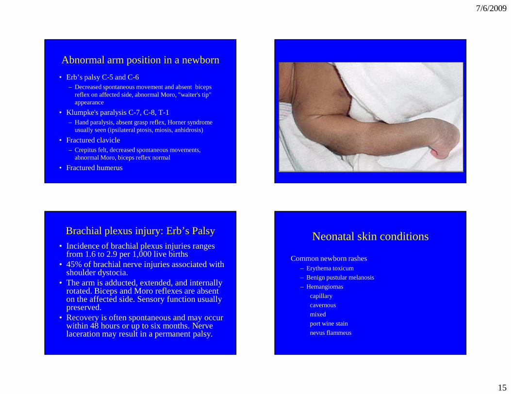
Abnormal arm position in a newborn
• Erb’s palsy C-5 and C-6– Decreased spontaneous movement and absent biceps
reflex on affected side, abnormal Moro, "waiter's tip" appearance
• Klumpke's paralysis C-7, C-8, T-1– Hand paralysis, absent grasp reflex, Horner syndrome
usually seen (ipsilateral ptosis, miosis, anhidrosis)
• Fractured clavicle – Crepitus felt, decreased spontaneous movements,
abnormal Moro, biceps reflex normal
• Fractured humerus
Brachial plexus injury: Erb’s Palsy• Incidence of brachial plexus injuries ranges
from 1.6 to 2.9 per 1,000 live births• 45% of brachial nerve injuries associated with
shoulder dystocia.• The arm is adducted, extended, and internally
rotated. Biceps and Moro reflexes are absent on the affected side. Sensory function usually preserved.
• Recovery is often spontaneous and may occur within 48 hours or up to six months. Nerve laceration may result in a permanent palsy.
Neonatal skin conditions
Common newborn rashes– Erythema toxicum
– Benign pustular melanosis
– Hemangiomas
capillary
cavernous
mixed
port wine stain
nevus flammeus
7/6/2009
16
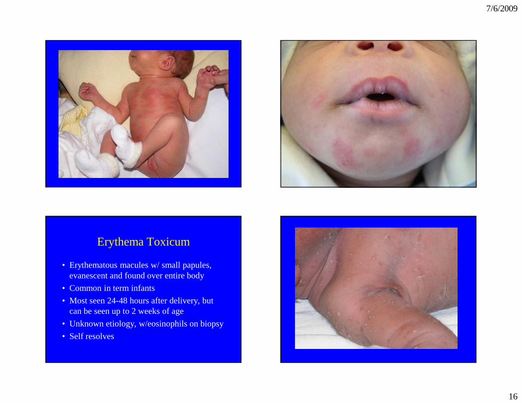
Erythema Toxicum
• Erythematous macules w/ small papules, evanescent and found over entire body
• Common in term infants
• Most seen 24-48 hours after delivery, but can be seen up to 2 weeks of age
• Unknown etiology, w/eosinophils on biopsy
• Self resolves
7/6/2009
17
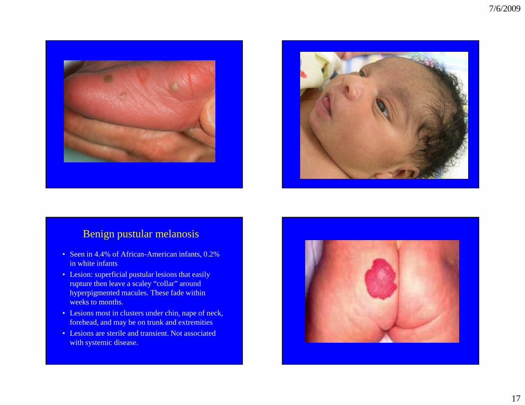
Benign pustular melanosis
• Seen in 4.4% of African-American infants, 0.2% in white infants
• Lesion: superficial pustular lesions that easily rupture then leave a scaley “collar” around hyperpigmented macules. These fade within weeks to months.
• Lesions most in clusters under chin, nape of neck, forehead, and may be on trunk and extremities
• Lesions are sterile and transient. Not associated with systemic disease.

7/6/2009
18
Hemangiomas• Strawberry hemangioma:
– 2.6% of infants (higher incidence in preterm infants)– May be seen at birth, but typically develop during first
few weeks of life and 90% seen by 1 mo of life– Start as small, discrete, well demarcated lesions. These
grow rapidly during infancy, and eventually involute. – Infants with large lesions, lesions on the face, eyelids,
airway, mouth, or cavernous lesions should be referred. • Flame nevus
– Very common, up to 40% of infants– “Salmon patch” on nape of neck, on eyelids, between
eyebrows– Do not grow during infancy and do not completely
disappear. Lesions fade and are less noticeable except during crying or exertion
Related Documents