RESEARCH Open Access In-depth proteomic analysis of a mollusc shell: acid-soluble and acid-insoluble matrix of the limpet Lottia gigantea Karlheinz Mann 1* , Eric Edsinger-Gonzales 2 and Matthias Mann 1 Abstract Background: Invertebrate biominerals are characterized by their extraordinary functionality and physical properties, such as strength, stiffness and toughness that by far exceed those of the pure mineral component of such composites. This is attributed to the organic matrix, secreted by specialized cells, which pervades and envelops the mineral crystals. Despite the obvious importance of the protein fraction of the organic matrix, only few in-depth proteomic studies have been performed due to the lack of comprehensive protein sequence databases. The recent public release of the gastropod Lottia gigantea genome sequence and the associated protein sequence database provides for the first time the opportunity to do a state-of-the-art proteomic in-depth analysis of the organic matrix of a mollusc shell. Results: Using three different sodium hypochlorite washing protocols before shell demineralization, a total of 569 proteins were identified in Lottia gigantea shell matrix. Of these, 311 were assembled in a consensus proteome comprising identifications contained in all proteomes irrespective of shell cleaning procedure. Some of these proteins were similar in amino acid sequence, amino acid composition, or domain structure to proteins identified previously in different bivalve or gastropod shells, such as BMSP, dermatopontin, nacrein, perlustrin, perlucin, or Pif. In addition there were dozens of previously uncharacterized proteins, many containing repeated short linear motifs or homorepeats. Such proteins may play a role in shell matrix construction or control of mineralization processes. Conclusions: The organic matrix of Lottia gigantea shells is a complex mixture of proteins comprising possible homologs of some previously characterized mollusc shell proteins, but also many novel proteins with a possible function in biomineralization as framework building blocks or as regulatory components. We hope that this data set, the most comprehensive available at present, will provide a platform for the further exploration of biomineralization processes in molluscs. Background Molluscan shells are extraordinarily stable biocomposites of calcium carbonate and an organic matrix consisting of polysaccharides and proteins. The organic matrix, although constituting a very minor fraction of the biocomposite by weight, is thought to be of utmost importance for the construction of the biocomposite and its final properties because it controls crystal nucleation, crystal growth, crystal shape and choice of calcium carbonate polymorph [1,2]. Previously established methods to identify new mollusc shell matrix proteins, such as isola- tion by chromatography and biochemical characterization or molecular biology approaches, have been comple- mented recently by mass spectrometry-based proteomic analysis or combination of proteomic and transcriptomic studies [3-11]. However, proteomic approaches depend on the comparison of experimentally determined spectra with theoretical spectra obtained by in silico digestion of proteins and in silico fragmentation of resulting peptides [12,13]. Therefore protein sequence databases that are as compre- hensive as possible, usually derived from genome sequen- cing, are presently indispensable for high-throughput proteomics. The need for a comprehensive database is highlighted by previously published proteomic studies of * Correspondence: [email protected] 1 Abteilung Proteomics und Signaltransduktion, Max-Planck-Institut für Biochemie, Am Klopferspitz 18, D-82152 Martinsried, Germany Full list of author information is available at the end of the article © 2012 Mann et al; licensee BioMed Central Ltd. This is an Open Access article distributed under the terms of the Creative Commons Attribution License (http://creativecommons.org/licenses/by/2.0), which permits unrestricted use, distribution, and reproduction in any medium, provided the original work is properly cited. Mann et al. Proteome Science 2012, 10:28 http://www.proteomesci.com/content/10/1/28

Welcome message from author
This document is posted to help you gain knowledge. Please leave a comment to let me know what you think about it! Share it to your friends and learn new things together.
Transcript

Mann et al. Proteome Science 2012, 10:28http://www.proteomesci.com/content/10/1/28
RESEARCH Open Access
In-depth proteomic analysis of a mollusc shell:acid-soluble and acid-insoluble matrix of thelimpet Lottia giganteaKarlheinz Mann1*, Eric Edsinger-Gonzales2 and Matthias Mann1
Abstract
Background: Invertebrate biominerals are characterized by their extraordinary functionality and physical properties,such as strength, stiffness and toughness that by far exceed those of the pure mineral component of suchcomposites. This is attributed to the organic matrix, secreted by specialized cells, which pervades and envelops themineral crystals. Despite the obvious importance of the protein fraction of the organic matrix, only few in-depthproteomic studies have been performed due to the lack of comprehensive protein sequence databases. The recentpublic release of the gastropod Lottia gigantea genome sequence and the associated protein sequence databaseprovides for the first time the opportunity to do a state-of-the-art proteomic in-depth analysis of the organic matrixof a mollusc shell.
Results: Using three different sodium hypochlorite washing protocols before shell demineralization, a total of 569proteins were identified in Lottia gigantea shell matrix. Of these, 311 were assembled in a consensus proteomecomprising identifications contained in all proteomes irrespective of shell cleaning procedure. Some of theseproteins were similar in amino acid sequence, amino acid composition, or domain structure to proteins identifiedpreviously in different bivalve or gastropod shells, such as BMSP, dermatopontin, nacrein, perlustrin, perlucin, or Pif.In addition there were dozens of previously uncharacterized proteins, many containing repeated short linear motifsor homorepeats. Such proteins may play a role in shell matrix construction or control of mineralization processes.
Conclusions: The organic matrix of Lottia gigantea shells is a complex mixture of proteins comprising possiblehomologs of some previously characterized mollusc shell proteins, but also many novel proteins with a possiblefunction in biomineralization as framework building blocks or as regulatory components. We hope that this data set,the most comprehensive available at present, will provide a platform for the further exploration of biomineralizationprocesses in molluscs.
BackgroundMolluscan shells are extraordinarily stable biocompositesof calcium carbonate and an organic matrix consisting ofpolysaccharides and proteins. The organic matrix,although constituting a very minor fraction of thebiocomposite by weight, is thought to be of utmostimportance for the construction of the biocomposite andits final properties because it controls crystal nucleation,crystal growth, crystal shape and choice of calciumcarbonate polymorph [1,2]. Previously established methods
* Correspondence: [email protected] Proteomics und Signaltransduktion, Max-Planck-Institut fürBiochemie, Am Klopferspitz 18, D-82152 Martinsried, GermanyFull list of author information is available at the end of the article
© 2012 Mann et al; licensee BioMed Central LCommons Attribution License (http://creativecoreproduction in any medium, provided the orig
to identify new mollusc shell matrix proteins, such as isola-tion by chromatography and biochemical characterizationor molecular biology approaches, have been comple-mented recently by mass spectrometry-based proteomicanalysis or combination of proteomic and transcriptomicstudies [3-11]. However, proteomic approaches depend onthe comparison of experimentally determined spectra withtheoretical spectra obtained by in silico digestion of proteinsand in silico fragmentation of resulting peptides [12,13].Therefore protein sequence databases that are as compre-hensive as possible, usually derived from genome sequen-cing, are presently indispensable for high-throughputproteomics. The need for a comprehensive database ishighlighted by previously published proteomic studies of
td. This is an Open Access article distributed under the terms of the Creativemmons.org/licenses/by/2.0), which permits unrestricted use, distribution, andinal work is properly cited.

Mann et al. Proteome Science 2012, 10:28 Page 2 of 18http://www.proteomesci.com/content/10/1/28
shell matrices in various molluscan species [3-11]. Thesestudies relied on translated EST databases contributed by anumber of groups [7,11,14-18] and usually less than 15proteins were identified from isolated organic matrices.Sometimes database searches were combined with de novomass spectrometric sequencing. However, de novo sequen-cing algorithms, which attempt to interpret spectra inde-pendently of a sequence database [19], are not compatiblewith high-throughput analysis at present. Transcriptomics,on the other hand, does not identify matrix proteinsdirectly, making additional techniques, such as immuno-histochemical localization, necessary to demonstrate theactual location of potential shell matrix proteins. Thus,although previous studies have identified several veryinteresting new matrix proteins, these studies may fail toshow the actual complexity of the shell matrix proteomeindicated by proteomic studies of biomineral matrices oforganisms with sequenced genomes, such as chicken [20]or the sea urchin Strongylocentrotus purpuratus [21-23].The first genome sequence of a mollusc, the limpet Lottia
gigantea, was made public recently (http://genome.jgi-psf.org/Lotgi1/Lotgi1.download.html) [24]. In the presentreport we used a protein sequence database derived fromthis genome sequence to perform a high-throughput in-depth proteomic analysis of the shell matrix of this marinesnail.The shell of Lottia and related limpets consists of five
layers [25,26], which are divided into 3 outer layers,M+ 1, M+ 2 and M+3 and separated from an innerlayer M-1 by the intermediate myostracum (M layer).The outermost layer, M+ 3, is reported to contain calciteas mineral phase. This layer appears eroded and oftendisappears altogether around the top of the shell. TheM+2 layer consists of flat prismatic crystals made ofaragonite, another common calcium carbonate mineral.The M+ 1 and M-1 layers are described to consist of la-mellar prisms similarly made of aragonite. Compared tothe other layers, the M layer, sandwiched between M+1and M-1, is very thin and has a prismatic structure ofaragonite. Organic matrix was visible in M+ 3 and M+2,but was not detected in other layers [25].Using LTQ Orbitrap Velos high-performance mass
spectrometers [27] in combination with the MaxQuant soft-ware package designed for analysis of large high-resolutionmass spectrometric data sets [28-30] we identified 311proteins in the organic matrix of the Lottia shell with veryhigh stringency. This is the first in-depth proteomic studyof a mollusc shell matrix.
Materials and methodsThe shells of freshly collected limpets were carefullycleaned manually and treated with sodium hypochloritesolution (Merck, Darmstadt; Germany; 6–14% activechlorine) to remove organic surface contaminants. Shells
were either treated with hypochlorite for 2 h at roomtemperature (A), for 2 h with two 5 min ultrasonic treat-ments at the start of each hour (B), or for 24 h with two5 min ultrasound bursts as before and one after 24 h (C).The shells were then washed with de-ionized water, dried,and crushed into small pieces using a hammer. The pieceswere demineralized in 50% acetic acid (20 ml/g of shell) ina cold room overnight, yielding a dark brown suspension.Acid-soluble and acid-insoluble matrix was separated bycentrifugation at 14000gav at 5°C for 1 h. The pellet waswashed twice by re-suspension in approximately 20volumes of 50% acetic acid, centrifugation for 30 min at14000gav, and lyophilized. The supernatant was dialyzedtwice against 10 volumes of 10% acetic acid followed bythree times 10 volumes of 5% acetic acid at 4–6°C(Spectra/Por 6 dialysis membrane, molecular weight cut-off 2000; Spectrum Europe, Breda, The Netherlands), andlyophilized.SDS-PAGE was done using pre-cast 4–12% Novex Bis-
Tris gels in MES buffer with reagents and protocolssupplied by the manufacturer (Invitrogen, Carlsbad, CA).Samples were suspended in 30 μl sample buffer/200 μgof organic matrix and heated to 95°C for 5 min. Samplebuffer-insoluble matrix was removed by centrifugation inan Eppendorf bench top centrifuge for 5 min at13000 rpm. Gels were loaded with 30 μl of matrixsample supernatant per lane and stained with colloidalCoomassie (Invitrogen) after electrophoresis. The proteinstandard used for molecular weight estimation wasNovex Sharp, pre-stained (Invitrogen). Gels were slicedinto 12 sections for in-gel digestion with trypsin [31]. Theeluted peptides were purified on C18 Stage Tips [32].Peptide mixtures were analyzed by on-line nanoflow
liquid chromatography using the EASY-nLC system(Proxeon Biosystems, Odense, Denmark; now ThermoFisher) with 15 cm capillary columns of an internaldiameter of 75 μm filled with 3 μm Reprosil-Pur C18-AQ resin (Dr. Maisch GmbH, Ammerbuch-Entringen,Germany). The gradient consisted of 5–30% acetonitrilein 0.5% acetic acid at a flow rate of 250 nl/min for85 min, 30–60% acetonitrile in 0.5% acetic acid at a flowrate of 250 nl/min and 60–80% acetonitrile in 0.5% aceticacid at a flow rate of 250 nl/min for 7 min. The eluatewas electrosprayed into an LTQ Orbitrap Velos (ThermoFisher Scientific, Bremen, Germany) through a Proxeonnanoelectrospray ion source. The Orbitrap Velos wasoperated in a HCD top 10 mode essentially as described[Olsen et al., 2009] at a resolution of 30,000 for full scansand of 7,500 (both at m/z 400) for MS/MS scans.Data analysis was performed with MaxQuant (v1.1.1.36)
[28,29], a computational proteomics platform based on theAndromeda search engine [30] (http://www.maxquant.org/), using the Lotgi1_GeneModels_Filtered Models1_aa.fasta.gz protein sequence database comprising 23,851 gene
Mann et al. Proteome Science 2012, 10:28 Page 3 of 18http://www.proteomesci.com/content/10/1/28
models at present (http://genome.jgi-psf.org/Lotgi1/Lotgi1.download.html) [24], together with the correspondingreversed database and the sequences of common contami-nants, including human keratins from IPIhuman. Carba-midomethylation was set as fixed modification. Variablemodifications were set as oxidation (M), N-acetyl (protein)and pyro-Glu/Gln (N-term). Initial peptide mass tolerancewas set to 7 ppm and fragment mass tolerance was20 ppm. Two missed cleavages were allowed and theminimal length required for peptide identification wasseven amino acids. The peptide and protein false discoveryrates (FDR) were both set to 0.01. The maximal posteriorerror probability (PEP) for peptides, which is the probabil-ity of each peptide to be a false hit considering identifica-tion score and peptide length [28,29], was set to 0.01. TheRe-quantify and Second Peptide [30] options were enabled.At least two MaxQuant group sequence-unique peptideswith a score >100 were required for protein identification.Furthermore, identifications were only accepted if thepeptides were identified in at least two replicates withinthe respective group A, B or C. Identifications with onlytwo unique peptides were manually validated consideringthe assignment of major peaks, occurrence of uninter-rupted y- or b-ion series of at least 4 consecutive aminoacids, preferred cleavages N-terminal to proline bonds, thepossible presence of a2/b2 ion pairs and immoniumions, and mass accuracy. The ProteinProspector MS-Product program (http://prospector.ucsf.edu/) was used tocalculate the theoretical masses of fragments of identifiedpeptides for manual validation. BLAST and FASTAsearches against non-redundant databases (all organisms)were performed using the programs provided by NCBI(http://www.ncbi.nlm.nih.gov/blast) and EBI http://www.ebi.ac.uk/Tools/sss/. Domains were predicted withInterProScan (http://www.ebi.ac.uk/Tools/pfa/iprscan/)and PROSITE (http://prosite.expasy.org/). For sequencealignments we employed Kalign (http://www.ebi.ac.uk/Tools/msa/kalign/) and ClustalW (http://www.ebi.ac.uk/Tools/msa/clustalw2/). Sequence repeats werepredicted using RADAR (http://www.ebi.ac.uk/Tools/Radar/index.html). The abundance of proteins wasestimated by calculating the exponentially modifiedprotein abundance index (emPAI) [33]. Observablepeptides were determined and counted with ProteinProspector (http://prospector.ucsf.edu/prospector/cgi-bin/msform.cgi? form=msdigest) using zero miss-cleavages, apeptide mass of 700–2800, and a minimal peptide lengthof seven amino acids. Observed unique parent ions with aminimal length of seven amino acids and a mass between700–2800 used for emPAI calculation included ions withup to two miss-cleavages, modifications specified forMaxQuant analysis (see above), different charges, andneutral losses [33]. Proteins with emPAI ≥9 were referredto as major proteins in this report.
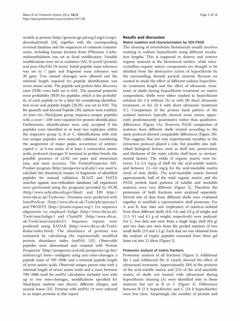
Results and discussionMatrix isolation and characterization by SDS-PAGEThe cleaning of invertebrate biominerals usually involveswashing in sodium hypochlorite using different incuba-tion lengths. This is supposed to destroy and removeorganic material at the biomineral surface, while intra-crystalline organic matrix components are thought to beshielded from the destructive action of hypochlorite bythe surrounding, densely packed, mineral. Because wewanted to study the effect of different sodium hypochlor-ite treatment length and the effect of ultrasonic treat-ment of shells during hypochlorite treatment on matrixcomposition, shells were either washed in hypochloritesolution for 2 h without (A) or with (B) short ultrasonictreatment, or for 24 h with short ultrasonic treatment(C). Comparison of the protein band pattern of theisolated matrices typically showed some minor, appar-ently predominantly quantitative rather than qualitative,differences (Figure 1A). However, PAGE comparison ofmatrices from different shells treated according to thesame protocol showed comparable differences (Figure 1B).This suggests that not only experimental variables in theextraction protocol played a role, but possibly also indi-vidual biological factors, such as shell size, preservationand thickness of the outer calcitic shell layer, or environ-mental factors. The yields of organic matrix were be-tween 2.2–5.3 mg/g of shell for the acid-soluble matrix,and between 2.1–4.6 mg/g for the acid-insoluble matrix(total of nine shells). The acid-insoluble matrix formedapproximately half of the total organic matrix and thePAGE protein band patterns of soluble and insolublematrices were very different (Figure 2). Therefore theproteomes of both fractions were analyzed separately.Several sets of data from different shells were evaluatedtogether to establish a representative shell proteome. ForA and B, four data sets (replicates) of matrices isolatedfrom three different shells (8.8, 5.6, and 3.8 g of weight and11.5, 9.1 and 4.1 g of weight, respectively) were analyzed.For C, two data sets were from a single large shell (8.6 g)and two data sets were from the pooled matrices of twosmall shells (2.9 and 1.5 g). Each data set was obtained fromthe analysis of tryptic peptides extracted from three gellanes cut into 12 slices (Figure 2).
Proteomic analysis of matrix fractionsProteomic analysis of all fractions (Figure 3; Additionalfile 1 and Additional file 2) clearly showed the effect ofultrasound treatment. Approximately 28% of the proteinsof the acid-soluble matrix and 21% of the acid-insolublematrix of shells not treated with ultrasound duringhypochlorite cleaning (A) were identified only in thesematrices but not in B or C (Figure 3). Differencesbetween B (2 h hypochlorite) and C (24 h hypochlorite)were less clear. Surprisingly the number of protein and

Figure 1 PAGE comparison of acid-soluble matrices from shells. Molecular weight markers are indicated at the left. Each lane was loadedwith 200 μg of matrix in a volume of 30 μl. A, matrices of shells cleaned with different sodium hypochlorite protocols. Lane A, 2 h hypochloriteat room temperature; lane B, 2 h hypochlorite with 2 x 5 min ultrasound treatment at the start of each hour; lane C, cleaned with hypochloritefor 24 h with 2 x 5 min ultrasound bursts as before and one after 24 h. B, matrices of different shells, all cleaned with hypochlorite according toprotocol B (2 h hypochlorite, 2 x 5 min ultrasound).
Mann et al. Proteome Science 2012, 10:28 Page 4 of 18http://www.proteomesci.com/content/10/1/28
peptide identifications in the soluble fraction of C wasgreater than that of B (Additional file 1). Most of theproteins distinct between the two preparations were notunique but also occurred in A. This was difficult toexplain, because all four replicates showed the sameeffect although they were prepared and analyzed atdifferent times, sometimes on different mass spectro-meters and often in sequence with replicates fromother preparations. However, the qualitative differencesbetween B and C were minor and focused almost exclu-sively on low abundance proteins. This may indicate thatultrasound treatment during cleaning with hypochloritemay have helped to solubilize and destroy proteins thatstuck tenaciously to the biomineral surface. The lengthof hypochlorite treatment, however, apparently didnot play a dominant role, at least after two hours oftreatment. This aspect of hypochlorite treatment maybecome more important with nacreous shell layers, asour experience with Haliotis laevigata has shown thatlengthy treatments start to degrade the matrix surroundingnacre plates, leading to a partial loss of the outermostnacre layers.Altogether 569 proteins were identified in matrices
obtained after different hypochlorite treatments. To obtaina representative, high-confidence, shell matrix proteome ofLottia gigantea, we assembled a consensus proteome
comprising all database entries identified in all three typesof samples (Figure 3). The consensus proteome of theacid-soluble fraction included 204 proteins and theconsensus proteome of the acid-insoluble fractioncontained 242 proteins. Given an overlap of 135, thissummed up to a total of 311 Lottia database entriescontaining shell matrix protein sequences. However, thesenumbers should not be regarded as final because somedatabase entries may eventually turn out to contain thesequence of more than one protein and some proteinsequences may be divided among several database entries.Furthermore, the identifications not comprised in theconsensus proteome are by no means to be considered asfalse positives but may be true shell matrix components. Inmost cases these were minor proteins and their absence orpresence in different fractions may be due to experimentalvariability or the still limited dynamic range of massspectrometers. Additional files 3, 4, 5, 6, 7, 8, 9, 10, 11, 12,13, and 14 contain protein and peptide details, such asaccession numbers of proteins sharing group-uniquepeptides, scores, masses, peptide sequences, and distribu-tion in gel slices (Additional files 3, 4, 5, 6, 7, 8, 9, 10, 11,12, 13, and 14). Unlike Additional file 1 and Additional file2 (Additional file 1 and Additional file 2), Additional files3, 4, 5, 6, 7, 8, 9, 10, 11, 12, 13, 14 contain data of allpeptides and proteins identified within the set thresholds
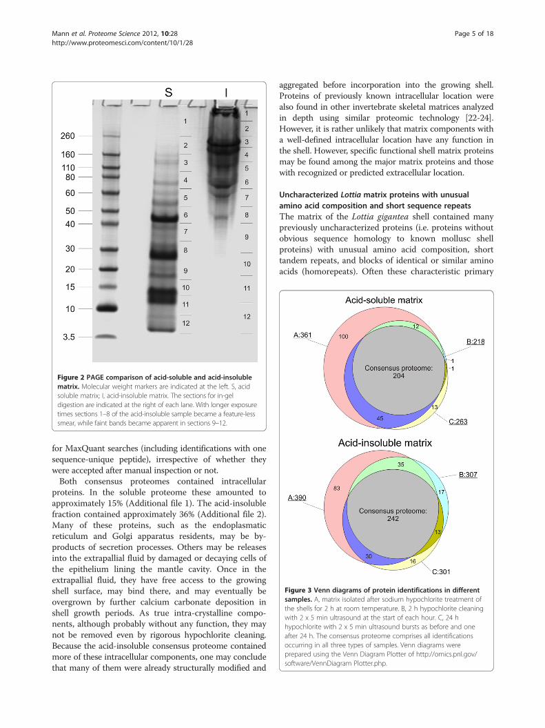
Figure 2 PAGE comparison of acid-soluble and acid-insolublematrix. Molecular weight markers are indicated at the left. S, acidsoluble matrix; I, acid-insoluble matrix. The sections for in-geldigestion are indicated at the right of each lane. With longer exposuretimes sections 1–8 of the acid-insoluble sample became a feature-lesssmear, while faint bands became apparent in sections 9–12.
Figure 3 Venn diagrams of protein identifications in differentsamples. A, matrix isolated after sodium hypochlorite treatment ofthe shells for 2 h at room temperature. B, 2 h hypochlorite cleaningwith 2 x 5 min ultrasound at the start of each hour. C, 24 hhypochlorite with 2 x 5 min ultrasound bursts as before and oneafter 24 h. The consensus proteome comprises all identificationsoccurring in all three types of samples. Venn diagrams wereprepared using the Venn Diagram Plotter of http://omics.pnl.gov/software/VennDiagram Plotter.php.
Mann et al. Proteome Science 2012, 10:28 Page 5 of 18http://www.proteomesci.com/content/10/1/28
for MaxQuant searches (including identifications with onesequence-unique peptide), irrespective of whether theywere accepted after manual inspection or not.Both consensus proteomes contained intracellular
proteins. In the soluble proteome these amounted toapproximately 15% (Additional file 1). The acid-insolublefraction contained approximately 36% (Additional file 2).Many of these proteins, such as the endoplasmaticreticulum and Golgi apparatus residents, may be by-products of secretion processes. Others may be releasesinto the extrapallial fluid by damaged or decaying cells ofthe epithelium lining the mantle cavity. Once in theextrapallial fluid, they have free access to the growingshell surface, may bind there, and may eventually beovergrown by further calcium carbonate deposition inshell growth periods. As true intra-crystalline compo-nents, although probably without any function, they maynot be removed even by rigorous hypochlorite cleaning.Because the acid-insoluble consensus proteome containedmore of these intracellular components, one may concludethat many of them were already structurally modified and
aggregated before incorporation into the growing shell.Proteins of previously known intracellular location werealso found in other invertebrate skeletal matrices analyzedin depth using similar proteomic technology [22-24].However, it is rather unlikely that matrix components witha well-defined intracellular location have any function inthe shell. However, specific functional shell matrix proteinsmay be found among the major matrix proteins and thosewith recognized or predicted extracellular location.
Uncharacterized Lottia matrix proteins with unusualamino acid composition and short sequence repeatsThe matrix of the Lottia gigantea shell contained manypreviously uncharacterized proteins (i.e. proteins withoutobvious sequence homology to known mollusc shellproteins) with unusual amino acid composition, shorttandem repeats, and blocks of identical or similar aminoacids (homorepeats). Often these characteristic primary

Mann et al. Proteome Science 2012, 10:28 Page 6 of 18http://www.proteomesci.com/content/10/1/28
sequence features are found in terminal regions of shellproteins that have been proposed to be structurallyunstable, unfolded domains able to adopt a specificstructure only upon binding to a ligand, such as a crystalsurface [34]. This proposition was based on experimentswith synthetic polypeptides confirming the intrinsicallydisordered conformation of such shell protein domains andthe in vitro interaction with calcium carbonate [35-39].However, most known features of such short linear motifsand homorepeats come from intracellular examples [40,41].Apart from occurring predominantly in natively disorderedstructures, such motifs mediate protein-protein interactionswith low affinity, which is usually compensated by frequentrepetition of the motif. Examples of major (average emPAI≥9) Lottia matrix proteins with peculiar primary sequencefeatures are shown in Table 1. Many of these proteins eitherdo not contain cysteines, which usually are disulfide-bondedin extracellular proteins and stabilize structured domains(except in the predicted signal peptide), or have cysteine-containing domains apart from the presumed intrinsicallydisordered sequence motifs. However, there are exceptions.Thus, in Lotgi1|173200, one of the most abundant proteinsof the acid-soluble matrix (Additional file 1), 30% of thesequence consists of Asn, Pro and Ser, but the sequencealso contains 20 Cys, indicating a well-ordered structurestabilized by disulfide bonds. Database searches indicatedsome similarity to the Pinctada fucata shell mpn88 proteinB7X6R9_PINFU (unpublished; submitted to EMBL byNogawa et al., 2007). The proteins showed 27% sequenceidentity, but none of the 20 cysteines of Lotgi1|173200 waspreserved in mpn88, which contains no cysteine at all inthe predicted mature sequence. Therefore we prefer toaccredit the similarity in database searches to regionsof similar amino acid composition, but not to sequencehomology. The same may be true for Lotgi1|231186(Table 1).Selected sequences and spectra of this group are
shown in Figures 4, 5, 6. Several of these proteins sharedtheir sequence features with recently discovered shellproteins. Thus, the very acidic protein in Lotgi1|233420,which is one of the most abundant proteins in Lottiashell matrix (Additional file 1 and Additional file 2),shows 36% sequence identity to aspein [42], but this isbased almost exclusively on alignment of aspartic acids.Extended Asp-rich sequences also occur in other shellproteins, such as MSP-1 [43] and asprich [44]. A verysimilar acidic domain was also contained in the C-terminal third of Lotgi1|239188, while the N-terminaldomain was similar to nacrein (Table 2). Glycine-richproteins may be relatives of shematrins [45]. However, inthe absence of significant sequence similarity in non-repetitive sequence regions a possible homology isdifficult to prove. The Lottia gigantea shell matrix alsocontained several proteins with sequence similarity
to previously identified mollusc shell proteins (Table 2)discussed below.
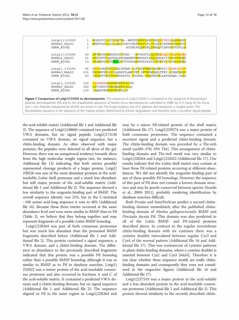
Proteins with possible homologs in other shellsDermatopontin, ependymin-like and gigasin-2-like proteinsThe first mollusc shell dermatopontin was isolated from thefreshwater snail Biomphalaria glabrata shell matrix [49].Since then several molluscan dermatopontin-encodinggenes have been identified and some of them were tran-scribed in mantle cells, implying the shell matrix as finaldestination [17,54,55]. A protein very similar to derma-topontin, Lotgi1|133595 (Figure 7), was identified atmoderate abundance in the acid-insoluble matrixconsensus proteome and in the soluble fraction of A andC (Additional file 1 and Additional file 2). The function ofthis protein remains unknown at present [55].A protein similar to the ependymin-related proteins
recently discovered in Haliotis asinina shells [6] wasfound in Lotgi1|233583, a minor protein of the acid-insoluble consensus proteome (Additional file 1 andAdditional file 2). It was also similar to an unpublishedHaliotis discus protein submitted to databases by Kanget al. (2006) under the name X-box binding protein withthe accession number B6RB39 (Additional file 15: FigureSA). The function of ependymin and related proteins isunknown at present.Entry Lotgi1|235548 contained a protein sequence
partially (~aa170-540) similar to the recently discoveredCrassostrea gigas shell protein gigasin-2 (Cgigas-IMSP-2)[9] and the related proteins EGF-like domain containingprotein-1 and −2 from Pinctada maxima [Jackson et al.,2009] (Additional file 15: Figure SB). Lotgi1|235548was a minor protein in both, acid-soluble and acid-insol-uble, consensus proteomes (Additional file 1 and Additionalfile 2).
Nacrein-like proteinsOne of the most important enzymes in biomineralizationevents is carbonic anhydrase, which catalyzes the forma-tion of hydrogen carbonate from CO2 and water. Thefirst carbonic anhydrase isolated from a mollusc shelland characterized at the molecular level was nacrein[46]. This protein, which was isolated from the nacreouslayer of Pinctada fucata shells, contained two carbonicanhydrase domains separated by a Gly-X-Asn repeatdomain. The same protein was also identified in theprismatic layer [56]. Since then nacrein-like proteins ornacrein-encoding genes have been identified in severalother molluscs [4,7,10,57,58].The Lottia shell matrix contained three entries that
showed some similarity to nacreins (Table 2). Of theseLotgi1|238082 belonged to the most abundant proteinsin the shell matrix (Additional file 1 and Additional file2) and its sequence was 25% identical to that of Mytilus

Table 1 Previously uncharacterized major Lottia shell matrix proteins with unusual primary sequence features
Accession Feature
Lotgi1|115147 14% P, 11% T, 6 repeats of ~30aa, starting with MITPE; pI: 4.7; 319aa
Lotgi1|142790 25% Q, 10% E, 17% P, 12% V, 10% N 10% L; 6 short repeats: k/qQQPxVELNKQQP; pI 5.2; 182aa
Lotgi1|142814 38% Q, 11% L, 10% P; 5 ~70aa repeats containing shorter repeat motifs like NQQQ and KQQQ; pI: 10.5; 322aa
Lotgi1|152688 20% G, 12% P; pI: 9.7; 137aa
Lotgi1|158113 11% P; Q-rich C-term (aa210–240); pI: 9.7; 258aa
Lotgi1|159331 26% E, 13% L,12% T; pI: 4; starting with aa156 8x SNLLQQPDa/tTQqLa/tTNeQQQ; (Figure 6)
Lotgi1|163637 17% D, 16% A; EFh, pI: 3.8; 643aa; 12 ca30aa repeats similar to AxVDNxxMADMIDTxQDxxEDAADNMADNIDTAQDAQbetween aa32–453
Lotgi1|171084 13% S; frequent doublets (SS, QQ, TT, YY, NN); G/E block aa322–337; pI: 4.4; 357aa
Lotgi1|172698 23% Q, 13% N, 13% S; aa130–702: 31 x 14aa repeats similar to QSNQQFNxxQSNQQF; pI: 7.1; 1184aa
Lotgi1|173200 ~10% of P, N and G; in aa107–170 10x GAMP/GSMP; pI: 9.6; 563aa
Lotgi1|174003 19% P in aa50–400 and 35% P in aa778–882; pI: 9.5; 882aa
Lotgi1|227783 aa17–126: 17% R+ K, 12% P, 11% L; pI: 11; 126aa
Lotgi1|228385 16% R, 11% S; pI: 11.7; 160aa; R/H-rich from aa103–150
Lotgi1|231186 19% G, 12% P; aa433–481: 27% M; pI: 4.6; 481aa; R/H-rich C-term half
Lotgi1|231509 aa26–230: 18% P; pI: 4.2; 230aa; acidic blocks in N-term half
Lotgi1|233397 A/P-rich motif aa150–170; H-rich motif aa171–185; pI: 8.8; 219aa
Lotgi1|233420 31% D, 10% E; pI: 3.6; similar to aspein?
Lotgi1|234884 42% Q in aa281–630; G/L/A-rich region aa631–928; pI: 9.2; 928aa
Lotgi1|235497 aa120–247: 20% P, 16% A, 10% Q; pI: 9.7; 247aa
Lotgi1|235610 15% P, 15% T; pI:5.7; 557aa
Lotgi1|235621 aa171–270: 33% G, 25% T, 15% P, 14% Q; 16 x GGQPs/tT; pI: 5.4; 303aa
Lotgi1|235812 24% P, 18% Q, 10% N; pI: 8.9; 729aa; aa57–376: 17 repeats of 16aa, similar to NNxa/vQPPxxQxxYQPt/p
Lotgi1|236689 19% P, 10% A, 10% V, 10% R; pI: 10; 317aa
Lotgi1|236690 21% Q, 18% P; aa268–356: 4 xAQPGAYQQP(x)2–4 GAYxQQP repeats; pI: 8.4; 440aa
Lotgi1|236691 22% P, 13% Q, 10% A; Q-rich regions: ~aa61–160 and~ aa721–990; P-rich: ~aa280–600 and ~780–970¸pI:8.8; 1035aa
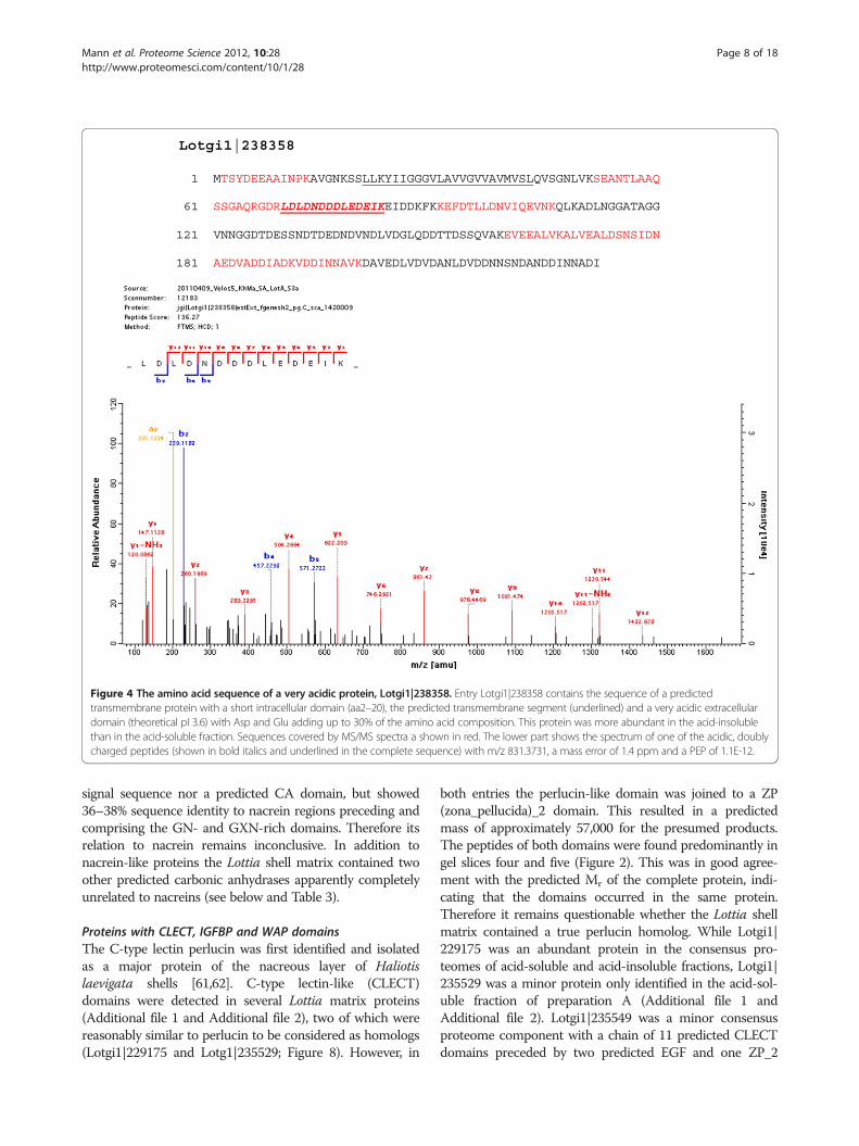
Lotgi1|238358 aa61–232: 32% D+ E, 12% N; pI: 3.7; 323aa; (Figure 4)
Lotgi1|238831 13% A, 11% R, 11% L; K/R/A-rich C-terminus (aa185–219); pI. 10.3; 219aa
Lotgi1|239170 16% G, 12% M, 10% Q; G blocks in N-term half; pI: 9.9; 145aa
Lotgi1|239174 20% G, 18%M, 12%A, 10% L; pI: 11.2; 186aa; some similarity to shematrins
Lotgi1|239339 13% T, 12% S, 10% P; blocks of T from aa185–240; pI: 9.7; 609aa
Lotgi1|239447 22% G, 12% N; pI:9.5; 191aa; some similarity to GAAP_HALAI (Figure 5)
Lotgi1|77105 19% P, 15% S; 12% G; 9 x g/dSQPGIYP and 4 x imperfect; pI: 4.5; 173aa
Lotgi1|84059 23% N, 15% P, 15%T, 11% S; 7 repeats similar to TPxxxNNVNPGSETPxTxNNVNPGSE and 2 incomplete;pI: 3.8; 234aa
For complete lists of matrix proteins see Additional file 1 and Additional file 2. Accessions in bold belong to the 26 most abundant proteins with average emPAI>1000 (Additional file 1 and Additional file 2).
Mann et al. Proteome Science 2012, 10:28 Page 7 of 18http://www.proteomesci.com/content/10/1/28
californianus nacrein-like protein [10] (Additional file15: Figure SC). It is comprised of a single α-CA_2 do-main preceded by a predicted secretion signal sequence.The peak of protein distribution along gels was in slice6. This was in agreement with the predicted proteinmass (44.7 kDa) and coincided with a major band in thePAGE pattern (Figure 2). A less abundant but still majorprotein was Lotgi1|239188. The sequence contained apredicted secretion signal sequence and a single α-CA_2
domain (aa87–411). This was followed by a region con-taining 26% Asp, 23% Gly, 22% Arg and 13% Asn thataligned with 32–37% identity to the GN- and GXN-richdomains of nacreins. The CA domain was 33% identicalto the sequence of an unpublished Haliotis tuberculataprotein (accession G0YY03 of UniProt, submitted as car-bonic anhydrase by LeRoy et al., 2011) and only 23% tothe sequence of Mytilus californianus nacrein-like pro-tein [10]. Lotgi1|233461 contained neither a secretion
Lotgi1|238358
1 MTSYDEEAAINPKAVGNKSSLLKYIIGGGVLAVVGVVAVMVSLQVSGNLVKSEANTLAAQ
61 SSGAQRGDRLDLDNDDDLEDEIKEIDDKFKKEFDTLLDNVIQEVNKQLKADLNGGATAGG
121 VNNGGDTDESSNDTDEDNDVNDLVDGLQDDTTDSSQVAKEVEEALVKALVEALDSNSIDN
181 AEDVADDIADKVDDINNAVKDAVEDLVDVDANLDVDDNNSNDANDDINNADI
Figure 4 The amino acid sequence of a very acidic protein, Lotgi1|238358. Entry Lotgi1|238358 contains the sequence of a predictedtransmembrane protein with a short intracellular domain (aa2–20), the predicted transmembrane segment (underlined) and a very acidic extracellulardomain (theoretical pI 3.6) with Asp and Glu adding up to 30% of the amino acid composition. This protein was more abundant in the acid-insolublethan in the acid-soluble fraction. Sequences covered by MS/MS spectra a shown in red. The lower part shows the spectrum of one of the acidic, doublycharged peptides (shown in bold italics and underlined in the complete sequence) with m/z 831.3731, a mass error of 1.4 ppm and a PEP of 1.1E-12.
Mann et al. Proteome Science 2012, 10:28 Page 8 of 18http://www.proteomesci.com/content/10/1/28
signal sequence nor a predicted CA domain, but showed36–38% sequence identity to nacrein regions preceding andcomprising the GN- and GXN-rich domains. Therefore itsrelation to nacrein remains inconclusive. In addition tonacrein-like proteins the Lottia shell matrix contained twoother predicted carbonic anhydrases apparently completelyunrelated to nacreins (see below and Table 3).
Proteins with CLECT, IGFBP and WAP domainsThe C-type lectin perlucin was first identified and isolatedas a major protein of the nacreous layer of Haliotislaevigata shells [61,62]. C-type lectin-like (CLECT)domains were detected in several Lottia matrix proteins(Additional file 1 and Additional file 2), two of which werereasonably similar to perlucin to be considered as homologs(Lotgi1|229175 and Lotg1|235529; Figure 8). However, in
both entries the perlucin-like domain was joined to a ZP(zona_pellucida)_2 domain. This resulted in a predictedmass of approximately 57,000 for the presumed products.The peptides of both domains were found predominantly ingel slices four and five (Figure 2). This was in good agree-ment with the predicted Mr of the complete protein, indi-cating that the domains occurred in the same protein.Therefore it remains questionable whether the Lottia shellmatrix contained a true perlucin homolog. While Lotgi1|229175 was an abundant protein in the consensus pro-teomes of acid-soluble and acid-insoluble fractions, Lotgi1|235529 was a minor protein only identified in the acid-sol-uble fraction of preparation A (Additional file 1 andAdditional file 2). Lotgi1|235549 was a minor consensusproteome component with a chain of 11 predicted CLECTdomains preceded by two predicted EGF and one ZP_2

Lotgi1|239447
1 MARFLPKEPTNQNQLPTLTIATSNVPVITKGNIIIADPTTGGGGNGGGNGGSNGGGGNNG
61 GGGNNGGWGNGGINGGSGNNGGGGNGGWGNNGGNNVGWPPFTNNPIFSIVDTMARKTVLR
121 RLKCKTVSQVYGFGKFLSPCYDGCPMTHNCIPIDPRRRRSIGYCCPYPVTEEWVKIKTML
181 SRYGTFSEMMS
Figure 5 The amino acid sequence of the Gly/Asn-rich protein in Lotgi1|239447. This was one of the most abundant proteins in theacid-soluble matrix. The sequence contained a Gly/Asn-rich domain (aa41–105; shaded yellow) consisting of 55% Gly and 28% Asn. This isfollowed by a cysteine-containing domain (cysteines shaded green) that can be presumed to have a more rigid structure stabilized by disulfidebonds. The Gly/Asn-rich domain did not yield a peptide because of the lack of tryptic cleavage sites. However, it is framed by MS/MS-sequencedpeptides. A very similar G/N-rich sequence region was found in the otherwise unrelated shell protein GAAP_HALAI, identified in Haliotis asinina [6]and in nacrein_like proteins [7,46]. Sequences covered by MS/MS are in red, the peptide giving rise to the spectrum is in bold italics andunderlined. The doubly charged peptide with m/z 994.4501 and a deviation from the calculated value of 0.1 ppm had a PEP of 4.7E-13. Verytypically, the most intense fragments, y8 and y10, were produced by preferential fragmentation N-terminal to Pro and in the +1 position of Pro.
Mann et al. Proteome Science 2012, 10:28 Page 9 of 18http://www.proteomesci.com/content/10/1/28
domains. Finally, in the predicted minor transmembraneprotein Lotgi1|156525 a single CLECT domain with limitedsimilarity to mollusc perlucins was joined by several CUB;Sushi and EGF domains. Perlucin was recently also detectedin the shell of aMytilus species [10].Compared to perlucin, the EGF- and insulin-binding
protein perlustrin was a minor component of the Haliotislaevigata shell nacre matrix [50,61]. However, its predictedhomolog (Figure 9) Lotgi|174065 was one of the mostabundant proteins in the Lottia matrix (Additional file 1and Additional file 2). A second perlustrin-like protein(Figure 9), Lotgi1|238970, was less abundant, but still amajor protein. To our knowledge no perlustrin-like protein
has been found in shells other than Haliotis laevigata andLottia gigantea.Another major protein of Haliotis laevigata nacre matrix
is perlwapin [51], which derives its name from three wheyacidic protein (WAP), also called four-disulfide coredomains. WAP domains are widespread among vertebratesand invertebrates [63] and proteins very similar to Haliotislaevigata perlwapin were recently identified in Haliotis asi-nia [6] and Mytilus galloprovincialis [10]. The Lottia shellmatrix contained three proteins with WAP domains(Figure 10). Lotgi1|143247 and Lotgi1|201804 were minorproteins of the acid-soluble consensus proteome, whileLotgi|239125 was a major constituent of both, acid-soluble

Lotgi1|159331
1 MKILSLIVLPLMAIHSTSGQEDIWLLLCLLRNCYQTPTQQTSYPSTYSYNEAFRPQQQQY
61 QQYTSTQQQPYQQPTATQQELDTIQPQEPNIIQPQQEPNIIQPQEPNIIQPQEPSIIQPQ
121 EPNTILTDTAQRQAIIAQAIESLKQTNTIENQQQSSNLLQQPDATQLLTTNEPQQLSNLL
181 QQPDTTQQLATNEQQQLSNLLQQPDATQQLTTTEQQQLSNLLQQPDSTQQLATTEQQQLS
241 NLLQQPDTTQQLATTEQQQLSNLLQQPDTTQQLTTNEQQQLSNLLQQPDTTQQLATNEQQ
301 QLSNLLQQPDTTQQLTTNEQQQLKGYNGNIGYRVMVISLSVNVKGKYI
Figure 6 The amino acid sequence of Lotgi1|159331, an acidic Gln-rich protein with multiple sequence repeats. The predicted secretionsignal sequence (aa1–19) is underlined. Sequences covered by MS/MS are in red, the peptide giving rise to the spectrum below is in bold italicsand underlined. The theoretical pI for this sequence is 4.0, and the amino acid composition includes 27% Gln, 13% leu and 12% Thr. Eight21aa-long Gln-rich sequence repeats are alternately shaded grey and yellow. No peptides from the repeat region were obtained because ofthe lack of tryptic cleavage sites. The doubly charged peptide with m/z 642.80 and a mass deviation of 0.6 ppm had a PEP of 6.2E-09.
Mann et al. Proteome Science 2012, 10:28 Page 10 of 18http://www.proteomesci.com/content/10/1/28
and acid-insoluble, consensus proteomes (Additional file 1and Additional file 2). Lotgi1|143247 contained fourcomplete WAP domains and what appeared to be a partialWAP domain at the N-terminus with four cysteines insteadof the canonical six-cysteine pattern. Lotgi1|201804contained eight WAP domains (Figure 10) separated intothree groups by predicted antistasin-like protease inhibitordomains. The peptides that identified this protein werealmost all derived from gel slices 3 and 4 in agreement withthe calculated mass of the intact protein of approximately85,000. Lotgi1|239125 contained two WAP domains at theN-terminus and an array of nine WAP domains in the C-
terminal half, the two groups being separated by proteinaseinhibitor/antistasin domains (Figure 10). As is usual withvery abundant proteins the peptides were derived fromseveral gel slices, but the distribution peaked in slice 3 andneighboring slices. This was compatible with a calculatedmass of approximately 103,000 and indicated that thedatabase entry comprised a single protein.
Pif- and BMSP-like proteinsSeveral identified Lottia proteins showed similarity tothe recently described acidic Pinctada fucata nacrematrix protein Pif [47] and its Mytilus galloprovincialis

Table 2 Lottia matrix proteins with possible sequence homologs in other shells
Accession Suggested homolog 1 Organism Reference Sequence identity 2 Alignment
Lotgi1|140660 BMSP (fragment)Pif(fragment)
M.galloprovincialisPinctadafucata
[47,48] 44% (5.0E–30)27%(1.3E–6)
Additional file 16
Lotgi1|173138 BMSP (fragment)Pif(fragment)
M.galloprovincialisPinctadafucata
[47,48] 37% (1.6E–33)27%(3.2E–13)
Additional file 16
Lotgi1|238526 BMSP 100 Mytilus galloprovincialis [48] 21% (4.0E–7) Additional file 16
Lotgi1|133595 dermatopontin Biomphalaria glabrata [49] 31% (6.6E–17) Figure 7
Lotgi1|233583 ependymin-related protein Haliotis asinina [6] 27% (6.5E–9) Additional file 15:Figure SA
Lotgi1|235548aa170–540 gigasin-2 Crassostrea gigas [9] 26% (8.6E–4) Additional file 15:Figure SB
Lotgi1|132911 Kunitz-type proteaseinhibitor KCP_HALAI
Haliotis asinina [6] 56% (3.6E–18)
Lotgi1|233461 nacrein B4/B3/A1/B2 Pinctada margaritifera [7] 36–38%(1.6E–9 – 5.2E-6)
Lotgi1|238082 nacrein-like protein Mytilus californianus [10] 25% (4.1E–13) Additional file 15:Figure SC
Lotgi1|239188(aa1–420) nacrein B2/B3/A1/B4;aa421–633 veryacidic, with similarity tosuch proteins as aspein
Mytilus californianus [10] 27–33%(4.1E-6 – 3.3E-5)
Lotgi1|229175(aa1–156) perlucin_like Mytilus galloprovincialis [10] 26% (1.3E-4) Figure 8
Lotgi1|235529(aa1–165) perlucin_like Mytilus galloprovincialis [10] 31% (1.0E-4) Figure 8
Lotgi1|174065 perlustrin Haliotis laevigata [50] 33% (0.076) Figure 9
Lotgi1|238970 perlustrin Haliotis laevigata [50] 39% (1.1E-7) Figure 9
Lotgi1|143247 perlwapin Haliotis laevigata [51] 31% (0.003)
Lotgi1|201804 perlwapin Haliotis asinina [6] 35% (1.2E-5)
Lotgi1|239125 perlwapin Haliotis laevigata [51] 40% (4.3E-9)
Lotgi1|228264 Pif (fragment)BMSP (fragment)
Pinctada fucataM.galloprovincialis
[47,48] 28% (5.8E-5)29%(1.1E-11)
Additional file 17
Lotgi1|232022 PifBMSP Pinctada fucata/Mytilusgalloprovincialis
[47,48] 24% (3.3E-15)32%(5.0E-12)
Additional file 17
Lotgi1|239574(~aa300–650)
BMSPPif Mytilus galloprovincialis/Pinctada fucata
[47,48] 22% (5.9E-9)28% (4.6E-4) Additional file 17
Lotgi1|237510 P86860Pif MytiluscalifornianusPinctadafucata
[10,48] 28% (2.0E-9)44%(1.0E-11)
Lotgi1|166196(aa1–400) tyrosinase Pinctada fucata [52,53] 35% (5.7E-5) Additional file 15:Figure SD
Lotgi1|231009 UP2 Haliotis asinina [6] 28% (2.9) Additional file 15:Figure SE
For complete lists of matrix proteins see Additional file 1 and Additional file 2. 1, identified in database searches against complete databases (UniProtKnowledgebase, NCBI non-redundant protein sequences) the suggested homolog was usually not the best match, but the best mollusc shell match. 2, sequenceidentity in regions of sequence similarity identified by database searches; E values for the FASTA results are shown in brackets. Accessions in bold belong to the 26most abundant proteins with average emPAI> 1000 (Additional file 1 and Additional file 2).
Mann et al. Proteome Science 2012, 10:28 Page 11 of 18http://www.proteomesci.com/content/10/1/28
homolog BMSP [48] (Table 2; Additional file 16 andAdditional file 17). Pif is synthesized as a large precursorcleaved into two products, Pif97 and Pif80. Pif97contains a von Willebrand type A (VWA) domain and achitin-binding peritrophin A domain. Pif80, which doesnot contain any known domain, induces the formation ofaragonite. Similarly, BMSP is cleaved into BMSP120,
which contains four VWA domains and a chitin-bindingdomain, and BMSP100, the calcium carbonate-bindingprotein. The sequence of Pif80 and BMSP100 weredescribed as completely different [48]. A Pif-relatedprotein was also identified in P. margaritifera [7].Lotgi1|140660 and Lotgi1|173138 were highly abundant
in the acid-insoluble matrix and moderately abundant in
Lotgi1|133595 1 MLKLFVCLLVVLPATVA--WMTEYDKPFLKECPSKQSVYWIKSQHSNSREB6RB40_HALDI 1 MMFPTAFLLLSLGAAVTSSYINKFDKPFLFLCPLGESIASWESYHSNYYE DERM_BIOGL 1 AYINLPGQPFDFQCPAGQVISRVSSVYDVLLE
Lotgi1|133595 49 DRVWDYRCRHSSKISTTSEI---NPYDGTILFQCPRKGALVGVQSYHSNHB6RB40_HALDI 51 DRRYRFTCQQTPQLFNCQWHNKVNNFKAPISFQCQHDAVITGVSSYHRNR DERM_BIOGL 33 DRQWEFGCRTENVTQTCSTSGYANDFGLPLSYSCPGKKVLTGIRSYHDNQ
Lotgi1_133595 96 HEDRIWSFKCCDVHLTRGAN-CLFTKSFVNQYDAVMDYKITPANNYLTGVB6RB40_HALDI 101 PEDREFKFLCCSLQGSPYLAHCVKT-DFVNDWDQRLTFRV-PIRHFLKGV DERM_BIOGL 83 IEDRRFTFRCCDVMSKATTG-CHVSEQ-VNQFNGPMLLEVSAGQA-IKGA
Lotgi1_133595 145 YSEHNNHREDRVWRFETCSIVHB6RB40_HALDI 149 YSTYSRYYKDRRWQFETCSIQ- DERM_BIOGL 130 ISQHDVAFEDRVWKFKLCK
Figure 7 Comparison of Lotgi1|133595 to dermatopontin. The sequence of Lotgi1|133595 is compared to the sequence of Biomphalariaglabrata dermatopontin [49] and to the unpublished sequence of Haliotis discus dermatopontin submitted to EMBL by H.-S. Kang, M. De Zoysaand J. Lee. Peptides sequenced by MS/MS are shown in red. The N-glycosylation site of B. glabrata dermatopontin is shaded green. TheBiomphalaria sequence is the sequence of the mature protein determined by Edman degradation and therefore lacks a secretion signal peptide.
Mann et al. Proteome Science 2012, 10:28 Page 12 of 18http://www.proteomesci.com/content/10/1/28
the acid-soluble matrix (Additional file 1 and Additional file2). The sequence of Lotgi1|140660 contained two predictedVWA domains, but no signal peptide. Lotgi1|173138contained no VWA domain, no signal sequence, but achitin-binding domain. As often observed with majorproteins, the peptides were detected in all slices of the gel.However, there was an unequivocal tendency towards slicesfrom the high molecular weight region (see, for instance,Additional file 13) indicating that both entries possiblyrepresented cleavage products of a larger protein. Lotgi1|238526 was one of the most abundant proteins in the acid-insoluble Lottia shell proteome and a much less abundant,but still major, protein of the acid-soluble matrix (Add-itional file 1 and Additional file 2). The sequence showed alow similarity to the aragonite-binding part of BMSP. Theoverall sequence identity was 21%, but in the C-terminal~100 amino acid-long sequence it rose to 40% (Additionalfile 16). Because these three entries occurred at the sameabundance level and were more similar to BMSP than to Pif(Table 2), we believe that they belong together and mayrepresent fragments of a possible Lottia BMSP homolog.Lotgi1|228264 was part of both consensus proteomes
but was much less abundant than the presumed BMSPfragments described before (Additional file 1 and Add-itional file 2). This protein contained a signal sequence, aVWA domain, and a chitin-binding domain. The differ-ence in abundance to the previously described fragmentsindicated that this protein was a possible Pif homolograther than a possible BMSP homolog, although it was assimilar to BMSP as to Pif in database searches. Lotgi1|232022 was a minor protein of the acid-insoluble consen-sus proteome and also occurred in fractions A and C ofthe acid-soluble matrix. It contained a predicted VWA do-main and a chitin-binding domain, but no signal sequence(Additional file 1 and Additional file 2). The sequencealigned to Pif in the same region as Lotgi1|228264 and
may be a minor Pif-related protein of the shell matrix(Additional file 17). Lotgi1|239574 was a major protein ofboth consensus proteomes. The sequence contained asecretion signal and a predicted chitin-binding domain.The chitin-binding domain was preceded by a Thr-richmotif (aa300–370; 59% Thr). This arrangement of chitin-binding domain and Thr-rich motif was very similar toLotgi1|228264 and Lotgi1|232022 (Additional file 17). Ourresults indicate that the Lottia shell matrix may contain atleast three Pif-related proteins occurring at different abun-dances. We did not identify the aragonite-binding part ofany of these possible Pif homologs. However, the sequenceof this part of Pif does not contain a known domain struc-ture and may be poorly conserved between species [Suzukiet al., 2009; 2011], probably rendering identification bydatabase searches difficult.Both Prosite and InterProScan predict a second chitin-
binding domain immediately after the published chitin-binding domain of Mytilus galloprovincialis BMSP andPinctada fucata Pif. This domain was also predicted inall of the Lottia BMSP- and Pif-related proteinsdescribed above. In contrast to the regular invertebratechitin-binding domain with six cysteines there was acysteine doublet intercalated between regular Cys3 andCys4 of the normal pattern (Additional file 16 and Add-itional file 17). This was reminiscent of cysteine patternsin plant chitin-binding domains, where a cysteine doublet isinserted between Cys2 and Cys3 [64,65]. Therefore it isnot clear whether these sequence motifs are really chitin-binding domains and consequently they were not consid-ered in the respective figures (Additional file 16 andAdditional file 17).Lotgi1|237510 was a major protein in the acid-soluble
and a less abundant protein in the acid-insoluble consen-sus proteome (Additional file 1 and Additional file 2). Thisprotein showed similarity to the recently described chitin-

Table 3 Other proteins with a possible or established link to biomineralization
Accession Protein Comment
Lotgi1|230492 Similar to calcineurin 30% identity in a 120aa overlap (Fasta E value: 0.37) withPinctada fucata calcineurin (C1ITK0_PINFU) [59,60]; EFh;
Lotgi1|205401 Carbonic anhydrase Minor protein; possibly intracellular
Lotgi1|66515 Carbonic anhydrase Major protein in acid-soluble shell proteoime; possibly intracellular
Lotgi1|159694 Chitin-binding Minor protein, 4 chitin-binding peritrophin A domains and 4–6 SRCR(scavenger receptor-related) domains
Lotgi1|160173 Chitin-binding Major protein, secreted; 2–3 chitin-binding peritrophin A domains
Lotgi1|231395 Chitin-binding Sequence contains predicted secretion signal sequence followed bytwo chitin-binding peritrophin A domains
Lotgi1|226726 Chitin-binding Major protein in acid-soluble, minor in acid-insoluble consensusproteome; chitin-binding_3 domain
Lotgi1|231869 Chitin-binding Major protein in acid soluble proteome; 10 chitin-binding perotrophin Adomains organized in two blocks separated by four Pro-rich extensin-likemotifs (aa470–600; 29% Pro, 16% Thr, 12% Gln, 12% Asn)
Lotgi1|232880 Chitin-binding/chitinase Major protein in acid-insoluble proteome; several SEA domains;chitin-binding peritrophin domain (aa2140–2200)with somesimilarity to chitinases
Lotgi1|234405 Chitin-binding Major protein in acid soluble proteome; four chitin-binding peritrophinA domains preceded by a predictedsecretion signal sequence
Lotgi1|238400 Chitin-binding Major protein in acid-insoluble proteome; predicted secretion signalsequence, VWA domain and Chitin-binding peritrophin A domain
Lotgi1|209107 Chitinase Lysosomal; chitin degradation; major protein
Lotgi1|181237 Chitin deacetylase Minor secreted protein
Lotgi1|156599 FAM20C/DMP4 Extracellular matrix protein; minor
Lotgi1|109908Lotgi1|176394 Osteonectin/SPARC/BM-40 Overlapping fragments; extracellular matrix protein; major in acid-solublematrix, minor in acid-insoluble matrix; Additional file 15: Figure SF
Accessions in bold belong to the 26 most abundant proteins with average emPAI> 1000 (Additional file 1 and Additional file 2). For complete lists of matrixproteins see Additional file 1 and Additional file 2.
Mann et al. Proteome Science 2012, 10:28 Page 13 of 18http://www.proteomesci.com/content/10/1/28
binding protein P86860 of different Mytilus species [10](Table 2) but part of it (aa1–100) was also predicted to besimilar to Pif in database searches.
Tyrosinase-like proteinsLotgi1|166196 encoded a minor protein of the acid-insoluble consensus proteome that was predicted tocontain a secretion signal sequence and a tyrosinasedomain. Database searches indicated similarity of ~ aa1–400 of this protein to several molluscan tyrosinasespreviously shown to occur in shells [7,52], or to besynthesized by mantle cells [17,53] indicating the shell asdestination (Additional file 15: Figure SD). In additionthe sequence was very similar to other molluscan tyro-sinase database entries, the known localization of which areeither not in shells or was not reported. The C-terminal halfof Lotgi1|166196 contained nine repeats of the typeGPPVNP (aa393–462). Tyrosinase was suggested to func-tion in periostracum formation of Pinctada fucata [53]. Asecond, unrelated, putative tyrosinase was found in Lotgi1|234481, but this protein was of low abundance, did notcontain a secretion signal sequence, and was only identifiedin acid-insoluble fractions A and C.
Miscellaneous proteinsLotgi1|171918 contained a sequence with high similarityto the protease inhibitor antistasin. However, thesequence was also similar to aa660–950 of the Haliotisrufescens shell protein lustrin A [66]. Two other entries,Lotgi1|231010 and Lotgi1|237013 matched to aa980–1420 of lustrin A in database searches. However, thesematches were not convincing and were probably due tosimilarities in amino acid composition. Most import-antly, the typical cysteine pattern of the lustrin Acysteine-rich repeats was not conserved in all of theseLottia sequences.Lotgi1|132911 contained a fragment of a Kunitz-
type protease inhibitor sequence similar to a recentlypublished Haliotis asinina shell protein (Table 2) [6].Lotgi1|231009, one of the most abundant proteins in theacid-soluble shell matrix, showed some similarity to theHaliotis asinina protein UP2 (Uncharacterized Protein 2;Table 2; Additional file 15: Figure SE) [6].
Other proteins of possible interest in biomineralizationLotgi1|230492 contained a sequence with 30% identity ina ~120aa overlap with Pinctada fucata calcineurin B [59]

Lotgi1|229175 1 MSIGNMLFKVLFLGLIHSLYGASYFDCPPGWKSYGEECWLALotgi1|235529 1 MGGLWPFTGLSHFETEPRQFQKPPPYILFGIPQYVCPEGWVKHKDNCMFS PLCL_MYTGA 1 MGKLTVVGILTLFIFYIVAASGKCTAPVNCPAGWKKYKTNCYFF PLC_HALLA 1 GCPLGFHQNRRSCYWF
Lotgi1|229175 42 STHQKS-WEKAYEFCRAQSPDGAFIDIRNNEENDAVEDMLKGS---NFEYLotgi1|235529 51 STQAKS-WSKARKLCLKNEPQGKLIEIHSAEENTVLTELVNGT---NFEY PLCL_MYTGA 45 SPDGKN-WHDAAKQC--QTMGGYLVKITDSEENSWVVDMITKSVKHKYGY PLC_HALLA 17 STI-KSSFAEAAGYCRYLESHLA--IISNKDEDSFIRGYATRLGE-AFNY
Lotgi1|229175 88 WFGLEVGNNNIPYVYNMYLRWNTTGTAVTEYGQQKLRMY--NSHSRQCGYLotgi1|235529 97 WIGLKDLHRRYTSNRAI-VWSTNSEIR-YLYNNFKTNTHHVR--EENCGFPLCL_MYTGA 92 WMGMADLKNEGDW------RWVNDSSAVS-YSNWHRGQP-NNANNEDCGH PLC_HALLA 63 WLGASDNIEG---------RWLWEGQRRMNYTNWSPGQPDNAGGIEHCLE
Lotgi1|229175 136 V-DDKGSW-FLTSSCNLRK-QFICQKE/Lotgi1|235529 142 L-DDRGLW-YLTKSCDLKKR-FLCQKK/PLCL_MYTGA 134 F-WSAVNYEWNDIVCNTDQMGYIC PLC_HALLA 105 LRRDLGNYLWNDYQCQ-KPSHFICEKE/
Figure 8 Sequence comparison of perlucin-like proteins. Peptides sequenced by MS/MS are shown in red. The sequence of PLCL_MYTGA isfrom [15] (P86854), PLC_HALLA is from [62] (P82596). This latter sequence had been determined by Edman degradation with the isolated matureprotein. Therefore there is no secretion signal sequence as in the other sequences.
Mann et al. Proteome Science 2012, 10:28 Page 14 of 18http://www.proteomesci.com/content/10/1/28
and a predicted secretion signal sequence. This proteinwas implicated in shell regeneration processes recently[60] and was a major component of the acid-solubleproteome (Additional file 1).Chitin is a major non-protein component of mollusc
shells [67-69] and the inhibition of chitin synthase hasdramatic effects on the structure of newly formed larvalshell [70]. This water-insoluble polysaccharide was sug-gested from structural studies to constitute a frameworkbinding silk-like and acidic proteins [71]. Apart fromproteins similar to Pif or BMSP described above, we haveretrieved several proteins with predicted chitin-bindingdomains but without significant similarity to known shellmatrix proteins in database searches (Table 3). Inaddition we identified a few putative chitin-degradingenzymes that could play a role in shell construction orrepair by modifying the chitin framework (Table 3).In addition to nacrein-like carbonic anhydrases we iden-
tified two putative carbonic anhydrases without obvioussimilarity to nacrein in sequence similarity searches(Table 3). Lotgi1|205401 was a minor carbonic anhydrasewith approximately 40% sequence identity to a Pinctadafucata enzyme recently submitted to databases by H.Miyamoto (E5RQ31_PINFU). Lotgi1|66515 contained
Lotgi1|238970 24 LSCL-PCDF-DTLKCSPLLotgi1|174065 23 LFCPRKCSIHDILACRPIPLS_HALLA 1 LSCA-SC---ENAACPAI
Lotgi1|238970 71 VRCHPDLVCVNATGFEKKLotgi1|174065 72 PRCRPNMVCQHINGIQSKPLS_HALLA 47 QRCQFDLWCLRRKGNKIE
Figure 9 Sequence comparison of perlustrin-like proteins. Peptides seqlaevigata perlustrin has no secretion signal sequence because the mature p
another predicted carbonic anhydrase, which was a mod-erately abundant protein in the acid-soluble matrix prote-ome (Additional file 1). The lack of a secretion signalsequence indicated an intracellular origin of this protein.Possible roles for these two carbonic anhydrases in themineralization process remain unclear at present.FAM20C, also known as dentin matrix protein 4, was
first detected in mouse dentin matrix [72] and may playa regulatory role in osteogenesis and odontogenesis ofthe mouse. However, similar proteins have also beendetected in invertebrates. The sequence in Lotgi1|156599was 41% identical to the mouse sequence and more than60% to an uncharacterized putative Daphnia pulexprotein (E9GAB5_DAPPU). The regulatory properties ofthis protein in vertebrates may implicate this minor shellprotein in Lottia shell production.Osteonectin was first isolated from bone matrix [73] but
was soon recognized to occur in many other tissues as well.Sequence comparisons established identity of osteonectinwith the basement membrane protein BM-40 [74] and aserum albumin-binding protein secreted by endothelial cellsin culture, later called SPARC [75]. Since then many func-tions have been proposed for this protein, including a regu-latory role in some biomineralization events in mammals
PDDDDCFP---AYTPCGCCPQCAGEEDDFCDNFTPKHEECFPTQ---PFCSCCKTCSGQMGSICNYKSGLP--CKPSEYVYTPCGCCPQCPLELGQPCGSFT
FVY-WYEF--DFKGTCQESELETEYEYEYEENE/VIYRWVPW---FTGRCQVMVDA-YKYVPWHLDFKGVCAR-VDV
uenced by MS/MS are shown in red. Unlike the Lottia proteins, Haliotisrotein had been sequenced by Edman degradation [50] (P82595).
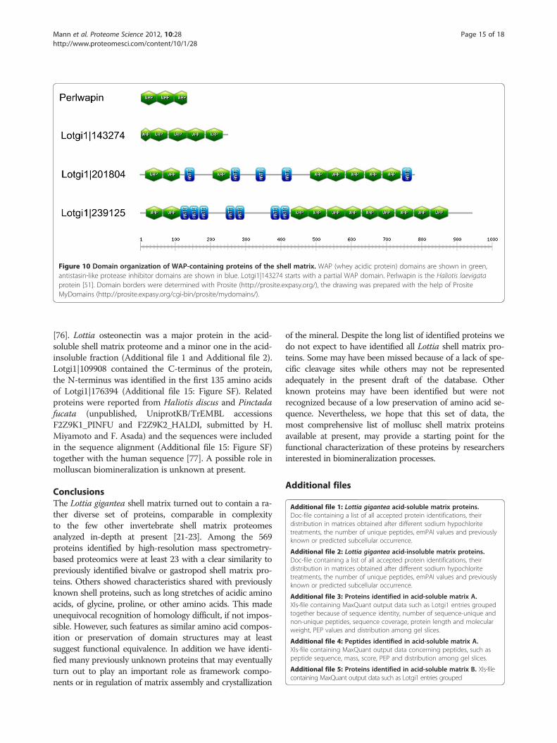
Figure 10 Domain organization of WAP-containing proteins of the shell matrix. WAP (whey acidic protein) domains are shown in green,antistasin-like protease inhibitor domains are shown in blue. Lotgi1|143274 starts with a partial WAP domain. Perlwapin is the Haliotis laevigataprotein [51]. Domain borders were determined with Prosite (http://prosite.expasy.org/), the drawing was prepared with the help of PrositeMyDomains (http://prosite.expasy.org/cgi-bin/prosite/mydomains/).
Mann et al. Proteome Science 2012, 10:28 Page 15 of 18http://www.proteomesci.com/content/10/1/28
[76]. Lottia osteonectin was a major protein in the acid-soluble shell matrix proteome and a minor one in the acid-insoluble fraction (Additional file 1 and Additional file 2).Lotgi1|109908 contained the C-terminus of the protein,the N-terminus was identified in the first 135 amino acidsof Lotgi1|176394 (Additional file 15: Figure SF). Relatedproteins were reported from Haliotis discus and Pinctadafucata (unpublished, UniprotKB/TrEMBL accessionsF2Z9K1_PINFU and F2Z9K2_HALDI, submitted by H.Miyamoto and F. Asada) and the sequences were includedin the sequence alignment (Additional file 15: Figure SF)together with the human sequence [77]. A possible role inmolluscan biomineralization is unknown at present.
ConclusionsThe Lottia gigantea shell matrix turned out to contain a ra-ther diverse set of proteins, comparable in complexityto the few other invertebrate shell matrix proteomesanalyzed in-depth at present [21-23]. Among the 569proteins identified by high-resolution mass spectrometry-based proteomics were at least 23 with a clear similarity topreviously identified bivalve or gastropod shell matrix pro-teins. Others showed characteristics shared with previouslyknown shell proteins, such as long stretches of acidic aminoacids, of glycine, proline, or other amino acids. This madeunequivocal recognition of homology difficult, if not impos-sible. However, such features as similar amino acid compos-ition or preservation of domain structures may at leastsuggest functional equivalence. In addition we have identi-fied many previously unknown proteins that may eventuallyturn out to play an important role as framework compo-nents or in regulation of matrix assembly and crystallization
of the mineral. Despite the long list of identified proteins wedo not expect to have identified all Lottia shell matrix pro-teins. Some may have been missed because of a lack of spe-cific cleavage sites while others may not be representedadequately in the present draft of the database. Otherknown proteins may have been identified but were notrecognized because of a low preservation of amino acid se-quence. Nevertheless, we hope that this set of data, themost comprehensive list of mollusc shell matrix proteinsavailable at present, may provide a starting point for thefunctional characterization of these proteins by researchersinterested in biomineralization processes.
Additional files
Additional file 1: Lottia gigantea acid-soluble matrix proteins.Doc-file containing a list of all accepted protein identifications, theirdistribution in matrices obtained after different sodium hypochloritetreatments, the number of unique peptides, emPAI values and previouslyknown or predicted subcellular occurrence.
Additional file 2: Lottia gigantea acid-insoluble matrix proteins.Doc-file containing a list of all accepted protein identifications, theirdistribution in matrices obtained after different sodium hypochloritetreatments, the number of unique peptides, emPAI values and previouslyknown or predicted subcellular occurrence.
Additional file 3: Proteins identified in acid-soluble matrix A.Xls-file containing MaxQuant output data such as Lotgi1 entries groupedtogether because of sequence identity, number of sequence-unique andnon-unique peptides, sequence coverage, protein length and molecularweight, PEP values and distribution among gel slices.
Additional file 4: Peptides identified in acid-soluble matrix A.Xls-file containing MaxQuant output data concerning peptides, such aspeptide sequence, mass, score, PEP and distribution among gel slices.
Additional file 5: Proteins identified in acid-soluble matrix B. Xls-filecontaining MaxQuant output data such as Lotgi1 entries grouped

Mann et al. Proteome Science 2012, 10:28 Page 16 of 18http://www.proteomesci.com/content/10/1/28
together because of sequence identity, number of sequence-unique andnon-unique peptides, sequence coverage, protein length and molecularweight, PEP values and distribution among gel slices.
Additional file 6: Peptides identified in acid-soluble matrix B. Xls-filecontaining MaxQuant output data concerning peptides, such as peptidesequence, mass, score, PEP and distribution among gel slices.
Additional file 7: Proteins identified in acid-soluble matrix C. Xls-filecontaining MaxQuant output data such as Lotgi1 entries groupedtogether because of sequence identity, number of sequence-unique andnon-unique peptides, sequence coverage, protein length and molecularweight, PEP values and gel slice origin of proteins.
Additional file 8: Peptides identified in acid-soluble matrix C. Xls-filecontaining MaxQuant output data concerning peptides, such as peptidesequence, mass, score, PEP and distribution in gel slices.
Additional file 9: Proteins identified in acid-insoluble matrix A.Xls-file containing MaxQuant output data such as Lotgi1 entries groupedtogether because of sequence identity, number of sequence-unique andnon-unique peptides, sequence coverage, protein length and molecularweight, PEP values and gel slice origin of proteins.
Additional file 10: Peptides identified in acid-insoluble matrix A.Xls-file containing MaxQuant output data concerning peptides, such as peptidesequence, mass, score, PEP and distribution of peptides among gel slices.
Additional file 11: Proteins identified in acid-insoluble matrix B.Xls-file containing MaxQuant output data such as Lotgi1 entries groupedtogether because of sequence identity, number of sequence-unique andnon-unique peptides, sequence coverage, protein length and molecular weight,PEP values and gel slices yielding peptides of the respective proteins.
Additional file 12: Peptides identified in acid-insoluble matrix B.Xls-file containing MaxQuant output data concerning peptides, such as peptidesequence, mass, score, PEP and distribution of peptides among gel slices.
Additional file 13: Proteins identified in acid-insoluble matrix C.Xls-file containing MaxQuant output data such as Lotgi1 entries groupedtogether because of sequence identity, number of sequence-unique andnon-unique peptides, sequence coverage, protein length and molecular weight,PEP values and gel slice origin of peptides for protein identification.
Additional file 14: Peptides identified in acid-insoluble matrix C.Xls-file containing MaxQuant output data concerning peptides, such as peptidesequence, mass, score, PEP and distribution among gel slices.
Additional file 15: Selected sequence alignments. Doc-file showingsequence alignments of ependymin-related protein, gigasin-2, nacrein-likeprotein, tyrosinase, UP2, and osteonectin to similar proteins identified in thisstudy.
Additional file 16: Sequence analysis of BMSP-related Lottia proteins.Doc-file showing the alignment of BMSP-related protein sequences to Mytilusgalloprovincialis BMSP (A) and the domain distribution in these sequences (B).
Additional file 17: Sequence analysis of Pif-related Lottia proteins.Doc-file showing the alignment of Pif-related protein sequences to Pinctadafucata Pif (A) and the domain distribution in these sequences (B).
AbbreviationsAa: Amino acid; BMSP: Blue Mussel Shell Protein; CA: Carbonic anhydrase;CLECT: C-type lectin; IGFBP: Insulin-like growth factor-binding protein;emPAI: Exponentially modified protein abundance index; FDR: False discoveryrate; HCD: Higher-energy collision-induced decomposition;PAGE: Polyacrylamide gel electrophoresis; PEP: Posterior error probability;VWA: Von Willebrand type A; WAP: Whey acidic protein.
Competing interestsThe authors declare that they have no competing interests.
AcknowledgementsWe thank Fred H. Wilt, Department of Molecular and Cell Biology, Universityof California, Berkeley, for drawing KM’s attention to the Lottia genomeproject and for bringing KM and EEG into contact.
Author details1Abteilung Proteomics und Signaltransduktion, Max-Planck-Institut fürBiochemie, Am Klopferspitz 18, D-82152 Martinsried, Germany. 2Departmentof Molecular and Cell Biology, University of California, Berkeley, 545 LifeSciences Addition, Berkeley, CA 94720, USA.
Authors’ contributionsKM conceived the study, performed sample preparation and data acquisition.EEG collected and mechanically cleaned Lottia shells and helped withdatabase search and annotation. MM supplied methodological expertise. Allauthors took part in the design of the study and were critically involved inmanuscript drafting. All authors read and approved the final manuscript.
Received: 19 January 2012 Accepted: 27 April 2012Published: 27 April 2012
References1. Addadi L, Joester D, Nudelman F, Weiner S: Mollusk shell formation: A
source of new concepts for understanding biomineralization processes.Chem Eur J 2006, 12:980–987.
2. Heinemann F, Launspach M, Gries K, Fritz M: Gastropod nacre: Structure,properties and growth – Biological, chemical and physical basis. BiophysChem 2011, 153:126–153.
3. Bédouet L, Marie A, Dubost L, Péduzzi J, Duplat D, Berland S, Puisségur M,Boulzaguet H, Rousseau M, Milet C, Lopez E: Proteomic analysis of thenacre soluble and insoluble proteins from the oyster Pinctadamargaritifera. Mar Biotechnol 2007, 9:638–649.
4. Marie B, Marin F, Marie A, Bédouet L, Dubost L, Alcaraz G, Milet C, Luquet G:Evolution of nacre: Biochemistry and proteomics of the shell organic matrixof the cephalopod Nautilus macromphalus. Chembiochem 2009, 10:1495–1506.
5. Marie B, Zanella-Cléon I, Le Roy N, Becchi M, Luquet G, Marin F: Proteomicanalysis of the acid-soluble nacre matrix of the bivalve Unio pictorum:Detection of a novel carbonic anhydrase and putative protease inhibitorproteins. Chembiochem 2010, 11:2138–2147.
6. Marie B, Marie A, Jackson DJ, Dubost L, Degnan B, Milet C, Marin F:Proteomic analysis of the organic matrix of the abalone Haliotis asiniacalcified shell. Proteome Sci 2010, 8:54.
7. Joubert C, Piquemal D, Marie B, Manchon L, Pierrat F, Zanella-Cléon I,Cochennec-Laureau N, Gueguen Y, Montagnani C: Transcriptome andproteome analysis of Pinctada margeritifera calcifying mantle and shell:focus on biomineralization. BMC Genomics 2010, 11:613.
8. Marie B, Trinkler N, Zanella-Cléon I, Guichard N, Becchi M, Paillard C, Marin F:Proteomic identification of novel proteins from the calcifying shellmatrix of the Manila clam Venerupis philippinarum. Mar Biotechnol 2011,13:955–962.
9. Marie B, Zanella-Cléon I, Guichars N, Becchi M, Marin F: Novel proteins fromthe calcifying matrix of the pacific oyster Crassostrea gigas. MarBiotechnol 2011, 13:1159–1168.
10. Marie B, LeRoy N, Zanella-Cléon I, Becchi M, Marin F: Molecular evolution ofmollusk shell proteins: Insights from proteomic analysis of the ediblemussel Mytilus. J Mol Evol 2011, 72:531–546.
11. Berland S, Marie A, Duplat D, Milet C, Sire JY, Bédouet L: Couplingproteomics and transcriptomics for the identification of novel andvariant forms of mollusk shell proteins: A study with P. margaritifera.Chembiochem 2011, 12:950–961.
12. Steen H, Mann M: The ABC’s (and XYZ’s) of peptide sequencing. Nat RevMol Cell Biol 2004, 5:699–711.
13. Cox J, Mann M: Quantitative, high-resolution proteomics for data-drivensystems biology. Annu Rev Biochem 2011, 80:273–299.
14. Jackson DJ, McDougall C, Green K, Simpson F, Wörheide G, Degnan BM: Arapidly evolving secretome builds and patterns a sea shell. BMC Biol 2006, 4:40.
15. Vernier P, De Pitta C, Pallavicini A, Marsano F, Varotto L, Romualdi C,Dondero F, Viarengo A, Lanfranchi G: Development of mussel mRNAprofiling: Can gene expression trends reveal coastal water pollution?Mutation Res 2006, 602:121–134.
16. Tanguy A, Bierne N, Saavedra C, Pina B, Bachere E, Kube M, Bazin E, BonhommeF, Boudry P, Boulo V, Boutet I, Cancela L, Dossat C, Favrel P, Huvet A, Jarque S,Jollivet D, Klages S, Lapegue S, Leite R, Moal J, Moraga D, Reinhardt R, Samain J,Zouros E, Canario A: Increasing genomic information in bivalves through
Mann et al. Proteome Science 2012, 10:28 Page 17 of 18http://www.proteomesci.com/content/10/1/28
new EST collections in four species: Development of new genetic markersfor environmental studies and genome evolution. Gene 2008, 408:27–36.
17. Jackson DJ, McDougall C, Woodcroft B, Moase P, Rose RA, Kube M,Reinhardt R, Rokhsar DS, Montagnani C, Joubert C, Piquemal D, Degnan BM:Parallel evolution of nacre building gene sets in mollusks. Mol Biol Evol2009, 27:591–608.
18. Kinoshita S, Wang N, Inoue H, Maeyama K, Okamoto K, Nagai K, Kondo H,Hirono I, Asakawa S, Watabe S: Deep sequencing of ESTs from nacreousand prismatic layer producing tissues and a screen for novel shellformation-related genes in the pearl oyster. PLoS One 2011, 6:e21238.
19. Seidler J, Zinn N, Boehm ME, Lehmann WD: De novo sequencing ofpeptides by MS/MS. Proteomics 2010, 10:1–16.
20. Mann K, Macek B, Olsen JV: Proteomic analysis of the acid-soluble organicmatrix of the chicken calcified eggshell layer. Proteomics 2006, 6:3801–3810.
21. Mann K, Poustka AJ, Mann M: The sea urchin (Strongylocentrotuspurpuratus) test and spine proteomes. Proteome Sci 2008, 6:22.
22. Mann K, Poustka AJ, Mann M: In-depth, high-accuracy proteomics of seaurchin tooth organic matrix. Proteome Sci 2008, 6:33.
23. Mann K, Wilt FH, Poustka AJ: Proteomic analysis of sea urchin(Strongylocentrotus purpuratus) spicule matrix. Proteome Sci 2010, 8:33.
24. Grigoriev IV, Nordberg H, Shabalov I, Aerts A, Cantor M, Goodstein D, Kuo A,Minovitsky S, Nikitin R, Ohm RA, Otillar R, Poliakov A, Ratnere I, Riley R,Smirnova T, Rokhsar D, Dubchak I: The Genome Portal of the Departmentof Energy Joint Genome Institute. Nucleic Acids Res 2011, 0:gkr947v1–gkr947.
25. Suzuki M, Kameda J, Sasaki T, Saruwatari K, Nagasawa H, Kogure T:Characterization of the multilayered shell of a limpet, Lottia kogamogai(Mollusca: Patellogastropoda), using SEM-EBSD and FIB-TEM techniques.J Struct Biol 2010, 171:223–230.
26. Suzuki M, Kogure T, Weiner S, Addadi L: Formation of aragonite crystals in thecrossed lamellar microstructure of limpet shells. Cryst Growth Des 2011,11:4850–4859.
27. Olsen JV, Schwartz JC, Griep-Raming J, Nielsen ML, Damoc E, Denisov E,Lange O, Remes P, Taylor D, Splendore M, Wouters ER, Senko M, Makarov A,Mann M, Horning S: A dual pressure linear ion trap-Orbitrap instrumentwith very high sequencing speed. Mol Cell Proteomics 2009, 8:2759–2769.
28. Cox J, Mann M: MaxQuant enables high peptide identification rates,individualized ppb-range mass accuracies and proteome-wide proteinquantification. Nat Biotechnol 2009, 26:1367–1372.
29. Cox J, Matic I, Hilger M, Nagaraj N, Selbach M, Olsen JV, Mann M: A practicalguide to the MaxQuant computational platform for SILAC-basedquantitative proteomics. Nat Protoc 2009, 4:698–705.
30. Cox J, Neuhauser N, Michalski A, Scheltema RA, Olsen JV, Mann M:Andromeda – a peptide search engine integrated into the MaxQuantenvironment. J Proteome Res 2011, 10:1794–1805.
31. Shevchenko A, Tomas H, Havlis J, Olsen JV, Mann M: (2006) In-gel digestionfor mass spectrometric characterization of proteins and proteomes. NatProtoc 2006, 1:2856–2860.
32. Rappsilber J, Mann M, Ishihama Y: Protocol for micro-purification,enrichment, pre-fractionation and storage of peptides for proteomicsusing StageTips. Nat Protoc 2007, 2:1896–1906.
33. Ishihama Y, Oda Y, Tabata T, Sato T, Nagasu T, Rappsilber J, Mann M:Exponentially modified protein abundance index (emPAI) for estimationof absolute protein amount in proteomics by the number of sequencedpeptides per protein. Mol Cell Proteomics 2005, 4:1265–1272.
34. Evans JS: “Tuning in” to mollusk shell nacre- and prismatic-associatedprotein terminal sequence. Implications for biomineralization and theconstruction of high performance inorganic–organic composites. ChemRev 2008, 108:4455–4462.
35. Amos FF, Evans JS: AP7, a partially disordered pseudo C-ring protein, iscapable of forming stabilized aragonite in vitro. Biochem 2009, 48:1332–1339.
36. Delak K, Collino S, Evans JS: Polyelectrolyte domains and intrinsic diaorderwithin the prismatic asprich protein family. Biochem 2009, 48:3669–3677.
37. Ndao M, Keene E, Amos FF, Rewari G, Ponce CB, Estroff L, Evans JS:Intrinsically disordered mollusk shell prismatic protein that modulatescalcium carbonate crystal growth. Biomacromol 2010, 11:2539–2544.
38. Keene EC, Evans JS, Estroff LA: Matrix interactions in biomineralization:aragonite nucleation by an intrinsically disordered nacre polypeptide,n16N, associated with a β-chitin substrate. Crys Growth Des 2010,10:1383–1389.
39. Amos FF, Destine E, Ponce CB, Evans JS: The N- and C-terminal regions ofthe pearl-associated EF hand protein, PFMG1, promote the formation ofthe aragonite polxmorph in vitro. Cryst Growth Des 2010, 10:4211–4216.
40. Davey NE, Van Roey K, Weatheritt RJ, Toedt G, Uyar B, Altenberg B, Budd A,Diella F, Dinkel H, Gibson TJ: Attributes of short linear motifs. Mol Biosyst2012, 8:268–281.
41. Lobanov MY, Galzitskaya OV: Occurrence of disordered patterns andhomorepeats in eukaryotic and bacterial proteomes. Mol Biosyst 2012,8:327–337.
42. Tsukamoto D, Sarashina I, Endo K: Structure and expression of anunusually acid matrix protein of pearl oyster shells. Biochem Biophys ResCommun 2004, 320:1175–1180.
43. Sarashina I, Endo K: Primary structure of a soluble matrix protein ofscallop shell: Implications for calcium carbonate biomineralization. AmMineralogist 1998, 83:1510–1515.
44. Gotliv BA, Kessler N, Sumerel JL, Morse DE, Tuross N, Addadi L, Weiner S:Asprich: A novel aspartic acid-rich protein family from the prismatic shellmatrix of the bivalve Atrina rigida. Chembiochem 2005, 6:304–314.
45. Yano M, Nagai K, Morimoto K, Miyamoto H: Shematrin: A family of glycine-rich structural proteins in the shell of the pearl oyster Pinctada fucata.Comp Biochem Physiol B 2006, 144:254–262.
46. Miyamoto H, Miyashita T, Okushima M, Nakano S, Morita T, Matsushiro A: Acarbonic anhydrase from the organic nacreous layer in oyster pearls. ProcNatl Acad Sci USA 1996, 93:9657–9660.
47. Suzuki M, Saruwatari K, Kogure T, Yamamoto Y, Nishimura T, Kato T,Nagasawa H: An acidic matrix protein, Pif, is a key macromolecule fornacre formation. Science 2009, 325:1388–1390.
48. Suzuki M, Iwashima A, Tsutsui N, Ohira T, Kogure T, Nagasawa H:Identification and characterization of a calcium carbonate-bindingprotein, blue mussel shell protein (BMSP), from the nacreous layer.Chembiochem 2011, 12:2478–2487.
49. Marxen JC, Nimtz M, Becker W, Mann K: The major soluble 19.6 kDaprotein of the organic shell matrix of the fresh water snail Biomphalariaglabrata is an N-glycosylated dermatopontin. Biochim Biophys Acta 2003,1650:92–98.
50. Weiss IM, Göhring W, Fritz M, Mann K: Perlustrin, a Haliotis laevigata(abalone) nacre protein, is homologous to the insulin-like growth factorbinding protein N-terminal module of vertebrates. Biochem Biophys ResCommun 2001, 285:244–249.
51. Treccani L, Mann K, Heinemann F, Fritz M: Perlwapin, an abalone nacreprotein with three four-disulfide core (whey acidic protein) domains,inhibits the growth of calcium carbonate crystals. Biophys J 2006,91:2601–2608.
52. Nagai K, Yano M, Morimoto K, Miyamoto H: Tyrosinase localization inmollusk shells. Comp Biochem Physiol B 2007, 146:207–214.
53. Zhang C, Xie L, Huang J, Chen L, Zhang R: A novel putative tyrosinaseinvolved in periostracum formation from the pearl oyster (Pinctadafucata). Biochem Biophys Res Commun 2006, 342:632–639.
54. Bouchut A, Roger E, Coustau C, Gourbal B, Mitta G: Compatibility in theBiomphalaria glabrata/Echinostoma caproni model: Potential involvementof adhesion genes. Int J Parasitol 2006, 36:175–184.
55. Sarashina I, Yamaguchi H, Haga T, Iijima M, Chiba S, Endo K: Molecularevolution and functionally important structures of molluscandermatopontin: Implications for the origins of molluscan shell matrixproteins. J Mol Evol 2006, 62:307–318.
56. Miyashita T, Takagi R, Miyamoto H, Matsushiro A: Identical carbonicanhydrase contributes to nacreous or prismatic layer formation inPinctada fucata (Mollusca: Bivalvia). Veliger 2002, 45:250–255.
57. Miyamoto H, Yano M, Miyashita T: Similarities in the structure of nacrein,the shell-matrix protein, in a bivalve and a gastropod. J Molluscan Stud2003, 69:87–89.
58. Norizuki M, Samata T: Distribution and function of the nacrein-relatedproteins inferred from structural analysis. Mar Biotech 2008, 10:234–241.
59. Li C, Huang J, Li S, Fan W, Hu Y, Wang Q, Zhu S, Xie L, Zhang R: Cloning,characterization and immunolocalization of two subunits of calcineurinfrom pearl oyster (Pinctada fucata). Comp Biochem Physiol B 2009,153:43–53.
60. Li C, Hu Y, Liang J, Kong Y, Huang J, Feng Q, Li S, Zhang G, Xie L, Zhang R:Calcineurin plays an important role in shell formation of pearl oyster

Mann et al. Proteome Science 2012, 10:28 Page 18 of 18http://www.proteomesci.com/content/10/1/28
(Pinctada fucata). Mar Biotech 2010, 12:100–110.61. Weiss IM, Kaufmann S, Mann K, Fritz M: Purification and characterization of
perlucin and perlustrin, two new proteins from the shell of the molluscHaliotis laevigata. Biochem Biophys Res Commun 2000, 267:17–21.
62. Mann K, Weiss IM, André S, Gabius HJ, Fritz M: The amino acid sequence ofabalone (Haliotis laevigata) nacre protein perlucin. Detection of afunctional C-type lectin domain with galactose/mannose specificity. Eur JBiochem 2000, 267:5257–5264.
63. Smith VJ: Phylogeny of whey acidic protein (WAP) four –disulfide coreproteins and their role in lower vertebrates and invertebrates. BiochemSoc Trans 2011, 39:1403–1408.
64. Shen Z, Jacobs-Lorena M: Evolution of chitin-binding proteins ininvertebrates. J Mol Evol 1999, 48:341–347.
65. Suetake T, Tsuda S, Kawabata S, Miura K, Iwanaga S, Hikichi K, Nitta K,Kawano K: Chitin-binding proteins in invertebrates and plants comprise acommon chitin-binding structural motif. J Biol Chem 2000, 275:17929–17932.
66. Shen X, Belcher AM, Hansma PK, Stucky GD, Morse DE: Molecular cloningand characterization of lustrin A, a matrix protein from shell and pearlnacre of Haliotis rufescens. J Biol Chem 1997, 51:32472–32481.
67. Peters W: Occurrence of chitin in mollusca. Comp Biochem Physiol B 1972,41:341–349.
68. Weiss IM, Schönitzer V: The distribution of chitin in larval shells of thebivalve mollusk Mytilus galloprovincialis. J Struct Biol 2006, 153:264–277.
69. Suzuki M, Sakuda S, Nagasawa H: Identification of chitin in the prismaticlayer of the shell and a chitin synthase gene from the Japanese pearloyster, Pinctada fucata. Biosci Biotechnol Biochem 2007, 71:1735–1744.
70. Schönitzer V, Weiss IM: The structure of mollusk larval shells formed inthe presence of the chitin synthase inhibitor nikkomycin Z. BMC StructBiol 2007, 7:71.
71. Levi-Kalisman Y, Falini G, Addadi L, Weiner S: Structure of the nacreousorganic matrix of a bivalve mollusk shell examined in the hydrated stateusing cryo-TEM. J Struct Biol 2001, 135:8–17.
72. Hao J, Narayanan K, Muni T, Ramachandran A, George A: Dentin matrixprotein 4, a novel secretory calcium-binding protein that modulatesodontoblast differentiation. J Biol Chem 2007, 282:15357–15365.
73. Termine JD, Kleinman HK, Whitson SW, Conn KM, McGarvey ML, Martin GR:Osteonectin, a bone-specific protein linking mineral to collagen. Cell1981, 26:99–105.
74. Mann K, Deutzmann R, Paulsson M, Timpl R: Solubilization of protein BM-40 from a basement membrane tumor with chelating agents andevidence for its identity with osteonectin and SPARC. FEBS Lett 1987,218:167–172.
75. Sage H, Johnson C, Bornstein P: Characterization of a novel serumalbumin-binding glycoprotein secreted by endothelial cells in culture.J Biol Chem 1984, 259:3993–4007.
76. Wallin R, Wajih N, Greenwood GT, Sane DC: Arterial calcification: A reviewof mechanisms, animal models, and the prospects for therapy. Med ResRev 2001, 21:274–301.
77. Lankat-Buttgereit B, Mann K, Deutzmann R, Timpl R, Krieg T: Cloning andcomplete amino acid sequence of human and murine basement membraneprotein BM-40 (SPARC, osteonectin). FEBS Lett 1988, 236:352–356.
doi:10.1186/1477-5956-10-28Cite this article as: Mann et al.: In-depth proteomic analysis of a molluscshell: acid-soluble and acid-insoluble matrix of the limpet Lottiagigantea. Proteome Science 2012 10:28.
Submit your next manuscript to BioMed Centraland take full advantage of:
• Convenient online submission
• Thorough peer review
• No space constraints or color figure charges
• Immediate publication on acceptance
• Inclusion in PubMed, CAS, Scopus and Google Scholar
• Research which is freely available for redistribution
Submit your manuscript at www.biomedcentral.com/submit
Related Documents