Research Article Revisiting the Factors Underlying Maxillary Midline Diastema Abdullah M. Zakria Jaija, 1 Amr Ragab El-Beialy, 2,3 and Yehya A. Mostafa 2,3 1 Private Practice, P.O. Box 376385, Riyadh 11335, Saudi Arabia 2 Department of Orthodontics and Dentofacial Orthopedics, Faculty of Oral and Dental Medicine, Cairo University, 11 El-Saraya Street, Manial, Cairo 12511, Egypt 3 MOrth Programs at Cairo University and Future University, Cairo, Egypt Correspondence should be addressed to Amr Ragab El-Beialy; [email protected] Received 5 December 2015; Accepted 17 March 2016 Academic Editor: William Maloney Copyright © 2016 Abdullah M. Zakria Jaija et al. is is an open access article distributed under the Creative Commons Attribution License, which permits unrestricted use, distribution, and reproduction in any medium, provided the original work is properly cited. Aim. e aim of this study is to analyze the etiological factors underlying the presence of maxillary midline diastema in a sample of orthodontic patients. Materials and Methods. One hundred patients who fulfill the inclusion criteria were selected from 1355 patients seeking orthodontic treatment. e pretreatment orthodontic records were analyzed. e width of the maxillary midline diastema was measured clinically with a digital caliper at two levels: the mesioincisal angles of the central incisors and five millimeters from the incisal edge. e two measurements were averaged, and patients with diastema of more than 0.5 millimeter in width were enrolled. Results. Diastema is a multifactorial clinical finding with more than one underlying etiological cause. e interrelationship between the familial pattern of midline diastema and the microdontia, macroglossia, labial frenum, and alveolar cleſt conforms was clear. e effect of a mesiodens and the upper lateral incisor whether bilaterally missing, unerupted, or peg shaped was minimal. Conclusion. Etiological factors underlying maxillary midline diastema are interconnected. Using a checklist as a guide during handling maxillary midline diastema is important in the different stages of treatment. 1. Introduction e presence of a midline diastema represents an esthetic and psychological impairment and distress for patients seeking orthodontic treatment [1]. Maxillary midline diastema is a clinical sign, which has a multitude of underlying etiological factors that might be interdependent or independent. Many etiological factors for maxillary midline diastema have been reported in the literature [2]. Among them are the physiological (developmental) self-limiting diastema, famil- ial background, mesiodens [3–5], abnormal labial frenum [6–8], missing or undersized lateral incisor, thumb sucking, mouth breathing, tongue thrust, ankylosed central incisor, flared or rotated central incisors, anodontia, macroglossia [9, 10], dentoalveolar disproportion, generalized spacing [11], localized spacing, closed bite, facial type, ethnic and familial characteristics [12, 13], interpremaxillary suture and transsep- tal fibers [14], midline pathology, midline submucosal alve- olar cleſt [15], tongue piercing [16], gingival recession, and pathological tooth migration [17]. e aim of this survey is to investigate the correlation between the different etiological factors underlying maxillary midline diastema and highlight their clinical implications. 2. Materials and Methods is survey was performed on patients undergoing orthodontic treatment at three orthodontic graduate programs. e pretreatment records of 1355 patients were examined, to collect 100 patients fulfilling the inclusion criterion (presence of maxillary midline diastema >0.5 mm) within an age range of 13–30 years. e pretreatment records included history, intra- and extraoral examination, and panoramic and periapical radiographs of the maxillary incisor region. e width of the maxillary midline diastema was measured clinically with a digital caliper at two levels: the mesioincisal angles of the central incisors and five millimeters from the incisal edge. e two measurements were averaged, and patients with diastema of more than 0.5 millimeter in Hindawi Publishing Corporation Scientifica Volume 2016, Article ID 5607594, 5 pages http://dx.doi.org/10.1155/2016/5607594

Welcome message from author
This document is posted to help you gain knowledge. Please leave a comment to let me know what you think about it! Share it to your friends and learn new things together.
Transcript

Research ArticleRevisiting the Factors Underlying Maxillary Midline Diastema
Abdullah M. Zakria Jaija,1 Amr Ragab El-Beialy,2,3 and Yehya A. Mostafa2,3
1Private Practice, P.O. Box 376385, Riyadh 11335, Saudi Arabia2Department of Orthodontics and Dentofacial Orthopedics, Faculty of Oral and Dental Medicine, Cairo University,11 El-Saraya Street, Manial, Cairo 12511, Egypt3MOrth Programs at Cairo University and Future University, Cairo, Egypt
Correspondence should be addressed to Amr Ragab El-Beialy; [email protected]
Received 5 December 2015; Accepted 17 March 2016
Academic Editor: William Maloney
Copyright © 2016 Abdullah M. Zakria Jaija et al.This is an open access article distributed under theCreativeCommonsAttributionLicense, which permits unrestricted use, distribution, and reproduction in anymedium, provided the originalwork is properly cited.
Aim. The aim of this study is to analyze the etiological factors underlying the presence of maxillary midline diastema in a sample oforthodontic patients.Materials andMethods. One hundred patients who fulfill the inclusion criteriawere selected from 1355 patientsseeking orthodontic treatment. The pretreatment orthodontic records were analyzed. The width of the maxillary midline diastemawas measured clinically with a digital caliper at two levels: the mesioincisal angles of the central incisors and five millimetersfrom the incisal edge.The two measurements were averaged, and patients with diastema of more than 0.5 millimeter in width wereenrolled.Results. Diastema is amultifactorial clinical finding withmore than one underlying etiological cause.The interrelationshipbetween the familial pattern of midline diastema and themicrodontia, macroglossia, labial frenum, and alveolar cleft conforms wasclear. The effect of a mesiodens and the upper lateral incisor whether bilaterally missing, unerupted, or peg shaped was minimal.Conclusion. Etiological factors underlying maxillary midline diastema are interconnected. Using a checklist as a guide duringhandling maxillary midline diastema is important in the different stages of treatment.
1. Introduction
Thepresence of amidline diastema represents an esthetic andpsychological impairment and distress for patients seekingorthodontic treatment [1]. Maxillary midline diastema is aclinical sign, which has a multitude of underlying etiologicalfactors that might be interdependent or independent.
Many etiological factors for maxillary midline diastemahave been reported in the literature [2]. Among them are thephysiological (developmental) self-limiting diastema, famil-ial background, mesiodens [3–5], abnormal labial frenum[6–8], missing or undersized lateral incisor, thumb sucking,mouth breathing, tongue thrust, ankylosed central incisor,flared or rotated central incisors, anodontia, macroglossia[9, 10], dentoalveolar disproportion, generalized spacing [11],localized spacing, closed bite, facial type, ethnic and familialcharacteristics [12, 13], interpremaxillary suture and transsep-tal fibers [14], midline pathology, midline submucosal alve-olar cleft [15], tongue piercing [16], gingival recession, andpathological tooth migration [17].
The aim of this survey is to investigate the correlationbetween the different etiological factors underlyingmaxillarymidline diastema and highlight their clinical implications.
2. Materials and Methods
This survey was performed on patients undergoingorthodontic treatment at three orthodontic graduateprograms. The pretreatment records of 1355 patients wereexamined, to collect 100 patients fulfilling the inclusioncriterion (presence of maxillary midline diastema >0.5mm)within an age range of 13–30 years. The pretreatment recordsincluded history, intra- and extraoral examination, andpanoramic and periapical radiographs of the maxillaryincisor region. The width of the maxillary midline diastemawasmeasured clinically with a digital caliper at two levels: themesioincisal angles of the central incisors and fivemillimetersfrom the incisal edge. The two measurements were averaged,and patients with diastema of more than 0.5 millimeter in
Hindawi Publishing CorporationScientificaVolume 2016, Article ID 5607594, 5 pageshttp://dx.doi.org/10.1155/2016/5607594

2 Scientifica
Figure 1: Maxillary midline diastema.
Sex Facial profile Facial type Dentition Axial inclination ProclinationMale Straight Brachycephalic Late mixed Convergence ProclinedFemale Convex Mesocephalic E. permanent Normal Normal
Concave Dolichocephalic L. permanent Divergence Retroclined
05
101520253035404550556065707580859095
100105
Sex Facial profile Facial type Dentition Axial inclination Proclination
Num
ber o
f pat
ient
s
Figure 2: Bar chart showing the distribution of the criteria of the sample.
width were enrolled (Figure 1).The examination wasmade bythe principle observer and repeated by the second observer.
Because of the physiological diastema, patients youngerthan 13 years were excluded, while patients above 30 yearswere excluded because of the possibility of diastema forma-tion due to periodontal involvement and migration of teeth.
The distribution of the criteria of the sample was ana-lyzed (Figure 2). The criteria represent the commonsensi-cal orthodontic categories that segregate the sample intocomparable subclasses. The etiological factors underlyingthe maxillary midline diastema were extracted from therecords and clinical examination of the patients (Table 1).These etiological foundations were separated into majoretiological factors and etiological factors of lesser influence.These factors represent all the etiological factors underlyingthe presence of the maxillary midline diastema that wereextracted from the research sample. The prevalence of eachfactor in percentage of the 100 cases enrolled was calculated.
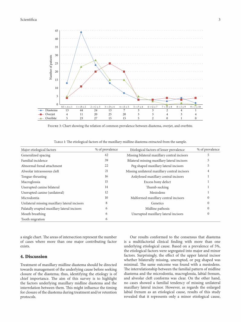
Additionally, the association between diastemawith over-jet and overbite is depicted through dividing the sampleinto 10 groups each representing 1mm regarding diastema,overjet, and overbite (Figure 3).
3. Results
The prevalence of the diastema was found to be 13.6%among the screened sample.The occurrence of the six criteria(Figure 2) demonstrated that the maxillary midline diastemais more observed in females, mesocephalic faces, convexfacial profiles, and the early permanent dentition. Maxillarymidline diastema is more prevalent with upright maxillarycentral incisors than convergent or divergent central incisors.The least prevalence of diastema occurs with retroclinedmaxillary incisors.
The relationship between themaxillarymidline diastema,overjet, and overbite depicted in Figure 3 shows that adiastemawidth of 1-2mm ismore prevalent (44 patients) thanother extents of diastema, and this prevalence decreases as theamount of overbite and overjet increases.
Etiological factors were segregated into major contribut-ing factors and factors of lesser contribution taking 5%prevalence as the limit (Table 1). The interrelation (overlap)between the major contributing factors is denoted by inter-secting circles charts (Figures 4–6). Factors that might be ofstrong developmental interrelation were linked together in
Scientifica 3
Diastema 13 44 24 13 7 3 3 2 4 1Overjet 4 11 20 25 20 3 3 4 3 4Overbite 5 23 27 15 15 5 2 0 1 0
0
5
10
15
20
25
30
35
40
45
Num
ber o
f pat
ient
s
9 < J ≤ 108 < I ≤ 97 < H ≤ 86 < G ≤ 75 < F ≤ 64 < E ≤ 53 < D ≤ 42 < C ≤ 31 < B ≤ 20.5 <A ≤ 1
Figure 3: Chart showing the relation of common prevalence between diastema, overjet, and overbite.
Table 1: The etiological factors of the maxillary midline diastema extracted from the sample.
Major etiological factors % of prevalence Etiological factors of lesser prevalence % of prevalenceGeneralized spacing 42 Missing bilateral maxillary central incisors 5Familial incidence 39 Bilateral missing maxillary lateral incisors 5Abnormal frenal attachment 22 Peg shaped maxillary lateral incisors 5Alveolar intraosseous cleft 21 Missing unilateral maxillary central incisors 4Tongue-thrusting 16 Ankylosed maxillary central incisors 1Macroglossia 15 Excess bony defect 1Unerupted canine bilateral 14 Thumb sucking 1Unerupted canine (unilateral) 12 Mesiodens 1Microdontia 10 Malformed maxillary central incisors 0Unilateral missing maxillary lateral incisors 8 Genetics 0Palatally erupted maxillary lateral incisors 6 Midline pathosis 0Mouth breathing 6 Unerupted maxillary lateral incisors 0Tooth migration 6
a single chart.The areas of intersection represent the numberof cases where more than one major contributing factorexists.
4. Discussion
Treatment of maxillary midline diastema should be directedtowards management of the underlying cause before seekingclosure of the diastema; thus, identifying the etiology is ofchief importance. The aim of this survey is to highlightthe factors underlying maxillary midline diastema and theinterrelation between them. This might influence the timingfor closure of the diastema during treatment and/or retentionprotocols.
Our results conformed to the consensus that diastemais a multifactorial clinical finding with more than oneunderlying etiological cause. Based on a prevalence of 5%,the etiological factors were segregated into major and minorfactors. Surprisingly, the effect of the upper lateral incisorwhether bilaterally missing, unerupted, or peg shaped wasminimal. The same outcome was found with a mesiodens.The interrelationship between the familial pattern of midlinediastema and the microdontia, macroglossia, labial frenum,and alveolar cleft conforms was clear. On the other hand,no cases showed a familial tendency of missing unilateralmaxillary lateral incisor. However, as regards the enlargedlabial frenum as an etiological cause, results of this studyrevealed that it represents only a minor etiological cause,

4 Scientifica
Table 2: Checklist showing the impact of each etiological factor of themaxillarymidline diastema upon the diagnosis, treatment, or retentionprotocol.
Factor Extra diagnostic tool Treatment modification Retention modificationGeneralized spacing r (Permanent)Familial incidence r (Family screening) r (Permanent)Abnormal frenal attachment r (Periapical radiograph) r (Frenotomy) r (Prolonged)Alveolar intraosseous cleft r (Periapical radiograph) r (Nonidentified)Tongue-thrusting r (Habit breaking appliance) r (Habit breaking)Macroglossia r (No encroaching on tongue) r (Permanent)Unerupted canine bilaterallyUnerupted canine unilaterallyMicrodontia r (Build-up)Unilateral missing maxillary lateral incisors r (Prosthesis)r (Canine substitution)Palatally erupted maxillary lateral incisors r (Root torquing)Mouth breathing r (ENT consultation) r (Habit breaking)Tooth migrationMissing bilateral maxillary central incisors r (Prosthesis)Bilateral missing maxillary lateral incisors r (Prosthesis)r (Canine substitution)Peg shape maxillary lateral incisors r (Build-up)Missing unilateral maxillary central incisors r (Prosthesis)Ankylosed maxillary central incisors r (Luxation, crowning, extraction)Excess bony defect r (Surgical)Thumb sucking r (Habit breaking appliance)Mesiodens r (Surgical extraction)Malformed maxillary central incisors r (Build-up)Midline pathosis r (Periapical radiograph) r (Surgical excision)Unerupted maxillary lateral incisors r (Periapical radiograph)
Familial
Alveola
r cleft
129
8
Microdontia 4
6
Labial frenum
Missing
unilateral 28
166Macroglossia
105
Figure 4: Intersecting circles chart depicting the common occur-rence between the major contributing factors.
an observation that conforms to the findings of Huang andCreath [2]. In addition, the interrelation between the alveolarcleft and abnormal labial frenum was an important finding.
Spacing
17
Macroglossia
Microd
ontia
3 9
7 7 Tongue thrust
4
11
Figure 5: Intersecting circles chart depicting the common occur-rence between spacing, microdontia, macroglossia, and tonguethrust.
Implementation of the findings of this survey is importantfrom the clinical sense. The impact of each etiological factorof the maxillary midline diastema upon the diagnosis, treat-ment, or retention protocol is summarized into a checklist.This checklist was designed to highlight the intervention at

Scientifica 5
Alveolarcleft
9
12
Labia
lfre
num
13
Figure 6: Intersecting circles chart depicting the common occur-rence between labial frenum and alveolar cleft.
the different stages of treatment for each etiological factor(Table 2).
5. Conclusion
Etiological factors underlyingmaxillarymidline diastema areinterconnected.
Using a checklist as a guide during handling maxillarymidline diastema is important in the different stages oftreatment.
Competing Interests
The authors declare that they have no competing interests.
References
[1] N. Gkantidis, O.-E. Kolokitha, and N. Topouzelis, “Manage-ment of maxillarymidline diastema with emphasis on etiology,”Journal of Clinical Pediatric Dentistry, vol. 32, no. 4, pp. 265–272,2008.
[2] W. J. Huang and C. J. Creath, “The midline diastema: a reviewof its etiology and treatment,” Pediatric Dentistry, vol. 17, no. 3,pp. 171–179, 1995.
[3] N. B. Nagaveni, K. V. Umashankara, Sreedevi, B. P. Reddy, N.B. Radhika, and T. S. Satisha, “Multi-lobed mesiodens with apalatal talon cusp: a rare case report,” Brazilian Dental Journal,vol. 21, no. 4, pp. 375–378, 2010.
[4] K. Gunduz, P. Celenk, Z. Zengin, and P. Sumer, “Mesiodens: aradiographic study in children,” Journal of Oral Science, vol. 50,no. 3, pp. 287–291, 2008.
[5] A. Alacam andM. Bani, “Mesiodens as a risk factor in treatmentof trauma cases,” Dental Traumatology, vol. 25, no. 2, pp. e25–e31, 2009.
[6] D. Belic and K. Obrez-Oblak, “Diastema verum and persistentlabial frenum,”Zobozdravstveni Vestnik, vol. 45, no. 4-5, pp. 107–109, 1990.
[7] J. G. Edwards, “The diastema, the frenum, the frenectomy: aclinical study,” American Journal of Orthodontics, vol. 71, no. 5,pp. 489–508, 1977.
[8] S. Bagga, K. M. Bhat, G. S. Bhat, and B. S. Thomas, “Estheticmanagement of the upper labial frenum: a novel frenectomytechnique,” Quintessence International, vol. 37, no. 10, pp. 819–823, 2006.
[9] N. Topouzelis, C. Iliopoulos, and O. E. Kolokitha, “Macroglos-sia,” International Dental Journal, vol. 61, no. 2, pp. 63–69, 2011.
[10] G. Farronato, S. Salvadori, L. Giannini, and C. Maspero, “Con-genital macroglossia: surgical and orthodontic management,”Progress in Orthodontics, vol. 13, no. 1, pp. 92–98, 2012.
[11] F. Popovich and G. W.Thompson, “Maxillary diastema: indica-tions for treatment,” American Journal of Orthodontics, vol. 75,no. 4, pp. 399–404, 1979.
[12] J. R. Gass, M. Valiathan, H. K. Tiwari, M. G. Hans, andR. C. Elston, “Familial correlations and heritability of maxil-lary midline diastema,” American Journal of Orthodontics andDentofacial Orthopedics, vol. 123, no. 1, pp. 35–39, 2003.
[13] M. Yamaoka, K. Furusawa, M. Tanaka, and H. Tanaka,“Unerupted canine without median diastema,” Journal of OralRehabilitation, vol. 24, no. 6, pp. 454–456, 1997.
[14] R. Stubley, “The influence of transseptal fibers on incisor posi-tion and diastema formation,” American Journal of Orthodon-tics, vol. 70, no. 6, pp. 645–662, 1976.
[15] H.-T. Liao, C.-H. Chen, L. Bergeron, E. W.-C. Ko, P. K. T. Chen,and Y.-R. Chen, “Alveolar bone grafting in the treatment ofmidline alveolar cleft and diastema in incomplete median cleftlip,” International Journal of Oral and Maxillofacial Surgery, vol.37, no. 10, pp. 886–891, 2008.
[16] S. Tabbaa, I. Guigova, and C. B. Preston, “Midline diastemacaused by tongue piercing,” Journal of Clinical Orthodontics, vol.44, no. 7, pp. 426–428, 2010.
[17] A. R. Pradeep and D. C. G. Sharma, “Gingival recession andpathologic migration due to an unusual habit,” Journal of theInternational Academy of Periodontology, vol. 8, no. 3, pp. 74–77, 2006.

Submit your manuscripts athttp://www.hindawi.com
Hindawi Publishing Corporationhttp://www.hindawi.com Volume 2014
Oral OncologyJournal of
DentistryInternational Journal of
Hindawi Publishing Corporationhttp://www.hindawi.com Volume 2014
Hindawi Publishing Corporationhttp://www.hindawi.com Volume 2014
International Journal of
Biomaterials
Hindawi Publishing Corporationhttp://www.hindawi.com Volume 2014
BioMed Research International
Hindawi Publishing Corporationhttp://www.hindawi.com Volume 2014
Case Reports in Dentistry
Hindawi Publishing Corporationhttp://www.hindawi.com Volume 2014
Oral ImplantsJournal of
Hindawi Publishing Corporationhttp://www.hindawi.com Volume 2014
Anesthesiology Research and Practice
Hindawi Publishing Corporationhttp://www.hindawi.com Volume 2014
Radiology Research and Practice
Environmental and Public Health
Journal of
Hindawi Publishing Corporationhttp://www.hindawi.com Volume 2014
The Scientific World JournalHindawi Publishing Corporation http://www.hindawi.com Volume 2014
Hindawi Publishing Corporationhttp://www.hindawi.com Volume 2014
Dental SurgeryJournal of
Drug DeliveryJournal of
Hindawi Publishing Corporationhttp://www.hindawi.com Volume 2014
Hindawi Publishing Corporationhttp://www.hindawi.com Volume 2014
Oral DiseasesJournal of
Hindawi Publishing Corporationhttp://www.hindawi.com Volume 2014
Computational and Mathematical Methods in Medicine
ScientificaHindawi Publishing Corporationhttp://www.hindawi.com Volume 2014
PainResearch and TreatmentHindawi Publishing Corporationhttp://www.hindawi.com Volume 2014
Preventive MedicineAdvances in
Hindawi Publishing Corporationhttp://www.hindawi.com Volume 2014
EndocrinologyInternational Journal of
Hindawi Publishing Corporationhttp://www.hindawi.com Volume 2014
Hindawi Publishing Corporationhttp://www.hindawi.com Volume 2014
OrthopedicsAdvances in
Related Documents


