Nature | Vol 607 | 7 July 2022 | 97 Article Whole-genome sequencing reveals host factors underlying critical COVID-19 Athanasios Kousathanas 1,556 , Erola Pairo-Castineira 2,3,556 , Konrad Rawlik 2 , Alex Stuckey 1 , Christopher A. Odhams 1 , Susan Walker 1 , Clark D. Russell 2,4 , Tomas Malinauskas 5 , Yang Wu 6 , Jonathan Millar 2 , Xia Shen 7,8 , Katherine S. Elliott 5 , Fiona Griffiths 2 , Wilna Oosthuyzen 2 , Kirstie Morrice 9 , Sean Keating 10 , Bo Wang 2 , Daniel Rhodes 1 , Lucija Klaric 3 , Marie Zechner 2 , Nick Parkinson 2 , Afshan Siddiq 1 , Peter Goddard 1 , Sally Donovan 1 , David Maslove 11 , Alistair Nichol 12 , Malcolm G. Semple 13,14 , Tala Zainy 1 , Fiona Maleady-Crowe 1 , Linda Todd 1 , Shahla Salehi 1 , Julian Knight 5 , Greg Elgar 1 , Georgia Chan 1 , Prabhu Arumugam 1 , Christine Patch 1 , Augusto Rendon 1 , David Bentley 15 , Clare Kingsley 15 , Jack A. Kosmicki 16 , Julie E. Horowitz 16 , Aris Baras 16 , Goncalo R. Abecasis 16 , Manuel A. R. Ferreira 16 , Anne Justice 17 , Tooraj Mirshahi 17 , Matthew Oetjens 17 , Daniel J. Rader 18 , Marylyn D. Ritchie 18 , Anurag Verma 18 , Tom A. Fowler 1,19 , Manu Shankar-Hari 20 , Charlotte Summers 21 , Charles Hinds 22 , Peter Horby 23 , Lowell Ling 24 , Danny McAuley 25,26 , Hugh Montgomery 27 , Peter J. M. Openshaw 28,29 , Paul Elliott 30 , Timothy Walsh 10 , Albert Tenesa 2,3,8 , GenOMICC investigators*, 23andMe investigators*, COVID-19 Human Genetics Initiative*, Angie Fawkes 9 , Lee Murphy 9 , Kathy Rowan 31 , Chris P. Ponting 3 , Veronique Vitart 3 , James F. Wilson 3,8 , Jian Yang 32,33 , Andrew D. Bretherick 3 , Richard H. Scott 1,34 , Sara Clohisey Hendry 2,557 , Loukas Moutsianas 1,557 , Andy Law 2,557 , Mark J. Caulfield 1,35,557 ✉ & J. Kenneth Baillie 2,3,4,10,557 ✉ Critical COVID-19 is caused by immune-mediated inflammatory lung injury. Host genetic variation influences the development of illness requiring critical care 1 or hospitalization 2–4 after infection with SARS-CoV-2. The GenOMICC (Genetics of Mortality in Critical Care) study enables the comparison of genomes from individuals who are critically ill with those of population controls to find underlying disease mechanisms. Here we use whole-genome sequencing in 7,491 critically ill individuals compared with 48,400 controls to discover and replicate 23 independent variants that significantly predispose to critical COVID-19. We identify 16 new independent associations, including variants within genes that are involved in interferon signalling (IL10RB and PLSCR1), leucocyte differentiation (BCL11A) and blood-type antigen secretor status (FUT2). Using transcriptome-wide association and colocalization to infer the effect of gene expression on disease severity, we find evidence that implicates multiple genes—including reduced expression of a membrane flippase (ATP11A), and increased expression of a mucin (MUC1)—in critical disease. Mendelian randomization provides evidence in support of causal roles for myeloid cell adhesion molecules (SELE, ICAM5 and CD209) and the coagulation factor F8, all of which are potentially druggable targets. Our results are broadly consistent with a multi-component model of COVID-19 pathophysiology, in which at least two distinct mechanisms can predispose to life-threatening disease: failure to control viral replication; or an enhanced tendency towards pulmonary inflammation and intravascular coagulation. We show that comparison between cases of critical illness and population controls is highly efficient for the detection of therapeutically relevant mechanisms of disease. Critical illness in COVID-19 is both an extreme disease phenotype and a relatively homogeneous clinical definition; it includes patients with hypoxaemic respiratory failure 5 with acute lung injury 6 , and excludes many patients with non-pulmonary clinical presentations 7 , who are known to have divergent responses to therapy 8 . In the UK, individu- als in the critically ill group are younger, less likely to have significant comorbidity and more severely affected than a general hospitalized cohort 5 , characteristics which may amplify observed genetic effects. In addition, as development of critical illness is in itself a key clinical end-point for therapeutic trials 8 , using critical illness as a phenotype in genetic studies enables the detection of directly therapeutically relevant genetic effects 1 . https://doi.org/10.1038/s41586-022-04576-6 Received: 2 September 2021 Accepted: 23 February 2022 Published online: 7 March 2022 Open access Check for updates A list of affiliations appears at the end of the paper.

Welcome message from author
This document is posted to help you gain knowledge. Please leave a comment to let me know what you think about it! Share it to your friends and learn new things together.
Transcript
Nature | Vol 607 | 7 July 2022 | 97
Article
Whole-genome sequencing reveals host factors underlying critical COVID-19
Athanasios Kousathanas1,556, Erola Pairo-Castineira2,3,556, Konrad Rawlik2, Alex Stuckey1, Christopher A. Odhams1, Susan Walker1, Clark D. Russell2,4, Tomas Malinauskas5, Yang Wu6, Jonathan Millar2, Xia Shen7,8, Katherine S. Elliott5, Fiona Griffiths2, Wilna Oosthuyzen2, Kirstie Morrice9, Sean Keating10, Bo Wang2, Daniel Rhodes1, Lucija Klaric3, Marie Zechner2, Nick Parkinson2, Afshan Siddiq1, Peter Goddard1, Sally Donovan1, David Maslove11, Alistair Nichol12, Malcolm G. Semple13,14, Tala Zainy1, Fiona Maleady-Crowe1, Linda Todd1, Shahla Salehi1, Julian Knight5, Greg Elgar1, Georgia Chan1, Prabhu Arumugam1, Christine Patch1, Augusto Rendon1, David Bentley15, Clare Kingsley15, Jack A. Kosmicki16, Julie E. Horowitz16, Aris Baras16, Goncalo R. Abecasis16, Manuel A. R. Ferreira16, Anne Justice17, Tooraj Mirshahi17, Matthew Oetjens17, Daniel J. Rader18, Marylyn D. Ritchie18, Anurag Verma18, Tom A. Fowler1,19, Manu Shankar-Hari20, Charlotte Summers21, Charles Hinds22, Peter Horby23, Lowell Ling24, Danny McAuley25,26, Hugh Montgomery27, Peter J. M. Openshaw28,29, Paul Elliott30, Timothy Walsh10, Albert Tenesa2,3,8, GenOMICC investigators*, 23andMe investigators*, COVID-19 Human Genetics Initiative*, Angie Fawkes9, Lee Murphy9, Kathy Rowan31, Chris P. Ponting3, Veronique Vitart3, James F. Wilson3,8, Jian Yang32,33, Andrew D. Bretherick3, Richard H. Scott1,34, Sara Clohisey Hendry2,557, Loukas Moutsianas1,557, Andy Law2,557, Mark J. Caulfield1,35,557 ✉ & J. Kenneth Baillie2,3,4,10,557 ✉
Critical COVID-19 is caused by immune-mediated inflammatory lung injury. Host genetic variation influences the development of illness requiring critical care1 or hospitalization2–4 after infection with SARS-CoV-2. The GenOMICC (Genetics of Mortality in Critical Care) study enables the comparison of genomes from individuals who are critically ill with those of population controls to find underlying disease mechanisms. Here we use whole-genome sequencing in 7,491 critically ill individuals compared with 48,400 controls to discover and replicate 23 independent variants that significantly predispose to critical COVID-19. We identify 16 new independent associations, including variants within genes that are involved in interferon signalling (IL10RB and PLSCR1), leucocyte differentiation (BCL11A) and blood-type antigen secretor status (FUT2). Using transcriptome-wide association and colocalization to infer the effect of gene expression on disease severity, we find evidence that implicates multiple genes—including reduced expression of a membrane flippase (ATP11A), and increased expression of a mucin (MUC1)—in critical disease. Mendelian randomization provides evidence in support of causal roles for myeloid cell adhesion molecules (SELE, ICAM5 and CD209) and the coagulation factor F8, all of which are potentially druggable targets. Our results are broadly consistent with a multi-component model of COVID-19 pathophysiology, in which at least two distinct mechanisms can predispose to life-threatening disease: failure to control viral replication; or an enhanced tendency towards pulmonary inflammation and intravascular coagulation. We show that comparison between cases of critical illness and population controls is highly efficient for the detection of therapeutically relevant mechanisms of disease.
Critical illness in COVID-19 is both an extreme disease phenotype and a relatively homogeneous clinical definition; it includes patients with hypoxaemic respiratory failure5 with acute lung injury6, and excludes many patients with non-pulmonary clinical presentations7, who are known to have divergent responses to therapy8. In the UK, individu-als in the critically ill group are younger, less likely to have significant
comorbidity and more severely affected than a general hospitalized cohort5, characteristics which may amplify observed genetic effects. In addition, as development of critical illness is in itself a key clinical end-point for therapeutic trials8, using critical illness as a phenotype in genetic studies enables the detection of directly therapeutically relevant genetic effects1.
https://doi.org/10.1038/s41586-022-04576-6
Received: 2 September 2021
Accepted: 23 February 2022
Published online: 7 March 2022
Open access
Check for updates
A list of affiliations appears at the end of the paper.

98 | Nature | Vol 607 | 7 July 2022
Article
Using microarray genotyping in 2,244 cases, we previously discov-ered that critical COVID-19 is associated with genetic variation in the host immune response to viral infection (OAS1, IFNAR2 and TYK2) and the inflammasome regulator DPP91. In collaboration with international groups, we extended these findings to include a variant near TAC4 (rs77534576)3. Several variants have been associated with milder phe-notypes, including the ABO blood-type locus2, a pleiotropic inversion in chr17q21.319 and associations in five additional loci, including the T lymphocyte-associated transcription factor, FOXP43. An enrichment of rare loss-of-function variants in candidate interferon signalling genes has been reported4, but this has yet to be replicated at genome-wide significance thresholds10,11.
In partnership with Genomics England, we performed whole-genome sequencing (WGS) to improve the resolution and deepen the fine-mapping of significant signals and thereby provide further bio-logical insight into critical COVID-19. Here we present results from a cohort of 7,491 critically ill patients from 224 intensive care units, compared with 48,400 control individuals, describing the discovery and validation of 23 gene loci for susceptibility to critical COVID-19 (Extended Data Fig. 1).
Genome-wide association study analysisAfter quality control procedures, we used a logistic mixed model regression, implemented in SAIGE12, to perform association analyses with unrelated individuals (critically ill cases, n = 7,491; controls, n = 48,400 (100,000 Genomes Project (100k) cohort, n = 46,770; mild COVID-19, n = 1,630) (Methods, Supplementary Table 2). A total of 1,339 of these cases were included in the primary analysis for our previous report1. Genome-wide association studies (GWASs) were performed separately for genetic ancestry groups (ncases/ncontrols: European (EUR) 5,989/42,891; South Asian (SAS) 788/3,793; African (AFR) 440/1,350; East Asian (EAS) 274/366), and combined by inverse-variance-weighted fixed effects meta-analysis using METAL (Methods). We established the independence of signals using GCTA-cojo, and we validated this with conditional analysis using individual-level data with SAIGE (Methods, Supplementary Table 6). To reduce the risk of spurious associations arising from genotyping or pipeline errors, we required supporting evidence from variants in linkage disequi-librium (LD) for all genome-wide-significant variants: observed z-scores for each variant were compared with imputed z-scores for the same variant, with discrepant values being excluded (see Methods, Supplementary Fig. 2).
Table 1 | Lead variants from independent association signals in the per-population GWAS and multi-ancestry meta-analysis
chr:pos (hg38) rsID REF ALT RAF OR ORCI P Phgib2.23m Preg Consequence Gene Cit.
1:155066988 rs114301457 C T* 0.0058 2.4 1.82–3.16 6.8× −10 10 0.00011* − Synonymous EFNA4 −
1:155175305‡ rs7528026 G A* 0.032 1.4 1.24–1.55 7.16× −10 9 0.00012* − Intron TRIM46 −
1:155197995 rs41264915 A* G 0.89 1.3 1.19–1.37 1.02× −10 12 1.51× −10 9* − Intron THBS3 3
2:60480453‡ rs1123573 A* G 0.61 1.1 1.09–1.18 9.85× −10 10 0.000018* − Intron BCL11A −
3:45796521 rs2271616 G T* 0.14 1.3 1.21–1.37 9.9 10 17× − 4.95 10 9× − * − 5′ UTR SLC6A20 3
3:45859597 rs73064425 C T* 0.077 2.7 2.51–2.94 1.97× −10 133 1.02 10 77× − * − Intron LZTFL1 2
3:146517122 rs343320 G A* 0.081 1.2 1.16–1.35 4.94 10 9× − 0.00028* − Missense PLSCR1 −
5:131995059 rs56162149 C T* 0.17 1.2 1.13–1.26 7.65× −10 11 0.00074* − Intron ACSL6 −
6:32623820 rs9271609 T* C 0.65 1.1 1.09–1.19 3.26× −10 9 0.89 − − HLA-DRB1 −
6:41515007‡ rs2496644 A* C 0.015 1.4 1.32–1.60 7.59 10 15× − 3.17× −10 7* − Intron LINC01276 3
9:21206606 rs28368148 C G* 0.013 1.7 1.45–2.09 1.93 10 9× − 0.0024 0.00089 Missense IFNA10 −
11:34482745 rs61882275 G* A 0.62 1.1 1.10–1.20 1.61 10 10× − 1.9× −10 10* − Intron ELF5 −
12:132489230 rs56106917 GC G* 0.49 1.1 1.09–1.18 2.08 10 9× − 0.00047* − Upstream FBRSL1 −
13:112889041 rs9577175 C T* 0.23 1.2 1.12–1.24 3.71× −10 11 1.29× −10 6* − Downstream ATP11A −
15:93046840‡ rs4424872 T* A 0.0079 2.4 1.87–3.01 8.61× −10 13 − 0.29 Intron RGMA −
16:89196249 rs117169628 G A* 0.15 1.2 1.12–1.26 4.4 10 9× − 6.57× −10 9* − Missense SLC22A31
17:46152620 rs2532300 T* C 0.77 1.2 1.10–1.22 4.19× −10 9 2.49 10 9× − * − Intron KANSL1 9
17:49863260 rs3848456 C A* 0.029 1.5 1.33–1.70 4.19× −10 11 1.34 10 7× − * − Regulatory . 3
19:4717660 rs12610495 A G* 0.31 1.3 1.27–1.38 3.91 10 36× − 5.74 10 19× − * − Intron DPP9 1
19:10305768 rs73510898 G A* 0.093 1.3 1.19–1.37 1.57 10 11× − 0.00016* − Intron ZGLP1 −
19:10352442 rs34536443 G C* 0.05 1.5 1.36–1.65 6.98× −10 17 4.06 10 11× − * − Missense TYK2 1
19:48697960 rs368565 C T* 0.44 1.1 1.1–1.2 3.55× −10 11 0.00087* − Intron FUT2 −
21:33230000 rs17860115 C A* 0.32 1.2 1.19–1.3 9.69× −10 22 1.77× −10 18* − 5′ UTR IFNAR2 1
21:33287378 rs8178521 C T* 0.27 1.2 1.12–1.23 3.53× −10 12 8.02 10 6× − * − Intron IL10RB −
21:33959662 rs35370143 T TAC* 0.083 1.3 1.17–1.36 1.24× −10 9 2.33× −10 7* − Intron LINC00649 −
Variants and the reference and alternative allele are reported according to GRCh38. The three variants discovered in multi-ancestry meta-analysis but not in the European ancestry GWAS are labelled with ‡, and † indicates genome-wide significant heterogeneity. REF and ALT columns indicate the reference and alternative alleles; an asterisk (*) indicates the risk allele. For each variant, we report the risk allele frequency in Europeans (RAF), the odds ratio and 95% confidence interval (OR and ORCI), and the association P value. ‘Consequence’ indicates the predicted worst consequence type across GENCODE basic transcripts predicted by VEP (v.104), and ‘Gene’ indicates the VEP-predicted gene, but not necessarily the causal mediator. For the HLA locus, the gene that was identified by HLA allele analysis is displayed. An asterisk (*) next to the replication P value (Phgib2.23m - HGI B2 and 23andMe; or Preg- Regeneron) indicates that the lead signal (from multi-ancestry meta-analysis) is replicated with a Bonferroni-corrected P < 0.002 (0.05/25) with a concordant direction of effect. The ‘Cit.’ column lists citation numbers for the first publication of confirmed genome-wide associations with critical illness or (in brackets) any COVID-19 phenotype.

Nature | Vol 607 | 7 July 2022 | 99
In population-specific analyses, we discovered 22 independent genome-wide-significant associations in the EUR ancestry group (Fig. 1, Supplementary Fig. 11, Table 1) at a P value threshold adjusted for mul-tiple testing (2.2 × 10−08; Supplementary Table 5). In multi-ancestry meta-analysis, we identified an additional three independent genome-wide-significant association signals (Fig. 1, Table 1).
To assess the sensitivity of our results to mismatches of demographic characteristics between cases and controls (Supplementary Figs. 9, 10), we performed an age-, sex- and body mass index (BMI)-matched case–control analysis (Supplementary Figs. 18–21). As there is a theoretical risk of mismatch between cases and 100,000 Genomes Project partici-pants in risk factors for exposure (for example, shielding behaviour) or susceptibility to critical COVID-19 (for example, immunosuppres-sion), we performed a sensitivity analysis using only the cohort with mild COVID-19 (see above; Supplementary Table 10). In both of these analyses, allele frequencies and directions of effect were concordant for all lead signals.
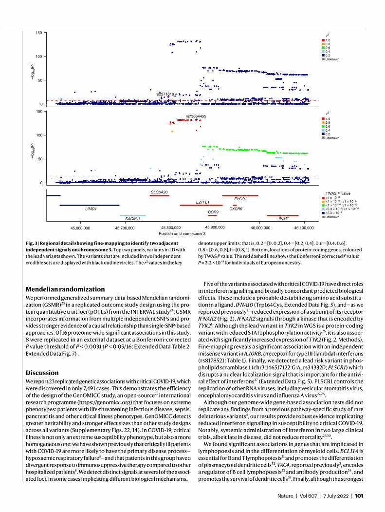
We inferred credible sets of variants using Bayesian fine-mapping with susieR13, by analysing the GWAS summaries of 17 regions of genomic length 3 Mb that were flanking groups of lead signals. We obtained 22 independent credible sets of variants for EUR and an addi-tional 2 from the trans-ancestry meta-analysis with a posterior inclusion probability greater than 0.95 (Extended Data Table 1, Supplementary Information). Fine-mapping of the association signals revealed puta-tive causal variants for both previously reported and novel association signals (see Supplementary Information, Extended Data Table 1). In 12 out of the 24 fine-mapped signals, the credible sets included 5 or fewer variants, and for 8 signals we detected variants with predicted missense or worse consequence across each credible set (Extended Data Table 1). We were able to fine-map multiple independent signals at previously identified loci (Fig. 3, Extended Data Figs. 2, 4). For example, the signal in the 3p21.31 region2, was fine-mapped into two independent associa-tions, with the credible set for the first refined to a single variant in the
5′ untranslated region (UTR) of SLC6A20 (chr3:45796521:G:T, rs2271616, odds ratio (OR): 1.29, 95% confidence interval (CI):1.21–1.37), and the second credible set including multiple variants in downstream and intronic regions of LZTFL1 (Fig. 3). Among the novel signals, at 3q24 and 9p21.3 we detected missense variants that affect PLSCR1 and IFNA10, respectively (chr3:146517122:G:A, rs343320, p.His262Tyr, OR: 1.24, 95% CI: 1.15–1.33, CADD: 22.6; chr9:21206606:C:G, rs28368148, p.Trp164Cys, OR:1.74, 95% CI: 1.45–2.09, CADD: 23.9). Both are predicted to be del-eterious by the Combined Annotation Dependent Depletion (CADD) tool14. Structural predictions for these variants suggest functional effects (Extended Data Fig. 5). We assessed whether the main signals of this study were underlain by rarer variants with a lower minor allele fre-quency (MAF) (less than 0.02%) than our GWAS default threshold (less than 0.5%), by including rarer variant summaries when fine-mapping, but no additional variants were added to the main credible sets (Sup-plementary Table 9).
Consistent with our expectation that genetic susceptibility has a stronger role in younger individuals, age-stratified analysis (individuals of younger than 60 years old versus individuals of 60 years old or above) in the EUR group revealed a signal in the 3p21.31 region with a signifi-cantly stronger effect in the younger age group (chr3:45801750:G:A, rs13071258, OR: 3.34, 95% CI: 2.98–3.75 versus OR: 2.1, 95% CI 1.88–2.34), which is in strong LD (r2 = 0.947) with the main GWAS signal indexed by rs73064425. Sex-specific analysis did not reveal significant effects (Supplementary Fig. 17).
ReplicationFor replication, we performed a meta-analysis of summary statistics generously shared by 23andMe and the COVID-19 Host Genetics Ini-tiative (HGI) data freeze 6 (B2). As a previous analysis of GenOMICC1 contributes a substantial part of the signal at each locus in HGI v.6, and leave-one-out analyses were not available, we removed the signal
EFNA4THBS3
SLC6A20
LZTFL1
PLSCR1ACSL6
HLA-DRB1 IFNA10 ELF5 FBRSL1ATP11A
RGMA
SLC22A31KANSL1
DPP9
ZGLP1 TYK2
FUT2
IFNAR2
IL10RB
LINC00649
2
4
8
16
32
64
128
192
1 2 3 4 5 6 7 8 9 10 11 12 13 14 15 16 17 18 19 20 21 22 XChromosome
–log
10(P
)
a= 1.04
0
50
100
0 2 4 6Expected –log10(P)
Ob
serv
ed –
log 10
(P)
EFNA4
TRIM46
THBS3BCL11A
SLC6A20
LZTFL1
PLSCR1ACSL6
HLA-DRB1
LINC01276
IFNA10ELF5 FBRSL1
ATP11A
SLC22A31
KANSL1
DPP9
ZGLP1
TYK2
FUT2
IFNAR2
IL10RB
LINC00649
2
4
8
16
32
64
128
192
1 2 3 4 5 6 7 8 9 10 11 12 13 14 15 16 17 18 19 20 21 22 XChromosome
b
0
50
100
150
0 2 4 6
–log
10(P
)
Ob
serv
ed –
log 10
(P)
Expected –log10(P)
= 1.01
Fig. 1 | GWAS results for the EUR ancestry group, and multi-ancestry meta-analysis. Manhattan plots are shown on the left and quantile–quantile (QQ) plots of observed versus expected P values on the right, with genomic inflation (λ) displayed for each analysis. Highlighted results in blue in the Manhattan plots indicate variants that are LD-clumped (r2 = 0.1, P2 = 0.01, EUR LD) with the lead variants at each locus. Gene name annotation indicates genes
that are affected by the predicted worst consequence type of each lead variant (annotation by Variant Effect Predictor (VEP)). For the HLA locus, the gene that was identified by HLA allele analysis is annotated. The GWAS was performed using logistic regression and meta-analysed by the inverse variant method. The red dashed line shows the Bonferroni-corrected P value: P = 2.2 × 10⁻⁸.
100 | Nature | Vol 607 | 7 July 2022
Article
from GenOMICC cases in HGI v.6 using mathematical subtraction to ensure independence (Methods). Using LD clumping to find variants genotyped in both the discovery and replication studies, we required P < 0.002 (0.05/25) and concordant direction of effect (Table 1, Sup-plementary Table 8) for replication. We interrogated two variants that failed replication in this set in a second GWAS meta-analysis of hospi-talized patients with COVID-19 from UK Biobank, AncestryDNA, Penn Medicine Biobank and Geisinger Health Systems, which included a total of 9,937 individuals who were hospitalized with COVID-19 and 1,059,390 control individuals. This led to a further successful replicated finding, in IFNA10 (Table 1).
We replicated 23 of the 25 significant associations that were iden-tified in the population-specific and/or multi-ancestry GWASs. One of the non-replicated signals (rs4424872) corresponds to a rare variant that may not be well represented in the replication datasets— which are dominated by single-nucleotide polymorphism (SNP) genotyp-ing data—but which also had significant heterogeneity among ancestries. The second non-replicated signal is within the human leukocyte antigen (HLA) locus, which has complex LD (see below).
HLA regionThe lead variant in the HLA region, rs9271609, lies upstream of the HLA-DQA1 and HLA-DRB1 genes. To investigate the contribution of specific HLA alleles to the observed association in the HLA region, we imputed HLA alleles at a four-digit (two-field) level using HIBAG15. The only allele that reached genome-wide significance was HLA-DRB1*04:01 (OR: 0.80, 95% CI: 0.75–0.86, P = 1.6 × 10⁻¹⁰ in EUR), which has a stronger P value than the lead SNP in the region (OR: 0.88, 95% CI: 0.84–0.92, P = 3.3 × 10⁻⁹ in EUR) and is a better fit to the data (Akaike information criterion (AIC): AICDRB1*04:01 = 30,241.34; AICleadSNP = 30,252.93) (Extended Data Fig. 6). HLA-DRB1*04:01 has been previ-ously reported to confer protection against severe disease in a small cohort of European ancestry16.
Gene burden testingTo assess the contribution of rare variants to critical illness, we performed gene-based analysis using SKAT-O as implemented in
SAIGE-GENE17 on a subset of 12,982 individuals from our cohort (7,491 individuals with critical COVID-19 and 5,391 control individuals), for which the genome-sequencing data were processed with the same alignment and variant calling pipeline. We tested the burden of rare (MAF < 0.5%) variants considering the predicted variant consequence type (tested variant counts provided in the Supplementary Informa-tion). We assessed burden using a strict definition for damaging vari-ants (high-confidence putative loss-of-function (pLoF) variants as identified by LOFTEE18) and a lenient definition (pLoF plus missense variants with CADD ≥ 10)14, but found no significant associations at a gene-wide-significance level. Moreover, all individual rare variants included in the tests had P values greater than 10−5.
Consistent with other recent work11, we did not find any significant gene burden test associations among the 13 genes previously reported from an interferon-pathway-focused study4 (tests for all genes had P > 0.05; Supplementary Information), and we did not replicate the reported association19–21 in TLR7 (EUR P = 0.30 for pLoF and P = 0.075 for missense variants).
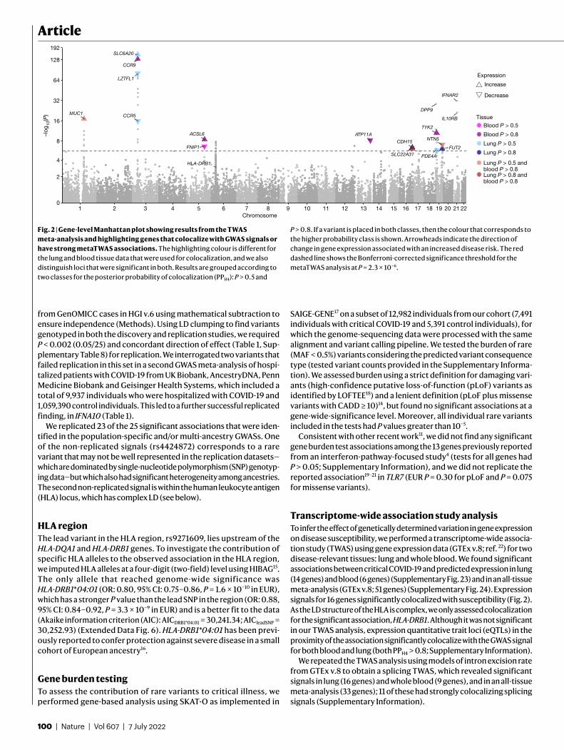
Transcriptome-wide association study analysisTo infer the effect of genetically determined variation in gene expression on disease susceptibility, we performed a transcriptome-wide associa-tion study (TWAS) using gene expression data (GTEx v.8; ref. 22) for two disease-relevant tissues: lung and whole blood. We found significant associations between critical COVID-19 and predicted expression in lung (14 genes) and blood (6 genes) (Supplementary Fig. 23) and in an all-tissue meta-analysis (GTEx v.8; 51 genes) (Supplementary Fig. 24). Expression signals for 16 genes significantly colocalized with susceptibility (Fig. 2). As the LD structure of the HLA is complex, we only assessed colocalization for the significant association, HLA-DRB1. Although it was not significant in our TWAS analysis, expression quantitative trait loci (eQTLs) in the proximity of the association significantly colocalize with the GWAS signal for both blood and lung (both PPH4 > 0.8; Supplementary Information).
We repeated the TWAS analysis using models of intron excision rate from GTEx v.8 to obtain a splicing TWAS, which revealed significant signals in lung (16 genes) and whole blood (9 genes), and in an all-tissue meta-analysis (33 genes); 11 of these had strongly colocalizing splicing signals (Supplementary Information).
ACSL6 ATP11A
CCR5
CCR9
CDH15
DPP9
FNIP1 FUT2
HLA-DRB1
IFNAR2
IL10RB
LZTFL1
MUC1
NTN5
PDE4ASLC22A31
SLC6A20
TYK2
0
2
4
8
16
32
64
128
192
1 2 3 4 5 6 7 8 9 10 11 12 13 14 15 16 17 18 19 20 21 22Chromosome
–log
10(P
)
Expression
Increase
Decrease
TissueBlood P > 0.5
Blood P > 0.8
Lung P > 0.5
Lung P > 0.8
Lung P > 0.5 andblood P > 0.8Lung P > 0.8 andblood P > 0.8
Fig. 2 | Gene-level Manhattan plot showing results from the TWAS meta-analysis and highlighting genes that colocalize with GWAS signals or have strong metaTWAS associations. The highlighting colour is different for the lung and blood tissue data that were used for colocalization, and we also distinguish loci that were significant in both. Results are grouped according to two classes for the posterior probability of colocalization (PPH4): P > 0.5 and
P > 0.8. If a variant is placed in both classes, then the colour that corresponds to the higher probability class is shown. Arrowheads indicate the direction of change in gene expression associated with an increased disease risk. The red dashed line shows the Bonferroni-corrected significance threshold for the metaTWAS analysis at P = 2.3 × 10⁻⁶.
Nature | Vol 607 | 7 July 2022 | 101
Mendelian randomizationWe performed generalized summary-data-based Mendelian randomi-zation (GSMR)23 in a replicated outcome study design using the pro-tein quantitative trait loci (pQTLs) from the INTERVAL study24. GSMR incorporates information from multiple independent SNPs and pro-vides stronger evidence of a causal relationship than single-SNP-based approaches. Of 16 proteome-wide-significant associations in this study, 8 were replicated in an external dataset at a Bonferroni-corrected P value threshold of P < 0.0031 (P < 0.05/16; Extended Data Table 2, Extended Data Fig. 7) .
DiscussionWe report 23 replicated genetic associations with critical COVID-19, which were discovered in only 7,491 cases. This demonstrates the efficiency of the design of the GenOMICC study, an open-source25 international research programme (https://genomicc.org) that focuses on extreme phenotypes: patients with life-threatening infectious disease, sepsis, pancreatitis and other critical illness phenotypes. GenOMICC detects greater heritability and stronger effect sizes than other study designs across all variants (Supplementary Figs. 22, 14). In COVID-19, critical illness is not only an extreme susceptibility phenotype, but also a more homogeneous one: we have shown previously that critically ill patients with COVID-19 are more likely to have the primary disease process—hypoxaemic respiratory failure5—and that patients in this group have a divergent response to immunosuppressive therapy compared to other hospitalized patients8. We detect distinct signals at several of the associ-ated loci, in some cases implicating different biological mechanisms.
Five of the variants associated with critical COVID-19 have direct roles in interferon signalling and broadly concordant predicted biological effects. These include a probable destabilizing amino acid substitu-tion in a ligand, IFNA10 (Trp164Cys, Extended Data Fig. 5), and—as we reported previously1—reduced expression of a subunit of its receptor IFNAR2 (Fig. 2). IFNAR2 signals through a kinase that is encoded by TYK21. Although the lead variant in TYK2 in WGS is a protein-coding variant with reduced STAT1 phosphorylation activity26, it is also associ-ated with significantly increased expression of TYK2 (Fig. 2, Methods). Fine-mapping reveals a significant association with an independent missense variant in IL10RB, a receptor for type III (lambda) interferons (rs8178521; Table 1). Finally, we detected a lead risk variant in phos-pholipid scramblase 1 (chr3:146517122:G:A, rs343320; PLSCR1) which disrupts a nuclear localization signal that is important for the antivi-ral effect of interferons27 (Extended Data Fig. 5). PLSCR1 controls the replication of other RNA viruses, including vesicular stomatitis virus, encephalomyocarditis virus and influenza A virus27,28.
Although our genome-wide gene-based association tests did not replicate any findings from a previous pathway-specific study of rare deleterious variants4, our results provide robust evidence implicating reduced interferon signalling in susceptibility to critical COVID-19. Notably, systemic administration of interferon in two large clinical trials, albeit late in disease, did not reduce mortality29,30.
We found significant associations in genes that are implicated in lymphopoesis and in the differentiation of myeloid cells. BCL11A is essential for B and T lymphopoiesis31 and promotes the differentiation of plasmacytoid dendritic cells32. TAC4, reported previously3, encodes a regulator of B cell lymphopoesis33 and antibody production34, and promotes the survival of dendritic cells35. Finally, although the strongest
1.00.80.60.40.2Unknown
Position on chromosome 345,600,000 45,800,000 46,000,000
SLC6A20
FYCO1
LIMD1
LZTFL1
CXCR6
SACM1L
CCR9
XCR1
TWAS P value<1 × 10–20
<1 × 10⁻¹⁵; ≥1 × 10⁻²⁰
<1 × 10⁻¹⁰; ≥1 × 10⁻¹⁵
<2.3 × 10⁻⁶; ≥1 × 10⁻¹⁰
≥2.3 × 10⁻⁶
Unknown
r21.00.80.60.40.2Unknown
0
50
100
150
0
50
100
150–l
og10
(P)
rs2271616
rs73064455
–log
10(P
)
r2
45,700,000 45,900,000 46,100,000
Fig. 3 | Regional detail showing fine-mapping to identify two adjacent independent signals on chromosome 3. Top two panels, variants in LD with the lead variants shown. The variants that are included in two independent credible sets are displayed with black outline circles. The r2 values in the key
denote upper limits; that is, 0.2 = [0, 0.2], 0.4 = [0.2, 0.4], 0.6 = [0.4, 0.6], 0.8 = [0.6, 0.8],1 = [0.8, 1]. Bottom, locations of protein-coding genes, coloured by TWAS P value. The red dashed line shows the Bonferroni-corrected P value: P = 2.2 × 10⁻⁸ for individuals of European ancestry.

102 | Nature | Vol 607 | 7 July 2022
Articlefine-mapping signal at 5q31.1 (chr5:131995059:C:T, rs56162149) is in an intron of ACSL6 with significant effects on expression (Supple-mentary Information), the credible set includes a missense variant in CSF2 (encoding granulocyte–macrophage colony stimulating factor; GM-CSF) of uncertain significance (chr5:132075767:T:C; Extended Data Table 1). We have previously shown that GM-CSF is strongly up-regulated in critical COVID-1936, and it is already under investigation as a target for therapy37. Mendelian randomization results are consistent with a direct link between the plasma levels of a closely related cytokine receptor subunit, IL3RA, and critical COVID-19 (Extended Data Table 2).
Fine-mapping, colocalization and TWAS analyses provide evidence for increased expression of MUC1 as the mediator of the association with rs41264915 (Supplementary Table 12). This suggests that mucins could have a therapeutically important role in the development of critical illness in COVID-19.
Mendelian randomization provides genetic evidence in support of a causal role for coagulation factors (F8) and platelet activation (PDGFRL) in critical COVID-19 (Extended Data Table 2, Extended Data Fig. 7), consist-ent with autopsy6, proteomic38 and therapeutic39 evidence. Perhaps more importantly, we identify specific and closely related intercellular adhesion molecules that have known roles in the recruitment of inflammatory cells to sites of inflammation, including E-selectin (SELE), intercellular adhesion molecule 5 (ICAM5) and DC-SIGN (dendritic-cell-specific ICAM3-grabbing non-integrin; CD209), which may provide additional therapeutic targets. DC-SIGN (CD209) mediates pathogen endocytosis and antigen presenta-tion, and is known to be involved in multiple viral infections, including SARS-CoV and influenza A virus. It has affinity for SARS-CoV-240,41.
Our previous report of an association between the OAS gene cluster and severe disease was robustly replicated in an external cohort1, but does not meet genome-wide significance in the present analysis (Sup-plementary Table 7). This may indicate a change in the observed effect size because any effect that is detected in GWASs is more likely to have been sampled from the larger end of the range of possible effect sizes —the ‘winner’s curse’. Alternatively, it may indicate either a change in the population of patients (cases or controls) or a change in the pathogen. For example it is possible that—as with the other coronaviruses that are known to infect humans42—more recent variants of SARS-CoV-2 have evolved to overcome this host antiviral defence mechanism.
LimitationsIn contrast to microarray genotyping, WGS is a rapidly evolving and rela-tively new technology for GWASs, with relatively few sources of popula-tion controls. We selected a control cohort from the 100,000 Genomes Project, which was sequenced and analysed using a different platform and bioinformatics pipeline compared with the case cohort (Extended Data Fig. 1). However, to minimize the risk of false-positive associations due to technical artifacts, extensive quality measures were used (Methods). In brief, we masked low-quality genotypes, filtered for genotype signal using a low threshold for missingness and performed a control–control relative allele frequency filter using a subset of samples processed with both bioinformatics pipelines. Finally, we required all significant associa-tions to be supported by local variants in LD, which may be excessively stringent (Methods). Although this approach may remove some true asso-ciations, our priority is to maximize confidence in the reported signals. Of 25 variants that meet this requirement, 23 are externally replicated, and the remaining 2 may be true associations that are yet to be replicated owing to a lack of coverage or power in the replication datasets.
The design of our study incorporates genetic signals for every stage in the disease progression into a single phenotype. This includes estab-lishment of infection, viral replication, inflammatory lung injury and hypoxaemic respiratory failure. Although we can have considerable confidence that the replicated associations with critical COVID-19 we report are robust, we cannot determine at which stage in the disease pro-cess, or in which tissue, the relevant biological mechanisms are active.
ConclusionsThese genetic associations identify biological mechanisms that may underlie the development of life-threatening COVID-19, several of which may be amenable to therapeutic targeting. Furthermore, we demon-strate the value of WGS for fine-mapping loci in a complex trait. In the context of the ongoing global pandemic, translation to clinical practice is an urgent priority. As with our previous work, biological and molecu-lar studies—and, where appropriate, large-scale randomized trials—will be essential before our findings can be translated into clinical practice.
Online contentAny methods, additional references, Nature Research reporting sum-maries, source data, extended data, supplementary information, acknowledgements, peer review information; details of author contri-butions and competing interests; and statements of data and code avail-ability are available at https://doi.org/10.1038/s41586-022-04576-6.
1. Pairo-Castineira, E. et al. Genetic mechanisms of critical illness in COVID-19. Nature 591, 92–98 (2021).
2. Ellinghaus, D. et al. Genomewide association study of severe Covid-19 with respiratory failure. N. Engl. J. Med. 383, 1522–1534 (2020).
3. COVID-19 Host Genetics Initiative. Mapping the human genetic architecture of COVID-19. Nature 600, 472–477 (2021).
4. Zhang, Q. et al. Inborn errors of type I IFN immunity in patients with life-threatening COVID-19. Science 370, eabd4570 (2020).
5. Docherty, A. B. et al. Features of 20,133 UK patients in hospital with covid-19 using the ISARIC WHO Clinical Characterisation Protocol: prospective observational cohort study. BMJ 369, m1985 (2020).
6. Dorward, D. A. et al. Tissue-specific immunopathology in fatal COVID-19. Am. J. Respir. Crit. Care Med. 203, 192–201 (2021).
7. Millar, J. E. et al. Distinct clinical symptom patterns in patients hospitalised with COVID-19 in an analysis of 59,011 patients in the ISARIC-4C study. Sci. Rep. 12, 6843 (2022).
8. The RECOVERY Collaborative Group. Dexamethasone in hospitalized patients with Covid-19. N. Engl. J. Med. 384, 693–704 (2021).
9. Degenhardt, F. et al. New susceptibility loci for severe COVID-19 by detailed GWAS analysis in European populations. Preprint at medRxiv https://doi.org/10.1101/ 2021.07.21.21260624 (2021).
10. Povysil, G. et al. Rare loss-of-function variants in type i IFN immunity genes are not associated with severe COVID-19. J. Clin. Invest. 131, e147834 (2021).
11. Kosmicki, J. A. et al. Pan-ancestry exome-wide association analyses of COVID-19 outcomes in 586,157 individuals. Am. J. Hum. Genet. 108, 1350–1355 (2021).
12. Zhou, W. et al. Efficiently controlling for case-control imbalance and sample relatedness in large-scale genetic association studies. Nat. Genet. 50, 1335–1341 (2018).
13. Wang, G., Sarkar, A., Carbonetto, P. & Stephens, M. A simple new approach to variable selection in regression, with application to genetic fine mapping. J. R. Stat. Soc. B 82, 1273–1300 (2020).
14. Rentzsch, P., Witten, D., Cooper, G. M., Shendure, J. & Kircher, M. CADD: predicting the deleteriousness of variants throughout the human genome. Nucleic Acids Res. 47, D886–D894 (2018).
15. Zheng, X. et al. HIBAG—HLA genotype imputation with attribute bagging. Pharmacogenomics J. 14, 192–200 (2014).
16. Langton, D. J. et al. The influence of HLA genotype on the severity of COVID-19 infection. HLA 98, 14–22 (2021).
17. Zhou, W. et al. Scalable generalized linear mixed model for region-based association tests in large biobanks and cohorts. Nat. Genet. 52, 634–639 (2020).
18. Karczewski, K. J. et al. The mutational constraint spectrum quantified from variation in 141,456 humans. Nature 581, 434–443 (2020).
19. Asano, T. et al. X-linked recessive TLR7 deficiency in ∼1% of men under 60 years old with life-threatening COVID-19. Sci. Immunol. 6, eabl4348 (2021).
20. Fallerini, C. et al. Association of toll-like receptor 7 variants with life-threatening COVID-19 disease in males: findings from a nested case-control study. eLife 10, e67569 (2021).
21. van der Made, C. I. et al. Presence of genetic variants among young men with severe COVID-19. J. Am. Med. Assoc. 324, 663–673 (2020).
22. The GTEx Consortium. The GTEx Consortium atlas of genetic regulatory effects across human tissues. Science 369, 1318–1330 (2020).
23. Zhu, Z. et al. Causal associations between risk factors and common diseases inferred from GWAS summary data. Nat. Commun. 9, 224 (2018).
24. Sun, B. B. et al. Genomic atlas of the human plasma proteome. Nature 558, 73–79 (2018).25. Dunning, J. W. et al. Open source clinical science for emerging infections. Lancet Infect.
Dis. 14, 8–9 (2014).26. Dendrou, C. A. et al. Resolving TYK2 locus genotype-to-phenotype differences in
autoimmunity. Sci. Transl. Med. 8, 363ra149 (2016).27. Dong, B. et al. Phospholipid scramblase 1 potentiates the antiviral activity of interferon.
J. Virol. 78, 8983–8993 (2004).28. Luo, W. et al. Phospholipid scramblase 1 interacts with influenza a virus NP, impairing its
nuclear import and thereby suppressing virus replication. PLoS Pathog. 14, e1006851 (2018).29. WHO Solidarity Trial Consortium. Repurposed antiviral drugs for Covid-19—interim WHO
Solidarity trial results. N. Engl. J. Med. 384, 497–511 (2021).
Nature | Vol 607 | 7 July 2022 | 103
30. Kalil, A. C. et al. Efficacy of interferon beta-1a plus remdesivir compared with remdesivir alone in hospitalised adults with COVID-19: a double-blind, randomised, placebo-controlled, phase 3 trial. Lancet Respir. Med. 12, 1365–1376 (2021).
31. Yu, Y. et al. Bcl11a is essential for lymphoid development and negatively regulates p53. J. Exp. Med. 209, 2467–2483 (2012).
32. Reizis, B. Plasmacytoid dendritic cells: development, regulation, and function. Immunity 50, 37–50 (2019).
33. Zhang, Y., Lu, L., Furlonger, C., Wu, G. E. & Paige, C. J. Hemokinin is a hematopoietic-specific tachykinin that regulates b lymphopoiesis. Nat. Immunol. 1, 392–397 (2000).
34. Wang, W. et al. Hemokinin-1 activates the MAPK pathway and enhances B cell proliferation and antibody production. J. Immunol. 184, 3590–3597 (2010).
35. Janelsins, B. M. et al. Proinflammatory tachykinins that signal through the neurokinin 1 receptor promote survival of dendritic cells and potent cellular immunity. Blood 113, 3017–3026 (2009).
36. Thwaites, R. S. et al. Inflammatory profiles across the spectrum of disease reveal a distinct role for GM-CSF in severe COVID-19. Sci. Immunol. 6, eabg9873 (2021).
37. Lang, F. M., Lee, K. M.-C., Teijaro, J. R., Becher, B. & Hamilton, J. A. GM-CSF-based treatments in COVID-19: reconciling opposing therapeutic approaches. Nat. Rev. Immunol. 20, 507–514 (2020).
38. Reyes, L. et al. A type I IFN, prothrombotic hyperinflammatory neutrophil signature is distinct for COVID-19 ARDS. Wellcome Open Res. 6, 38 (2021).
39. Lawler, P. R. et al. Therapeutic anticoagulation with heparin in noncritically ill patients with Covid-19. N. Engl. J. Med. 385, 790–802 (2021).
40. Amraei, R. et al. CD209L/L-SIGN and CD209/DC-SIGN act as receptors for SARS-CoV-2. ACS Cent. Sci. 7, 1156–1165 (2021).
41. Thépaut, M. et al. DC/L-SIGN recognition of spike glycoprotein promotes SARS-CoV-2 trans-infection and can be inhibited by a glycomimetic antagonist. PLoS Pathog. 17, e1009576 (2021).
42. Silverman, R. H. & Weiss, S. R. Viral phosphodiesterases that antagonize double-stranded RNA signaling to RNase L by degrading 2-5A. J. Interferon Cytokine Res. 34, 455–463 (2014).
Publisher’s note Springer Nature remains neutral with regard to jurisdictional claims in published maps and institutional affiliations.
Open Access This article is licensed under a Creative Commons Attribution 4.0 International License, which permits use, sharing, adaptation, distribution and reproduction in any medium or format, as long as you give appropriate
credit to the original author(s) and the source, provide a link to the Creative Commons license, and indicate if changes were made. The images or other third party material in this article are included in the article’s Creative Commons license, unless indicated otherwise in a credit line to the material. If material is not included in the article’s Creative Commons license and your intended use is not permitted by statutory regulation or exceeds the permitted use, you will need to obtain permission directly from the copyright holder. To view a copy of this license, visit http://creativecommons.org/licenses/by/4.0/.
© The Author(s) 2022
1Genomics England, London, UK. 2Roslin Institute, University of Edinburgh, Edinburgh, UK. 3MRC Human Genetics Unit, Institute of Genetics and Cancer, University of Edinburgh, Western General Hospital, Edinburgh, UK. 4Centre for Inflammation Research, The Queen’s Medical Research Institute, University of Edinburgh, Edinburgh, UK. 5Wellcome Centre for Human Genetics, University of Oxford, Oxford, UK. 6Institute for Molecular Bioscience, The University of Queensland, Brisbane, Queensland, Australia. 7Biostatistics Group, Greater Bay Area Institute of Precision Medicine (Guangzhou), Fudan University, Guangzhou, China. 8Centre for Global Health Research, Usher Institute of Population Health Sciences and Informatics, Edinburgh, UK. 9Edinburgh Clinical Research Facility, Western General Hospital, University of Edinburgh, Edinburgh, UK. 10Intensive Care Unit, Royal Infirmary of Edinburgh, Edinburgh, UK. 11Department of Critical Care Medicine, Queen’s University and Kingston Health Sciences Centre, Kingston, Ontario, Canada. 12Clinical Research Centre at St Vincent’s University Hospital, University College Dublin, Dublin, Ireland. 13NIHR Health Protection Research Unit for Emerging and Zoonotic Infections, Institute of Infection, Veterinary and Ecological Sciences, University of Liverpool, Liverpool, UK. 14Respiratory Medicine and Institute in the Park, Alder Hey Children’s Hospital and University of Liverpool, Liverpool, UK. 15Illumina Cambridge, Great Abington, UK. 16Regeneron Genetics Center, Tarrytown, NY, USA. 17Geisinger, Danville, PA, USA. 18Department of Genetics, Perelman School of Medicine, University of Pennsylvania, Philadelphia, PA, USA. 19Test and Trace, the Health Security Agency, Department of Health and Social Care, London, UK. 20Department of Intensive Care Medicine, Guy’s and St Thomas’ NHS Foundation Trust, London, UK. 21Department of Medicine, University of Cambridge, Cambridge, UK. 22William Harvey Research Institute, Barts and the London School of Medicine and Dentistry, Queen Mary University of London, London, UK. 23Centre for Tropical Medicine and Global Health, Nuffield Department of Medicine, University of Oxford, Oxford, UK. 24Department of Anaesthesia and Intensive Care, The Chinese University of Hong Kong, Prince of Wales Hospital, Hong Kong, China. 25Wellcome–Wolfson Institute for Experimental Medicine, Queen’s University Belfast, Belfast, UK. 26Department of Intensive Care Medicine, Royal Victoria Hospital, Belfast, UK. 27UCL Centre for Human Health and Performance, London, UK. 28National Heart and Lung Institute, Imperial College London, London, UK. 29Imperial College Healthcare NHS Trust: London, London, UK. 30Imperial College, London, UK. 31Intensive Care National Audit and Research Centre, London, UK. 32School of Life Sciences, Westlake University, Hangzhou, China. 33Westlake Laboratory of Life Sciences and Biomedicine, Hangzhou, China. 34Great Ormond Street Hospital, London, UK. 35William Harvey Research Institute, Queen Mary University of London, London, UK. 556These authors contributed equally: Athanasios Kousathanas, Erola Pairo-Castineira. 557These authors jointly supervised this work: Sara Clohisey Hendry, Loukas Moutsianas, Andy Law, Mark J. Caulfield, J. Kenneth Baillie. *Lists of authors and their affiliations appear online. ✉e-mail: [email protected]; [email protected]

ArticleGenOMICC investigatorsGenOMICC co-investigatorsJ. Kenneth Baillie36,37, Colin Begg38, Sara Clohisey Hendry36, Charles Hinds39, Peter Horby40, Julian Knight41, Lowell Ling42, David Maslove43, Danny McAuley44,45, Johnny Millar36, Hugh Montgomery46, Alistair Nichol47, Peter J. M. Openshaw48,49, Alexandre C. Pereira50, Chris P. Ponting51, Kathy Rowan52, Malcolm G. Semple53,54, Manu Shankar-Hari55, Charlotte Summers56 & Timothy Walsh37
Management, laboratory and data teamLatha Aravindan57, Ruth Armstrong36, J. Kenneth Baillie36,37, Heather Biggs58, Ceilia Boz36, Adam Brown36, Richard Clark59, Sara Clohisey Hendry36, Audrey Coutts59, Judy Coyle36, Louise Cullum36, Sukamal Das57, Nicky Day36, Lorna Donnelly59, Esther Duncan36, Angie Fawkes59, Paul Finernan36, Max Head Fourman36, Anita Furlong58, James Furniss36, Bernadette Gallagher36, Tammy Gilchrist59, Ailsa Golightly36, Fiona Griffiths36, Katarzyna Hafezi59, Debbie Hamilton36, Ross Hendry36, Andy Law36, Dawn Law36, Rachel Law36, Sarah Law36, Rebecca Lidstone-Scott36, Louise Macgillivray59, Alan Maclean59, Hanning Mal36, Sarah McCafferty59, Ellie Mcmaster36, Jen Meikle36, Shona C. Moore53, Kirstie Morrice59, Lee Murphy59, Sheena Murphy57, Mybaya Hellen36, Wilna Oosthuyzen36, Chenqing Zheng60, Jiantao Chen60, Nick Parkinson36, Trevor Paterson36, Katherine Schon58, Andrew Stenhouse36, Mihaela Das57, Maaike Swets36,61, Helen Szoor-McElhinney36, Filip Taneski36, Lance Turtle53, Tony Wackett36, Mairi Ward36, Jane Weaver36, Nicola Wrobel59, Marie Zechner36 & Mybaya Hellen36
Guy’s and St Thomas’ Hospital teamGill Arbane62, Aneta Bociek62, Sara Campos62, Neus Grau62, Tim Owen Jones62, Rosario Lim62, Martina Marotti62, Marlies Ostermann62, Manu Shankar-Hari62 & Christopher Whitton62
Barts Health NHS Trust teamZoe Alldis63, Raine Astin-Chamberlain63, Fatima Bibi63, Jack Biddle63, Sarah Blow63, Matthew Bolton63, Catherine Borra63, Ruth Bowles63, Maudrian Burton63, Yasmin Choudhury63, David Collier63, Amber Cox63, Amy Easthope63, Patrizia Ebano63, Stavros Fotiadis63, Jana Gurasashvili63, Rosslyn Halls63, Pippa Hartridge63, Delordson Kallon63, Jamila Kassam63, Ivone Lancoma-Malcolm63, Maninderpal Matharu63, Peter May63, Oliver Mitchelmore63, Tabitha Newman63, Mital Patel63, Jane Pheby63, Irene Pinzuti63, Zoe Prime63, Oleksandra Prysyazhna63, Julian Shiel63, Melanie Taylor63, Carey Tierney63, Suzanne Wood63, Anne Zak63 & Olivier Zongo63
James Cook University Hospital teamStephen Bonner64, Keith Hugill64, Jessica Jones64, Steven Liggett64 & Evie Headlam64
Royal Stoke University Hospital teamNageswar Bandla65, Minnie Gellamucho65, Michelle Davies65 & Christopher Thompson65
North Middlesex University Hospital NHS Trust teamMarwa Abdelrazik66, Dhanalakshmi Bakthavatsalam66, Munzir Elhassan66, Arunkumar Ganesan66, Anne Haldeos66, Jeronimo Moreno-Cuesta66, Dharam Purohit66, Rachel Vincent66, Kugan Xavier66, Rohit Kumar67, Alasdair Frater66, Malik Saleem66, David Carter66, Samuel Jenkins66, Zoe Lamond66 & Alanna Wall66
The Royal Liverpool University Hospital teamJaime Fernandez-Roman68, David O. Hamilton68, Emily Johnson68, Brian Johnston68, Maria Lopez Martinez68, Suleman Mulla68, David Shaw68, Alicia A. C. Waite68, Victoria Waugh68, Ingeborg D. Welters68 & Karen Williams68
King’s College Hospital teamAnna Cavazza69, Maeve Cockrell69, Eleanor Corcoran69, Maria Depante69, Clare Finney69, Ellen Jerome69, Mark McPhail69, Monalisa Nayak69, Harriet Noble69, Kevin O’Reilly69, Evita Pappa69, Rohit Saha69, Sian Saha69, John Smith69 & Abigail Knighton69
Charing Cross Hospital teamDavid Antcliffe70, Dorota Banach70, Stephen Brett70, Phoebe Coghlan70, Ziortza Fernandez70, Anthony Gordon70, Roceld Rojo70, Sonia Sousa Arias70 & Maie Templeton70
Nottingham University Hospital teamMegan Meredith71, Lucy Morris71, Lucy Ryan71, Amy Clark71, Julia Sampson71, Cecilia Peters71, Martin Dent71, Margaret Langley71, Saima Ashraf71, Shuying Wei71 & Angela Andrew71
John Radcliffe Hospital teamArchana Bashyal72, Neil Davidson72, Paula Hutton72, Stuart McKechnie72 & Jean Wilson72
Kingston Hospital teamDavid Baptista73, Rebecca Crowe73, Rita Fernandes73, Rosaleen Herdman-Grant73, Anna Joseph73, Denise O’Connor74, Meryem Allen73, Adam Loveridge73, India McKenley73, Eriko Morino73, Andres Naranjo73, Richard Simms73, Kathryn Sollesta73, Andrew Swain73, Harish Venkatesh73, Jacyntha Khera73 & Jonathan Fox73
Royal Infirmary of Edinburgh teamGillian Andrew75, J. Kenneth Baillie75, Lucy Barclay75, Marie Callaghan75, Rachael Campbell75, Sarah Clark75, Dave Hope75, Lucy Marshall75, Corrienne McCulloch75, Kate Briton75, Jo Singleton75 & Sophie Birch75
Queen Alexandra Hospital teamLutece Brimfield76, Zoe Daly76, David Pogson76, Steve Rose76 & Angela Nown76
Morriston Hospital teamCeri Battle77, Elaine Brinkworth77, Rachel Harford77, Carl Murphy77, Luke Newey77, Tabitha Rees77, Marie Williams77 & Sophie Arnold77
Addenbrooke’s Hospital teamPetra Polgarova78, Katerina Stroud78, Charlotte Summers78, Eoghan Meaney78, Megan Jones78, Anthony Ng78, Shruti Agrawal78, Nazima Pathan78, Deborah White78, Esther Daubney78 & Kay Elston78
BHRUT (Barking Havering)—Queen’s Hospital and King George Hospital teamLina Grauslyte79, Musarat Hussain79, Mandeep Phull79, Tatiana Pogreban79, Lace Rosaroso79, Erika Salciute79, George Franke79, Joanna Wong79 & Aparna George79
Royal Sussex County Hospital teamLaura Ortiz-Ruiz de Gordoa80, Emily Peasgood80 & Claire Phillips80
Queen Elizabeth Hospital teamMichelle Bates81, Jo Dasgin81, Jaspret Gill81, Annette Nilsson81, James Scriven81, Amy Collins82, Waqas Khaliq82 & Estefania Treus Gude82
St George’s Hospital teamCarlos Castro Delgado83, Deborah Dawson83, Lijun Ding83, Georgia Durrant83, Obiageri Ezeobu83, Sarah Farnell-Ward83, Abiola Harrison83, Rebecca Kanu83, Susannah Leaver83, Elena Maccacari83, Soumendu Manna83, Romina Pepermans Saluzzio83, Joana Queiroz83, Tinashe Samakomva83, Christine Sicat83, Joana Texeira83, Edna Fernandes Da Gloria83, Ana Lisboa83, John Rawlins83, Jisha Mathew83, Ashley Kinch83, William James Hurt83, Nirav Shah83, Victoria Clark83, Maria Thanasi83, Nikki Yun83 & Kamal Patel83
Stepping Hill Hospital teamSara Bennett84, Emma Goodwin84, Matthew Jackson84, Alissa Kent84, Clare Tibke84, Wiesia Woodyatt84 & Ahmed Zaki84
Countess of Chester Hospital teamAzmerelda Abraheem85, Peter Bamford85, Kathryn Cawley85, Charlie Dunmore85, Maria Faulkner85, Rumanah Girach85, Helen Jeffrey85, Rhianna Jones85, Emily London85, Imrun Nagra85, Farah Nasir85, Hannah Sainsbury85 & Clare Smedley85
Royal Blackburn Teaching Hospital teamTahera Patel86, Matthew Smith86, Srikanth Chukkambotla86, Aayesha Kazi86, Janice Hartley86, Joseph Dykes86, Muhammad Hijazi86, Sarah Keith86, Meherunnisa Khan86, Janet Ryan-Smith86, Philippa Springle86, Jacqueline Thomas86, Nick Truman86, Samuel Saad86, Dabheoc Coleman86, Christopher Fine86, Roseanna Matt86, Bethan Gay86, Jack Dalziel86, Syamlan Ali86, Drew Goodchild86, Rhiannan Harling86, Ravi Bhatterjee86, Wendy Goddard86, Chloe Davison86, Stephen Duberly86, Jeanette Hargreaves86 & Rachel Bolton86
The Tunbridge Wells Hospital and Maidstone Hospital teamMiriam Davey87, David Golden87 & Rebecca Seaman87
Royal Gwent Hospital teamShiney Cherian88, Sean Cutler88, Anne Emma Heron88, Anna Roynon-Reed88, Tamas Szakmany88, Gemma Williams88, Owen Richards88 & Yusuf Cheema88
Pinderfields General Hospital teamHollie Brooke89, Sarah Buckley89, Jose Cebrian Suarez89, Ruth Charlesworth89, Karen Hansson89, John Norris89, Alice Poole89, Alastair Rose89, Rajdeep Sandhu89, Brendan Sloan89, Elizabeth Smithson89, Muthu Thirumaran89, Veronica Wagstaff89 & Alexandra Metcalfe89
Royal Berkshire NHS Foundation Trust teamMark Brunton90, Jess Caterson90, Holly Coles90, Matthew Frise90, Sabi Gurung Rai90, Nicola Jacques90, Liza Keating90, Emma Tilney90, Shauna Bartley90 & Parminder Bhuie90
Broomfield Hospital teamSian Gibson91, Amanda Lyle91, Fiona McNeela91, Jayachandran Radhakrishnan91 & Alistair Hughes91
Northumbria Healthcare NHS Foundation Trust teamBryan Yates92, Jessica Reynolds92, Helen Campbell92, Maria Thompsom92, Steve Dodds92 & Stacey Duffy92
Whiston Hospital teamSandra Greer93, Karen Shuker93 & Ascanio Tridente93
Croydon University Hospital teamReena Khade94, Ashok Sundar94 & George Tsinaslanidis94
York Hospital teamIsobel Birkinshaw95, Joseph Carter95, Kate Howard95, Joanne Ingham95, Rosie Joy95, Harriet Pearson95, Samantha Roche95 & Zoe Scott95
Heartlands Hospital teamHollie Bancroft96, Mary Bellamy96, Margaret Carmody96, Jacqueline Daglish96, Faye Moore96, Joanne Rhodes96, Mirriam Sangombe96, Salma Kadiri96 & James Scriven96
Ashford and St Peter’s Hospital teamMaria Croft97, Ian White97, Victoria Frost97 & Maia Aquino97
Barnet Hospital teamRajeev Jha98, Vinodh Krishnamurthy98, Lai Lim98, Rajeev Jha98, Vinodh Krishnamurthy98 & Li Lim98
East Surrey Hospital teamEdward Combes99, Teishel Joefield99, Sonja Monnery99, Valerie Beech99 & Sallyanne Trotman99
Ninewells Hospital teamChristine Almaden-Boyle100, Pauline Austin100, Louise Cabrelli100, Stephen Cole100, Matt Casey100, Susan Chapman100, Stephen Cole100 & Clare Whyte100
Worthing Hospital teamYolanda Baird101,102, Aaron Butler101,102, Indra Chadbourn101,102, Linda Folkes101,102, Heather Fox101,102, Amy Gardner101,102, Raquel Gomez101,102, Gillian Hobden101,102, Luke Hodgson101,102, Kirsten King101,102, Michael Margarson101,102, Tim Martindale101,102, Emma Meadows101,102, Dana Raynard101,102, Yvette Thirlwall101,102, David Helm101,102 & Jordi Margalef101,102
Southampton General Hospital teamKristine Criste103, Rebecca Cusack103, Kim Golder103, Hannah Golding103, Oliver Jones103, Samantha Leggett103, Michelle Male103, Martyna Marani103, Kirsty Prager103, Toran Williams103, Belinda Roberts103 & Karen Salmon103
The Alexandra Hospital teamPeter Anderson104, Katie Archer104, Karen Austin104, Caroline Davis104, Alison Durie104, Olivia Kelsall104, Jessica Thrush104, Charlie Vigurs104, Laura Wild104, Hannah-Louise Wood104, Helen Tranter104, Alison Harrison104, Nicholas Cowley104, Michael McAlindon104, Andrew Burtenshaw104, Stephen Digby104, Emma Low104, Aled Morgan104, Naiara Cother104, Tobias Rankin104, Sarah Clayton104 & Alex McCurdy104
Sandwell General Hospital and City Hospital teamCecilia Ahmed105, Balvinder Baines105, Sarah Clamp105, Julie Colley105, Risna Haq105, Anne Hayes105, Jonathan Hulme105, Samia Hussain105, Sibet Joseph105, Rita Kumar105, Zahira Maqsood105 & Manjit Purewal105
Blackpool Victoria Hospital teamLeonie Benham106, Zena Bradshaw106, Joanna Brown106, Melanie Caswell106, Jason Cupitt106, Sarah Melling106, Stephen Preston106, Nicola Slawson106, Emma Stoddard106 & Scott Warden106
Royal Glamorgan Hospital teamBethan Deacon107, Ceri Lynch107, Carla Pothecary107, Lisa Roche107, Gwenllian Sera Howe107, Jayaprakash Singh107, Keri Turner107, Hannah Ellis107 & Natalie Stroud107
The Royal Oldham Hospital teamJodie Hunt108, Joy Dearden108, Emma Dobson108, Andy Drummond108, Michelle Mulcahy108, Sheila Munt108, Grainne O’Connor108, Jennifer Philbin108, Chloe Rishton108, Redmond Tully108 & Sarah Winnard108
Glasgow Royal Infirmary teamSusanne Cathcart109, Katharine Duffy109, Alex Puxty109, Kathryn Puxty109, Lynne Turner109, Jane Ireland109 & Gary Semple109
St James’s University Hospital and Leeds General Infirmary teamKate Long110, Simon Whiteley110, Elizabeth Wilby110 & Bethan Ogg110
University Hospital North Durham teamAmanda Cowton111,112, Andrea Kay111,112, Melanie Kent111,112, Kathryn Potts111,112, Ami Wilkinson111,112, Suzanne Campbell111,112 & Ellen Brown111,112
Fairfield General Hospital teamJulie Melville113, Jay Naisbitt113, Rosane Joseph113, Maria Lazo113, Olivia Walton113 & Alan Neal113
Wythenshawe Hospital teamPeter Alexander114, Schvearn Allen114, Joanne Bradley-Potts114, Craig Brantwood114, Jasmine Egan114, Timothy Felton114, Grace Padden114, Luke Ward114, Stuart Moss114 & Susannah Glasgow114
Royal Alexandra Hospital teamLynn Abel115, Michael Brett115, Brian Digby115, Lisa Gemmell115, James Hornsby115, Patrick MacGoey115, Pauline O’Neil115, Richard Price115, Natalie Rodden115, Kevin Rooney115, Radha Sundaram115 & Nicola Thomson115
Good Hope Hospital teamBridget Hopkins116, James Scriven116, Laura Thrasyvoulou116 & Heather Willis116
Tameside General Hospital teamMartyn Clark117, Martina Coulding117, Edward Jude117, Jacqueline McCormick117, Oliver Mercer117, Darsh Potla117, Hafiz Rehman117, Heather Savill117 & Victoria Turner117
Royal Derby Hospital teamCharlotte Downes118, Kathleen Holding118, Katie Riches118, Mary Hilton118, Mel Hayman118, Deepak Subramanian118 & Priya Daniel118
Medway Maritime Hospital teamOluronke Adanini119, Nikhil Bhatia119, Maines Msiska119 & Rebecca Collins119
Royal Victoria Infirmary teamIan Clement120, Bijal Patel120, A. Gulati120, Carole Hays120, K. Webster120, Anne Hudson120, Andrea Webster120, Elaine Stephenson120, Louise McCormack120, Victoria Slater120, Rachel Nixon120, Helen Hanson120, Maggie Fearby120, Sinead Kelly120, Victoria Bridgett120 & Philip Robinson120
Poole Hospital teamJulie Camsooksai121, Charlotte Humphrey121, Sarah Jenkins121, Henrik Reschreiter121, Beverley Wadams121 & Yasmin Death121
Bedford Hospital teamVictoria Bastion122, Daphene Clarke122, Beena David122, Harriet Kent122, Rachel Lorusso122, Gamu Lubimbi122, Sophie Murdoch122, Melchizedek Penacerrada122, Alastair Thomas122, Jennifer Valentine122, Ana Vochin122, Retno Wulandari122 & Brice Djeugam122
Queens Hospital Burton teamGillian Bell123, Katy English123, Amro Katary123 & Louise Wilcox123
North Manchester General Hospital teamMichelle Bruce124, Karen Connolly124, Tracy Duncan124, Helen T. Michael124, Gabriella Lindergard124, Samuel Hey124, Claire Fox124, Jordan Alfonso124, Laura Jayne Durrans124, Jacinta Guerin124, Bethan Blackledge124, Jade Harris124, Martin Hruska124, Ayaa Eltayeb124, Thomas Lamb124, Tracey Hodgkiss124, Lisa Cooper124 & Joanne Rothwell124
Aberdeen Royal Infirmary teamAngela Allan125, Felicity Anderson125, Callum Kaye125, Jade Liew125, Jasmine Medhora125, Teresa Scott125, Erin Trumper125 & Adriana Botello125
Derriford Hospital teamLiana Lankester126, Nikitas Nikitas126, Colin Wells126, Bethan Stowe126 & Kayleigh Spencer126
Manchester Royal Infirmary teamCraig Brandwood127, Lara Smith127, Richard Clark127, Katie Birchall127, Laurel Kolakaluri127, Deborah Baines127 & Anila Sukumaran127
Salford Royal Hospital teamElena Apetri128, Cathrine Basikolo128, Bethan Blackledge128, Laura Catlow128, Bethan Charles128, Paul Dark128, Reece Doonan128, Jade Harris128, Alice Harvey128, Daniel Horner128, Karen Knowles128, Stephanie Lee128, Diane Lomas128, Chloe Lyons128, Tracy Marsden128, Danielle McLaughlan128, Liam McMorrow128, Jessica Pendlebury128, Jane Perez128, Maria Poulaka128, Nicola Proudfoot128, Melanie Slaughter128, Kathryn Slevin128, Melanie Taylor128, Vicky Thomas128, Danielle Walker128, Angiy Michael128 & Matthew Collis128
William Harvey Hospital teamTracey Cosier129, Gemma Millen129, Neil Richardson129, Natasha Schumacher129, Heather Weston129 & James Rand129
Queen Elizabeth University Hospital teamNicola Baxter130, Steven Henderson130, Sophie Kennedy-Hay130, Christopher McParland130, Laura Rooney130, Malcolm Sim130 & Gordan McCreath130
Bradford Royal Infirmary teamLouise Akeroyd131, Shereen Bano131, Matt Bromley131, Lucy Gurr131, Tom Lawton131, James Morgan131, Kirsten Sellick131, Deborah Warren131, Brian Wilkinson131, Janet McGowan131, Camilla Ledgard131, Amelia Stacey131, Kate Pye131, Ruth Bellwood131 & Michael Bentley131
Bristol Royal Infirmary teamJeremy Bewley132, Zoe Garland132, Lisa Grimmer132, Bethany Gumbrill132, Rebekah Johnson132, Katie Sweet132, Denise Webster132 & Georgia Efford132
Norfolk and Norwich University Hospital (NNUH) teamKaren Convery133, Deirdre Fottrell-Gould133, Lisa Hudig133, Jocelyn Keshet-Price133, Georgina Randell133 & Katie Stammers133
Queen Elizabeth Hospital Gateshead teamMaria Bokhari134, Vanessa Linnett134, Rachael Lucas134, Wendy McCormick134, Jenny Ritzema134, Amanda Sanderson134 & Helen Wild134

ArticleSunderland Royal Hospital t ea mAnthony Rostron135, Alistair Roy135, Lindsey Woods135, Sarah Cornell135, Fiona Wakinshaw135, Kimberley Rogerson135 & Jordan Jarmain135
Aintree University Hospital teamRobert Parker136, Amie Reddy136, Ian Turner-Bone136, Laura Wilding136 & Peter Harding136
Hull Royal Infirmary teamCaroline Abernathy137, Louise Foster137, Andrew Gratrix137, Vicky Martinson137, Priyai Parkinson137, Elizabeth Stones137 & Llucia Carbral-Ortega138
University College Hospital teamGeorgia Bercades139, David Brealey139, Ingrid Hass139, Niall MacCallum139, Gladys Martir139, Eamon Raith139, Anna Reyes139 & Deborah Smyth139
Royal Devon and Exeter Hospital teamLetizia Zitter140, Sarah Benyon140, Suzie Marriott140, Linda Park140, Samantha Keenan140, Elizabeth Gordon140, Helen Quinn140 & Kizzy Baines140
The Royal Papworth Hospital teamLenka Cagova141, Adama Fofano141, Lucie Garner141, Helen Holcombe141, Sue Mepham141, Alice Michael Mitchell141, Lucy Mwaura141, Krithivasan Praman141, Alain Vuylsteke141 & Julie Zamikula141
Ipswich Hospital teamBally Purewal142, Vanessa Rivers142 & Stephanie Bell142
Southmead Hospital teamHayley Blakemore143, Borislava Borislavova143, Beverley Faulkner143, Emma Gendall143, Elizabeth Goff143, Kati Hayes143, Matt Thomas143, Ruth Worner143, Kerry Smith143 & Deanna Stephens143
Milton Keynes University Hospital teamLouise Mew144, Esther Mwaura144, Richard Stewart144, Felicity Williams144, Lynn Wren144 & Sara-Beth Sutherland144
Royal Hampshire County Hospital teamEmily Bevan145, Jane Martin145, Dawn Trodd145, Geoff Watson145 & Caroline Wrey Brown145
Great Ormond St Hospital and UCL Great Ormond St Institute of Child Health NIHR Biomedical Research Centre teamOlugbenga Akinkugbe146, Alasdair Bamford146, Emily Beech146, Holly Belfield146, Michael Bell146, Charlene Davies146, Gareth A. L. Jones146, Tara McHugh146, Hamza Meghari146, Lauran O’Neill146, Mark J. Peters146, Samiran Ray146 & Ana Luisa Tomas146
Stoke Mandeville Hospital teamIona Burn147, Geraldine Hambrook147, Katarina Manso147, Ruth Penn147, Pradeep Shanmugasundaram147, Julie Tebbutt147 & Danielle Thornton147
University Hospital of Wales teamJade Cole148, Michelle Davies148, Rhys Davies148, Donna Duffin148, Helen Hill148, Ben Player148, Emma Thomas148 & Angharad Williams148
Basingstoke and North Hampshire Hospital teamDenise Griffin149, Nycola Muchenje149, Mcdonald Mupudzi149, Richard Partridge149, Jo-Anna Conyngham149, Rachel Thomas149, Mary Wright149 & Maria Alvarez Corral149
Arrowe Park Hospital teamReni Jacob150, Cathy Jones150 & Craig Denmade150
Chesterfield Royal Hospital Foundation Trust teamSarah Beavis151, Katie Dale151, Rachel Gascoyne151, Joanne Hawes151, Kelly Pritchard151, Lesley Stevenson151 & Amanda Whileman151
Musgrove Park Hospital teamPatricia Doble152, Joanne Hutter152, Corinne Pawley152, Charmaine Shovelton152 & Marius Vaida152
Peterborough City Hospital teamDeborah Butcher153,154, Susie O’Sullivan153,154 & Nicola Butterworth-Cowin153,154
Royal Hallamshire Hospital and Northern General Hospital teamNorfaizan Ahmad155, Joann Barker155, Kris Bauchmuller155, Sarah Bird155, Kay Cawthron155, Kate Harrington155, Yvonne Jackson155, Faith Kibutu155, Becky Lenagh155, Shamiso Masuko155, Gary H. Mills155, Ajay Raithatha155, Matthew Wiles155, Jayne Willson155, Helen Newell155, Alison Lye155, Lorenza Nwafor155, Claire Jarman155, Sarah Rowland-Jones155, David Foote155, Joby Cole155, Roger Thompson155, James Watson155, Lisa Hesseldon155, Irene Macharia155, Luke Chetam155, Jacqui Smith155, Amber Ford155, Samantha Anderson155, Kathryn Birchall155, Kay Housley155, Sara Walker155, Leanne Milner155, Helena Hanratty155, Helen Trower155, Patrick Phillips155, Simon Oxspring155 & Ben Donne155
Dumfries and Galloway Royal Infirmary teamCatherine Jardine156, Dewi Williams156 & Alasdair Hay156
Royal Bolton Hospital teamRebecca Flanagan157, Gareth Hughes157, Scott Latham157, Emma McKenna157, Jennifer Anderson157, Robert Hull157 & Kat Rhead157
Lister Hospital teamCarina Cruz158 & Natalie Pattison158
Craigavon Area Hospital teamRob Charnock159, Denise McFarland159 & Denise Cosgrove159
Southport and Formby District General Hospital teamAshar Ahmed160, Anna Morris160, Srinivas Jakkula160 & Arvind Nune160
Calderdale Royal Hospital teamAsifa Ali161,162, Megan Brady161,162, Sam Dale161,162, Annalisa Dance161,162, Lisa Gledhill161,162, Jill Greig161,162, Kathryn Hanson161,162, Kelly Holdroyd161,162, Marie Home161,162, Diane Kelly161,162, Ross Kitson161,162, Lear Matapure161,162, Deborah Melia161,162, Samantha Mellor161,162, Tonicha Nortcliffe161,162, Jez Pinnell161,162, Matthew Robinson161,162, Lisa Shaw161,162, Ryan Shaw161,162, Lesley Thomis161,162, Alison Wilson161,162, Tracy Wood161,162, Lee-Ann Bayo161,162, Ekta Merwaha161,162, Tahira Ishaq161,162 & Sarah Hanley161,162
Prince Charles Hospital teamBethan Deacon163, Meg Hibbert163, Carla Pothecary163, Dariusz Tetla163, Christopher Woodford163, Latha Durga163 & Gareth Kennard-Holden163
Royal Bournemouth Hospital teamDebbie Branney164, Jordan Frankham164, Sally Pitts164 & Nigel White164
Royal Preston Hospital teamShondipon Laha165, Mark Verlander165 & Alexandra Williams165
Whittington Hospital teamAbdelhakim Altabaibeh166, Ana Alvaro166, Kayleigh Gilbert166, Louise Ma166, Loreta Mostoles166, Chetan Parmar166, Kathryn Simpson166, Champa Jetha166, Lauren Booker166 & Anezka Pratley166
Princess Royal Hospital teamColene Adams167, Anita Agasou167, Tracie Arden167, Amy Bowes167, Pauline Boyle167, Mandy Beekes167, Heather Button167, Nigel Capps167, Mandy Carnahan167, Anne Carter167, Danielle Childs167, Denise Donaldson167, Kelly Hard167, Fran Hurford167, Yasmin Hussain167, Ayesha Javaid167, James Jones167, Sanal Jose167, Michael Leigh167, Terry Martin167, Helen Millward167, Nichola Motherwell167, Rachel Rikunenko167, Jo Stickley167, Julie Summers167, Louise Ting167, Helen Tivenan167, Louise Tonks167, Rebecca Wilcox167, Denise Skinner168, Jane Gaylard168, Dee Mullan168 & Julie Newman168
Macclesfield District General Hospital teamMaureen Holland169, Natalie Keenan169, Marc Lyons169, Helen Wassall169, Chris Marsh169, Mervin Mahenthran169, Emma Carter169 & Thomas Kong169
Royal Surrey County Hospital teamHelen Blackman170, Ben Creagh-Brown170, Sinead Donlon170, Natalia Michalak-Glinska170, Sheila Mtuwa170, Veronika Pristopan170, Armorel Salberg170, Eleanor Smith170, Sarah Stone170, Charles Piercy170, Jerik Verula170, Dorota Burda170, Rugia Montaser170, Lesley Harden170, Irving Mayangao170, Cheryl Marriott170, Paul Bradley170 & Celia Harris170
Hereford County Hospital teamSusan Anderson171, Eleanor Andrews171, Janine Birch171, Emma Collins171, Kate Hammerton171 & Ryan O’Leary171
University Hospital of North Tees teamMichele Clark172 & Sarah Purvis172
Lincoln County Hospital teamRussell Barber173, Claire Hewitt173, Annette Hilldrith173, Karen Jackson-Lawrence173, Sarah Shepardson173, Maryanne Wills173, Susan Butler173, Silvia Tavares173, Amy Cunningham173, Julia Hindale173 & Sarwat Arif173
Royal Cornwall Hospital teamSarah Bean174, Karen Burt174 & Michael Spivey174
Royal United Hospital teamCarrie Demetriou175, Charlotte Eckbad175, Sarah Hierons175, Lucy Howie175, Sarah Mitchard175, Lidia Ramos175, Alfredo Serrano-Ruiz175, Katie White175 & Fiona Kelly175
Royal Brompton Hospital teamDaniele Cristiano176, Natalie Dormand176, Zohreh Farzad176, Mahitha Gummadi176, Kamal Liyanage176, Brijesh Patel176, Sara Salmi176, Geraldine Sloane176, Vicky Thwaites176, Mathew Varghese176 & Anelise C. Zborowski176
University Hospital Crosshouse teamJohn Allan177, Tim Geary177, Gordon Houston177, Alistair Meikle177 & Peter O’Brien177
Basildon Hospital teamMiranda Forsey178, Agilan Kaliappan178, Anne Nicholson178, Joanne Riches178, Mark Vertue178, Miranda Forsey178, Agilan Kaliappan178, Anne Nicholson178, Joanne Riches178 & Mark Vertue178
Glan Clwyd Hospital teamElizabeth Allan179, Kate Darlington179, Ffyon Davies179, Jack Easton179, Sumit Kumar179, Richard Lean179, Daniel Menzies179, Richard Pugh179, Xinyi Qiu179, Llinos Davies179, Hannah Williams179, Jeremy Scanlon179, Gwyneth Davies179, Callum Mackay179, Joanne Lewis179 & Stephanie Rees179
West Middlesex Hospital teamMetod Oblak180, Monica Popescu180 & Mini Thankachen180
Royal Lancaster Infirmary teamAndrew Higham181, Kerry Simpson181 & Jayne Craig181
Western General Hospital teamRosie Baruah182, Sheila Morris182, Susie Ferguson182 & Amy Shepherd182
Chelsea and Westminster NHS Foundation Trust teamLuke Stephen Prockter Moore183, Marcela Paola Vizcaychipi183, Laura Gomes de Almeida Martins183 & Jaime Carungcong183
The Queen Elizabeth Hospital teamInthakab Ali Mohamed Ali184, Karen Beaumont184, Mark Blunt184, Zoe Coton184, Hollie Curgenven184, Mohamed Elsaadany184, Kay Fernandes184, Sameena Mohamed Ally184, Harini Rangarajan184, Varun Sarathy184, Sivarupan Selvanayagam184, Dave Vedage184 & Matthew White184
King’s Mill Hospital teamMandy Gill185, Paul Paul185, Valli Ratnam185, Sarah Shelton185 & Inez Wynter185
Watford General Hospital teamSiobhain Carmody186 & Valerie Joan Page186
University Hospital Wishaw teamClaire Marie Beith187, Karen Black187, Suzanne Clements187, Alan Morrison187, Dominic Strachan187, Margaret Taylor187, Michelle Clarkson187, Stuart D’Sylva187 & Kathryn Norman187
Forth Valley Royal Hospital teamFiona Auld188, Joanne Donnachie188, Ian Edmond188, Lynn Prentice188, Nikole Runciman188, Dario Salutous188, Lesley Symon188, Anne Todd188, Patricia Turner188, Abigail Short188, Laura Sweeney188, Euan Murdoch188 & Dhaneesha Senaratne188
George Eliot Hospital NHS Trust teamMichaela Hill189, Thogulava Kannan189 & Laura Wild189
Barnsley Hospital teamRikki Crawley190, Abigail Crew190, Mishell Cunningham190, Allison Daniels190, Laura Harrison190, Susan Hope190, Ken Inweregbu190, Sian Jones190, Nicola Lancaster190, Jamie Matthews190, Alice Nicholson190 & Gemma Wray190
The Great Western Hospital teamHelen Langton191, Rachel Prout191, Malcolm Watters191 & Catherine Novis191
Harefield Hospital teamAnthony Barron192, Ciara Collins192, Sundeep Kaul192, Heather Passmore192, Claire Prendergast192, Anna Reed192, Paula Rogers192, Rajvinder Shokkar192, Meriel Woodruff192, Hayley Middleton192, Oliver Polgar192, Claire Nolan192, Vicky Thwaites192 & Kanta Mahay192
Rotherham General Hospital teamDawn Collier193, Anil Hormis193, Victoria Maynard193, Cheryl Graham193, Rachel Walker193 & Victoria Maynard193
Ysbyty Gwynedd teamEllen Knights194, Alicia Price194, Alice Thomas194 & Chris Thorpe194
Diana Princess of Wales Hospital teamTeresa Behan195, Caroline Burnett195, Jonathan Hatton195, Elaine Heeney195, Atideb Mitra195, Maria Newton195, Rachel Pollard195 & Rachael Stead195
Russell’s Hall Hospital teamVishal Amin196, Elena Anastasescu196, Vikram Anumakonda196, Komala Karthik196, Rizwana Kausar196, Karen Reid196, Jacqueline Smith196, Janet Imeson-Wood196, Denise Skinner168, Jane Gaylard168, Dee Mullan168 & Julie Newman168
St Mary’s Hospital teamAlison Brown197, Vikki Crickmore197, Gabor Debreceni197, Joy Wilkins197 & Liz Nicol197
University Hospital Lewisham teamWaqas Khaliq198, Rosie Reece-Anthony198 & Mark Birt198
Colchester General Hospital teamAlison Ghosh199 & Emma Williams199
Queen Elizabeth the Queen Mother Hospital teamLouise Allen200, Eva Beranova200, Nikki Crisp200, Joanne Deery200, Tracy Hazelton200, Alicia Knight200, Carly Price200, Sorrell Tilbey200, Salah Turki200 & Sharon Turney200
Royal Albert Edward Infirmary teamJoshua Cooper201, Cheryl Finch201, Sarah Liderth201, Alison Quinn201 & Natalia Waddington201
Victoria Hospital teamTina Coventry202, Susan Fowler202, Michael MacMahon202 & Amanda McGregor202
Eastbourne District General Hospital teamAnne Cowley203,204 & Judith Highgate203,204
Cumberland Infirmary teamAlison Brown205, Jane Gregory205, Susan O’Connell205, Tim Smith205 & Luigi Barberis205
New Cross Hospital teamShameer Gopal206, Nichola Harris206, Victoria Lake206, Stella Metherell206 & Elizabeth Radford206
The Princess Alexandra Hospital teamAmelia Daniel207, Joanne Finn207, Rajnish Saha207, Nikki White207 & Amy Easthope207
Salisbury District Hospital teamPhil Donnison208, Fiona Trim208 & Beena Eapen208
Dorset County Hospital teamJenny Birch209, Laura Bough209, Josie Goodsell209, Rebecca Tutton209, Patricia Williams209, Sarah Williams209 & Barbara Winter-Goodwin209
University College Dublin teamAilstair Nichol210, Kathy Brickell210, Michelle Smyth210 & Lorna Murphy210
Glangwili General Hospital teamSamantha Coetzee211, Alistair Gales211, Igor Otahal211, Meena Raj211 & Craig Sell211
Gloucestershire Royal Hospital teamPaula Hilltout212, Jayne Evitts212, Amanda Tyler212 & Joanne Waldron212
Yeovil Hospital teamKate Beesley213, Sarah Board213, Agnieszka Kubisz-Pudelko213, Alison Lewis213, Jess Perry213, Lucy Pippard213, Di Wood213 & Clare Buckley213
Leicester Royal Infirmary teamPeter Barry214, Neil Flint214, Patel Rekha214 & Dawn Hales214
Royal Manchester Children’s Hospital teamLara Bunni215, Claire Jennings215, Monica Latif215, Rebecca Marshall215 & Gayathri Subramanian215
Royal Victoria Hospital teamPeter J. McGuigan216, Christopher Wasson216, Stephanie Finn216, Jackie Green216, Erin Collins216 & Bernadette King216
Wrexham Maelor Hospital teamAndy Campbell217, Sara Smuts217, Joseph Duffield217, Oliver Smith217, Lewis Mallon217 & Claire Watkins217
Walsall Manor Hospital teamLiam Botfield218, Joanna Butler218, Catherine Dexter218, Jo Fletcher218, Atul Garg218, Aditya Kuravi218, Poonam Ranga218 & Emma Virgilio218
Darent Valley Hospital teamZakaula Belagodu219, Bridget Fuller219, Anca Gherman219, Olumide Olufuwa219, Remi Paramsothy219, Carmel Stuart219, Naomi Oakley219, Charlotte Kamundi219, David Tyl219, Katy Collins219, Pedro Silva219, June Taylor219, Laura King219, Charlotte Coates219, Maria Crowley219, Phillipa Wakefield219, Jane Beadle219, Laura Johnson219, Janet Sargeant219 & Madeleine Anderson219
Warrington General Hospital teamAilbhe Brady220, Rebekah Chan220, Jeff Little220, Shane McIvor220, Helena Prady220, Helen Whittle220 & Bijoy Mathew220

ArticleWarwick Hospital teamBen Attwood221 & Penny Parsons221
University Hospitals Coventry and Warwickshire NHS Trust teamGeraldine Ward222 & Pamela Bremmer222
University Hospital Monklands teamWest Joe223, Baird Tracy223 & Ruddy Jim223
Princess of Wales Hospital teamEllie Davies224, Lisa Roche224 & Sonia Sathe224
Northwick Park Hospital teamCatherine Dennis225, Alastair McGregor225, Victoria Parris225, Sinduya Srikaran225 & Anisha Sukha225
Raigmore Hospital teamRachael Campbell226, Noreen Clarke226, Jonathan Whiteside226, Mairi Mascarenhas226, Avril Donaldson226, Joanna Matheson226, Fiona Barrett226, Marianne O’Hara226, Laura Okeefe226 & Clare Bradley226
Royal Free Hospital teamChristine Eastgate-Jackson227, Helder Filipe227, Daniel Martin227, Amitaa Maharajh227, Sara Mingo Garcia227, Glykeria Pakou227 & Mark De Neef227
Scunthorpe General Hospital teamKathy Dent228, Elizabeth Horsley228, Muhammad Nauman Akhtar228, Sandra Pearson228, Dorota Potoczna228 & Sue Spencer228
West Cumberland Hospital teamMelanie Clapham229, Rosemary Harper229, Una Poultney229, Polly Rice229, Tim Smith229, Rachel Mutch229 & Luigi Barberis229
Airedale General Hospital teamLisa Armstrong230, Hayley Bates230, Emma Dooks230, Fiona Farquhar230, Brigid Hairsine230, Chantal McParland230 & Sophie Packham230
Birmingham Children’s Hospital teamRehana Bi231, Barney Scholefield231 & Lydia Ashton231
Liverpool Heart and Chest Hospital teamLinsha George232, Sophie Twiss232 & David Wright232
Pilgrim Hospital teamManish Chablani233, Amy Kirkby233 & Kimberley Netherton233
Prince Philip Hospital teamKim Davies234, Linda O’Brien234, Zohra Omar234, Igor Otahal234, Emma Perkins234, Tracy Lewis234 & Isobel Sutherland234
Furness General Hospital teamKaren Burns235 & Andrew Higham235
Scarborough General Hospital teamBen Chandler236, Kerry Elliott236, Janine Mallinson236 & Alison Turnbull236
Southend University Hospital teamPrisca Gondo237, Bernard Hadebe237, Abdul Kayani237 & Bridgett Masunda237
Alder Hey Children’s Hospital teamTaya Anderson238, Dan Hawcutt238, Laura O’Malley238, Laura Rad238, Naomi Rogers238, Paula Saunderson238, Kathryn Sian Allison238, Deborah Afolabi238, Jennifer Whitbread238, Dawn Jones238 & Rachael Dore238
Torbay Hospital teamMatthew Halkes239, Pauline Mercer239 & Lorraine Thornton239
Borders General Hospital teamJoy Dawson240, Sweyn Garrioch240, Melanie Tolson240 & Jonathan Aldridge240
Kent and Canterbury Hospital teamRitoo Kapoor241, David Loader241 & Karen Castle241
West Suffolk Hospital teamSally Humphreys242 & Ruth Tampsett242
James Paget University Hospital NHS Trust teamKatherine Mackintosh243, Amanda Ayers243, Wendy Harrison243 & Julie North243
The Christie NHS Foundation Trust teamSuzanne Allibone244, Roman Genetu244, Vidya Kasipandian244, Amit Patel244, Ainhi Mac244, Anthony Murphy244, Parisa Mahjoob244, Roonak Nazari244, Lucy Worsley244 & Andrew Fagan244
The Royal Marsden Hospital teamThomas Bemand245, Ethel Black245, Arnold Dela Rosa245, Ryan Howle245, Shaman Jhanji245, Ravishankar Rao Baikady245, Kate Colette Tatham245 & Benjamin Thomas245
University Hospital Hairmyres teamDina Bell246, Rosalind Boyle246, Katie Douglas246, Lynn Glass246, Emma Lee246, Liz Lennon246 & Austin Rattray246
Withybush General Hospital teamAbigail Taylor247, Rachel Anne Hughes247, Helen Thomas247, Alun Rees247, Michaela Duskova247, Janet Phipps247, Suzanne Brooks247 & Michelle Edwards247
Ealing Hospital teamVictoria Parris248, Sheena Quaid248 & Ekaterina Watson248
North Devon District Hospital teamAdam Brayne249, Emma Fisher249, Jane Hunt249, Peter Jackson249, Duncan Kaye249, Nicholas Love249, Juliet Parkin249, Victoria Tuckey249, Lynne van Koutrik249, Sasha Carter249, Benedict Andrew249, Louise Findlay249 & Katie Adams249
St John’s Hospital Livingston teamJen Service250, Alison Williams250, Claire Cheyne250, Anne Saunderson250, Sam Moultrie250 & Miranda Odam250
Northampton General Hospital NHS Trust teamKathryn Hall251, Isheunesu Mapfunde251, Charlotte Willis251 & Alex Lyon251
Harrogate and District NHS Foundation Trust teamChunda Sri-Chandana252, Joslan Scherewode252, Lorraine Stephenson252 & Sarah Marsh252
National Hospital for Neurology and Neurosurgery teamDavid Brealey253, John Hardy253, Henry Houlden253, Eleanor Moncur253, Eamon Raith253, Ambreen Tariq253 & Arianna Tucci253
Bronglais General Hospital teamMaria Hobrok254, Ronda Loosley254, Heather McGuinness254, Helen Tench254 & Rebecca Wolf-Roberts254
Golden Jubilee National Hospital teamVal Irvine255 & Benjamin Shelley255
Homerton University Hospital Foundation NHS Trust teamAmy Easthope256, Claire Gorman256, Abhinav Gupta256, Elizabeth Timlick256 & Rebecca Brady256
Royal Hospital for Children teamColin Begg38 & Barry Milligan38
Sheffield Children’s Hospital teamArianna Bellini257, Jade Bryant257, Anton Mayer257, Amy Pickard257, Nicholas Roe257, Jason Sowter257 & Alex Howlett257
The Royal Alexandra Children’s Hospital teamKaty Fidler258, Emma Tagliavini258 & Kevin Donnelly258
36Roslin Institute, University of Edinburgh, Edinburgh, UK. 37Intensive Care Unit, Royal Infirmary of Edinburgh, Edinburgh, UK. 38Royal Hospital for Children, Glasgow, UK. 39William Harvey Research Institute, Barts and the London School of Medicine and Dentistry, Queen Mary University of London, London, UK. 40Centre for Tropical Medicine and Global Health, Nuffield Department of Medicine, University of Oxford, Oxford, UK. 41Wellcome Centre for Human Genetics, University of Oxford, Oxford, UK. 42Prince of Wales Hospital, Hong Kong, China. 43Department of Critical Care Medicine, Queen’s University and Kingston Health Sciences Centre, Kingston, Ontario, Canada. 44Wellcome–Wolfson Institute for Experimental Medicine, Queen’s University Belfast, Belfast, UK. 45Department of Intensive Care Medicine, Royal Victoria Hospital, Belfast, UK. 46UCL Centre for Human Health and Performance, London, UK. 47Clinical Research Centre at St Vincent’s University Hospital, University College Dublin, Dublin, Ireland. 48National Heart and Lung Institute, Imperial College London, London, UK. 49Imperial College Healthcare NHS Trust: London, London, UK. 50Heart Institute, University of São Paulo, São Paulo, Brazil. 51MRC Human Genetics Unit, Institute of Genetics and Molecular Medicine, University of Edinburgh, Western General Hospital, Edinburgh, UK. 52Intensive Care National Audit and Research Centre, London, UK. 53NIHR Health Protection Research Unit for Emerging and Zoonotic Infections, Institute of Infection, Veterinary and Ecological Sciences, University of Liverpool, Liverpool, UK. 54Respiratory Medicine and Institute in the Park, Alder Hey Children’s Hospital and University of Liverpool, Liverpool, UK. 55Department of Intensive Care Medicine, Guy’s and St Thomas’ NHS Foundation Trust, London, UK. 56Department of Medicine, University of Cambridge, Cambridge, UK. 57NIHR Clinical Research Network (CRN), Hammersmith Hospital, London, UK. 58Cambridge University Hospitals NHS Foundation Trust, Cambridge, UK. 59Edinburgh Clinical Research

Facility, Western General Hospital, University of Edinburgh, Edinburgh, UK. 60Biostatistics Group, State Key Laboratory of Biocontrol, School of Life Sciences, Sun Yat-sen University, Guangzhou, China. 61Department of Infectious Diseases, Leiden University Medical Center, Leiden, The Netherlands. 62Guys and St Thomas’ Hospital, London, UK. 63Barts Health NHS Trust, London, UK. 64James Cook University Hospital, Middlesbrough, UK. 65Royal Stoke University Hospital, Stoke-on-Trent, UK. 66North Middlesex University Hospital NHS Trust, London, UK. 67North Middlesex University Hospital NHS Trust, London, UK. 68The Royal Liverpool University Hospital, Liverpool, UK. 69King’s College Hospital, London, UK. 70Charing Cross Hospital, St Mary’s Hospital and Hammersmith Hospital, London, UK. 71Nottingham University Hospital, Nottingham, UK. 72John Radcliffe Hospital, Oxford, UK. 73Kingston Hospital, Kingston-upon-Thames, UK. 74Kingston Hospital, Kingston-upon-Thames, UK. 75Royal Infirmary of Edinburgh, Edinburgh, UK. 76Queen Alexandra Hospital, Portsmouth, UK. 77Morriston Hospital, Swansea, UK. 78Addenbrooke’s Hospital, Cambridge, UK. 79BHRUT (Barking Havering)—Queen’s Hospital and King George Hospital, Romford, UK. 80Royal Sussex County Hospital, Brighton, UK. 81Queen Elizabeth Hospital, Birmingham, UK. 82Queen Elizabeth Hospital, Woolwich, London, UK. 83St George’s Hospital, London, UK. 84Stepping Hill Hospital, Stockport, UK. 85Countess of Chester Hospital, Chester, UK. 86Royal Blackburn Teaching Hospital, Blackburn, UK. 87The Tunbridge Wells Hospital and Maidstone Hospital, Tunbridge Wells, UK. 88Royal Gwent Hospital, Newport, UK. 89Pinderfields General Hospital, Wakefield, UK. 90Royal Berkshire NHS Foundation Trust, Reading, UK. 91Broomfield Hospital, Chelmsford, UK. 92Northumbria Healthcare NHS Foundation Trust, North Shields, UK. 93Whiston Hospital, Prescot, UK. 94Croydon University Hospital, Croydon, UK. 95York Hospital, York, UK. 96Heartlands Hospital, Birmingham, UK. 97Ashford and St Peter’s Hospital, Chertsey, UK. 98Barnet Hospital, London, UK. 99East Surrey Hospital, Redhill, UK. 100Ninewells Hospital, Dundee, UK. 101Worthing Hospital, Worthing, UK. 102St Richard’s Hospital, Chichester, UK. 103Southampton General Hospital, Southampton, UK. 104The Alexandra Hospital, Redditch and Worcester Royal Hospital, Worcester, UK. 105Sandwell General Hospital and City Hospital, Birmingham, UK. 106Blackpool Victoria Hospital, Blackpool, UK. 107Royal Glamorgan Hospital, Pontyclun, UK. 108The Royal Oldham Hospital, Manchester, UK. 109Glasgow Royal Infirmary, Glasgow, UK. 110St James’s University Hospital and Leeds General Infirmary, Leeds, UK. 111University Hospital North Durham, Durham, UK. 112Darlington Memorial Hospital, Darlington, UK. 113Fairfield General Hospital, Bury, UK. 114Wythenshawe Hospital, Manchester, UK. 115Royal Alexandra Hospital, Paisley, UK. 116Good Hope Hospital, Birmingham, UK. 117Tameside General Hospital, Ashton-under-Lyne, UK. 118Royal Derby Hospital, Derby, UK. 119Medway Maritime Hospital, Gillingham, UK. 120Royal Victoria Infirmary, Newcastle-upon-Tyne, UK. 121Poole Hospital, Poole, UK. 122Bedford Hospital, Bedford, UK. 123Queens Hospital Burton, Burton-on-Trent, UK. 124North Manchester General Hospital, Manchester, UK. 125Aberdeen Royal Infirmary, Aberdeen, UK. 126Derriford Hospital, Plymouth, UK. 127Manchester Royal Infirmary, Manchester, UK. 128Salford Royal Hospital, Manchester, UK. 129William Harvey Hospital, Ashford, UK. 130Queen Elizabeth University Hospital, Glasgow, UK. 131Bradford Royal Infirmary, Bradford, UK. 132Bristol Royal Infirmary, Bristol, UK. 133Norfolk and Norwich University Hospital (NNUH), Norwich, UK. 134Queen Elizabeth Hospital Gateshead, Gateshead, UK. 135Sunderland Royal Hospital, Sunderland, UK. 136Aintree University Hospital, Liverpool, UK. 137Hull Royal Infirmary, Hull, UK. 138Hull Royal Infirmary, Hull, UK. 139University College Hospital, London, UK. 140Royal Devon and Exeter Hospital, Exeter, UK. 141The Royal Papworth Hospital, Cambridge, UK. 142Ipswich Hospital, Ipswich, UK. 143Southmead Hospital, Bristol, UK. 144Milton Keynes University Hospital, Milton Keynes, UK. 145Royal Hampshire County Hospital, Winchester, UK. 146Great Ormond St Hospital and UCL Great Ormond St Institute of Child Health NIHR Biomedical Research Centre, London, UK. 147Stoke Mandeville Hospital, Aylesbury, UK. 148University Hospital of Wales, Cardiff, UK. 149Basingstoke and North Hampshire Hospital, Basingstoke, UK. 150Arrowe Park Hospital, Wirral, UK. 151Chesterfield Royal Hospital Foundation Trust, Chesterfield, UK. 152Musgrove Park Hospital, Taunton, UK. 153Peterborough City Hospital, Peterborough, UK. 154Hinchingbrooke Hospital, Huntingdon, UK. 155Royal Hallamshire Hospital and Northern General Hospital, Sheffield, UK. 156Dumfries and Galloway Royal Infirmary, Dumfries, UK. 157Royal Bolton Hospital, Bolton, UK. 158Lister Hospital, Stevenage, UK. 159Craigavon Area Hospital, Craigavon, UK. 160Southport and Formby District General Hospital, Ormskirk, UK. 161Calderdale Royal Hospital, Halifax, UK. 162Huddersfield Royal Infirmary, Huddersfield, UK. 163Prince Charles Hospital, Merthyr Tydfil, UK. 164Royal Bournemouth Hospital, Bournemouth, UK. 165Royal Preston Hospital, Preston, UK. 166Whittington Hospital, London, UK. 167Princess Royal Hospital, Telford and Royal Shrewsbury Hospital, Shrewsbury, UK. 168Princess Royal Hospital, Haywards Heath, UK. 169Macclesfield District General Hospital, Macclesfield, UK. 170Royal Surrey County Hospital, Guildford, UK. 171Hereford County Hospital, Hereford, UK. 172University Hospital of North Tees, Stockton-on-Tees, UK. 173Lincoln County Hospital, Lincoln, UK. 174Royal Cornwall Hospital, Truro, UK. 175Royal United Hospital, Bath, UK. 176Royal Brompton Hospital, London, UK. 177University Hospital Crosshouse, Kilmarnock, UK. 178Basildon Hospital, Basildon, UK. 179Glan Clwyd Hospital, Bodelwyddan, UK. 180West Middlesex Hospital, Isleworth, UK. 181Royal Lancaster Infirmary, Lancaster, UK. 182Western General Hospital, Edinburgh, UK. 183Chelsea and Westminster NHS Foundation Trust, London, UK. 184The Queen Elizabeth Hospital, King’s Lynn, UK. 185King’s Mill Hospital, Nottingham, UK. 186Watford General Hospital, Watford, UK. 187University Hospital Wishaw, Wishaw, UK. 188Forth Valley Royal Hospital, Falkirk, UK. 189George Eliot Hospital NHS Trust, Nuneaton, UK. 190Barnsley Hospital, Barnsley, UK. 191The Great Western Hospital, Swindon, UK. 192Harefield Hospital, London, UK. 193Rotherham General Hospital, Rotherham, UK. 194Ysbyty Gwynedd, Bangor, UK. 195Diana Princess of Wales Hospital, Grimsby, UK. 196Russell’s Hall Hospital, Dudley, UK. 197St Mary’s Hospital, Newport, UK. 198University Hospital Lewisham, London, UK. 199Colchester General Hospital, Colchester, UK. 200Queen Elizabeth the Queen Mother Hospital, Margate, UK. 201Royal Albert Edward Infirmary, Wigan, UK. 202Victoria Hospital, Kirkcaldy, UK. 203Eastbourne District General Hospital, Eastbourne, UK. 204Conquest Hospital, St Leonards-on-Sea, UK. 205Cumberland Infirmary, Carlisle, UK. 206New Cross Hospital, Wolverhampton, UK. 207The Princess Alexandra Hospital, Harlow, UK. 208Salisbury District Hospital, Salisbury, UK. 209Dorset County Hospital, Dorchester, UK. 210University College Dublin, St Vincent’s University Hospital, Dublin, Ireland. 211Glangwili General Hospital, Carmarthen, UK. 212Gloucestershire Royal Hospital, Gloucester, UK. 213Yeovil Hospital, Yeovil, UK. 214Leicester Royal Infirmary, Leicester, UK. 215Royal Manchester Children’s Hospital, Manchester, UK. 216Royal Victoria Hospital, Belfast, UK. 217Wrexham Maelor Hospital, Wrexham, UK. 218Walsall Manor Hospital, Walsall, UK. 219Darent Valley Hospital, Dartford, UK. 220Warrington General Hospital, Warrington, UK. 221Warwick
Hospital, Warwick, UK. 222University Hospitals Coventry and Warwickshire NHS Trust, Coventry, UK. 223University Hospital Monklands, Airdrie, UK. 224Princess of Wales Hospital, Llantrisant, UK. 225Northwick Park Hospital, London, UK. 226Raigmore Hospital, Inverness, UK. 227Royal Free Hospital, London, UK. 228Scunthorpe General Hospital, Scunthorpe, UK. 229West Cumberland Hospital, Whitehaven, UK. 230Airedale General Hospital, Keighley, UK. 231Birmingham Children’s Hospital, Birmingham, UK. 232Liverpool Heart and Chest Hospital, Liverpool, UK. 233Pilgrim Hospital, Lincoln, UK. 234Prince Philip Hospital, Llanelli, UK. 235Furness General Hospital, Barrow-in-Furness, UK. 236Scarborough General Hospital, Scarborough, UK. 237Southend University Hospital, Westcliff-on-Sea, UK. 238Alder Hey Children’s Hospital, Liverpool, UK. 239Torbay Hospital, Torquay, UK. 240Borders General Hospital, Melrose, UK. 241Kent and Canterbury Hospital, Canterbury, UK. 242West Suffolk Hospital, Bury St Edmunds, UK. 243James Paget University Hospital NHS Trust, Great Yarmouth, UK. 244The Christie NHS Foundation Trust, Manchester, UK. 245The Royal Marsden Hospital, London, UK. 246University Hospital Hairmyres, East Kilbride, UK. 247Withybush General Hospital, Haverfordwest, Wales, UK. 248Ealing Hospital, Southall, UK. 249North Devon District Hospital, Barnstaple, UK. 250St John’s Hospital Livingston, Livingston, UK. 251Northampton General Hospital NHS Trust, Northampton, UK. 252Harrogate and District NHS Foundation Trust, Harrogate, UK. 253National Hospital for Neurology and Neurosurgery, London, UK. 254Bronglais General Hospital, Aberystwyth, UK. 255Golden Jubilee National Hospital, Clydebank, UK. 256Homerton University Hospital Foundation NHS Trust, London, UK. 257Sheffield Children’s Hospital, Sheffield, UK. 258The Royal Alexandra Children’s Hospital, Brighton, U K .
23andMe investigators
Janie F. Shelton259, Anjali J. Shastri259, Chelsea Ye259, Catherine H. Weldon259, Teresa Filshtein-Sonmez259, Daniella Coker259, Antony Symons259, Jorge Esparza-Gordillo260, Stella Aslibekyan259 & Adam Auton259
25923andMe, Sunnyvale, CA, USA. 260Human Genetics R&D and Target Sciences R&D, GSK Medicines Research Centre, Stevenage, UK.
COVID-19 Human Genetics Initiative
Gita A. Pathak261, Juha Karjalainen262, Christine Stevens263, Shea J. Andrews264, Masahiro Kanai263, Mattia Cordioli262, Renato Polimanti261, Matti Pirinen262, Nadia Harerimana264, Kumar Veerapen263, Brooke Wolford265, Huy Nguyen263, Matthew Solomonson263, Rachel G. Liao263, Karolina Chwialkowska266, Amy Trankiem263, Mary K. Balaconis263, Caroline Hayward267, Anne Richmond267, Archie Campbell267, Marcela Morris268, Chloe Fawns-Ritchie267, Joseph T. Glessner269,270, Douglas M. Shaw271, Xiao Chang269, Hannah Polikowski271, Lauren E. Petty271, Hung-Hsin Chen271, Zhu Wanying271, Hakon Hakonarson269,270, David J. Porteous267, Jennifer Below271, Kari North272, Joseph B. McCormick268, Paul R. H. J. Timmers267, James F. Wilson267, Albert Tenesa267,273, Kenton D’Mellow273, Shona M. Kerr267, Mari E. K. Niemi262, Lindokuhle Nkambul263,274, Kathrin Aprile von Hohenstaufen275, Ali Sobh276, Madonna M. Eltoukhy277, Amr M. Yassen278, Mohamed A. F. Hegazy279, Kamal Okasha280, Mohammed A. Eid281, Hanteera S. Moahmed282, Doaa Shahin283, Yasser M. El-Sherbiny283,284, Tamer A. Elhadidy285, Mohamed S. Abd Elghafar286, Jehan J. El-Jawhari283,284, Attia A. S. Mohamed277, Marwa H. Elnagdy287, Amr Samir279, Mahmoud Abdel-Aziz288, Walid T. Khafaga289, Walaa M. El-Lawaty282, Mohamed S. Torky282, Mohamed R. El-shanshory290, Chiara Batini291, Paul H. Lee291, Nick Shrine291, Alexander T. Williams291, Martin D. Tobin291,292, Anna L. Guyatt291, Catherine John291, Richard J. Packer291, Altaf Ali291, Robert C. Free293, Xueyang Wang291, Louise V. Wain291, Edward J. Hollox294, Laura D. Venn291, Catherine E. Bee291, Emma L. Adams291, Ahmadreza Niavarani295, Bahareh Sharififard295, Rasoul Aliannejad296, Ali Amirsavadkouhi297, Zeinab Naderpour296, Hengameh Ansari Tadi298, Afshar Etemadi Aleagha299, Saeideh Ahmadi300, Seyed Behrooz Mohseni Moghaddam301, Alireza Adamsara302, Morteza Saeedi303, Hamed Abdollahi304, Abdolmajid Hosseini305, Pajaree Chariyavilaskul306,307, Monpat Chamnanphon306,308, Thitima B. Suttichet306, Vorasuk Shotelersuk309,310, Monnat Pongpanich311,312, Chureerat Phokaew309,310,313, Wanna Chetruengchai309,310, Watsamon Jantarabenjakul314,315, Opass Putchareon314,316, Pattama Torvorapanit314,316, Thanyawee Puthanakit315,317, Pintip Suchartlikitwong317,318, Nattiya Hirankarn319,320, Voraphoj Nilaratanakul316,321, Pimpayao Sodsai319,320, Ben M. Brumpton322,323,324, Kristian Hveem322,323, Cristen Willer265,325,326, Wei Zhou274,327, Tormod Rogne328,329,330, Erik Solligard328,330, Bjørn Olav Åsvold322,323,324, Malak Abedalthagafi331, Manal Alaamery332,333, Saleh Alqahtani334,335, Duna Barakeh336 ✉, Fawz Al Harthi331, Ebtehal Alsolm331, Leen Abu Safieh331, Albandary M. Alowayn331, Fatimah Alqubaishi331, Amal Al Mutairi331, Serghei Mangul337, Abdulraheem Alshareef338, Mona Sawaji339, Mansour Almutairi332,333, Nora Aljawini340, Nour Albesher340, Yaseen M. Arabi341, Ebrahim S. Mahmoud341, Amin K. Khattab342, Roaa T. Halawani342, Ziab Z. Alahmadey342, Jehad K. Albakri342, Walaa A. Felemban342, Bandar A. Suliman338, Rana Hasanato336, Laila Al-Awdah343, Jahad Alghamdi344, Deema AlZahrani345, Sameera AlJohani346, Hani Al-Afghani347, May Alrashed348, Nouf AlDhawi345, Hadeel AlBardis331, Sarah Alkwai340, Moneera Alswailm340, Faisal Almalki345, Maha Albeladi345, Iman Almohammed340, Eman Barhoush349, Anoud Albader345, Salam Massadeh332,333, Abdulaziz AlMalik350, Sara Alotaibi331, Bader Alghamdi351, Junghyun Jung352, Mohammad S. Fawzy331, Yunsung Lee353, Per Magnus353, Lill-Iren S. Trogstad354, Øyvind Helgeland355, Jennifer R. Harris355, Massimo Mangino356,357, Tim D. Spector356, Emma Duncan356, Sandra P. Smieszek358, Bartlomiej P. Przychodzen358, Christos Polymeropoulos358, Vasilios Polymeropoulos358, Mihael H. Polymeropoulos358, Israel Fernandez-Cadenas359, Jordi Perez-Tur360,361,362, Laia Llucià-Carol359,363, Natalia Cullell359,364, Elena Muiño359, Jara Cárcel-Márquez359, Marta L. DeDiego365, Lara Lloret Iglesias366, Anna M. Planas363,367, Alex Soriano368, Veronica Rico368, Daiana Agüero368, Josep L. Bedini368, Francisco Lozano369, Carlos Domingo368, Veronica Robles368, Francisca Ruiz-Jaén370, Leonardo Márquez371, Juan Gomez372, Eliecer Coto372, Guillermo M. Albaiceta372, Marta García-Clemente372, David Dalmau373, Maria J. Arranz373, Beatriz Dietl373, Alex Serra-Llovich373, Pere Soler374, Roger Colobrán374,
ArticleAndrea Martín-Nalda374, Alba Parra Martínez374, David Bernardo375, Silvia Rojo376, Aida Fiz-López375, Elisa Arribas375, Paloma de la Cal-Sabater375, Tomás Segura377, Esther González-Villa377, Gemma Serrano-Heras377, Joan Martí-Fàbregas378, Elena Jiménez-Xarrié378, Alicia de Felipe Mimbrera379, Jaime Masjuan379, Sebastian García-Madrona379, Anna Domínguez-Mayoral380,381, Joan Montaner Villalonga380,381, Paloma Menéndez-Valladares380,381, Daniel I. Chasman382,383, Julie E. Buring382,383, Paul M. Ridker382,383, Giulianini Franco382, Howard D. Sesso382,383, JoAnn E. Manson382,383, Joseph R. Glessner269,384, Hakon Hakonarson269,384,385, Carolina Medina-Gomez386, Andre G. Uitterlinden386, M. Arfan Ikram386, Kati Kristiansson387, Sami Koskelainen387, Markus Perola387,388, Kati Donner262, Katja Kivinen262, Aarno Palotie262, Samuli Ripatti262,263,389, Sanni Ruotsalainen262, Mari Kaunisto262, Tomoko Nakanishi390,391,392,393, Guillaume Butler-Laporte390,391, Vincenzo Forgetta390, David R. Morrison390, Biswarup Ghosh390, Laetitia Laurent390, Alexandre Belisle390, Danielle Henry390, Tala Abdullah390, Olumide Adeleye390, Noor Mamlouk390, Nofar Kimchi390, Zaman Afrasiabi390, Nardin Rezk390, Branka Vulesevic390, Meriem Bouab390, Charlotte Guzman390, Louis Petitjean390, Chris Tselios390, Xiaoqing Xue390, Erwin Schurr390, Jonathan Afilalo390, Marc Afilalo390, Maureen Oliveira390, Bluma Brenner390, Pierre Lepage390, Jiannis Ragoussis390, Daniel Auld390, Nathalie Brassard390, Madeleine Durand390, Michaël Chassé390, Daniel E. Kaufmann390, G. Mark Lathrop390, Vincent Mooser390, J. Brent Richards390, Rui Li390, Darin Adra390, Souad Rahmouni394, Michel Georges394, Michel Moutschen395, Benoit Misset394,395, Gilles Darcis394,395, Julien Guiot394,395, Julien Guntz395, Samira Azarzar394,395, Stéphanie Gofflot396, Yves Beguin396, Sabine Claassen397, Olivier Malaise395, Pascale Huynen395, Christelle Meuris395, Marie Thys395, Jessica Jacques395, Philippe Léonard395, Frederic Frippiat395, Jean-Baptiste Giot395, Anne-Sophie Sauvage395, Christian von Frenckell395, Yasmine Belhaj394, Bernard Lambermont395, Mari E. K. Niemi262, Sara Pigazzini262, Lindokuhle Nkambule263,263,274, Michelle Daya398, Jonathan Shortt398, Nicholas Rafaels398, Stephen J. Wicks398, Kristy Crooks398, Kathleen C. Barnes398, Christopher R. Gignoux398, Sameer Chavan398, Triin Laisk399, Kristi Läll399, Maarja Lepamets399, Reedik Mägi399, Tõnu Esko399, Ene Reimann399, Lili Milani399, Helene Alavere399, Kristjan Metsalu399, Mairo Puusepp399, Andres Metspalu399, Paul Naaber400, Edward Laane401,402, Jaana Pesukova401, Pärt Peterson403, Kai Kisand403, Jekaterina Tabri404, Raili Allos404, Kati Hensen404, Joel Starkopf402, Inge Ringmets405, Anu Tamm402, Anne Kallaste402, Pierre-Yves Bochud406, Carlo Rivolta407,408, Stéphanie Bibert406, Mathieu Quinodoz407,408, Dhryata Kamdar407,408, Noémie Boillat406, Semira Gonseth Nussle409, Werner Albrich410, Noémie Suh411, Dionysios Neofytos412, Véronique Erard413, Cathy Voide414, Rafael de Cid415, Iván Galván-Femenía415, Natalia Blay415, Anna Carreras415, Beatriz Cortés415, Xavier Farré415, Lauro Sumoy415, Victor Moreno416, Josep Maria Mercader417, Marta Guindo-Martinez418, David Torrents418, Manolis Kogevinas419,420,421,422, Judith Garcia-Aymerich419,421,422, Gemma Castaño-Vinyals419,420,421,422, Carlota Dobaño419,422, Alessandra Renieri423,424,425, Francesca Mari423,424,425, Chiara Fallerini423,425, Sergio Daga423,425, Elisa Benetti425, Margherita Baldassarri423,425, Francesca Fava423,424,425, Elisa Frullanti423,425, Floriana Valentino423,425, Gabriella Doddato423,425, Annarita Giliberti423,425, Rossella Tita424, Sara Amitrano424, Mirella Bruttini423,424,425, Susanna Croci423,425, Ilaria Meloni423,425, Maria Antonietta Mencarelli424, Caterina Lo Rizzo424, Anna Maria Pinto424, Giada Beligni423,425, Andrea Tommasi426, Laura Di Sarno423,425, Maria Palmieri423,425, Miriam Lucia Carriero423,425, Diana Alaverdian423,425, Stefano Busani427, Raffaele Bruno428,429, Marco Vecchia428, Mary Ann Belli430, Nicola Picchiotti431,432, Maurizio Sanarico433, Marco Gori432, Simone Furini425, Stefania Mantovani428, Serena Ludovisi434, Mario Umberto Mondelli428,429, Francesco Castelli435, Eugenia Quiros-Roldan435, Melania Degli Antoni435, Isabella Zanella436,437, Massimo Vaghi438, Stefano Rusconi439,440, Matteo Siano440, Francesca Montagnani425,441, Arianna Emiliozzi442, Massimiliano Fabbiani441, Barbara Rossetti441, Elena Bargagli443, Laura Bergantini443, Miriana D’Alessandro443, Paolo Cameli443, David Bennett443, Federico Anedda444, Simona Marcantonio444, Sabino Scolletta444, Federico Franchi444, Maria Antonietta Mazzei445, Susanna Guerrini445, Edoardo Conticini446, Luca Cantarini446, Bruno Frediani446, Danilo Tacconi447, Chiara Spertilli447, Marco Feri448, Alice Donati448, Raffaele Scala449, Luca Guidelli449, Genni Spargi450, Marta Corridi450, Cesira Nencioni451, Leonardo Croci451, Maria Bandini452, Gian Piero Caldarelli453, Paolo Piacentini452, Elena Desanctis452, Silvia Cappelli452, Anna Canaccini454, Agnese Verzuri454, Valentina Anemoli454, Agostino Ognibene455, Alessandro Pancrazzi455, Maria Lorubbio455, Antonella D’Arminio Monforte456, Federica Gaia Miraglia456, Massimo Girardis427, Sophie Venturelli427, Andrea Cossarizza457, Andrea Antinori442, Alessandra Vergori442, Arianna Gabrieli440, Agostino Riva439,440, Daniela Francisci426,458, Elisabetta Schiaroli426,458, Francesco Paciosi458, Pier Giorgio Scotton459, Francesca Andretta459, Sandro Panese460, Renzo Scaggiante461, Francesca Gatti461, Saverio Giuseppe Parisi462, Stefano Baratti462, Matteo Della Monica463, Carmelo Piscopo463, Mario Capasso464,465,466, Roberta Russo464,465, Immacolata Andolfo464,465, Achille Iolascon464,465, Giuseppe Fiorentino467, Massimo Carella468, Marco Castori468, Giuseppe Merla464,469, Gabriella Maria Squeo469, Filippo Aucella470, Pamela Raggi471, Carmen Marciano471, Rita Perna471, Matteo Bassetti472,473, Antonio Di Biagio473, Maurizio Sanguinetti474,475, Luca Masucci474,475, Serafina Valente476, Marco Mandalà477, Alessia Giorli477, Lorenzo Salerni477, Patrizia Zucchi478, Pierpaolo Parravicini478, Elisabetta Menatti479, Tullio Trotta480, Ferdinando Giannattasio480, Gabriella Coiro480, Fabio Lena481, Domenico A. Coviello482, Cristina Mussini483, Enrico Martinelli484, Sandro Mancarella430, Luisa Tavecchia430, Lia Crotti485,486,487,487, Chiara Gabbi433, Marco Rizzi488, Franco Maggiolo488, Diego Ripamonti488, Tiziana Bachetti489, Maria Teresa La Rovere490, Simona Sarzi-Braga491, Maurizio Bussotti492, Stefano Ceri493, Pietro Pinoli493, Francesco Raimondi494, Filippo Biscarini495, Alessandra Stella495, Kristina Zguro425, Katia Capitani425,496, Claudia Suardi497, Simona Dei498, Gianfranco Parati485,486, Sabrina Ravaglia499, Rosangela Artuso500, Giordano Bottà501, Paolo Di Domenico501, Ilaria Rancan441, Antonio Perrella502, Francesco Bianchi425,502, Davide Romani452, Paola Bergomi503, Emanuele Catena503, Riccardo Colombo503, Marco Tanfoni432, Antonella Vincenti504, Claudio Ferri505, Davide Grassi505, Gloria Pessina506, Mario Tumbarello425,507, Massimo Di Pietro508, Ravaglia Sabrina499, Sauro Luchi509, Chiara Barbieri510, Donatella Acquilini511, Elena Andreucci500, Francesco Vladimiro Segala512, Giusy Tiseo510, Marco Falcone510, Mirjam Lista423,425, Monica Poscente506, Oreste De Vivo476,
Paola Petrocelli509, Alessandra Guarnaccia474, Silvia Baroni513, Albert V. Smith265, Andrew P. Boughton265, Kevin W. Li265, Jonathon LeFaive265, Aubrey Annis265, Anne E. Justice514, Tooraj Mirshahi515, Geetha Chittoor514, Navya Shilpa Josyula514, Jack A. Kosmicki516, Manuel A. R. Ferreira516, Joseph B. Leader517, Dave J. Carey515, Matthew C. Gass517, Julie E. Horowitz516, Michael N. Cantor516, Ashish Yadav516, Aris Baras516, Goncalo R. Abecasis516, David A. van Heel518, Karen A. Hunt518, Dan Mason519, Qin Qin Huang520, Sarah Finer518, Bhavi Trivedi518, Christopher J. Griffiths518, Hilary C. Martin520, John Wright519, Richard C. Trembath521, Nicole Soranzo522,523,524, Jing Hua Zhao525, Adam S. Butterworth523,525,526,527, John Danesh522,523,525,526,527, Emanuele Di Angelantonio523,525,526,527, Lude Franke528, Marike Boezen528, Patrick Deelen529, Annique Claringbould528, Esteban Lopera528, Robert Warmerdam528, Judith M. Vonk530, Irene van Blokland528, Pauline Lanting531, Anil P. S. Ori528,532, Sebastian Zöllner265, Jiongming Wang265, Andrew Beck265, Gina Peloso533,534, Yuk-Lam Ho535, Yan V. Sun536, Jennifer E. Huffman534, Christopher J. O’Donnell534, Kelly Cho535, Phil Tsao537, J. Michael Gaziano535, Michel Nivard538, Eco de Geus538, Meike Bartels538, Jouke Jan Hottenga538, Scott T. Weiss382, Elizabeth W. Karlson382, Jordan W. Smoller417, Robert C. Green539, Yen-Chen Anne Feng417, Josep Mercader539, Shawn N. Murphy417, James B. Meigs417, Ann E. Woolley382, Emma F. Perez417, Daniel Rader540, Anurag Verma540, Marylyn D. Ritchie540, Binglan Li537, Shefali S. Verma540, Anastasia Lucas540, Yuki Bradford540, Hugo Zeberg541,542, Robert Frithiof543, Michael Hultström543,544, Miklos Lipcsey543,543, Lindo Nkambul263,274,545, Nicolas Tardif546, Olav Rooyackers546, Jonathan Grip546, Tomislav Maricic542, Konrad J. Karczewski263,417, Elizabeth G. Atkinson263,417, Kristin Tsuo263,417, Nikolas Baya263,417, Patrick Turley263,417, Rahul Gupta263,417, Shawneequa Callier547, Raymond K. Walters263,417, Duncan S. Palmer263,417, Gopal Sarma263,417, Nathan Cheng263,417, Wenhan Lu263,417, Sam Bryant263,417, Claire Churchhouse263,417, Caroline Cusick263, Jacqueline I. Goldstein263,417, Daniel King263,417, Cotton Seed263,417, Hilary Finucane263,417, Alicia R. Martin263,417, F. Kyle Satterstrom263,417, Daniel J. Wilson548, Jacob Armstrong548, Justine K. Rudkin548, Gavin Band549, Sarah G. Earle548, Shang-Kuan Lin548, Nicolas Arning548, Derrick W. Crook550, David H. Wyllie551, Anne Marie O’Connell552, Chris C. A. Spencer553, Nils Koelling553, Mark J. Caulfield554, Richard H. Scott554, Tom Fowler554, Loukas Moutsianas554, Athanasios Kousathanas554, Dorota Pasko554, Susan Walker554, Augusto Rendon554, Alex Stuckey554, Christopher A. Odhams554, Daniel Rhodes554, Georgia Chan554, Prabhu Arumugam554, Catherine A. Ball555, Eurie L. Hong555, Kristin Rand555, Ahna Girshick555, Harendra Guturu555, Asher Haug Baltzell555, Genevieve Roberts555, Danny Park555, Marie Coignet555, Shannon McCurdy555, Spencer Knight555, Raghavendran Partha555, Brooke Rhead555, Miao Zhang555, Nathan Berkowitz555, Michael Gaddis555, Keith Noto555, Luong Ruiz555, Milos Pavlovic555, Laura G. Sloofman264, Alexander W. Charney264, Noam D. Beckmann264, Eric E. Schadt264, Daniel M. Jordan264, Ryan C. Thompson264, Kyle Gettler264, Noura S. Abul-Husn264, Steven Ascolillo264, Joseph D. Buxbaum264, Kumardeep Chaudhary264, Judy H. Cho264, Yuval Itan264, Eimear E. Kenny264, Gillian M. Belbin264, Stuart C. Sealfon264, Robert P. Sebra264, Irene Salib264, Brett L. Collins264, Tess Levy264, Bari Britvan264, Katherine Keller264, Lara Tang264, Michael Peruggia264, Liam L. Hiester264, Kristi Niblo264, Alexandra Aksentijevich264, Alexander Labkowsky264, Avromie Karp264, Menachem Zlatopolsky264, Michael Preuss264, Ruth J. F. Loos264, Girish N. Nadkarni264, Ron Do264, Clive Hoggart264, Sam Choi264, Slayton J. Underwood264, Paul O’Reilly264, Laura M. Huckins264, Marissa Zyndorf264, Mark J. Daly262,263, Benjamin M. Neale263 & Andrea Ganna262,263
261Yale University, New Haven, CT, USA. 262Institute for Molecular Medicine Finland (FIMM), University of Helsinki, Helsinki, Finland. 263Broad Institute of MIT and Harvard, Cambridge, MA, USA. 264Icahn School of Medicine at Mount Sinai, New York, NY, USA. 265University of Michigan, Ann Arbor, MI, USA. 266Centre for Bioinformatics and Data Analysis, Medical University of Bialystok, Bialystok, Poland. 267Institute of Genetics and Cancer, University of Edinburgh, Western General Hospital, Edinburgh, UK. 268University of Texas Health, Houston, TX, USA. 269Center for Applied Genomics, Children’s Hospital of Philadelphia, Philadelphia, PA, USA. 270Department of Pediatrics, Perelman School of Medicine, University of Pennsylvania, Philadelphia, PA, USA. 271Vanderbilt University Medical Center, Nashville, TN, USA. 272University of North Carolina at Chapel Hill, Chapel Hill, NC, USA. 273Roslin Institute, The Royal (Dick) School of Veterinary Studies, University of Edinburgh, Edinburgh, UK. 274Analytic and Translational Genetics Unit, Massachusetts General Hospital, Boston, MA, USA. 275Genolier Innovation Network and Hub, Swiss Medical Network, Genolier Healthcare Campus, Genolier, Switzerland. 276Department of Pediatrics, Faculty of Medicine, Mansoura University, Mansoura, Egypt. 277Department of Clinical Pathology, Faculty of Medicine, Tanta University, Tanta, Egypt. 278Department of Anaethesia and Critical Care, Faculty of Medicine, Mansoura University, Mansoura, Egypt. 279Department of Surgery, Faculty of Medicine, Mansoura University, Mansoura, Egypt. 280Department of Internal Medicine, Faculty of Medicine, Tanta University, Tanta, Egypt. 281Faculty of Science, Tanta University, Tanta, Egypt. 282Chest Department, Faculty of Medicine, Tanta University, Tanta, Egypt. 283Department of Clinical Pathology, Faculty of Medicine, Mansoura University, Mansoura, Egypt. 284Department of Biosciences, School of Science and Technology, Nottingham Trent University, Nottingham, UK. 285Chest Department, Faculty of Medicine, Mansoura University, Mansoura, Egypt. 286Anesthesia, Surgical Intensive Care and Pain Management Department, Faculty of Medicine, Tanta University, Tanta, Egypt. 287Department of Medical Biochemistry, Faculty of Medicine, Mansoura University, Mansoura, Egypt. 288Department of Tropical Medicine, Faculty of Medicine, Mansoura University, Mansoura, Egypt. 289Pediatric and Neonatology, Kafr El-Zayat General Hospital, Kafr El-Zayat, Egypt. 290Pediatrics Department, Faculty of Medicine, Tanta University, Tanta, Egypt. 291Department of Health Sciences, University of Leicester, Leicester, UK. 292Leicester NIHR Biomedical Research Centre, Leicester, UK. 293Department of Respiratory Sciences, University of Leicester, Leicester, UK. 294University of Leicester, Leicester, UK. 295Digestive Oncology Research Center, Digestive Disease Research Institute, Shariati Hospital, Tehran University of Medical Sciences, Tehran, Iran. 296Department of Pulmonology, School of Medicine, Shariati Hospital, Tehran University of Medical Sciences, Tehran, Iran. 297Department of Critical Care Medicine, Noorafshar Hospital, Tehran, Iran. 298Department of Emergency Intensive Care Unit, School of Medicine, Shariati Hospital, Tehran University of Medical Sciences, Tehran, Iran. 299Department of Anesthesiology,

School of Medicine, Amir Alam Hospital, Tehran University of Medical Sciences, Tehran, Iran. 300Department of Pulmonology, School of Medicine, Tehran University of Medical Sciences, Tehran, Iran. 301Department of Pathology, Parseh Pathobiology and Genetics Laboratory, Tehran, Iran. 302Department of Microbiology, Health and Family Research Center, NIOC Hospital, Tehran, Iran. 303Department of Emergency Medicine, School of Medicine, Shariati Hospital, Tehran University of Medical Sciences, Tehran, Iran. 304Department of Anesthesiology, School of Medicine, Tehran University of Medical Sciences, Tehran, Iran. 305Department of Pathology, Faculty of Medicine, Tehran Azad University, Tehran, Iran. 306Clinical Pharmacokinetics and Pharmacogenomics Research Unit, Faculty of Medicine, Chulalongkorn University, Bangkok, Thailand. 307Department of Pharmacology, Faculty of Medicine, Chulalongkorn University, Bangkok, Thailand. 308Department of Pathology, Faculty of Medicine, Nakornnayok, Srinakharinwirot University, Bangkok, Thailand. 309Center of Excellence for Medical Genomics, Medical Genomics Cluster, Faculty of Medicine, Chulalongkorn University, Bangkok, Thailand. 310Excellence Center for Genomics and Precision Medicine, King Chulalongkorn Memorial Hospital, The Thai Red Cross Society, Bangkok, Thailand. 311Department of Mathematics and Computer Science, Faculty of Science, Chulalongkorn University, Bangkok, Thailand. 312Omics Sciences and Bioinfomatics Center, Faculty of Science, Chulalongkorn University, Bangkok, Thailand. 313Research Affairs, Faculty of Medicine, Chulalongkorn University, Bangkok, Thailand. 314Thai Red Cross Emerging Infectious Diseases Clinical Centre, King Chulalongkorn Memorial Hospital, Bangkok, Thailand. 315Department of Pediatrics, Faculty of Medicine, Chulalongkorn University, Bangkok, Thailand. 316Division of Infectious Diseases, Department of Medicine, Faculty of Medicine, Chulalongkorn University, Bangkok, Thailand. 317Center of Excellence in Pediatric Infectious Diseases and Vaccines, Chulalongkorn University, Bangkok, Thailand. 318Department of Microbiology, Faculty of Medicine, Chulalongkorn University, Bangkok, Thailand. 319Immunology Division, Department of Microbiology, Faculty of Medicine, Chulalongkorn University, Bangkok, Thailand. 320Center of Excellence in Immunology and Immune-mediated Diseases, Department of Microbiology, Faculty of Medicine, Chulalongkorn University, Bangkok, Thailand. 321Healthcare-associated Infection Research Group STAR (Special Task Force for Activating Research), Chulalongkorn University, Bangkok, Thailand. 322K.G. Jebsen Center for Genetic Epidemiology, Department of Public Health and Nursing, Norwegian University of Science and Technology (NTNU), Trondheim, Norway. 323HUNT Research Center, Department of Public Health and Nursing, Norwegian University of Science and Technology (NTNU), Levanger, Norway. 324Clinic of Medicine, St Olav’s Hospital, Trondheim University Hospital, Trondheim, Norway. 325Division of Cardiovascular Medicine, Department of Internal Medicine, University of Michigan, Ann Arbor, MI, USA. 326Department of Computational Medicine and Bioinformatics, University of Michigan, Ann Arbor, MI, USA. 327Program in Medical and Population Genetics, Broad Institute of Harvard and MIT, Cambridge, MA, USA. 328Gemini Center for Sepsis Research, Department of Circulation and Medical Imaging, Norwegian University of Science and Technology (NTNU), Trondheim, Norway. 329Department of Chronic Disease Epidemiology and Center for Perinatal, Pediatric and Environmental Epidemiology, Yale School of Public Health, New Haven, CT, USA. 330Clinic of Anaesthesia and Intensive Care, St Olav’s Hospital, Trondheim University Hospital, Trondheim, Norway. 331Genomics Research Department, Saudi Human Genome Project, King Fahad Medical City and King Abdulaziz City for Science and Technology (KACST), Riyadh, Saudi Arabia. 332Developmental Medicine Department, King Abdullah International Medical Research Center, King Saud Bin Abdulaziz University for Health Sciences, Ministry of National Guard Health Affairs, Riyadh, Saudi Arabia. 333Saudi Human Genome Project (SHGP), King Abdulaziz City for Science and Technology (KACST), Satellite Lab at King Abdulaziz Medical City, Ministry of National Guard Health Affairs, Riyadh, Saudi Arabia. 334The Liver Transplant Unit, King Faisal Specialist Hospital and Research Centre, Riyadh, Saudi Arabia. 335The Division of Gastroenterology and Hepatology, Johns Hopkins University, Baltimore, MD, USA. 336Department of Pathology, College of Medicine, King Saud University, Riyadh, Saudi Arabia. 337Titus Family Department of Clinical Pharmacy, USC School of Pharmacy, University of Southern California, Los Angeles, CA, USA. 338College of Applied Medical Sciences, Taibah University, Madina, Saudi Arabia. 339Developmental Medicine Department, King Abdullah International Medical Research Center, King Saud Bin Abdulaziz University for Health Sciences, Ministry of National Guard Health Affairs, Riyadh, Saudi Arabia. 340KACST-BWH Centre of Excellence for Biomedicine, Joint Centers of Excellence Program, King Abdulaziz City for Science and Technology (KACST), Riyadh, Saudi Arabia. 341Ministry of the National Guard Health Affairs, King Abdullah International Medical Research Center and King Saud Bin Abdulaziz University for Health Sciences, Riyadh, Saudi Arabia. 342Ohud Hospital, Ministry of Health, Madinah, Saudi Arabia. 343Pediatric Infectious Diseases, Children’s Specialized Hospital, King Fahad Medical City, Riyadh, Saudi Arabia. 344The Saudi Biobank, King Abdullah International Medical Research Center, King Saud bin Abdulaziz University for Health Sciences, Ministry of National Guard Health Affairs, Riyadh, Saudi Arabia. 345Developmental Medicine Department, King Abdullah International Medical Research Center and King Saud Bin Abdulaziz University for Health Sciences, King Abdulaziz Medical City, Ministry of National Guard Health Affairs, Riyadh, Saudi Arabia. 346Department of Pathology and Laboratory Medicine, King Abdulaziz Medical City, Ministry of National Guard Health Affairs, King Saud Bin Abdulaziz University for Health Sciences and King Abdullah International Medical Research Center, Riyadh, Saudi Arabia. 347Laboratory Department, Security Forces Hospital, General Directorate of Medical Services, Ministry of Interior, Riyadh, Saudi Arabia. 348Department of Clinical Laboratory Sciences, College of Applied Medical Sciences, King Saud University, Riyadh, Saudi Arabia. 349King Abdulaziz City for Science and Technology (KACST), Riyadh, Saudi Arabia. 350Life Science and Environmental Institute, King Abdulaziz City for Science and Technology (KACST), Riyadh, Saudi Arabia. 351Department of Developmental Medicine, King Abdullah International Medical Research Center, King Saud Bin Abdulaziz University for Health Sciences, King Abdulaziz Medical City, Ministry of National Guard Health Affairs, Riyadh, Saudi Arabia. 352Titus Family Department of Clinical Pharmacy, USC School of Pharmacy University of Southern California, Los Angeles, CA, USA. 353Centre for Fertility and Health, Norwegian Institute of Public Health, Oslo, Norway. 354Department of Method Development and Analytics, Norwegian Institute of Public Health, Oslo, Norway. 355Department of Genetics and Bioinformatics, Norwegian Institute of Public Health, Oslo, Norway. 356Department of Twin Research and Genetic Epidemiology, King’s College London, London, UK. 357NIHR Biomedical Research Centre at Guy’s and St Thomas’ Foundation Trust, London, UK. 358Vanda Pharmaceuticals, London, UK. 359Stroke Pharmacogenomics and
Genetics, Biomedical Research Institute Sant Pau, Sant Pau Hospital, Barcelona, Spain. 360Institute of Biomedicine of Valencia (IBV), National Spanish Research Council (CSIC), València, Spain. 361Network Center for Biomedical Research on Neurodegenerative Diseases (CIBERNED), València, Spain. 362Neurology and Genetic Mixed Unit, La Fe Health Research Institute, València, Spain. 363Institute for Biomedical Research of Barcelona (IIBB), National Spanish Research Council (CSIC), Barcelona, Spain. 364Department of Neurology, Hospital Universitari MútuaTerrassa, Fundació Docència i Recerca MútuaTerrassa, Terrassa, Spain. 365Department of Molecular and Cell Biology, Centro Nacional de Biotecnología (CNB-CSIC), Campus Universidad Autónoma de Madrid, Madrid, Spain. 366Instituto de Física de Cantabria (IFCA-CSIC), Santander, Spain. 367Institut d’Investigacions Biomèdiques August Pi i Sunyer (IDIBAPS), Barcelona, Spain. 368Hospital Clínic, Barcelona, Spain. 369Hospital Clínic, IDIBAPS, School of Medicine, University of Barcelona, Barcelona, Spain. 370IDIBAPS, Barcelona, Spain. 371IIBB-CSIC, Barcelona, Spain. 372Servicio de Salud del Principado de Asturias, Oviedo, Spain. 373Hospital Mutua de Terrassa, Terrassa, Spain. 374Hospital Valle Hebrón, Barcelona, Spain. 375Instituto de Biomedicina y Genética Molecular (IBGM), CSIC-Universidad de Valladolid, Valladolid, Spain. 376Hospital Clínico Universitario de Valladolid (SACYL), Valladolid, Spain. 377University Hospital of Albacete, Albacete, Spain. 378Department of Neurology, Biomedical Research Institute Sant Pau (IIB Sant Pau), Hospital de la Santa Creu i Sant Pau, Barcelona, Spain. 379Hospital Universitario Ramon y Cajal, IRYCIS, Madrid, Spain. 380Institute of Biomedicine of Seville (IBiS), Hospital Universitario Virgen del Rocío, CSIC and University of Seville, Seville, Spain. 381Department of Neurology, Hospital Universitario Virgen Macarena, Seville, Spain. 382Brigham and Women’s Hospital, Boston, MA, USA. 383Harvard Medical School, Boston, MA, USA. 384Division of Human Genetics, Department of Pediatrics, The Perelman School of Medicine, University of Pennsylvania, Philadelphia, PA, USA. 385Faculty of Medicine, University of Iceland, Reykjavik, Iceland. 386Erasmus MC, Rotterdam, The Netherlands. 387Finnish Institute for Health and Welfare (THL), Helsinki, Finland. 388University of Helsinki, Faculty of Medicine, Clinical and Molecular Metabolism Research Program, Helsinki, Finland. 389Public Health, Faculty of Medicine, University of Helsinki, Helsinki, Finland. 390Department of Human Genetics, McGill University, Montréal, Québec, Canada. 391Lady Davis Institute, Jewish General Hospital, McGill University, Montréal, Québec, Canada. 392Kyoto–McGill International Collaborative School in Genomic Medicine, Graduate School of Medicine, Kyoto University, Kyoto, Japan. 393Research Fellow, Japan Society for the Promotion of Science, Kyoto, Japan. 394University of Liege, Liege, Belgium. 395CHU of Liege, Liege, Belgium. 3965BHUL (Liege Biobank), CHU of Liege, Liege, Belgium. 397CHC Mont-Legia, Liege, Belgium. 398University of Colorado Anschutz Medical Campus, Aurora, CO, USA. 399Estonian Genome Centre, Institute of Genomics, University of Tartu, Tartu, Estonia. 400University of Tartu, Tartu, Estonia. 401Kuressaare Hospital, Kuressaare, Estonia. 402Tartu University Hospital, Tartu, Estonia. 403Institute of Biomedicine and Translational Medicine, University of Tartu, Tartu, Estonia. 404West Tallinn Central Hospital, Tallinn, Estonia. 405Estonian Health Insurance Fund, Tallinn, Estonia. 406Infectious Diseases Service, Department of Medicine, University Hospital and University of Lausanne, Lausanne, Switzerland. 407Institute of Molecular and Clinical Ophthalmology Basel (IOB), Basel, Switzerland. 408Department of Ophthalmology, University of Basel, Basel, Switzerland. 409Centre for Primary Care and Public Health, University of Lausanne, Lausanne, Switzerland. 410Division of Infectious Diseases and Hospital Epidemiology, Cantonal Hospital St Gallen, St Gallen, Switzerland. 411Division of Intensive Care, Geneva University Hospitals and the University of Geneva Faculty of Medicine, Geneva, Switzerland. 412Infectious Disease Service, Department of Internal Medicine, Geneva University Hospital, Geneva, Switzerland. 413Clinique de Médecine et Spécialités, Infectiologie, HFR-Fribourg, Fribourg, Switzerland. 414Infectious Diseases Division, University Hospital Centre of the canton of Vaud, Hospital of Valais, Sion, Switzerland. 415GCAT-Genomes for Life, Germans Trias i Pujol Health Sciences Research Institute (IGTP), Badalona, Spain. 416Catalan Institute of Oncology, Bellvitge Biomedical Research Institute, Consortium for Biomedical Research in Epidemiology and Public Health and University of Barcelona, Barcelona, Spain. 417Massachusetts General Hospital, Boston, MA, USA. 418Life and Medical Sciences, Barcelona Supercomputing Center–Centro Nacional de Supercomputación (BSC-CNS), Barcelona, Spain. 419ISGlobal, Barcelona, Spain. 420IMIM (Hospital del Mar Medical Research Institute), Barcelona, Spain. 421Universitat Pompeu Fabra (UPF), Barcelona, Spain. 422CIBER Epidemiología y Salud Pública (CIBERESP), Madrid, Spain. 423Medical Genetics, University of Siena, Siena, Italy. 424Genetica Medica, Azienda Ospedaliero-Universitaria Senese, Siena, Italy. 425Med Biotech Hub and Competence Center, Department of Medical Biotechnologies, University of Siena, Siena, Italy. 426Infectious Diseases Clinic, Department of Medicine 2, Azienda Ospedaliera di Perugia and University of Perugia, Santa Maria Hospital, Perugia, Italy. 427Department of Anesthesia and Intensive Care, University of Modena and Reggio Emilia, Modena, Italy. 428Division of Infectious Diseases and Immunology, Fondazione IRCCS Policlinico San Matteo, Pavia, Italy. 429Department of Internal Medicine and Therapeutics, University of Pavia, Pavia, Italy. 430U.O.C. Medicina, ASST Nord Milano, Ospedale Bassini, Milan, Italy. 431Department of Mathematics, University of Pavia, Pavia, Italy. 432University of Siena, DIISM-SAILAB, Siena, Italy. 433Independent researcher, Milan, Italy. 434Fondazione IRCCS Ca’ Granda Ospedale Maggiore Policlinico, Milan, Italy. 435Department of Infectious and Tropical Diseases, University of Brescia and ASST Spedali Civili Hospital, Brescia, Italy. 436Department of Molecular and Translational Medicine, University of Brescia, Brescia, Italy. 437Clinical Chemistry Laboratory, Cytogenetics and Molecular Genetics Section, Diagnostic Department, ASST Spedali Civili di Brescia, Brescia, Italy. 438Chirurgia Vascolare, Ospedale Maggiore di Crema, Crema, Italy. 439III Infectious Diseases Unit, ASST-FBF-Sacco, Milan, Italy. 440Department of Biomedical and Clinical Sciences Luigi Sacco, University of Milan, Milan, Italy. 441Department of Specialized and Internal Medicine, Tropical and Infectious Diseases Unit, Azienda Ospedaliera Universitaria Senese, Siena, Italy. 442HIV/AIDS Department, National Institute for Infectious Diseases Lazzaro Spallanzani, IRCCS, Rome, Italy. 443Unit of Respiratory Diseases and Lung Transplantation, Department of Internal and Specialist Medicine, University of Siena, Siena, Italy. 444Unit of Intensive Care Medicine. Departments of Emergency and Urgency, Medicine, Surgery and Neurosciences, Siena University Hospital, Siena, Italy. 445Unit of Diagnostic Imaging, Departments of Medical, Surgical and Neurosciences and Radiological Sciences, University of Siena, Siena, Italy. 446Rheumatology Unit, Department of Medicine, Surgery and Neurosciences, University of Siena, Policlinico Le Scotte, Siena, Italy. 447Infectious Diseases Unit, Department of Specialized and Internal Medicine, San Donato Hospital Arezzo, Arezzo, Italy. 448Anesthesia Unit, Department of Emergency, San Donato Hospital, Arezzo, Italy. 449Pneumology Unit and UTIP, Department of

112 | Nature | Vol 607 | 7 July 2022
ArticleSpecialized and Internal Medicine, San Donato Hospital, Arezzo, Italy. 450Anesthesia Unit, Department of Emergency, Misericordia Hospital, Grosseto, Italy. 451Infectious Diseases Unit, Department of Specialized and Internal Medicine, Misericordia Hospital, Grosseto, Italy. 452Department of Preventive Medicine, Azienda USL Toscana Sud Est, Tuscany, Italy. 453Clinical Chemical Analysis Laboratory, Misericordia Hospital, Grosseto, Italy. 454Territorial Scientific Technician Department, Azienda USL Toscana Sud Est, Arezzo, Italy. 455Clinical Chemical Analysis Laboratory, San Donato Hospital, Arezzo, Italy. 456Department of Health Sciences, Clinic of Infectious Diseases, ASST Santi Paolo e Carlo, University of Milan, Milan, Italy. 457Department of Medical and Surgical Sciences for Children and Adults, University of Modena and Reggio Emilia, Modena, Italy. 458Infectious Diseases Clinic, Santa Maria Hospital, University of Perugia, Perugia, Italy. 459Department of Infectious Diseases, Treviso Hospital, Treviso, Italy. 460Clinical Infectious Diseases, Mestre Hospital, Venezia, Italy. 461Infectious Diseases Clinic, Belluno, Italy. 462Department of Molecular Medicine, University of Padova, Padua, Italy. 463Medical Genetics and Laboratory of Medical Genetics Unit, A.O.R.N. Antonio Cardarelli Hospital, Naples, Italy. 464Department of Molecular Medicine and Medical Biotechnology, University of Naples Federico II, Naples, Italy. 465CEINGE Biotecnologie Avanzate, Naples, Italy. 466IRCCS SDN, Naples, Italy. 467Unit of Respiratory Physiopathology, AORN dei Colli, Monaldi Hospital, Naples, Italy. 468Division of Medical Genetics, Fondazione IRCCS Casa Sollievo della Sofferenza Hospital, San Giovanni Rotondo, Italy. 469Laboratory of Regulatory and Functional Genomics, Fondazione IRCCS Casa Sollievo della Sofferenza Hospital, San Giovanni Rotondo, Italy. 470Department of Medical Sciences, Fondazione IRCCS Casa Sollievo della Sofferenza Hospital, San Giovanni Rotondo, Italy. 471Clinical Trial Office, Fondazione IRCCS Casa Sollievo della Sofferenza Hospital, San Giovanni Rotondo, Italy. 472Department of Health Sciences, University of Genova, Genova, Italy. 473Infectious Diseases Clinic, Policlinico San Martino Hospital, IRCCS for Cancer Research, Genova, Italy. 474Microbiology, Fondazione Policlinico Universitario Agostino Gemelli IRCCS, Catholic University of Medicine, Rome, Italy. 475Department of Laboratory Sciences and Infectious Diseases, Fondazione Policlinico Universitario A. Gemelli IRCCS, Rome, Italy. 476Department of Cardiovascular Diseases, University of Siena, Siena, Italy. 477Otolaryngology Unit, University of Siena, Siena, Italy. 478Department of Internal Medicine, ASST Valtellina e Alto Lario, Sondrio, Italy. 479Oncologia Medica e Ufficio Flussi Sondrio, Sondrio, Italy. 480First Aid Department, Luigi Curto Hospital, Polla, Salerno, Italy. 481Local Health Unit, Pharmaceutical Department of Grosseto, Toscana Sud Est Local Health Unit, Grosseto, Italy. 482U.O.C. Laboratorio di Genetica Umana, IRCCS Istituto Giannina Gaslini, Genoa, Italy. 483Infectious Diseases Clinics, University of Modena and Reggio Emilia, Modena, Italy. 484Department of Respiratory Diseases, Azienda Ospedaliera di Cremona, Cremona, Italy. 485Department of Cardiovascular, Neural and Metabolic Sciences, Istituto Auxologico Italiano, IRCCS, San Luca Hospital, Milan, Italy. 486Department of Medicine and Surgery, University of Milano-Bicocca, Milan, Italy. 487Laboratory of Cardiovascular Genetics, Istituto Auxologico Italiano, IRCCS, Milan, Italy. 488Unit of Infectious Diseases, ASST Papa Giovanni XXIII Hospital, Bergamo, Italy. 489Direzione Scientifica, Istituti Clinici Scientifici Maugeri IRCCS, Pavia, Italy. 490Department of Cardiology, Istituti Clinici Scientifici Maugeri IRCCS, Institute of Montescano, Pavia, Italy. 491Department of Cardiac Rehabilitation, Institute of Tradate (VA) and Istituti Clinici Scientifici Maugeri IRCCS, Pavia, Italy. 492Department of Cardiology, Istituti Clinici Scientifici Maugeri IRCCS, Institute of Milan, Milan, Italy. 493Department of Electronics, Information and Bioengineering (DEIB), Politecnico di Milano, Milan, Italy. 494Scuola Normale Superiore, Pisa, Italy. 495CNR-Consiglio Nazionale delle Ricerche, Istituto di Biologia e Biotecnologia Agraria (IBBA), Milano, Italy. 496Core Research Laboratory, ISPRO, Florence, Italy. 497Fondazione per la Ricerca Ospedale di Bergamo, Bergamo, Italy. 498Health Management, Azienda USL Toscana Sud Est, Tuscany, Italy. 499IRCCS Mondino Foundation, Pavia, Italy. 500Medical Genetics Unit, Meyer Children’s University Hospital, Florence, Italy. 501Allelica, New York, NY, USA. 502Pneumology Unit, Department of Medicine, Misericordia Hospital, Grosseto, Italy. 503Intensive Care Unit and Department of Anesthesia, ASST Fatebenefratelli Sacco, Luigi Sacco Hospital, Polo Universitario, University of Milan, Milan, Italy. 504Infectious Disease Unit, Hospital of Massa,
Massa, Italy. 505Department of Clinical Medicine, Public Health, Life and Environment Sciences, University of L’Aquila, L’Aquila, Italy. 506UOSD Laboratorio di Genetica Medica—ASL Viterbo, San Lorenzo, Italy. 507Department of Medical Sciences, Infectious and Tropical Diseases Unit, Azienda Ospedaliera Universitaria Senese, Siena, Italy. 508Unit of Infectious Diseases, Santa Maria Annunziata Hospital, Florence, Italy. 509Infectious Disease Unit, Hospital of Lucca, Lucca, Italy. 510Infectious Diseases Unit, Department of Clinical and Experimental Medicine, University of Pisa, Pisa, Italy. 511Infectious Disease Unit, Santo Stefano Hospital, AUSL Toscana Centro, Prato, Italy. 512Clinic of Infectious Diseases, Catholic University of the Sacred Heart, Rome, Italy. 513Department of Diagnostic and Laboratory Medicine, Institute of Biochemistry and Clinical Biochemistry, Fondazione Policlinico Universitario A. Gemelli IRCCS, Catholic University of the Sacred Heart, Rome, Italy. 514Department of Population Health Sciences, Geisinger Health System, Danville, PA, USA. 515Department of Molecular and Functional Genomics, Geisinger Health System, Danville, PA, USA. 516Regeneron Genetics Center, Tarrytown, NY, USA. 517Phenomic Analytics and Clinical Data Core, Geisinger Health System, Danville, PA, USA. 518Queen Mary University of London, London, UK. 519Bradford Institute for Health Research, Bradford Teaching Hospitals National Health Service (NHS) Foundation Trust, Bradford, UK. 520Medical and Population Genomics, Wellcome Sanger Institute, Hinxton, UK. 521School of Basic and Medical Biosciences, Faculty of Life Sciences and Medicine, King’s College London, London, UK. 522Department of Human Genetics, Wellcome Sanger Institute, Hinxton, UK. 523National Institute for Health Research Blood and Transplant Research Unit in Donor Health and Genomics, University of Cambridge, Cambridge, UK. 524Department of Haematology, University of Cambridge, Cambridge, UK. 525British Heart Foundation Cardiovascular Epidemiology Unit, Department of Public Health and Primary Care, University of Cambridge, Cambridge, UK. 526British Heart Foundation Centre of Research Excellence, University of Cambridge, Cambridge, UK. 527Health Data Research UK Cambridge, Wellcome Genome Campus, University of Cambridge, Cambridge, UK. 528Department of Genetics, University Medical Centre Groningen, University of Groningen, Groningen, The Netherlands. 529Department of Genetics, University Medical Centre Utrecht, Utrecht, The Netherlands. 530Department of Epidemiology, University Medical Center Groningen, University of Groningen, Groningen, The Netherlands. 531Department of Genetics, University Medical Centre Groningen, University of Groningen, Groningen, The Netherlands. 532Department of Psychiatry, University Medical Center Groningen, Groningen, The Netherlands. 533Department of Biostatistics, Boston University School of Public Health, Boston, MA, USA. 534Center for Population Genomics, MAVERIC, VA Boston Healthcare System, Boston, MA, USA. 535MAVERIC, VA Boston Healthcare System, Boston, MA, USA. 536Department of Epidemiology, Emory University Rollins School of Public Health, Atlanta, GA, USA. 537Stanford University, Stanford, CA, USA. 538Vrije Universiteit Amsterdam, Amsterdam, The Netherlands. 539Broad Institute of MIT and Harvard, Boston, MA, USA. 540Department of Genetics, University of Pennsylvania Perelman School of Medicine, Philadelphia, PA, USA. 541Department of Neuroscience, Karolinska Institutet, Stockholm, Sweden. 542Max Planck Institute for Evolutionary Anthropology, Leipzig, Germany. 543Anaesthesiology and Intensive Care Medicine, Department of Surgical Sciences, Uppsala University, Uppsala, Sweden. 544Integrative Physiology, Department of Medical Cell Biology, Uppsala University, Uppsala, Sweden. 545Stanley Center for Psychiatric Research and Program in Medical and Population Genetics, Muscatine, IA, USA. 546Division Anesthesiology and Intensive Care, CLINTEC, Karolinska Institutet, Stockholm, Sweden. 547Department of Clinical Research and Leadership, George Washington University, Washington, DC, USA. 548Big Data Institute, Nuffield Department of Population Health, University of Oxford, Oxford, UK. 549Wellcome Centre for Human Genetics, University of Oxford, Oxford, UK. 550Nuffield Department of Medicine, Experimental Medicine Division, University of Oxford, John Radcliffe Hospital, Oxford, UK. 551Public Health England, Field Service, Addenbrooke’s Hospital, Cambridge, UK. 552Public Health England, Data and Analytical Services, National Infection Service, London, UK. 553Genomics PLC, Oxford, UK. 554Genomics England, London, UK. 555Ancestry, Lehi, UT, USA. ✉e-mail: [email protected]
Methods
EthicsGenOMICC study: GenOMICC was approved by the following research ethics committees: Scotland ‘A’ Research Ethics Commit-tee (15/SS/0110) and Coventry and Warwickshire Research Ethics Committee (England, Wales and Northern Ireland) (19/WM/0247). Current and previous versions of the study protocol are available at https://genomicc.org/protocol/. 100,000 Genomes project: the 100,000 Genomes project was approved by the East of England—Cambridge Central Research Ethics Committee (REF 20/EE/0035). Only individuals from the 100,000 Genomes project for whom WGS data were available and who consented for their data to be used for research purposes were included in the analyses. UK Biobank study: ethical approval for the UK Biobank was previously obtained from the North West Centre for Research Ethics Committee (11/NW/0382). The work described herein was approved by UK Biobank under application number 26041. Geisinger Health Systems (GHS) study: approval for DiscovEHR analyses was provided by the GHS Institu-tional Review Board under project number 2006-0258. AncestryDNA study: all data for this research project were from individuals who provided prior informed consent to participate in AncestryDNA’s Human Diversity Project, as reviewed and approved by our external institutional review board, Advarra (formerly Quorum). All data were de-identified before use. Penn Medicine Biobank study: appropriate consent was obtained from each participant regarding the storage of biological specimens, genetic sequencing and genotyping, and access to all available EHR data. This study was approved by the institutional review board of the University of Pennsylvania and complied with the principles set out in the Declaration of Helsinki. Informed consent was obtained for all study participants. 23andMe study: participants in this study were recruited from the customer base of 23andMe, a personal genetics company. All individuals included in the analyses provided informed consent and answered surveys online according to the 23andMe protocol for research in humans, which was reviewed and approved by Ethical and Independ-ent Review Services, a private institutional review board (http://www.eandireview.com).
Recruitment of cases (patients with COVID-19)Patients were recruited to the GenOMICC study in 224 UK intensive care units (https://genomicc.org). All individuals had confirmed COVID-19 according to local clinical testing and were deemed, in the view of the treating clinician, to require continuous cardiorespiratory monitoring. In UK practice this kind of monitoring is undertaken in high-dependency or intensive care units.
Recruitment of control individualsMild or asymptomatic control individuals. Participants were recruited to the mild COVID-19 cohort on the basis of having experienced mild (non-hospitalized) or asymptomatic COVID-19. Participants volun-teered to take part in the study via a microsite and were required to self-report the details of a positive COVID-19 test. Volunteers were prioritized for genome sequencing on the basis of demographic match-ing with the critical COVID-19 cohort considering self-reported ances-try, sex, age and location within the UK. We refer to this cohort as the COVID-19 mild cohort.
Control individuals from the 100,000 Genomes project. Partici-pants were enrolled in the 100,000 Genomes Project from families with a broad range of rare diseases, cancers and infection by 13 regional NHS Genomic Medicine Centres across England and in Northern Ire-land, Scotland and Wales. For this analysis, participants for whom a positive SARS-CoV-2 test had been recorded as of March 2021 were not included owing to uncertainty in the severity of COVID-19 symptoms.
Only participants for whom genome sequencing was performed from blood-derived DNA were included and participants with haematologi-cal malignancies were excluded to avoid potential tumour contami-nation.
DNA extractionFor severe cases of COVID-19 and mild cohort controls, DNA was extracted from whole blood either manually using a Nucleon Kit (Cytiva) and resuspended in 1 ml TE buffer pH 7.5 (10 mM Tris-Cl pH 7.5, 1 mM EDTA pH 8.0), or automated on the Chemagic 360 platform using the Chemagic DNA blood kit (PerkinElmer) and re-suspended in 400 μl elution buffer. The yield of the DNA was measured using Qubit and normalized to 50 ng μl−1 before sequencing. For the 100,000 Genomes Project samples, DNA was extracted from whole blood at designated extraction centres following sample handling guidance provided by Genomics England and NHS England.
WGSSequencing libraries were generated using the Illumina TruSeq DNA PCR-Free High Throughput Sample Preparation kit and sequenced with 150-bp paired-end reads in a single lane of an Illumina Hiseq X instru-ment (for 100,000 Genomes Project samples) or a NovaSeq instrument (for the COVID-19 critical and mild cohorts).
Sequencing data quality control. All genome sequencing data were re-quired to meet minimum quality metrics and quality control measures were applied for all genomes as part of the bioinformatics pipeline. The minimum data requirements for all genomes were: more than 85 × 10−9 bases with Q ≥ 30 and at least 95% of the autosomal genome covered at 15× or higher calculated from reads with mapping quality greater than 10 after removing duplicate reads and overlapping bases, after adaptor and quality trimming. Assessment of germline cross-sample contamina-tion was performed using VerifyBamID and samples with more than 3% contamination were excluded. Sex checks were performed to confirm that the sex reported for a participant was concordant with the sex inferred from the genomic data.
WGS alignment and variant callingCOVID-19 cohorts. For the critical and mild COVID-19 cohorts, se-quencing data alignment and variant calling were performed with Genomics England pipeline 2.0, which uses the DRAGEN software (v.3.2.22). Alignment was performed to genome reference GRCh38 including decoy contigs and alternative haplotypes (ALT contigs), with ALT-aware mapping and variant calling to improve specificity.
100,000 Genomes Project cohort. All genomes from the 100,000 Genomes Project cohort were analysed with the Illumina North Star Version 4 Whole Genome Sequencing Workflow (NSV4, v.2.6.53.23); which comprises the iSAAC Aligner (v.03.16.02.19) and Starling Small Variant Caller (v.2.4.7). Samples were aligned to the Homo Sapiens NCBI GRCh38 assembly with decoys.
A subset of the genomes from the cancer program of the 100,000 Genomes Project were reprocessed (alignment and variant calling) using the same pipeline used for the COVID-19 cohorts (DRAGEN v.3.2.22) for equity of alignment and variant calling.
AggregationAggregation was conducted separately for the samples analysed with Genomics England pipeline 2.0 (severe cohort, mild cohort, cancer-realigned 100,000 Genomes Project) and those analysed with the Illumina North Star Version 4 pipeline (100,000 Genomes Project).
For the first three, the WGS data were aggregated from single-sample gVCF files to multi-sample VCF files using GVCFGenotyper (GG) v.3.8.1, which accepts gVCF files generated by the DRAGEN pipeline as input. GG
Articleoutputs multi-allelic variants (several ALT variants per position on the same row), and for downstream analyses the output was decomposed to bi-allelic variants per row using the software vt v.0.57721. We refer to the aggregate as aggCOVID_vX, in which X is the specific freeze.The analysis in this manuscript uses data from freeze v.4.2 and the respec-tive aggregate is referred to as aggCOVID_v4.2.
Aggregation for the 100,000 Genomes Project cohort was performed using Illumina’s gvcfgenotyper v.2019.02.26, merged with bcftools v.1.10.2 and normalized with vt v.0.57721.
Sample quality controlSamples that failed any of the following four BAM-level quality control filters: freemix contamination > 3%, mean autosomal coverage < 25×, per cent mapped reads < 90% or per cent chimeric reads > 5% were excluded from the analysis.
In addition, a set of VCF-level quality control filters were applied after aggregation on all autosomal bi-allelic single-nucleotide variants (SNVs) (akin to gnomAD v.3.1)18. Samples were filtered out on the basis of the residuals of eleven quality control metrics (calculated using bcftools) after regressing out the effects of sequencing platform and the first three ancestry assignment principal components (PCs) (including all linear, quadratic and interaction terms) taken from the sample projections onto the SNP loadings from the individuals of 1000 Genomes Project phase 3 (1KGP3). Samples were removed that were four median absolute deviations (MADs) above or below the median for the following metrics: ratio of heterozygous to homozygous, ratio of insertions to deletions, ratio of transitions to transversions, total deletions, total insertions, total heterozygous SNPs, total homozy-gous SNPs, total transitions and total transversions. For the number of total singletons (SNPs), samples were removed that were more than 8 MADs above the median. For the ratio of heterozygous to homozygous alternative SNPs, samples were removed that were more than 4 MADs above the median.
After quality control, 79,803 individuals were included in the analy-sis with the breakdown according to cohort shown in Supplementary Table 2.
Selection of high-quality independent SNPsWe selected high-quality independent variants for inferring kinship coefficients, performing PCA, assigning ancestry and for the condition-ing on the genetic relatedness matrix by the logistic mixed model of SAIGE and SAIGE-GENE. To avoid capturing platform and/or analysis pipeline effects for these analyses, we performed very stringent variant quality control as described below.
High-quality common SNPs. We started with autosomal, bi-allelic SNPs which had a frequency of higher than 5% in aggV2 (100,000 Ge-nomes Project participant aggregate) and in the 1KGP3. We then re-stricted to variants that had missingness < 1%, median genotype quality control > 30, median depth (DP) ≥ 30 and at least 90% of heterozygote genotypes passing an ABratio binomial test with P value > 10−2 for aggV2 participants. We also excluded variants in complex regions from the list available in https://genome.sph.umich.edu/wiki/Regions_of_high_link-age_disequilibrium_(LD) (lifted over for GRCh38), and variants where the REF/ALT combination was CG or AT (C/G, G/C, A/T, T/A). We also removed all SNPs that were out of Hardy–Weinberg equilibrium (HWE) in any of the AFR, EAS, EUR or SAS super-populations of aggV2, with a P value cut-off of PHWE < 10−5. We then LD-pruned using PLINK v.1.9 with r2 = 0.1 and in 500-kb windows. This resulted in a total of 63,523 high-quality sites from aggV2.
We then extracted these high-quality sites from the aggCOVID_v4.2 aggregate and further applied variant quality filters (missingness < 1%, median quality control > 30, median depth ≥ 30 and at least 90% of hete-rozygote genotypes passing an ABratio binomial test with P value > 10−2), per batch of sequencing platform (that is, HiseqX, NovaSeq6000).
After applying variant filters in aggV2 and aggCOVID_v4.2, we merged the genomic data from the two aggregates for the intersection of the variants, which resulted in a final total of 58,925 sites.
High-quality rare SNPs. We selected high-quality rare (MAF < 0.005) bi-allelic SNPs to be used with SAIGE for aggregate variant testing (AVT) analysis. To create this set, we applied the same variant quality control procedure as with the common variants: We selected variants that had missingness < 1%, median quality control > 30, median depth ≥ 30 and at least 90% of heterozygote genotypes passing an ABratio binomial test with P value > 10−2 per batch of sequencing and genotyping platform (that is, HiSeq + NSV4, HiSeq + Pipeline 2.0, NovaSeq + Pipeline 2.0). We then subsetted those to the following groups of minor allele count (MAC) and MAF categories: MAC 1, 2, 3, 4, 5, 6–10, 11–20, MAC 20–MAF 0.001, MAF 0.001–0.005.
Relatedness, ancestry and principal componentsKinship. We calculated kinship coefficients among all pairs of samples using the software PLINK v.2.0 and its implementation of the KING ro-bust algorithm. We used a kinship cut-off of <0.0442 to select unrelated individuals with argument “–king-cutoff”.
Genetic ancestry prediction. To infer the ancestry of each individual, we performed principal component analysis (PCA) on unrelated 1KGP3 individuals with GCTA v.1.93.1_beta software using high-quality common SNPs43, and inferred the first 20 PCs. We calculated loadings for each SNP, which we used to project aggV2 and aggCOVID_v4.2 individuals onto the 1KGP3 PCs. We then trained a random forest algorithm from the R package randomForest with the first 10 1KGP3 PCs as features and the super-population ancestry of each individual as labels. These were ‘AFR’ for individuals of African ancestry, ‘AMR’ for individuals of American ancestry, ‘EAS’ for individuals of East Asian ancestry, ‘EUR’ for individuals of European ancestry and ‘SAS’ for individuals of South Asian ancestry. We used 500 trees for the training. We then used the trained model to assign a probability of belonging to a certain super-population class for each individual in our cohorts. We assigned individuals to a super-population when class probability ≥ 0.8. Individuals for whom no class had probability ≥ 0.8 were labelled as ‘unassigned’ and were not included in the analyses.
PCA. After labelling each individual with predicted genetic ancestry, we calculated ancestry-specific PCs using GCTA v.1.93.1_beta43. We computed 20 PCs for each of the ancestries that were used in the as-sociation analyses (AFR, EAS, EUR and SAS).
Variant quality controlVariant quality control was performed to ensure high quality of vari-ants and to minimize batch effects due to using samples from different sequencing platforms (NovaSeq6000 and HiseqX) and different variant callers (Strelka2 and DRAGEN). We first masked low-quality genotypes setting them to missing, merged aggregate files and then performed additional variant quality control separately for the two major types of association analyses, GWAS and AVT, which concerned common and rare variants, respectively.
Masking. Before any analysis, we masked low-quality genotypes using the bcftools setGT module. Genotypes with DP < 10, genotype quality (GQ) < 20 and heterozygote genotypes failing an ABratio binomial test with P value < 10−3 were set to missing.
We then converted the masked VCF files to PLINK and bgen format using PLINK v.2.0.
Merging of aggregate samples. Merging of aggV2 and aggCOVID_v4.2 samples was done using PLINK files with masked genotypes and the merge function of PLINK v.1.944. for variants that were found in both aggregates.

GWAS analysesVariant quality control. We restricted all GWAS analyses to common variants applying the following filters using PLINK v.1.9: MAF > 0 in both cases and controls, MAF > 0.5% and MAC > 20, missingness < 2%, differential missingness between cases and controls, mid-P value < 10−5, HWE deviations on unrelated controls, mid-P value < 10−6. Multi-allelic variants were in addition required to have MAF > 0.1% in both aggV2 and aggCOVID_v4.2.
Control–control quality control filter. 100,000 Genomes Project aggV2 samples that were aligned and genotype called with the Illumina North Star version 4 pipeline represented the majority of control sam-ples in our GWAS analyses, whereas all of the cases were aligned and called with Genomics England pipeline 2.0 (Supplementary Table 1). Therefore, the alignment and genotyping pipelines partially match the case–control status, which necessitates additional filtering for adjusting for between-pipeline differences in alignment and variant calling. To control for potential batch effects, we used the overlap of 3,954 samples from the Genomics England 100,000 Genomes Project participants that were aligned and called with both pipelines. For each variant, we computed and compared between platforms the inferred allele frequency for the population samples. We then filtered out all variants that had >1% relative difference in allele frequency between platforms. The relative difference was computed on a per-population basis for EUR (n = 3,157), SAS (n = 373), AFR (n = 354) and EAS (n = 81).
Model. We used a two-step logistic mixed model regression approach as implemented in SAIGE v.0.44.5 for single-variant association analy-ses. In step 1, SAIGE fits the null mixed model and covariates. In step 2, single-variant association tests are performed with the saddlepoint approximation (SPA) correction to calibrate unbalanced case–control ratios. We used the high-quality common variant sites for fitting the null model and sex, age, age2, age-by-sex and 20 PCs as covariates in step 1. The PCs were computed separately by predicted genetic ancestry (that is, EUR-specific, AFR-specific and so on), to capture subtle structure effects.
Analyses. All analyses were done on unrelated individuals with a pair-wise kinship coefficient < 0.0442. We conducted GWAS analyses per predicted genetic ancestry, for all populations for which we had more than 100 cases and more than 100 controls (AFR, EAS, EUR and SAS).
Multiple testing correction. As our study is testing variants that were directly sequenced by WGS and not imputed, we calculated the P value significance threshold by estimating the effective number of tests. After selecting the final filtered set of tested variants for each popula-tion, we LD-pruned in a window of 250 kb and r2 = 0.8 with PLINK 1.9. We then computed the Bonferroni-corrected P value threshold as 0.05 divided by the number of LD-pruned variants tested in the GWAS. The P value thresholds that were used for declaring statistical significance are provided in Supplementary Table 5.
LD-clumping. We used PLINK v.1.9 to do clumping of variants that were genome-wide significant for each analysis with P1 set to per-population P value from Supplementary Table 5, P2 = 0.01, clump distance 1,500 kb and r2 = 0.1.
Conditional analysis and signal independence. To find the set of independent variants in the per-population analyses, we performed a step-wise conditional analysis with the GWAS summary statistics for each population using GCTA 1.9.3 –cojo-slct function43. The parameters for the function were pval = 2.2 × 10⁻⁸, a distance of 10,000 kb and a colinear threshold of 0.9 (ref. 45). For establishing independence of multi-ancestry meta-analysis signals from per-population discovered signals, we performed LD-clumping using the meta-analysis summaries
and identified signals with no overlap with the LD-clumped results from the per-population analyses. In addition to the GCTA-cojo analysis, we also performed confirmatory individual-level conditional analysis as implemented in SAIGE. For every lead variant signal (including the multi-ancestry meta-analysis signals), we conditioned on the lead vari-ants of all other signals identified as independent by GCTA-cojo and located on the same chromosome with option –condition of SAIGE (Supplementary Table 6).
Fine-mapping. We performed fine-mapping for genome-wide- significant signals using theR package SusieR v.0.11.4213. For each genome- wide-significant variant locus, we selected the variants 1.5 Mbp on each side and computed the correlation matrix among them with PLINK v.1.9. We then ran the susieR summary-statistics-based function susie_rss and provided the summary z scores from SAIGE (that is, effect size divided by its standard error) and the correlation matrix computed with the same samples that were used for the corresponding GWAS. We required coverage ≥``{=html}0.95 for each identified credible set and minimum and median absolute correlation coefficients (purity) of r = 0.1 and 0.5, respectively.
Functional annotation of credible sets. We annotated all variants included in each credible set identified by SusieR using the online Vari-ant Effect Predictor (VEP) v.104 and selected the worst consequence across GENCODE basic transcripts (Supplementary Information). We also ranked each variant within each credible set according to the pre-dicted consequence and the ranking was based on the table provided by Ensembl: https://www.ensembl.org/info/genome/variation/predic-tion/predicted_data.html.
Multi-ancestry meta-analysis. We performed a meta-analysis across all ancestries using an inverse-variance weighting method and control for population stratification for each separate analysis in the METAL software46. The meta-analysed variants were filtered for variants with heterogeneity P value P < 2.22 × 10⁻⁸ and variants that are not present in at least half of the individuals. We used the meta R package to plot forest plots of the clumped multi-ancestry meta-analysis variants47.
LD-based validation of lead GWAS signals. To quantify the support for genome-wide-significant signals from nearby variants in LD, we assessed the internal consistency of GWAS results of the lead variants and their surroundings. To this end, we compared observed z-scores at lead variants with the expected z-scores based on those observed at neighbouring variants. Specifically, we computed the observed z-score for a variant i as s β σ= ˆ/ ˆi β̂ and, following a previous approach48, the imputed z-score at a target variant t as
s λˆ = ( + )t t P P P P, ,−1Σ Σ I s
where sP are the observed z-scores at a set P of predictor variants, x y,Σ is the empirical correlation matrix of dosage coded genotypes com-puted on the GWAS sample between the variants in x and y, and λ is a regularization parameter set to 10−5. The set P of predictor variants consisted of all variants within 100 kb of the target variant with a gen-otype correlation with the target variant greater than 0.25. This approach is similar to one proposed recently49.
Stratified analysis. We performed sex-specific analysis (male and female individuals separately) as well as analysis stratified by age (that is, participants of younger than 60 years old and 60 years old or above) for the EUR ancestry group. To compare the effect of variants within groups for the age- and sex-stratified analysis we first adjusted the effect and error of each variant for the standard deviation of the trait in each stratified group and then used the following t-statistic, as in previous studies50,51
Article
tb b
r r=
−
se + se − 2 × se × se1 2
12
22
1 2
where b1 is the adjusted effect for group 1, b2 is the adjusted effect for group 2, se1 and se2 are the adjusted standard errors for groups 1 and 2, respectively, and r is the Spearman rank correlation between groups across all genetic variants.
Replication. To generate a replication set, we conducted a meta-analysis of data from 23andMe, together with a meta-analysis of the COVID-19 HGI data freeze 6 (hospitalized COVID versus population) GWAS (B2 analysis), including all genetic ancestries. Although the HGI pro-gramme included an analysis designed to mirror the GenOMICC study (analysis ‘A2’), most of these cases come from GenOMICC and are already included in the discovery cohort. We therefore used the broader hospitalized phenotype (‘B2’) for replication.
To account for signal due to sample overlap we performed a math-ematical subtraction from HGI v.6 B2, of the GenOMICC GWAS of Euro-pean genetic ancestry. Publicly available HGI data were downloaded from https://www.covid19hg.org/results/r6/. The subtraction was performed using the MetaSubtract package (v.1.60) for R (v.4.0.2) after removing variants with the same genomic position and using the lambda.cohorts with genomic inflation calculated on the GenOMICC summary statistics.
We calculated a multi-ancestry meta-analysis for the three ancestries with summary statistics in 23andMe—African, Latino and European—using variants that passed the 23andMe ancestry quality control, with imputation score > 0.6 and with MAF > 0.005, before performing a final meta-analysis of 23andMe and HGI B2 without GenOMICC to create the final replication set. Meta-analysis was performed using METAL46, with the inverse-variance weighting method (STDERR mode) and genomic control ON. We considered that a hit was replicated if the direction of effect in the GenOMICC-subtracted HGI summary statistics was the same as in our GWAS, and the P value was significant after Bonferroni correction for the number of attempted replications (pval < 0.05/25). If the main hit was not present in the HGI–23andMe meta-analysis or if the hit was not replicating, we looked for replication in variants in high LD with the top variant (r2 > 0.9), which helped replicate two regions.
To attempt additional replication of two associations, we performed a multi-ancestry meta-analysis across five continental ancestry groups in the UK Biobank, AncestryDNA, Penn Medicine Biobank and GHS, totalling 9,937 hospitalized cases of COVID-19 and 1,059,390 controls (COVID-19 negative or unknown). Hospitalization status (positive, negative or unknown) was determined on the basis of COVID-19-related ICD10 codes U071, U072, U073 in variable ‘diag_icd10’ (table ‘hesin_diag’) in the UK Biobank study; self-reported hospitalization due to COVID-19 in the AncestryDNA study; and medical records in the GHS and Penn Medicine Biobank studies. Association analyses in each study were performed using the genome-wide Firth logistic regres-sion test implemented in REGENIE. In this implementation, Firth’s approach is applied when the P value from a standard logistic regres-sion score test is less than 0.05. We included in step 1 of REGENIE (that is, prediction of individual trait values based on the genetic data) directly genotyped variants with MAF > 1%, missingness < 10%, HWE test P > 1 × 10⁻¹⁵ and LD-pruning (1,000 variant windows, 100 variant sliding windows and r2 < 0.9). The association model used in step 2 of REGENIE included as covariates age, age2, sex, age-by-sex, and the first 10 ancestry-informative PCs derived from the analysis of a stricter set of LD-pruned (50 variant windows, 5 variant sliding windows and r2 < 0.5) common variants from the array (imputed for the GHS study) data. Within each study, association analyses were performed sepa-rately for five different continental ancestries defined on the basis of the array data: African (AFR), Hispanic or Latin American (HLA),
East Asian (EAS), European (EUR) and South Asian (SAS). Results were subsequently meta-analysed across studies and ancestries using an inverse-variance-weighted fixed-effects meta-analysis.
HLA imputation and association analysisHLA types were imputed at two-field (four-digit) resolution for all sam-ples within aggV2 and aggCOVID_v4.2 for the following seven loci: HLA-A, HLA-C, HLA-B, HLA-DRB1, HLA-DQA1, HLA-DQB1 and HLA-DPB1, using the HIBAG package in R15. At the time of writing, HLA types were also imputed for
∼8 2% of samples using HLA*LA52. Inferred HLA alleles
between HIBAG and HLA*LA were more than 96% identical at four-digit resolution. HLA association analysis was run under an additive model using SAIGE, in an identical manner to the SNV GWAS. The multi-sample VCF of aggregated HLA type calls from HIBAG was used as input in cases in which any allele call with posterior probability (T) < 0.5 were set to missing.
AVTAVT on aggCOVID_v4.2 was performed using SKAT-O as implemented in SAIGE-GENE v.0.44.517 on all protein-coding genes. Variant and sample quality control for the preparation and masking of the aggregate files have been described elsewhere. We further excluded SNPs with differ-ential missingness between cases and controls (mid-P value < 10−5) or a site-wide missingness above 5%. Only bi-allelic SNPs with MAF < 0.5% were included.
We filtered the variants to include in the AVT by applying two func-tional annotation filters: a putative loss of function (pLoF) filter, in which only variants that are annotated by LOFTEE18 as high-confidence loss of function were included; and a more lenient (missense) filter, in which variants that have a consequence of missense or worse as annotated by VEP, with a CADD_PHRED score of ≥10, were also included. All variants were annotated using VEP v99. SAIGE-GENE was run with the same covariates used in the single variant analysis: sex, age, age2, age-by-sex and 20 (population-specific) PCs generated from common variants (MAF ≥ 5%).
We ran the tests separately by genetically predicted ancestry, as well as across all four ancestries as a mega-analysis. We considered a gene-wide-significant threshold on the basis of the genes tested per ancestry, correcting for the two masks (pLoF and missense; Supple-mentary Table 14).
Post-GWAS analysisTWASs. We performed TWASs in the MetaXcan framework and the GTEx v.8 eQTL and splicing quantitative trait loci (sQTL) MASHR-M models available for download in http://predictdb.org/. We first cal-culated, using the European summary statistics, individual TWASs for whole blood and lung with the S-PrediXcan function53,54. Then we performed a metaTWAS including data from all tissues to increase sta-tistical power using s-MultiXcan55. We applied the Bonferroni correction to the results to choose significant genes and introns for each analysis.
Colocalization analysis. Significant genes from the TWAS, splicing TWAS, metaTWAS and splicing metaTWAS, as well as genes for which one of the top variants was a significant eQTL or sQTL, were selected for a colocalization analysis using the coloc R package56. We chose the lead SNPs from the European ancestry GWAS summary statistics and a region of ±200 kb around each SNP to do the colocalization with the identified genes in the region. GTEx v.8 whole-blood and lung tissue summary statistics and eqtlGen (which has blood eQTL summary sta-tistics for more than 30,000 individuals) were used for the analysis22,57. We first performed a sensitivity analysis of the posterior probability of colocalization (PPH4) on the prior probability of colocalization (P12), go-ing from P12 = 10−8 to P12 = 10−4, with the default threshold being P12 = 10−5. eQTL signal and GWAS signals were deemed to colocalize if these two criteria were met: (1) at P12 = 5 × 10⁻⁵ the probability of colocalization
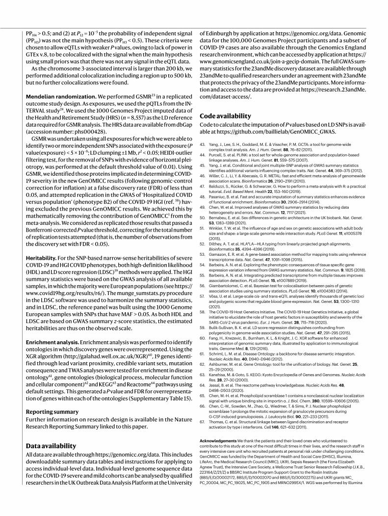
PPH4 > 0.5; and (2) at P12 = 10−5 the probability of independent signal (PPH3) was not the main hypothesis (PPH3 < 0.5). These criteria were chosen to allow eQTLs with weaker P values, owing to lack of power in GTEx v.8, to be colocalized with the signal when the main hypothesis using small priors was that there was not any signal in the eQTL data.
As the chromosome 3-associated interval is larger than 200 kb, we performed additional colocalization including a region up to 500 kb, but no further colocalizations were found.
Mendelian randomization. We performed GSMR23 in a replicated outcome study design. As exposures, we used the pQTLs from the IN-TERVAL study24. We used the 1000 Genomes Project imputed data of the Health and Retirement Study (HRS) (n = 8,557) as the LD reference data required for GSMR analysis. The HRS data are available from dbGap (accession number: phs000428).
GSMR was undertaken using all exposures for which we were able to identify two or more independent SNPs associated with the exposure (P value(exposure) < 5 × 10⁻⁸; LD clumping ±1 Mb, r2 < 0.05; HEIDI-outlier filtering test, for the removal of SNPs with evidence of horizontal plei-otropy, was performed at the default threshold value of 0.01). Using GSMR, we identified those proteins implicated in determining COVID-19 severity in the new GenOMICC results (following genomic-control correction for inflation) at a false discovery rate (FDR) of less than 0.05, and attempted replication in the GWAS of ‘Hospitalized COVID versus population’ (phenotype B2) of the COVID-19 HGI (ref. 58) hav-ing excluded the previous GenOMICC results. We achieved this by mathematically removing the contribution of GenOMICC1 from the meta-analysis. We considered as replicated those results that passed a Bonferroni-corrected P value threshold, correcting for the total number of replication tests attempted (that is, the number of observations from the discovery set with FDR < 0.05).
Heritability. For the SNP-based narrow-sense heritabilities of severe COVID-19 and HGI COVID phenotypes, both high-definition likelihood (HDL) and LD score regression (LDSC)59 methods were applied. The HGI summary statistics were based on the GWAS analysis of all available samples, in which the majority were European populations (see https://www.covid19hg.org/results/r6/). The munge_sumstats.py procedure in the LDSC software was used to harmonize the summary statistics, and in LDSC, the reference panel was built using the 1000 Genome European samples with SNPs that have MAF > 0.05. As both HDL and LDSC are based on GWAS summary z-score statistics, the estimated heritabilities are thus on the observed scale.
Enrichment analysis. Enrichment analysis was performed to identify ontologies in which discovery genes were overrepresented. Using the XGR algorithm (http://galahad.well.ox.ac.uk/XGR)60, 19 genes identi-fied through lead variant proximity, credible variant sets, mutation consequence and TWAS analyses were tested for enrichment in disease ontology61, gene ontologies (biological process, molecular function and cellular component)62 and KEGG63 and Reactome64 pathways using default settings. This generated a P value and FDR for overrepresenta-tion of genes within each of the ontologies (Supplementary Table 15).
Reporting summaryFurther information on research design is available in the Nature Research Reporting Summary linked to this paper.
Data availabilityAll data are available through https://genomicc.org/data. This includes downloadable summary data tables and instructions for applying to access individual-level data. Individual-level genome sequence data for the COVID-19 severe and mild cohorts can be analysed by qualified researchers in the UK Outbreak Data Analysis Platform at the University
of Edinburgh by application at https://genomicc.org/data. Genomic data for the 100,000 Genomes Project participants and a subset of COVID-19 cases are also available through the Genomics England research environment, which can be accessed by application at https://www.genomicsengland.co.uk/join-a-gecip-domain. The full GWAS sum-mary statistics for the 23andMe discovery dataset are available through 23andMe to qualified researchers under an agreement with 23andMe that protects the privacy of the 23andMe participants. More informa-tion and access to the data are provided at https://research.23andMe.com/dataset-access/.
Code availabilityCode to calculate the imputation of P values based on LD SNPs is avail-able at https://github.com/baillielab/GenOMICC_GWAS. 43. Yang, J., Lee, S. H., Goddard, M. E. & Visscher, P. M. GCTA: a tool for genome-wide
complex trait analysis. Am. J. Hum. Genet. 88, 76–82 (2011).44. Purcell, S. et al. PLINK: a tool set for whole-genome association and population-based
linkage analyses. Am. J. Hum. Genet. 81, 559–575 (2007).45. Yang, J. et al. Conditional and joint multiple-SNP analysis of GWAS summary statistics
identifies additional variants influencing complex traits. Nat. Genet. 44, 369–375 (2012).46. Willer, C. J., Li, Y. & Abecasis, G. R. METAL: fast and efficient meta-analysis of genomewide
association scans. Bioinformatics 26, 2190–2191 (2010).47. Balduzzi, S., Rücker, G. & Schwarzer, G. How to perform a meta-analysis with R: a practical
tutorial. Evid. Based Ment. Health 22, 153–160 (2019).48. Pasaniuc, B. et al. Fast and accurate imputation of summary statistics enhances evidence
of functional enrichment. Bioinformatics 30, 2906–2914 (2014).49. Chen, W. et al. Improved analyses of GWAS summary statistics by reducing data
heterogeneity and errors. Nat. Commun. 12, 7117 (2021).50. Bernabeu, E. et al. Sex differences in genetic architecture in the UK biobank. Nat. Genet.
53, 1283–1289 (2021).51. Winkler, T. W. et al. The influence of age and sex on genetic associations with adult body
size and shape: a large-scale genome-wide interaction study. PLoS Genet. 11, e1005378 (2015).
52. Dilthey, A. T. et al. HLA*LA—HLA typing from linearly projected graph alignments. Bioinformatics 35, 4394–4396 (2019).
53. Gamazon, E. R. et al. A gene-based association method for mapping traits using reference transcriptome data. Nat. Genet. 47, 1091–1098 (2015).
54. Barbeira, A. N. et al. Exploring the phenotypic consequences of tissue specific gene expression variation inferred from GWAS summary statistics. Nat. Commun. 9, 1825 (2018).
55. Barbeira, A. N. et al. Integrating predicted transcriptome from multiple tissues improves association detection. PLoS Genet. 15, e1007889 (2019).
56. Giambartolomei, C. et al. Bayesian test for colocalisation between pairs of genetic association studies using summary statistics. PLoS Genet. 10, e1004383 (2014).
57. Võsa, U. et al. Large-scale cis- and trans-eQTL analyses identify thousands of genetic loci and polygenic scores that regulate blood gene expression. Nat. Genet. 53, 1300–1310 (2021).
58. The COVID-19 Host Genetics Initiative. The COVID-19 Host Genetics Initiative, a global initiative to elucidate the role of host genetic factors in susceptibility and severity of the SARS-CoV-2 virus pandemic. Eur. J. Hum. Genet. 28, 715–718 (2020).
59. Bulik-Sullivan, B. K. et al. LD score regression distinguishes confounding from polygenicity in genome-wide association studies. Nat. Genet. 47, 291–295 (2015).
60. Fang, H., Knezevic, B., Burnham, K. L. & Knight, J. C. XGR software for enhanced interpretation of genomic summary data, illustrated by application to immunological traits. Genome Med. 8, 129 (2016).
61. Schriml, L. M. et al. Disease Ontology: a backbone for disease semantic integration. Nucleic Acids Res. 40, D940–D946 (2012).
62. Ashburner, M. et al. Gene Ontology: tool for the unification of biology. Nat. Genet. 25, 25–29 (2000).
63. Kanehisa, M. & Goto, S. KEGG: Kyoto Encyclopedia of Genes and Genomes. Nucleic Acids Res. 28, 27–30 (2000).
64. Jassal, B. et al. The reactome pathway knowledgebase. Nucleic Acids Res. 48, D498–D503 (2020).
65. Chen, M.-H. et al. Phospholipid scramblase 1 contains a nonclassical nuclear localization signal with unique binding site in importin α. J. Biol. Chem. 280, 10599–10606 (2005).
66. Chen, C.-W., Sowden, M., Zhao, Q., Wiedmer, T. & Sims, P. J. Nuclear phospholipid scramblase 1 prolongs the mitotic expansion of granulocyte precursors during G-CSF-induced granulopoiesis. J. Leukocyte Biol. 90, 221–233 (2011).
67. Thomas, C. et al. Structural linkage between ligand discrimination and receptor activation by type i interferons. Cell 146, 621–632 (2011).
Acknowledgements We thank the patients and their loved ones who volunteered to contribute to this study at one of the most difficult times in their lives, and the research staff in every intensive care unit who recruited patients at personal risk under challenging conditions. GenOMICC was funded by the Department of Health and Social Care (DHSC), Illumina, LifeArc, the Medical Research Council (MRC), UKRI, Sepsis Research (the Fiona Elizabeth Agnew Trust), the Intensive Care Society, a Wellcome Trust Senior Research Fellowship (J.K.B., 223164/Z/21/Z) a BBSRC Institute Program Support Grant to the Roslin Institute (BBS/E/D/20002172, BBS/E/D/10002070 and BBS/E/D/30002275) and UKRI grants MC_PC_20004, MC_PC_19025, MC_PC_1905 and MRNO2995X/1. WGS was performed by Illumina
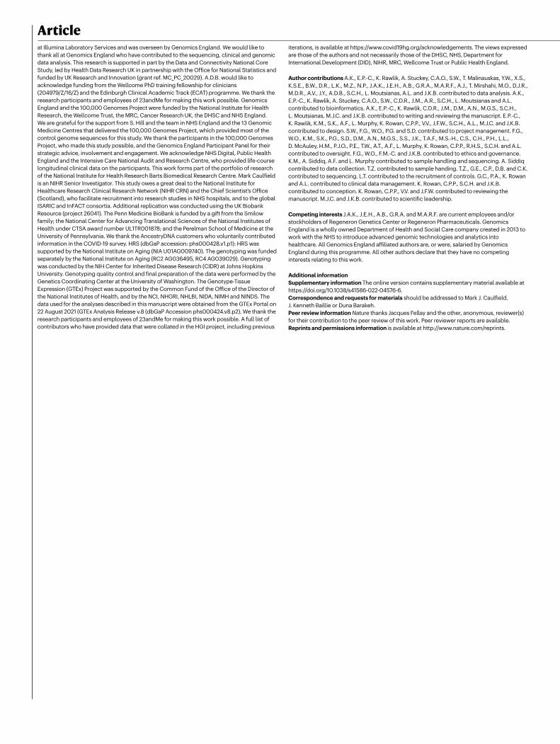
Articleat Illumina Laboratory Services and was overseen by Genomics England. We would like to thank all at Genomics England who have contributed to the sequencing, clinical and genomic data analysis. This research is supported in part by the Data and Connectivity National Core Study, led by Health Data Research UK in partnership with the Office for National Statistics and funded by UK Research and Innovation (grant ref. MC_PC_20029). A.D.B. would like to acknowledge funding from the Wellcome PhD training fellowship for clinicians (204979/Z/16/Z) and the Edinburgh Clinical Academic Track (ECAT) programme. We thank the research participants and employees of 23andMe for making this work possible. Genomics England and the 100,000 Genomes Project were funded by the National Institute for Health Research, the Wellcome Trust, the MRC, Cancer Research UK, the DHSC and NHS England. We are grateful for the support from S. Hill and the team in NHS England and the 13 Genomic Medicine Centres that delivered the 100,000 Genomes Project, which provided most of the control genome sequences for this study. We thank the participants in the 100,000 Genomes Project, who made this study possible, and the Genomics England Participant Panel for their strategic advice, involvement and engagement. We acknowledge NHS Digital, Public Health England and the Intensive Care National Audit and Research Centre, who provided life-course longitudinal clinical data on the participants. This work forms part of the portfolio of research of the National Institute for Health Research Barts Biomedical Research Centre. Mark Caulfield is an NIHR Senior Investigator. This study owes a great deal to the National Institute for Healthcare Research Clinical Research Network (NIHR CRN) and the Chief Scientist’s Office (Scotland), who facilitate recruitment into research studies in NHS hospitals, and to the global ISARIC and InFACT consortia. Additional replication was conducted using the UK Biobank Resource (project 26041). The Penn Medicine BioBank is funded by a gift from the Smilow family; the National Center for Advancing Translational Sciences of the National Institutes of Health under CTSA award number UL1TR001878; and the Perelman School of Medicine at the University of Pennsylvania. We thank the AncestryDNA customers who voluntarily contributed information in the COVID-19 survey. HRS (dbGaP accession: phs000428.v1.p1): HRS was supported by the National Institute on Aging (NIA U01AG009740). The genotyping was funded separately by the National Institute on Aging (RC2 AG036495, RC4 AG039029). Genotyping was conducted by the NIH Center for Inherited Disease Research (CIDR) at Johns Hopkins University. Genotyping quality control and final preparation of the data were performed by the Genetics Coordinating Center at the University of Washington. The Genotype-Tissue Expression (GTEx) Project was supported by the Common Fund of the Office of the Director of the National Institutes of Health, and by the NCI, NHGRI, NHLBI, NIDA, NIMH and NINDS. The data used for the analyses described in this manuscript were obtained from the GTEx Portal on 22 August 2021 (GTEx Analysis Release v.8 (dbGaP Accession phs000424.v8.p2). We thank the research participants and employees of 23andMe for making this work possible. A full list of contributors who have provided data that were collated in the HGI project, including previous
iterations, is available at https://www.covid19hg.org/acknowledgements. The views expressed are those of the authors and not necessarily those of the DHSC, NHS, Department for International Development (DID), NIHR, MRC, Wellcome Trust or Public Health England.
Author contributions A.K., E.P.-C., K. Rawlik, A. Stuckey, C.A.O., S.W., T. Malinauskas, Y.W., X.S., K.S.E., B.W., D.R., L.K., M.Z., N.P., J.A.K., J.E.H., A.B., G.R.A., M.A.R.F., A.J., T. Mirshahi, M.O., D.J.R., M.D.R., A.V., J.Y., A.D.B., S.C.H., L. Moutsianas, A.L. and J.K.B. contributed to data analysis. A.K., E.P.-C., K. Rawlik, A. Stuckey, C.A.O., S.W., C.D.R., J.M., A.R., S.C.H., L. Moutsianas and A.L. contributed to bioinformatics. A.K., E.P.-C., K. Rawlik, C.D.R., J.M., D.M., A.N., M.G.S., S.C.H., L. Moutsianas, M.J.C. and J.K.B. contributed to writing and reviewing the manuscript. E.P.-C., K. Rawlik, K.M., S.K., A.F., L. Murphy, K. Rowan, C.P.P., V.V., J.F.W., S.C.H., A.L., M.J.C. and J.K.B. contributed to design. S.W., F.G., W.O., P.G. and S.D. contributed to project management. F.G., W.O., K.M., S.K., P.G., S.D., D.M., A.N., M.G.S., S.S., J.K., T.A.F., M.S.-H., C.S., C.H., P.H., L.L., D. McAuley, H.M., P.J.O., P.E., T.W., A.T., A.F., L. Murphy, K. Rowan, C.P.P., R.H.S., S.C.H. and A.L. contributed to oversight. F.G., W.O., F.M.-C. and J.K.B. contributed to ethics and governance. K.M., A. Siddiq, A.F. and L. Murphy contributed to sample handling and sequencing. A. Siddiq contributed to data collection. T.Z. contributed to sample handing. T.Z., G.E., C.P., D.B. and C.K. contributed to sequencing. L.T. contributed to the recruitment of controls. G.C., P.A., K. Rowan and A.L. contributed to clinical data management. K. Rowan, C.P.P., S.C.H. and J.K.B. contributed to conception. K. Rowan, C.P.P., V.V. and J.F.W. contributed to reviewing the manuscript. M.J.C. and J.K.B. contributed to scientific leadership.
Competing interests J.A.K., J.E.H., A.B., G.R.A. and M.A.R.F. are current employees and/or stockholders of Regeneron Genetics Center or Regeneron Pharmaceuticals. Genomics England is a wholly owned Department of Health and Social Care company created in 2013 to work with the NHS to introduce advanced genomic technologies and analytics into healthcare. All Genomics England affiliated authors are, or were, salaried by Genomics England during this programme. All other authors declare that they have no competing interests relating to this work.
Additional informationSupplementary information The online version contains supplementary material available at https://doi.org/10.1038/s41586-022-04576-6.Correspondence and requests for materials should be addressed to Mark J. Caulfield, J. Kenneth Baillie or Duna Barakeh.Peer review information Nature thanks Jacques Fellay and the other, anonymous, reviewer(s) for their contribution to the peer review of this work. Peer reviewer reports are available.Reprints and permissions information is available at http://www.nature.com/reprints.
Extended Data Fig. 1 | Analysis workflow for GWAS and AVT analyses of this study. The cohorts displayed in yellow and green in the top box were processed with Genomics England Pipeline 2.0 and Illumina NSV4, respectively (see Methods on WGS Alignment and variant calling for details on differences between pipelines). We used individuals that were processed with either pipeline for the GWAS analyses and individuals processed only with Genomics
England Pipeline 2.0 for the AVT analyses. The definition of the cases and controls was the same for GWAS and AVT, cases were the COVID-19 severe individuals for both, and controls included individuals from the 100,000 Genomes Project (100,000 Genomes Project) and also COVID-19 positive individuals that were recruited for this study and experienced only mild symptoms (COVID-mild).
Article
Extended Data Fig. 2 | Regional detail showing fine-mapping to identify three adjacent independent signals on chromosome 1. Top two panels: variants in LD with the lead variants shown. The variants that are included in two independent credible sets are displayed with black outline circles. r 2 values
in the legend denote upper limits, 0.2=[0,0.2], 0.4=[0.2,0.4], 0.6=[0.4,0.6], 0.8=[0.6,0.8],1=[0.8,1]. Bottom panel: locations of protein-coding genes, coloured by TWAS P-value. The red dashed line shows the Bonferroni-corrected P-value=2.2 × 10⁻⁸ for Europeans.

Extended Data Fig. 3 | Regional detail showing fine-mapping to identify two adjacent independent signals on chromosome 19. Top two panels: variants in LD with the lead variants shown. The variants that are included in two independent credible sets are displayed with black outline circles. r 2 values
in the legend denote upper limits, 0.2=[0,0.2], 0.4=[0.2,0.4], 0.6=[0.4,0.6], 0.8=[0.6,0.8],1=[0.8,1]. Bottom panel: locations of protein-coding genes, coloured by TWAS P-value. The red dashed line shows the Bonferroni-corrected P-value=2.2 × 10⁻⁸ for Europeans.

Article
Extended Data Fig. 4 | Regional detail showing fine-mapping to identify three adjacent independent signals on chromosome 21. Top three panels: variants in LD with the lead variants shown. The variants that are included in three independent credible sets are displayed with black outline circles.
r 2 values in the legend denote upper limits, 0.2=[0,0.2], 0.4=[0.2,0.4], 0.6=[0.4,0.6], 0.8=[0.6,0.8],1=[0.8,1]. Bottom panel: locations of protein- coding genes, coloured by TWAS P-value. The red dashed line shows the Bonferroni-corrected P-value=2.2 × 10⁻⁸ for Europeans.

Extended Data Fig. 5 | Predicted structural consequences of lead variants at PLSCR1 and IFNA10. (a) Crystal structure of PLSCR1 nuclear localization signal (orange, Gly257–Ile266, numbering correspond to UniProt entry O15162) in complex with Importin α (blue), Protein Data Bank (PDB) ID 1Y2A (ref. 65). Side chains of PLSCR1 are shown as connected spheres with carbon atoms coloured in orange, nitrogens in blue and oxygens in red. Hydrogen atoms were not determined at this resolution (2.20) and are not shown. (b) Close-up view showing side chains of PLSCR1 Ser260, His262 and Importin Glu107 as sticks. Distance (in) between selected atoms (PLSCR1 His262 Nϵ2 and Importin Glu107 carboxyl O) is indicated. A hydrogen bond between PLSCR1 His262 and Importin Glu107 is indicated with a dashed line. The risk variant is predicted to eliminate this bond, disrupting nuclear import, an essential step for effect on antiviral signalling27 and neutrophil maturation66. (c) Because there is very
strong sequence conservation between IFNA10 and the gene encoding IFNω, we used existing crystal structure data (Protein Data Bank ID 3SE4 (ref. 67)) for IFNω (cyan) to display a ternary complex with interferon α/β receptor IFNAR1 (blue), IFNAR2 (red). The side chain of Trp164 is shown as spheres and indicated with a black line. (d) The hydrophobic core of IFNω with Trp164 shielded from the solvent in the center. Trp164-surrounding residues of IFNω are numbered and correspond to UniProt entry P05000. Trp164 and surrounding residues are conserved in IFNA10 (UniProt ID P01566) and share the same numbering as in IFNω (P05000). Side chains of four residues are shown as sticks. Carbon and nitrogen atoms coloured in cyan and blue, respectively. The critical COVID-19- associated mutation, Trp164Cys, would replace an evolutionarily conserved, bulky side chain in the hydrophobic core of IFNA10 with a smaller one, which may destabilize IFNA10.
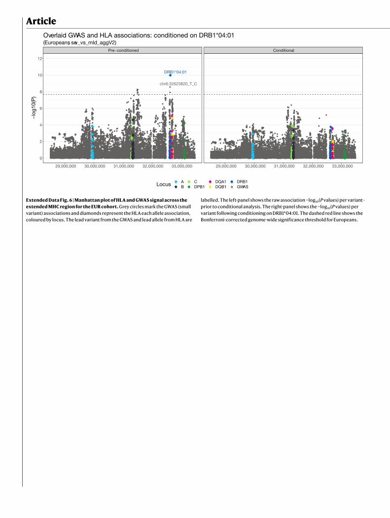
Article
chr6:32623820_T_C
DRB1*04:01
Pre−conditioned Conditional
29,000,000 30,000,000 31,000,000 32,000,000 33,000,000 29,000,000 30,000,000 31,000,000 32,000,000 33,000,000
0
2
4
6
8
10
12
−lo
g10(
P)
Locus aa
aa
aa
aa
AB
CDPB1
DQA1DQB1
DRB1GWAS
(Europeans, sev_vs_mld_aggV2)Overlaid GWAS and HLA associations: conditioned on DRB1*04:01
Extended Data Fig. 6 | Manhattan plot of HLA and GWAS signal across the extended MHC region for the EUR cohort. Grey circles mark the GWAS (small variant) associations and diamonds represent the HLA each allele association, coloured by locus. The lead variant from the GWAS and lead allele from HLA are
labelled. The left-panel shows the raw association −log10(P values) per variant - prior to conditional analysis. The right-panel shows the −log10(P values) per variant following conditioning on DRB1*04:01. The dashed red line shows the Bonferroni-corrected genome-wide significance threshold for Europeans.
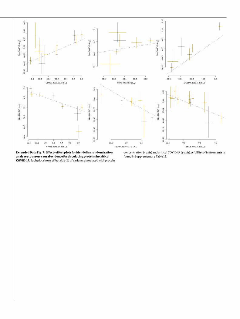
Extended Data Fig. 7 | Effect–effect plots for Mendelian randomization analyses to assess causal evidence for circulating proteins in critical COVID-19. Each plot shows effect size (β) of variants associated with protein
concentration (x axis) and critical COVID-19 ( y axis). A full list of instruments is found in Supplementary Table 13.

ArticleExtended Data Table 1 | Fine-mapping results for lead variants and worst consequence variant in each credible set
Fine-mapping was performed in EUR for all variants except chr6:41515007:A:C, which was fine-mapped in the SAS population for which the signal was strongest among the per-population analyses. The lead variant chr2:60480453:A:G (rs1123573) that was discovered in multi-ancestry meta-analysis is not included in the table as fine-mapping did not generate any credible sets with the required posterior inclusion probability of >0.95 for any of the populations. Focal CS is the index SNP that was used for fine-mapping with SusieR, 1.5 Mb on each side. nCS indicates the number of variants included in each credible set. Consequence annotation for all variants across credible sets was generated using VEP v.104 and the worst consequence across GENCODE basic transcripts was chosen. All variants were ranked according to their consequence type and chr:poshg38:refhg38:alt, P value and CADD score are provided for the variant with the worst consequence across all variants in each credible set.
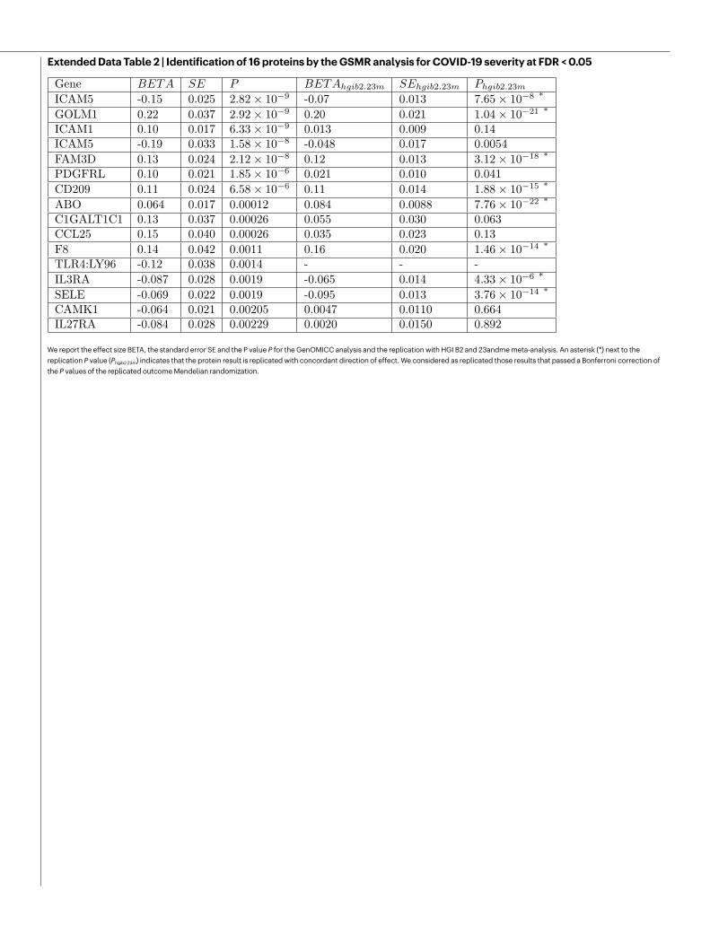
Extended Data Table 2 | Identification of 16 proteins by the GSMR analysis for COVID-19 severity at FDR < 0.05
We report the effect size BETA, the standard error SE and the P value P for the GenOMICC analysis and the replication with HGI B2 and 23andme meta-analysis. An asterisk (*) next to the replication P value (Phgib2.23m) indicates that the protein result is replicated with concordant direction of effect. We considered as replicated those results that passed a Bonferroni correction of the P values of the replicated outcome Mendelian randomization.


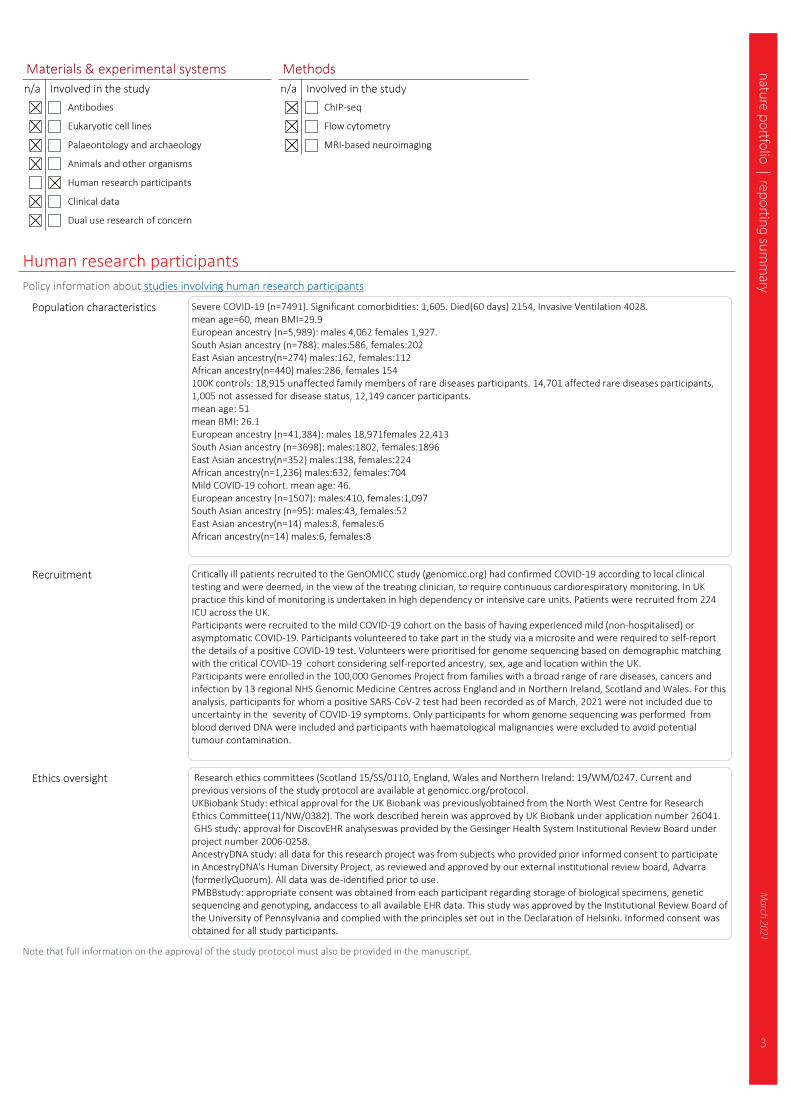
Related Documents