REVIEW Open Access Relevance of immune cell and tumor microenvironment imaging in the new era of immunotherapy Filippo Galli 1* , Jesus Vera Aguilera 2 , Belinda Palermo 3 , Svetomir N. Markovic 2 , Paola Nisticò 3 and Alberto Signore 1 Abstract Tumor-infiltrating immune cells play a key role against cancer. However, malignant cells are able to evade the immune response and establish a very complex balance in which different immune subtypes may drive tumor progression, metastatization and resistance to therapy. New immunotherapeutic approaches aim at restoring the natural balance and increase immune response against cancer by different mechanisms. The complexity of these interactions and the heterogeneity of immune cell subpopulations are a real challenge when trying to develop new immunotherapeutics and evaluate or predict their efficacy in vivo. To this purpose, molecular imaging can offer non-invasive diagnostic tools like radiopharmaceuticals, contrast agents or fluorescent dyes. These agents can be useful for preclinical and clinical purposes and can overcome [ 18 F]FDG limitations in discriminating between true- progression and pseudo-progression. This review provides a comprehensive overview of immune cells involved in microenvironment, available immunotherapies and imaging agents to highlight the importance of new therapeutic biomarkers and their in vivo evaluation to improve the management of cancer patients. Keywords: molecular imaging, tumor microenvironment, onco-immunology, immunotherapy, lymphocytes Background Immunotherapy is the most appealing anti-cancer ap- proach of the modern era and researchers are continu- ously exploring new ways to reprogram immune cells of the host against cancer [1]. Despite the initial hype, due to promising results, playing with the immune system raised important issues in many treated patients together with controversial results. Indeed, the removal of the intrinsic immune suppression can trigger a cascade of events with serious adverse effects [2]. Moreover, because of the complex and dynamic nature of the interactions between cancer and immune cells, a high inter- and intra-patient heterogeneity is observed, sometimes leading to failure of the treatment [3]. That is why there is an urgent need of diagnostic tools to help physician in predicting and evaluating treatment re- sponse at very early stages. This will help to accurately select patients for specific therapies and to promptly suspend or change the therapeutic approach if needed. Indeed, the possibility to characterize in vivo each tumor lesion opens the door to true personalized-medicine that we might even define as “lesion based-medicine” [4]. In this scenario, molecular medicine imaging offers plenty of tools to specifically follow immune cell sub- types in a non-invasive manner [5]. This is not only thanks to availability of many radiopharmaceutical and probes to target specific cell subtypes, but also to high sensitivity technologies that can allow us to detect even limited numbers of cancer infiltrating cells. In this review we will give an overview of tumor © The Author(s). 2020 Open Access This article is licensed under a Creative Commons Attribution 4.0 International License, which permits use, sharing, adaptation, distribution and reproduction in any medium or format, as long as you give appropriate credit to the original author(s) and the source, provide a link to the Creative Commons licence, and indicate if changes were made. The images or other third party material in this article are included in the article's Creative Commons licence, unless indicated otherwise in a credit line to the material. If material is not included in the article's Creative Commons licence and your intended use is not permitted by statutory regulation or exceeds the permitted use, you will need to obtain permission directly from the copyright holder. To view a copy of this licence, visit http://creativecommons.org/licenses/by/4.0/. The Creative Commons Public Domain Dedication waiver (http://creativecommons.org/publicdomain/zero/1.0/) applies to the data made available in this article, unless otherwise stated in a credit line to the data. * Correspondence: [email protected] 1 Nuclear Medicine Unit, Department of Medical-Surgical Sciences and of Translational Medicine, “Sapienza” University of Rome, S. Andrea University Hospital, Roma, Italy Full list of author information is available at the end of the article Galli et al. Journal of Experimental & Clinical Cancer Research (2020) 39:89 https://doi.org/10.1186/s13046-020-01586-y

Welcome message from author
This document is posted to help you gain knowledge. Please leave a comment to let me know what you think about it! Share it to your friends and learn new things together.
Transcript
REVIEW Open Access
Relevance of immune cell and tumormicroenvironment imaging in the new eraof immunotherapyFilippo Galli1* , Jesus Vera Aguilera2, Belinda Palermo3, Svetomir N. Markovic2, Paola Nisticò3 and Alberto Signore1
Abstract
Tumor-infiltrating immune cells play a key role against cancer. However, malignant cells are able to evade theimmune response and establish a very complex balance in which different immune subtypes may drive tumorprogression, metastatization and resistance to therapy. New immunotherapeutic approaches aim at restoring thenatural balance and increase immune response against cancer by different mechanisms. The complexity of theseinteractions and the heterogeneity of immune cell subpopulations are a real challenge when trying to develop newimmunotherapeutics and evaluate or predict their efficacy in vivo. To this purpose, molecular imaging can offernon-invasive diagnostic tools like radiopharmaceuticals, contrast agents or fluorescent dyes. These agents can beuseful for preclinical and clinical purposes and can overcome [18F]FDG limitations in discriminating between true-progression and pseudo-progression. This review provides a comprehensive overview of immune cells involved inmicroenvironment, available immunotherapies and imaging agents to highlight the importance of new therapeuticbiomarkers and their in vivo evaluation to improve the management of cancer patients.
Keywords: molecular imaging, tumor microenvironment, onco-immunology, immunotherapy, lymphocytes
BackgroundImmunotherapy is the most appealing anti-cancer ap-proach of the modern era and researchers are continu-ously exploring new ways to reprogram immune cells ofthe host against cancer [1]. Despite the initial hype, dueto promising results, playing with the immune systemraised important issues in many treated patients togetherwith controversial results. Indeed, the removal of theintrinsic immune suppression can trigger a cascade ofevents with serious adverse effects [2].Moreover, because of the complex and dynamic nature
of the interactions between cancer and immune cells, ahigh inter- and intra-patient heterogeneity is observed,
sometimes leading to failure of the treatment [3]. That iswhy there is an urgent need of diagnostic tools to helpphysician in predicting and evaluating treatment re-sponse at very early stages. This will help to accuratelyselect patients for specific therapies and to promptlysuspend or change the therapeutic approach if needed.Indeed, the possibility to characterize in vivo each tumorlesion opens the door to true personalized-medicine thatwe might even define as “lesion based-medicine” [4].In this scenario, molecular medicine imaging offers
plenty of tools to specifically follow immune cell sub-types in a non-invasive manner [5]. This is not onlythanks to availability of many radiopharmaceutical andprobes to target specific cell subtypes, but also to highsensitivity technologies that can allow us to detect evenlimited numbers of cancer infiltrating cells. In thisreview we will give an overview of tumor
© The Author(s). 2020 Open Access This article is licensed under a Creative Commons Attribution 4.0 International License,which permits use, sharing, adaptation, distribution and reproduction in any medium or format, as long as you giveappropriate credit to the original author(s) and the source, provide a link to the Creative Commons licence, and indicate ifchanges were made. The images or other third party material in this article are included in the article's Creative Commonslicence, unless indicated otherwise in a credit line to the material. If material is not included in the article's Creative Commonslicence and your intended use is not permitted by statutory regulation or exceeds the permitted use, you will need to obtainpermission directly from the copyright holder. To view a copy of this licence, visit http://creativecommons.org/licenses/by/4.0/.The Creative Commons Public Domain Dedication waiver (http://creativecommons.org/publicdomain/zero/1.0/) applies to thedata made available in this article, unless otherwise stated in a credit line to the data.
* Correspondence: [email protected] Medicine Unit, Department of Medical-Surgical Sciences and ofTranslational Medicine, “Sapienza” University of Rome, S. Andrea UniversityHospital, Roma, ItalyFull list of author information is available at the end of the article
Galli et al. Journal of Experimental & Clinical Cancer Research (2020) 39:89 https://doi.org/10.1186/s13046-020-01586-y

microenvironment, new therapies and the added valueof molecular imaging towards a personalized medicineapproach.
The tumor microenvironmentCancers are not a mass of transformed cells butrather a new organ composed of various non-malignant cells comprising a large portion of thetumor mass, which have become wayward and lostthe ability to maintain a dialogue enabling homeosta-sis of the tissue architecture [6]. These cells includefibroblasts, adipocytes, pericytes, vascular endothelialcells, and, as main players, immune cells [7]. Tumorand stromal cells co-evolve, similarly to what occursin organogenesis during development, and the inter-action among the different components leads to acontinuous phenotypic and functional plasticity. Dy-namic reciprocal communication between cells andmicroenvironment is conducted via junctions andreceptors plus a plethora of signals produced by themultiple cell types encased in a three-dimensional
extracellular matrix (ECM). This includes glycopro-teins, proteoglycans, cytokines and growth factors,together with ECM-remodeling enzymes, providingboth structural support and appropriate information[8]. The disruption of tissue homeostasis creates dy-namic changes in the cellular metabolism and func-tion of both stromal and immune cells [9]. Thishighly trafficked network constitutes the tumormicroenvironment (TME) (Fig. 1), and cancerresearch has to make a major effort to draw up amultidimensional map that will elucidate the high-ways and byways of the cancer battlefield.
Cells of the tumor microenvironmentImmune cells - T lymphocytesT lymphocytes are the most potent mediators of adap-tive anti-tumor immune response. The cytotoxic CD8+T cell population, supported by CD4+ T helper (Th1)cells through the production of IL2 and IFNγ, generatesthe final effector mechanism leading to tumor elimin-ation and are associated with a good prognosis [10, 11].
Fig. 1 The trafficking in the tumor microenvironment
Galli et al. Journal of Experimental & Clinical Cancer Research (2020) 39:89 Page 2 of 21

Whereas, the CD4+ T cell subsets Th2 and Th17, pro-ducing IL4, IL5, IL13 or IL17A, IL17F, IL21 and IL22 re-spectively, are generally associated with tissueinflammation and a pro-tumorigenic effect. The CD8-mediated immune response is modulated by an im-munosuppressive class of CD4+ T cells known as Tregulatory (Treg), which expresses the CD25 and FOXP3molecules, governing peripheral immune tolerance [12].In the TME, high amount of Tregs is often present andtheir main role is to suppress the anti-tumor response.However, due to the Treg subpopulation diversity alongwith different functional pathways, their role in cancerdevelopment and progression is ambiguous and still notfully understood [13, 14].The successful control of tumor progression, mediated
by T lymphocytes, firstly requires that they infiltrate thetumors. In fact, the immune contexture and the T cellabundance, functional activity and spatial distribution inthe TME are crucial prognostic and predictive factors[15], as recently proposed for the immune checkpointblockades (ICB) [16, 17].Compartmentation of the immune response into
three major phenotypes - inflamed, immune-excludedand immune-desert phenotypes - has been proposedas the major predictor of response to different cancertreatments in the new era of immune inhibitory re-ceptor blockades [17, 18]. The inflamed phenotypecomprises the concurrent presence of both CD8+ andCD4+ T cells with inhibitory cells (i.e., macrophages,fibroblasts, Treg, suppressor myeloid cells and B cells)in the tumor parenchyma. These cells affect T cellfunctionality up-regulating several inhibitory receptorsand leading to T cell dysfunction and exhaustion [19].The immune-excluded phenotype has been associatedto mesenchymal traits, which have been proposed asputative biomarkers of response to ICB [20]. Thisphenotype is characterized by a huge number of im-mune cells in the stroma surrounding tumor nests, asdictated by physical barriers (i.e., stiffened tissue withhigh matrix fiber mass and dense collagen network)[21] or the low expression of specific chemokines in-volved in T cell recruitment [22, 23].The above-mentioned suppressive cells, accompanied
by the hindrance of lymphocyte infiltration and traffick-ing, may be modulated in multiple ways. Indeed, solublemolecules, such as vascular endothelial growth factor(VEGF) and the consequent abnormal neovasculature[24] as well as down-modulation by tumor cells of adhe-sion and chemotactic signals on the tumor endothelium,may participate in an immune-suppressive TME [25]. Tcell exclusion may also be mediated by cancer-associatedfibroblasts, which produce the C-X-C motif chemokine12 (CXCL12). This chemokine inhibition in a mousemodel of pancreatic ductal adenocarcinoma (PDAC) has
been shown to revert the immune exclusion and syner-gize with the anti-PD1 therapy [26].The lack of an endogenous anti-tumor response in
TME described as the third immune-desert phenotype,may be due to insufficient T cell priming, immunologicalignorance or induction of tolerance. This immune con-texture is characterized by the presence of Treg, MDSCand macrophages, which wire a circuit inhibiting den-dritic cell (DC) maturation and hamper T cell expansionand activation [27].
B lymphocytesRecent findings have assessed a role for B cells in theanti-tumor immune response [28] B cell infiltration intothe TME occurs as occasionally localized at the invasivemargin of tumors, but more often localized in draininglymph nodes and tertiary lymphoid structures (TLS),and may be associated to both positive and negative ef-fects in tumor immunity. The anti-tumor role of B cellshas been reported in murine models, indicating that Bcells increase T cell functionality [29]. In different hu-man tumors, such as ovarian, non-small cell lung cancer(NSCLC), gastric and cervical cancer, the presence oftumor-infiltrating CD20+ B cells is associated with goodprognosis [30–33]. Despite this protective role, B cellsmay negatively regulate anti-tumor immunity, as re-ported in a murine model of squamous carcinogenesis[34]. Similarly, Ammirante et al. showed that B cells, re-cruited by the chemokine CXCL13, promote the pro-gression of castrate-resistant prostate cancer byproducing lymphotoxin [35]. Furthermore, the immuno-genic effect of chemotherapy in mouse and humanprostate tumors, requires the removal of an immuno-suppressive B cell subtype, plasmocytes that expressIgA, interleukin (IL)-10 and programmed death ligand 1(PD-L1) - the appearance of which depends on TGFβreceptor signalling - that induce CD8+ T cell exhaustionand suppress anti-tumor CTL responses [36].
Natural killer cellsTumor stroma may be infiltrated by innate cytotoxiclymphocytes, the natural killer (NK) cells. NK cells notonly recognize and kill cancer cells through the releaseof cytolytic granules, but also greatly impact the adaptiveanti-tumor immune response by producing chemokinesand cytokines. NK cells are highly heterogeneous, andthe availability of different combination markers hasallowed researchers to identify distinct subpopulationswith definite functionality [37]. Otherwise, in commonwith tumor-associated immune cells, NK cells can alsonegatively influence anti-cancer responses by modulatingDC and T cells. Recently, Glasner et al. reported a newanti-tumor role of NK cells by modulating the immuneresponse. The authors demonstrated that the activation
Galli et al. Journal of Experimental & Clinical Cancer Research (2020) 39:89 Page 3 of 21

of the NK natural cytotoxic receptor 1 (mouse) andNKp46 (human) induces IFNγ production which, inturn, modulates fibronectin 1 expression on tumor cells,preventing metastatic spread [38]. However, differenttumor-related soluble factors (i.e. IL-10, IDO, PGE2,TGF-β1) produced by different tumor-infiltrating im-mune cells (i.e. M2-macrophages, MDSC, DC, Treg),may negatively affect NK cell activity [39].
Dendritic cellsDCs are antigen-presenting cells (APC) able to captureantigens in the form of peptide-major histocompatibility(MHC) molecule complexes and present them to the Tcells [40]. They are a ubiquitous population of myeloidcells, heterogeneous in terms of morphology, ontogenyand immunological features [41]. The different DC sub-sets are related to specific immunological functions: a)DC processing and presenting antigens; b) epidermalLangerhans cells specializing in priming CD8+ T cell im-munity and interstitial/dermal (CD14+) DCs endorsinghumoral immunity; c) plasmacytoid (pDCs) secretinghigh amount of type I IFN. DCs exist in immature state(iDCs) in the absence of maturation signals, eliciting im-munological tolerance and/or suppression. Several cues,such as microbe-associated molecular patterns or en-dogenous damage-associated molecular patterns, canlead iDC to a mature state [42].
Tertiary lymphoid structuresTLS are lymphoid aggregates induced postnatally innon-lymphoid tissues that resemble the organizationof lymph nodes, characterized by clusters of matureDCs and T cells juxtaposing B-cell follicles and highendothelial venules without encapsulation. Similar tolymph nodes, they are assumed to provide the mainlymphocytic functional environments for both cellularand humoral immunity [43]. The TLS architecture iscoordinated by homeostatic chemokines, i.e. CCL19,CCL21, CXCL13 and CXCL12, the same found in thesecondary lymphoid organ. The presence of peri-and/or intra-tumoral TLS has been correlated with agood prognosis and prolonged patient's survival in 12different types of cancer. Further studies are neededto elucidate the immune mechanisms that are acti-vated within these structures and the driver mecha-nisms of their development within the tumor. Fromthe clinical point of view, it is urgent to understandwhether the presence and localization of TLS in pre-treatment or longitudinal tumor tissue samples duringand post-treatment, may be validated as prognostic/predictive of responses to checkpoint blockade, withfar reaching clinical implications, as recently reported[44, 45].
MacrophagesMost of the immune cell populations within the tumorstroma are made up of tumor-associated macrophages(TAMs), major players in orchestrating cancer-relatedinflammation. Pre-clinical and clinical evidences demon-strated that an abundance of TAMs in the TME is asso-ciated with a poor prognosis [46]. The bi-directionalcommunication between macrophages and TME affectstheir phenotype, and is strictly dependent on the diseasestage and the involved tissue. Indeed, pro-inflammatorymacrophages, which play a key role against pathogensare driven by cytokines, such as IFNγ, TNFα and micro-bial products, and are referred to as the M1 subtype.This subtype in turn favours a Th1 response. On theother hand, IL-4 or IL-13 determine the M2 subtypepolarization, related to tumor-promotion and contrib-utes to an immune-suppressive TME, hampering T cellfunctionality [47]. TAMs have shown to negatively affectT cell responses in hepatocellular [48] and ovarian can-cer [49], through PD-L1 and B7-H4, respectively. Be-sides, TAMs can also express PD-L2 along with B7-H4and VISTA immune checkpoint inhibitory molecules.Overall, the activity of TAMs in cancer is usually pro-tumorigenic, closely related to the colony-stimulatingfactor (CSF)-1 secretion by cancer cells that recruitTAMs, which in turn, by releasing EGF, edit cancer cellsand favour cell migration, extravasation and metastases[50]. Several pharmacological agents targeting macro-phages in tumor have been successfully tested in experi-mental tumor indicating the rationale to move intoclinical trial [51].
NeutrophilsNeutrophils constitute 50-70% of all circulating leuko-cytes and representing the traditional front line ofdefense against infection. Inside the TME, a number ofkey molecular mechanisms can promote neutrophilpolarization in two opposite subpopulations of anti-tumorigenic (N1) and pro-tumorigenic (N2) tumor-associated neutrophils (TANs) [52]. In particular, TGF-βsecreted by cancer associated fibroblasts (CAFs) is re-sponsible for both the recruitment and activation of N2[53] and the suppression of N1 neutrophils [54]. It hasbeen suggested that the degree of tumor development isthe primary determinant of the resulting TAN pheno-type [55]. N2 TANs through the secretion of MMPs andinterleukin (IL)-1β activates endothelial cells and inhibitsNK cells, promoting tumor cell plasticity [56] and cancermigration [57]. Disseminating cancer cells interact withneutrophils in the metastatic sites and it is crucial tounderstand the neutrophils contribution to the meta-static processes, keeping in mind that many cancerpatients who are undergoing chemotherapy are alsotreated with neutrophil-stimulating factors [58].
Galli et al. Journal of Experimental & Clinical Cancer Research (2020) 39:89 Page 4 of 21

Furthermore, in melanoma patients, high levels of circu-lating neutrophils and neutrophil-to-lymphocyte ratiohave been associated with resistance to anti-CTLA-4,indicating the main role that these cells may exert ininhibiting immune response [59].
Myeloid-derived suppressor cellsMyeloid-derived suppressor cells MDSCs are classed asone of the major sub-populations of inhibitory immunecells that are frequently found in several mouse and hu-man cancers and show a plastic phenotype [60, 61] ren-dering their tracking difficult. Monocytic-MDSCs induceprocesses related to cancer invasion, such as the epithe-lial mesenchymal transition (EMT), a dynamic processregulated by microenvironmental stimuli [62] promotingtumor invasiveness by impairing anti-tumor innate andadaptive responses [63]. On the contrary, granulocytic-MDSCs suppress EMT [64]. Furthermore, the presenceof MDSC in TME has been linked to ECM modification,as shown in a murine model of breast cancer, highly ex-pressing the secreted protein acidic and cysteine rich[65]. Of clinical relevance, these cells are clearly involvedin resistance to ICB therapy in patients [66]. MDSCtumor-infiltration is mediated by CSF-1 and the combin-ation of CSF-1/CSF-1R signalling inhibition with anti-CTLA-4 has been recently proposed [67].
Cancer associated fibroblastsAmong the non-neoplastic cells in the TME, CAFs arethe most prominent stromal component and key playersin cancer progression [68]. CAFs secrete growth factorswith the TGF-β as the major player favouring EMTthrough the biomechanical and biochemical remodellingof the ECM. In the context of tumor-stroma coevolu-tion, CAFs are linked to cancer progression, giving mes-enchymal traits to tumor cells and contribute totherapeutic outcome [69]. Among the soluble factorsproduced by CAFs, the IL-6 cytokine mediates a dy-namic crosstalk between tumor cells and CAFs, drivingmesenchymal tumor phenotype and chemo-resistance[70]. The major contribution of fibroblast composition istheir ability to secrete ECM components and its re-modelling enzymes [69, 71]. Fibroblast activation proteinis expressed in a CAF subtype associated with ECM re-modelling and tumor-promoting inflammation [72, 73].Depletion of these cells determines INFγ production,reverting immunosuppression [74].
The immunosuppressive TME: ECM, hypoxia andmetabolismThe complex mixture of immune cells, non-cancerouscells and cancer cells are embedded in the extracellularmatrix. The dysregulation of ECM composition, struc-ture, stiffness and quantity, by regulating mechanical
and biochemical cues in the TME, is crucial in cancerprogression, invasion and immunosuppression [75]. Arecent elegant work has reported an ECM-associatedmolecular signature predictive of the extent of thedisease in ovarian cancer [76].ECM deposition and remodelling are strictly linked to
a reduction of the oxygen level, known as hypoxia. Rapidgrowth and poor vasculature development frequentlylead to hypoxic microenvironments within the tumor.The association between hypoxia and ECM remodellingis mediated by hypoxia inducible factor-1 and 2, whichregulate the expression of enzymes related to bio-synthesis fibres in collagen degradation [77]. To survivein hypoxic conditions, cancer cells adopt strategies ofmetabolic shift from oxidative phosphorylation to gly-colysis [78]. Glycolysis within tumor cells has been re-ported to compete with glucose availability to T cells,associated with an inhibition of effector function [79].These data pave the way for new studies aimed at meas-uring the effect of ICB therapy on available intra-tumoral nutrients for immune cell metabolism in treatedpatients and their clinical response.The overview of this amazing complexity surely justi-
fies a great multidisciplinary effort in cancer researchand new methodologies to track the immune cells in thehighly trafficked highways and byways of the cancer roadmap.
Cancer immunotherapyDrugs stimulating the host immune responseThe ability of the host immune system to identifyand eradicate malignant cells with minimal systemictoxicity remains the holy grail of cancer immunother-apy [80]. The first immunotherapy for the treatmentof malignant tumors began in 1891 by William B.Coley (Coley’s toxin). Dr. Coley injected live bacteria(streptococcal organisms) directly into tumors, muscletissue or intravenously, in patients with soft tissuesarcomas “in order to cause erysipelas and stimulatethe immune system” to attack the cancer [81]. Severetoxicity and lack of reproducible results ultimately, inthe face of emerging clinical use of chemotherapy andradiation therapy ultimately lead to discontinuation ofits use 40 years later [82]. Nonetheless, Coley’s earlyobservations remain as the foundation of cancer im-munotherapy to this day, suggesting that activation ofimmunity can indeed result in tumor rejection. Thefirst of the modern applications of Coley’s principlecame about in the 1970s when Morales et al. estab-lished the effectiveness of the bacterium BacillusCalmette-Guérin (BCG) in the treatment of superficialbladder cancer [83]. The underpinnings for this clin-ical trial include a 1959 study by Old et al. showingthe anti-tumor effects of BCG in a mouse model [84].
Galli et al. Journal of Experimental & Clinical Cancer Research (2020) 39:89 Page 5 of 21

Besides his work on BCG, Old also performed exten-sive research and was involved in the description oftumor necrosis factor in 1975 [85]; however the ideathat the immune system could play an important rolein the treatment of many cancers still remained aconcept solidly external to the purview of mainstreamoncology [86].The discovery and characterization of dendritic cells
by Ralph Steinman in 1973, the description of MHCrestriction in 1974 by Zinkernagel and Doherty’s, thedocumentation of NK cell activity in 1975 by EvaKlein’s, the investigation in large-scale of cytokines inbreast cancer, renal cell cancer (RCC), glioblastoma,lymphoma, and melanoma in the 1980s, initiated themodern immune-based cancer treatments in clinicalmedicine [86, 87].
Monoclonal antibodiesDuring the past 20 years, mAbs have been a major com-ponent of treatment for many cancers, including breast,lymphoma, and colo-rectal cancer malignancies [88].The prospect of using human mAbs for the preventionor treatment of human diseases was evident early on andwas the driving force behind intense effort put into thedevelopment of human hybridoma methods [89].The challenge of identifying antigen-specific cells and
expanding them to numbers that enabled researchers toovercome the barrier of low fusion efficiency would,however, require several more decades of investigation.The principal advantage of the use of human hybridomatechnology for mAb generation is that this approachpreserves the authentic sequence and pairing of antibodyDNA from a natural B cell for the expression of a natur-ally occurring full-length human mAb [90]. TherapeuticmAbs are typically of the IgG class and are composed ofa fragment antibody-binding and a fragment constantcomponent. A mAb can be “naked,” meaning it is notcombined with any other drug, or conjugated. Conju-gated mAbs are joined with chemotherapy drugs, radio-active particles, or toxins so that they can act as a toolto lead these agents into cancer cells [91, 92]. The Foodand Drug Administration (FDA) has approved manytherapeutic mAbs to treat different types of cancer. In1997, rituximab (Rituxan, Genentech) became the firstmAb approved for clinical use, indicated in patients withselected B-cell malignancies. Numerous other mAbshave been approved since then, among them trastuzu-mab (Herceptin, Genentech), alemtuzumab (Campath,Genzyme), ibritumomab tiuxetan (Zevalin, SpectrumPharmaceuticals), cetuximab (Erbitux, Lilly), bevacizu-mab (Avastin, Genentech), panitumumab (Vectibix,Amgen), ofatumumab (Arzerra, Novartis), ipilimumab(Yervoy, Bristol-Myers Squibb), brentuximab vedotin(Adcetris, Seattle Genetics), nivolumab (Opdivo, Bristol-
Myers Squibb), and pembrolizumab (Keytruda, MerckSharp & Dohme Corp.). Others are under regulatoryreviewing at the FDA or are in phase III clinical trials. In2017, pembrolizumab, an anti-PD-L1 antibody, receivedapproval for any solid tumor with microsatellite instabil-ity or mismatch repair deficiency (dMMR) [92]. This isdiscussed in more details in the next paragraph.
Immune checkpoint inhibitorsImmune checkpoint inhibitors constitute an importantbreakthrough positively influencing treatment outcomesin cancer patients [93]. Treatment with checkpoint in-hibitors involve antibodies generated against the cyto-toxic T lymphocyte associated protein 4 (CTLA-4), theprogrammed death receptor 1 (PD-1) or its ligand; thus,immune checkpoint inhibitors modulate the interactionbetween tumor cells and cytotoxic T lymphocytes in theTME [94]. Targeting with CTLA-4, PD-1 or PD-L1 anti-bodies reverses the exhaustion of cytotoxic T lympho-cytes thus leading to the elimination of tumor cells viathe re-induction of the “natural” function of the T cellpopulation. Interestingly, some of the clinical resultswhen using anti PD-1 and anti PD-L1 antibodies may bealso due to additional effects on T cells including theirtargeting of B7.1 [95].Brunet and colleagues in 1987 described for the first
time CTLA-4, also known as CD152, a co-inhibitorymolecule that functions to regulate T cell activation andits effect in melanoma were described by Jim Allison’sgroup in 1995; fourteen years later the FDA approvedthe revolutionary checkpoint inhibitor ipilimumab amAb for the treatment of stage IV melanoma.More recently, the PD-L1 interaction was described
as a major pathway used by tumors to suppress im-mune control [96]. PD-1 receptor (encoded by thegene Pdcd1) is an Ig superfamily member related toCD28 and CTLA-4. It is expressed on the cell surfaceof activated T cells under normal conditions, by bind-ing to its ligand (PD-L1 and PD-L2), PD-1 down-regulates T cell activation and therefore dampensunwarranted and excessive immune responses, includ-ing autoimmunity [97]. The interaction between PD-L1 expressed on tumor and stromal cells and PD-1on T cells can trigger inhibitory signalling pathwaysthat reduce effector cell functions and T cell-killingcapacity [96]. Blocking the PD-1/PD-L1 with mAbshas been shown to potentiate tumor-specific CD8+ Tcell infiltration and effector T cell activation thatpromote tumor rejection [98, 99].Anti PD-1 or anti PD-L1 antibodies are currently reg-
istered by the FDA for metastatic malignant melanoma,non-small cell lung cancer (NSCLC), renal cell cancer,head and neck cancer, urothelial carcinoma and Hodg-kin’s lymphoma in various stages of the respective
Galli et al. Journal of Experimental & Clinical Cancer Research (2020) 39:89 Page 6 of 21

disease and in the context of varying treatment histories[83]. Many other malignancies (e. g. hepatocellular car-cinoma, ovarian cancer, mesothelioma, gastric cancer, Bcell non-Hodgkin lymphoma) are currently under clin-ical investigation to determine a possible efficacy ofcheckpoint inhibition [94, 100].Anti-CTLA-4 antibodies (ipilimumab and tremelimu-
mab), anti-PD-1 antibodies (nivolumab and pembrolizu-mab), and anti-PD-L1 antibodies (atezolizumab,avelumab and durvalumab) have produced remarkableresults regarding tumor control in many malignancies;however, response is often followed by relapse and dis-ease progression.In this context, potential antitumor targets are regula-
tory T cells (Treg cells). It was proposed that they impairactivation, survival and expansion of antitumor T cellsthrough the production of immunosuppressive cyto-kines, such as transforming growth factor-β (TGFβ) andinterleukin-10 (IL-10), and the CTLA4 [101]. Depletionof Treg cells or disruption of their differentiation mayrestore anti-tumour T cell responses and immunosur-veillance against cancer cells in mice [101]. Although in-creased intra-tumoural expression of chemokines suchas CC-chemokine ligand 17 (CCL17), CCL22 andCCL28 facilitates the recruitment of Tregs, it is still un-clear how the TME supports excessive Tregs suppressiveactivity or whether their differentiation from naive or ef-fector CD4+ T cells takes place in the TME [101, 102].
Drugs promoting immune cell recruitment into the tumorInflammatory infiltrates in tumors are considered to be ahost attempt at the detection of emerging tumor cellsand their elimination, for this reason researchers are try-ing to identify new drugs to increase this immunologicalinfiltrate [103].
Oncolytic virusesFor this purpose, viruses have been used based on theobservation that some of them could infect and killleukemic peripheral blood cells in vitro [104]; while mostoncolytic viruses are given by direct injection into estab-lished tumors, several viruses can be delivered by theintravenous route avoiding the need for tumorlocalization and/or complex interventional administra-tion strategies [105]. To date, the virus that has gainedthe most attention is an attenuated herpes simplex virus,type 1 (HSV-1) engineered to express humangranulocyte-macrophage colony-stimulating factor (GM-CSF), termed Talimogene laherparepvec (T-VEC; Imly-gic™) [105]. Based on a randomized phase III clinical trialin which a significant improvement in durable andobjective response rates were seen in patients with ad-vanced melanoma, T-VEC became the first oncolyticvirus to achieve regulatory approval in the United States,
Europe and Australia [105, 106]. T-VEC replicateswithin neoplastic cells, and accumulation of the virionsleads to lysis of the cancer cell, causing necrosis and celldeath, releasing tumor-associated antigens and anti-tumor T cell responses, the local release of GM-CSF re-cruits dendritic cells and macrophages into the tumorand promotes their maturation allowing the presentationof tumor antigen to T cells in the regional lymph nodes,where stimulation of tumor-specific CD8+ T cells oc-curs, additional particles are released when tumor cellslyse, such as damage-associated molecular patterns andpathogen associated molecular patterns that also attractand stimulate inflammatory cells [105, 107, 108].
CytokinesCytokines, such as interferons, interleukins, chemokines,and growth factors, are immune modulators that areproduced naturally by numerous cell types [109]. Certaincytokines can directly enhance or suppress T cell re-sponse against cancer cells, so it is not surprising thatthe systemic administration of cytokines (initially inter-ferons and interleukins) was among the first approachesto cancer immunotherapy [110]. Early cytokine-basedtreatments were made possible by the development ofrecombinant DNA technology using genetically engi-neered Escherichia coli strains. This enabled the large-scale production of purified recombinant human cyto-kines that are suitable for systemic administration topatients.Although IFN-α and IL-2 have been best characterized
and used for cancer treatment, many additional cyto-kines are being investigated for use in cancer immuno-therapy [110]; the discovery and early clinical use thatinterferon-α (IFN-α) was approved as therapy for hairycell leukaemia and in 1995 it became the first immuno-therapy approved by the US Food and Drug Administra-tion (FDA) for the adjuvant treatment of stage IIB/IIImelanoma [87].IL-2 is one of the key cytokines with pleiotropic effects
on the immune system and it was an early candidate forcancer immunotherapy, approved for the treatment ofmetastatic renal cell carcinoma (1992) and later formetastatic melanoma (1998) by FDA. Although highdoses of IL-2 showed promising results in metastaticrenal cell carcinoma and melanoma, the toxicity andcost limited its application in a large population [110].Thus, some investigators evaluated the efficacy ofregimens containing low-dose IL-2 combined with othercytokines, such as interferon α (IFN-α).Interferons are agents with antiviral, antiproliferative,
and immunomodulatory properties. IFN-α has shownantitumor and antiviral efficacy and FDA approval wasgranted for the treatment of patients with hairy cell leu-kaemia, acquired immune deficiency syndrome-related
Galli et al. Journal of Experimental & Clinical Cancer Research (2020) 39:89 Page 7 of 21

Kaposi's sarcoma, and condylomata acuminata. AlthoughIFNs are effective as single agents in certain clinicalpathologic entities, increasing experience with these cy-tokines suggests that their greatest therapeutic potentialmay be realized in combination with other biological re-sponse modifiers, cytotoxic, or antiviral agents [110].While IFN-α appears to be moderately effective in cer-tain diseases, the flu-like syndrome associated with itsuse is a major limiting factor for its clinical application.It is notable to mention that the overwhelming majorityof these interventions rely on T cells against tumors.
Cancer vaccinesTherapeutic vaccines represent a viable option for activeimmunotherapy of cancers that aim to treat late stagedisease by using a patient's own immune system. Thepromising results from clinical trials recently led to theapproval by the FDA of sipuleucel-T, a dendritic cellvaccine, for the treatment of stage IV metastatic butasymptomatic castrate-resistant prostate, the first thera-peutic cancer vaccine [111]. Based on their format/con-tent, they may be classified into several major categories,which include cell vaccines (tumor or immune cell), pro-tein/peptide vaccines, and genetic (DNA, RNA and viral)vaccines [112].One goal of cancer vaccines is to stimulate the im-
mune system to attack and eradicate cancer cells. Tothis end, cancer vaccines contain whole cancer cells,parts of cancer cells, or purified antigens that enhancethe immune response against cancer cells. In this con-text, cancer vaccines exhibit high specificity and low tox-icity, but their therapeutic efficacy had been very lowwith a reported overall objective response rate of only3.3%; tumor eradication has been achieved in models ofcancer by intratumoral or peritumoral application ofcytokines or by implantation of tumor cells expressingcytokines [113].Autologous tumor vaccines prepared using patient-
derived tumor cells represent one of the first types ofcancer vaccines to be tested [114]. These tumor cells aretypically irradiated, combined with an immunostimula-tory adjuvant (e.g., BCG), and then administered to theindividual from whom the tumor cells were isolated; onemajor advantage of whole tumor cell vaccines is itspotential to present the entire spectrum of tumor-associated antigens to the patient's immune system[114]. However, preparation of autologous tumor cellvaccines requires sufficient tumor specimen, whichlimits this technology to only certain tumor types orstages [112, 114].Allogeneic whole tumor cell vaccines typically con-
tain two or three established human tumor cell lines,may be used to overcome many limitations ofautologous tumor cell vaccines [115]. These include
limitless sources of tumor antigens, standardized andlarge-scale vaccine production, reliable analysis ofclinical outcomes, easy manipulation for expressionof immunostimulatory molecules and cost-effectiveness [112]. However, two multi-institutionalrandomized phase III trials in patients with stage IIIand IV melanoma failed to achieve a determinationof vaccine efficacy, and therefore, these trials werediscontinued [116]. Tumor-infiltrating professionalAPCs are infrequent within the TME and these cellsoften show a tolerogenic phenotype with only low-level expression of co-stimulatory membrane pro-teins such as CD80 and CD86, which hinders effi-cient activation of antitumor T cells. It is likely thatre-educating APCs to become mature APCs, as wellas the development of new approaches to boost therecruitment and the activation of professional APCs,will improve the generation and the function of anti-tumor T cells [89]; for this reason, DCs have beenused in the past by exposing these cells to someform of tumor antigen in vitro, and then returningantigen-loaded DCs to the patient to stimulate anti-tumor immunity [117–119]. Clinical trials of DCimmunotherapy have suggested that this approachcan result in significant stimulation of the immuneresponse against many different forms of cancer[120–122] (Table 1).The availability of patient's samples or specimens and
the complex procedure of preparing individualized vac-cines greatly limit the broad use of autologous cancervaccines, including whole tumor cells or DCs [112]. Re-combinant vaccines, which are based on peptides fromdefined tumor-associated antigens, and usually adminis-tered together with an adjuvant or an immune modula-tor, clearly have advantages. MAGE-1 is the first genethat was reported to encode a human tumor antigenrecognized by T cells [123]. Most peptide-based vaccinesin clinical trials target cancer-testis antigens,differentiation-associated antigens, or certain oncofoetalantigens (CEA, MUC-1) [112]. Although these vaccineswere able to induce antigen-specific T cell responses,clinical outcomes have been disappointing; for example,in the phase III study that led to the approval of ipilimu-mab, no difference in overall survival was observed inpatients with unresectable stage III or IV melanoma be-tween the ipilimumab group and ipilimumab plus gp100group [124]. However, Schwartzentruber et, al. in 2011,reported encouraging results from a randomized phaseIII trial involving patients with stage IV or locally ad-vanced stage III cutaneous melanoma) in which thegroup treated with the gp100 (210M) peptide in Monta-nide ISA-51 adjuvant plus IL-2 demonstrated a statisti-cally significant improvement in overall clinical response(16% vs. 6%, P = 0.03), longer progression-free survival
Galli et al. Journal of Experimental & Clinical Cancer Research (2020) 39:89 Page 8 of 21

(2.2 months vs. 1.6 months, P = 0.008) and improvedmedian overall survival (OS = 17.8 vs. 11.1 months;P = 0.06) compared with the IL-2 group [125].
Drugs inducing metabolic changes in the tumormicroenvironmentIt is proposed that myeloid-derived suppressor cells(MDSCs) aberrantly infiltrate the TME and effectivelypromote T cell dysfunction through production of nitricoxide and reactive oxygen species and expression ofindoleamine-2,3-dioxygenase (IDO) and arginase 1 inmice. In this context, IDO, a tryptophan-catabolizing en-zyme plays a key role in the normal regulation of periph-eral immune tolerance.This was first suggested when inhibition of IDO in
pregnant mice caused spontaneous immune rejection ofallogeneic foetuses [126]. In tumors, inhibition of theIDO pathway is theorized to help ameliorate a state ofimmune privilege created by tumor cells enhancing
endogenous T cell mediated response against the tumor[127, 128]. The mechanism of “cancer immunoediting”is the direct consequence of a T cell-dependent immu-noselection process that drives the formation of IDO1+tumors [129]. IDO1 inhibitors could be administered asco-therapeutic agents in the presence of redox regula-tors, IFN-γ, or anti-IL-6. Combining IDO1 drugs withthe inhibition of specific transcription factors regulatingIDO1 activity (e.g., AhR) may also improve the effective-ness and specificity of chemotherapies. Current genomeediting and exome sequencing technologies offer pro-mising new strategies to identify novel tumor-specificmutational antigens and thus expand the repertoire oftumor-specific immunotherapies [129].
Cellular therapy of cancerRecently, the chimeric antigen receptor T (CAR-T) hasbeen identified as a potential target in several malignan-cies. CAR-T cells recognize specific tumor antigens in a
Table 1 Examples of clinical trials testing vaccination with ex vivo DCs
Vaccine and antigen Indication Key observations
GM-CSF–IL-4 DCs with or without HLA-A*0201-restricted peptides or peptidesalone
Metastatic prostate cancer One of the first studies that tested the immunogenicity of DCs
GM-CSF–IL-4 DCs with peptides, tumourlysates or autologous tumour-elutedpeptides
Stage IV melanoma, renal cellcarcinoma and malignantglioma
Loading DCs with complex antigen preparations; Objective clinicalresponses
Blood DCs and idiotype antigens Multiple myeloma Immunogenicity of DCs; Tumour regression
Mature GM-CSF–IL-4 DCs and peptides Stage IV melanoma Well-controlled and validated vaccine manufacture process; Testingmature DCs; Immunogenicity; Objective clinical responses
CD34+ HPC-derived DCs and peptides Stage IV melanoma One of the first studies to test CD34+ HPC-derived DCs; Loadingvaccines with a mixture of well-defined peptides; Durable immuneresponses in long-term survivors; Objective clinical responses
FLT3 ligand-expanded blood DCs and al-tered peptides
Advanced CEA+ cancer Immunogenicity; Objective clinical responses
Immature GM-CSF–IL-4 DCs Healthy volunteers Antigen-specific inhibition of effector T cell function after injection ofimmature DCs
GM-CSF–IL-4 DCs and tumour lysates Refractory pediatric solidtumors
Immunogenicity; Objective clinical responses
Mature cryopreserved GM-CSF–IL-4 DCs Stage IV melanoma Immunogenicity
DCs loaded with autologous tumour RNA Colon cancer Feasibility; Immunogenicity
DCs loaded with killed allogeneic tumourcells
Stage IV melanoma Immunogenicity; Durable objective clinical responses; Long-termsurvival
Monocyte-derived DCs loaded with the NKT cell ligand α-galactosylceramide
Advanced cancer Adjuvant effect of NK cell activation on CD8+ T cell-mediated immuneresponse
Monocyte-derived DCs Melanoma In vivo identification of antigen-specific immune response by PETimaging in patients
Comparative study of CD34+ HPC-derivedLangerhans cells versus monocyte-derivedDCs
Melanoma Langerhans cell-based vaccines stimulated significantly greatertyrosinase-HLA-A*0201 tetramer reactivity than the monocyte-derivedDC vaccines
Type 1-polarized monocyte-derived DCs Glioma Combination of DC vaccination with polyICLC to trigger systemicinflammation driven by type I interferon family members
CEA carcinoembryonic antigen; DC dendritic cell; IL-4 interleukin-4; GM-CSF granulocyte–macrophage colony-stimulating factor; HLA human leukocyte antigen; HPChaematopoietic progenitor cell; NK cell natural killer cell; PET positron emission tomography; polyICLC polyinosinic–polycytidylic acid stabilized with poly-L-lysineand carboxymethylcellulose
Galli et al. Journal of Experimental & Clinical Cancer Research (2020) 39:89 Page 9 of 21

MHC-independent manner, which lead to the activationand execution of its antitumor function [130]. OnceCAR specifically binds with tumor-associated antigens,T cells are activated through the phosphorylation of im-mune receptor tyrosine-based activation motifs and sub-sequently induce cytokine secretion, T cell proliferation,and cytotoxicity [131]. Chimeric immunoreceptor-activated T lymphocytes perform cytotoxicity throughtwo predominant pathways: (1) secretion of perforin andgranzyme granules and (2) activation of death receptorsignalling via Fas/Fas-ligand or TNF/TNF-R [131]. Manystrategies have been employed to potentiate the func-tions of CAR-T cells. It has been demonstrated thatCAR-T cells with multiple signalling receptors could im-prove amplification, cytokine production, and cytotox-icity of T cells, as well as reduce antigen-induced celldeath in vitro and in vivo [132]. Based on this mechan-ism, CAR-T antigens in solid tumors, focusing on thecommon targets of EGFR, HER2, and mesothelin havebeen implemented in preclincal trials [130, 133]. Al-though the curative effect in CAR-T treatments ofhematological malignancies are reported, the results ofpilot clinical trials on solid cancers are below expect-ation. Several obstacles remain to be overcome for a suc-cessful application of CAR-T cells in solid tumor,including the lack of ideal TAAs, inefficient traffickingof CAR-T cells to tumor sites, hostile solid tumor micro-environment, and the risk of developing on-target/off-tumor toxicities [130, 133].Adoptive cell therapy is a particularly promising ap-
proach that utilizes endogenous tumor-infiltrating lym-phocytes (TIL), which are expanded in vitro from asurgically resected tumor and then re-infused back intothe patient [134]. This therapy for metastatic melanomapatients is associated with a 20 % complete responselasting beyond 3 years [135, 136]. When adoptive TILtherapy was applied to other solid tumors, includingthose of the uterus, cervix, lung, and gastrointestinaltract, some patients also showed excellent clinicalresponses [136, 137].One of the major constraints of TIL therapy is the
complex TIL-manufacturing process. The procedurestarts with multi-well cultures of tumor fragments orsingle-cell suspensions obtained from disaggregated tu-mors, in the presence of high dose of IL-2 [138]. Afterthis initial culture lasting 3–5 weeks, the tumor reactiv-ity of different wells is tested by coculturing TIL sampleswith autologous tumor cells, the reactive sublines arethen chosen for large-scale secondary polyclonal expan-sion during two additional weeks to generate the finalproduct, this method is known as the “selected TIL” ap-proach and has been the basis of most of the TIL clinicaltrials performed in melanoma patients at the NationalCancer Institute [138, 139].
TIL therapy will not most likely be a standalone ther-apy but will need to be part of a larger combination regi-men with checkpoint inhibitors. The need to performthis combination may be also critical when using PD-1-selected TILs, given the fact that these cells maintain arelatively high expression of PD-1 after expansion [138].
Other new immunotherapy drugs and unmetrequirementsThe use of combination therapies that integrate im-munotherapy with chemotherapy, radiation therapy,and targeted molecular therapy are under active in-vestigation. For example, pembrolizumab in combin-ation with platinum-doublet chemotherapy wasevaluated in KN-021, a multi-center phase I/II study,that demonstrated that the combination group statis-tically significant improved objective response rate of55% compared with 29% for chemotherapy alone (P =0.0016) in non-small cell lung carcinoma. The rate ofobjective responses was similar among patients with aPD-L1 TPS <1% (57%) and those with a score of 1%or greater (54%) [139]. The potential mechanism ofaction of this synergism may rely in two major ways:(a) inducing immunogenic cell death as part of itsintended therapeutic effect; and (b) disrupting strat-egies that tumors use to evade the immune response.It is known that anthracyclines activate expression ofthe pattern recognition receptor toll-like receptor-3,the rapid secretion of type I IFNs, and the release ofthe chemokine CXCL10; a type I IFN gene signaturepredicted response to anthracycline therapy in breastcancer patients [140]. Loss of function polymorphismsin TLR4 or P2RX7 fail to impact clinical outcome inpatients with non-small cell lung cancer, suggestingthat tumor biology, chemotherapeutic agent, or bothmay influence whether tumor cell death is immuno-genic, and which cell death pathway is activated.Similar results have been reported with nivolumab,atezolizumab and durvalumab; given these promisingresults, ongoing phase III studies are being conductedto evaluate first-line immunotherapy in combinationwith chemotherapy versus chemotherapy or immuno-therapy in advanced NSCLS [141].Despite these advances, obstacles still exist for the field
of cancer immunotherapy; these include the inability topredict treatment efficacy and patient response; the needfor additional biomarkers; the development of resistanceto cancer immunotherapies; the lack of clinical study de-signs that are optimized to determine efficacy; and hightreatment costs [142]. The field of cancer immunother-apy is expected to advance rapidly in the coming years,moving away from cancer immunotherapies that broadlyactivate the immune system toward more targeted ap-proaches that enhance efficacy and reduce toxicity [133].
Galli et al. Journal of Experimental & Clinical Cancer Research (2020) 39:89 Page 10 of 21
Since the responses are quite variable and anatomicimaging showing an increased tumor size (pseudopro-gression) may occur, there is an urgent need to developtechnologies and imaging approaches, which may imple-ment immune response criteria. This may help cliniciansto decide whether to continue, pause or interrupt thetreatment.
Targets and radiopharmaceuticals for imaging tumor-infiltrating cellsImaging of the immune cells in tumor microenviron-ment is very challenging because many cell subtypes cancoexist in different phases of activation, also playing dif-ferent roles. Therefore, achievement of an accurateevaluation of TME and its cellular components is a verycomplex task. In vivo imaging currently offers quantita-tive and sensitive modalities that exploit long-livedtracers for metabolic phenotypes, specific targets rele-vant for therapy or critical for their effector function. Inthis paragraph we will highlight these aspects of imagingspecific immune cell populations in cancer lesions.A diverse range of molecular imaging techniques
and cell-labelling strategies are available for preclinicaland clinical studies. Modalities that are currently usedin clinical settings include positron emission tomog-raphy (PET) and single-photon emission computedtomography (SPECT) radionuclide imaging, as well asnon-nuclear imaging techniques e.g. magnetic reson-ance imaging, ultrasound. In preclinical settings, op-tical imaging techniques, e.g. fluorescence andbioluminescence play an important role, as well asphotoacoustic imaging [143]. However, the penetra-tion depth of the signals derived from these tech-niques is currently too low for detection of labelledimmune cells in clinical practice, therefore the follow-ing paragraph will mainly focus on nuclear medicineimaging.
Cell labelling strategiesIn vivo tracking of a particular cell subset can be accom-plished either by direct or indirect labelling. With thedirect labelling approach is possible to isolate the cellsand radiolabel them in vitro prior to re-administeringthem in the subject (ex vivo labelling) or to inject in vivoa radiopharmaceutical that binds to a membrane specificantigen (in vivo labelling). The indirect labelling methodrelies on the transduction of a reporter gene into thecells prior their reinfusion. This leads to the expressionof a specific enzyme or transporter that can be exploitedto image cells after administration of appropriate sub-strates or probes [144]. The use of such radioactive com-pounds, able to diffuse through the plasma membrane, isone of the most common direct strategies, especially in aclinical setting. However, also other imaging techniques
are emerging as valid alternatives, but with limited suc-cess [145].
Ex vivo labellingDirect cell/ex vivo labelling is routinely performed to ra-diolabel leukocytes for white blood cell scintigraphy.Cells are isolated from the blood of patients and incu-bated with either 99mTc-hexamethylpropyleneamine ox-ime (99mTc-HMPAO) or 111In-oxine prior to re-infusion[146]. This is a well-established technique and offers theadvantage of a lower background, since the radiophar-maceutical is already inside the cells and the signal fromits physiological uptake in non-target organs is signifi-cantly reduced. However, specific training and equip-ment is required and when trying to radiolabel specificimmune cell subtypes, additional purification steps leadto a cumbersome and time-consuming procedure. More-over, administered activity results to be low because ofthe small percentage of each cell subpopulation in thetotal white blood cells (WBCs) and because of leakage ofthe radiopharmaceutical as cells die, with the subsequentuptake in non-target tissues at later time point. Similarissues, like the dilution effect caused by cell division,have been also observed when trying to label cells usinga non-radioactive probe, thus limiting the sensitivity ofthese approaches [147].
In vivo labellingA much more specific and straightforward approach isto inject in the subject a radiopharmaceutical that is ableto bind to specific antigens expressed on the plasmamembrane of each immune cell subtype. In general, thisis accomplished by using radiolabelled mAbs and it is acommon trend to select a therapeutic one (e.g. PD-1/PD-L1) so that the immunotherapeutic drug and the ra-diopharmaceutical share the same target. This strategyhas been explored also for other pathologies with prom-ising results. However, non-specific uptake by non-targetorgans like liver, spleen and bone marrow is usually pro-nounced and together with the long plasma half-life ofmAbs limit their use for early time points and with themost common short-lived radioisotopes. This leads tohigher-radiation doses to patients and, in some cases, asuboptimal target-to-background ratio [148].
Imaging tumor-infiltrating lymphocytesEx vivo labellingAccumulation of lymphocytes in tumor lesions has beenalready shown after labelling with 111In-oxine, but thoseold study had no real follow-up mainly because of lowsensitivity and poor spatial resolution of indium-111. Toovercome these limitations radiolabelling with PETisotopes has been explored for image quality andquantitative imaging. First attempts with [18F]
Galli et al. Journal of Experimental & Clinical Cancer Research (2020) 39:89 Page 11 of 21

Fluorodeoxyglucose ([18F]FDG) trying to exploit glucosetransporters were not successful because of slow accu-mulation of cells in the tumors, leakage of the radio-pharmaceutical and high accumulation of injected cellsin the lungs at early time points [149]. Zirconium-89 canbe a suitable alternative, with its longer half-life (3.3 d)and can be used to radiolabel oxine or other compoundsable to diffuse through the plasma membrane. Despite alow labelling efficiency, Sato et al. reported that 89Zr-oxine labelling of cytotoxic lymphocytes is feasible, butwhen compared with 111In-oxine it suffers from similarlimitations. Indeed, the radioisotope is eventually re-leased from cells causing accumulation in the bones withconsequent bone marrow irradiation [150–153]. In amelanoma model, it was observed accumulation of cyto-toxic cells in the tumor lesion, with reduction of tumorvolume over time, nevertheless images are not very im-pressive, maybe due to the small number of cells infil-trating the tumor. Copper-64 is another valid alternative,due to its intermediate half-life (12.7 h) that has alreadybeen proposed to radiolabel WBCs in place oftechnetium-99m or indium-111 for PET applications[154]. This isotope can be delivered inside the cellsthrough the use of pyruvaldehyde-bis(N4-methylthiose-micarbazone a lipophilic compound in a manner similarto HMPAO or oxine. Release of the radioactive com-pounds from the cytoplasm was observed also in thiscase, thus confirming that the ex vivo approach is stillcharacterized by important limitations. Attempts to use64Cu-gold nanoparticles previously trapped in the cyto-plasm of T lymphocytes did not solve this issue, whichcurrently is an open challenge.Imaging of T cell trafficking can be also achieved using
other modalities like magnetic resonance imaging. Tothis purpose, the most common approach is to use smalliron oxide particles (SPIO) that have to be vehiculatedinside the cells by electroporation, transfection agents ormolecules able to penetrate the cell membrane [155].Then, like other particles, they remain trapped in thecytoplasm. Studies performed with SPIO-labelled lym-phocytes in mice bearing ovalbumin-expressing tumorsdemonstrated the feasibility of this approach. Cell viabil-ity was not significantly affected by the procedure andsignal from ovalbumin-expressing tumors, due tolymphocyte infiltration, remained high up to 72 h [156].However, limitation of SPIO-based techniques derivesfrom possible alteration of biodistribution of labelledcells or from the dilution effect caused by cell division.This also applies to other particle or fluorine-19 basedtechniques, like 19F-perfluorcarbon. In these cases, thelabeling compound enters the circulation and is gener-ally metabolized by liver or RES thus providing alteredimages. For this reason, scan at late time points is notadvisable [157].
In vivo labellingSince the majority of ex vivo approaches suffers fromlow specificity and none or weak binding to a specificbiomarker, in vivo methods proved to be the morepromising even though more challenging. Indeed, inaddition to the specific signal due to the presence of thetarget of interest, images will display also the unspecificsignal that derives from physiologic biodistribution ofthe injected radiopharmaceutical. Still this approach ishighly specific and easier to implement in clinical prac-tice. If we focus on TILs, we know that after activationthey express peculiar receptors that can be used as bio-markers to follow their trafficking. In particular, it ispossible to produce different mAbs against their clustersof differentiation (CD antigens). This has already beenperformed, for example, to target CD3, CD4 or CD8,with both PET and SPECT radiopharmaceuticals. By fol-lowing this very well established “magic bullets” conceptit is possible to virtually target any receptor on theplasma membrane of TILs [158]. Another recent ap-proach was described by Griessinger et al. that exploitedthe turnover of a 64Cu-mAb-TCR complex to stably ra-diolabel T cells and follow their homing in mice [159].This approach is promising but still limited to preclinicalstudies. Finally, since many immunotherapeutics aremAbs-based, many attempts have been made to radiola-bel those very same antibodies to develop radiopharma-ceuticals that share the same target with the anti-cancerdrug. This is a key example of how it could be possibleto non-invasively evaluate the expression status of a spe-cific biomarker and make the most appropriate thera-peutic choice. This particularly important for mAbsagainst immune check-point inhibitors like anti-PD-1 oranti-PD-L1. These two antibodies have been radiola-belled with PET or SPECT isotopes with promising re-sults, yet none of them was able to enter in the clinicalpractice [160, 161].To overcome the long circulating half-life of mAbs,
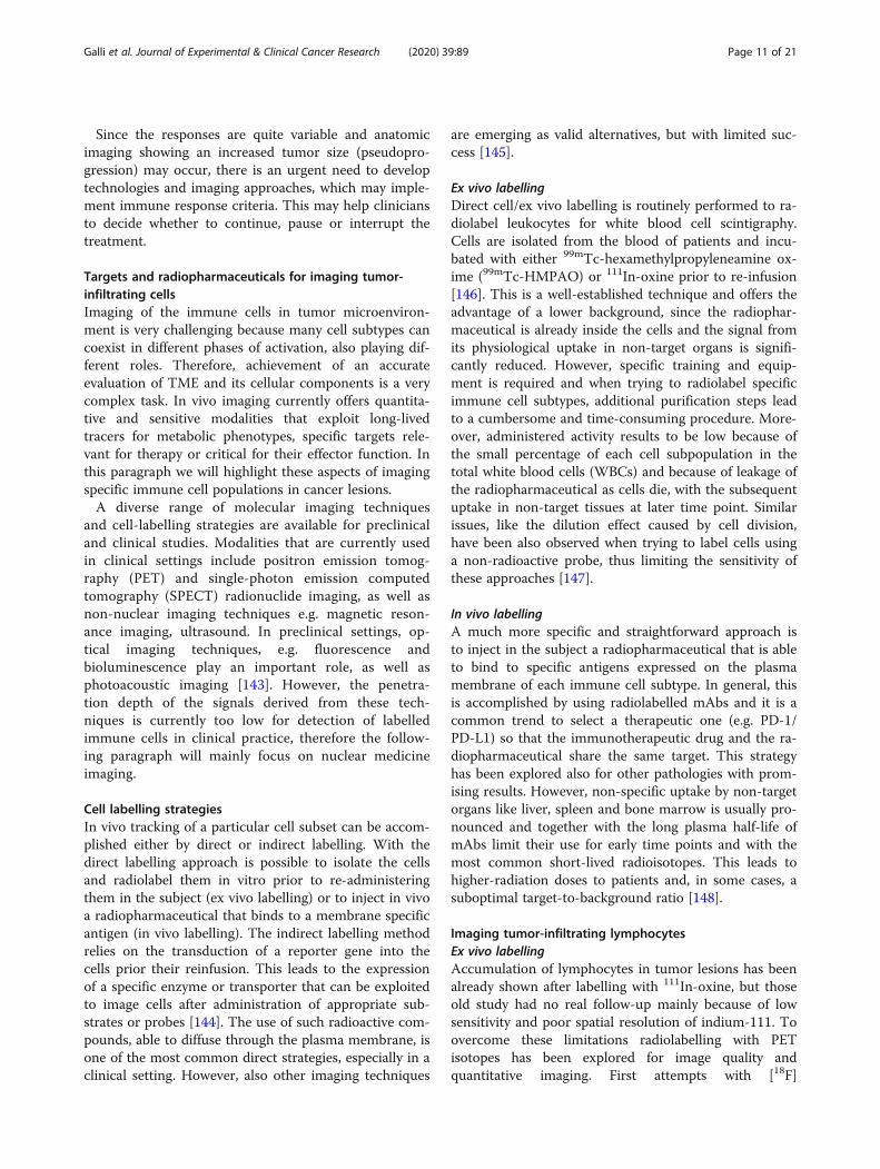
smaller molecules can be used and they include peptidesor small proteins like cytokines. In particular, radio-labelled IL2 is one of the most studied cytokine-basedradiopharmaceuticals. Its receptor, the CD25, is over-expressed on activated T lymphocytes and it drives theirproliferation and inflammatory response. Therefore,radioactive IL-2 as a radiopharmaceutical to target Tcells in vivo has been pioneered by Signore et al. inmany autoimmune pathologies. A recent study, con-ducted in patients affected by metastatic melanoma andundergoing immunotherapy with either pembrolizumabor ipilimumab, demonstrated the feasibility of its use asa candidate-imaging tool to evaluate TILs into tumors[162]. Indeed, in some patients, lesions with high SUV atthe pre-therapy scan positively responded to the therapy.However, what emerged from this study is that intra-
Galli et al. Journal of Experimental & Clinical Cancer Research (2020) 39:89 Page 12 of 21
patient heterogeneity is a true open challenge, since inthe same patient, differential uptake in studied lesionsover the course of the therapy were observed (Fig. 2).This leads to the need of more accurate studies in ahigher cohort of patients, to understand common pat-terns of uptake and understand the mechanisms thatcause therapy response or failure. As an alternative tointact mAbs, radiolabelled fragments like diabodies orminibodies offers a lower half-life (2-5 h or 5-12 h re-spectively) with faster clearance from the blood pool.However, lower specificity and stability is a commonissue that should be taken into account.This approach has been investigated by Tavarè et al. that
developed an anti-CD8 cys-diabody radiolabelled withzirconium-89. This radiopharmaceutical showed specificityto activated T cells and allowed the authors to follow theirinfiltration of EL4-Ova tumors in an OT-I adoptive T celltherapy model. Moreover, they were able to demonstrateits potential by treating the same mice with an immune ac-tivating mAb (anti-CD137). Indeed, treated mice showedhigher uptake of the radiopharmaceutical than controls,due to higher infiltration of tumor lesions [163, 164].
Imaging tumor-infiltrating NK cellsEx vivo labellingApproaches to radiolabel tumor-infiltrating NK cells aresimilar to those described for T lymphocytes. Indeed, 111In-
oxine, 99mTc-HMPAO or [18F]FDG has been attempted tofollow NK infiltration in patients undergoing immunother-apy or in pre-clinical models, but with limited success. Is-sues related to these techniques like poor sensitivity oraltered biodistribution are amplified by the low number ofNK cells and the cumbersome purification procedure priortheir labelling and injection. This has been confirmed byMeller et al. that analysed NK cell number after 3 d fromtheir administration in patients with renal cell carcinomathat received 111In-oxine-labelled and unlabelled NK cellsfrom allogeneic donors [165]. They observed accumulationof labelled cells in two out of four metastases, but also sig-nificant circulating activity due to indium-111 releasedfrom dying cells. Other techniques like 11C-methyl-iodideor fluorescent labelling are described in the literature andpotentially applicable, but they are still limited to early pre-clinical phases [166, 167]. Also, the use of SPIOs showedthe typical signal reduction caused by cell division and de-creased cell viability. From these studies emerged that injec-tion of engineered NK cells against cancer specific antigens,was followed by a decrease in the signal at tumor site, thusconfirming the strong anti-cancer activity of NKs and thepotential of immunotherapies.
In vivo labellingVery few papers describe the use of radiopharmaceuti-cals that binds to NKs in vivo. The most recent study
Fig. 2 99mTc-IL2 SPECT-CT in patients affected by metastatic melanoma before (top) and after (bottom) immunotherapy with ipilimumab. a)Patient with a 99mTc-IL2-positive lesion that responded to therapy. b) Multimetastatic patient with different degree of uptake of 99mTc-IL2
Galli et al. Journal of Experimental & Clinical Cancer Research (2020) 39:89 Page 13 of 21

investigated the use of 99mTc-anti-CD56 mAb, being thisantigen a distinctive marker of NK lineage. In the study,SCID mice bearing a human tumor xenograft derivedfrom an aggressive cell line have been injected with NKcells, followed by injection of the radiopharmaceutical.Results showed uptake of the radiolabelled mAb intumor lesions of mice that received NKs but not in con-trols that did not receive the cells. Immunohistochemis-try confirmed the presence of tumor-infiltrating NKcells and their amount positively correlated with T/B ra-tios against the contralateral leg. In line with findingsfrom other studies, the more the tumors were infiltrated,the more necrosis occurred due to active killing of can-cer cells from NKs [168].
Imaging tumor-associated macrophagesEx vivo labellingAs for T and NK cells, macrophages can be cultured anddifferentiated ex vivo prior to radiolabelling with 111In-oxine or 18[F]FDG. However, in a study by Quillienet al., 111In-oxine-labelled macrophages, after in vitro ex-pansion, accumulated in only 1 lesion out of 15 patientsstudied by SPECT imaging. They analysed cell’s pheno-type after culturing them ex vivo and hypothesized thatculturing conditions might have influenced their homingproperties [169].
Given the innate phagocytic activity of macro-phages, new approaches consist in the use of nano-particles loaded with different reporter agents. Forthis purpose, SPIO nanoparticles were the mostused for magnetic resonance imaging [170], but also19F-loaded and/or fluorescent polymeric nanoparti-cles were used [171, 172]. All the limitations de-scribed above apply also for macrophage imaging,but the use of long-lived radioisotopes likezirconium-89 could lead to improved sensitivity andhigh T/B ratio. In the literature we can find rHDL,polymeric or cross-linked dextran nanoparticlesradiolabelled with zirconium-89 and with differentsizes. All of them showed high tumor uptake, butno correlation with number of TAMs subpopula-tions reflecting a possible unspecific uptake causedmore from the EPR effect than from phagocyticactivity [173, 174].
In vivo labellingA well-known radiopharmaceutical for in vivo im-aging of macrophages is the [11C]-(R)PK11195 thatbinds the translocator protein (TSPO) expressed athigh grade in the mitochondrial membrane ofmacrophages and microglial cells. This has beenmainly studied to image neuroinflammation, butmay have application in imaging tumor-associated
Table 2 Immunotherapeutic drugs approved for human use
Drug Target Clinical use Mechanism of action Labellingagent
Rituximab CD20 B-Cell non-Hodgkin lymphoma, Chronic lymphocyticleukemia.
Direct induction of apoptosis. 99mTc
Ipilimumab/Tremelimumab
CTLA-4 Metastatic melanoma, renal cell carcinoma, hepatocellularcarcinoma.
Inhibition of CTLA-4 signaling 64Cu-DOTA
Pembrolizumab/Nivolumab
PD-1 Melanoma, non-small-cell lung cancer, renal cell carcin-oma, Hodgkin lymphoma, squamous cell carcinoma ofthe head and neck, gastric cancer, cervical cancer, urothe-lial carcinoma, colorectal cancer with microsatelliteinstability-high (MSI-H) or mismatch repair deficient(dMMR)metastatic colorectal cancer.
Inhibition of PD-1 (expressed in lympho-cytes), induction of tumor-specific T cellCD8+ activation against cancer
64Cu-DOTA;89Zr-DFO;111In-DTPA
Atezolizumab PD-L1 Urothelial cancer, non-small cell lung cancer, small celllung cancer, triple negative breast cancer.
Inhibition of PD-L1 (expressed in tumorcells), induction of tumor-specific T cellCD8+ activation against cancer
89Zr-DFO;111In-DTPA
Durvalumab PD-L1 Urothelial carcinoma, non-small cell lung cancer. Inhibition of PD-L1 (expressed in tumorcells), induction of tumor-specific T cellCD8+ activation against cancer
89Zr-DFO
Avelumab PD-L1 Merkel -cell carcinoma, renal cell carcinoma, urothelialcarcinoma.
Inhibition of PD-L1 (expressed in tumorcells), induction of tumor-specific T cellCD8+ activation against cancer
89Zr-DFO
Interleukin-2 IL2receptors
Metastatic renal cell carcinoma and metastatic melanoma T cell activation and expansion 123I; 99mTc;18F
Interferon alfa-2B
INF- αreceptors
Hairy cell leukemia, Malignant melanoma, follicularlymphoma, AIDS related Kaposi Sarcoma.
Immunomodulating activities, includingcytotoxicity of lymphocytes. Upregulationof Th1 T-helper cell subsets
131I
Source. https://www.fda.gov/
Galli et al. Journal of Experimental & Clinical Cancer Research (2020) 39:89 Page 14 of 21

macrophages (TAMs). In vivo studies in mice thatwere not able to correlate the radiopharmaceuticaluptake with TSPO expression, revealed by immuno-histochemistry [175] and data in humans is verylimited. An alternative approach has been proposedby Movahedi et al that used a 99mTc-radiolabelednanobody against the mannose receptor, which isexpressed by macrophages. They were able to dem-onstrate uptake of the radiopharmaceutical in man-nose receptor-expressing tumors as compared withcontrol mice bearing negative tumors [176]. Similarresults were obtained using a [18F]-SFB-counterpartthat showed higher sensitivity and better biodistri-bution. A more recent approach exploits the use of3′-Aza-2′-[18F]-fluoro-folic acid, also known as[18F]-AzaFol, which was previously used to imagemacrophages in various diseases [177]. This radio-pharmaceutical has more advantages than TSPO,but its use in tumor associated macrophages hasnot been investigated yet.
Indirect labellingThe indirect labelling approach is based on the insertionof a gene encoding for specific receptors or enzymes thatallows the labelled probe to enter the cell and being spe-cifically trapped inside. This strategy greatly reducesbackground and can be controlled by placing the genesunder control of specific promotors. Moreover, the dilu-tion effect is not an issue, since the construct will bemaintained after cell division. On the other hand, it isvery difficult to apply this strategy in clinical practicedue to the need of genetic modification and cellmanipulation.The HSV1-tk reporter gene is a common technique
that exploit the specificity of this enzyme for 9-[4-[18F]3-(hydroxymethyl)butyl] guanine ([18F]FHBG), 2-deoxy-2-[18F]5-ethyl-1-D-arabinofuranosyluracil ([18F]FEAU) or2-deoxy-2-[18F]5-iodo-1-D-arabino-furanosyluracil([18F]FIAU). These compounds are taken up by nucleo-side transporters and then are phosphorylated by theenzyme remaining trapped in the cytoplasm [178]. This
Table 3 Other potential radiopharmaceuticals to image tumor infiltrating immune cells
Compound Labelling agent Target/Mechanism Application
T lymphocytes 111In-oxine Tumor infiltration/Cytokine production Evaluation of immunotherapy/adoptive cell transferefficacy89Zr-oxine
[18F]FDG64Cu-goldnanoparticles
SPIO19F-Perfluorcarbon
mAb-TCR-complex 64Cu Tumor infiltration T cell homing
Interleukin-2 123I Interleukin-2 receptors on activatedlymphocytes
Evaluation of immunotherapy/adoptive cell transferefficacy99mTc
18F
Anti-CD8 cys diabody 89Zr CD8 on activated T cells Evaluation of immunotherapy efficacy
NK cells 111In-oxine NK cell infiltration Evaluation of adoptive cell transfer efficacy – NKcell homing89Zr-oxine
[18F]FDG
SPIO
Anti-CD56 mAb 99mTc CD56 on NK cells Evaluation of adoptive cell transfer efficacy – NKcell homing
Macrophages 111In-oxine Tumor infiltration by macrophages Pre-clinical evaluation of TAMs89Zr-Nanoparticles
[18F]FDG19F-Nanoparticles
SPIO
(R)PK11195 11C translocator protein (TSPO) expressed byTAMs
Pre-clinical evaluation of TAMs
Anti-Mannose receptornanobody
99mTc Mannose receptor on TAMs Pre-clinical evaluation of TAMs18F
Galli et al. Journal of Experimental & Clinical Cancer Research (2020) 39:89 Page 15 of 21

allows following cell trafficking in vivo by PET withoutthe limitations of the short half-life of fluorine-18. Thisvery same strategy can be applied by transducing the so-dium/iodine symporter gene and administering iodine-124 for PET or sodium pertechnetate-99m for gammacamera imaging. However, this approach requiresspecific training and equipment due to genetic cellmanipulation and is less suitable for routine humanapplications.
ConclusionIn the present review, we wanted to give an overview ofthe immune cells that are involved in tumor microenvir-onment infiltration to highlight why imaging of theirtrafficking is so crucial with so many new immunother-apies entering the clinical practice. The main issue whenevaluating tumor response to cancer immunotherapy isthe enlargement due to infiltrating immune cells thateventually leads to tumor shrinkage and death. The sameenlargement occurs in case of tumor progression due tocancer cell growth and in both situations increased up-take of [18F]FDG is observed. This limits the use ofcurrent available criteria and new ones are under defin-ition with limited success. That is why we need newnon-invasive tools to rely on and molecular imagingoffers the most suitable approach.Unfortunately, to fully achieve this goal we still have
to face many open challenges like the many immune cellsubtypes, small number and dynamic behaviour. Thisimplies that radiopharmaceuticals of choice should behighly specific for biomarkers expressed by different im-mune cells. Molecular imaging can guide basic research,drug development and clinical follow up. It can also helpresearchers to elucidate mechanisms of pathology, effectof new drugs and predict efficacy of immunotherapies.There is still a long way to go, but many tools, summa-rized in Table 2 and 3, are already available and underinvestigation with different pros and cons. Antibodiesare still “the magic bullets”, but their long circulatinghalf-life and need of humanization at high costs are stillthe limiting factors. To overcome these issues fragmentscan be developed with loss of specificity but increasedT/B ratio at earlier time points. This permits to useshort-lived PET isotopes like gallium-68 or fluorine-18in place of zirconium-89, thus reducing radiation doseto patients.Radiolabelled immune checkpoint inhibitor mAbs
showed great results in vivo, but to date they are stilllimited to pre-clinical studies. Also, cytokines like IL2showed great potential and pilot human studies havealready been performed with interesting results. Thenext step, for each of these radiopharmaceuticals, wouldbe to increase the number of enrolled patients and de-fine patterns of uptake to define new criteria for therapy
decision-making and follow-up. This is particularlyimportant, since inter- and intra-patient tumor hetero-geneity is a real concern that may even require a“lesionalised therapy”.This will require multimodal approaches as interac-
tions in tumor microenvironment and different mecha-nisms of evasion from immune response are toocomplex to be unfolded by a single imaging tool.
AbbreviationsAPC: antigen-presenting cells; BCG: Bacillus Calmette-Guérin; CAF: Cancerassociated fibroblasts; CAR-T: Chimeric antigen receptor T; CSF: Colony-stimulating factor; CXCL12: C-X-C motif chemokine 12; CTLA-4: Cytotoxic Tlymphocyte associated protein 4; DC: Dendritic cells; ECM: Extracellularmatrix; EMT: Epithelial mesenchymal transition; FDA: Food and DrugAdministration; [18F]FDG: [18F]Fluorodeoxyglucose; GM-CSF: Granulocyte-macrophage colony-stimulating factor; HMPAO: Hexamethylpropyleneamineoxime; ICB: Immune checkpoint blockades; IDO: Indoleamine-2,3-dioxygenase; IFN-α: Interferon α; MHC: Major histocompatibility complex;MDSCs: Myeloid-derived suppressor cells; NK: Natural killer; NSCLC: Non-smallcell lung cancer; PDAC: pancreatic ductal adenocarcinoma; PD-L1: Programmed death ligand 1; PD-1: Programmed death receptor 1;PET: Positron emission tomography; SPECT: Single-photon emissioncomputed tomography; SPIO: Small iron oxide particles; TAMs: Tumor-associated macrophages; TAN: Tumor-associated neutrophils;TGFβ: transforming growth factor-β; TILs: Tumor-infiltrating lymphocytes;TME: Tumor microenvironment; TSPO: Translocator protein; VEGF: Vascularendothelial growth factor; WBCs: white blood cells
AcknowledgementsNot applicable
Availability of data and materialNot applicable
Authors’ contributionsBP and PA edited the section on tumor infiltrating immune cells andmicroenvironment. JA and SM edited the section on current and newimmunotherapeutic approaches against cancer. FG and AS edited theimaging section and coordinated the work. All authors read and approvedthe final manuscript.
FundingFunding has been provided by AIRC IG-Grant 2017 20411 (AS), AIRC IG 19822and AIRC 5X1000 12182 (PN).
Ethics approval and consent to participateNot applicable
Consent for publicationNot applicable
Competing interestsAuthors declare no conflict of interests.
Author details1Nuclear Medicine Unit, Department of Medical-Surgical Sciences and ofTranslational Medicine, “Sapienza” University of Rome, S. Andrea UniversityHospital, Roma, Italy. 2Department of oncology and Department ofImmunology, Mayo Clinic, (MN), Rochester, USA. 3Tumor Immunology andImmunotherapy Unit, Department of Research, Advanced Diagnostics andTechnological Innovation, IRCCS Regina Elena National Cancer Institute,Rome, Italy.
Galli et al. Journal of Experimental & Clinical Cancer Research (2020) 39:89 Page 16 of 21

Received: 13 February 2020 Accepted: 28 April 2020
References1. Wei SC, Duffy CR, Allison JP. Fundamental Mechanisms of Immune
Checkpoint Blockade Therapy. Cancer Discov. 2018;8:1069–86.2. Urwyler P, Earnshaw I, Bermudez M, Perucha E, Wu W, Ryan S, Mcdonald L,
Karagiannis SN, Taams LS, Powell N, Cope A, Papa S. Mechanisms ofcheckpoint inhibition induced adverse events. Clin Exp Immunol. 2020.https://doi.org/10.1111/cei.13421.
3. Miranda A, Hamilton PT, Zhang AW, Pattnaik S, Becht E, Mezheyeuski A,Bruun J, Micke P, de Reynies A, Nelson BH. Cancer stemness, intratumoralheterogeneity, and immune response across cancers. PNAS. 2019;116:9020–9.
4. Gwin WR, Disis ML, Ruiz-Garcia E. Immuno-Oncology in the Era ofPersonalized Medicine. Adv Exp Med Biol. 2019;1168:117–29.
5. Verhoeff SR, van den Heuvel MM, van Herpen CML, Piet B, Aarntzen EHJG,Heskamp S. Programmed Cell Death-1/Ligand-1 PET Imaging: A Novel Toolto Optimize Immunotherapy? PET Clin. 2020;15:35–43.
6. Bissell MJ, Radisky D. Putting tumours in context. Nat Rev Cancer.2001;1:46–54.
7. Hanahan D, Coussens LM. Accessories to the crime: functions of cellsrecruited to the tumor microenvironment. Cancer Cell. 2012;21:309–22.
8. Pickup MW, Mouw JK, Weaver VM. The extracellular matrix modulates thehallmarks of cancer. EMBO Rep. 2014;15:1243–53.
9. Buck MD, Sowell RT, Kaech SM, Pearce EL. Metabolic Instruction ofImmunity. Cell. 2017;169:570–86.
10. Fridman WH, Pagès F, Sautès-Fridman C, Galon J. The immune contexturein human tumours: impact on clinical outcome. Nat Rev Cancer. 2012;12:298–306.
11. Fridman WH, Zitvogel L, Sautès-Fridman C, Kroemer G. The immunecontexture in cancer prognosis and treatment. Nat Rev Clin Oncol.2017;14:717–34.
12. Josefowicz SZ, Lu LF, Rudensky AY. Regulatory T cells: mechanisms ofdifferentiation and function. Annu Rev Immunol. 2012;30:531–64.
13. Pacella I, Piconese S. Immunometabolic Checkpoints of Treg Dynamics:Adaptation Microenvironmental Opportunities and Challenges. FrontImmunol. 2019;10:1889.
14. Zhang Y, Lazarus J, Steele NG, Yan W, Lee HJ, Nwosu ZC, Halbrook CJ,Menjivar RE, Kemp SB, Sirihorachai V, Velez-Delgado A, Donahue K,Carpenter ES, Brown KL, Irizarry-Negron V, Nevison AC, Vinta A, AndersonMA, Crawford HC, Lyssiotis CA, Frankel TL, Bednar F, Pasca di Magliano M.Regulatory T cell depletion alters the tumor microenvironment andaccelerates pancreatic carcinogenesis. Cancer Discov. 2020. pii: CD-19-0958.
15. Galon J, Costes A, Sanchez-Cabo F, Kirilovsky A, Mlecnik B, Lagorce-Pagès C,Tosolini M, Camus M, Berger A, Wind P, Zinzindohoué F, Bruneval P,Cugnenc PH, Trajanoski Z, Fridman WH, Pagès F. Type, density, and locationof immune cells within human colorectal tumors predict clinical outcome.Science. 2006;313:1960–4.
16. Sharma P, Allison JP. The future of immune checkpoint therapy. Science.2015;348:56–61.
17. Binnewies M, Roberts EW, Kersten K, Chan V, Fearon DF, Merad M, CoussensLM, Gabrilovich DI, Ostrand-Rosenberg S, Hedrick CC, Vonderheide RH, PittetMJ, Jain RK, Zou W, Howcroft TK, Woodhouse EC, Weinberg RA, KrummelMF. Understanding the tumor immune microenvironment (TIME) foreffective therapy. Nat Med. 2018;24:541–50.
18. Chen DS, Mellman I. Elements of cancer immunity and the cancer-immuneset point. Nature. 2017;541:321–30.
19. Jiang P, Gu S, Pan D, Fu J, Sahu A, Hu X, Li Z, Traugh N, Bu X, Li B, Liu J,Freeman GJ, Brown MA, Wucherpfennig KW, Liu XS. Signatures of T celldysfunction and exclusion predict cancer immunotherapy response. NatMed. 2018;24:1550–8.
20. Trono P, Sistigu A, Palermo B, Ciliberto G, Nisticò P. Mesenchymal traits atthe convergence of tumor-intrinsic and -extrinsic mechanisms of resistanceto immune checkpoint blockers Emerging Topics in Life Sciences. 2017;1:471-486.
21. Levental KR, Yu H, Kass L, Lakins JN, Egeblad M, Erler JT, Fong SF, Csiszar K,Giaccia A, Weninger W, Yamauchi M, Gasser DL, Weaver VM. Matrixcrosslinking forces tumor progression by enhancing integrin signaling. Cell.2009;139:891–906.
22. Joyce JA, Fearon DT. T cell exclusion, immune privilege, and the tumormicroenvironment. Science. 2015;348:74–80.
23. Anderson KG, Stromnes IM, Greenberg PD. Obstacles Posed by the TumorMicroenvironment to T cell Activity: A Case for Synergistic Therapies. CancerCell. 2017;31:311–25.
24. Zhang L, Conejo-Garcia JR, Katsaros D, Gimotty PA, Massobrio M, Regnani G,Makrigiannakis A, Gray H, Schlienger K, Liebman MN, Rubin SC, Coukos G.Intratumoral T cells, recurrence, and survival in epithelial ovarian cancer. NEngl J Med. 2003;348:203–13.
25. Johansson-Percival A, He B, Ganss R. Immunomodulation of Tumor Vessels:It Takes Two to Tango. Trends Immunol. 2018;39:801–14.
26. Feig C, Jones JO, Kraman M, Wells RJ, Deonarine A, Chan DS, Connell CM,Roberts EW, Zhao Q, Caballero OL, Teichmann SA, Janowitz T, Jodrell DI,Tuveson DA, Fearon DT. Targeting CXCL12 from FAP-expressing carcinoma-associated fibroblasts synergizes with anti-PD-L1 immunotherapy inpancreatic cancer. Proc Natl Acad Sci U S A. 2013;110:20212–7.
27. Sprooten J, Ceusters J, Coosemans A, Agostinis P, De Vleeschouwer S,Zitvogel L, Kroemer G, Galluzzi L, Garg AD. Trial watch: dendritic cellvaccination for cancer immunotherapy. Oncoimmunology. 2019;8:e1638212.
28. Petitprez F, de Reyniès A, Keung EZ, Chen TW, Sun CM, Calderaro J, JengYM, Hsiao LP, Lacroix L, Bougoüin A, Moreira M, Lacroix G, Natario I, Adam J,Lucchesi C, Laizet YH, Toulmonde M, Burgess MA, Bolejack V, Reinke D,Wani KM, Wang WL, Lazar AJ, Roland CL, Wargo JA, Italiano A, Sautès-Fridman C, Tawbi HA, Fridman WH. B cells are associated with survival andimmunotherapy response in sarcoma. Nature. 2020. https://doi.org/10.1038/s41586-019-1906-8.
29. DiLillo DJ, Yanaba K, Tedder TF. B cells are required for optimal CD4+ andCD8+ T cell tumor immunity: therapeutic B cell depletion enhances B16melanoma growth in mice. J Immunol. 2010;184:4006–16.
30. Nielsen JS, Sahota RA, Milne K, Kost SE, Nesslinger NJ, Watson PH, NelsonBH. CD20+ tumor-infiltrating lymphocytes have an atypical CD27- memoryphenotype and together with CD8+ T cells promote favorable prognosis inovarian cancer. Clin Cancer Res. 2012;18:3281–92.
31. Al-Shibli KI, Donnem T, Al-Saad S, Persson M, Bremnes RM, Busund LT.Prognostic effect of epithelial and stromal lymphocyte infiltration in non-small cell lung cancer. Clin Cancer Res. 2008;14:5220–7.
32. Svensson MC, Warfvinge CF, Fristedt R, Hedner C, Borg D, Eberhard J, MickeP, Nodin B, Leandersson K, Jirström K. The integrative clinical impact oftumor-infiltrating T lymphocytes and NK cells in relation to B lymphocyteand plasma cell density in esophageal and gastric adenocarcinoma.Oncotarget. 2017;8:72108–26.
33. Nedergaard BS, Ladekarl M, Nyengaard JR, Nielsen K. A comparative studyof the cellular immune response in patients with stage IB cervicalsquamous cell carcinoma. Low numbers of several immune cell subtypesare strongly associated with relapse of disease within 5 years. GynecolOncol. 2008;108:106–11.
34. de Visser KE, Korets LV, Coussens LM. De novo carcinogenesis promoted bychronic inflammation is B lymphocyte dependent. Cancer Cell. 2005;7:411–23.
35. Ammirante M, Luo JL, Grivennikov S, Nedospasov S, Karin M. B-cell-derivedlymphotoxin promotes castration-resistant prostate cancer. Nature. 2010;464:302–5.
36. Shalapour S, Font-Burgada J, Di Caro G, Zhong Z, Sanchez-Lopez E, Dhar D,Willimsky G, Ammirante M, Strasner A, Hansel DE, Jamieson C, Kane CJ,Klatte T, Birner P, Kenner L, Karin M. Immunosuppressive plasma cellsimpede T-cell-dependent immunogenic chemotherapy. Nature. 2015;521:94–8.
37. Zingoni A, Fionda C, Borrelli C, Cippitelli M, Santoni A, Soriani A. NaturalKiller Cell Response to Chemotherapy-Stressed Cancer Cells: Role in TumorImmunosurveillance. Front Immunol. 2017;8:1194.
38. Glasner A, Levi A, Enk J, Isaacson B, Viukov S, Orlanski S, Scope A, Neuman T,Enk CD, Hanna JH, Sexl V, Jonjic S, Seliger B, Zitvogel L, Mandelboim O.NKp46 Receptor-Mediated Interferon-γ Production by Natural Killer CellsIncreases Fibronectin 1 to Alter Tumor Architecture and Control Metastasis.Immunity. 2018;48:107–119.e4.
39. Sungur CM, Murphy WJ. Positive and negative regulation by NK cells incancer. Crit Rev Oncog. 2014;19:57–66.
40. Steinman RM, Cohn ZA. Identification of a novel cell type in peripherallymphoid organs of mice. I. Morphology, quantitation, tissue distribution. JExp Med. 1973;137:1142–62.
41. Banchereau J, Steinman RM. Dendritic cells and the control of immunity.Nature. 1998;392:245–52.
Galli et al. Journal of Experimental & Clinical Cancer Research (2020) 39:89 Page 17 of 21
42. Martinek J, Wu TC, Cadena D, Banchereau J, Palucka K. Interplay betweendendritic cells and cancer cells. Int Rev Cell Mol Biol. 2019;348:179–215.
43. Sautès-Fridman C, Petitprez F, Calderaro J, Fridman WH. Tertiary lymphoidstructures in the era of cancer immunotherapy. Nat Rev Cancer. 2019;19:307–25.
44. Helmink BA, Reddy SM, Gao J, Zhang S, Basar R, Thakur R, Yizhak K, Sade-Feldman M, Blando J, Han G, Gopalakrishnan V, Xi Y, Zhao H, Amaria RN,Tawbi HA, Cogdill AP, Liu W, VS LB, Kugeratski FG, Patel S, Davies MA, HwuP, Lee JE, Gershenwald JE, Lucci A, Arora R, Woodman S, Keung EZ,Gaudreau PO, Reuben A, Spencer CN, Burton EM, Haydu LE, Lazar AJ,Zapassodi R, Hudgens CW, Ledesma DA, Ong S, Bailey M, Warren S, Rao D,Krijgsman O, Rozeman EA, Peeper D, Blank CU, Schumacher TN, ButterfieldLH, Zelazowska MA, KM MB, Kalluri R, Allison J, Petitprez F, Fridman WH,Sautès-Fridman C, Hacohen N, Rezvani K, Sharma P, Tetzlaff MT, Wang L,Wargo JA. B cells and tertiary lymphoid structures promote immunotherapyresponse. Nature. 2020;577:549–55.
45. Cabrita R, Lauss M, Sanna A, Donia M, Skaarup Larsen M, Mitra S, JohanssonI, Phung B, Harbst K, Vallon-Christersson J, van Schoiack A, Lövgren K,Warren S, Jirström K, Olsson H, Pietras K, Ingvar C, Isaksson K, SchadendorfD, Schmidt H, Bastholt L, Carneiro A, Wargo JA, Svane IM, Jönsson G.Tertiary lymphoid structures improve immunotherapy and survival inmelanoma. Nature. 2020;577:561–5.
46. Mantovani A, Marchesi F, Malesci A, Laghi L, Allavena P. Tumour-associatedmacrophages as treatment targets in oncology. Nat Rev Clin Oncol. 2017;14:399–416.
47. Murray PJ, Allen JE, Biswas SK, Fisher EA, Gilroy DW, Goerdt S, Gordon S,Hamilton JA, Ivashkiv LB, Lawrence T, Locati M, Mantovani A, Martinez FO,Mege JL, Mosser DM, Natoli G, Saeij JP, Schultze JL, Shirey KA, Sica A, SuttlesJ, Udalova I, van Ginderachter JA, Vogel SN, Wynn TA. Macrophageactivation and polarization: nomenclature and experimental guidelines.Immunity. 2014;41:14–20.
48. Kuang DM, Zhao Q, Peng C, Xu J, Zhang JP, Wu C, Zheng L. Activatedmonocytes in peritumoral stroma of hepatocellular carcinoma fosterimmune privilege and disease progression through PD-L1. J Exp Med. 2009;206:1327–37.
49. Kryczek I, Zou L, Rodriguez P, Zhu G, Wei S, Mottram P, Brumlik M, Cheng P,Curiel T, Myers L, Lackner A, Alvarez X, Ochoa A, Chen L, Zou W. B7-H4expression identifies a novel suppressive macrophage population in humanovarian carcinoma. J Exp Med. 2006;203:871–81.
50. Sun X, Ingman WV. Cytokine networks that mediate epithelial cell-macrophage crosstalk in the mammary gland: implications for developmentand cancer. J Mammary Gland Biol Neoplasia. 2014;19:191–201.
51. Pathria P, Louis TL, Varner JA. Targeting Tumor-Associated Macrophages inCancer. Trends Immunol. 2019;40:310–27.
52. Lecot P, Sarabi M, Pereira Abrantes M, Mussard J, Koenderman L, Caux C,Bendriss-Vermare N, Michallet MC. Neutrophil Heterogeneity in Cancer:From Biology to Therapies. Front Immunol. 2019;10:2155.
53. Fridlender ZG, Sun J, Kim S, Kapoor V, Cheng G, Ling L, Worthen GS, AlbeldaSM. Polarization of tumor-associated neutrophil phenotype by TGF-beta:"N1" versus "N2" TAN. Cancer Cell. 2009;16:183–94.
54. Granot Z, Jablonska J. Distinct Functions of Neutrophil in Cancer and ItsRegulation. Mediators Inflamm. 2015;2015:701067.
55. Mishalian I, Bayuh R, Levy L, Zolotarov L, Michaeli J, Fridlender ZG. Tumor-associated neutrophils (TAN) develop pro-tumorigenic properties duringtumor progression. Cancer Immunol Immunother. 2013;62:1745–56.
56. Sade-Feldman M, Kanterman J, Klieger Y, Ish-Shalom E, Olga M, Saragovi A,Shtainberg H, Lotem M, Baniyash M. Clinical Significance of CirculatingCD33+CD11b+HLA-DR- Myeloid Cells in Patients with Stage IV MelanomaTreated with Ipilimumab. Clin Cancer Res. 2016;22:5661–72.
57. Martens A, Wistuba-Hamprecht K, Geukes Foppen M, Yuan J, Postow MA,Wong P, Romano E, Khammari A, Dreno B, Capone M, Ascierto PA, Di GiacomoAM, Maio M, Schilling B, Sucker A, Schadendorf D, Hassel JC, Eigentler TK,Martus P, Wolchok JD, Blank C, Pawelec G, Garbe C, Weide B. BaselinePeripheral Blood Biomarkers Associated with Clinical Outcome ofAdvanced Melanoma Patients Treated with Ipilimumab. Clin Cancer Res.2016;22:2908–18.
58. Park J, Wysocki RW, Amoozgar Z, Maiorino L, Fein MR, Jorns J, Schott AF,Kinugasa-Katayama Y, Lee Y, Won NH, Nakasone ES, Hearn SA, Küttner V,Qiu J, Almeida AS, Perurena N, Kessenbrock K, Goldberg MS, Egeblad M.Cancer cells induce metastasis-supporting neutrophil extracellular DNAtraps. Sci Transl Med. 2016;8:361ra138.
59. Ferrucci PF, Ascierto PA, Pigozzo J, Del Vecchio M, Maio M, AntoniniCappellini GC, Guidoboni M, Queirolo P, Savoia P, Mandalà M, Simeone E,Valpione S, Altomonte M, Spagnolo F, Cocorocchio E, Gandini S, GiannarelliD, Martinoli C. Baseline neutrophils and derived neutrophil-to-lymphocyteratio: prognostic relevance in metastatic melanoma patients receivingipilimumab. Ann Oncol. 2016;27:732–8.
60. Bronte V, Brandau S, Chen SH, Colombo MP, Frey AB, Greten TF,Mandruzzato S, Murray PJ, Ochoa A, Ostrand-Rosenberg S, Rodriguez PC,Sica A, Umansky V, Vonderheide RH, Gabrilovich DI. Recommendations formyeloid-derived suppressor cell nomenclature and characterizationstandards. Nat Commun. 2016;7:12150.
61. Tcyganov E, Mastio J, Chen E, Gabrilovich DI. Plasticity of myeloid-derivedsuppressor cells in cancer. Curr Opin Immunol. 2018;51:76–82.
62. Sistigu A, Di Modugno F, Manic G, Nisticò P. Deciphering the loop ofepithelial-mesenchymal transition, inflammatory cytokines and cancerimmunoediting. Cytokine Growth Factor Rev. 2017;36:67–77.
63. Nagaraj S, Youn JI, Gabrilovich DI. Reciprocal relationship between myeloid-derived suppressor cells and T cells. J Immunol. 2013;191:17–23.
64. Mulder PO, de Vries EG, Koops HS, Splinter T, Maas A, van der Geest S,Mulder NH, Sleijfer DT. Chemotherapy with maximally tolerable doses of VP16-213 and cyclophosphamide followed by autologous bone marrowtransplantation for the treatment of relapsed or refractory germ cell tumors.Eur J Cancer Clin Oncol. 1988;24:675–9.
65. Sangaletti S, Tripodo C, Santangelo A, Castioni N, Portararo P, Gulino A, BottiL, Parenza M, Cappetti B, Orlandi R, Tagliabue E, Chiodoni C, Colombo MP.Mesenchymal Transition of High-Grade Breast Carcinomas Depends onExtracellular Matrix Control of Myeloid Suppressor Cell Activity. Cell Rep.2016 Sep 27;17(1):233–48.
66. Gebhardt C, Sevko A, Jiang H, Lichtenberger R, Reith M, Tarnanidis K,Holland-Letz T, Umansky L, Beckhove P, Sucker A, Schadendorf D, Utikal J,Umansky V. Myeloid Cells and Related Chronic Inflammatory Factors asNovel Predictive Markers in Melanoma Treatment with Ipilimumab. ClinCancer Res. 2015;21:5453–9.
67. Holmgaard RB, Brachfeld A, Gasmi B, Jones DR, Mattar M, Doman T, MurphyM, Schaer D, Wolchok JD, Merghoub T. Timing of CSF-1/CSF-1R signalingblockade is critical to improving responses to CTLA-4 basedimmunotherapy. Oncoimmunology. 2016;5:e1151595.
68. Biffi G, Tuveson DA. Deciphering cancer fibroblasts. J Exp Med. 2018 3;215:2967-2968.
69. Gascard P, Tlsty TD. Carcinoma-associated fibroblasts: orchestrating thecomposition of malignancy. Genes Dev. 2016;30:1002–19.
70. Shintani Y, Fujiwara A, Kimura T, Kawamura T, Funaki S, Minami M, OkumuraM. IL-6 Secreted from Cancer-Associated Fibroblasts MediatesChemoresistance in NSCLC by Increasing Epithelial-Mesenchymal TransitionSignaling. J Thorac Oncol. 2016;11:1482–92.
71. Nissen NI, Karsdal M, Willumsen N. Collagens and Cancer associatedfibroblasts in the reactive stroma and its relation to Cancer biology. J ExpClin Cancer Res. 2019 6;38:115.
72. 3D models in the new era of immune oncology: focus on T cells, CAF andECM. Di Modugno F, Colosi C, Trono P, Antonacci G, Ruocco G, Nisticò P. JExp Clin Cancer Res. 2019;38:117.
73. Jacob M, Chang L, Puré E. Fibroblast activation protein in remodelingtissues. Curr Mol Med. 2012;12:1220–43.
74. Kraman M, Bambrough PJ, Arnold JN, Roberts EW, Magiera L, Jones JO,Gopinathan A, Tuveson DA, Fearon DT. Suppression of antitumor immunityby stromal cells expressing fibroblast activation protein-alpha. Science. 2010;330:827–30.
75. Looi CK, Chung FF, Leong CO, Wong SF, Rosli R, Mai CW. Therapeuticchallenges and current immunomodulatory strategies in targeting theimmunosuppressive pancreatic tumor microenvironment. J Exp Clin CancerRes. 2019;38:162.
76. Pearce OMT, Delaine-Smith RM, Maniati E, Nichols S, Wang J, Böhm S,Rajeeve V, Ullah D, Chakravarty P, Jones RR, Montfort A, Dowe T, Gribben J,Jones JL, Kocher HM, Serody JS, Vincent BG, Connelly J, Brenton JD, ChelalaC, Cutillas PR, Lockley M, Bessant C, Knight MM, Balkwill FR. Deconstructionof a Metastatic Tumor Microenvironment Reveals a Common MatrixResponse in Human Cancers. Cancer Discov. 2018;8:304–19.
77. Gilkes DM, Semenza GL, Wirtz D. Hypoxia and the extracellular matrix:drivers of tumour metastasis. Nat Rev Cancer. 2014;14:430–9.
78. Sukumar M, Roychoudhuri R, Restifo NP. Nutrient Competition: A New Axisof Tumor Immunosuppression. Cell. 2015;162:1206–8.
Galli et al. Journal of Experimental & Clinical Cancer Research (2020) 39:89 Page 18 of 21

79. Chang CH, Qiu J, O'Sullivan D, Buck MD, Noguchi T, Curtis JD, Chen Q,Gindin M, Gubin MM, van der Windt GJ, Tonc E, Schreiber RD, Pearce EJ,Pearce EL. Metabolic Competition in the Tumor Microenvironment Is aDriver of Cancer Progression. Cell. 2015;162:1229–41.
80. Marin-Acevedo JA, Soyano AE, Dholaria B, Knutson KL, Lou Y. Cancerimmunotherapy beyond immune checkpoint inhibitors. J Hematol Oncol.2018;11:8.
81. McCarthy EF. The toxins of William B. Coley and the treatment of bone andsoft-tissue sarcomas. Iowa Orthop J. 2006;26:154–8.
82. Parish CR. Cancer immunotherapy: the past, the present and the future.Immunol Cell Biol. 2003;81:106–13.
83. Morales A, Eidinger D, Bruce AW. Intracavitary Bacillus Calmette-Guerin inthe Treatment of Superficial Bladder Tumors. J Urol. 2017;197:S142–s145.
84. Old LJ, Clarke DA, Benacerraf B. Effect of Bacillus Calmette-Guerin infectionon transplanted tumours in the mouse. Nature. 1959;184:291–2.
85. Sarraf C. Tumor-necrosis-factor and cell-death in tumors (review). Int JOncol. 1994;5:1333–9.
86. Decker WK, da Silva RF, Sanabria MH, Angelo LS, Guimarães F, Burt BM,Kheradmand F, Paust S. Cancer Immunotherapy: Historical Perspective of aClinical Revolution and Emerging Preclinical Animal Models. Front Immunol.2017;8:829.
87. Egen JG, Ouyang W, Wu LC. Human Anti-tumor Immunity: Insights fromImmunotherapy Clinical Trials. Immunity. 2020;52:36–54.
88. Weiner LM. Cancer immunology for the clinician. Clin Adv Hematol Oncol.2015;3:299–306.
89. Smith SA, Crowe JE Jr. Use of Human Hybridoma Technology To IsolateHuman Monoclonal Antibodies. Microbiol Spectr. 2015;3:AID-0027-2014.
90. Klener P Jr, Otáhal P, Lateckova L, Klener P. Immunotherapy Approaches inCancer Treatment. Curr Pharm Biotechnol. 2015;16:771–81.
91. Karlitepe A, Ozalp O, Avci CB. New approaches for cancer immunotherapy.Tumour Biol. 2015;36:4075–8.
92. Ventola CL. Cancer Immunotherapy, Part 1: Current Strategies and Agents. PT. 2017;42:375–83.
93. Abdel-Wahab N, Shah M, Lopez-Olivo MA, Suarez-Almazor ME. Use ofImmune Checkpoint Inhibitors in the Treatment of Patients With Cancerand Preexisting Autoimmune Disease: A Systematic Review. Ann InternMed. 2018;168:121–30.
94. Thallinger C, Füreder T, Preusser M, Heller G, Müllauer L, Höller C, Prosch H,Frank N, Swierzewski R, Berger W, Jäger U, Zielinski C. Review of cancertreatment with immune checkpoint inhibitors : Current concepts,expectations, limitations and pitfalls. Wien Klin Wochenschr. 2018;130:85–91.
95. Sheng J, Fang W, Yu J, Chen N, Zhan J, Ma Y, Yang Y, Huang Y, Zhao H, ZhangL. Expression of programmed death ligand-1 on tumor cells varies pre andpost chemotherapy in non-small cell lung cancer. Sci Rep. 2016;6:20090.
96. Ishida Y, Agata Y, Shibahara K, Honjo T. Induced expression of PD-1, a novelmember of the immunoglobulin gene superfamily, upon programmed celldeath. The EMBO journal. 1992;11(11):3887–95.
97. Tumeh PC, Harview CL, Yearley JH, Shintaku IP, Taylor EJ, Robert L,Chmielowski B, Spasic M, Henry G, Ciobanu V, West AN, Carmona M, KivorkC, Seja E, Cherry G, Gutierrez AJ, Grogan TR, Mateus C, Tomasic G, Glaspy JA,Emerson RO, Robins H, Pierce RH, Elashoff DA, Robert C, Ribas A. PD-1blockade induces responses by inhibiting adaptive immune resistance.Nature. 2014;515:568–71.
98. Pardoll DM. The blockade of immune checkpoints in cancerimmunotherapy. Nature reviews. Cancer. 2012;12:252–64.
99. Larkin J, Hatswell AJ, Nathan P, Lebmeier M, Lee D. The Predicted Impact ofIpilimumab Usage on Survival in Previously Treated Advanced or MetastaticMelanoma in the UK. PLoS One. 2015;10:e0145524.
100. Speiser DE, Ho PC, Verdeil G. Regulatory circuits of T cell function in cancer.Nat Rev Immunol. 2016;16:599–611.
101. Mizukami Y, Kono K, Kawaguchi Y, Akaike H, Kamimura K, Sugai H, Fujii H.CCL17 and CCL22 chemokines within tumor microenvironment are relatedto accumulation of Foxp3+ regulatory T cells in gastric cancer. Int J Cancer.2008;122:2286–93.
102. Zitvogel LA. Tesniere, and G. Kroemer, Cancer despite immunosurveillance:immunoselection and immunosubversion. Nat Rev Immunol. 2006;6:715–27.
103. Shen F, Su H, Fan Y, Chen Y, Zhu Y, Liu W, Young WL, Yang GY. Adeno-associated viral-vector-mediated hypoxia-inducible vascular endothelialgrowth factor gene expression attenuates ischemic brain injury after focalcerebral ischemia in mice. Stroke. 2006;37:2601–6.
104. Rehman H, Silk AW, Kane MP, Kaufman HL. Into the clinic: Talimogenelaherparepvec (T-VEC), a first-in-class intratumoral oncolytic viral therapy. JImmunother Cancer. 2016;4:53.
105. Zheng M, Huang J, Tong A, Yang H. Oncolytic Viruses for Cancer Therapy:Barriers and Recent Advances. Mol Ther Oncolytics. 2019;15:234–47.
106. Hawkins LK, Lemoine NR, Kirn D. Oncolytic biotherapy: a novel therapeuticplatform. Lancet Oncol. 2002;3:17–26.
107. Toda M, Martuza RL, Rabkin SD. Tumor growth inhibition by intratumoralinoculation of defective herpes simplex virus vectors expressinggranulocyte-macrophage colony-stimulating factor. Mol Ther. 2000;2:324–9.
108. Alatrash G, Jakher H, Stafford PD, Mittendorf EA. Cancer immunotherapies,their safety and toxicity. Expert Opin Drug Saf. 2013;12:631–45.
109. Jiang T, Zhou C, Ren S. Role of IL-2 in cancer immunotherapy.Oncoimmunology. 2016;5:e1163462.
110. Kirkwood J. Cancer immunotherapy: the interferon-alpha experience. SeminOncol. 2002;29:18–26.
111. Bartlett DL, Liu Z, Sathaiah M, Ravindranathan R, Guo Z, He Y, Guo ZS.Oncolytic viruses as therapeutic cancer vaccines. Mol Cancer. 2013;12:103.
112. Pérez-Torres A, Vera-Aguilera J, Sahaza JH, Vera-Aguilera C, Moreno-AguileraE, Pulido-Camarillo E, Nuñez-Ochoa L, Jeganathan P. Hematological Effects,Serum, and Pulmonary Cytokine Profiles in a Melanoma Mouse ModelTreated with GK1. Cancer Biother Radiopharm. 2015;30:247–54.
113. Hanna MG Jr, Peters LC. Specific immunotherapy of established visceralmicrometastases by BCG-tumor cell vaccine alone or as an adjunct tosurgery. Cancer. 1978;42:2613–25.
114. Morton DL, Foshag LJ, Hoon DS, Nizze JA, Famatiga E, Wanek LA, Chang C,Davtyan DG, Gupta RK, Elashoff R, et al. Prolongation of survival inmetastatic melanoma after active specific immunotherapy with a newpolyvalent melanoma vaccine. Ann Surg. 1992;216:463–82.
115. Sondak VK, Sabel MS, Mule JJ. Allogeneic and autologous melanomavaccines: where have we been and where are we going? Clin Cancer Res.2006;12:2337s–41s.
116. Steinman RM. The dendritic cell system and its role in immunogenicity.Annu Rev Immunol. 1991;9:271–96.
117. Steinman RM. The control of immunity and tolerance by dendritic cell.Pathol Biol. 2003;51:59–60.
118. Boczkowski D, Nair SK, Snyder D, Gilboa E. Dendritic cells pulsed with RNA arepotent antigen-presenting cells in vitro and in vivo. J Exp Med. 1996;184:465–72.
119. Palucka K, Banchereau J. Dendritic-cell-based therapeutic cancer vaccines.Immunity. 2013;39:38–48.
120. Plantinga M, de Haar C, Nierkens S, Boelens JJ. Dendritic Cell Therapy in anAllogeneic-Hematopoietic Cell Transplantation Setting: An Effective Strategytoward Better Disease Control? Frontiers in Immunology. 2014;5:218.
121. Boudewijns S, Bol KF, Schreibelt G, Westdorp H, Textor JC, van Rossum MM,Scharenborg NM, de Boer AJ, van de Rakt MW, Pots JM, van Oorschot TG,Duiveman-de Boer T, Olde Nordkamp MA, van Meeteren WS, van der GraafWT, Bonenkamp JJ, de Wilt JH, Aarntzen EH, Punt CJ, Gerritsen WR, FigdorCG, de Vries IJ. Adjuvant dendritic cell vaccination induces tumor-specificimmune responses in the majority of stage III melanoma patients.Oncoimmunology. 2016;5:e1191732.
122. van der Bruggen P, Traversari C, Chomez P, Lurquin C, De Plaen E, Van denEynde B, Knuth A, Boon T. A gene encoding an antigen recognized bycytolytic T lymphocytes on a human melanoma. Science. 1991;254:1643–7.
123. Hodi FS, Butler M, Oble DA, Seiden MV, Haluska FG, Kruse A, Macrae S,Nelson M, Canning C, Lowy I, Korman A, Lautz D, Russell S, Jaklitsch MT,Ramaiya N, Chen TC, Neuberg D, Allison JP, Mihm MC, Dranoff G.Immunologic and clinical effects of antibody blockade of cytotoxic Tlymphocyte-associated antigen 4 in previously vaccinated cancer patients.Proc Natl Acad Sci USA. 2008;105:3005–10.
124. Schwartzentruber DJ, Lawson DH, Richards JM, Conry RM, Miller DM,Treisman J, Gailani F, Riley L, Conlon K, Pockaj B, Kendra KL, White RL,Gonzalez R, Kuzel TM, Curti B, Leming PD, Whitman ED, Balkissoon J,Reintgen DS, Kaufman H, Marincola FM, Merino MJ, Rosenberg SA, ChoykeP, Vena D, Hwu P. gp100 peptide vaccine and interleukin-2 in patients withadvanced melanoma. N Engl J Med. 2011;364:2119–27.
125. Munn DH, Zhou M, Attwood JT, Bondarev I, Conway SJ, Marshall B, BrownC, Mellor AL. Prevention of allogeneic fetal rejection by tryptophancatabolism. Science. 1998;281:1191–3.
126. Johnson BA 3rd, Baban B, Mellor AL. Targeting the immunoregulatoryindoleamine 2,3 dioxygenase pathway in immunotherapy. Immunotherapy.2009;1:645–61.
Galli et al. Journal of Experimental & Clinical Cancer Research (2020) 39:89 Page 19 of 21
127. Mangaonkar A, Mondal AK, Fulzule S, Pundkar C, Park EJ, Jillella A, Kota V,Xu H, Savage NM, Shi H, Munn D, Kolhe R. A novel immunohistochemicalscore to predict early mortality in acute myeloid leukemia patients basedon indoleamine 2,3 dioxygenase expression. Sci Rep. 2017;7:12892.
128. Hornyák L, Dobos N, Koncz G, Karányi Z, Páll D, Szabó Z, Halmos G,Székvölgyi L. The Role of Indoleamine-2,3-Dioxygenase in CancerDevelopment, Diagnostics, and Therapy. Front Immunol. 2018;9:151.
129. Cartellieri M, Bachmann M, Feldmann A, Bippes C, Stamova S, Wehner R,Temme A, Schmitz M. Chimeric antigen receptor-engineered T cells forimmunotherapy of cancer. J Biomed Biotechnol. 2010;2010:956304.
130. Kershaw MH, Westwood JA, Slaney CY, Darcy PK. Clinical application ofgenetically modified T cells in cancer therapy. Clin Transl Immunology.2014;3:e16.
131. Long AH, Haso WM, Shern JF, Wanhainen KM, Murgai M, Ingaramo M,Smith JP, Walker AJ, Kohler ME, Venkateshwara VR, Kaplan RN, Patterson GH,Fry TJ, Orentas RJ, Mackall CL. 4-1BB costimulation ameliorates T cellexhaustion induced by tonic signaling of chimeric antigen receptors. NatMed. 2015;21:581–90.
132. Zhang E, Xu H. A new insight in chimeric antigen receptor-engineered Tcells for cancer immunotherapy. J Hematol Oncol. 2017;10:1.
133. Hall M, Liu H, Malafa M, Centeno B, Hodul PJ, Pimiento J, Pilon-Thomas S,Sarnaik AA. Expansion of tumor-infiltrating lymphocytes (TIL) from humanpancreatic tumors. J Immunother Cancer. 2016;4:61.
134. Rosenberg SA. CCR 20th Anniversary Commentary: Autologous T Cells-TheUltimate Personalized Drug for the Immunotherapy of Human Cancer. ClinCancer Res. 2015;21:5409–11.
135. Rosenberg SA. Cell transfer immunotherapy for metastatic solid cancer--what clinicians need to know. Nat Rev Clin Oncol. 2011;8:577–85.
136. Lee GR. Phenotypic and Functional Properties of Tumor-InfiltratingRegulatory T Cells. Mediators Inflamm. 2017;2017:5458178.
137. Fernandez-Poma SM, Salas-Benito D, Lozano T, Casares N, Riezu-Boj JI,Mancheño U, Elizalde E, Alignani D, Zubeldia N, Otano I, Conde E, Sarobe P,Lasarte JJ, Hervas-Stubbs S. Expansion of Tumor-Infiltrating CD8(+) T cellsExpressing PD-1 Improves the Efficacy of Adoptive T cell Therapy. CancerRes. 2017;77:3672–84.
138. Dudley ME, Rosenberg SA. Adoptive cell transfer therapy. Semin Oncol.2007;34:524–31.
139. Mathew M, Enzler T, Shu CA, Rizvi NA. Combining chemotherapy with PD-1blockade in NSCLC. Pharmacol Ther. 2018;186:130–7.
140. Sistigu A, Yamazaki T, Vacchelli E, Chaba K, Enot DP, Adam J, et al. Cancercell-autonomous contribution of type I interferon signaling to the efficacyof chemotherapy. Nat Med. 2014;20:1301–9.
141. Hellmann MD, Rizvi NA, Goldman JW, Gettinger SN, Borghaei H, Brahmer JR,Ready NE, Gerber DE, Chow LQ, Juergens RA, Shepherd FA, Laurie SA,Geese WJ, Agrawal S, Young TC, Li X, Antonia SJ. Nivolumab plusipilimumab as first-line treatment for advanced non-small-cell lung cancer(CheckMate 012): results of an open-label, phase 1, multicohort study.Lancet Oncol. 2017;18:31–41.
142. Ventola CL. Cancer Immunotherapy, Part 3: Challenges and Future Trends.Pharmacy and Therapeutics. 2017;42:514–21.
143. Ehlerding EB, England CG, McNeel DG, Cai W. Molecular Imaging ofImmunotherapy Targets in Cancer. J Nucl Med. 2016;57:1487–92.
144. Wolfs E, Verfaillie CM, Van Laere K, Deroose CM. Radiolabeling strategies forradionuclide imaging of stem cells. Stem Cell Rev. 2015;11:254–74.
145. Jin WN, Yang X, Li Z, Li M, Shi SX, Wood K, Liu Q, Fu Y, Han W, Xu Y, Shi FD,Liu Q. Non-invasive tracking of CD4+ T cells with a paramagnetic andfluorescent nanoparticle in brain ischemia. J Cereb Blood Flow Metab. 2016;36:1464–76.
146. Djekidel M, Brown RK, Piert M. Benefits of hybrid SPECT/CT for (111)In-oxine-and Tc-99m-hexamethylpropylene amine oxime-labeled leukocyte imaging.Clin Nucl Med. 2011;36:e50.
147. Yongtao Z, Jiongwei H, Tongming Z, Ronggang L, Zhifu W, Fukai M,Jianhong Z. Stem Cell Tracking Technologies for Neurological RegenerativeMedicine Purposes. Stem Cells Int. 2017;2017:2934149.
148. Zeelen C, Paus C, Draper D, Heskamp S, Signore A, Galli F, Griessinger CM,Aarntzen EH. In-vivo imaging of tumor-infiltrating immune cells:implications for cancer immunotherapy. Q J Nucl Med Mol Imaging. 2018;62:56–77.
149. Ritchie D, Mileshkin L, Wall D, Bartholeyns J, Thompson M, Coverdale J, et al.In vivo tracking of macrophage activated killer cells to sites of metastaticovarian carcinoma. Cancer Immunol Immunother. 2007;56:155–63.
150. Danpure HJ, Osman S, Brady F. The labelling of blood cells in plasma with111In-tropolonate. Br J Radiol. 1982;55:247–9.
151. Bansal A, Pandey MK, Demirhan YE, Nesbitt JJ, Crespo-Diaz RJ, Terzic A,Behfar A, DeGrado TR. Novel (89) Zr cell labeling approach for PET-basedcell trafficking studies. EJNMMI Res. 2015;5:19.
152. Gildehaus FJ, Haasters F, Drosse I, Wagner E, Zach C, Mutschler W,Cumming P, Bartenstein P, Schieker M. Impact of indium-111 oxine labellingon viability of human mesenchymal stem cells in vitro, and 3D cell-trackingusing SPECT/CT in vivo. Mol Imaging Biol. 2011;13:1204–14.
153. Nowak B, Weber C, Schober A, Zeiffer U, Liehn EA, von Hundelshausen P,Reinartz P, Schaefer WM, Buell U. Indium-111 oxine labelling affects thecellular integrity of haematopoietic progenitor cells. Eur J Nucl Med MolImaging. 2007;34:715–21.
154. Bhargava KK, Gupta RK, Nichols KJ. Palestro CJ. In-vitro human leukocytelabeling with 64Cu: An intraindividual comparison with 111In-oxine and[18F]FDG. Nucl Med Biol. 2009;36:545–549.
155. Ahrens ET, Bulte JW. Tracking immune cells in vivo using magneticresonance imaging. Nature Reviews Immunology. 2013;13:755–63.
156. Smirnov PP. In vivo cellular imaging of lymphocyte trafficking by MRI: atumor model approach to cell-based anticancer therapy. MagneticResonance in Medicine. 2006;56:498–508.
157. Janjic JM, Srinivas M, Kadayakkara DK, Ahrens ET. Self-deliveringnanoemulsions for dual fluorine-19 MRI and fluorescence detection. Journalof the American Chemical Society. 2008;130:2832–41.
158. Malviya G, Galli F, Sonni I, Signore A. Imaging T-lymphocytes ininflammatory diseases: a nuclear medicine approach. Q J Nucl Med MolImaging. 2014;58:237–57.
159. Griessinger C, Maurer A, Kesenheimer C, Kehlbach R, Reischl G, EhrlichmannW, Bukala D, Harant M, Cay F, Brück J, Nordin R, Kohlhofer U, RammenseeHG, Quintanilla-Martinez L, Schaller M, Röcken M, Pichler BJ, Kneilling M.64Cu antibody-targeting of the T cell receptor and subsequentinternalization enables in vivo tracking of lymphocytes by PET. Proc NatlAcad Sci U S A. 2015;112:1161–6.
160. Beckford Vera DR, Smith CC, Bixby LM, Glatt DM, Dunn SS, Saito R, Kim WY,Serody JS, Vincent BG, Parrott MC. Immuno-PET imaging of tumor-infiltratinglymphocytes using zirconium-89 radiolabeled anti-CD3 antibody in immune-competent mice bearing syngeneic tumors. PLoS One. 2018;13:e0193832.
161. England CG, Jiang D, Ehlerding EB, Rekoske BT, Ellison PA, Hernandez R,Barnhart TE, McNeel DG, Huang P, Cai W. 89Zr-labeled nivolumab forimaging of T-cell infiltration in a humanized murine model of lung cancer.Eur J Nucl Med Mol Imaging. 2018;45:110–20.
162. Markovic SN, Galli F, Suman VJ, Nevala WK, Paulsen AM, Hung JC, GansenDN, Erickson LA, Marchetti P, Wiseman GA, Signore A. Non-invasivevisualization of tumor infiltrating lymphocytes in patients with metastaticmelanoma undergoing immune checkpoint inhibitor therapy: a pilot study.Oncotarget. 2018;9:30268–78.
163. Tavaré R, McCracken MN, Zettlitz KA, Salazar FB, Olafsen T, Witte ON, WuAM. Immuno-PET of Murine T Cell Reconstitution Postadoptive Stem CellTransplantation Using Anti-CD4 and Anti-CD8 Cys-Diabodies. J Nucl Med.2015;56(8):1258–64.
164. Tavaré R, Escuin-Ordinas H, Mok S, McCracken MN, Zettlitz KA, Salazar FB,Witte ON, Ribas A, Wu AM. An Effective Immuno-PET Imaging Method toMonitor CD8-Dependent Responses to Immunotherapy. Cancer Research.2016;76:73–82.
165. Meller B, Frohn C, Brand JM, Lauer I, Schelper LF, von Hof K, Kirchner H,Richter E, Baehre M. Monitoring of a new approach of immunotherapy withallogenic (111)In-labelled NK cells in patients with renal cell carcinoma. Eur JNucl Med Mol Imaging. 2004;31:403–7.
166. Melder RJ, Brownell AL, Shoup TM, Brownell GL, Jain RK. Imaging ofactivated natural killer cells in mice by positron emission tomography:preferential uptake in tumors. Cancer Res. 1993 15;53:5867-5871.
167. Tavri S, Jha P, Meier R, Henning TD, Müller T, Hostetter D, Knopp C,Johansson M, Reinhart V, Boddington S, Sista A, Wels WS, Daldrup-Link HE.Optical imaging of cellular immunotherapy against prostate cancer. MolImaging. 2009;8:15–26.
168. Galli F, Rapisarda AS, Stabile H, Malviya G, Manni I, Bonanno E, Piaggio G,Gismondi A, Santoni A, Signore A. In Vivo Imaging of Natural Killer CellTrafficking in Tumors. J Nucl Med. 2015;56:1575–80.
169. Quillien V, Moisan A, Lesimple T, Leberre C, Toujas L. Biodistribution of111indium-labeled macrophages infused intravenously in patients withrenal carcinoma. Cancer Immunol Immunother. 2001;50:477–82.
Galli et al. Journal of Experimental & Clinical Cancer Research (2020) 39:89 Page 20 of 21

170. Weissleder R, Nahrendorf M, Pittet MJ. Imaging macrophages withnanoparticles. Nat Mater. 2014;13:125–38.
171. Srinivas M, Boehm-Sturm P, Figdor CG, de Vries IJ, Hoehn M. Labeling cellsfor in vivo tracking using (19) F MRI. Biomaterials. 2012;33:8830–40.
172. Getts DR, Shea LD, Miller SD, King NJC. Harnessing nanoparticles forimmune modulation: (Trends in Immunology 36, 419-427; July 2016). TrendsImmunol. 2015;36:419–27.
173. Pérez-Medina C, Tang J, Abdel-Atti D, Hogstad B, Merad M, Fisher EA, FayadZA, Lewis JS, Mulder WJ, Reiner T. PET Imaging of Tumor-AssociatedMacrophages with 89Zr-Labeled High-Density Lipoprotein Nanoparticles. JNucl Med. 2015;56:1272–7.
174. Keliher EJ, Yoo J, Nahrendorf M, Lewis JS, Marinelli B, Newton A, Pittet MJ,Weissleder R. 89Zr-labeled dextran nanoparticles allow in vivo macrophageimaging. Bioconjug Chem. 2011;22:2383–9.
175. Zheng J, Boisgard R, Siquier-Pernet K, Decaudin D, Dollé F, Tavitian B.Differential expression of the 18 kDa translocator protein (TSPO) byneoplastic and inflammatory cells in mouse tumors of breast cancer. MolPharm. 2011;8:823–32.
176. Movahedi K, Schoonooghe S, Laoui D, Houbracken I, Waelput W, BreckpotK, Bouwens L, Lahoutte T, De Baetselier P, Raes G, Devoogdt N, VanGinderachter JA. Nanobody-based targeting of the macrophage mannosereceptor for effective in vivo imaging of tumor-associated macrophages.Cancer Res. 2012;72:4165–77.
177. Schniering J, Benešová M, Brunner M, Haller S, Cohrs S, Frauenfelder T, VrugtB, Feghali-Bostwick C, Schibli R, Distler O, Müller C, Maurer B. 18F-AzaFol forDetection of Folate Receptor-β Positive Macrophages in ExperimentalInterstitial Lung Disease-A Proof-of-Concept Study. Front Immunol.2019;10:2724.
178. Gambhir SS, Barrio JR, Wu L, Iyer M, Namavari M, Satyamurthy N, et al.Imaging of adenoviral-directed herpes simplex virus type 1 thymidinekinase reporter gene expression in mice with radiolabeled ganciclovir. JNucl Med. 1998;39:2003–11.
Publisher’s NoteSpringer Nature remains neutral with regard to jurisdictional claims inpublished maps and institutional affiliations.
Galli et al. Journal of Experimental & Clinical Cancer Research (2020) 39:89 Page 21 of 21
Related Documents


![Review Open Access - Microsoft · microenvironment like PD-L1[13]. 2. Tumour microenvironment consists of regulatory immune cells, extracellular matrix proteins, and fibroblasts (cancer-associated](https://static.cupdf.com/doc/110x72/5ea091bddee5aa7e442e2d43/review-open-access-microsoft-microenvironment-like-pd-l113-2-tumour-microenvironment.jpg)