© Copyright 2021. Korean Association for the Study of Intestinal Diseases. All rights reserved. This is an Open Access article distributed under the terms of the Creative Commons Attribution Non-Commercial License (https://creativecommons.org/licenses/by-nc/4.0/) which permits unrestricted non-commercial use, distribution, and reproduction in any medium, provided the original work is properly cited. 1 been no significant change in the genetic background in re- cent decades, but the prevalence of IBD has dramatically in- creased. is suggests a significant role for environmental fac- tors such as hygiene, dietary habits, and gut microbiota rather than genetic factors in the pathogenesis of IBD. 4-6 Dysbiosis in the gut is defined as negative alterations of the microbial community, which is associated with health and disease. 7 Previous studies using fecal or mucosal samples iden- tified dysbiosis in IBD, which is characterized by the reduced abundance of the phylum Firmicutes (e.g., Faecalibacterium, Roseburia, and Ruminococcus) and an increase of the phylum Proteobacteria (e.g., Enterobacteriaceae). 7-11 ese changes re- pISSN 1598-9100 • eISSN 2288-1956 https://doi.org/10.5217/ir.2021.00054 Intest Res, Published online May 14, 2021 Relationship between the gut microbiota and bile acid composition in the ileal mucosa of Crohn’s disease Shigeki Bamba 1 , Osamu Inatomi 1 , Atsushi Nishida 1 , Masashi Ohno 1 , Takayuki Imai 1 , Kenichiro Takahashi 1 , Yuji Naito 2 , Junichi Iwamoto 3 , Akira Honda 4 , Naohiro Inohara 5 , Akira Andoh 1 1 Division of Gastroenterology, Shiga University of Medical Science, Otsu; 2 Department of Gastroenterology and Hepatology, Kyoto Prefectural University of Medicine, Kyoto; 3 Department of Gastroenterology and Hepatology and 4 Joint Research Center, Tokyo Medical University Ibaraki Medical Center, Ibaraki, Japan; 5 Department of Pathology, University of Michigan Medical School, Ann Arbor, MI, USA Background/Aims: Crosstalk between the gut microbiota and bile acid plays an important role in the pathogenesis of gastroin- testinal disorders. We investigated the relationship between microbial structure and bile acid metabolism in the ileal mucosa of Crohn’s disease (CD). Methods: Twelve non-CD controls and 38 CD patients in clinical remission were enrolled. Samples were collected from the distal ileum under balloon-assisted enteroscopy. Bile acid composition was analyzed by liquid chromatog- raphy-mass spectrometry. e gut microbiota was analyzed by 16S rRNA gene sequencing. Results: e Shannon evenness index was significantly lower in endoscopically active lesions than in non-CD controls. β-Diversity, evaluated by the UniFrac metric, revealed a significant difference between the active lesions and non-CD controls (P = 0.039). e relative abundance of Escherichia was significantly higher and that of Faecalibacterium and Roseburia was significantly lower in CD samples than in non-CD controls. e increased abundance of Escherichia was more prominent in active lesions than in inactive lesions. e proportion of conjugated bile acids was significantly higher in CD patients than in non-CD controls, but there was no differ- ence in the proportion of primary or secondary bile acids. e genera Escherichia and Lactobacillus were positively correlated with the proportion of conjugated bile acids. On the other hand, Roseburia, Intestinibacter, and Faecalibacterium were nega- tively correlated with the proportion of conjugated bile acids. Conclusions: Mucosa-associated dysbiosis and the alteration of bile acid composition were identified in the ileum of CD patients. ese may play a role in the pathophysiology of ileal lesions in CD patients. (Intest Res, Published online) Key Words: Single-balloon enteroscopy; Crohn disease; Mucosa associated microbiota Received March 30, 2021. Revised April 9, 2021. Accepted April 12, 2021. Correspondence to Akira Andoh, Department of Medicine, Shiga University of Medical Science, Seta-Tsukinowa, Otsu 520-2192, Japan. Tel: +81-77- 548-2899, Fax: +81-77-548-2499, E-mail: [email protected] ORIGINAL ARTICLE INTRODUCTION Inflammatory bowel diseases (IBDs), which include Crohn’s disease (CD) and ulcerative colitis, are chronic inflammatory disorders of the gastrointestinal tract. Although the precise eti- ology of IBD remains unknown, it is believed to be caused by a combination of immune, dietary, and gut microbial factors in genetically susceptible individuals. 1-3 In Japan, there has

Welcome message from author
This document is posted to help you gain knowledge. Please leave a comment to let me know what you think about it! Share it to your friends and learn new things together.
Transcript

© Copyright 2021. Korean Association for the Study of Intestinal Diseases. All rights reserved. This is an Open Access article distributed under the terms of the Creative Commons Attribution Non-Commercial License (https://creativecommons.org/licenses/by-nc/4.0/) which permits unrestricted non-commercial use, distribution, and reproduction in any medium, provided the original work is properly cited.
1
been no significant change in the genetic background in re-
cent decades, but the prevalence of IBD has dramatically in-
creased. This suggests a significant role for environmental fac-
tors such as hygiene, dietary habits, and gut microbiota rather
than genetic factors in the pathogenesis of IBD.4-6
Dysbiosis in the gut is defined as negative alterations of the
microbial community, which is associated with health and
disease.7 Previous studies using fecal or mucosal samples iden-
tified dysbiosis in IBD, which is characterized by the reduced
abundance of the phylum Firmicutes (e.g., Faecalibacterium,
Roseburia, and Ruminococcus) and an increase of the phylum
Proteobacteria (e.g., Enterobacteriaceae).7-11 These changes re-
pISSN 1598-9100 • eISSN 2288-1956https://doi.org/10.5217/ir.2021.00054Intest Res, Published online May 14, 2021
Relationship between the gut microbiota and bile acid composition in the ileal mucosa of Crohn’s disease
Shigeki Bamba1, Osamu Inatomi1, Atsushi Nishida1, Masashi Ohno1, Takayuki Imai1, Kenichiro Takahashi1, Yuji Naito2, Junichi Iwamoto3, Akira Honda4, Naohiro Inohara5, Akira Andoh1
1Division of Gastroenterology, Shiga University of Medical Science, Otsu; 2Department of Gastroenterology and Hepatology, Kyoto Prefectural University of Medicine, Kyoto; 3Department of Gastroenterology and Hepatology and 4Joint Research Center, Tokyo Medical University Ibaraki Medical Center, Ibaraki, Japan; 5Department of Pathology, University of Michigan Medical School, Ann Arbor, MI, USA
Background/Aims: Crosstalk between the gut microbiota and bile acid plays an important role in the pathogenesis of gastroin-testinal disorders. We investigated the relationship between microbial structure and bile acid metabolism in the ileal mucosa of Crohn’s disease (CD). Methods: Twelve non-CD controls and 38 CD patients in clinical remission were enrolled. Samples were collected from the distal ileum under balloon-assisted enteroscopy. Bile acid composition was analyzed by liquid chromatog-raphy-mass spectrometry. The gut microbiota was analyzed by 16S rRNA gene sequencing. Results: The Shannon evenness index was significantly lower in endoscopically active lesions than in non-CD controls. β-Diversity, evaluated by the UniFrac metric, revealed a significant difference between the active lesions and non-CD controls (P = 0.039). The relative abundance of Escherichia was significantly higher and that of Faecalibacterium and Roseburia was significantly lower in CD samples than in non-CD controls. The increased abundance of Escherichia was more prominent in active lesions than in inactive lesions. The proportion of conjugated bile acids was significantly higher in CD patients than in non-CD controls, but there was no differ-ence in the proportion of primary or secondary bile acids. The genera Escherichia and Lactobacillus were positively correlated with the proportion of conjugated bile acids. On the other hand, Roseburia, Intestinibacter, and Faecalibacterium were nega-tively correlated with the proportion of conjugated bile acids. Conclusions: Mucosa-associated dysbiosis and the alteration of bile acid composition were identified in the ileum of CD patients. These may play a role in the pathophysiology of ileal lesions in CD patients. (Intest Res, Published online )
Key Words: Single-balloon enteroscopy; Crohn disease; Mucosa associated microbiota
Received March 30, 2021. Revised April 9, 2021. Accepted April 12, 2021.Correspondence to Akira Andoh, Department of Medicine, Shiga University of Medical Science, Seta-Tsukinowa, Otsu 520-2192, Japan. Tel: +81-77-548-2899, Fax: +81-77-548-2499, E-mail: [email protected]
ORIGINAL ARTICLE
INTRODUCTION
Inflammatory bowel diseases (IBDs), which include Crohn’s
disease (CD) and ulcerative colitis, are chronic inflammatory
disorders of the gastrointestinal tract. Although the precise eti-
ology of IBD remains unknown, it is believed to be caused by
a combination of immune, dietary, and gut microbial factors
in genetically susceptible individuals.1-3 In Japan, there has
Shigeki Bamba, et al. • Ileal gut microbiota and bile acids in CD
2 www.irjournal.org
Silvio Danese, et al. • iSTART consensus recommendations
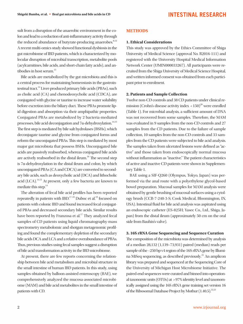
sult from a disruption of the anaerobic environment in the co-
lon and lead to a reduction of anti-inflammatory activity through
the reduced abundance of butyrate-producing anaerobes.8,12
A recent multi-omics study showed functional dysbiosis in the
gut microbiome of IBD patients, which is characterized by mo-
lecular disruption of microbial transcription, metabolite pools
(acylcarnitines, bile acids, and short-chain fatty acids), and an-
tibodies in host serum.13
Bile acids are metabolized by the gut microbiota and this is
a central process for maintaining homeostasis in the gastroin-
testinal tract.14 Liver-produced primary bile acids (PBAs), such
as cholic acid (CA) and chenodeoxycholic acid (CDCA), are
conjugated with glycine or taurine to increase water solubility
before excretion into the biliary duct. These PBAs promote lip-
id digestion and absorption via their amphipathic properties.
Conjugated PBAs are metabolized by 2 bacteria-mediated
processes, bile acid deconjugation and 7α-dehydroxylation.14,15
The first step is mediated by bile salt hydrolases (BSHs), which
deconjugate taurine and glycine from conjugated forms and
reform the unconjugated PBAs. This step is mediated by most
major gut microbiota that possess BSHs. Unconjugated bile
acids are passively reabsorbed, whereas conjugated bile acids
are actively reabsorbed in the distal ileum.16 The second step
is 7α-dehydroxylation in the distal ileum and colon, by which
unconjugated PBAs (CA and CDCA) are converted to second-
ary bile acids, such as deoxycholic acid (DCA) and lithocholic
acid (LCA).14,15 At present, only a few bacteria are known to
mediate this step.14
The alteration of fecal bile acid profiles has been reported
repeatedly in patients with IBD.17-19 Duboc et al.19 focused on
patients with colonic IBD and found increased fecal conjugat-
ed PBAs and decreased secondary bile acids. Similar results
have been reported by Franzosa et al.17 They analyzed fecal
samples of CD patients using liquid chromatography-mass
spectrometry metabolomic and shotgun metagenomic profil-
ing and found the complementary depletion of the secondary
bile acids DCA and LCA and a relative overabundance of PBAs.
Thus, previous studies using fecal samples suggest a disruption
of bile acid transformation activity in the IBD microbiome.
At present, there are few reports concerning the relation-
ship between bile acid metabolism and microbial structure in
the small intestine of human IBD patients. In this study, using
samples obtained by balloon-assisted enteroscopy (BAE), we
comprehensively analyzed the mucosa-associated microbi-
ome (MAM) and bile acid metabolites in the small intestine of
patients with CD.
METHODS
1. Ethical ConsiderationsThis study was approved by the Ethics Committee of Shiga
University of Medical Science (approval No. R2016-111) and
registered with the University Hospital Medical Information
Network Center (UMIN000033267). All participants were re-
cruited from the Shiga University of Medical Science Hospital,
and written informed consent was obtained from each partici-
pant prior to enrolment.
2. Patients and Sample CollectionTwelve non-CD controls and 38 CD patients under clinical re-
mission (Crohn’s disease activity index < 150)20 were enrolled
(Table 1). For microbial analysis, a sufficient amount of DNA
was not recovered from some samples. Therefore, the MAM
was evaluated in 9 samples from the non-CD controls and 27
samples from the CD patients. Due to the failure of sample
collection, 10 samples from the non-CD controls and 33 sam-
ples from the CD patients were subjected to bile acid analysis.
The samples taken from ulcerated lesions were defined as “ac-
tive” and those taken from endoscopically normal mucosa
without inflammation as “inactive.” The patient characteristics
of active and inactive CD patients were shown in Supplemen-
tary Table 1.
BAE using a SIF-Q260 (Olympus, Tokyo, Japan) was per-
formed via the anal route with a polyethylene glycol-based
bowel preparation. Mucosal samples for MAM analysis were
obtained by gentle brushing of mucosal surfaces using a cytol-
ogy brush (CCB-7-240-3-S; Cook Medical, Bloomington, IN,
USA). Intestinal fluid for bile acid analysis was aspirated using
an endoscopic catheter (ES-825H; Yasec Co., Ltd., Shiga, Ja-
pan) from the distal ileum (approximately 50 cm on the oral
side from Bauhin’s valve).
3. 16S rRNA Gene Sequencing and Sequence CurationThe composition of the microbiota was determined by analysis
of a median 28,132 (1,139–73,931) paired (median) reads per
sample of the ~250 bp v4 region of the 16S rRNA gene by Illumi-
na MiSeq sequencing, as described previously.21 An amplicon
library was prepared and sequenced at the Sequencing Core of
the University of Michigan Host Microbiome Initiative. The
paired-end sequences were curated and binned into operation-
al taxonomic units (OTUs) at > 97% identity level and taxonom-
ically assigned using the 16S rRNA gene training set version 16
of the Ribosomal Database Project by Mothur (1.40.5).22,23

Intest Res, Published online
3www.irjournal.org
<doi> • <doi 1>
4. Bile Acid Analysis of Intestinal FluidBile acid concentrations of intestinal fluid were determined by
liquid chromatography-tandem mass spectrometry (LC-MS/
MS) as described previously.24 Briefly, after the addition of in-
ternal standards and 2 mL of 0.5 M potassium phosphate buf-
fer (pH 7.4) to 20 μL intestinal fluids, bile acids were extracted
with Bond Elut C18 cartridges (Agilent Technologies, Santa
Clara, CA, USA).25 An aliquot of the extract was subjected to
LC-MS/MS using a TSQ Vantage triple stage quadrupole mass
spectrometer (Thermo Fisher Scientific, Waltham, MA, USA)
equipped with an HESI-II probe and a Prominence ultra-fast
liquid chromatography system (Shimadzu, Kyoto, Japan). Chro-
matographic separation was performed using a Hypersil GOLD
column (150 × 2.1 mm, 3 μm; Thermo Fisher Scientific) at 40°C.
5. Statistical AnalysisNonparametric data were compared between groups with the
Mann-Whitney U test using Prism version 8.01 (GraphPad,
San Diego, CA, USA) and JMP software version 14.0 (SAS In-
stitute, Cary, NC, USA). Spearman rank correlation coefficients
were used to evaluate the correlations between the parame-
ters. P-values were two-sided, with statistical significance set
at P < 0.05. The observed species and the Chao1 and Shannon
phylogenic diversity indices were calculated using PRIMER7,
version 7.0.13 (PRIMER-e, Auckland, New Zealand). β-Diversity
was estimated using the UniFrac metric to calculate the dis-
tances between the samples, and then visualized using non-
metric multidimensional scaling (NMDS) ordination and sta-
tistically analyzed by permutational multivariate analysis of
variance (PERMANOVA) using QIIME version 1.9. Linear dis-
criminant analysis coupled with effect size measurements
values of OTUs was performed using Mothur.22,23
RESULTS
Microbial analysis revealed a total of 592 OTUs among 36 sam-
ples. The Shannon evenness index was significantly lower in
endoscopically active lesions than in inactive lesions and non-
CD controls (Fig. 1A-C). However, there was no difference in
the observed species and Chao-1 index (richness markers).
β-Diversity was evaluated using the UniFrac metric to calcu-
late the distance between the samples (Fig. 1D). A significant
Table 1. Background Characteristics of the Subjects
Characteristics Non-CD controls (n=12) CD patients (n=38)
Sex (male/female) 6/6 31/7
Age (yr), median (IQR) 60.3 (31.4–68.3) 37.1 (32.3–47.6)
Body mass index (kg/m2), median (IQR) 20.3 (18.4–23.9) 21.7 (19.4–24.4)
Smoking status (never/previous/current) 9/2/1 28/5/5
History of intestinal resection (yes/no) 1/11 8/30
Disease duration (yr), median (IQR) - 9.5 (4.1–15.5)
Disease location (L1/L2/L3) - 16/4/18
Disease behavior (B1/B2/B3) - 16/15/7
CDAI, median (IQR) - 63.6 (41.0–90.5)
Disease
CD - 38
Gastrointestinal bleeding 5 -
Intestinal neoplasia 3 -
Other 4 -
Medication, No. (%)
5-ASA/SASP 1 (8.3) 26 (68.4)
Prednisolone 2 (16.7) 1 (2.6)
Immunomodulators 0 18 (47.4)
Biologics 1 (8.3) 13 (34.2)
CD, Crohn’s disease; IQR, interquartile range; L1, ileal; L2, colonic; L3, ileocolonic; B1, non-stricturing, non-penetrating; B2, stricturing; B3, penetrating; CDAI, Crohn’s disease activity index; 5-ASA, 5-aminosalicylates; SASP, sulfasalazine.

Shigeki Bamba, et al. • Ileal gut microbiota and bile acids in CD
4 www.irjournal.org
Silvio Danese, et al. • iSTART consensus recommendations
difference was detected only between active lesions and non-
CD controls (P = 0.039, PERMANOVA). There was no signifi-
cant difference between endoscopically inactive lesions and
non-CD controls (Fig. 1D).
As shown in Fig. 2, the relative abundance of the phylum
Firmicutes was significantly lower in active lesions than in non-
CD controls. The relative abundance of the phylum Fusobac-
teria was significantly higher in inactive lesions than in non-
CD controls. The relative abundance of the phylum Actino-
bacteria was significantly lower in active lesions than in inac-
tive lesions.
Representative taxa showing a significant difference in abun-
dance are shown in Fig. 3. When comparing CD samples and
non-CD controls, the relative abundance of the genus Esche-
richia was significantly higher in CD samples, while the gen-
era Faecalibacterium and Roseburia were significantly lower
in CD patients (Fig. 3A). The relative abundance of the genera
Escherichia, Edwardsiella, and Cryptobacterium was signifi-
cantly higher in active lesions than in inactive lesions, and the
genera Veillonella and Prevotella were significantly less abun-
dant in active lesions than in inactive lesions (Fig. 3B).
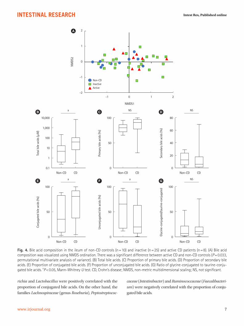
Bile acid composition expressed by non-metric multidimen-
sional scaling was significantly different between the active
CD and non-CD samples (P = 0.033, PERMANOVA) (Fig. 4A).
Fig. 1. Comparative analysis of the gut microbial communities in non-CD controls (n=9) and inactive (n=18) and active lesions (n=9) of CD patients. (A) Observed species. (B) Chao1 index. (C) Shannon index. aP<0.05, Mann-Whitney U test. (D) β-Diversity estimated using the UniFrac metric and visualized using NMDS ordination. There was a significant difference between active lesions and non-CD samples (P=0.039, permutational multivariate analysis of variance). CD, Crohn’s disease; NMDS, non-metric multidimensional scaling; NS, not significant.
0.5
0.4
0.3
0.2
0.1
0
-0.1
-0.2
-0.3
-0.4
-0.5 -0.3 -0.2 -0.1 0 0.1 0.2 0.3 0.4
NMDS1
Non-CD
Inactive
Active
NM
DS2
D
250
200
150
100
50
0
Non-C
D
Inacti
veAc
tive
NS
NS NS
Obse
rved
spe
cies
A
150
100
50
0
Non-C
D
Inacti
veAc
tive
NS
NS NS
Chao
1 in
dex
B
4
3
2
1
0
Non-C
D
Inacti
veAc
tive
a
NS a
Shan
non
inde
x
C

Intest Res, Published online
5www.irjournal.org
<doi> • <doi 1>
Total bile acid concentrations were significantly higher in CD
samples than in non-CD samples (38.4 μM vs. 4.55 μM, re-
spectively, P = 0.01) (Fig. 4B). There was no significant differ-
ence between both groups in the proportion of primary or
secondary bile acids (Fig. 4C and D). The proportion of conju-
gated bile acids was significantly higher in CD patients than in
non-CD controls (Fig. 4E), while the proportion of unconju-
gated bile acids was significantly lower in CD patients (Fig. 4F).
There was no difference in the ratio of glycine-conjugated to
taurine-conjugated bile acids between both groups (Fig. 4G).
Fig. 2. Comparative analysis of the taxonomic composition of the microbial community at the phylum level in non-CD controls (n=9) and inactive (n=18) and active lesions (n=9) of CD patients. (A) Firmicutes. (B) Bacteroidetes. (C) Proteobacteria. (D) Fusobacteria. (E) Ac-tinobacteria. aP<0.05, Mann-Whitney U test. CD, Crohn’s disease; NS, not significant.
100
80
60
40
20
0
Non-C
D
Inacti
veAc
tive
a
NS NS
Firmicutes
(%)
A
60
40
20
0
Non-C
D
Inacti
veAc
tive
NS
NS NS
Bacteroidetes
(%)
B
100
80
60
40
20
0
Non-C
D
Inacti
veAc
tive
NS
NS NS
Proteobacteria
(%)
C
40
30
20
10
0
Non-C
D
Inacti
veAc
tive
NSNSa
Fusobacteria
(%)
D
15
10
5
0
Non-C
D
Inacti
veAc
tive
NSaNS
Actinobacteria
(%)
E
Table 2. Association between the Bile Acid Fraction and CD
Bile acids (%)Median (IQR)
P-valueNon-CD (n=10) CD (n=33)
CA 42.8 (12.6–63.2) 11.2 (4.51–43.7) 0.041
GCA 11.7 (1.3–43.8) 33.0 (16.9–55.6) 0.047
TCA 0.831 (0.579–2.790) 2.500 (0.753–4.700) 0.127
CDCA 4.35 (2.11–8.78) 2.11 (0.80–6.68) 0.186
GCDCA 9.69 (2.12–19.90) 11.50 (5.62–21.10) 0.604
TCDCA 0.975 (0.513–2.500) 1.580 (0.348–4.360) 0.527
DCA 4.820 (0.955–9.620) 1.070 (0.232–3.390) 0.054
GDCA 0.6150 (0.0440–3.3700) 0.2980 (0.0175–3.7300) 0.795
TDCA 0.0969 (0.0166–0.2980) 0.0496 (0.0045–0.2450) 0.372
LCA 6.360 (0.956–9.580) 0.639 (0.260–3.830) 0.014
GLCA 0.02350 (0.00640–0.03100) 0.00298 (0.00136–0.02000) 0.031
TLCA 0.04200 (0.01080–0.14400) 0.00699 (0.00217–0.04590) 0.047
UDCA 1.600 (0.540–3.430) 0.451 (0.102–1.230) 0.057
GUDCA 0.856 (0.155–4.560) 0.744 (0.163–4.080) 0.954
TUDCA 0.1660 (0.0904–0.2890) 0.0299 (0.0107–0.3020) 0.065
CD, Crohn’s disease; IQR, interquartile range; CA, cholic acid; GCA, glycocholic acid; TCA, taurocholic acid; CDCA, chenodeoxycholic acid; GCDCA, glyco-chenodeoxycholic acid; TCDCA, taurochenodeoxycholic acid; DCA, deoxycholic acid; GDCA, glycodeoxycholic acid; TDCA, taurodeoxycholic acid; LCA, lithocholic acid; GLCA, glycolithocholic acid; TLCA, taurolithocholic acid; UDCA, ursodeoxycholic acid; GUDCA, glycoursodeoxycholic acid; TUDCA, taur-oursodeoxycholic acid.P-values were calculated using the Mann-Whitney U test.

Shigeki Bamba, et al. • Ileal gut microbiota and bile acids in CD
6 www.irjournal.org
Silvio Danese, et al. • iSTART consensus recommendations
As shown in Table 2, analysis of the bile acid fraction indi-
cated that the proportion of all unconjugated bile acids was
relatively lower in CD patients than in non-CD controls. In
particular, the proportions of CA and LCA were significantly
lower in CD patients than in non-CD controls. The proportion
of DCA tended to be lower in CD patients but not significant
(P = 0.054). On the other hand, the proportions of conjugated
bile acids such as glycocholic acid (GCA) were significantly
higher in CD patients than in non-CD controls. The compari-
son of the bile acid fraction between active and inactive CD
revealed significantly decreased glycodeoxycholic acid (GDCA)
in active CD than in inactive CD (Supplementary Table 2).
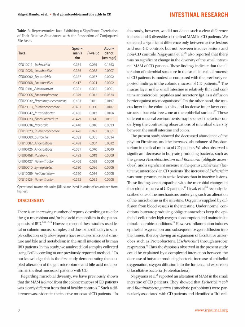
Bile acid metabolism is closely associated with the gut mi-
crobiome.14 Therefore, we evaluated whether there was a cor-
relation between the relative abundance of taxa and the pro-
portion of conjugated bile acids. Representative taxa whose
abundance was significantly correlated with the proportion of
conjugated bile acids are shown in Table 3. The genera Esche-
Fig. 3. Comparative analysis of the taxonomic composition of the microbial community at the genus level using linear discriminant anal-ysis effect size. (A) Comparison between non-CD (n=9) and CD samples (n=27). (B) Comparison between inactive (n=18) and active le-sions (n=9) of CD patients. CD, Crohn’s disease.
-6 -4 -2 0 2 4 6
Linear discriminant analysis score
Non-CD
CD
A g_Escherichiap_Tenericutesc_Mollicutesg_Abiotrophiaf_Aerococcaceae
g_Brevundimonasf_Clostridiales_unclassified
g_Clostridiales_unclassified_unclassifiedf_Helicobacteraceae
g_Helicobacterg_Intestinimonas
f_Burkholderiales_unclassifiedg_Burkholderiales_unclassified_unclassified
g_Roseburiag_Faecalibacterium
-6 -4 -2 0 2 4 6
Linear discriminant analysis score
Inactive
Active
B g_Escherichiag_Edwardsiellag_Cryptobacteriumg_Cupriavidusf_Burkholderiales_unclassifiedg_Burkholderiales_unclassified_unclassifiedg_Parabacteroidesf_Pseudomonadaceaeg_Pseudomonas
g_Lactobacillales_unclassified_unclassifiedf_Lactobacillales_unclassified
c_Actinobacteriap_Actinobacteria
g_Prevotellaf_Prevotellaceae
g_Veillonellac_Negativicutes
o_Selenomonadalesf_Veillonellaceae
Intest Res, Published online
7www.irjournal.org
<doi> • <doi 1>
richia and Lactobacillus were positively correlated with the
proportion of conjugated bile acids. On the other hand, the
families Lachnospiraceae (genus Roseburia), Peptostreptococ-
caceae (Intestinibacter) and Ruminococcaceae (Faecalibacteri-
um) were negatively correlated with the proportion of conju-
gated bile acids.
Fig. 4. Bile acid composition in the ileum of non-CD controls (n=10) and inactive (n=25) and active CD patients (n=8). (A) Bile acid composition was visualized using NMDS ordination. There was a significant difference between active CD and non-CD controls (P=0.033, permutational multivariate analysis of variance). (B) Total bile acids. (C) Proportion of primary bile acids. (D) Proportion of secondary bile acids. (E) Proportion of conjugated bile acids. (F) Proportion of unconjugated bile acids. (G) Ratio of glycine-conjugated to taurine-conju-gated bile acids. aP<0.05, Mann-Whitney U test. CD, Crohn’s disease; NMDS, non-metric multidimensional scaling; NS, not significant.
10,000
1,000
100
10
1
0.1Non-CD CD
a
Tota
l bile
aci
ds (μ
M)
B
100
50
0Non-CD CD
NS
Prim
ary
bile
aci
ds (%
)
C
80
60
40
20
0Non-CD CD
NS
Seco
ndar
y bi
le a
cids
(%)
D
100
50
0Non-CD CD
a
Conj
ugat
ed b
ile a
cids
(%)
E
100
50
0Non-CD CD
a
Unco
njug
ated
bile
aci
ds (%
)
F
100
50
0Non-CD CD
NS
Glyc
ine-
conj
ugat
ed/t
aurin
e-co
njug
ated
G
2
1
0
-1
-2 -1 0 1 2
NMDS1
Non-CDInactiveActive
NM
DS2
A
Shigeki Bamba, et al. • Ileal gut microbiota and bile acids in CD
8 www.irjournal.org
Silvio Danese, et al. • iSTART consensus recommendations
DISCUSSION
There is an increasing number of reports describing a role for
the gut microbiota and/or bile acid metabolism in the patho-
genesis of IBD.7-11,17-19 However, most of these studies used fe-
cal or colonic mucosa samples, and due to the difficulty in sam-
ple collection, only a few reports have evaluated microbial struc-
ture and bile acid metabolism in the small intestine of human
IBD patients. In this study, we analyzed ileal samples collected
using BAE according to our previously reported method.11 To
our knowledge, this is the first study demonstrating the cou-
pled alteration of the gut microbiome and bile acid metabo-
lism in the ileal mucosa of patients with CD.
Regarding microbial diversity, we have previously shown
that the MAM isolated from the colonic mucosa of CD patients
was clearly different from that of healthy controls.11 Such a dif-
ference was evident in the inactive mucosa of CD patients.11 In
this study, however, we did not detect such a clear difference
in the α- and β-diversities of the ileal MAM in CD patients. We
detected a significant difference only between active lesions
and non-CD controls, but not between inactive lesions and
non-CD controls. Nagayama et al.26 also reported that there
was no significant change in the diversity of the small intesti-
nal MAM of CD patients. These findings indicate that the al-
teration of microbial structure in the small intestinal mucosa
of CD patients is modest as compared with the previously re-
ported findings in the colonic mucosa of CD patients.11 The
mucus layer in the small intestine is relatively thin and con-
tains antimicrobial peptides and secretory IgA as a diffusion
barrier against microorganisms.27 On the other hand, the mu-
cus layer in the colon is thick and its dense inner layer con-
structs a bacteria-free zone at the epithelial surface.27 These
different mucosal environments may be one of the factors un-
derlying the contrasting observations of microbial diversity
between the small intestine and colon.
The present study showed the decreased abundance of the
phylum Firmicutes and the increased abundance of Fusobac-
terium in the ileal mucosa of CD patients. We also observed a
significant decrease in butyrate-producing bacteria, such as
the genera Faecalibacterium and Roseburia (obligate anaer-
obes), and a significant increase in the genus Escherichia (fac-
ultative anaerobes) in CD patients. The increase of Escherichia
was more prominent in active lesions than in inactive lesions.
These findings are compatible with the microbial changes in
the colonic mucosa of CD patients.11 Litvak et al.28 recently de-
scribed one of the mechanisms underlying such an alteration
of the microbiome in the intestine. Oxygen is supplied by dif-
fusion from blood vessels in the intestine. Under normal con-
ditions, butyrate-producing obligate anaerobes keep the epi-
thelial cells under high oxygen consumption and maintain lu-
minal anaerobic conditions.28 However, inflammation induces
epithelial oxygenation and subsequent oxygen diffusion into
the lumen, thereby driving an expansion of facultative anaer-
obes such as Proteobacteria (Escherichia) through aerobic
respiration.12 Thus, the dysbiosis observed in the present study
could be explained by a complexed interaction between the
decrease of butyrate-producing bacteria, increase of epithelial
oxygenation, oxygen diffusion into the lumen, and expansion
of facultative bacteria (Proteobacteria).
Nagayama et al.26 reported an alteration of MAM in the small
intestine of CD patients. They showed that Escherichia coli
and Ruminococcus gnavus (mucolytic pathobiont) were par-
ticularly associated with CD patients and identified a Th1 cell-
Table 3. Representative Taxa Exhibiting a Significant Correlation of Their Relative Abundance with the Proportion of Conjugated Bile Acids
TaxaSpear-man’s rho
P-valueAbun-dance
(average)
OTU10013_Escherichia 0.384 0.039 0.1863
OTU10026_Lactobacillus 0.386 0.038 0.0007
OTU00092_Leptotrichia 0.387 0.037 0.0002
OTU00208_Lactobacillus 0.417 0.024 0.0002
OTU10191_Alloscardovia 0.391 0.035 0.0001
OTU20005_Lachnospiraceae –0.379 0.042 0.0524
OTU20032_Peptostreptococcaceae –0.463 0.011 0.0197
OTU20013_Ruminococcaceae –0.401 0.030 0.0187
OTU00047_Intestinibacter –0.456 0.012 0.0166
OTU00023_Faecalibacterium –0.429 0.020 0.0113
OTU00036_Prevotella –0.440 0.016 0.0093
OTU10020_Ruminococcaceae –0.426 0.021 0.0051
OTU00089_Sutterella –0.392 0.035 0.0034
OTU10067_Anaerostipes –0.488 0.007 0.0012
OTU00125_Anaerostipes –0.381 0.040 0.0010
OTU00158_Roseburia –0.432 0.019 0.0009
OTU00127_Flavonifractor –0.406 0.028 0.0006
OTU50010_Synergistetes –0.390 0.036 0.0005
OTU10059_Fretibacterium –0.390 0.036 0.0005
OTU10129_Flavonifractor –0.392 0.035 0.0005
Operational taxonomic units (OTUs) are listed in order of abundance from highest.
Intest Res, Published online
9www.irjournal.org
<doi> • <doi 1>
inducing E. coli strain. However, they did not observe changes
of butyrate-producing obligate anaerobes. They used BAE for
sample collection, but their method was different from ours.
We collected samples from the distal ileum (approximately 50
cm on the oral side from Bauhin’s valve), but Nagayama et al.
harvested samples from active lesions of the middle small in-
testine (jejunum or proximal ileum). It is likely that the differ-
ences in sampling locations might have led to the different ob-
servations in these studies, since anaerobic conditions are dif-
ferent between the middle (facultatively anaerobic) and ter-
minal (obligately anaerobic) small intestine.
In parallel with microbiome analyses, we analyzed bile acid
composition in the ileum. We found a significant elevation of
the levels of total bile acids and a significant increase in the
proportion of conjugated bile acids (e.g., GCA and taurocholic
acid) in CD patients. These results are partially compatible
with the previous report of fecal samples by Duboc et al.19 They
reported an increase of conjugated bile acids as well as an in-
crease of secondary bile acids in fecal samples from active IBD
patients.19 The composition of bile acids have been reported to
be determined by several processes and factors, such as the
balance of passive and active absorption and bacterial bile
acid modifications.16 Since mucosal inflammation continues
even without endoscopic mucosal findings in CD patients,29
the elevation of the total amount of bile acids suggests the mal-
absorption of bile acids over a wide area of the small intestine.
The reabsorption of conjugated bile acids is dependent on
their recognition by active transport sites in the terminal ile-
um,16 but unconjugated bile acids bind with a lower affinity to
the transport sites and pass into the colon.16 The terminal ile-
um is one of the most commonly affected sites in CD, and the
active absorption of conjugated bile acids may be disrupted in
the ileum of CD patients, leading to an increase in the propor-
tion of conjugated bile acids.
Bacterial modification of bile acids is another process that
affects bile acid composition. In this regard, deconjugation
mediated by bacterial BSHs has a particular importance.30 As
described in the review by Ridlon et al.,30 BSH activity is wide-
spread in commensal bacteria in the small intestine and co-
lon. Gram-positive gut bacteria including Clostridium, Entero-
coccus, Bifidobacterium, and Lactobacillus have the most di-
verse distribution of BSHs, while the distribution of BSHs in
Gram-negative bacteria is only detected in members of the
genus Bacteroides.30 In the present study, the abundance of
the genera Escherichia and Lactobacillus was positively corre-
lated with the proportion of conjugated bile acids. On the oth-
er hand, the abundance of the family Lachnospiraceae (the ge-
nus Roseburia), Peptostreptococcaceae (Intestinibacter), and
Ruminococcaceae (Faecalibacterium) was negatively correlat-
ed with the proportion of conjugated bile acids. These results
suggest that the proportion of conjugated bile acids may be
increased in association with the increase of Gram-negative,
BSH-lacking Escherichia and the decrease of various BSH-posi-
tive, Gram-positive bacteria such as Roseburia and Faecalibac-
terium.
At present, there are little clinical data on the relationship
between bile acid composition and the activity of CD. Sinha et
al.15 revealed that secondary bile acids have an inhibitory ef-
fect on intestinal inflammation via transmembrane G protein-
coupled receptor 5 (TGR5). In our study, we found a decrease
in GDCA, a conjugated secondary bile acid, in active CD, sug-
gesting that it may influence disease activity.
This study has some limitations based on the small number
of samples. First, since it was difficult to perform per anal BAE
without stress in healthy individuals, we could not obtain truly
normal small intestine fluid. Second, our results may be affect-
ed by the bowel cleaning preparation prior to BAE. To clear
the bowel, all participants took a laxative the night before sam-
pling, and a cleaning liquid composed of polyethylene glycol
on the day of sampling. These procedures might cause dilu-
tion effects on the samples of the small intestine used in this
study.
In conclusion, dysbiosis in the ileum of CD patients was
confirmed, but it was relatively modest as compared to previ-
ously reported findings in the colon or feces of IBD patients. In
addition, dysbiosis in the ileum was accompanied with an al-
teration of bile acid composition. The precise role of the cou-
pled changes of the microbiome and bile acid composition in
the pathophysiology of CD should be investigated further in
the future.
ADDITIONAL INFORMATION
Funding Source This work was supported by the Japan Agency for Medical Re-
search and Development (AMED) under grant numbers JP-
20gm1010008h9904 (Andoh A) and 20ek0410056 (Andoh
A), in part by Health and Labor Sciences Research Grants for
Research on Intractable Diseases from the Ministry of Health,
Labour and Welfare of Japan under grant number 20FC1037
(Andoh A), and in part by Grants-in-Aid for Scientific Research
from the Ministry of Education, Culture, Sports, Science and

Shigeki Bamba, et al. • Ileal gut microbiota and bile acids in CD
10 www.irjournal.org
Silvio Danese, et al. • iSTART consensus recommendations
Technology of Japan under grant numbers 18K10990 (Bamba
S) and 18K08002 (Andoh A).
Conflict of InterestAndoh A is an editorial board member of the journal but did
not involve in the peer reviewer selection, evaluation, or deci-
sion process of this article. No other potential conflicts of inter-
est relevant to this article were reported.
Author ContributionConceptualization: Bamba S, Andoh A . Data curation: Bamba
S. Methodology: Bamba S, Inatomi O, Ohno M, Honda A, Ino-
hara N. Formal analysis: Bamba S, Inohara N. Investigations:
Bamba S, Inatomi O, Nishida A, Ohno M, Imai T, Takahashi K,
Naito Y, Iwamoto J, Honda A, Inohara N, Andoh A. Writing -
original draft: Bamba S, Andoh A. Writing - review & editing:
Andoh A. Supervision: Nishida A, NI, Andoh A. Approval of fi-
nal manuscript: all authors.
OthersThe authors thank the Sequencing Core of the University of
Michigan Host Microbiome Initiative for support.
ORCIDBamba S https://orcid.org/0000-0002-4108-5894
Inatomi O https://orcid.org/0000-0002-5837-6575
Nishida A https://orcid.org/0000-0002-1288-3272
Ohno M https://orcid.org/0000-0003-3505-7835
Imai T https://orcid.org/0000-0002-9170-4075
Takahashi K https://orcid.org/0000-0002-3522-9740
Naito Y https://orcid.org/0000-0001-5443-788X
Iwamoto J https://orcid.org/0000-0002-5238-3272
Honda A https://orcid.org/0000-0003-0902-8272
Inohara N https://orcid.org/0000-0002-4215-9349
Andoh A https://orcid.org/0000-0001-8533-2669
Supplementary MaterialSupplementary materials are available at the Intestinal Re-
search website (https://www.irjournal.org).
REFERENCES
1. Sheehan D, Moran C, Shanahan F. The microbiota in inflam-
matory bowel disease. J Gastroenterol 2015;50:495-507.
2. Goldsmith JR, Sartor RB. The role of diet on intestinal micro-
biota metabolism: downstream impacts on host immune func-
tion and health, and therapeutic implications. J Gastroenterol
2014;49:785-798.
3. Nishida A, Inoue R, Inatomi O, Bamba S, Naito Y, Andoh A.
Gut microbiota in the pathogenesis of inflammatory bowel
disease. Clin J Gastroenterol 2018;11:1-10.
4. Mak WY, Zhao M, Ng SC, Burisch J. The epidemiology of in-
flammatory bowel disease: east meets west. J Gastroenterol
Hepatol 2020;35:380-389.
5. Ananthakrishnan AN. Epidemiology and risk factors for IBD.
Nat Rev Gastroenterol Hepatol 2015;12:205-217.
6. Kaplan GG, Ng SC. Understanding and preventing the global
increase of inflammatory bowel disease. Gastroenterology
2017;152:313-321.
7. Sartor RB. Microbial influences in inflammatory bowel dis-
eases. Gastroenterology 2008;134:577-594.
8. Takahashi K, Nishida A, Fujimoto T, et al. Reduced abundance
of butyrate-producing bacteria species in the fecal microbial
community in Crohn’s disease. Digestion 2016;93:59-65.
9. Nagalingam NA, Lynch SV. Role of the microbiota in inflam-
matory bowel diseases. Inflamm Bowel Dis 2012;18:968-984.
10. Frank DN, Robertson CE, Hamm CM, et al. Disease pheno-
type and genotype are associated with shifts in intestinal-as-
sociated microbiota in inflammatory bowel diseases. Inflamm
Bowel Dis 2011;17:179-184.
11. Nishino K, Nishida A, Inoue R, et al. Analysis of endoscopic
brush samples identified mucosa-associated dysbiosis in in-
flammatory bowel disease. J Gastroenterol 2018;53:95-106.
12. Litvak Y, Byndloss MX, Tsolis RM, Bäumler AJ. Dysbiotic Pro-
teobacteria expansion: a microbial signature of epithelial dys-
function. Curr Opin Microbiol 2017;39:1-6.
13. Lloyd-Price J, Arze C, Ananthakrishnan AN, et al. Multi-omics
of the gut microbial ecosystem in inflammatory bowel diseas-
es. Nature 2019;569:655-662.
14. Jia W, Xie G, Jia W. Bile acid-microbiota crosstalk in gastroin-
testinal inflammation and carcinogenesis. Nat Rev Gastroen-
terol Hepatol 2018;15:111-128.
15. Sinha SR, Haileselassie Y, Nguyen LP, et al. Dysbiosis-induced
secondary bile acid deficiency promotes intestinal inflamma-
tion. Cell Host Microbe 2020;27:659-670.
16. Begley M, Gahan CG, Hill C. The interaction between bacteria
and bile. FEMS Microbiol Rev 2005;29:625-651.
17. Franzosa EA, Sirota-Madi A, Avila-Pacheco J, et al. Gut micro-
biome structure and metabolic activity in inflammatory bow-
el disease. Nat Microbiol 2019;4:293-305.
18. Lavelle A, Sokol H. Gut microbiota-derived metabolites as key
actors in inflammatory bowel disease. Nat Rev Gastroenterol

Intest Res, Published online
11www.irjournal.org
<doi> • <doi 1>
Hepatol 2020;17:223-237.
19. Duboc H, Rajca S, Rainteau D, et al. Connecting dysbiosis, bile-
acid dysmetabolism and gut inflammation in inflammatory
bowel diseases. Gut 2013;62:531-539.
20. Best WR, Becktel JM, Singleton JW, Kern F Jr. Development of
a Crohn’s disease activity index. National Cooperative Crohn’s
Disease Study. Gastroenterology 1976;70:439-444.
21. Hasegawa M, Inohara N. Regulation of the gut microbiota by
the mucosal immune system in mice. Int Immunol 2014;26:
481-487.
22. Schloss PD, Westcott SL, Ryabin T, et al. Introducing Mothur:
open-source, platform-independent, community-supported
software for describing and comparing microbial communi-
ties. Appl Environ Microbiol 2009;75:7537-7541.
23. Costello EK, Lauber CL, Hamady M, Fierer N, Gordon JI, Knight
R. Bacterial community variation in human body habitats across
space and time. Science 2009;326:1694-1697.
24. Murakami M, Iwamoto J, Honda A, et al. Detection of gut dys-
biosis due to reduced Clostridium subcluster XIVa using the
fecal or serum bile acid profile. Inflamm Bowel Dis 2018;24:
1035-1044.
25. Shoda J, Mahara R, Osuga T, et al. Similarity of unusual bile
acids in human umbilical cord blood and amniotic fluid from
newborns and in sera and urine from adult patients with cho-
lestatic liver diseases. J Lipid Res 1988;29:847-858.
26. Nagayama M, Yano T, Atarashi K, et al. TH1 cell-inducing Esch-
erichia coli strain identified from the small intestinal mucosa of
patients with Crohn’s disease. Gut Microbes 2020;12:1788898.
27. Johansson ME, Hansson GC. Immunological aspects of intes-
tinal mucus and mucins. Nat Rev Immunol 2016;16:639-649.
28. Litvak Y, Byndloss MX, Bäumler AJ. Colonocyte metabolism
shapes the gut microbiota. Science 2018;362:eaat9076.
29. Baumgart DC, Sandborn WJ. Inflammatory bowel disease:
clinical aspects and established and evolving therapies. Lan-
cet 2007;369:1641-1657.
30. Ridlon JM, Harris SC, Bhowmik S, Kang DJ, Hylemon PB. Con-
sequences of bile salt biotransformations by intestinal bacte-
ria. Gut Microbes 2016;7:22-39.

Shigeki Bamba, et al. • Ileal gut microbiota and bile acids in CD
www.irjournal.org
Silvio Danese, et al. • iSTART consensus recommendations
Supplementary Table 1. Background Characteristics of Crohn’s Disease
Characteristics Inactive (n=18) Active (n=9)
Sex (male/female) 14/4 7/2
Age (yr), median (IQR) 35.1 (31.9–47.0) 47.4 (35.3–60.7)
Body mass index (kg/m2), median (IQR) 21.2 (18.6–24.5) 21.7 (20.2–23.5)
Smoking status (never/previous/current) 12/3/3 7/2/0
History of intestinal resection (yes/no) 3/15 0/9
Disease duration (yr), median (IQR) 9.5 (6.6–14.5) 14.6 (1.6–33.0)
Disease location (L1/L2/L3) 9/1/8 4/0/5
Disease behavior (B1/B2/B3) 9/7/2 1/5/3
CDAI, median (IQR) 62.0 (46.5–83.7) 66.0 (48.0–96.0)
Disease
Crohn’s disease 18 9
Gastrointestinal bleeding - -
Intestinal neoplasia - -
Other - -
Medication, No. (%)
Proton pump inhibitors 3 (16.7) 2 (22.2)
Probiotics 6 (33.3) 4 (44.4)
5-ASA/SASP 11 (61.1) 7 (77.8)
Prednisolone 1 (5.6) 0
Immunomodulators 10 (55.6) 3 (33.3)
Biologics 6 (33.3) 3 (33.3)
IQR, interquartile range; L1, ileal; L2, colonic; L3, ileocolonic; B1, non-stricturing, non-penetrating; B2, stricturing; B3, penetrating; CDAI, Crohn’s disease activity index; 5-ASA, 5-aminosalicylates; SASP, sulfasalazine.
See “Relationship between the gut microbiota and bile acid composition in the ileal mucosa of Crohn’s disease” on page 1-11.

Intest Res, Published online
www.irjournal.org
<doi> • <doi 1>
Supplementary Table 2. Comparison of Bile Acid Fraction between Active and Inactive CD
Bile acids (%)Median (IQR)
P-valueInactive (n=25) Active (n=8)
CA 11.10 (4.51–47.70) 12.10 (4.68–38.80) 0.983
GCA 33.0 (11.3–55.7) 40.9 (26.3–54.2) 0.883
TCA 2.890 (0.654–4.580) 2.090 (0.983–6.700) 0.412
CDCA 2.00 (0.71–5.63) 3.47 (1.28–18.00) 0.185
GCDCA 10.80 (4.33–18.60) 18.60 (8.18–26.30) 0.265
TCDCA 1.140 (0.287–4.030) 2.690 (0.692–4.580) 0.462
DCA 1.920 (0.267–7.270) 0.840 (0.158–1.490) 0.185
GDCA 0.46400 (0.04670–3.95000) 0.01680 (0.00399–0.23800) 0.046
TDCA 0.07270 (0.00815–0.31900) 0.01210 (0.00298–0.15200) 0.344
LCA 0.639 (0.212–4.400) 0.529 (0.304–1.810) 0.629
GLCA 0.00443 (0.00145–0.02680) 0.00217 (0.00169–0.00667) 0.159
TLCA 0.01000 (0.00217–0.07730) 0.00614 (0.00252–0.00887) 0.437
UDCA 0.531 (0.037–1.380) 0.387 (0.191–1.160) 0.850
GUDCA 0.331 (0.134–4.080) 1.710 (0.508–5.830) 0.185
TUDCA 0.0356 (0.0107–0.3360) 0.0225 (0.0116–0.3100) 0.659
CD, Crohn’s disease; IQR, interquartile range; CA, cholic acid; GCA, glycocholic acid; TCA, taurocholic acid; CDCA, chenodeoxycholic acid; GCDCA, glyco-chenodeoxycholic acid; TCDCA, taurochenodeoxycholic acid; DCA, deoxycholic acid; GDCA, glycodeoxycholic acid; TDCA, taurodeoxycholic acid; LCA, litho cholic acid; GLCA, glycolithocholic acid; TLCA, taurolithocholic acid; UDCA, ursodeoxycholic acid; GUDCA, glycoursodeoxycholic acid; TUDCA, tauro-ursodeoxycholic acid.P-values were calculated using the Mann-Whitney U test.
Related Documents