REHABILITATION TECHNIQUES FOR SPORTS MEDICINE AND ATHLETIC TRAINING WILLIAM E. PRENTICE Restoring Range of Motion and Improving Flexibility

REHABILITATION TECHNIQUES FOR SPORTS MEDICINE AND ATHLETIC TRAINING WILLIAM E. PRENTICE Restoring Range of Motion and Improving Flexibility.
Dec 14, 2015
Welcome message from author
This document is posted to help you gain knowledge. Please leave a comment to let me know what you think about it! Share it to your friends and learn new things together.
Transcript

REHABILITATION TECHNIQUES FOR SPORTS MEDICINE AND ATHLETIC
TRAINING
WILLIAM E. PRENTICE
Restoring Range of Motion and Improving Flexibility

Introduction
Loss of motion occurs after injury Due to pain, swelling, muscle guarding or spasm
Shortening of connective tissue and muscle Loss of neuromuscular control
Restoring normal range of motion is one of primary goals of rehabilitation program
Flexibility Ability to move a joint through a full, non-restricted,
pain free range of motion Dependent on combination of joint range of motion and
muscle flexibility
Introduction
Joint range of motion Limited by shape of articulating surface and capsular
and ligamentous structuresMuscle flexibility
Ability of musculotendinous unit to lengthen
Lack of flexibility in one joint can effect the entire kinetic chain

Importance of flexibility
Essential to normal daily living
Functional activities require relatively “normal” amounts of flexibility Some activities require more flexibility for superior
performance
Decreased flexibility creates uncoordinated/awkward movement patterns Result of loss of neuromuscular control
Importance of Flexibility
Generally accepted that flexibility is essential for improved performance Recent studies conflicting and inconclusive Stretching has shown to decrease performance
parameters Strength, endurance, power, joint position sense and
reaction times
Decrease incidence of injury Recent studies fail to find true cause and effect
relationship

Anatomic Factors that Limit Flexibility
Muscles, tendons and their surrounding fascial sheaths Stretching attempts to take advantage of highly elastic
properties of muscle Overtime it is possible to increase elasticity , or the length
a given muscle can be stretched
Connective tissue (ligaments and joint capsule) Become shortened and stiff during periods of
immobilization People can also be loose jointed from slack or increased
laxity in connective tissue Creates some instability

Anatomic Factors that Limit Flexibility
Bony structures Restrict end point in the range of motion
Good for stability After fracture excess calcium can develop which
interferes with normal range
Fat Excess fatty tissue can restrict range of motion
For example; excess abdominal fat can restrict trunk movement

Anatomic Factors that Limit Flexibility
Skin Inelastic scar tissue can develop after surgery or injury
Incapable of stretching with joint movement Overtime can improve elasticity to varying degrees through
stretching
Neural tissue Tightness develops in neural tissues from acute
compression, chronic repetitive microtrauma, muscle imbalances, joint dysfunctions, or poor posture Can create morphological changes in tissue that can cause pain Pain can cause muscle guarding and spasm Can eventually lad to neural fibrosis or scarring

Active and Passive Range of Motion
Active range of motion Dynamic flexibility Degree to which a joint can be moved by a muscle
contraction Not necessarily a good indicator of joint stiffness or
looseness because movement of joint has little resistance
Passive range of motion Static flexibility Degree to which a joint can be passively moved to end
points of range of motion No muscle contraction involved

Active and Passive ROM
Many situations in activity when muscle is forced beyond its normal active limits If muscle does not have elasticity to compensate,
injury to musculotendinous unit may occur
Assessment of ROM
Goniometers Large protractors w/ measurements in degrees Align arms of goniometer along longitudinal axis of 2
segments Reasonably accurate measurement of ROM Standardization of measurement techniques and
recording AROM & PROM have been developed Immobile arm is lined up along immobile segment Mobile arm is lined up along mobile segment
Neurophysiologic Basis of Stretching
Mechanoreceptors in muscle tell CNS what is happening within that muscle
2 of these are important in the stretch reflex
Muscle spindle and the Golgi tendon organ (GTO) Sensitive to changes in muscle length GTO also sensitive to change in muscle tension

Neurophysiologic Basis of Stretching
Muscle spindle initially sends sensory impulse to spinal cord which then sends a message back to muscle spindle causing the muscle to reflexively contract
If stretch last longer than 6 seconds, impulses from GTO begin to override muscle spindle • autogenic inhibition, or a reflex relaxation of the antagonist
muscle• Protective mechanism to allow stretch to avoid damage to
muscle fibers
Reciprocal inhibition• Contraction of agonist causes a reflex relaxation in the
antagonist muscle• Allows antagonist to stretch and protects from injury
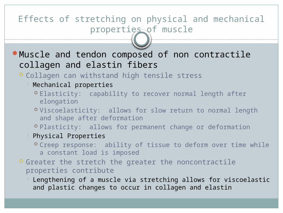
Effects of stretching on physical and mechanical properties of muscle
Muscle and tendon composed of non contractile collagen and elastin fibers Collagen can withstand high tensile stress
Mechanical properties Elasticity: capability to recover normal length after
elongation Viscoelasticity: allows for slow return to normal length and
shape after deformation Plasticity: allows for permanent change or deformation
Physical Properties Creep response: ability of tissue to deform over time while a
constant load is imposed Greater the stretch the greater the noncontractile properties
contribute Lengthening of a muscle via stretching allows for viscoelastic
and plastic changes to occur in collagen and elastin

Effects of stretching on the kinetic chain
Muscle tightness has significant impact on neuromuscular control Effects normal length-tension relationships Compensations and adaptations occur that affect
neuromuscular efficiency through kinetic chain Reciprocal inhibition
For example: if psoas is tight or hyperactive the antagonist gluteus maximus can be inhibited due to decreased neural drive
The synergist, hamstrings (muscle that assist glut max); the stabilizers, erector spinae; and the neutralizers, piriformis become overactive• Creates abnormal joint stress and decreased neuromuscular
control during functional movement

Importance of increasing muscle temp. prior to stretching
Muscle temperature should be increased prior to stretching Positive effect of collagen and elastin components to
deform Capability of GTO to reflexively relax is enhanced
Can be achieved through low intensity warm up or through various therapeutic modalities However exercise is recommended over modalities If muscle guarding occurs cold therapy can also be
used prior to stretching

Stretching techniques
Agonist muscle: muscle that contracts to allow movement through a joint
Antagonist muscle: the muscle that is being stretched in response to contraction of agonist Balance between the agonist and antagonist is
necessary for normal, smooth, coordinated movement As well as reduce muscle strains secondary to muscular
imbalances Comprehension of this synergistic muscle action is
essential to understanding various stretching techniques

Ballistic stretching
Repetitive contractions of agonist used to produce quick stretches of antagonist Safety questioned because of risk of microtears in
musculotendinous unit Not recommended for sedentary individuals or those
recovering from muscle injury Most physical activities are dynamic and repetitive
contraction of agonist with eccentric resistance from antagonist occurs May be implemented in later stages of rehab during
reconditioning phase Progressive velocity flexibility program has been proposed
• Slow static to slow end range to slow full range to fast end range to fast full range

Static stretching
Extremely effective and widely used method of stretching Passively stretching antagonist or actively contracting
agonist to stretch antagonist to maximal position and holding for extended time Recommended to hold for 15 to 30 seconds is most
effective to increase flexibility Can be used early on in rehabilitation program
ATC, teammate, other assistive devices Best to do after muscle temperature is increased
May be more efficient to do after activity and not before


Proprioceptive Neuromuscular Facilitation (PNF)
First used by ATC’s rehabilitating neuromuscular disorders More recently used to increase flexibility 3 types of PNF stretching techniques
Contract relax: beneficial to athletes where ROM is limited by muscle tightness Athlete actively contracts agonist to point of limitation,
athlete then instructed to contract antagonist (muscle to be stretched) isotonically (through range of motion), athlete then relaxes as ATC passively moves part to point of limitation. Stretch is then repeated

Proprioceptive Neuromuscular Facilitation (PNF)
Hold relax: Similar to contract relax except antagonist goes through isometric contraction (contraction w/o movement) Hold for at least 6 seconds Can be used for agonist or antagonist
Slow reversal-hold-relax Begins with isotonic contraction of agonist, followed by
isometric contraction of antagonist During relax phase antagonist are relaxed while agonist
are contracting
..\..\..\..\Downloads\url2.htm

Dynamic stretching
Stretching through series of movement patters Progressive slow controlled movements to faster movements
Muscle activation of agonist and muscle stretching of antagonist Posture and form important Increases core and muscle temperature Increases neuromuscular control Increases balance Core stability Effective for increasing flexibility
Better way to stretch prior to activity• Shown to increase flexibility, decrease injury and increase force
and power output• Mimics sport activity, so more functional• Can initially cause some muscle soreness

..\..\..\..\Downloads\url.htm

Comparing stretch techniques
All have been shown to increase flexibility PNF is capable of producing greater improvement in flexibility
Requires a partner Static
Has to be done often and held for extended periods of time Prior to exercise may decrease core and muscle temperature when
compared to dynamic stretching More appropriate in rehab and as a cool down method after
activity Dynamic
Research supports use prior to activity Ballistic
Can cause injury in untrained or already injured person Can maintain flexibility w/ stretching 1 day a week, but to
improve flexibility must perform 3-5 times/week

Pilates and Yoga
Pilates Extremely popular and widely used method Developed by Joseph Pilates prior to WWII Conditioning program that improves muscle control,
flexibility, coordination, strength and tone. Concentrated on body alignment, breathing, lengthening
of all muscles while building endurance and strength


Pilates and Yoga
Yoga Originated in India 6000 years ago Philosophy that most illness related to poor mental
attitude, posture and diet Aimed to unite body and mind through various body
postures and meditative breathing Start simple and progress to more complex movements Increase mobility and flexibility


Myofascial release stretching
Relieve soft tissue from abnormal grip of tight fascia (fibrous membrane that covers, supports and separates muscles, tendons, bones and organs) Localize restriction and move into direction of
restriction Releasing myofascial restrictions over large treatment
area can have significant impact on joint mobility Progression of superficial restrictions to deeper
restrictions Stretching techniques can be incorporated after
extensibility is improved in myofascia Can use foam rollers to assist with myofascial release


Other methods to restore ROM
MassageStrain-counter strainSoft tissue mobilizationGraston techniqueMassage

Benefits of ROM exercises
Inreased flexibilityIncreased mobilityIncreased blood flow to area
Facilitate tissue healing processDecreased swellingDecreased adhesion formationDecreased pain
Related Documents