OPEN Regulation of anti-apoptotic signaling by Kruppel-like factors 4 and 5 mediates lapatinib resistance in breast cancer MK Farrugia 1,2 , SB Sharma 1,2 , C-C Lin 1,3 , SL McLaughlin 3 , DB Vanderbilt 1,2 , AG Ammer 3 , MA Salkeni 3,4 , P Stoilov 1,2,3 , YM Agazie 1,2,3 , CJ Creighton 5 and JM Ruppert* ,1,2,3 The Kruppel-like transcription factors (KLFs) 4 and 5 (KLF4/5) are coexpressed in mouse embryonic stem cells, where they function redundantly to maintain pluripotency. In mammary carcinoma, KLF4/5 can each impact the malignant phenotype, but potential linkages to drug resistance remain unclear. In primary human breast cancers, we observed a positive correlation between KLF4/5 transcript abundance, particularly in the human epidermal growth factor receptor 2 (HER2)-enriched subtype. Furthermore, KLF4/5 protein was rapidly upregulated in human breast cancer cells following treatment with the HER2/epidermal growth factor receptor inhibitor, lapatinib. In addition, we observed a positive correlation between these factors in the primary tumors of genetically engineered mouse models (GEMMs). In particular, the levels of both factors were enriched in the basal-like tumors of the C3(1) TAg (SV40 large T antigen transgenic mice under control of the C3(1)/prostatein promoter) GEMM. Using tumor cells derived from this model as well as human breast cancer cells, suppression of KLF4 and/or KLF5 sensitized HER2-overexpressing cells to lapatinib. Indicating cooperativity, greater effects were observed when both genes were depleted. KLF4/5-deficient cells had reduced basal mRNA and protein levels of the anti-apoptotic factors myeloid cell leukemia 1 (MCL1) and B-cell lymphoma-extra large (BCL-XL). Moreover, MCL1 was upregulated by lapatinib in a KLF4/5-dependent manner, and enforced expression of MCL1 in KLF4/5-deficient cells restored drug resistance. In addition, combined suppression of KLF4/5 in cultured tumor cells additively inhibited anchorage-independent growth, resistance to anoikis and tumor formation in immunocompromised mice. Consistent with their cooperative role in drug resistance and other malignant properties, KLF4/5 levels selectively stratified human HER2- enriched breast cancer by distant metastasis-free survival. These results identify KLF4 and KLF5 as cooperating protumorigenic factors and critical participants in resistance to lapatinib, furthering the rationale for combining anti-MCL1/BCL-XL inhibitors with conventional HER2-targeted therapies. Cell Death and Disease (2015) 6, e1699; doi:10.1038/cddis.2015.65; published online 19 March 2015 In mouse embryonic stem (ES) cells, pluripotency is main- tained by the redundant function of three Kruppel-like transcription factors, KLF2, KLF4 and KLF5. 1 Furthermore, as determined by chromatin immunoprecipitation combined with high-throughput sequence analysis (ChIP-seq), KLF4 and KLF5 (KLF4/5) have both overlapping and distinct target genes. 2 Depletion of Klf4 or Klf5 in the anterior eye elicits similar developmental phenotypes, whereas in other tissues they exert opposing influences. 3–5 For example, KLF4/5 differentially affect the expression of several cell cycle regulatory proteins, such as CCND1, CCNB1 and p21 Waf1/Cip1 . 3 In adult tissues, KLF4 and/or KLF5 are induced by a variety of stress stimuli and can promote cell survival in diverse contexts. 3,6–9 In breast cancer, KLF4/5 protein levels or mRNA abundance are elevated in aggressive primary tumors. 10–13 Consistent with these results, promoter demethylation of KLF4 or KLF5 in breast tumors is associated with an unfavorable clinical course. 14 Individually, both KLF4/5 exert oncogenic functions in experimental models of cancer such as cellular transforma- tion, migration, invasion and xenograft formation. 15–19 Although signaling mechanisms remain to be elucidated, KLF4 directly regulates the transcription of microRNA-206 (miR-206) to promote tumor cell survival and tumor initiation in 1 Department of Biochemistry, West Virginia University Health Sciences Center, Morgantown, WV 26506, USA; 2 Program in Cancer Cell Biology, West Virginia University, Morgantown, WV 26506, USA; 3 The Mary Babb Randolph Cancer Center, West Virginia University, Morgantown, WV 26506, USA; 4 Department of Medicine, West Virginia University, Morgantown, WV 26506, USA and 5 Department of Medicine and Dan L Duncan Cancer Center, Division of Biostatistics, Baylor College of Medicine, Houston, TX 77030, USA *Corresponding author: JM Ruppert, Department of Biochemistry, West Virginia University Health Sciences Center, 1 Medical Center Drive, PO Box 9142, Room 212 Biomedical Research, Morgantown, WV 26506, USA. Tel: +1 304 293 5246; Fax: +1 304 293 4667; E-mail: [email protected] Received 12.11.14; revised 10.2.15; accepted 12.2.15; Edited by J Chipuk Abbreviations: BCL2, B-cell lymphoma 2; BCL-XL, B-cell lymphoma-extra large; C3(1) Tag, SV40 large Tantigen transgenic mice under control of the C3(1)/prostatein promoter; ChIP-seq, chromatin immunoprecipitation combined with high-throughput sequence; CI, confidence interval; DAPI, 4',6-diamidino-2-phenylindole; DFS, disease- free survival; DMFS, distant metastasis-free survival; ES cells, embryonic stem cells; EGFR, epidermal growth factor receptor; GEMM, genetically engineered mouse model; HER2, human epidermal growth factor receptor 2; KLF, Kruppel-like factor; KLF4/5, KLF4 and KLF5; MAPK, mitogen-activated protein kinase; miR-206, microRNA-206; MMI, mitochondrial membrane integrity; MMTV, mouse mammary tumor virus promoter; MCL1, myeloid cell leukemia 1; PI3K, phosphatidylinositide 3- kinase; qRT-PCR, quantitative reverse transcription and polymerase chain reaction; RTK, receptor tyrosine kinase; TCGA, The Cancer Genome Atlas; TNBC, triple- negative breast cancer Citation: Cell Death and Disease (2015) 6, e1699; doi:10.1038/cddis.2015.65 & 2015 Macmillan Publishers Limited All rights reserved 2041-4889/15 www.nature.com/cddis

Welcome message from author
This document is posted to help you gain knowledge. Please leave a comment to let me know what you think about it! Share it to your friends and learn new things together.
Transcript
OPEN
Regulation of anti-apoptotic signaling by Kruppel-likefactors 4 and 5 mediates lapatinib resistance inbreast cancer
MK Farrugia1,2, SB Sharma1,2, C-C Lin1,3, SL McLaughlin3, DB Vanderbilt1,2, AG Ammer3, MA Salkeni3,4, P Stoilov1,2,3, YM Agazie1,2,3,CJ Creighton5 and JM Ruppert*,1,2,3
The Kruppel-like transcription factors (KLFs) 4 and 5 (KLF4/5) are coexpressed in mouse embryonic stem cells, where theyfunction redundantly to maintain pluripotency. In mammary carcinoma, KLF4/5 can each impact the malignant phenotype, butpotential linkages to drug resistance remain unclear. In primary human breast cancers, we observed a positive correlation betweenKLF4/5 transcript abundance, particularly in the human epidermal growth factor receptor 2 (HER2)-enriched subtype. Furthermore,KLF4/5 protein was rapidly upregulated in human breast cancer cells following treatment with the HER2/epidermal growth factorreceptor inhibitor, lapatinib. In addition, we observed a positive correlation between these factors in the primary tumors ofgenetically engineered mouse models (GEMMs). In particular, the levels of both factors were enriched in the basal-like tumors ofthe C3(1) TAg (SV40 large T antigen transgenic mice under control of the C3(1)/prostatein promoter) GEMM. Using tumor cellsderived from this model as well as human breast cancer cells, suppression of KLF4 and/or KLF5 sensitized HER2-overexpressingcells to lapatinib. Indicating cooperativity, greater effects were observed when both genes were depleted. KLF4/5-deficient cellshad reduced basal mRNA and protein levels of the anti-apoptotic factors myeloid cell leukemia 1 (MCL1) and B-cell lymphoma-extralarge (BCL-XL). Moreover, MCL1 was upregulated by lapatinib in a KLF4/5-dependent manner, and enforced expression of MCL1 inKLF4/5-deficient cells restored drug resistance. In addition, combined suppression of KLF4/5 in cultured tumor cells additivelyinhibited anchorage-independent growth, resistance to anoikis and tumor formation in immunocompromised mice. Consistentwith their cooperative role in drug resistance and other malignant properties, KLF4/5 levels selectively stratified human HER2-enriched breast cancer by distant metastasis-free survival. These results identify KLF4 and KLF5 as cooperating protumorigenicfactors and critical participants in resistance to lapatinib, furthering the rationale for combining anti-MCL1/BCL-XL inhibitors withconventional HER2-targeted therapies.Cell Death and Disease (2015) 6, e1699; doi:10.1038/cddis.2015.65; published online 19 March 2015
In mouse embryonic stem (ES) cells, pluripotency is main-tained by the redundant function of three Kruppel-liketranscription factors, KLF2, KLF4 and KLF5.1 Furthermore,as determined by chromatin immunoprecipitation combinedwith high-throughput sequence analysis (ChIP-seq), KLF4and KLF5 (KLF4/5) have both overlapping and distinct targetgenes.2 Depletion of Klf4 or Klf5 in the anterior eye elicitssimilar developmental phenotypes, whereas in other tissuesthey exert opposing influences.3–5 For example, KLF4/5differentially affect the expression of several cell cycleregulatory proteins, such as CCND1, CCNB1 andp21Waf1/Cip1.3 In adult tissues, KLF4 and/or KLF5 are induced
by a variety of stress stimuli and can promote cell survival indiverse contexts.3,6–9
In breast cancer, KLF4/5 protein levels or mRNAabundanceare elevated in aggressive primary tumors.10–13 Consistentwith these results, promoter demethylation of KLF4 or KLF5 inbreast tumors is associated with an unfavorable clinicalcourse.14 Individually, both KLF4/5 exert oncogenic functionsin experimental models of cancer such as cellular transforma-tion, migration, invasion and xenograft formation.15–19
Although signaling mechanisms remain to be elucidated,KLF4 directly regulates the transcription of microRNA-206(miR-206) to promote tumor cell survival and tumor initiation in
1Department of Biochemistry, West Virginia University Health Sciences Center, Morgantown, WV 26506, USA; 2Program in Cancer Cell Biology, West Virginia University,Morgantown, WV 26506, USA; 3The Mary Babb Randolph Cancer Center, West Virginia University, Morgantown, WV 26506, USA; 4Department of Medicine, West VirginiaUniversity, Morgantown, WV 26506, USA and 5Department of Medicine and Dan L Duncan Cancer Center, Division of Biostatistics, Baylor College of Medicine, Houston,TX 77030, USA*Corresponding author: JM Ruppert, Department of Biochemistry, West Virginia University Health Sciences Center, 1 Medical Center Drive, PO Box 9142, Room 212Biomedical Research, Morgantown, WV 26506, USA. Tel: +1 304 293 5246; Fax: +1 304 293 4667; E-mail: [email protected]
Received 12.11.14; revised 10.2.15; accepted 12.2.15; Edited by J Chipuk
Abbreviations: BCL2, B-cell lymphoma 2; BCL-XL, B-cell lymphoma-extra large; C3(1) Tag, SV40 large Tantigen transgenic mice under control of the C3(1)/prostateinpromoter; ChIP-seq, chromatin immunoprecipitation combined with high-throughput sequence; CI, confidence interval; DAPI, 4',6-diamidino-2-phenylindole; DFS, disease-free survival; DMFS, distant metastasis-free survival; ES cells, embryonic stem cells; EGFR, epidermal growth factor receptor; GEMM, genetically engineeredmouse model; HER2, human epidermal growth factor receptor 2; KLF, Kruppel-like factor; KLF4/5, KLF4 and KLF5; MAPK, mitogen-activated protein kinase; miR-206,microRNA-206; MMI, mitochondrial membrane integrity; MMTV, mouse mammary tumor virus promoter; MCL1, myeloid cell leukemia 1; PI3K, phosphatidylinositide 3-kinase; qRT-PCR, quantitative reverse transcription and polymerase chain reaction; RTK, receptor tyrosine kinase; TCGA, The Cancer Genome Atlas; TNBC, triple-negative breast cancer
Citation: Cell Death and Disease (2015) 6, e1699; doi:10.1038/cddis.2015.65& 2015 Macmillan Publishers Limited All rights reserved 2041-4889/15
www.nature.com/cddis
athymic mice (manuscript submitted).18,20 Although indepen-dently KLF4/5 have important roles in breast cancer, therelationship between the two genes in this disease remainsunderstudied.We observed a positive correlation of KLF4/5 expression in
the human epidermal growth factor receptor 2 (HER2)-enriched breast cancer subtype. In addition, in these patientsthe median expression of both KLF4/5 significantly stratifiedthe distant metastasis-free survival (DMFS). Clinicallyapproved HER2-targeted therapies such as lapatinib andtrastuzumab (Herceptin) have significantly improved thedisease-free survival (DFS) of patients with HER2-amplifiedbreast cancers.21,22 However, eventual resistance to thesetherapies is observed in the majority of cases, representing amajor obstacle to long-term cures.23–26
Several mechanisms of resistance have been described,often involving sustained signaling through dimerization withother receptor tyrosine kinases (RTKs) or activatingmutations indownstream effectors, namely RAS pathway components.27–32
Although numerous pathways to resistance have been char-acterized, it is unclear which of these mechanisms predominatein patients and how they are specifically regulated.Interestingly, neutralization of apoptotic signaling contributes to
anti-HER2 therapeutic failure.33–35 For example, phosphorylationof BAD or overexpression of B-cell lymphoma-extra large (BCL-XL) reduces the efficacy of these drugs. One such drug islapatinib, a HER2/epidermal growth factor receptor (EGFR)inhibitor that has activity not only in the HER2+ cancers ofpatients, but also shows efficacy in combination with other agentsin basal-like breast cancermodels.36,37Weobserved that lapatinibtreatment of HER2-amplified tumor cells resulted in the rapidinduction of KLF4/5. Subsequent experiments demonstrated anovel role for the endogenous KLFs in the regulation of anti-apoptotic factors. As shown by shRNA studies, in the presence oflapatinib KLF4/5 coregulated the expression of myeloid leukemiacell 1 (MCL1) and cooperated to confer lapatinib resistance. Evenin the absence of drug treatment, the endogenous KLFs werecritical for maintaining basal levels of the anti-apoptotic factorsMCL1 and BCL-XL, and collaboratively promoted the malignantphenotype. KLF4/5 were positively correlated with MCL1 inprimary breast tumors, and enforced expression of MCL1 wassufficient to rescue the lapatinib sensitivity of KLF4/5-deficientcells. These results identify KLF4/5 as inducible regulators oflapatinib resistance through mediation of anti-apoptotic signaling.
Results
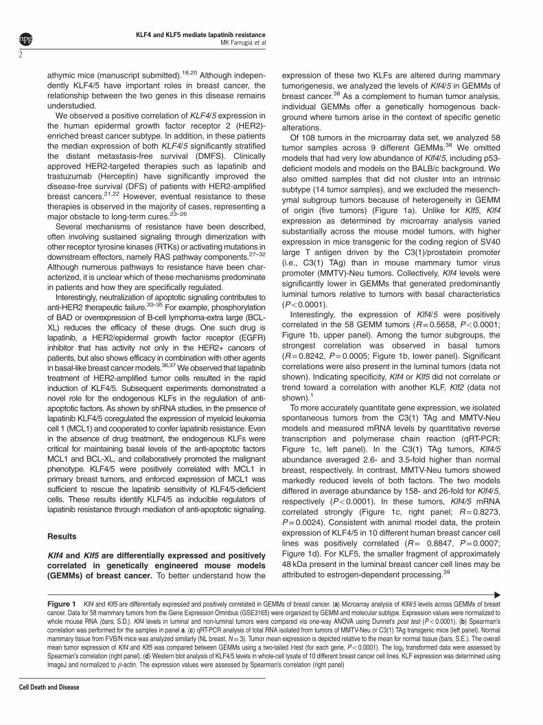
Klf4 and Klf5 are differentially expressed and positivelycorrelated in genetically engineered mouse models(GEMMs) of breast cancer. To better understand how the
expression of these two KLFs are altered during mammarytumorigenesis, we analyzed the levels of Klf4/5 in GEMMs ofbreast cancer.38 As a complement to human tumor analysis,individual GEMMs offer a genetically homogenous back-ground where tumors arise in the context of specific geneticalterations.Of 108 tumors in the microarray data set, we analyzed 58
tumor samples across 9 different GEMMs.38 We omittedmodels that had very low abundance of Klf4/5, including p53-deficient models and models on the BALB/c background. Wealso omitted samples that did not cluster into an intrinsicsubtype (14 tumor samples), and we excluded the mesench-ymal subgroup tumors because of heterogeneity in GEMMof origin (five tumors) (Figure 1a). Unlike for Klf5, Klf4expression as determined by microarray analysis variedsubstantially across the mouse model tumors, with higherexpression in mice transgenic for the coding region of SV40large T antigen driven by the C3(1)/prostatein promoter(i.e., C3(1) TAg) than in mouse mammary tumor viruspromoter (MMTV)-Neu tumors. Collectively, Klf4 levels weresignificantly lower in GEMMs that generated predominantlyluminal tumors relative to tumors with basal characteristics(Po0.0001).Interestingly, the expression of Klf4/5 were positively
correlated in the 58 GEMM tumors (R=0.5658, Po0.0001;Figure 1b, upper panel). Among the tumor subgroups, thestrongest correlation was observed in basal tumors(R=0.8242, P=0.0005; Figure 1b, lower panel). Significantcorrelations were also present in the luminal tumors (data notshown). Indicating specificity, Klf4 or Klf5 did not correlate ortrend toward a correlation with another KLF, Klf2 (data notshown).1
To more accurately quantitate gene expression, we isolatedspontaneous tumors from the C3(1) TAg and MMTV-Neumodels and measured mRNA levels by quantitative reversetranscription and polymerase chain reaction (qRT-PCR;Figure 1c, left panel). In the C3(1) TAg tumors, Klf4/5abundance averaged 2.6- and 3.5-fold higher than normalbreast, respectively. In contrast, MMTV-Neu tumors showedmarkedly reduced levels of both factors. The two modelsdiffered in average abundance by 158- and 26-fold for Klf4/5,respectively (Po0.0001). In these tumors, Klf4/5 mRNAcorrelated strongly (Figure 1c, right panel; R= 0.8273,P= 0.0024). Consistent with animal model data, the proteinexpression of KLF4/5 in 10 different human breast cancer celllines was positively correlated (R= 0.8847, P= 0.0007;Figure 1d). For KLF5, the smaller fragment of approximately48 kDa present in the luminal breast cancer cell lines may beattributed to estrogen-dependent processing.39
Figure 1 Klf4 and Klf5 are differentially expressed and positively correlated in GEMMs of breast cancer. (a) Microarray analysis of Klf4/5 levels across GEMMs of breastcancer. Data for 58 mammary tumors from the Gene Expression Omnibus (GSE3165) were organized by GEMM and molecular subtype. Expression values were normalized towhole mouse RNA (bars, S.D.). Klf4 levels in luminal and non-luminal tumors were compared via one-way ANOVA using Dunnet’s post test (Po0.0001). (b) Spearman’scorrelation was performed for the samples in panel a. (c) qRT-PCR analysis of total RNA isolated from tumors of MMTV-Neu or C3(1) TAg transgenic mice (left panel). Normalmammary tissue from FVB/N mice was analyzed similarly (NL breast, N= 3). Tumor mean expression is depicted relative to the mean for normal tissue (bars, S.E.). The overallmean tumor expression of Klf4 and Klf5 was compared between GEMMs using a two-tailed t-test (for each gene, Po0.0001). The log2 transformed data were assessed bySpearman’s correlation (right panel). (d) Western blot analysis of KLF4/5 levels in whole-cell lysate of 10 different breast cancer cell lines. KLF expression was determined usingImageJ and normalized to β-actin. The expression values were assessed by Spearman’s correlation (right panel)
KLF4 and KLF5 mediate lapatinib resistanceMK Farrugia et al
2
Cell Death and Disease
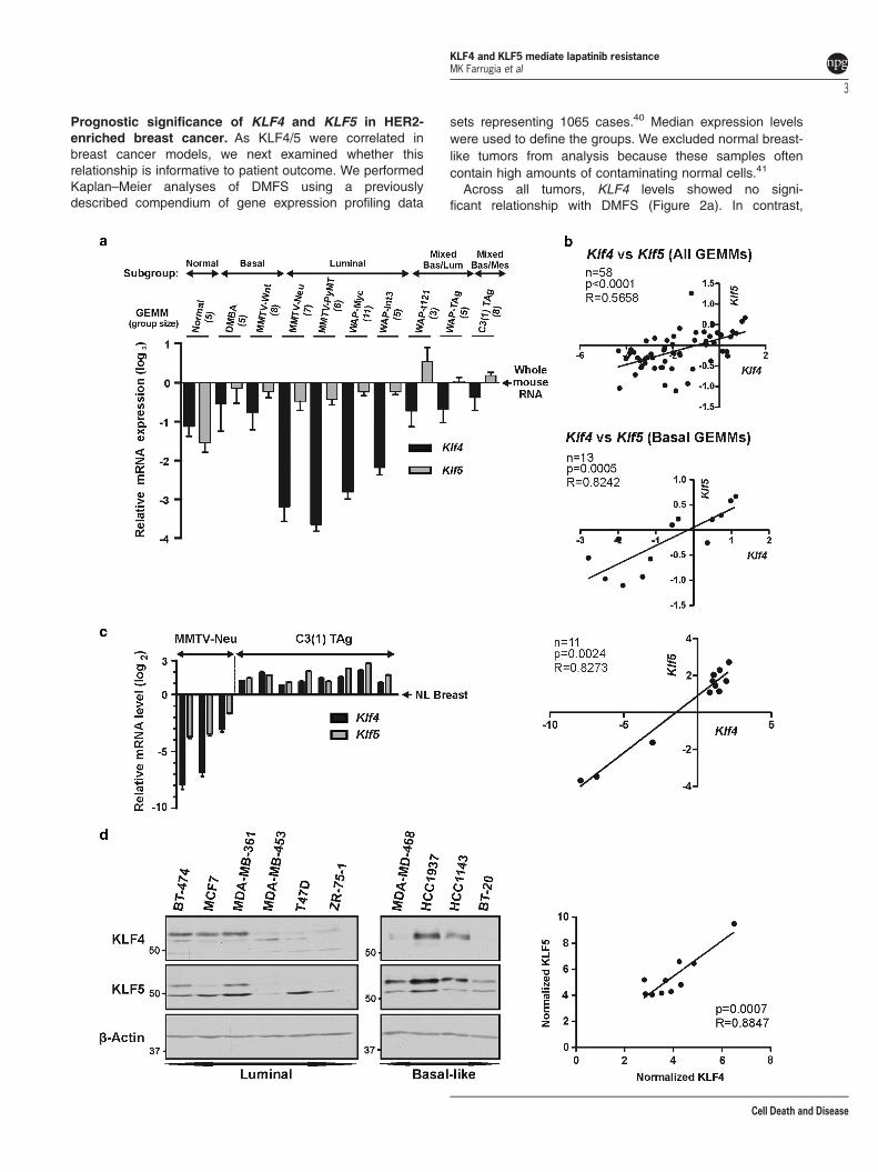
Prognostic significance of KLF4 and KLF5 in HER2-enriched breast cancer. As KLF4/5 were correlated inbreast cancer models, we next examined whether thisrelationship is informative to patient outcome. We performedKaplan–Meier analyses of DMFS using a previouslydescribed compendium of gene expression profiling data
sets representing 1065 cases.40 Median expression levelswere used to define the groups. We excluded normal breast-like tumors from analysis because these samples oftencontain high amounts of contaminating normal cells.41
Across all tumors, KLF4 levels showed no signi-ficant relationship with DMFS (Figure 2a). In contrast,
KLF4 and KLF5 mediate lapatinib resistanceMK Farrugia et al
3
Cell Death and Disease
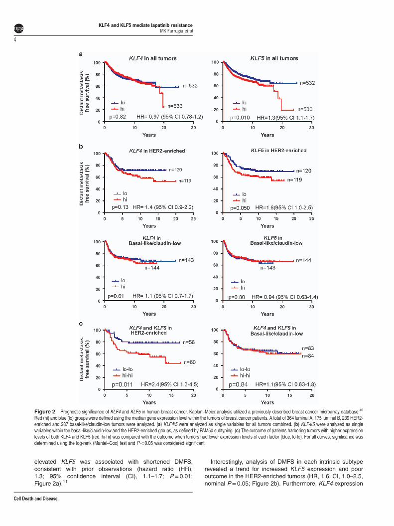
elevated KLF5 was associated with shortened DMFS,consistent with prior observations (hazard ratio (HR),1.3; 95% confidence interval (CI), 1.1–1.7; P= 0.01;Figure 2a).11
Interestingly, analysis of DMFS in each intrinsic subtyperevealed a trend for increased KLF5 expression and pooroutcome in the HER2-enriched tumors (HR, 1.6; CI, 1.0–2.5,nominal P=0.05; Figure 2b). Furthermore, KLF4 expression
Figure 2 Prognostic significance of KLF4 and KLF5 in human breast cancer. Kaplan–Meier analysis utilized a previously described breast cancer microarray database.40
Red (hi) and blue (lo) groups were defined using the median gene expression level within the tumors of breast cancer patients. A total of 364 luminal A, 175 luminal B, 239 HER2-enriched and 287 basal-like/claudin-low tumors were analyzed. (a) KLF4/5 were analyzed as single variables for all tumors combined. (b) KLF4/5 were analyzed as singlevariables within the basal-like/claudin-low and the HER2-enriched groups, as defined by PAM50 subtyping. (c) The outcome of patients harboring tumors with higher expressionlevels of both KLF4 and KLF5 (red, hi-hi) was compared with the outcome when tumors had lower expression levels of each factor (blue, lo-lo). For all curves, significance wasdetermined using the log-rank (Mantel–Cox) test and Po0.05 was considered significant
KLF4 and KLF5 mediate lapatinib resistanceMK Farrugia et al
4
Cell Death and Disease
showed a similar trend in this context (HR, 1.4; CI, 0.9–2.2,P= 0.13). No such relationships were observed in a similarlyhigh-risk group, basal-like/claudin-low (Figure 2b), nor in theluminal A or luminal B groups (Supplementary Figure 1).Furthermore, the expression of KLF4/5 together (i.e.,o median levels of each gene versus ≥ median levelsof each gene) considerably stratified DMFS in the HER2-enriched subtype (HR, 2.4; CI, 1.2–4.5, P=0.011; Figure 2c).HER2-enriched tumors with elevated KLF4/5 levels hadDMFS similar to 43% at 15 years post-diagnosis. This effectwas unique to the HER2-enriched subtype, as no other groupshowed any trend (Figure 2c and Supplementary Figure 1). Toassess survival in patients with elevated expression of onlyone of the two KLFs, we analyzed the four survival curvesdefined by 2 × 2 contingency using the median scores forKLF4/5 (Supplementary Figure 1). The log-rank test for trendidentified a significant trend between KLF4/5 expression andmedian DMFS (P=0.016).
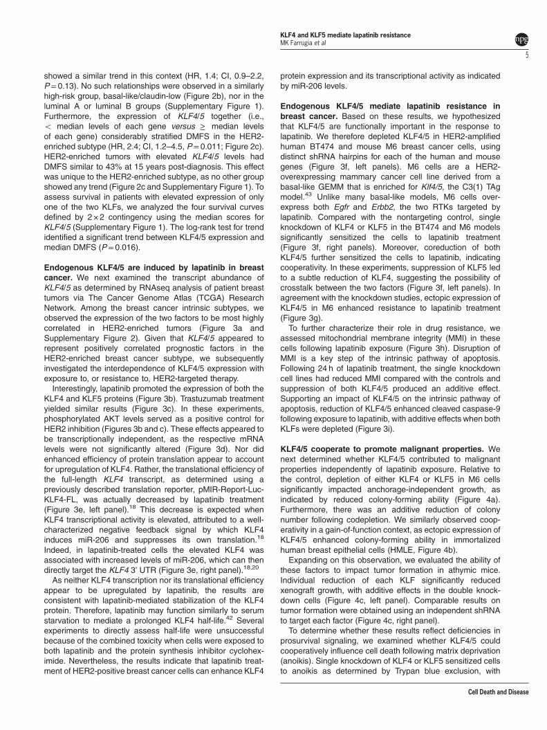
Endogenous KLF4/5 are induced by lapatinib in breastcancer. We next examined the transcript abundance ofKLF4/5 as determined by RNAseq analysis of patient breasttumors via The Cancer Genome Atlas (TCGA) ResearchNetwork. Among the breast cancer intrinsic subtypes, weobserved the expression of the two factors to be most highlycorrelated in HER2-enriched tumors (Figure 3a andSupplementary Figure 2). Given that KLF4/5 appeared torepresent positively correlated prognostic factors in theHER2-enriched breast cancer subtype, we subsequentlyinvestigated the interdependence of KLF4/5 expression withexposure to, or resistance to, HER2-targeted therapy.Interestingly, lapatinib promoted the expression of both the
KLF4 and KLF5 proteins (Figure 3b). Trastuzumab treatmentyielded similar results (Figure 3c). In these experiments,phosphorylated AKT levels served as a positive control forHER2 inhibition (Figures 3b and c). These effects appeared tobe transcriptionally independent, as the respective mRNAlevels were not significantly altered (Figure 3d). Nor didenhanced efficiency of protein translation appear to accountfor upregulation of KLF4. Rather, the translational efficiency ofthe full-length KLF4 transcript, as determined using apreviously described translation reporter, pMIR-Report-Luc-KLF4-FL, was actually decreased by lapatinib treatment(Figure 3e, left panel).18 This decrease is expected whenKLF4 transcriptional activity is elevated, attributed to a well-characterized negative feedback signal by which KLF4induces miR-206 and suppresses its own translation.18
Indeed, in lapatinib-treated cells the elevated KLF4 wasassociated with increased levels of miR-206, which can thendirectly target the KLF4 3’ UTR (Figure 3e, right panel).18,20
As neither KLF4 transcription nor its translational efficiencyappear to be upregulated by lapatinib, the results areconsistent with lapatinib-mediated stabilization of the KLF4protein. Therefore, lapatinib may function similarly to serumstarvation to mediate a prolonged KLF4 half-life.42 Severalexperiments to directly assess half-life were unsuccessfulbecause of the combined toxicity when cells were exposed toboth lapatinib and the protein synthesis inhibitor cyclohex-imide. Nevertheless, the results indicate that lapatinib treat-ment of HER2-positive breast cancer cells can enhance KLF4
protein expression and its transcriptional activity as indicatedby miR-206 levels.
Endogenous KLF4/5 mediate lapatinib resistance inbreast cancer. Based on these results, we hypothesizedthat KLF4/5 are functionally important in the response tolapatinib. We therefore depleted KLF4/5 in HER2-amplifiedhuman BT474 and mouse M6 breast cancer cells, usingdistinct shRNA hairpins for each of the human and mousegenes (Figure 3f, left panels). M6 cells are a HER2-overexpressing mammary cancer cell line derived from abasal-like GEMM that is enriched for Klf4/5, the C3(1) TAgmodel.43 Unlike many basal-like models, M6 cells over-express both Egfr and Erbb2, the two RTKs targeted bylapatinib. Compared with the nontargeting control, singleknockdown of KLF4 or KLF5 in the BT474 and M6 modelssignificantly sensitized the cells to lapatinib treatment(Figure 3f, right panels). Moreover, coreduction of bothKLF4/5 further sensitized the cells to lapatinib, indicatingcooperativity. In these experiments, suppression of KLF5 ledto a subtle reduction of KLF4, suggesting the possibility ofcrosstalk between the two factors (Figure 3f, left panels). Inagreement with the knockdown studies, ectopic expression ofKLF4/5 in M6 enhanced resistance to lapatinib treatment(Figure 3g).To further characterize their role in drug resistance, we
assessed mitochondrial membrane integrity (MMI) in thesecells following lapatinib exposure (Figure 3h). Disruption ofMMI is a key step of the intrinsic pathway of apoptosis.Following 24 h of lapatinib treatment, the single knockdowncell lines had reduced MMI compared with the controls andsuppression of both KLF4/5 produced an additive effect.Supporting an impact of KLF4/5 on the intrinsic pathway ofapoptosis, reduction of KLF4/5 enhanced cleaved caspase-9following exposure to lapatinib, with additive effects when bothKLFs were depleted (Figure 3i).
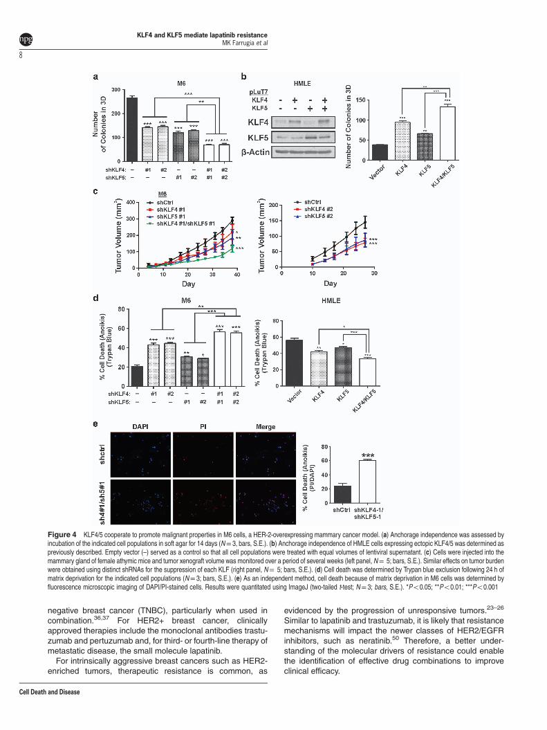
KLF4/5 cooperate to promote malignant properties. Wenext determined whether KLF4/5 contributed to malignantproperties independently of lapatinib exposure. Relative tothe control, depletion of either KLF4 or KLF5 in M6 cellssignificantly impacted anchorage-independent growth, asindicated by reduced colony-forming ability (Figure 4a).Furthermore, there was an additive reduction of colonynumber following codepletion. We similarly observed coop-erativity in a gain-of-function context, as ectopic expression ofKLF4/5 enhanced colony-forming ability in immortalizedhuman breast epithelial cells (HMLE, Figure 4b).Expanding on this observation, we evaluated the ability of
these factors to impact tumor formation in athymic mice.Individual reduction of each KLF significantly reducedxenograft growth, with additive effects in the double knock-down cells (Figure 4c, left panel). Comparable results ontumor formation were obtained using an independent shRNAto target each factor (Figure 4c, right panel).To determine whether these results reflect deficiencies in
prosurvival signaling, we examined whether KLF4/5 couldcooperatively influence cell death following matrix deprivation(anoikis). Single knockdown of KLF4 or KLF5 sensitized cellsto anoikis as determined by Trypan blue exclusion, with
KLF4 and KLF5 mediate lapatinib resistanceMK Farrugia et al
5
Cell Death and Disease
KLF4 and KLF5 mediate lapatinib resistanceMK Farrugia et al
6
Cell Death and Disease
cooperative effects following codepletion (Figure 4d, leftpanel). Conversely, overexpression of these factors in HMLEenhanced anoikis resistance (Figure 4d, right panel). Deter-mination of cell death by an independent method yieldedsimilar results (Figure 4e).
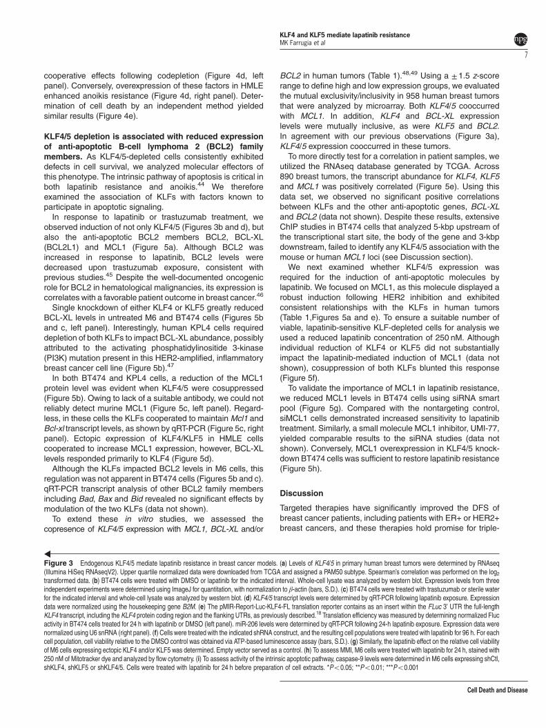
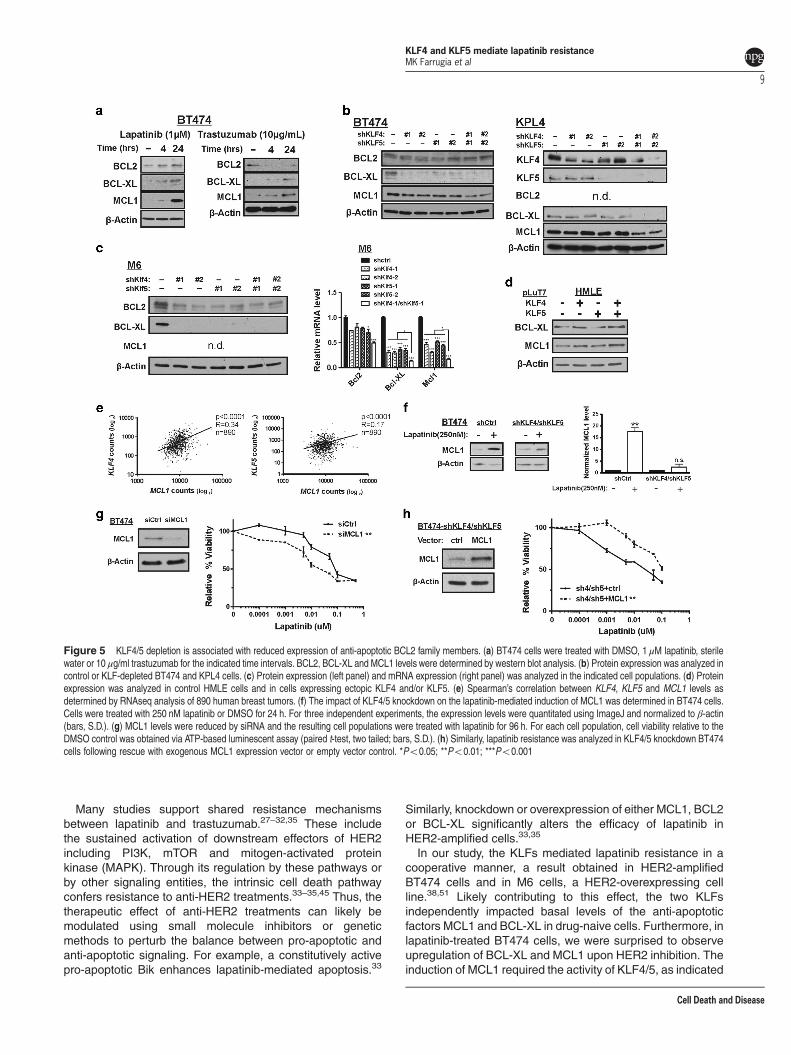
KLF4/5 depletion is associated with reduced expressionof anti-apoptotic B-cell lymphoma 2 (BCL2) familymembers. As KLF4/5-depleted cells consistently exhibiteddefects in cell survival, we analyzed molecular effectors ofthis phenotype. The intrinsic pathway of apoptosis is critical inboth lapatinib resistance and anoikis.44 We thereforeexamined the association of KLFs with factors known toparticipate in apoptotic signaling.In response to lapatinib or trastuzumab treatment, we
observed induction of not only KLF4/5 (Figures 3b and d), butalso the anti-apoptotic BCL2 members BCL2, BCL-XL(BCL2L1) and MCL1 (Figure 5a). Although BCL2 wasincreased in response to lapatinib, BCL2 levels weredecreased upon trastuzumab exposure, consistent withprevious studies.45 Despite the well-documented oncogenicrole for BCL2 in hematological malignancies, its expression iscorrelates with a favorable patient outcome in breast cancer.46
Single knockdown of either KLF4 or KLF5 greatly reducedBCL-XL levels in untreated M6 and BT474 cells (Figures 5band c, left panel). Interestingly, human KPL4 cells requireddepletion of both KLFs to impact BCL-XL abundance, possiblyattributed to the activating phosphatidylinositide 3-kinase(PI3K) mutation present in this HER2-amplified, inflammatorybreast cancer cell line (Figure 5b).47
In both BT474 and KPL4 cells, a reduction of the MCL1protein level was evident when KLF4/5 were cosuppressed(Figure 5b). Owing to lack of a suitable antibody, we could notreliably detect murine MCL1 (Figure 5c, left panel). Regard-less, in these cells the KLFs cooperated to maintain Mcl1 andBcl-xl transcript levels, as shown by qRT-PCR (Figure 5c, rightpanel). Ectopic expression of KLF4/KLF5 in HMLE cellscooperated to increase MCL1 expression, however, BCL-XLlevels responded primarily to KLF4 (Figure 5d).Although the KLFs impacted BCL2 levels in M6 cells, this
regulation was not apparent in BT474 cells (Figures 5b and c).qRT-PCR transcript analysis of other BCL2 family membersincluding Bad, Bax and Bid revealed no significant effects bymodulation of the two KLFs (data not shown).To extend these in vitro studies, we assessed the
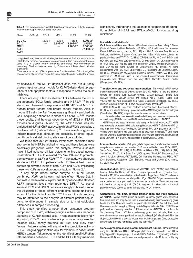
copresence of KLF4/5 expression with MCL1, BCL-XL and/or
BCL2 in human tumors (Table 1).48,49 Using a ± 1.5 z-scorerange to define high and low expression groups, we evaluatedthe mutual exclusivity/inclusivity in 958 human breast tumorsthat were analyzed by microarray. Both KLF4/5 cooccurredwith MCL1. In addition, KLF4 and BCL-XL expressionlevels were mutually inclusive, as were KLF5 and BCL2.In agreement with our previous observations (Figure 3a),KLF4/5 expression cooccurred in these tumors.To more directly test for a correlation in patient samples, we
utilized the RNAseq database generated by TCGA. Across890 breast tumors, the transcript abundance for KLF4, KLF5and MCL1 was positively correlated (Figure 5e). Using thisdata set, we observed no significant positive correlationsbetween KLFs and the other anti-apoptotic genes, BCL-XLand BCL2 (data not shown). Despite these results, extensiveChIP studies in BT474 cells that analyzed 5-kbp upstream ofthe transcriptional start site, the body of the gene and 3-kbpdownstream, failed to identify any KLF4/5 association with themouse or human MCL1 loci (see Discussion section).We next examined whether KLF4/5 expression was
required for the induction of anti-apoptotic molecules bylapatinib. We focused on MCL1, as this molecule displayed arobust induction following HER2 inhibition and exhibitedconsistent relationships with the KLFs in human tumors(Table 1,Figures 5a and e). To ensure a suitable number ofviable, lapatinib-sensitive KLF-depleted cells for analysis weused a reduced lapatinib concentration of 250 nM. Althoughindividual reduction of KLF4 or KLF5 did not substantiallyimpact the lapatinib-mediated induction of MCL1 (data notshown), cosuppression of both KLFs blunted this response(Figure 5f).To validate the importance of MCL1 in lapatinib resistance,
we reduced MCL1 levels in BT474 cells using siRNA smartpool (Figure 5g). Compared with the nontargeting control,siMCL1 cells demonstrated increased sensitivity to lapatinibtreatment. Similarly, a small molecule MCL1 inhibitor, UMI-77,yielded comparable results to the siRNA studies (data notshown). Conversely, MCL1 overexpression in KLF4/5 knock-down BT474 cells was sufficient to restore lapatinib resistance(Figure 5h).
Discussion
Targeted therapies have significantly improved the DFS ofbreast cancer patients, including patients with ER+ or HER2+breast cancers, and these therapies hold promise for triple-
Figure 3 Endogenous KLF4/5 mediate lapatinib resistance in breast cancer models. (a) Levels of KLF4/5 in primary human breast tumors were determined by RNAseq(Illumina HiSeq RNAseqV2). Upper quartile normalized data were downloaded from TCGA and assigned a PAM50 subtype. Spearman’s correlation was performed on the log2transformed data. (b) BT474 cells were treated with DMSO or lapatinib for the indicated interval. Whole-cell lysate was analyzed by western blot. Expression levels from threeindependent experiments were determined using ImageJ for quantitation, with normalization to β-actin (bars, S.D.). (c) BT474 cells were treated with trastuzumab or sterile waterfor the indicated interval and whole-cell lysate was analyzed by western blot. (d) KLF4/5 transcript levels were determined by qRT-PCR following lapatinib exposure. Expressiondata were normalized using the housekeeping gene B2M. (e) The pMIR-Report-Luc-KLF4-FL translation reporter contains as an insert within the FLuc 3’ UTR the full-lengthKLF4 transcript, including the KLF4 protein coding region and the flanking UTRs, as previously described.18 Translation efficiency was measured by determining normalized Flucactivity in BT474 cells treated for 24 h with lapatinib or DMSO (left panel). miR-206 levels were determined by qRT-PCR following 24-h lapatinib exposure. Expression data werenormalized using U6 snRNA (right panel). (f) Cells were treated with the indicated shRNA construct, and the resulting cell populations were treated with lapatinib for 96 h. For eachcell population, cell viability relative to the DMSO control was obtained via ATP-based luminescence assay (bars, S.D.). (g) Similarly, the lapatinib effect on the relative cell viabilityof M6 cells expressing ectopic KLF4 and/or KLF5 was determined. Empty vector served as a control. (h) To assess MMI, M6 cells were treated with lapatinib for 24 h, stained with250 nM of Mitotracker dye and analyzed by flow cytometry. (i) To assess activity of the intrinsic apoptotic pathway, caspase-9 levels were determined in M6 cells expressing shCtl,shKLF4, shKLF5 or shKLF4/5. Cells were treated with lapatinib for 24 h before preparation of cell extracts. *Po0.05; **Po0.01; ***Po0.001
KLF4 and KLF5 mediate lapatinib resistanceMK Farrugia et al
7
Cell Death and Disease
negative breast cancer (TNBC), particularly when used incombination.36,37 For HER2+ breast cancer, clinicallyapproved therapies include the monoclonal antibodies trastu-zumab and pertuzumab and, for third- or fourth-line therapy ofmetastatic disease, the small molecule lapatinib.For intrinsically aggressive breast cancers such as HER2-
enriched tumors, therapeutic resistance is common, as
evidenced by the progression of unresponsive tumors.23–26
Similar to lapatinib and trastuzumab, it is likely that resistancemechanisms will impact the newer classes of HER2/EGFRinhibitors, such as neratinib.50 Therefore, a better under-standing of the molecular drivers of resistance could enablethe identification of effective drug combinations to improveclinical efficacy.
Figure 4 KLF4/5 cooperate to promote malignant properties in M6 cells, a HER-2-overexpressing mammary cancer model. (a) Anchorage independence was assessed byincubation of the indicated cell populations in soft agar for 14 days (N= 3, bars, S.E.). (b) Anchorage independence of HMLE cells expressing ectopic KLF4/5 was determined aspreviously described. Empty vector (–) served as a control so that all cell populations were treated with equal volumes of lentiviral supernatant. (c) Cells were injected into themammary gland of female athymic mice and tumor xenograft volume was monitored over a period of several weeks (left panel, N= 5; bars, S.E.). Similar effects on tumor burdenwere obtained using distinct shRNAs for the suppression of each KLF (right panel, N= 5; bars, S.E.). (d) Cell death was determined by Trypan blue exclusion following 24 h ofmatrix deprivation for the indicated cell populations (N= 3; bars, S.E.). (e) As an independent method, cell death because of matrix deprivation in M6 cells was determined byfluorescence microscopic imaging of DAPI/PI-stained cells. Results were quantitated using ImageJ (two-tailed t-test; N= 3; bars, S.E.). *Po0.05; **Po0.01; ***Po0.001
KLF4 and KLF5 mediate lapatinib resistanceMK Farrugia et al
8
Cell Death and Disease
Many studies support shared resistance mechanismsbetween lapatinib and trastuzumab.27–32,35 These includethe sustained activation of downstream effectors of HER2including PI3K, mTOR and mitogen-activated proteinkinase (MAPK). Through its regulation by these pathways orby other signaling entities, the intrinsic cell death pathwayconfers resistance to anti-HER2 treatments.33–35,45 Thus, thetherapeutic effect of anti-HER2 treatments can likely bemodulated using small molecule inhibitors or geneticmethods to perturb the balance between pro-apoptotic andanti-apoptotic signaling. For example, a constitutively activepro-apoptotic Bik enhances lapatinib-mediated apoptosis.33
Similarly, knockdown or overexpression of either MCL1, BCL2or BCL-XL significantly alters the efficacy of lapatinib inHER2-amplified cells.33,35
In our study, the KLFs mediated lapatinib resistance in acooperative manner, a result obtained in HER2-amplifiedBT474 cells and in M6 cells, a HER2-overexpressing cellline.38,51 Likely contributing to this effect, the two KLFsindependently impacted basal levels of the anti-apoptoticfactors MCL1 and BCL-XL in drug-naive cells. Furthermore, inlapatinib-treated BT474 cells, we were surprised to observeupregulation of BCL-XL and MCL1 upon HER2 inhibition. Theinduction of MCL1 required the activity of KLF4/5, as indicated
Figure 5 KLF4/5 depletion is associated with reduced expression of anti-apoptotic BCL2 family members. (a) BT474 cells were treated with DMSO, 1 μM lapatinib, sterilewater or 10 μg/ml trastuzumab for the indicated time intervals. BCL2, BCL-XL and MCL1 levels were determined by western blot analysis. (b) Protein expression was analyzed incontrol or KLF-depleted BT474 and KPL4 cells. (c) Protein expression (left panel) and mRNA expression (right panel) was analyzed in the indicated cell populations. (d) Proteinexpression was analyzed in control HMLE cells and in cells expressing ectopic KLF4 and/or KLF5. (e) Spearman’s correlation between KLF4, KLF5 and MCL1 levels asdetermined by RNAseq analysis of 890 human breast tumors. (f) The impact of KLF4/5 knockdown on the lapatinib-mediated induction of MCL1 was determined in BT474 cells.Cells were treated with 250 nM lapatinib or DMSO for 24 h. For three independent experiments, the expression levels were quantitated using ImageJ and normalized to β-actin(bars, S.D.). (g) MCL1 levels were reduced by siRNA and the resulting cell populations were treated with lapatinib for 96 h. For each cell population, cell viability relative to theDMSO control was obtained via ATP-based luminescent assay (paired t-test, two tailed; bars, S.D.). (h) Similarly, lapatinib resistance was analyzed in KLF4/5 knockdown BT474cells following rescue with exogenous MCL1 expression vector or empty vector control. *Po0.05; **Po0.01; ***Po0.001
KLF4 and KLF5 mediate lapatinib resistanceMK Farrugia et al
9
Cell Death and Disease
by analysis of the KLF4/5-deficient cells. We are currentlyassessing other tumor models for KLF4/5-dependent upregu-lation of anti-apoptotic factors in response to small moleculeinhibitors.There are only a few established links between the KLFs,
anti-apoptotic BCL2 family proteins and HER2.52–54 In thisstudy, we observed coexpression of KLF4/5 and MCL1 inhuman breast tumors and breast cancer models. Also, inmouse ES cells the Mcl1 locus was significantly enriched viaChIP-seq using antibodies to either KLF4 or KLF5.2,55 Despitethese results, and the clear dependence of MCL1 on KLF4/5expression (Figures 5b and c), the MCL1 locus was notenriched in KLF4/5 ChIP assays, in whichMIR206 served as apositive control (data not shown).20 These results suggest anindirect relationship, although the possibility of direct regula-tion through a distal binding site cannot be excluded.We found that KLF4/5 mRNA levels were correlated most
strongly in the HER2-enriched tumors, and these factors wereselectively prognostic within this subtype. Previous studieshave linked adverse clinical outcome to increased nuclearlocalization of KLF4, to elevated KLF5 mRNA and to promoterdemethylation of KLF4 or KLF5.10–14 In our study, we observedshortened DMFS for patients with HER2-enriched tumorscontaining elevated levels of both KLF4 and KLF5, implicatingthese two KLFs as novel prognostic factors (Figure 2d).In any single breast tumor subtype or in all tumors
combined, KLF4 on its own had little effect (Figure 2b). Incontrast to these results, a previous study associated elevatedKLF4 transcript levels with prolonged DFS.56 As overallsurvival, DFS and DMFS correlate strongly in breast cancer,the utilization of these different endpoints seems unlikely toaccount for the distinct results.57–59 Instead, the discrepancymay be attributed to the analysis of different patient popula-tions, to differences in sample size or to methodologicaldifferences in sample processing.This study identifies a novel drug resistance program
composed of KLF4/5, with likely origins in the stress responsesignaling of KLFs in normal cells. In response to deficient RTKsignaling, KLF4/5 can coordinate a prosurvival response thatincludes BCL2 family proteins, miR-206 and likely otherfactors. Future studies will examine the predictive utility ofKLF4/5 for guiding patient therapy, for example, in patientswithHER2+ tumors. Taken together, the identification of KLF4/5 asintermediaries between HER2 and the BCL2 family members
significantly strengthens the rationale for combined therapeu-tic inhibition of HER2 and BCL-XL/MCL1 to combat drugresistance.
Materials and MethodsCell lines and tissue culture. M6 cells were obtained from Jeffrey E Green(National Cancer Institute, Bethesda, MD, USA), KPL4 cells were from AfsanehKeyhani (MD Anderson, Houston, TX, USA) and HMLE cells were from Robert AWeinberg (Whitehead Institute, Cambridge, MA, USA). Cells were cultured aspreviously described.51,60,61 T47D, BT-20, ZR-75-1, MCF7, HCC1937, BT474 andHCC1143 cell lines were purchased from ATCC (Manassas, VA, USA) and culturedin RPMI 1640. MDA-MB-453 cells were cultured in DMEM, whereas MDA-MB-361and MDA-MB-468 were cultured in DMEM/Ham’s F12 50 : 50. All weresupplemented with 10% (v/v) FBS (Hyclone, GE Healthcare Life Sciences, Logan,UT, USA), penicillin and streptomycin. Lapatinib (Selleck, Boston, MA, USA) wasdissolved in DMSO and used at the indicated concentrations. Trastuzumab(Herceptin) was obtained from the Mary Babb Randolph Cancer Center(Morgantown, WV, USA).
Transfections and retroviral transduction. The control shRNA vector(nonsilencing-GIPZ lentiviral shRNA control (shCtrl); RHS4346) and the shRNAvectors for murine Klf4 (V3LMM_459916, V3LMM_524009), murine Klf5(V3LMM_489119, V2LMM_73715) and human KLF5 (V2LHS_150118 andV2LHS_150120) were purchased from Open Biosystems (Pittsburgh, PA, USA).shRNAs targeting human KLF4 have been previously described.20
siMCL1 ON-TARGETplus smart pool was purchased from Dharmacon (Lafayette,CO, USA) and transfected using Lipofectamine RNAiMAX Reagent, Life Technologies(Grand Island, NY, USA), as per the manufacturer’s recommendations.Luciferase-based reporter assay of translational efficiency was performed as previously
reported, using pMIR-Report-Luc-KLF4-FL and with normalization to pRL-TK.18
KLF4/5 were expressed in human cells using the lentiviral vector, pLuT7. KLF4was ectopically expressed in M6 cells using pBabe-puro-KLF4.18 pBabe-puro MCL1was a gift from Roger Davis (Addgene, Cambridge, MA, USA; plasmid # 25371).62
Vectors were packaged into viral particles as previously described.18 Cells wereinfected with viral supernatant supplemented with 10 μg/ml polybrene, centrifuged at2500 r.p.m. for 1.5 h at 30 °C and selected in puromycin (1.0 μg/ml).
Immunoblot analysis. Cell lysis, gel electrophoresis, transfer and immunoblotanalysis was performed as described.18 Primary antibodies were KLF4 (H180,Santa Cruz, Dallas, TX, USA), KLF5 (Millipore, Billerica, MA, USA), MCL1 (S19,Santa Cruz), BCL-XL (L19, Santa Cruz), BCL2 (clone 7/Bcl-2, BD Biosciences, SanJose, CA, USA), phospho-AKT(Ser473, Cell Signaling, Danvers, MA, USA), AKT(Cell Signaling), Caspase-9 (Cell Signaling, 9502) and β-actin (C-4, Sigma,St. Louis, MO, USA).
Animal studies. C3(1) TAg mice and MMTV-Neu mice (202Mul) were obtainedfrom Jax Labs (Bar Harbor, ME, USA). Female athymic nude mice (Charles River,Frederick, MD, USA) were obtained at 6–8 weeks of age. In all, 2.0 × 10⁶ cells wereinjected into the fourth mammary fat pad in 100 μl of DMEM. Caliper measurementswere performed twice per week to measure tumor volume. Tumor volume wascalculated according to π(L1 × L22)/6 (L1, long axis; L2, short axis). All animalprocedures were performed under an approved ACUC protocol.
Quantitative, real-time, reverse transcription and PCR analysisof mRNA. Mouse breast tumors or normal mammary glands were harvestedfrom killed mice and snap frozen. Tissue was mechanically dissociated using glassbeads and total RNA was isolated as previously described.63 For cell lines, totalRNA was extracted using the RNeasy minikit (Qiagen, Valencia, CA). qRT-PCR wasperformed as previously described.18 Primers used for qRT-PCR can be found inSupplementary Table 1. Of the housekeeping genes analyzed using RNA fromnormal mouse mammary gland and tumors, including Rplp0, Gapdh and B2m, theRplp0 levels showed the best correlation with total RNA quantity. Gene expressionassays were therefore normalized using this transcript.64
Gene expression analysis of human breast tumors. Data generatedusing the UNC Illumina HiSeq RNAseqV2 platform were downloaded from TCGA(http://apps.nhlbi.nih.gov/grasp/, 11 March 2013). Statistical programming softwareR (version 3.0.1) was used to assemble and process the data. Molecular subtyping
Table 1 The expression levels of KLF4/5 in breast cancer are mutually inclusivewith the anti-apoptotic BCL2 family members
Gene BCL-XL BCL2 MCL1 KLF5 KLF4
BCL-XL — 1.22E-1 1.59E-1 1.78E-1 1.00E-2a
BCL2 — 6.98E-4a 2.30E-5a 2.52E-1MCL1 — 1.28E-2a 9.00E-6a
KLF5 — o1.00E-6a
KLF4 —
Using cBioPortal, the mutual inclusivity/exclusivity of KLF4/5 and anti-apoptoticBCL2 family member expression was assessed in 958 human breast tumorsusing a ± 1.5 z-score range. Transcript abundance was determined bymicroarray. P-values were obtained by Fisher’s exact T-test (bold indicatessignificance)aDenotes an odds ratio (OR) range of 2–10. For this analysis, OR42.0 signifiescooccurrence of expression within the tumor subsets as defined by the z-score
KLF4 and KLF5 mediate lapatinib resistanceMK Farrugia et al
10
Cell Death and Disease

was accomplished using the Bioconductor 2.12 genefu R package.65 At the time ofthis study, patient follow-up data for TCGA samples were not sufficiently mature forsurvival analysis. For survival analyses, the compendium of gene expression arraydata sets of breast cancer was previously compiled, with subtypes assigned asdescribed elsewhere.40,66 Probes used for KLF4/5 were 221841_s_at and209211_at, respectively.
Luminescence based cell viability assay. In all, 2 × 10³ cells perwell (BT474) or 5 × 10² cells per well (M6) were transferred to 96-well plates andcultured for 96 h in the indicated concentrations of lapatinib. Five replicateswere used for each concentration of drug. Fresh media and lapatinib was added tothe cells after 48 h. Viable cell number was determined via the ATPliteLuminescence ATP Detection Assay System (PerkinElmer, Waltham, MA, USA).Relative percent viability was determined by normalizing each condition to DMSOtreatment only.
Anoikis sensitivity assays. The anoikis procedure and quantitation of celldeath by Trypan blue exclusion was previously described.20 Alternatively, cell deathwas determined using fluorescent microscopic analysis (Zeiss Axio Imager Z2,Oberkochen, Germany) of cells stained with propidium iodide (PI) and 4',6-diamidino-2-phenylindole (DAPI) to identify dead cells and total cell number,respectively.67 Quantitation of PI/DAPI-stained cells was performed using Image J(National Institutes of Health, Bethesda, MD, USA).
Soft agar colony formation. SeaPlaque agarose (Lonza, Anaheim, CA,USA) was dissolved in 1X PBS and autoclaved. The agar layers contained completegrowth media and consisted of 1.0 ml 0.5% (w/v) agar underlay, 2.0 ml of 0.5% agarcell layer containing 1.5 × 10³ cells/ml and 1 ml of 0.3% agar overlay per well of asix-well plate. In all, 250 μl of growth media was added onto the top layer. Theplates were wrapped in parafilm and incubated at 37 °C for 14 days, with anadditional 250 μl of complete growth media added after 7 days. Colonies werevisualized with a Perfection vV700 Photo scanner (Epson, Long Beach, CA, USA)and colonies 4200 μm were counted.
Flow cytometry. Following 24- h treatment with 1 μM lapatinib, 5.0 × 10⁵ M6cells were washed twice with PBS, resuspended in 300 μl PBS containing 250 nMMitotracker Deep Red dye (Invitrogen/GE Healthcare, Logan, UT, USA), andincubated at room temperature for 30 min. Stained cells were centrifuged at lowspeed and then resuspended in PBS and analyzed in a BD FACS Calibur flowcytometer (BD Biosciences) using BD CellQuest Pro software (BD Biosciences).Plots were generated using FCS Express 4 (De novo software, Glendale, CA, USA).
Statistical analysis. Statistical analyses were performed in GraphPad Prism 6(GraphPad Software, San Diego, CA, USA). RNA samples were independentlyanalyzed by qRT-PCR three times in duplicate manner (column, mean; bars, S.E.).Correlations were obtained using Spearman’s correlation. Xenograft and drugsensitivity assays were analyzed by repeated measures one-way analysis ofvariance (ANOVA) followed by Tukey’s multiple comparison post test. Other assaysincluding soft agar growth and anoikis were assessed by one-way ANOVA followedby Tukey’s post test. Differences were considered significant when two-sidedanalysis yielded P o 0.05.
Conflict of InterestThe authors declare no conflict of interest.
Acknowledgements. This work was supported by grants NCI RO1 CA127405(to JMR), the Jo and Ben Statler Chair in Breast Cancer Research, and the Wilmer Vand Helen B Morley Memorial Fund at the Mary Babb Randolph Cancer Center(MBRCC). Flow cytometry experiments were performed in the West VirginiaUniversity Flow Cytometry Core Facility, which is supported by NIH equipment grantRR020866 and the Institutional Development Award (IDeA) from the National Instituteof General Medical Sciences under grants P30GM103488 (CoBRE) andP20GM103434 (INBRE). Animal work and imaging experiments were performed inthe West Virginia University Microscope Imaging Facility and West Virginia UniversityAnimal Models and Imaging Facility, which was supported by the MBRCC and NIHgrants P20 RR016440, P30 RR032138/GM103488, P20 RR016477 and S10RR026378. Work performed at the Statistics and Bioinformatics Core Resources of
the Dan L Duncan Cancer, Baylor College of Medicine, was supported by NCI grantP30 CA125123. We thank William Petros for the generous gift of trastuzumab(MBRCC) and Fengju Chen (Baylor College of Medicine) for assistance with geneexpression analysis. In addition, we thank Jeffrey E Green for C3(1) TAg mice and M6cells, Laura F. Gibson for sharing several antibodies, Afsaneh Keyhani for the KPL4cell line and Robert A.Weinberg for donating HMLE cells.
1. Jiang J, Chan YS, Loh YH, Cai J, Tong GQ, Lim CA et al. A core Klf circuitry regulates self-renewal of embryonic stem cells. Nat Cell Biol 2008; 10: 353–360.
2. Aksoy I, Giudice V, Delahaye E, Wianny F, Aubry M, Mure M et al. Klf4 and Klf5 differentiallyinhibit mesoderm and endoderm differentiation in embryonic stem cells. Nat Commun 2014;5: 3719.
3. McConnell BB, Ghaleb AM, Nandan MO, Yang VW. The diverse functions of Kruppel-likefactors 4 and 5 in epithelial biology and pathobiology. Bioessays 2007; 29: 549–557.
4. Swamynathan SK, Katz JP, Kaestner KH, Ashery-Padan R, Crawford MA, Piatigorsky J.Conditional deletion of the mouse Klf4 gene results in corneal epithelial fragility, stromaledema, and loss of conjunctival goblet cells. Mol Cell Biol 2007; 27: 182–194.
5. Kenchegowda D, Swamynathan S, Gupta D, Wan H, Whitsett J, Swamynathan SK.Conditional disruption of mouse Klf5 results in defective eyelids with malformed meibomianglands, abnormal cornea and loss of conjunctival goblet cells. Dev Biol 2011; 356: 5–18.
6. Pedersen TX, Leethanakul C, Patel V, Mitola D, Lund LR, Dano K et al. Laser capturemicrodissection-based in vivo genomic profiling of wound keratinocytes identifies similaritiesand differences to squamous cell carcinoma. Oncogene 2003; 22: 3964–3976.
7. Zhao Y, Hamza MS, Leong HS, Lim CB, Pan YF, Cheung E et al. Kruppel-like factor 5modulates p53-independent apoptosis through Pim1 survival kinase in cancer cells.Oncogene 2008; 27: 1–8.
8. Li X, Liu X, Xu Y, Liu J, Xie M, Ni W et al. KLF5 promotes hypoxia-induced survival andinhibits apoptosis in non-small cell lung cancer cells via HIF-1alpha. Int J Oncol 2014; 45:1507–1514.
9. Talmasov D, Xinjun Z, Yu B, Nandan MO, Bialkowska AB, Elkarim E et al. Kruppel-like factor4 is a radioprotective factor for the intestine following gamma-radiation-induced gut injuryin mice. Am J Physiol Gastrointest Liver Physiol 2015; 308: G121–G138.
10. Pandya AY, Talley LI, Frost AR, Fitzgerald TJ, Trivedi V, Chakravarthy M et al. Nuclearlocalization of KLF4 is associated with an aggressive phenotype in early-stagebreast cancer. Clin Cancer Res 2004; 10: 2709–2719.
11. Tong D, Czerwenka K, Heinze G, Ryffel M, Schuster E, Witt A et al. Expression of KLF5 is aprognostic factor for disease-free survival and overall survival in patients with breast cancer.Clin Cancer Res 2006; 12: 2442–2448.
12. Ben-Porath I, Thomson MW, Carey VJ, Ge R, Bell GW, Regev A et al. An embryonic stemcell-like gene expression signature in poorly differentiated aggressive human tumors. NatGenet 2008; 40: 499–507.
13. Chen CJ, Lin SE, Lin YM, Lin SH, Chen DR, Chen CL. Association of expression of Kruppel-like Factor 4 and Kruppel-like Factor 5 with the clinical manifestations of breast cancer.Pathol Oncol Res 2012; 18: 161–168.
14. Kamalakaran S, Varadan V, Giercksky Russnes HE, Levy D, Kendall J, Janevski A et al.DNA methylation patterns in luminal breast cancers differ from non-luminal subtypes and canidentify relapse risk independent of other clinical variables. Mol Oncol 2011; 5: 77–92.
15. Rowland BD, Bernards R, Peeper DS. The KLF4 tumour suppressor is a transcriptionalrepressor of p53 that acts as a context-dependent oncogene. Nat Cell Biol 2005; 7:1074–1082.
16. Zheng HQ, Zhou Z, Huang J, Chaudhury L, Dong JT, Chen C. Kruppel-like factor 5 promotesbreast cell proliferation partially through upregulating the transcription of fibroblast growthfactor binding protein 1. Oncogene 2009; 28: 3702–3713.
17. Yu F, Li J, Chen H, Fu J, Ray S, Huang S et al. Kruppel-like factor 4 (KLF4) is required formaintenance of breast cancer stem cells and for cell migration and invasion. Oncogene2011; 30: 2161–2172.
18. Lin CC, Liu LZ, Addison JB, Ivanov AV, Ruppert JM. A KLF4-miRNA-206 autoregulatoryfeedback loop can promote or inhibit protein translation depending upon cell context.Mol CellBiol 2011; 31: 2513–2527.
19. Zhao D, Zhi X, Zhou Z, Chen C. TAZ antagonizes the WWP1-mediated KLF5 degradationand promotes breast cell proliferation and tumorigenesis. Carcinogenesis 2012; 33: 59–67.
20. Sharma SB, Lin CC, Farrugia MK, McLaughlin SL, Ellis EJ, Brundage KM et al. microRNAs-206 and -21 cooperate to promote RAS-ERK signaling by suppressing the translation ofRASA1 and SPRED1. Mol Cell Biol 2014; 34: 4143–4164.
21. Cobleigh MA, Vogel CL, Tripathy D, Robert NJ, Scholl S, Fehrenbacher L et al. Multinationalstudy of the efficacy and safety of humanized anti-HER2 monoclonal antibody in women whohave HER2-overexpressing metastatic breast cancer that has progressed afterchemotherapy for metastatic disease. J Clin Oncol 1999; 17: 2639–2648.
22. Vogel C, Cobleigh MA, Tripathy D, Gutheil JC, Harris LN, Fehrenbacher L et al. First-line,single-agent Herceptin(R) (trastuzumab) in metastatic breast cancer. a preliminary report.Eur J Cancer 2001; 37: 25–29.
23. Slamon DJ, Leyland-Jones B, Shak S, Fuchs H, Paton V, Bajamonde A et al. Use ofchemotherapy plus a monoclonal antibody against HER2 for metastatic breast cancer thatoverexpresses HER2. N Engl J Med 2001; 344: 783–792.
KLF4 and KLF5 mediate lapatinib resistanceMK Farrugia et al
11
Cell Death and Disease
24. Geyer CE, Forster J, Lindquist D, Chan S, Romieu CG, Pienkowski T et al. Lapatinib pluscapecitabine for HER2-positive advanced breast cancer. N Engl J Med 2006; 355:2733–2743.
25. Verma S, Miles D, Gianni L, Krop IE, Welslau M, Baselga J et al. Trastuzumab emtansine forHER2-positive advanced breast cancer. N Engl J Med 2012; 367: 1783–1791.
26. Baselga J, Cortes J, Kim SB, Im SA, Hegg R, Im YH et al. Pertuzumab plus trastuzumab plusdocetaxel for metastatic breast cancer. N Engl J Med 2012; 366: 109–119.
27. Nagata Y, Lan KH, Zhou X, Tan M, Esteva FJ, Sahin AA et al. PTEN activation contributes totumor inhibition by trastuzumab, and loss of PTEN predicts trastuzumab resistance inpatients. Cancer Cell 2004; 6: 117–127.
28. Nahta R, Yuan LX, Du Y, Esteva FJ. Lapatinib induces apoptosis in trastuzumab-resistantbreast cancer cells: effects on insulin-like growth factor I signaling. Mol Cancer Ther 2007; 6:667–674.
29. Berns K, Horlings HM, Hennessy BT, Madiredjo M, Hijmans EM, Beelen K et al. A functionalgenetic approach identifies the PI3K pathway as a major determinant of trastuzumabresistance in breast cancer. Cancer Cell 2007; 12: 395–402.
30. Eichhorn PJ, Gili M, Scaltriti M, Serra V, Guzman M, Nijkamp W et al. Phosphatidylinositol 3-kinase hyperactivation results in lapatinib resistance that is reversed by the mTOR/phosphatidylinositol 3-kinase inhibitor NVP-BEZ235. Cancer Res 2008; 68: 9221–9230.
31. Esteva FJ, Guo H, Zhang S, Santa-Maria C, Stone S, Lanchbury JS et al. PTEN, PIK3CA,p-AKT, and p-p70S6K status: association with trastuzumab response and survival in patientswith HER2-positive metastatic breast cancer. Am J Pathol 2010; 177: 1647–1656.
32. Dave B, Migliaccio I, Gutierrez MC, Wu MF, Chamness GC, Wong H et al. Loss ofphosphatase and tensin homolog or phosphoinositol-3 kinase activation and response totrastuzumab or lapatinib in human epidermal growth factor receptor 2-overexpressing locallyadvanced breast cancers. J Clin Oncol 2011; 29: 166–173.
33. Lang JY, Hsu JL, Meric-Bernstam F, Chang CJ, Wang Q, Bao Y et al. BikDD eliminatesbreast cancer initiating cells and synergizes with lapatinib for breast cancer treatment.Cancer Cell 2011; 20: 341–356.
34. Valabrega G, Capellero S, Cavalloni G, Zaccarello G, Petrelli A, Migliardi G et al. HER2-positive breast cancer cells resistant to trastuzumab and lapatinib lose reliance upon HER2and are sensitive to the multitargeted kinase inhibitor sorafenib. Breast Cancer Res Treat2011; 130: 29–40.
35. Moody SE, Schinzel AC, Singh S, Izzo F, Strickland MR, Luo L et al. PRKACA mediatesresistance to HER2-targeted therapy in breast cancer cells and restores anti-apoptoticsignaling. Oncogene 2014; e-pub ahead of print 9 June 2014; doi:10.1038/onc.2014.153.
36. Liu T, Yacoub R, Taliaferro-Smith LD, Sun SY, Graham TR, Dolan R et al. Combinatorialeffects of lapatinib and rapamycin in triple-negative breast cancer cells. Mol Cancer Ther2011; 10: 1460–1469.
37. Nowsheen S, Cooper T, Stanley JA, Yang ES. Synthetic lethal interactions between EGFRand PARP inhibition in human triple negative breast cancer cells. PLoS One 2012; 7: e46614.
38. Herschkowitz JI, Simin K, Weigman VJ, Mikaelian I, Usary J, Hu Z et al. Identification ofconserved gene expression features between murine mammary carcinoma models andhuman breast tumors. Genome Biol 2007; 8: R76.
39. Liu R, Zhou Z, Zhao D, Chen C. The induction of KLF5 transcription factor by progesteronecontributes to progesterone-induced breast cancer cell proliferation and dedifferentiation.Mol Endocrinol 2011; 25: 1137–1144.
40. Kessler JD, Kahle KT, Sun T, Meerbrey KL, Schlabach MR, Schmitt EM et al. ASUMOylation-dependent transcriptional subprogram is required for Myc-driven tumorigen-esis. Science 2012; 335: 348–353.
41. Peppercorn J, Perou CM, Carey LA. Molecular subtypes in breast cancer evaluation andmanagement: divide and conquer. Cancer Invest 2008; 26: 1–10.
42. Chen ZY, Wang X, Zhou Y, Offner G, Tseng CC. Destabilization of Kruppel-like factor 4protein in response to serum stimulation involves the ubiquitin-proteasome pathway. CancerRes 2005; 65: 10394–10400.
43. Yoshidome K, Shibata MA, Couldrey C, Korach KS, Green JE. Estrogen promotes mammarytumor development in C3(1)/SV40 large T-antigen transgenic mice: paradoxical loss of estrogenreceptor α expression during tumor progression. Cancer Res 2000; 60: 6901–6910.
44. Cory S, Huang DC, Adams JM. The Bcl-2 family: roles in cell survival and oncogenesis.Oncogene 2003; 22: 8590–8607.
45. Milella M, Trisciuoglio D, Bruno T, Ciuffreda L, Mottolese M, Cianciulli A et al. Trastuzumabdown-regulates Bcl-2 expression and potentiates apoptosis induction by Bcl-2/Bcl-XLbispecific antisense oligonucleotides in HER-2 gene–amplified breast cancer cells. ClinCancer Res 2004; 10: 7747–7756.
46. Dawson SJ, Makretsov N, Blows FM, Driver KE, Provenzano E, Le QJ et al. BCL2 in breastcancer: a favourable prognostic marker across molecular subtypes and independent ofadjuvant therapy received. Br J Cancer 2010; 103: 668–675.
47. Huw LY, O'Brien C, Pandita A, Mohan S, Spoerke JM, Lu S et al. Acquired PIK3CAamplification causes resistance to selective phosphoinositide 3-kinase inhibitors inbreast cancer. Oncogenesis 2013; 2: e83.
48. Cerami E, Gao J, Dogrusoz U, Gross BE, Sumer SO, Aksoy BA et al. The cBio cancergenomics portal: an open platform for exploring multidimensional cancer genomics data.Cancer Discov 2012; 2: 401–404.
49. Gao J, Aksoy BA, Dogrusoz U, Dresdner G, Gross B, Sumer SO et al. Integrative analysis ofcomplex cancer genomics and clinical profiles using the cBioPortal. Sci Signal 2013; 6: l1.
50. Burstein HJ, Sun Y, Dirix LY, Jiang Z, Paridaens R, Tan AR et al. Neratinib, an irreversibleErbB receptor tyrosine kinase inhibitor, in patients with advanced ErbB2-positivebreast cancer. J Clin Oncol 2010; 28: 1301–1307.
51. Holzer RG, MacDougall C, Cortright G, Atwood K, Green JE, Jorcyk CL. Development andcharacterization of a progressive series of mammary adenocarcinoma cell lines derived fromthe C3(1)/SV40 Large T-antigen transgenic mouse model. Breast Cancer Res Treat 2003;77: 65–76.
52. Beckers J, Herrmann F, Rieger S, Drobyshev AL, Horsch M, Hrabé de Angelis M et al.Identification and validation of novel ERBB2 (HER2, NEU) targets including genes involved inangiogenesis. Int J Cancer 2005; 114: 590–597.
53. Li Z, Zhao J, Li Q, Yang W, Song Q, Li W et al. KLF4 promotes hydrogen-peroxide-inducedapoptosis of chronic myeloid leukemia cells involving the bcl-2/bax pathway. Cell StressChaperones 2010; 15: 905–912.
54. Mohan N, Ai W, Chakrabarti M, Banik NL, Ray SK. KLF4 overexpression and apigenintreatment down regulated anti-apoptotic Bcl-2 proteins and matrix metalloproteinases tocontrol growth of human malignant neuroblastoma SK-N-DZ and IMR-32 cells. Mol Oncol2013; 7: 464–474.
55. Chen X, Xu H, Yuan P, Fang F, Huss M, Vega VB et al. Integration of external signalingpathways with the core transcriptional network in embryonic stem cells. Cell 2008; 133:1106–1117.
56. Tiwari N, Meyer-Schaller N, Arnold P, Antoniadis H, Pachkov M, van Nimwegen E et al. Klf4is a transcriptional regulator of genes critical for EMT, including Jnk1 (Mapk8). PLoS One2013; 8: e57329.
57. Ilknur GB, Hilmi A, Tulay C, Oguz C, Selma S, Serdar S et al. The importance ofextracapsular extension of axillary lymph node metastases in breast cancer. Tumori 2004;90: 107–111.
58. Gruber G, Greiner RH, Hlushchuk R, Aebersold DM, Altermatt HJ, Berclaz G et al. Hypoxia-inducible factor 1 alpha in high-risk breast cancer: an independent prognostic parameter?Breast Cancer Res 2004; 6: R191–R198.
59. Wang SL, Li YX, Song YW, Wang WH, Jin J, Liu YP et al. Triple-negative or HER2-positivestatus predicts higher rates of locoregional recurrence in node-positive breast cancerpatients after mastectomy. Int J Radiat Oncol Biol Phys 2011; 80: 1095–1101.
60. Kurebayashi J, Otsuki T, Tang CK, Kurosumi M, Yamamoto S, Tanaka K et al. Isolation andcharacterization of a new human breast cancer cell line, KPL-4, expressing the Erb B familyreceptors and interleukin-6. Br J Cancer 1999; 79: 707–717.
61. Elenbaas B, Spirio L, Koerner F, Fleming MD, Zimonjic DB, Donaher JL et al. Human breastcancer cells generated by oncogenic transformation of primary mammary epithelial cells.Genes Dev 2001; 15: 50–65.
62. Morel C, Carlson SM, White FM, Davis RJ. Mcl-1 integrates the opposing actions of signalingpathways that mediate survival and apoptosis. Mol Cell Biol 2009; 29: 3845–3852.
63. Chomczynski P, Sacchi N. Single-step method of RNA isolation by acid guanidiniumthiocyanate-phenol-chloroform extraction. Anal Biochem 1987; 162: 156–159.
64. Blanquicett C, Johnson MR, Heslin M, Diasio RB. Housekeeping gene variability in normaland carcinomatous colorectal and liver tissues: applications in pharmacogenomic geneexpression studies. Anal Biochem 2002; 303: 209–214.
65. Haibe-Kains B, Schroeder M, Bontempi G, Soririou C, Quackenbush J Genefu: relevantfunctions for gene expression analysis, especially in breast cancer 2012.
66. Creighton CJ. The molecular profile of luminal B breast cancer. Biologics 2012; 6:289–297.
67. Howe EN, Cochrane DR, Cittelly DM, Richer JK. miR-200c targets a NF-kappaB up-regulated TrkB/NTF3 autocrine signaling loop to enhance anoikis sensitivity in triple negativebreast cancer. PLoS One 2012; 7: e49987.
Cell Death and Disease is an open-access journalpublished by Nature Publishing Group. This work is
licensed under a Creative Commons Attribution 4.0 InternationalLicense. The images or other third party material in this article areincluded in the article’s Creative Commons license, unless indicatedotherwise in the credit line; if the material is not included under theCreative Commons license, users will need to obtain permission fromthe license holder to reproduce the material. To view a copy of thislicense, visit http://creativecommons.org/licenses/by/4.0/
Supplementary Information accompanies this paper on Cell Death and Disease website (http://www.nature.com/cddis)
KLF4 and KLF5 mediate lapatinib resistanceMK Farrugia et al
12
Cell Death and Disease
Related Documents