Reelin and Stk25 Have Opposing Roles in Neuronal Polarization and Dendritic Golgi Deployment Tohru Matsuki 1 , Russell T. Matthews 1 , Jonathan A. Cooper 3 , Marcel P. van der Brug 2,a , Mark R. Cookson 2 , John A. Hardy 2,b , Eric C. Olson 1 , and Brian W. Howell 1,* 1 Department of Neuroscience and Physiology, SUNY Upstate Medical University, Syracuse, NY 13210 2 Laboratory of Neurogenetics, National Institute on Aging, National Institutes of Health, Bethesda, MD 20892 3 Division of Basic Sciences, Fred Hutchinson Cancer Research Center, Seattle, WA 98109 Summary The Reelin ligand regulates a Dab1-dependent signaling pathway required for brain lamination and normal dendritogenesis, but the specific mechanisms underlying these actions remain unclear. We find that Stk25, a modifier of Reelin-Dab1 signaling, regulates Golgi morphology and neuronal polarization as part of an LKB1-Stk25-Golgi matrix protein 130 (GM130) signaling pathway. Overexpression of Stk25 induces Golgi condensation and multiple axons, both of which are rescued by Reelin treatment. Reelin stimulation of cultured neurons induces the extension of the Golgi into dendrites, which is suppressed by Stk25 overexpression. In vivo, Reelin and Dab1 are required for the normal extension of the Golgi apparatus into the apical dendrites of hippocampal and neocortical pyramidal neurons. This demonstrates that the balance between Reelin-Dab1 signaling and LKB1-Stk25-GM130 regulates Golgi dispersion, axon specification and dendrite growth, and provides insights into the importance of the Golgi apparatus for cell polarization. Introduction The development of the exquisite morphology of neurons is a carefully orchestrated process that optimizes the ability of individual neurons to receive signals, integrate them and transmit the output to target cells. Neuronal polarization, first observed as the rapid growth of a process that will ultimately become an axon, followed by the asymmetrical development of dendrites, are key steps in morphological and functional maturation (Arimura and Kaibuchi, 2005). Interestingly, the Golgi apparatus has been implicated in these different aspects of neuronal polarity. In the nascent neuron, the position of the Golgi and the adjoined centrosome correlates with the site of axon emergence, which becomes the future basal side of a mature pyramidal neuron (de Anda et al., 2010; de Anda et al., 2005; Zmuda and Rivas, 1998). Later, the Golgi apparatus is positioned on the apical side of * Corresponding author: Contact information: Brian Howell, SUNY Upstate Medical University, 750 E. Adams St., Syracuse, NY 13210 , ph 315-464-8154 fx 315-464-7712, [email protected]. a Current addresses: Department of Neuroscience, The Scripps Research Institute, Jupiter, FL 33458 b Current addresses: Department of Molecular Neuroscience and Reta Lila Weston Laboratories, University College, Queens Square House, London WC1 3BG. Publisher's Disclaimer: This is a PDF file of an unedited manuscript that has been accepted for publication. As a service to our customers we are providing this early version of the manuscript. The manuscript will undergo copyediting, typesetting, and review of the resulting proof before it is published in its final citable form. Please note that during the production process errors may be discovered which could affect the content, and all legal disclaimers that apply to the journal pertain. NIH Public Access Author Manuscript Cell. Author manuscript; available in PMC 2011 November 24. Published in final edited form as: Cell. 2010 November 24; 143(5): 826–836. doi:10.1016/j.cell.2010.10.029. NIH-PA Author Manuscript NIH-PA Author Manuscript NIH-PA Author Manuscript

Welcome message from author
This document is posted to help you gain knowledge. Please leave a comment to let me know what you think about it! Share it to your friends and learn new things together.
Transcript

Reelin and Stk25 Have Opposing Roles in Neuronal Polarizationand Dendritic Golgi Deployment
Tohru Matsuki1, Russell T. Matthews1, Jonathan A. Cooper3, Marcel P. van der Brug2,a,Mark R. Cookson2, John A. Hardy2,b, Eric C. Olson1, and Brian W. Howell1,*
1 Department of Neuroscience and Physiology, SUNY Upstate Medical University, Syracuse, NY132102 Laboratory of Neurogenetics, National Institute on Aging, National Institutes of Health,Bethesda, MD 208923 Division of Basic Sciences, Fred Hutchinson Cancer Research Center, Seattle, WA 98109
SummaryThe Reelin ligand regulates a Dab1-dependent signaling pathway required for brain laminationand normal dendritogenesis, but the specific mechanisms underlying these actions remain unclear.We find that Stk25, a modifier of Reelin-Dab1 signaling, regulates Golgi morphology andneuronal polarization as part of an LKB1-Stk25-Golgi matrix protein 130 (GM130) signalingpathway. Overexpression of Stk25 induces Golgi condensation and multiple axons, both of whichare rescued by Reelin treatment. Reelin stimulation of cultured neurons induces the extension ofthe Golgi into dendrites, which is suppressed by Stk25 overexpression. In vivo, Reelin and Dab1are required for the normal extension of the Golgi apparatus into the apical dendrites ofhippocampal and neocortical pyramidal neurons. This demonstrates that the balance betweenReelin-Dab1 signaling and LKB1-Stk25-GM130 regulates Golgi dispersion, axon specificationand dendrite growth, and provides insights into the importance of the Golgi apparatus for cellpolarization.
IntroductionThe development of the exquisite morphology of neurons is a carefully orchestrated processthat optimizes the ability of individual neurons to receive signals, integrate them andtransmit the output to target cells. Neuronal polarization, first observed as the rapid growthof a process that will ultimately become an axon, followed by the asymmetricaldevelopment of dendrites, are key steps in morphological and functional maturation(Arimura and Kaibuchi, 2005). Interestingly, the Golgi apparatus has been implicated inthese different aspects of neuronal polarity. In the nascent neuron, the position of the Golgiand the adjoined centrosome correlates with the site of axon emergence, which becomes thefuture basal side of a mature pyramidal neuron (de Anda et al., 2010; de Anda et al., 2005;Zmuda and Rivas, 1998). Later, the Golgi apparatus is positioned on the apical side of
*Corresponding author: Contact information: Brian Howell, SUNY Upstate Medical University, 750 E. Adams St., Syracuse, NY13210 , ph 315-464-8154 fx 315-464-7712, [email protected] addresses: Department of Neuroscience, The Scripps Research Institute, Jupiter, FL 33458bCurrent addresses: Department of Molecular Neuroscience and Reta Lila Weston Laboratories, University College, Queens SquareHouse, London WC1 3BG.Publisher's Disclaimer: This is a PDF file of an unedited manuscript that has been accepted for publication. As a service to ourcustomers we are providing this early version of the manuscript. The manuscript will undergo copyediting, typesetting, and review ofthe resulting proof before it is published in its final citable form. Please note that during the production process errors may bediscovered which could affect the content, and all legal disclaimers that apply to the journal pertain.
NIH Public AccessAuthor ManuscriptCell. Author manuscript; available in PMC 2011 November 24.
Published in final edited form as:Cell. 2010 November 24; 143(5): 826–836. doi:10.1016/j.cell.2010.10.029.
NIH
-PA Author Manuscript
NIH
-PA Author Manuscript
NIH
-PA Author Manuscript

pyramidal neurons, proximal to the major apical dendritic tree and opposite to the axon andminor basal dendrites (Horton et al., 2005). Dispersion of the Golgi apparatus away from theapical pole leads to a loss of dendrite asymmetry in these cells, with equal-sized apical andbasal dendrites (Horton et al., 2005). Furthermore, specialized Golgi outposts, whichpopulate dendrites, promote the elaboration of dendritic branches (Ye et al., 2007).However, it remains to be determined how Golgi positioning within neurons is regulated.
Mutations in the genes encoding the Reelin-Dab1 signaling pathway lead to profounddefects in neuronal positioning and dendritogenesis during brain development (Niu et al.,2004; Rice et al., 2001). The lamination of the cerebral cortex, hippocampus and cerebellumis disorganized and appears approximately inverted compared to normal. Reelin is a secretedligand that is produced in discreet layers in the developing brain (D'Arcangelo et al., 1995;Ogawa et al., 1995). Genetic and biochemical studies have shown that it regulates a signaltransduction pathway requiring the ApoE receptors ApoER2 and VLDLR (D'Arcangelo etal., 1999; Hiesberger et al., 1999; Trommsdorff et al., 1999), the cytoplasmic adaptor proteinDab1 (Howell et al., 2000), and Src family kinases (Arnaud et al., 2003; Bock and Herz,2003). Disparate functions have been proposed for Reelin-Dab1 signaling, though a clearbiological response to clarify its role in brain development is lacking (Chai et al., 2009;Cooper, 2008; Forster et al., 2010; Sanada et al., 2004).
The severity of Dab1-dependent phenotypes depends on the genetic background (Brich etal., 2003). We have recently identified stk25 as a modifier of dab1-mutant phenotypes(manuscript in preparation). Here we characterize the role of Stk25 (also YSK1, Sok1) innervous system development. Previous work has implicated Stk25 in regulating Golgimorphology through the Golgi matrix protein GM130 (Preisinger et al., 2004), which weconfirm here. GM130 regulates the fusion of ER-to-Golgi vesicles with the Golgi cisternaeand the fusion of Golgi cisternae into elongated ribbons (Barr and Short, 2003; Puthenveeduet al., 2006). Depletion or mitotic phosphorylation of GM130 leads to Golgi fragmentationand reduced efficiency of biosynthetic processing (Lowe et al., 1998; Marra et al., 2007;Puthenveedu et al., 2006).
The protein kinase LKB1 and its associated factors STRAD and MO25 are known to beimportant for neuronal polarization, axon specification and dendrite growth (Asada et al.,2007; Barnes et al., 2007; Shelly et al., 2007). In this study, we find that Stk25 is part of anLKB1 cell polarization pathway. Stk25, LKB1 and GM130 are shown to regulate Golgimorphology and axon initiation. In addition, we show that Stk25 and Reelin-Dab1 signalinghave antagonistic effects on neuronal polarization and the morphology and subcellulardistribution of the Golgi. Since the position of the Golgi plays roles in cell polarization,process extension and cell migration (Fidalgo et al., 2010; Horton et al., 2005; Yadav et al.,2009; Ye et al., 2007), this evidence is fundamental for understanding the molecular controlof neuronal morphogenesis and provides new insights into the biological role of Reelin-Dab1 signaling.
ResultsStk25 regulates neuronal polarity
Stk25 has previously been shown to regulate the polarized migration of epithelial cells.Since other Ste20-like kinases have roles in neuronal polarization (Jacobs et al., 2007;Preisinger et al., 2004), we sought to assess a role for Stk25 in neuronal polarization usinghippocampal neuronal cultures (Dotti and Banker, 1987). These neurons have a stereotypicmorphology and program of differentiation, and respond to Reelin-Dab1 signaling (Matsukiet al., 2008). Soon after plating, they extend short uniform processes that have the potentialto develop into either axons or dendrites (Arimura and Kaibuchi, 2007). By stage III, 48 to
Matsuki et al. Page 2
Cell. Author manuscript; available in PMC 2011 November 24.
NIH
-PA Author Manuscript
NIH
-PA Author Manuscript
NIH
-PA Author Manuscript

72 h later, one of the processes can be identified as an axon while the other processesdifferentiate into dendrites.
We reduced Stk25 levels by infection with a lentivirus carrying GFP and Stk25 shRNA, andidentified axons 6 days later using SMI-312, a pan-axonal neurofilament marker. Depletionof Stk25 inhibited axon specification. At least 30% of the Stk25 shRNA lentivirus-infected,GFP-positive neurons lacked an axon (Fig. 1B, 1F lane 2), while axons were detected in allneurons infected with either empty vector (EV) or control shRNA vectors (Fig 1A, 1F lane1, 3 and insets). The longest process in Stk25 shRNA-expressing cells was also muchshorter than the long axons of control cells (Fig. 1A, 1B, 1F, lane 2), consistent with afailure to induce an axon.
To assess whether axon absence was specifically caused by reduced Stk25 expression, wetested for rescue by Stk25 overexpression. Both kinase-active and kinase-inactive versionsof an shRNA-resistant Stk25 (Stk25*) were expressed as red fluorescent protein (RFP)fusion proteins in cultures that were also infected with the GFP-expressing, Stk25 shRNAvirus (Fig. S1A-S1D). Both kinase-active and kinase-inactive Stk25*-RFP rescued the axon-less phenotype caused by Stk25 knockdown (Fig. 1F, lanes 7–9). This suggests that theaxon-less phenotype in Stk25 shRNA-expressing cells was the specific result of reducingStk25 expression and that Stk25 kinase activity is not required for axon production.
To investigate whether Stk25 affected axon initiation or maintenance, we examined stage IIIhippocampal neurons (Fig. 1D, 1E). We found that 56±5% of Stk25 knockdown neuronslacked an axon compared to only 7±8% of control samples (Fig. 1G). The longest neurite inStk25 knockdown neurons was also significantly shorter than the incipient axon in controlcultures. Moreover, overexpression of Stk25 induced multiple axons. Expression of eitherthe wild-type or kinase-inactive Stk25*-RFP fusion proteins, or an Stk25-green fluorescentprotein (GFP) fusion that has previously been shown to be biologically active (Preisinger etal., 2004; Fig. S2B-S2E), induced multiple SMI-positive axons in approximately 45% –50% of neurons as compared to 15±3% in GFP-alone expressing controls (Fig. 1C, 1F lanes5, 6, 8, 9). Stk25 overexpression did not increase axon length (Fig. 1F). Taken together, theresults show that Stk25 regulates axon initiation but not axon growth in cultured neurons.
Reelin-Dab1 signaling suppresses multiple axon productionStk25 is expressed at relatively high levels in Reelin-Dab1 responsive cells in thedeveloping cortical plate (Fig. S1E) and in the adult hippocampus and cerebellar Purkinjecells (Fig. S1F). Since we identified stk25 in a screen for modifiers of dab1 mutantphenotypes (manuscript in preparation), we examined whether Reelin-Dab1 signaling mighthave an undiscovered role in axon initiation. Hippocampal neurons were cultured fromdab1−/− mutant embryos and infected with GFP-expressing lentiviruses to survey theirmorphology. Surprisingly, approximately 30% of the dab1−/− mutant neurons producedmultiple axons as compared to approximately 15% of the wild-type neurons (Fig. 1H). Todetermine if the multiple axon phenotype in dab1−/− mutant neurons was sensitive to Stk25expression level, we examined the effect of knocking down Stk25. Significantly fewerdab1−/− mutant neurons infected with the Stk25 shRNA-expressing lentivirus producedmultiple axons than the GFP-expressing control sample (Fig. 1H). In addition, a significantnumber of the Stk25 shRNA-expressing neurons completely lacked axons. This shows thatReelin-Dab1 signaling regulates axon initiation and that the multiple axon phenotype indab1−/− mutant mice is dependent upon Stk25 expression.
Congruent with this result, growth of neurons in the presence of Reelin suppressed themultiple axon phenotype caused by Stk25 overexpression (Fig. 1I). This treatment did not,however, lead to the loss of axon production, which would be expected if Stk25 function
Matsuki et al. Page 3
Cell. Author manuscript; available in PMC 2011 November 24.
NIH
-PA Author Manuscript
NIH
-PA Author Manuscript
NIH
-PA Author Manuscript
was abolished. None of these treatments affected axon length. Therefore, Reelin-Dab1signaling appears to counteract the effects of high Stk25 expression without completelyblocking its function in axon induction.
Stk25 regulates axon formation and dendrite asymmetry in vivoTo investigate whether Stk25 regulates neuronal differentiation in vivo, we electroporatedthe Stk25 shRNA-expressing vector into the hippocampi of fetal mice. The brains of thesemice were analyzed for GFP expression and neuronal polarization of Ctip2-positive,pyramidal neurons in the CA1 region of the hippocampus at postnatal day 7 (P7). Stk25shRNA did not interfere with the positioning of neurons but their apical dendrites weresignificantly longer (Fig 2A, 2B, 2E). In addition, approximately 40% of the strongly GFP-positive, Stk25 shRNA-expressing neurons lacked identifiable axon initial segments,detected using anti-phospho-IκBα antibodies, suggesting that axons were either absent orfailed to mature normally (Fig. 2D, 2F, movie S1). By comparison, all of the GFP-positive,EV-control electroporated neurons examined had axon initial segments (Fig. 2C, 2F, movieS1). This suggests that Stk25 regulates axon specification and dendrite growth inhippocampal pyramidal neurons in vivo.
In addition to having longer apical dendrites, the basal dendrites of Stk25 shRNA-expressingneurons were also atypical. Normal pyramidal neurons have long, thick apical dendrites andmuch thinner and shorter basal dendrites (Horton et al., 2005; Fig. 2G, 2K, movie S2). Theapical dendrites of Stk25 shRNA-expressing neurons had normal thickness, but the basaldendrites were thicker than normal (Fig. 2H, 2K, movie S2). We were not able to measurethe length of the basal dendrites. Therefore, there is evidence for growth of both apical andbasal dendrites, and this reduced the distinction between apical and basal dendrites in termsthickness. Therefore, Stk25 is needed for normal axon production and dendrite asymmetryin vivo.
Stk25 interacts with STRADα and acts on the LKB1 signaling pathwayThe functions of Stk25 resemble those reported for LKB1-STRAD signaling (Barnes et al.,2007; Kishi et al., 2005; Shelly et al., 2007). This pathway has a prominent role in cellpolarity control across numerous cell types from Caenorhabditis elegans to man. LKB1 ispartially regulated by binding STRAD, which both shuttles it from the nucleus to thecytoplasm and stabilizes it. We therefore investigated whether Stk25 associates with theLKB1-STRAD signaling complex. By immunoprecipitating tagged fusion proteinscoexpressed in HEK293T cells, we found that both wild-type and kinase-inactive HA-Stk25coimmunoprecipitated with myc-STRADα (Fig. S2A). Identifying Stk25 as a direct orindirect STRAD-binding protein suggests a potential role for Stk25 on the LKB1 pathway.
To investigate whether Stk25 is important for LKB1 function, we took two approaches. Weexamined whether 1) Stk25 is required for LKB1-STRAD regulated epithelial cellpolarization and 2) Stk25 overexpression rescues the LKB1 knockdown phenotype inneurons.
We first tested whether reduced Stk25 expression would inhibit the LKB1-STRAD-dependent polarization of W4 intestinal epithelial cells. These cells have been engineered toconstitutively express LKB1 and express STRAD in response to doxycyline, which leads totheir polarization (Baas et al., 2004). Most W4 cells infected with EV and control shRNAlentiviruses became polarized within 24 h of doxycycline treatment (Fig. S2C, S2E). Incontrast, only 20% of cells infected by the humanized (h) Stk25 shRNA lentivirus werepolarized by doxycycline treatment (Fig. S2C, S2E). Furthermore, expression of either wild-type or kinase-inactive Stk25*-RFP rescued STRAD-induced polarization in Stk25 shRNA-
Matsuki et al. Page 4
Cell. Author manuscript; available in PMC 2011 November 24.
NIH
-PA Author Manuscript
NIH
-PA Author Manuscript
NIH
-PA Author Manuscript

expressing W4 epithelial cells (Fig. S2F). Collectively, these experiments show that theStk25 protein, not its kinase activity, is required for LKB1-STRAD regulated epithelial cellpolarization.
We then confirmed that LKB1 knockdown leads to a loss of axon initiation in culturedhippocampal neurons (Fig. 3A, Barnes et al., 2007;Shelly et al., 2007). We tested whetherStk25 can rescue or bypass the LKB1 requirement by overexpressing Stk25* wt-RFP inLKB1 shRNA-expressing neurons (Fig. 3B). Ninety-two percent of LKB1 knockdownneurons that expressed Stk25* wt-RFP produced at least one axon compared to only 48% ofRFP-, LKB1 shRNA-coexpressing neurons (Fig. 3E). These results are consistent with a roleof Stk25 on the LKB1 pathway to regulate axon induction.
GM130 interacts with Stk25 and regulates axon inductionThe Golgi matrix protein GM130, which has critical roles in regulating Golgi dynamics, wasidentified as an Stk25 binding partner in a yeast two-hybrid screen (Preisinger et al., 2004).We confirmed this interaction by coimmunoprecipitating tagged fusions of GM130 andStk25 (Fig. S2B). Interestingly, kinase-inactive Stk25 consistently immunoprecipitated withGM130 more efficiently than wild-type, suggesting that Stk25-dependent phosphorylationmay destabilize the complex.
Stk25 colocalizes with GM130 at the Golgi apparatus of HeLa cells (Preisinger et al., 2004).To determine if Stk25 localizes to the Golgi complex in neurons, we raised an antibody to aregion of Stk25 that is divergent from the close relatives Mst3 and Mst4 (extendedexperimental procedures). Endogenous Stk25 expression overlapped with the GM130-positive cis-Golgi in neurons at stage III, coincident with axon specification (Fig. S2D).
To asses if GM130 plays a role in neuronal differentiation, we examined GM130 shRNA-expressing neurons for defects in polarity. Similar to Stk25 and LKB1 knockdown neurons,knockdown of GM130 reduced axon number at 6DIV (Fig. 3C). GM130 knockdown alsocaused a significant reduction in axon initiation in stage III (2DIV) neurons (data notshown). Stk25*-RFP overexpression in GM130-deficient cells did not rescue axon numberat 6DIV (Fig. 3D), which suggests that GM130 is required for neuronal polarizationdownstream of Stk25.
Stk25, GM130 and LKB1 regulate Golgi distributionPreviously it was shown that GM130 regulates Golgi morphology in HeLa cells(Puthenveedu et al., 2006). Since Stk25, LKB1 and GM130 regulate axon initiation, and theposition of the Golgi apparatus early in differentiation normally coincides with axonallocalization (de Anda et al., 2010; de Anda et al., 2005), we examined whether Stk25, LKB1and GM130 regulate Golgi morphology (Fig. 4). Individually knocking down Stk25, LKB1and GM130 in stage III primary hippocampal neurons resulted in dispersion of Golgielements in a high percentage of cells, in contrast to the typical elongated morphologyobserved in the EV-control neurons (Fig. 4A, 4B, movie S3).
Interestingly, the Golgi fragmentation caused by LKB1 knockdown was rescued by Stk25*-RFP overexpression (Fig. 4B), suggesting that Stk25 overexpression can compensate forreductions in LKB1 signaling. In contrast, Golgi fragmentation in GM130 shRNA-expressing cells was not rescued by Stk25 overexpression (Fig. 4B). Overexpression ofeither Stk25 wt-GFP or Stk25 K49R-GFP led to the condensation of the Golgi into a smallervolume (Fig. 4C, neurobasal). Therefore, increasing or decreasing Stk25 expression fromendogenous levels has different consequences for Golgi morphology, in addition to havingthe opposite effects on axon production. These results suggest an LKB1-Stk25-GM130pathway for Golgi regulation in cultured neurons.
Matsuki et al. Page 5
Cell. Author manuscript; available in PMC 2011 November 24.
NIH
-PA Author Manuscript
NIH
-PA Author Manuscript
NIH
-PA Author Manuscript

Importantly, Stk25 knockdown in hippocampal pyramidal neurons also caused Golgifragmentation in vivo, as determined by use of in utero electroporation. Normally, the Golgiis strictly localized to the apical side of the soma and forms outposts in the apical dendrite(Horton et al., 2005; Fig. 2G, 2J, movie S2). However, in Stk25 shRNA-expressing Ctip2-positive neurons, the Golgi apparatus was often broadly distributed throughout the soma(Fig. 2H, 2J, movie S2).
In summary, these results indicate that, Stk25, LKB1 and GM130 are required for normalGolgi morphology in neurons at a time when axons are first appearing. Furthermore, thefragmented Golgi phenotype correlated with the loss of axon production in neurons and bothphenotypes were rescued by Stk25 overexpression in LKB1 knockdown cells.
Reelin signaling regulates Golgi morphologySince Stk25 and Reelin have opposing effects on axon initiation (Fig. 1H) and Stk25 affectsGolgi morphology (Fig. 4A, 4B), we investigated the role of Reelin in regulating Golgimorphology.
First we examined the appearance of the Golgi apparatus in hippocampal and neocorticalpyramidal neurons of reelin−/− and dab1−/− mutant mice. In the pyramidal layer of thewild-type CA1 zone and in developing neocortical layers, the Golgi apparati were linearlyorganized and extended tens of microns into the apical processes (Fig. 5D, S4D, S4G,insets). The Golgi of the reelin-/− and dab1−/− mutants often appear convoluted near thenucleus rather than extended into a dendrite (Fig. 5E, 5F, S4E, S4F, insets). The distancefrom the Ctip2-positive nucleus to the tip of the Golgi ribbon was significantly decreased inreelin−/− and dab1−/− mutants as compared to wild-type (Fig. 5G, S4G), indicating that thereelin and dab1 genes either directly or indirectly regulate Golgi extension into the apicalprocess of pyramidal neurons.
Since Reelin and Dab1 also regulate the proper layering of hippocampal pyramidal neurons(Caviness and Sidman, 1973; Goffinet, 1984; Rice et al., 2001; Fig. 5B, 5C), the effects ofReelin and Dab1 on Golgi deployment may be indirect. Therefore, we tested whetherReelin-Dab1 signaling acutely induces changes in Golgi morphology or localization bytreating hippocampal neuron cultures with Reelin for 30 min. Hippocampal pyramidalneurons were infected with a low titer GFP-expressing lentivirus to help visualize individualneurons. The Golgi was largely localized close to the nucleus in control-CM andNeurobasal-treated Ctip2-positive pyramidal neurons (Fig. 6A, 6C). However, inapproximately 80±5% of Reelin-CM treated neurons, the Golgi apparati extended into thelargest dendritic process (Fig. 6A, 6C). The distance between the nucleus and the most distalportion of the Golgi ribbon from randomly selected Ctip2-positive neurons was significantlylarger in the Reelin-CM treated samples compared to the control-CM and neurobasal treatedsamples (Fig. 6B). The Golgi apparatus is therefore rapidly deployed into dendrites inresponse to Reelin stimulation.
We next evaluated whether the Golgi response to Reelin was sensitive to elevated Stk25expression levels. Hippocampal neurons were infected with Stk25 wt-GFP or Stk25 K49R-GFP expressing viruses after 72 h in culture and treated analogously to experimentsdescribed above. Expression of either Stk25 wt-GFP and Stk25 K49R-GFP reduced but didnot eliminate the Golgi extension in response to Reelin (Fig. 6B, 6C). Under theseconditions, linear Golgi ribbons were observed extending into the dendrites, but on averagethis was approximately 50% the distance observed in the Reelin-treated, GFP-expressingcells (Fig. 6B). Furthermore, Reelin signaling suppressed Golgi compaction induced byStk25 overexpression (Fig. 4C, D). In cultures that were grown in Reelin CM for two days(Fig. 4), we did not observe Golgi deployment into dendrites. This is not surprising since
Matsuki et al. Page 6
Cell. Author manuscript; available in PMC 2011 November 24.
NIH
-PA Author Manuscript
NIH
-PA Author Manuscript
NIH
-PA Author Manuscript

components of the Reelin-Dab1 pathway begin to be degraded with in a few hours. In 60day-old animals Golgi extension into dendrites was also reduced (data not shown).Therefore Golgi deployment appears to be a transient, developmental phenomemon. Thus,similar to the manifestation of the multiple axon phenotype caused by Stk25 overexpressionor loss of dab1 gene function, the degree of Golgi extension seems to be determined by acompetition between Reelin-Dab1 signaling and Stk25 levels.
DiscussionIn this study, we find that Reelin-Dab1 signaling acts in an opposing manner to LKB1,GM130, and Stk25 to regulate the polarization of axons, dendrites, and Golgi apparati ofhippocampal neurons, as shown in Fig. 7. Knocking down these three proteins led to Golgifragmentation and inhibited axon initiation (Fig. 1, 3, 4). In contrast, Stk25 overexpressioncaused Golgi condensation and the formation of multiple axons (Fig. 1, 4). It also rescuedaxon production and Golgi fragmentation caused by LKB1 knockdown but did not rescueeither phenotype caused by reduced GM130 expression (Fig. 3), suggesting that Stk25functions as an intermediary between LKB1 and GM130. Stk25 directly or indirectly bindsto the LKB1-STRAD complex and GM130, and may play a scaffolding role to link LKB1signaling to GM130 and Golgi regulation (Fig. S2). Reelin-Dab1 signaling antagonizes theeffects of Stk25 overexpression on Golgi morphology and neuronal polarization as well asinducing polarized deployment of the Golgi into the apical dendrite (Fig. 1, 4, 6, S1).Together this implicates the LKB1 pathway, GM130, Stk25, and Reelin-Dab1 signaling inGolgi regulation during neuronal polarization.
Involvement of the Golgi apparatus in neuronal polarizationThe Golgi apparatus and centrosomes reorient as neurons migrate into the cortical plate (deAnda et al., 2010; Nichols and Olson, 2010). At the time of axon initiation, the centrosomeis near the basal pole (rear) of the cell. It then moves to the opposite pole (front) and isimportant for extending an apical process that is used for radial migration (de Anda et al.,2010). The apical process subsequently transforms into the apical dendritic tree, with theGolgi and centrosomes at its base (Barnes et al., 2008; Horton et al., 2005). The same eventspresumably occur during migration of hippocampal pyramidal neurons in vivo. Whenhippocampal neurons are cultured, the centrosome position determines which neuritebecomes an axon (de Anda et al., 2005). Later, the apical localization of the Golgi apparatuspromotes the asymmetric growth of the apical compared to the basal dendrites (Horton et al.,2005). Consistent with this, Stk25 knockdown led to Golgi disorganization, inhibited axoninduction, and lessened the asymmetry between the long, thick apical dendrite and short,slender basal dendrites (Fig. 2E, H, J, K).
The Golgi may influence axon initiation through nucleating microtubules, regulatingsecretory trafficking or interacting with the centrosome (Efimov et al., 2007; Pfenninger,2009; Rosso et al., 2004; Sutterlin and Colanzi, 2010). It seems less likely that the Golgi isrequired to supply materials to sustain axon growth, since none of our manipulationsaffected axon length, only axon number. Therefore, the Golgi probably has a signaling ormicrotubule nucleation role in axon specification. Indeed, microtubule stabilization has beenshown to enhance axon formation (Witte et al., 2008), and inhibiting post-Golgi traffickingdisrupts axo-dendritic polarization (Bisbal et al., 2008; Yin et al., 2008). In dendrites,however, the Golgi may have a role in supplying materials for dendrite growth, since wedetected effects on dendrite thickness and length (Fig. 2E, 2K). Deployment of the Golgiinto the apical dendrite may initiate the formation of dendritic Golgi outposts, which havebeen shown to promote dendrite growth and branching (Horton et al., 2005; Ye et al., 2007).
Matsuki et al. Page 7
Cell. Author manuscript; available in PMC 2011 November 24.
NIH
-PA Author Manuscript
NIH
-PA Author Manuscript
NIH
-PA Author Manuscript

We found that Stk25 functions in Golgi morphology and axon specification as part of anLKB1 pathway (Fig. 3, 4). LKB1, the mammalian Par-4 homolog, is an evolutionarilyconserved cell polarity protein that is known to regulate axo-dendritic polarity in neurons(Barnes et al., 2008). LKB1 is activated upon binding STRAD and MO25 (Alessi et al.,2006). STRAD stabilized LKB1 in processes prior to axon production and in the nascentaxon, suggesting a role in axon specification (Shelly et al., 2007). As a master kinase, LKB1activates several downstream kinases that regulate various aspects of cell polarity. Theseinclude the Sad A and Sad B kinases, which are required for neuronal polarization (Barneset al., 2007;Kishi et al., 2005). Mst4, another downstream kinase, is closely related to Stk25.Like Stk25, it binds to GM130 and is enriched in the Golgi apparatus (Preisinger et al.,2004). Both Mst4 and Stk25 are required downstream of LKB1-STRAD induction forpolarized brush border formation in epithelial cells (ten Klooster et al., 2009;Fig. S2).However, while Mst4 kinase activity is required during this process, the kinase activity ofStk25 is not required to induce polarized brush border formation, regulate Golgimorphogenesis or polarization of hippocampal neurons (Fig. 1F,4C, 4D). This suggests akinase-independent scaffolding function for Stk25 (Fig. 7), which is reminiscent of thepseudokinase STRAD (Lizcano et al., 2004). GM130 appears to be necessary for Stk25effects on Golgi and neuronal polarization; however, it may not be sufficient. By linkingLKB1 signaling to GM130, Stk25 may directly regulate GM130 or indirectly modulate theactivity of other Golgi proteins.
Reelin-Dab1 signaling regulates neuronal polarization and Golgi deploymentOur work also identifies an effect of Reelin-Dab1 signaling on Golgi morphology and axonformation, acting in opposition to LKB1-Stk25-GM130. The absence of Reelin or Dab1inhibited Golgi deployment into the apical dendrite in vivo (Fig. 5, S4), and long-termgrowth in Reelin opposed Golgi condensation induced by Stk25 overexpression in vitro(Fig. 4). Similarly, Dab1 absence induced supernumerary axons in vitro (Fig. 1H), theopposite effect to depleting Stk25. However, Reelin-Dab1 and LKB1-Stk25-GM130 do notfit into a simple epistatic relationship. For example, Stk25 depletion reduces axon numbereven when Dab1 is absent, suggesting that Stk25 does not require Dab1 to regulate axonnumber (Fig. 1). This indicates that LKB1-Stk25-GM130 and Reelin-Dab1 act on the Golgiand axon initiation through different pathways, and the balance between the two pathwaysdetermines the outcome. In this respect, Golgi distribution is a quantitative trait, not all ornone, and may be influenced by other factors. Indeed, extended Golgi were observed in asubset of neurons in reelin−/− and dab1−/− mutant brains (Fig. 5, S4). One possibility isthat Reelin-Dab1 and LKB1-Stk25-GM130 regulate different aspects of Golgi morphologythrough different mechanisms. For example, Reelin-Dab1 may regulate ER-Golgi vesiclemovement, and LKB1-Stk25-GM130 may affect vesicle fusion.
In sum, we have characterized Stk25, a modifier of the Reelin-Dab1 pathway, and shown itacts on the LKB1-STRAD pathway to regulate Golgi morphology and neuronal polarization.Stk25 may play a scaffolding role to link LKB1-STRAD to Golgi regulation throughbinding GM130, since the kinase activity was shown to be dispensable for neuronalpolarization and Golgi morphogenesis. We find that Reelin-Dab1 signaling regulates Golgimorphology and deployment into dendrites in a competitive manner with Stk25. Golgiposition has been shown to enhance local secretory trafficking (Horton et al., 2005; Ye et al.,2007); thus, this competition may regulate membrane and protein cargo flow into proximaldendrites. Our findings provide new insights into the regulation of morphogenic changes inneurons that drive neuronal polarization and brain lamination.
Matsuki et al. Page 8
Cell. Author manuscript; available in PMC 2011 November 24.
NIH
-PA Author Manuscript
NIH
-PA Author Manuscript
NIH
-PA Author Manuscript

Experimental ProceduresExpression vectors
The lentiviral vectors used in this study were based on pLentiLox 3.7 (pLL3.7) vectors(Rubinson et al., 2003) with the following substitutions: 1) For shRNA experiments, insteadof the CMV promoter, the CMV enhancer/Chicken β-actin promoter (Niwa et al., 1991)directs GFP expression, 2) For fusion protein experiments, instead of the U6 promoter theCMV enhancer/chicken β-actin promoter directs expression. The shRNA constructs includeStk25 shRNA AGGAGCTCCTGAAGCACAAAT and control shRNAAGTAGCTCCTAAAGCACACAT. The lentivirus production was as previously described(Matsuki et al., 2008). The knockdown viruses were confirmed to reduce expression ofeither Stk25, LKB1 or GM130 (Fig. S1, S3). The Stk25 K49R mutant has previously beenreported to be kinase inactive, which we confirmed (Preisinger et al., 2004; and data notshown).
AnimalsAll animals were used in accordance with protocols approved by the Animal Care and UseCommittees of SUNY Upstate Medical University, National Institutes of NeurologicalDisorders and Stroke, and the Fred Hutchinson Cancer Research Center, following NIHguidelines. Time pregnant mice (C57BL/6 for in vitro experiments and Swiss Webster for inutero electroporations) and rats (Sprague Dawley) were purchased from Charles RiverLaboratories and Taconic. The dab1 −/− (Howell et al., 1997) and reelin −/− (JacksonLabs) mice were on the C57BL/6 strain.
ImmunocytochemistryImmunocytochemistry was done according to published methods (Matsuki et al., 2008) andis detailed in the extended experimental procedures along with a list of the antibodies used.To measure Golgi volumes and length of the longest Golgi ribbon we immunostained theneurons with anti-GRASP65, anti-GFP, and anti-Ctip2, which recognizes a CA1 and layer Vpyramidal neuron-specific transcription factor. The area of the Golgi apparatus wascalculated for each Z-plain (Image Examiner, Zeiss), multiplied by the thickness of thesection and summed to determine the volume.
Cell cultureHippocampal neuronal cultures were isolated from E17.5 mice or E18.5 rats and grown inneurobasal supplemented with 2% B27 (Invitrogen, Matsuki et al., 2008). For polaritystudies, neurons (1X10⁴ cells per cm2) were infected with the respective viruses on the dayof culturing, and replated 2 days later on poly-L-lysine coated coverslips placed over amonolayer of astrocytes. Axons were quantified at 2DIV or 6DIV as indicated, followingstandard criteria (Shelly et al., 2007). For Golgi deployment assays, rat cultured neurons(3X10⁵ cells per cm2) were infected with low titer virus on day 3 and treated and fixed onday 6 in culture. Similar results were obtained with mouse neurons (data not shown). Thecontrol- and Reelin-conditioned media were collected and concentrated as previouslydescribed (Matsuki et al., 2008).
Analysis of in utero electroporated brainsTo knock down Stk25 expression, DNA was injected into the lateral ventricle of E17.5embryos of Swiss Webster mice in utero and electroporated (70 mV) as previously described(Olson et al., 2006) with the electrode paddles oriented to direct the DNA into thehippocampus. Perfused brains were processed for analysis on P7. Floating sections (70–100μm) were immunostained with antibodies described in figure legends. Confocal images were
Matsuki et al. Page 9
Cell. Author manuscript; available in PMC 2011 November 24.
NIH
-PA Author Manuscript
NIH
-PA Author Manuscript
NIH
-PA Author Manuscript
collected with overlapping optical sections through 30 μm, which were flattened for display.We assessed whether axon initial segments or Golgi elements belonged to a particular GFP-positive neuron (Fig. 2), by examining movies of either 3D-rendered images or Z-sections(movies S1, S2). Golgi areas (Fig. 2G, 2H) were produced by thresholding (AdobePhotoshop) flattened, 2D-negative images to match the GRASP65 signal channel in theoriginal and discarding the signal extraneous to the GFP-positive cells (movie S2). Processdiameters were measured 12 μm from the nucleus (Fig. 2K). These measurements were doneusing Image Examiner (Zeiss). Measurement of dendrite lengths was done using thesoftWoRx (AppliedPrecision).
Supplementary MaterialRefer to Web version on PubMed Central for supplementary material.
AcknowledgmentsWe would like to thank Zainab Mansaray and Kristin Giamanco for experimental assistance, Michael Zuber forcomments on the manuscript, Hans Clevers for cell lines, Louis Cantley and Jun-ichi Miyazaki for DNA vectors,Arvydas Matiukas and Melissa Pepling for assistance with confocal microscopy and Bonnie Lee Howell for editing.This work was supported by funds from the NINDS intramural program and SUNY Upstate Medical University toB.W.H. and NIH grants NS066071 to E.C.O., NS069660 to R.T.M., CA41072 to J.A.C. and NIA intramural fundsfor M.R.C. None of the authors have financial conflicts of interest.
ReferencesAlessi DR, Sakamoto K, Bayascas JR. LKB1-dependent signaling pathways. Annu Rev Biochem.
2006; 75:137–163. [PubMed: 16756488]Arimura N, Kaibuchi K. Key regulators in neuronal polarity. Neuron. 2005; 48:881–884. [PubMed:
16364893]Arimura N, Kaibuchi K. Neuronal polarity: from extracellular signals to intracellular mechanisms. Nat
Rev Neurosci. 2007; 8:194–205. [PubMed: 17311006]Arnaud L, Ballif BA, Förster E, Cooper JA. Fyn tyrosine kinase is a critical regulator of Disabled-1
during brain development. Curr Biol. 2003; 13:9–17. [PubMed: 12526739]Asada N, Sanada K, Fukada Y. LKB1 regulates neuronal migration and neuronal differentiation in the
developing neocortex through centrosomal positioning. J Neurosci. 2007; 27:11769–11775.[PubMed: 17959818]
Baas AF, Kuipers J, van der Wel NN, Batlle E, Koerten HK, Peters PJ, Clevers HC. Completepolarization of single intestinal epithelial cells upon activation of LKB1 by STRAD. Cell. 2004;116:457–466. [PubMed: 15016379]
Barnes AP, Lilley BN, Pan YA, Plummer LJ, Powell AW, Raines AN, Sanes JR, Polleux F. LKB1 andSAD kinases define a pathway required for the polarization of cortical neurons. Cell. 2007;129:549–563. [PubMed: 17482548]
Barnes AP, Solecki D, Polleux F. New insights into the molecular mechanisms specifying neuronalpolarity in vivo. Curr Opin Neurobiol. 2008; 18:44–52. [PubMed: 18514505]
Barr FA, Short B. Golgins in the structure and dynamics of the Golgi apparatus. Curr Opin Cell Biol.2003; 15:405–413. [PubMed: 12892780]
Bisbal M, Conde C, Donoso M, Bollati F, Sesma J, Quiroga S, Diaz Anel A, Malhotra V, MarzoloMP, Caceres A. Protein kinase D regulates trafficking of dendritic membrane proteins indeveloping neurons. J Neurosci. 2008; 28:9297–9308. [PubMed: 18784310]
Bock HH, Herz J. Reelin activates Src family tyrosine kinases in neurons. Curr Biol. 2003; 13:18–26.[PubMed: 12526740]
Brich J, Shie FS, Howell BW, Li R, Tus K, Wakeland EK, Jin LW, Mumby M, Churchill G, Herz J, etal. Genetic modulation of tau phosphorylation in the mouse. J Neurosci. 2003; 23:187–192.[PubMed: 12514215]
Matsuki et al. Page 10
Cell. Author manuscript; available in PMC 2011 November 24.
NIH
-PA Author Manuscript
NIH
-PA Author Manuscript
NIH
-PA Author Manuscript

Caviness VSJ, Sidman RL. Retrohippocampal, hippocampal and related structures of the forebrain inthe reeler mutant mouse. J Comp Neurol. 1973; 147:235–254. [PubMed: 4682775]
Chai X, Forster E, Zhao S, Bock HH, Frotscher M. Reelin stabilizes the actin cytoskeleton of neuronalprocesses by inducing n-cofilin phosphorylation at serine3. J Neurosci. 2009; 29:288–299.[PubMed: 19129405]
Cooper JA. A mechanism for inside-out lamination in the neocortex. Trends Neurosci. 2008; 31:113–119. [PubMed: 18255163]
D'Arcangelo G, Homayouni R, Keshvara L, Rice DS, Sheldon M, Curran T. Reelin is a ligand forlipoprotein receptors. Neuron. 1999; 24:471–479. [PubMed: 10571240]
D'Arcangelo G, Miao GG, Chen SC, Soares HD, Morgan JI, Curran T. A protein related toextracellular matrix proteins deleted in the mouse mutant reeler. Nature. 1995; 374:719–723.[PubMed: 7715726]
de Anda FC, Meletis K, Ge X, Rei D, Tsai LH. Centrosome motility is essential for initial axonformation in the neocortex. J Neurosci. 2010; 30:10391–10406. [PubMed: 20685982]
de Anda FC, Pollarolo G, Da Silva JS, Camoletto PG, Feiguin F, Dotti CG. Centrosome localizationdetermines neuronal polarity. Nature. 2005; 436:704–708. [PubMed: 16079847]
Dotti CG, Banker GA. Experimentally induced alteration in the polarity of developing neurons.Nature. 1987; 330:254–256. [PubMed: 3313064]
Efimov A, Kharitonov A, Efimova N, Loncarek J, Miller PM, Andreyeva N, Gleeson P, Galjart N,Maia AR, McLeod IX, et al. Asymmetric CLASP-dependent nucleation of noncentrosomalmicrotubules at the trans-Golgi network. Dev Cell. 2007; 12:917–930. [PubMed: 17543864]
Fidalgo M, Fraile M, Pires A, Force T, Pombo C, Zalvide J. CCM3/PDCD10 stabilizes GCKIIIproteins to promote Golgi assembly and cell orientation. J Cell Sci. 2010; 123:1274–1284.[PubMed: 20332113]
Forster E, Bock HH, Herz J, Chai X, Frotscher M, Zhao S. Emerging topics in Reelin function. Eur JNeurosci. 2010; 31:1511–1518. [PubMed: 20525064]
Goffinet AM. Events governing organization of postmigratory neurons: studies on brain developmentin normal and reeler mice. Brain Res. 1984; 319:261–296. [PubMed: 6383524]
Hiesberger T, Trommsdorff M, Howell BW, Goffinet A, Mumby MC, Cooper JA, Herz J. Directbinding of Reelin to VLDL receptor and ApoE receptor 2 induces tyrosine phosphorylation ofdisabled-1 and modulates tau phosphorylation. Neuron. 1999; 24:481–489. [PubMed: 10571241]
Horton AC, Racz B, Monson EE, Lin AL, Weinberg RJ, Ehlers MD. Polarized secretory traffickingdirects cargo for asymmetric dendrite growth and morphogenesis. Neuron. 2005; 48:757–771.[PubMed: 16337914]
Howell BW, Hawkes R, Soriano P, Cooper JA. Neuronal position in the developing brain is regulatedby mouse disabled-1. Nature. 1997; 389:733–737. [PubMed: 9338785]
Howell BW, Herrick TM, Hildebrand JD, Zhang Y, Cooper JA. Dab1 tyrosine phosphorylation sitesrelay positional signals during mouse brain development. Curr Biol. 2000; 10:877–885. [PubMed:10959835]
Jacobs T, Causeret F, Nishimura YV, Terao M, Norman A, Hoshino M, Nikolic M. Localizedactivation of p21-activated kinase controls neuronal polarity and morphology. J Neurosci. 2007;27:8604–8615. [PubMed: 17687038]
Kishi M, Pan YA, Crump JG, Sanes JR. Mammalian SAD kinases are required for neuronalpolarization. Science. 2005; 307:929–932. [PubMed: 15705853]
Lizcano JM, Goransson O, Toth R, Deak M, Morrice NA, Boudeau J, Hawley SA, Udd L, Makela TP,Hardie DG, et al. LKB1 is a master kinase that activates 13 kinases of the AMPK subfamily,including MARK/PAR-1. EMBO J. 2004; 23:833–843. [PubMed: 14976552]
Lowe M, Rabouille C, Nakamura N, Watson R, Jackman M, Jamsa E, Rahman D, Pappin DJ, WarrenG. Cdc2 kinase directly phosphorylates the cis-Golgi matrix protein GM130 and is required forGolgi fragmentation in mitosis. Cell. 1998; 94:783–793. [PubMed: 9753325]
Marra P, Salvatore L, Mironov A Jr, Di Campli A, Di Tullio G, Trucco A, Beznoussenko G, MironovA, De Matteis MA. The biogenesis of the Golgi ribbon: the roles of membrane input from the ERand of GM130. Mol Biol Cell. 2007; 18:1595–1608. [PubMed: 17314401]
Matsuki et al. Page 11
Cell. Author manuscript; available in PMC 2011 November 24.
NIH
-PA Author Manuscript
NIH
-PA Author Manuscript
NIH
-PA Author Manuscript
Matsuki T, Pramatarova A, Howell BW. Reduction of Crk and CrkL expression blocks reelin-induceddendritogenesis. J Cell Sci. 2008; 121:1869–1875. [PubMed: 18477607]
Nichols AJ, Olson EC. Reelin Promotes Neuronal Orientation and Dendritogenesis during PreplateSplitting. Cereb Cortex. 2010; 20:2213–2223. [PubMed: 20064940]
Niu S, Renfro A, Quattrocchi CC, Sheldon M, D'Arcangelo G. Reelin Promotes HippocampalDendrite Development through the VLDLR/ApoER2-Dab1 Pathway. Neuron. 2004; 41:71–84.[PubMed: 14715136]
Niwa H, Yamamura K, Miyazaki J. Efficient selection for high-expression transfectants with a noveleukaryotic vector. Gene. 1991; 108:193–199. [PubMed: 1660837]
Ogawa M, Miyata T, Nakajima K, Yagyu K, Seike M, Ikenaka K, Yamamoto H, Mikoshiba K. Thereeler gene-associated antigen on Cajal-Retzius neurons is a crucial molecule for laminarorganization of cortical neurons. Neuron. 1995; 14:899–912. [PubMed: 7748558]
Olson EC, Kim S, Walsh CA. Impaired neuronal positioning and dendritogenesis in the neocortex aftercell-autonomous Dab1 suppression. J Neurosci. 2006; 26:1767–1775. [PubMed: 16467525]
Pfenninger KH. Plasma membrane expansion: a neuron's Herculean task. Nat Rev Neurosci. 2009;10:251–261. [PubMed: 19259102]
Preisinger C, Short B, De Corte V, Bruyneel E, Haas A, Kopajtich R, Gettemans J, Barr FA. YSK1 isactivated by the Golgi matrix protein GM130 and plays a role in cell migration through itssubstrate 14-3-3zeta. J Cell Biol. 2004; 164:1009–1020. [PubMed: 15037601]
Puthenveedu MA, Bachert C, Puri S, Lanni F, Linstedt AD. GM130 and GRASP65-dependent lateralcisternal fusion allows uniform Golgi-enzyme distribution. Nat Cell Biol. 2006; 8:238–248.[PubMed: 16489344]
Rice DS, Nusinowitz S, Azimi AM, Martinez A, Soriano E, Curran T. The reelin pathway modulatesthe structure and function of retinal synaptic circuitry. Neuron. 2001; 31:929–941. [PubMed:11580894]
Rosso S, Bollati F, Bisbal M, Peretti D, Sumi T, Nakamura T, Quiroga S, Ferreira A, Caceres A.LIMK1 regulates Golgi dynamics, traffic of Golgi-derived vesicles, and process extension inprimary cultured neurons. Mol Biol Cell. 2004; 15:3433–3449. [PubMed: 15090620]
Rubinson DA, Dillon CP, Kwiatkowski AV, Sievers C, Yang L, Kopinja J, Rooney DL, Ihrig MM,McManus MT, Gertler FB, et al. A lentivirus-based system to functionally silence genes inprimary mammalian cells, stem cells and transgenic mice by RNA interference. Nat Genet. 2003;33:401–406. [PubMed: 12590264]
Sanada K, Gupta A, Tsai LH. Disabled-1-regulated adhesion of migrating neurons to radial glial fibercontributes to neuronal positioning during early corticogenesis. Neuron. 2004; 42:197–211.[PubMed: 15091337]
Sanchez-Ponce D, Tapia M, Munoz A, Garrido JJ. New role of IKK alpha/beta phosphorylated I kappaB alpha in axon outgrowth and axon initial segment development. Mol Cell Neurosci. 2008;37:832–844. [PubMed: 18321726]
Shelly M, Cancedda L, Heilshorn S, Sumbre G, Poo MM. LKB1/STRAD promotes axon initiationduring neuronal polarization. Cell. 2007; 129:565–577. [PubMed: 17482549]
Sutterlin C, Colanzi A. The Golgi and the centrosome: building a functional partnership. J Cell Biol.2010; 188:621–628. [PubMed: 20212314]
ten Klooster JP, Jansen M, Yuan J, Oorschot V, Begthel H, Di Giacomo V, Colland F, de Koning J,Maurice MM, Hornbeck P, et al. Mst4 and Ezrin induce brush borders downstream of the Lkb1/Strad/Mo25 polarization complex. Dev Cell. 2009; 16:551–562. [PubMed: 19386264]
Trommsdorff M, Borg JP, Margolis B, Herz J. Interaction of cytosolic adaptor proteins with neuronalapolipoprotein E receptors and the amyloid precursor protein. J Biol Chem. 1998; 273:33556–33560. [PubMed: 9837937]
Trommsdorff M, Gotthardt M, Hiesberger T, Shelton J, Stockinger W, Nimpf J, Hammer RE,Richardson JA, Herz J. Reeler/Disabled-like disruption of neuronal migration in knockout micelacking the VLDL receptor and ApoE receptor-2. Cell. 1999:689–701. [PubMed: 10380922]
Witte H, Neukirchen D, Bradke F. Microtubule stabilization specifies initial neuronal polarization. JCell Biol. 2008; 180:619–632. [PubMed: 18268107]
Matsuki et al. Page 12
Cell. Author manuscript; available in PMC 2011 November 24.
NIH
-PA Author Manuscript
NIH
-PA Author Manuscript
NIH
-PA Author Manuscript

Yadav S, Puri S, Linstedt AD. A primary role for Golgi positioning in directed secretion, cell polarity,and wound healing. Mol Biol Cell. 2009; 20:1728–1736. [PubMed: 19158377]
Ye B, Zhang Y, Song W, Younger SH, Jan LY, Jan YN. Growing dendrites and axons differ in theirreliance on the secretory pathway. Cell. 2007; 130:717–729. [PubMed: 17719548]
Yin DM, Huang YH, Zhu YB, Wang Y. Both the establishment and maintenance of neuronal polarityrequire the activity of protein kinase D in the Golgi apparatus. J Neurosci. 2008; 28:8832–8843.[PubMed: 18753385]
Zmuda JF, Rivas RJ. The Golgi apparatus and the centrosome are localized to the sites of newlyemerging axons in cerebellar granule neurons in vitro. Cell Motil Cytoskeleton. 1998; 41:18–38.[PubMed: 9744296]
Matsuki et al. Page 13
Cell. Author manuscript; available in PMC 2011 November 24.
NIH
-PA Author Manuscript
NIH
-PA Author Manuscript
NIH
-PA Author Manuscript

Figure 1. Stk25 expression regulates axon differentiation in cultureA Primary hippocampal neurons (E17.5) infected with the GFP-expressing EV-control virushad typical pyramidal neuron morphologies, including a long SMI-positive axon (inset a)and shorter dendrites. B Neurons infected with the Stk25 shRNA virus had shorter processesand frequently lacked long (>250 μm) SMI-positive processes that meet the criteria foraxons (inset b). An SMI-positive process (arrowhead) from a non-infected neuron runsparallel to the GFP-positive process (arrow). C Cells overexpressing Stk25 wild-type (wt)-GFP had multiple SMI-positive axons (insets c, c’). D At stage III (2DIV), EV-controlinfected neurons have one dominant SMI-positive axon. E In contrast, Stk25 shRNA-expressing neurons often lacked SMI-positive, axon-like processes. F The number ofneurons with 0, 1, 2 or more axons and the length of the longest processes were determinedfor neurons infected with the indicated viruses. For rescue experiments, neurons werecoinfected with the Stk25 shRNA (GFP positive) and either RFP, Stk25* wt-RFP, or Stk25*K49R-RFP expressing viruses (lanes 7-9, Fig. S2). G At stage III (2DIV) many Stk25shRNA-expressing neurons lacked axons as compared to a small percentage of EV-controlinfected neurons H The number of neurons with multiple axons was increased in dab1 −/−(lane 2) compared to wild-type neurons (lane 1, duplicated from F) and this was reduced byStk25 shRNA expression (lanes 3). I Primary hippocampal neurons that were infected witheither GFP or Stk25 wt-GFP expressing viruses were split into three groups and grown ineither neurobasal (NB), control-conditioned (CCM) or Reelin-conditioned (RCM) media forsix days. Statistical significance (*,**,***, p <0.0001, Student’s t-test, compared betweenthe sample pairs: for F 1:2; 4:5,6,7; 7:8,9, for G 1:2, for H 1:2, 2:3; n >60; I 5:6 n indicatedin bars). Bars C 50 μm; a, c’ 5 μm; E 20 μm. See also Fig. S1.
Matsuki et al. Page 14
Cell. Author manuscript; available in PMC 2011 November 24.
NIH
-PA Author Manuscript
NIH
-PA Author Manuscript
NIH
-PA Author Manuscript

Figure 2. Stk25 regulates neuronal polarity during brain developmentA EV-control vector (GFP positive, green) electroporated at E16.5 in utero was expressed inCtip2-positive (red), hippocampal-pyramidal neurons at P7. B Stk25 shRNA-expressingneurons (GFP positive) were appropriately positioned in the CA1 layer, and their apicaldendrites extended further than EV-control. C GFP-expressing, EV-control transfected CA1neurons had the typical pyramidal shape and phospho-IκBα- (red), GFP-positive (green)axon initial segments (Sanchez-Ponce et al., 2008; movie S1). D In contrast, a highpercentage of strongly GFP-positive, Stk25 shRNA-expressing neurons were oftenmisshapen and lacked axon initial segments (movie S1). E Quantification of apical dendritelength in EV-control and Stk25 shRNA hippocampi. F Quantification of the number ofGFP-, Ctip2-positive pyramidal neurons that had axon initial segments (n indicated in bar.)G In EV-control neurons, the Golgi apparatus (trace of GRASP65 signal) is concentrated onthe apical side of the neuron (movie S2). H In Stk25 shRNA-expressing neurons, the Golgiapparatus was broadly distributed throughout the neuron (movie S2). I Scheme used todetermine Golgi distribution in J. J The Golgi distribution in apical, lateral (combined) orbasal quadrants was quantified. K The diameter of the largest apical and basal processes wasdetermined (*p <0.0005, Student’s t-test, n≥12, neurons from 3 animals). Bars B 200 μm; D,H 10 μm. Error bars indicate SEM in all figures.
Matsuki et al. Page 15
Cell. Author manuscript; available in PMC 2011 November 24.
NIH
-PA Author Manuscript
NIH
-PA Author Manuscript
NIH
-PA Author Manuscript

Figure 3. Stk25-RFP overexpression rescues the neuronal polarization defect caused by LKB1but not by GM130 knockdownA Expression of LKB1 shRNA (GFP positive, green) in hippocampal neurons led to anincrease in the number of neurons that lack an axon at 6DIV in cells also expressing RFP(red). a Longest process lacks SMI immunoreactivity. B In contrast, overexpressing Stk25*wt-RFP in LKB1 knockdown neurons rescued axon production. b Long, axon-like process isSMI-positive. C GM130 knockdown (GFP positive) also caused a reduction in axonproduction in RFP-positive cells. c No SMI-imunoreactivity was detected in processes of theGFP-, RFP- positive neuron. D Stk25* wt-RFP expression did not rescue axonogenesis inGM130 knockdown neurons. d Longest process is SMI negative. E Axon number and thelength of the longest processes were quantified for the indicated treatment groups. (Lane 1was duplicated from Fig. 1F lane 1). (* p<0.005 compared to lane 1, ** p=0.01 compared tolane 2, Student’s t-test) Bars: D, 50 μm; d, 5 μm. See also Fig. S2.
Matsuki et al. Page 16
Cell. Author manuscript; available in PMC 2011 November 24.
NIH
-PA Author Manuscript
NIH
-PA Author Manuscript
NIH
-PA Author Manuscript

Figure 4. Golgi apparatus morphology is regulated by Stk25, LKB1 and GM130 expression andReelin signalingA Stage III neurons that were infected with the EV-control virus had typical cis-Golgiribbons (GRASP65, movie S3). In contrast, the cis-Golgi in Stk25 shRNA-, LKB1 shRNA-or GM130 shRNA-expressing neurons was fragmented (movie S5). GFP signal was omittedfor clarity. B Significantly more Stk25 knockdown neurons had fragmented Golgicomplexes compared to the EV-control and the control shRNA (n, as indicated). LKB1 andGM130 knockdown also caused significant Golgi fragmentation as compared to EV-controlinfected neurons. Stk25*-RFP expression rescued Golgi fragmentation in LKB1 shRNA butnot GM130 shRNA-expressing neurons. C Neurons overexpressing either Stk25 wt-GFP orStk25 K49R-GFP had condensed cis-Golgi (GRASP65 signal) compared to EV-controlswhen grown in either neurobasal or control-CM. Growth in Reelin-CM partially rescued theGolgi appearance in Stk25-overexpressing cells. GM130 and GRASP65 colocalized underall conditions (not shown). D Golgi volume (upper panel) and the length of the longestGolgi ribbon (lower panel) were determined (* p< 0.0001, Student’s t-test, n indicated inbars). Bars 5 μm. See also Fig. S3.
Matsuki et al. Page 17
Cell. Author manuscript; available in PMC 2011 November 24.
NIH
-PA Author Manuscript
NIH
-PA Author Manuscript
NIH
-PA Author Manuscript

Figure 5. The Golgi apparatus extends into an apical process in neonatal hippocampus in areelin- and dab1- dependent mannerA Ctip2-positive CA1 neurons are organized into a tight lamella in wild-type brain. BHomozygous disruption of reelin or C dab1 causes dispersion of these neurons. D Confocalimaging through the CA1 region of the wild-type hippocampus revealed that the Golgiapparatus (white, or green, inset) extends radially into the presumptive apical dendrite ofCtip2-positive neurons (red, inset). E In equivalent reelin−/− or F dab1−/− mutant sections,the Golgi is more often convoluted proximal to the nucleus (inset). Insets were selected fromregions where isolated cells could be distinquished. G The Golgi phenotype was quantifiedby measuring the distance from the nucleus to the furthest tip of the Golgi ribbon. (*p<0.0001, Student’s t-test, n indicated in bar from 3 animals per group). Bar 200 μm in C,20 μm in F and 2 μm in inset. See also Fig. S4.
Matsuki et al. Page 18
Cell. Author manuscript; available in PMC 2011 November 24.
NIH
-PA Author Manuscript
NIH
-PA Author Manuscript
NIH
-PA Author Manuscript
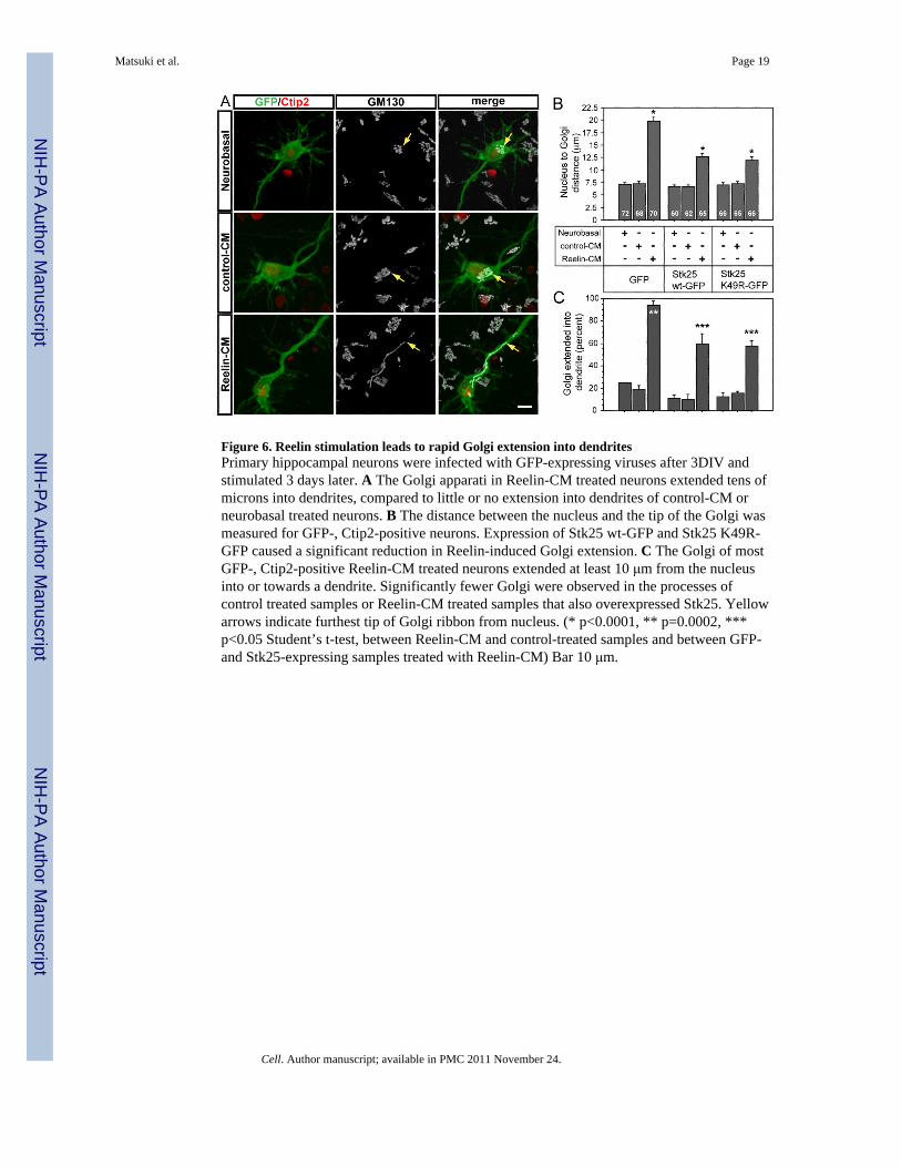
Figure 6. Reelin stimulation leads to rapid Golgi extension into dendritesPrimary hippocampal neurons were infected with GFP-expressing viruses after 3DIV andstimulated 3 days later. A The Golgi apparati in Reelin-CM treated neurons extended tens ofmicrons into dendrites, compared to little or no extension into dendrites of control-CM orneurobasal treated neurons. B The distance between the nucleus and the tip of the Golgi wasmeasured for GFP-, Ctip2-positive neurons. Expression of Stk25 wt-GFP and Stk25 K49R-GFP caused a significant reduction in Reelin-induced Golgi extension. C The Golgi of mostGFP-, Ctip2-positive Reelin-CM treated neurons extended at least 10 μm from the nucleusinto or towards a dendrite. Significantly fewer Golgi were observed in the processes ofcontrol treated samples or Reelin-CM treated samples that also overexpressed Stk25. Yellowarrows indicate furthest tip of Golgi ribbon from nucleus. (* p<0.0001, ** p=0.0002, ***p<0.05 Student’s t-test, between Reelin-CM and control-treated samples and between GFP-and Stk25-expressing samples treated with Reelin-CM) Bar 10 μm.
Matsuki et al. Page 19
Cell. Author manuscript; available in PMC 2011 November 24.
NIH
-PA Author Manuscript
NIH
-PA Author Manuscript
NIH
-PA Author Manuscript

Figure 7. Model of Stk25 as a scaffolding protein acting competitively with Reelin-Dab1signalingLKB1 is known to act in complex with STRAD to regulate cellular polarity (Alessi et al.,2006). Reelin, the receptors ApoER2 and VLDLR, and Dab1 also form a signaling complex(Hiesberger et al., 1999; Trommsdorff et al., 1998). STK25 coimmunoprecipitates withSTRAD and GM130 (Fig. 2S). Overexpression of LKB1, and STRAD are known to inducethe formation of multiple axons (Barnes et al., 2007; Shelly et al., 2007). Independent of itskinase activity, STK25 does so also and induces Golgi condensation (Fig. 1F, 4A).Knocking down LKB1, Stk25 or GM130 causes Golgi fragmentation/dispersion and lostaxon production; the opposite to Golgi condensation and multiple axon formation (Fig. 1, 3,4, Barnes et al., 2007; Shelly et al., 2007). The overexpression phenotypes are suppressed byReelin stimulation. Dab1−/− neurons (Reelin signaling deficient) have multiple axons andshorter dendrites (Fig. 1F, Niu et al., 2004). Reelin stimulation induces Golgi deploymentand dendrite growth, phenotypes suppressed by Stk25 expression/overexpression (Fig. 2, 6).
Matsuki et al. Page 20
Cell. Author manuscript; available in PMC 2011 November 24.
NIH
-PA Author Manuscript
NIH
-PA Author Manuscript
NIH
-PA Author Manuscript
Related Documents