Recombinant Antigens from Phlebotomus perniciosus Saliva as Markers of Canine Exposure to Visceral Leishmaniases Vector Jan Drahota 1. * , Ines Martin-Martin 2. , Petra Sumova 1 , Iva Rohousova 1 , Maribel Jimenez 2 , Ricardo Molina 2 , Petr Volf 1 1 Department of Parasitology, Faculty of Science, Charles University in Prague, Prague, Czech Republic, 2 Unidad de Entomologı ´a Me ´ dica, Servicio de Parasitologı ´a, Centro Nacional de Microbiologı ´a, Instituto de Salud Carlos III, Majadahonda, Madrid, Spain Abstract Background: Phlebotomus perniciosus is the main vector in the western Mediterranean area of the protozoan parasite Leishmania infantum, the causative agent of canine and human visceral leishmaniases. Infected dogs serve as a reservoir of the disease, and therefore measuring the exposure of dogs to sand fly bites is important for estimating the risk of L. infantum transmission. In bitten hosts, sand fly saliva elicits a specific antibody response that reflects the intensity of sand fly exposure. As screening of specific anti-saliva antibodies is limited by the availability of salivary gland homogenates, utilization of recombinant salivary proteins is a promising alternative. In this manuscript we show for the first time the use of recombinant salivary proteins as a functional tool for detecting P. perniciosus bites in dogs. Methodology/Principal Findings: The reactivity of six bacterially-expressed recombinant salivary proteins of P. perniciosus, yellow-related protein rSP03B, apyrases rSP01B and rSP01, antigen 5-related rSP07, ParSP25-like protein rSP08 and D7- related protein rSP04, were tested with sera of mice and dogs experimentally bitten by this sand fly using immunoblots and ELISA. In the immunoblots, both mice and canine sera gave positive reactions with yellow-related protein, both apyrases and ParSP25-like protein. A similar reaction for recombinant salivary proteins was observed by ELISA, with the reactivity of yellow-related protein and apyrases significantly correlated with the antibody response of mice and dogs against the whole salivary gland homogenate. Conclusions/Significance: Three recombinant salivary antigens of P. perniciosus, yellow-related protein rSP03B and the apyrases rSP01B and rSP01, were identified as the best candidates for evaluating the exposure of mice and dogs to P. perniciosus bites. Utilization of these proteins, or their combination, would be beneficial for screening canine sera in endemic areas of visceral leishmaniases for vector exposure and for estimating the risk of L. infantum transmission in dogs. Citation: Drahota J, Martin-Martin I, Sumova P, Rohousova I, Jimenez M, et al. (2014) Recombinant Antigens from Phlebotomus perniciosus Saliva as Markers of Canine Exposure to Visceral Leishmaniases Vector. PLoS Negl Trop Dis 8(1): e2597. doi:10.1371/journal.pntd.0002597 Editor: Shaden Kamhawi, National Institutes of Health, United States of America Received June 20, 2013; Accepted November 4, 2013; Published January 2, 2014 Copyright: ß 2014 Drahota et al. This is an open-access article distributed under the terms of the Creative Commons Attribution License, which permits unrestricted use, distribution, and reproduction in any medium, provided the original author and source are credited. Funding: This research was supported by Czech Science Foundation (GACR 13-05292S, http://www.gacr.cz/) and by EU grant 2011-261504 EDENEXT (http:// www.edenext.eu/) and the paper is catalogued by the EDENext Steering Committee as EDENextXXX. The funders had no role in study design, data collection and analysis, decision to publish, or preparation of the manuscript. Competing Interests: The authors have declared that no competing interests exist. * E-mail: [email protected] . These authors contributed equally to this work. Introduction Canine leishmaniasis (CanL) is a systemic disease with variable clinical symptoms. Its causative agent, the protozoan parasite Leishmania infantum, is transmitted by phlebotomine sand flies (Diptera: Phlebotominae). CanL occurs frequently around the Mediterranean Basin and in many countries in Latin America, where the prevalence of infection often exceeds 25%. Dogs with inapparent infections often play a role in the circulation of the parasite, as they are able to infect sand flies (reviewed in [1]). New cases of autochthonous leishmaniasis caused by L. infantum have been occurring in various countries, suggesting an expansion of CanL towards new biotopes at higher latitudes and higher altitudes (reviewed in [2,3,4]). Importantly, CanL is not just a veterinary problem; infected dogs serve as a reservoir host of human visceral leishmaniasis and there is a correlation between the prevalence of leishmaniasis in the canine population and the human disease in many countries [1]. Two sand fly genera are involved in L. infantum transmission, Lutzomyia in the New World and Phlebotomus in the Old World. Seven species of the genus Phlebotomus, subgenus Larroussius, are proven or probable vectors of CanL in different places around the Mediterranean Basin [5]. Of these, Phlebotomus perniciosus has the widest distribution, with ranges in both the southern and northern parts of the Mediterranean, from Morocco and Portugal in the west to Italy in the east and Germany in the north [5]. Measuring the exposure of dogs to sand fly bites is important for estimating the risk of L. infantum transmission. Recently, it has been PLOS Neglected Tropical Diseases | www.plosntds.org 1 January 2014 | Volume 8 | Issue 1 | e2597

Welcome message from author
This document is posted to help you gain knowledge. Please leave a comment to let me know what you think about it! Share it to your friends and learn new things together.
Transcript
Recombinant Antigens from Phlebotomus perniciosusSaliva as Markers of Canine Exposure to VisceralLeishmaniases VectorJan Drahota1.*, Ines Martin-Martin2., Petra Sumova1, Iva Rohousova1, Maribel Jimenez2,
Ricardo Molina2, Petr Volf1
1 Department of Parasitology, Faculty of Science, Charles University in Prague, Prague, Czech Republic, 2 Unidad de Entomologıa Medica, Servicio de Parasitologıa, Centro
Nacional de Microbiologıa, Instituto de Salud Carlos III, Majadahonda, Madrid, Spain
Abstract
Background: Phlebotomus perniciosus is the main vector in the western Mediterranean area of the protozoan parasiteLeishmania infantum, the causative agent of canine and human visceral leishmaniases. Infected dogs serve as a reservoir ofthe disease, and therefore measuring the exposure of dogs to sand fly bites is important for estimating the risk of L.infantum transmission. In bitten hosts, sand fly saliva elicits a specific antibody response that reflects the intensity of sand flyexposure. As screening of specific anti-saliva antibodies is limited by the availability of salivary gland homogenates,utilization of recombinant salivary proteins is a promising alternative. In this manuscript we show for the first time the use ofrecombinant salivary proteins as a functional tool for detecting P. perniciosus bites in dogs.
Methodology/Principal Findings: The reactivity of six bacterially-expressed recombinant salivary proteins of P. perniciosus,yellow-related protein rSP03B, apyrases rSP01B and rSP01, antigen 5-related rSP07, ParSP25-like protein rSP08 and D7-related protein rSP04, were tested with sera of mice and dogs experimentally bitten by this sand fly using immunoblots andELISA. In the immunoblots, both mice and canine sera gave positive reactions with yellow-related protein, both apyrasesand ParSP25-like protein. A similar reaction for recombinant salivary proteins was observed by ELISA, with the reactivity ofyellow-related protein and apyrases significantly correlated with the antibody response of mice and dogs against the wholesalivary gland homogenate.
Conclusions/Significance: Three recombinant salivary antigens of P. perniciosus, yellow-related protein rSP03B and theapyrases rSP01B and rSP01, were identified as the best candidates for evaluating the exposure of mice and dogs to P.perniciosus bites. Utilization of these proteins, or their combination, would be beneficial for screening canine sera inendemic areas of visceral leishmaniases for vector exposure and for estimating the risk of L. infantum transmission in dogs.
Citation: Drahota J, Martin-Martin I, Sumova P, Rohousova I, Jimenez M, et al. (2014) Recombinant Antigens from Phlebotomus perniciosus Saliva as Markers ofCanine Exposure to Visceral Leishmaniases Vector. PLoS Negl Trop Dis 8(1): e2597. doi:10.1371/journal.pntd.0002597
Editor: Shaden Kamhawi, National Institutes of Health, United States of America
Received June 20, 2013; Accepted November 4, 2013; Published January 2, 2014
Copyright: � 2014 Drahota et al. This is an open-access article distributed under the terms of the Creative Commons Attribution License, which permitsunrestricted use, distribution, and reproduction in any medium, provided the original author and source are credited.
Funding: This research was supported by Czech Science Foundation (GACR 13-05292S, http://www.gacr.cz/) and by EU grant 2011-261504 EDENEXT (http://www.edenext.eu/) and the paper is catalogued by the EDENext Steering Committee as EDENextXXX. The funders had no role in study design, data collection andanalysis, decision to publish, or preparation of the manuscript.
Competing Interests: The authors have declared that no competing interests exist.
* E-mail: [email protected]
. These authors contributed equally to this work.
Introduction
Canine leishmaniasis (CanL) is a systemic disease with variable
clinical symptoms. Its causative agent, the protozoan parasite
Leishmania infantum, is transmitted by phlebotomine sand flies
(Diptera: Phlebotominae). CanL occurs frequently around the
Mediterranean Basin and in many countries in Latin America,
where the prevalence of infection often exceeds 25%. Dogs with
inapparent infections often play a role in the circulation of the
parasite, as they are able to infect sand flies (reviewed in [1]). New
cases of autochthonous leishmaniasis caused by L. infantum have
been occurring in various countries, suggesting an expansion of
CanL towards new biotopes at higher latitudes and higher
altitudes (reviewed in [2,3,4]). Importantly, CanL is not just a
veterinary problem; infected dogs serve as a reservoir host of
human visceral leishmaniasis and there is a correlation between
the prevalence of leishmaniasis in the canine population and the
human disease in many countries [1].
Two sand fly genera are involved in L. infantum transmission,
Lutzomyia in the New World and Phlebotomus in the Old World.
Seven species of the genus Phlebotomus, subgenus Larroussius, are
proven or probable vectors of CanL in different places around the
Mediterranean Basin [5]. Of these, Phlebotomus perniciosus has the
widest distribution, with ranges in both the southern and northern
parts of the Mediterranean, from Morocco and Portugal in the
west to Italy in the east and Germany in the north [5].
Measuring the exposure of dogs to sand fly bites is important for
estimating the risk of L. infantum transmission. Recently, it has been
PLOS Neglected Tropical Diseases | www.plosntds.org 1 January 2014 | Volume 8 | Issue 1 | e2597
demonstrated that experimental exposure of dogs to L. longipalpis
or P. perniciosus bites elicits the production of specific anti-saliva
IgG (measured by ELISA with whole SGH ) that positively
correlates with the number of sand fly bites [6,7]. The elicitation of
IgG antibody levels persists for at least five weeks after last
exposure of dogs to P. perniciosus [7] or even nineteen weeks after
dogs are exposed to L. longipalpis [6]. Therefore, monitoring canine
IgG levels specific for sand fly saliva could indicate their exposure
to sand fly bites. Such a monitoring technique would be useful for
evaluating the need for, and effectiveness of, anti-vector campaigns
[6,8]. However, obtaining sufficient native antigens through sand
fly dissections for the upscaled production of such antigens
necessary for ELISA is not feasible; thus there is a need to replace
native antigens in the ELISAs by recombinant antigens. To date,
recombinant salivary proteins from Phlebotomus papatasi have been
used to detect the antibody response in mice experimentally
exposed to this sand fly species [9], and L. longipalpis recombinant
antigens have been tested for reactivity with the sera of naturally
bitten humans, dogs and foxes [10,11].
Recent studies [7,12] have shown that the sera of dogs bitten by
P. perniciosus recognize up to thirteen antigens in the salivary gland
homogenate (SGH) of this species. The most intense reaction has
repeatedly been observed against the 43 kDa yellow-related
protein PpeSP03B (referred to further in the text as yellow protein
rSP03B), the 35.3 kDa PpeSP01B and 35.5 kDa PpeSP01 kDa
apyrases (apyrase rSP01B and rSP01, respectively), the 30 kDa
antigen 5-related protein PpeSP07 (antigen 5 rSP07), the 29 kDa
ParSP25-like protein PpeSP08 (ParSP25 protein rSP08), and the
24.5 kDa D7-related salivary protein PpeSP04 (D7 protein rSP04).
The recombinant forms of these six proteins were therefore chosen
in the present work as the most promising candidates for markers
of P. perniciosus exposure.
Materials and Methods
1. Ethics statementSKH1-hr mice were maintained and handled in the animal
facility of Charles University in Prague in accordance with
institutional guidelines and Czech legislation (Act No. 246/1992
coll. on Protection of Animals against Cruelty in present statutes at
large), which complies with all relevant European Union and
international guidelines for experimental animals. The experi-
ments were approved by the Committee on the Ethics of Animal
Experiments of the Charles University in Prague (Permit Number:
24773/2008-10001) and were performed under the Certificate of
Competency (Registration Number: CZU 934/05; CZU 307/09)
in accordance with the Examination Order approved by Central
Commission for Animal Welfare of the Czech Republic.
2. Sand flies and salivary gland homogenateTwo Phlebotomus (Larroussius) perniciosus sand fly colonies origi-
nating from Spain – from Murcia and from Madrid – were used,
and maintained under standard conditions described in [13,14].
Phlebotomus perniciosus salivary glands for immunoblots and
ELISA assays were dissected from 3 to 5-day-old females, and
pools of 20 salivary glands in 20 ml of Tris-NaCl buffer (20 mM
Tris, 150 mM NaCl, pH 7.6) were stored at 280uC. Salivary
gland homogenate (SGH) was obtained by disruption of the glands
with repeated freezing and thawing cycles. Salivary glands used for
isolation of mRNA were dissected from 1-day-old female sand
flies, and were stored in groups of 20 salivary glands in 20 ml of
RNAlater (Qiagen) and kept at 280uC until use.
3. Mice and canine seraHyperimmune mice sera were obtained by repeated exposure of
three SKH1hr mice to bites of uninfected P. perniciosus females;
three other mice of the same strain served as non-exposed controls.
Canine sera from dogs (beagles) experimentally exposed to P.
perniciosus bites were selected from those previously used for studies
on SGH [7]. Sera were chosen to cover a wide range of anti-saliva
antibody levels: six originated from highly exposed dogs (bitten by
150–190 sand fly females once a week), six from dogs with lower
exposure (20–70 females once a week) and six from unexposed
dogs (before exposure). The sera of exposed dogs were collected
seven days after the fourth exposure.
4. Recombinant salivary proteinsSix bacterially-expressed recombinant antigens expressed in
nine forms were studied: yellow protein rSP03B (KF257369), two
apyrases - rSP01B (KF257364, KF257366, KF178455) and rSP01
(KF257365, KF257367), antigen 5 protein rSP07 (KF257368),
ParSP25 protein rSP08 (KF178457) and D7 protein rSP04
(KF178456). Coding sequences of SP01 and SP01B (referred to
further in the text as rSP01B/1 in pET42b and rSP01/1 in
pET42b, respectively), SP07 and SP03B were obtained from
salivary glands of P. perniciosus (Murcia colony). After dissection
into RNAlater, mRNA was isolated by a Roche High Pure RNA
Tissue Kit and transcripted to cDNA by a Roche High Fidelity
cDNA Synthesis Kit using polyA primers. The second cDNA
strands of SP01B and SP01 were amplified by PCR using specific
primers that were synthesized according to the sequences of the
mature protein (without signal peptide). These sequences have
been published with the following accession numbers [15]:
rSP01B/1 - DQ192491 and SP01/1 - DQ192490. Afterwards,
we followed the procedure that was described in [9] – briefly,
genes were expressed in pET42b (Novagen) with a His tag
containing 8 histidines – the E.coli BL21 (DE3) expression system.
In parallel, single-stranded cDNA transcripted from salivary
glands as described above was sent to Apronex s.r.o. (Prague) for
preparation of rSP01, rSP01B, rSP03B and rSP07 proteins in the
recombinant form according to sequences published in the cDNA
library [15] - rSP03B (DQ150622), rSP01B/2 (DQ192491),
rSP01/2 (DQ192490), rSP07 (DQ153101). All four proteins were
expressed in the E.coli BL21 (DE3) expression system; apyrases
rSP01B/2 and rSP01/2 were expressed in the pET42b vector
Author Summary
The protozoan parasite Leishmania infantum is a causativeagent of zoonotic visceral leishmaniasis, an important andpotentially fatal human disease. The main reservoir hostsof this Leishmania species are dogs, and the only provenvectors are phlebotominae sand flies, Phlebotomus perni-ciosus being considered the major vector in the westernMediterranean area. During feeding on the host, sand fliesspit saliva into the host skin; hosts develop a specificantibody response directed against sand fly salivaryproteins and levels of these antibodies reflect the intensityof sand fly exposure. As the availability of salivary glandhomogenate is limited, recombinant salivary proteins havebeen suggested as antigens suitable for measuring specificantibody levels. In the present work, we expressed six ofthe most-antigenic salivary proteins, and studied the miceand canine humoral immune responses to these recom-binant proteins. We demonstrated that three proteins, ayellow-related protein and two apyrases, are suitableantigens for measuring anti-P. perniciosus antibody levelsand estimating the host exposure to this sand fly species.
Phlebotomus perniciosus Recombinant Antigens
PLOS Neglected Tropical Diseases | www.plosntds.org 2 January 2014 | Volume 8 | Issue 1 | e2597
(Novagen) with 2 His tags - the first containing 6 histidines and the
second 8 histidines, and a 1.4 kDa adaptor, while yellow protein
rSP03B and antigen 5 protein rSP07 were expressed in the
pET28b vector (Novagen) with 1 His tag containing 6 histidines.
All proteins were isolated under denaturing conditions with 8M
urea.
In addition, salivary coding sequences of SP01B, SP04 and
SP08 were obtained from a cDNA library constructed from the
salivary glands of P. perniciosus from Madrid – GenBank accession
numbers are: SP01B - HE974345.1, SP04 - HE980444.1, SP08 -
HE974347.1; in contrast to the other proteins tested, they contain
signal sequences (thus they seem to be about 3 kDa heavier on
immunoblots). Recombinant proteins rSP01B and rSP04 were
expressed in the pQE31 vector (Qiagen) with a His tag containing
6 histidines in E. coli M15 cells, and purified under denaturing
conditions with 8M urea. Protein rSP08 was cloned into the
pGEX4T3 vector (Amersham Biotech), expressed in E. coli Arctic
Express cells (Agilent), and purified by polyacrylamide gel
extraction in PBS. As rSP08 was expressed as a fusion protein
with gluthathione S-transferase (GST), the latter was also obtained
and used as a control in immunoblots and ELISA. In all these
cases, protein refolding was performed using dialysis against PBS
using SnakeSkin Dialysis Tubing (10 kDa MWCO, Thermo
Scientific Goettiengen, Germany).
The concentration of all proteins was quantified by the Lowry
method (Bio-Rad) following the manufacturer’s protocol.
5. ImmunoblotsThe immunogenicity of the recombinant proteins was tested by
the immunoblot technique. Recombinant salivary proteins were
separated by SDS PAGE on 12% polyacrylamide gels under non-
reducing conditions using a Mini-protean apparatus (Bio-Rad).
Protein concentration was optimized using preliminary experi-
ments; the list of proteins and their quantity per well of the 5-well
comb are given in Table 1. Proteins were either stained with
Coomassie Blue (Invitrogen) or transferred from the gel to
nitrocellulose membranes using an iBLOT dry system (Invitrogen).
Membranes were cut into strips (area corresponding to one well
was cut into 5 strips), blocked for 1–2 hours with 5% milk in Tris
buffer with 0.05% Tween (Tris-Tw) and then incubated for 1 hour
with either mice or canine sera diluted in Tris-Tw. Mice sera were
diluted 1:200 for denatured proteins rSP01B and rSP01 in
pET42b and 1:100 in the case of other proteins; canine sera were
used at a dilution of 1:50. After washing in Tris-Tw, the strips were
incubated with peroxidase conjugated anti-mouse IgG (1:1000,
AbD Serotec) or anti-dog IgG antibodies (1:1000, Bethyl) and
reacting protein bands were visualized using the substrate solution
with diaminobenzidine.
6. ELISAThe ELISA test described elsewhere [7,16] was modified as
follows. Covalink plates (Nunc) were coated with 100 ml of either
salivary gland homogenate (40 ng of protein per well, corresponds
to 1/5 of a salivary gland) or recombinant salivary protein, both in
0.1 M carbonate-bicarbonate buffer (pH 9.5), overnight at 4uC.
The optimal concentrations of the recombinant proteins are given
in Table 1. After washing in PBS with 0.05% Tween (PBS-Tw),
plates were blocked with 6% milk in PBS-Tw for 1–2 h at 37uC.
Mice and canine sera were diluted in 2% milk PBS-Tw.
Dilution of mice sera 1:1600 was optimal for rSP03B, 1:400 for
apyrases in pET42b with 1 His tag, and 1:200 for the other
proteins tested. All canine sera were used at a dilution of 1:50. Sera
were incubated for 90 min at 37uC. Following washing cycles,
plates were incubated with peroxidase-conjugated anti-mouse IgG
(AbD Serotec) or anti-dog IgG (Bethyl) and the color reaction was
developed in the substrate solution with orthophenylendiamine.
Absorbance values (OD) were recorded at 492 nm using a
Multiscan RC ELISA reader (Labsystems).
7. Statistical analysisThe non-parametric Spearman test was used to assess
correlations between total anti-SGH and anti-recombinants
antibody levels in GraphPad Prism version 6 (GraphPad Software,
Inc., San Diego, CA). Statistical significance was considered when
the p-value was ,0.05.
8. Accession numbersThe sequences of apyrases rSP01B/1 (in pET42b), rSP01B/2
(in pET42b), rSP01/1 (in pET42b) and rSP01/2 (in pET42b),
yellow protein rSP03B (in pET28b) and antigen 5 protein rSP07
(in pET28b) were based on sequences from a published cDNA
library of P. perniciosus [15]: rSP01B - DQ192491, rSP01 -
DQ192490, rSP03B - DQ150622) and rSP07 - DQ153101. The
other sequences were published directly in GenBank: rSP01B (in
pQE31) - HE974345.1, rSP04 (in pQE31) - HE980444.1, rSP08
(pGEX4T3 ) - HE974347.1. The expressed sequences were
published with the following GenBank accession numbers: yellow
protein rSP03B (KF257369), two apyrases - rSP01B (KF257364,
KF257366, KF178455) and rSP01 (KF257365, KF257367),
antigen 5 protein rSP07 (KF257368), ParSP25 protein rSP08
(KF178457) and D7 protein rSP04 (KF178456).
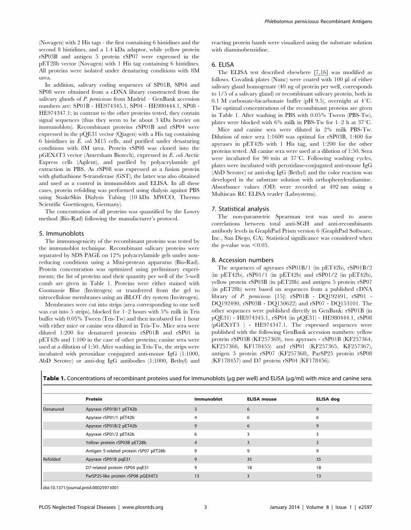
Table 1. Concentrations of recombinant proteins used for immunoblots (mg per well) and ELISA (mg/ml) with mice and canine sera.
Protein Immunoblot ELISA mouse ELISA dog
Denatured Apyrase rSP01B/1 pET42b 3 6 9
Apyrase rSP01/1 pET42b 4 6 6
Apyrase rSP01B/2 pET42b 9 6 9
Apyrase rSP01/2 pET42b 6 3 3
Yellow protein rSP03B pET28b 4 3 3
Antigen 5-related protein rSP07 pET28b 9 9 9
Refolded Apyrase rSP01B pqE31 9 35 35
D7-related protein rSP04 pqE31 9 18 18
ParSP25-like protein rSP08 pGEX4T3 13 3 13
doi:10.1371/journal.pntd.0002597.t001
Phlebotomus perniciosus Recombinant Antigens
PLOS Neglected Tropical Diseases | www.plosntds.org 3 January 2014 | Volume 8 | Issue 1 | e2597
Phlebotomus perniciosus Recombinant Antigens
PLOS Neglected Tropical Diseases | www.plosntds.org 4 January 2014 | Volume 8 | Issue 1 | e2597
Results
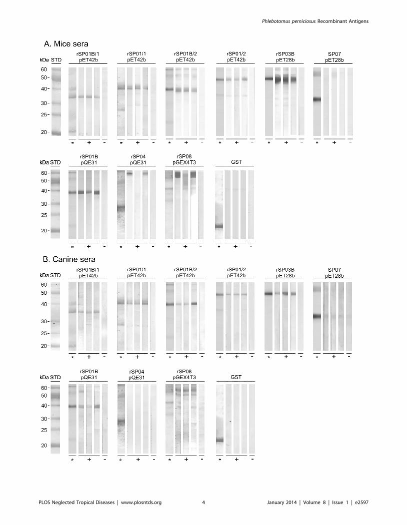
1. Immunoblots with mice and canine seraAll recombinant proteins except antigen 5 protein rSP07 and
D7 protein rSP04 were recognized by the sera of all three
repeatedly exposed mice; control sera and the GST tag were
negative (Fig. 1A). A similar reactivity of recombinant antigens was
found with the sera of the three dogs repeatedly exposed to P.
perniciosus (Fig. 1B): all recombinant proteins except antigen 5
protein rSP07 and D7 protein rSP04 were recognized by the sera
of repeatedly exposed dogs. In comparison with mice sera, the
reaction of canine sera was less intense for some proteins (yellow
protein rSP03B and the apyrases rSP01B/2 and rSP01/2 in
pET42b) and fewer nonspecific bands appeared in the immuno-
blots. Control canine sera were negative (Fig. 1B).
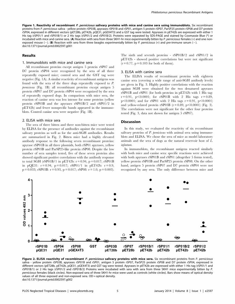
2. ELISA with mice seraThe sera of three bitten and three non-bitten mice were tested
by ELISA for the presence of antibodies against the recombinant
salivary proteins as well as for the anti-SGH antibodies. Results
are summarized in Fig. 2. Bitten mice had a highly elevated
antibody response to the following seven recombinant proteins:
apyrase rSP01B in all three plasmids, both rSP01 apyrases, yellow
protein rSP03B and ParSP25-like protein rSP08. Despite the low
number of sera samples tested, five of these seven proteins also
showed significant positive correlations with the antibody response
to total SGH (rSP01B/1 in pET42b: r = 0.94, p = 0.017; rSP01B
in pQE31: r = 0.94, p = 0.017; rSP01/1 in pET42b: r = 0.9,
p = 0.033; rSP03B: r = 0.93, p = 0.017; rSP08: r = 1.0, p = 0.003).
The sixth and seventh proteins - rSP01B/2 and rSP01/2 in
pET42b - showed positive correlations but were not significant
(r = 0.77, p = 0.103 for both of them).
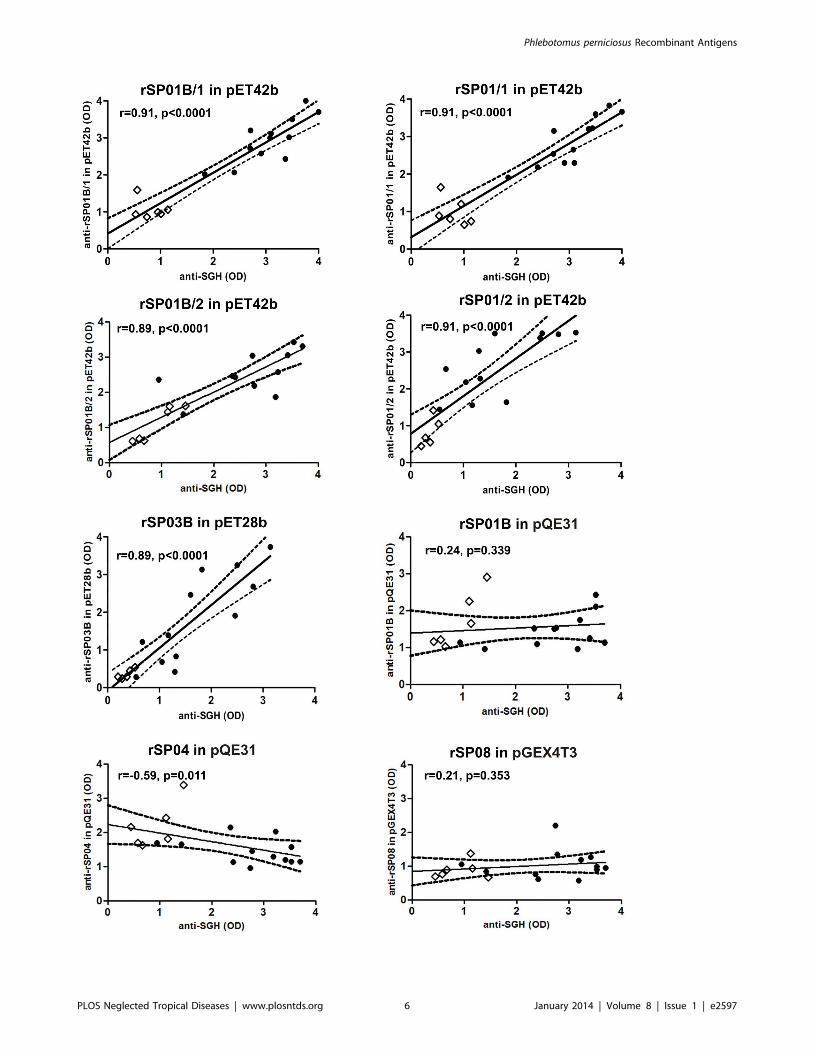
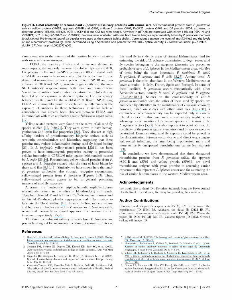
3. ELISA with canine seraThe ELISA results of recombinant proteins with eighteen
canine sera (covering a wide range of anti-SGH antibody levels)
are given in Fig. 3. Highly positive correlations with the reaction
against SGH were obtained for the two denatured apyrases
rSP01B and rSP01 (for both proteins in pET42b with 1 His tag:
r = 0.91, p,0.0001; for rSP01B with 2 His tags r = 0.89,
p,0.0001; and for rSP01 with 2 His tags r = 0.91, p,0.0001)
and yellow-related protein rSP03B (r = 0.89, p,0.0001) (Fig. 3).
The correlations were not significant for the other four proteins
tested (Fig. 3, data not shown for antigen 5 rSP07).
Discussion
In this study, we evaluated the reactivity of six recombinant
salivary proteins of P. perniciosus with animal sera using immuno-
blots and ELISA. We chose the sera of mice as model laboratory
animals and the sera of dogs as the natural reservoir host of L.
infantum.
In immunoblots, the recombinant antigens reacted similarly
with both mice and canine sera: specific reactions were achieved
with both apyrases rSP01B and rSP01 (altogether 5 forms tested),
yellow protein rSP03B and ParSP25 protein rSP08. On the other
hand, antigen 5 protein rSP07 and D7 protein rSP04 were not
recognized by any sera. The only difference between mice and
Figure 1. Reactivity of recombinant P. perniciosus salivary proteins with mice and canine sera using immunoblots. Six recombinantproteins from P. perniciosus saliva - yellow protein rSP03B, apyrases rSP01B and rSP01, antigen 5 protein rSP07, ParSP25 protein rSP08 and D7 proteinrSP04, expressed in different vectors (pET28b, pET42b, pQE31, pGEX4T3) and a GST tag were tested. Apyrases in pET42b are expressed with either 1His tag (rSP01/1 and rSP01B/1) or 2 His tags (rSP01/2 and rSP01B/2). Proteins were separated by SDS-PAGE and stained by Coomassie Blue (*) orincubated with mice and canine sera. (A) Reaction with sera from three SKH1-hr mice experimentally bitten by P. perniciosus females (+) and one non-exposed mouse (2). (B) Reaction with sera from three beagles experimentally bitten by P. perniciosus (+) and pre-immune serum (2).doi:10.1371/journal.pntd.0002597.g001
Figure 2. ELISA reactivity of recombinant P. perniciosus salivary proteins with mice sera. Six recombinant proteins from P. perniciosussaliva - yellow protein rSP03B, apyrases rSP01B and rSP01, antigen 5 protein rSP07, ParSP25 protein rSP08 and D7 protein rSP04, expressed indifferent vectors (pET28b, pET42b, pQE31, pGEX4T3) and GST tag were tested. Apyrases in pET42b are expressed with either 1 His tag (rSP01/1 andrSP01B/1) or 2 His tags (rSP01/2 and rSP01B/2) Proteins were incubated with sera with sera from three SKH1 mice experimentally bitten by P.perniciosus females (black circles). Non-exposed sera of three SKH1-hr mice were used as controls (white circles). Bars show means of optical densityvalues of all three exposed and non-exposed sera. OD = optical density.doi:10.1371/journal.pntd.0002597.g002
Phlebotomus perniciosus Recombinant Antigens
PLOS Neglected Tropical Diseases | www.plosntds.org 5 January 2014 | Volume 8 | Issue 1 | e2597
Phlebotomus perniciosus Recombinant Antigens
PLOS Neglected Tropical Diseases | www.plosntds.org 6 January 2014 | Volume 8 | Issue 1 | e2597
canine sera was in the intensity of the positive bands – reactions
with mice sera were stronger.
In ELISA, the reactivity of mice and canine sera differed in
some aspects; the antibody response to refolded apyrase rSP01B,
D7 protein rSP04 and ParSP25 protein rSP08 correlated with
anti-SGH response only in mice sera. On the other hand, three
denatured recombinant proteins, yellow protein rSP03B and two
apyrases, rSP01B and rSP01, correlated significantly with the anti-
SGH antibody response using both mice and canine sera.
Variations in antigen conformation (denatured vs. refolded) may
have led to the exposure of different epitopes. The discrepancy
between results found for the refolded rSP01B and rSP08 using
ELISA vs. immunoblot could be explained by differences in the
exposure of antigens in these techniques; a similar lack of
concordance has already been observed between ELISA and
immunoblots with mice antibodies against Phlebotomus sergenti saliva
[17].
Yellow-related proteins were found in the saliva of all sand fly
species studied [18,19,20,21]. They were shown to have hemag-
glutination and lectin-like properties [22]. They also act as high
affinity binders of proinflammatory biogenic amines such as
serotonin, catecholamines and histamine, suggesting that these
proteins may reduce inflammation during sand fly blood-feeding
[23]. In L. longipalpis, yellow-related protein LJM11 has been
proven to have immunogenic properties leading to protective
cellular immunity in C57BL/6 mice against leishmaniasis caused
by L. major [23,24]. Recombinant yellow-related proteins from P.
papatasi and L. longipalpis reacted with the sera of hosts bitten by
these sand flies [9,10,11]. Similarly, we have shown here that anti-
P. perniciosus antibodies also strongly recognize recombinant
yellow-related protein from P. perniciosus (Figures 1–3). Thus,
yellow-related proteins appear to be, in general, promising
markers of sand fly exposure.
Apyrases are nucleoside triphosphate-diphosphohydrolases
ubiquitously present in the saliva of blood-sucking arthropods.
They hydrolyze ADP and ATP in a Ca2+-dependent manner and
inhibit ADP-induced platelet aggregation and inflammation to
facilitate the blood feeding [18]. In sand fly host models, mouse
and hamster antibodies elicited by P. duboscqi or P. perniciosus saliva
recognized bacterially expressed apyrases of P. duboscqi and P.
perniciosus, respectively [25,26].
The three recombinant salivary proteins from P. perniciosus are
primarily designed for measuring the canine exposure to bites of
this sand fly in endemic areas of visceral leishmaniases, and for
estimating the risk of L. infantum transmission to dogs. Seven sand
fly species belonging to the subgenus Larroussius are proven or
probable vectors of L. infantum in the Mediterranean area, with five
of them being the most important: P. perniciosus, P. ariasi,
P. perfiliewi, P. neglectus and P. tobbi [5,27]. Among them, P.
perniciosus is the most abundant in the Western Mediterranean at
lower altitudes - in Italy, France, Spain and Portugal. In some of
these localities, P. perniciosus occurs sympatrically with other
Larroussius vectors, namely P. ariasi, P. perfiliewi and P. neglectus
[27,28,29,30,31]. Studies on the cross-reactivity of anti-P.
perniciosus antibodies with the saliva of these sand fly species are
hampered by difficulties in the maintenance of Larroussius colonies;
however, based on studies with other sand flies [16,17,32], a
certain level of cross-reactivity can be expected only in closely-
related species. In this case, such cross-reactivity might be an
advantage as all mentioned Larroussius species are known to be
L. infantum vectors [5,27]. It is also important to point out that the
specificity of the protein against sympatric sand fly species needs to
be studied. Demonstrating sand fly exposure could be pivotal in
the discrimination between vector-borne and direct (e.g. congen-
ital, sexual) infections, the latter being hypothesized more and
more to justify unexpected autochthonous canine leishmaniasis
[33].
In conclusion, we have demonstrated that three denatured
recombinant proteins from P. perniciosus saliva, the apyrases
rSP01B and rSP01 and yellow protein rSP03B, are novel
recombinant antigens with great promise in screening canine
exposure to this important L. infantum vector and for estimating the
risk of canine leishmaniases in the western Mediterranean area.
Acknowledgments
We would like to thank Dr. Dorothee Stanneck from the Bayer Animal
Health GmbH, Leverkusen, Germany for providing the canine sera.
Author Contributions
Conceived and designed the experiments: PV MJ RM IR. Performed the
experiments: JD IMM PS. Analyzed the data: JD IMM IR PV.
Contributed reagents/materials/analysis tools: PV MJ RM. Wrote the
paper: JD IMM PV MJ RM IR. Created figures: JD IMM. Created
striking still image: JD.
References
1. Baneth G, Koutinas AF, Solano-Gallego L, Bourdeau P, Ferrer L (2008). Canine
leishmaniosis - new concepts and insights on an expanding zoonosis: part one.
Trends Parasitol 24: 324–30.
2. Schantz PM, Steurer FJ, Duprey ZH, Kurpel KP, Barr SC, et al. (2005).
Autochthonous visceral leishmaniasis in dogs in North America. J Am Vet Med
Assoc 226: 1316–22.
3. Dujardin JC, Campino L, Canavate C, Dedet JP, Gradoni L, et al. (2008).
Spread of vector-borne diseases and neglect of Leishmaniasis, Europe. Emerg
Infect Dis 14: 1013–8.
4. Carranza-Tamayo CO, Carvalho Mdo S, Bredt A, Bofil MI, Rodrigues RM,
Silva AD, et al. (2010). Autochthonous visceral leishmaniasis in Brasilia, Federal
District, Brazil. Rev Soc Bras Med Trop 43: 396–9.
5. Killick-Kendrick R (1999). The biology and control of phlebotomine sand flies.
Clin Dermatol 17: 279–89.
6. Hostomska J, Rohousova I, Volfova V, Stanneck D, Mencke N, et al. (2008).
Kinetics of canine antibody response to saliva of the sand fly Lutzomyia
longipalpis. Vector Borne Zoonotic Dis 8: 443–50.
7. Vlkova M, Rohousova I, Drahota J, Stanneck D, Kruedewagen EM, et al.
(2011). Canine antibody response to Phlebotomus perniciosus bites negatively
correlates with the risk of Leishmania infantum transmission. PLoS Negl Trop
Dis 5: e1344.
8. Gomes RB, Mendonca IL, Silva VC, Ruas J, Silva MB, et al. (2007). Antibodies
against Lutzomyia longipalpis saliva in the fox Cerdocyon thousand the sylvatic
cycle of Leishmania chagasi. Trans R Soc Trop Med Hyg 101: 127–33.
Figure 3. ELISA reactivity of recombinant P. perniciosus salivary proteins with canine sera. Six recombinant proteins from P. perniciosussaliva - yellow protein rSP03B, apyrases rSP01B and rSP01, antigen 5 protein rSP07, ParSP25 protein rSP08 and D7 protein rSP04, expressed indifferent vectors (pET28b, pET42b, pQE31, pGEX4T3) and GST tag were tested. Apyrases in pET42b are expressed with either 1 His tag (rSP01/1 andrSP01B/1) or 2 His tags (rSP01/2 and rSP01B/2). Proteins were incubated with sera from twelve beagles experimentally bitten by P. perniciosus females(black circles). Pre-immune sera of six beagles were used as the controls (white circles). Correlations between the levels of anti-SGH IgG and the levelsof anti-recombinant proteins IgG were performed using a Spearman non-parametric test. OD = optical density, r = correlation index, p = p-value.doi:10.1371/journal.pntd.0002597.g003
Phlebotomus perniciosus Recombinant Antigens
PLOS Neglected Tropical Diseases | www.plosntds.org 7 January 2014 | Volume 8 | Issue 1 | e2597
9. Vlkova M, Rohousova I, Hostomska J, Pohankova L, Zidkova L, et al. (2012).
Kinetics of antibody response in BALB/c and C57BL/6 mice bitten byPhlebotomus papatasi. PLoS Negl Trop Dis 6: e1719.
10. Souza AP, Andrade BB, Aquino D, Entringer P, Miranda JC, et al. (2010).
Using recombinant proteins from Lutzomyia longipalpis saliva to estimatehuman vector exposure in visceral Leishmaniasis endemic areas. PLoS Negl
Trop Dis 4: e649.11. Teixeira C, Gomes R, Collin N, Reynoso D, Jochim R, et al. (2010). Discovery
of markers of exposure specific to bites of Lutzomyia longipalpis, the vector of
Leishmania infantum chagasi in Latin America. PLoS Negl Trop Dis 4: e638.12. Martin-Martin I, Molina R, Jimenez M (2012). An insight into the Phlebotomus
perniciosus saliva by a proteomic approach. Acta Trop 123: 22–30.13. Volf P, Volfova V (2011). Establishment and maintenance of sand fly colonies.
J Vector Ecol 36 Suppl 1: S1–9.14. Molina R (1991). Laboratory adaptation of an autochthonous colony of
Phlebotomus perniciosus Newstead, 1911(Diptera: Psychodidae). Research and
Reviews in Parasitology 51: 87–89.15. Anderson JM, Oliveira F, Kamhawi S, Mans BJ, Reynoso D, et al. (2006).
Comparative salivary gland transcriptomics of sandfly vectors of visceralleishmaniasis. BMC Genomics 7: 52.
16. Rohousova I, Ozensoy S, Ozbel Y, Volf P (2005). Detection of species-specific
antibody response of humans and mice bitten by sand flies. Parasitology 130:493–9.
17. Drahota J, Lipoldova M, Volf P, Rohousova I (2009). Specificity of anti-salivaimmune response in mice repeatedly bitten by Phlebotomus sergenti. Parasite
Immunol 12: 766–70.18. Ribeiro JM, Mans BJ, Arca B (2010). An insight into the sialome of blood-
feeding Nematocera. Insect Biochem Mol Biol 40: 767–84
19. Oliveira F, Kamhawi S, Seitz AE, Pham VM, Guigal PM, et al. (2006). Fromtranscriptome to immunome: identification of DTH inducing proteins from a
Phlebotomus ariasi salivary gland cDNA library. Vaccine 24: 374–90.20. Hostomska J, Volfova V, Mu J, Garfield M, Rohousova I, et al. (2009). Analysis
of salivary transcripts and antigens of the sand fly Phlebotomus arabicus. BMC
Genomics 10: 282.21. Rohousova I, Subrahmanyam S, Volfova V, Mu J, Volf P, Valenzuela JG, et al.
(2012). Salivary gland transcriptomes and proteomes of Phlebotomus tobbi andPhlebotomus sergenti, vectors of leishmaniasis. PLoS Negl Trop Dis 6: e1660.
22. Volf P, Skarupova S, Man P (2002). Characterization of the lectin from femalesof Phlebotomus duboscqi sand flies. Eur J Biochem 269: 6294–301.
23. Xu X, Oliveira F, Chang BW, Collin N, Gomes R, et al. (2011). Structure and
function of a ‘‘yellow’’ protein from saliva of the sand fly Lutzomyia longipalpis
that confers protective immunity against Leishmania major infection. J Biol
Chem 286: 32383–93.
24. Gomes R, Oliveira F, Teixeira C, Meneses C, Gilmore DC, et al. (2012).
Immunity to sand fly salivary protein LJM11 modulates host response to vector-
transmitted leishmania conferring ulcer-free protection. J Invest Dermatol 132:
2735–43
25. Martin-Martin I, Molina R, Jimenez M (2013). Molecular and Immunogenic
Properties of Apyrase SP01B and D7-Related SP04 Recombinant Salivary
Proteins of Phlebotomus perniciosus from Madrid, Spain. BioMed Research
International 2013: 526069.
26. Ryoichi Hamasaki, Hirotomo Kato, Yoshimi Terayama, Hiroyuki Iwata,
Jesus G. Valenzuela (2009). Functional characterization of a salivary apyrase
from the sand fly, Phlebotomus duboscqi, a vector of Leishmania major. J Insect
Physiol 55: 1044–1049.
27. Ready PD (2010). Leishmaniasis emergence in Europe. Euro Surveill 15: 19505.
28. Maroli M, Rossi L, Baldelli R, Capelli G, Ferroglio E, et al. (2008). The
northward spread of leishmaniasis in Italy: evidence from retrospective and
ongoing studies on the canine reservoir and phlebotomine vectors. Trop Med Int
Health 13: 256–64.
29. Baron SD, Morillas-Marquez F, Morales-Yuste M, Diaz-Saez V, Irigaray C, et
al. (2011). Risk maps for the presence and absence of Phlebotomus perniciosus in
an endemic area of leishmaniasis in southern Spain: implications for the control
of the disease. Parasitology 138: 1234–44.
30. Hartemink N, Vanwambeke SO, Heesterbeek H, Rogers D, Morley D, et al.
(2011). Integrated mapping of establishment risk for emerging vector-borne
infections: a case study of canine leishmaniasis in southwest France. PLoS One 6:
e20817.
31. Branco S, Alves-Pires C, Maia C, Cortes S, Cristovao JM, et al. (2013).
Entomological and ecological studies in a new potential zoonotic leishmaniasis
focus in Torres Novas municipality, Central Region, Portugal. Acta Trop 125:
339–48.
32. Volf P, Rohousova I (2001). Species-specific antigens in salivary glands of
phlebotomine sandflies. Parasitology 122: 37–41.
33. Boggiatto PM, Gibson-Corley KN, Metz K, Gallup JM, Hostetter JM, et al.
(2011). Transplacental transmission of Leishmania infantum as a means for
continued disease incidence in North America. PLoS Negl Trop Dis 5(4):e1019.
Phlebotomus perniciosus Recombinant Antigens
PLOS Neglected Tropical Diseases | www.plosntds.org 8 January 2014 | Volume 8 | Issue 1 | e2597
Related Documents