Ward 8. Silberatdn. M.D. 19811 &en, K.D. bpne L. Saenger, l4.D. Jue G. breiakea, Ph.D. RCCl.958039.001 i ,- University of Cincinnati College of Hedicine Cincinxiati, Ohio Supported in part by the Defame Atomic Support Nancy, Cantract No. DASA 01-69-C-0131. Uuhington, D.C.; the Albertine 0. Sckoepf Research Fund of the College of Hmdicine Univermity of Cincinmti urd USW PR- . 05408 General mearch' Support Grant of the College of Wdicine University of Cincinnati.

Welcome message from author
This document is posted to help you gain knowledge. Please leave a comment to let me know what you think about it! Share it to your friends and learn new things together.
Transcript
Ward 8 . Silberatdn. M.D. 19811 &en, K.D. b p n e L. Saenger, l4.D. J u e G. breiakea, Ph.D.
RCCl.958039.001
i ,-
University of Cincinnati College of Hedicine Cincinxiati, Ohio
Supported in part by the Defame Atomic Support Nancy, Cantract No. DASA 01-69-C-0131. Uuhington, D.C.; the Albertine 0. Sckoepf Research Fund of the College of Hmdicine Univermity of Cincinmti urd USW PR- . 05408 General mearch' Support Grant of the College of Wdicine University of Cincinnati.
ABSTRACT
The resu l t of radiat ion interact ing w i t h l iv ing tiraue is the depooition of energy therein. Tbim mer= trimers numerous ch.nica1 nac t iocu within tho rn1ecul.r of 'the &asget t i . s u u . We have u u u r e d fn u n the remultr of soma of theme reactions a t dome. up t o 300 rad.: i n the kine t ics of rpec i f i c human c e l l p 0 p d A t i O M ; change. in 37 biochemical coamtituents OK nema andlor urine.
p a r t i a l l y perfected but there are numerow d i a c r e p ~ n c i u in data between d i f f e ren t l a b o r a t o r i a . Etiocholanolone c.o be wed t o evaluate mnrrov in jury before the white c e l l count f a l l s below 5000 per ku. m. W a t b i o e k d c l l doairneterr evaluated gave negative or incormistent resul ts . Elowever, sa i ivarg amylase is a p?Od6ing ind ica tor of buman radiation injury from domes as low as 100 rad6.
c h r o a i o w aberratiaum; . I te ra t ions
The u t i l i z a t i o n of ehmmos- AB a biological dosimeter is
Y
L
’L
Damage due t o ionizing radiation i n biological systems is the r e s u l t of deposition of anerglr within the t i asue of the animal irra- diated. -Y rece ive , in jury from blast and bum an well as radiat ion, and a l l th ree modalities involve the t r r m f e r of caergy t o tissue, the in- portance of finding a radii t ion-mpecific dosimeter cannot be over- Mtfpated. d g h t be r e l a t ive ly non-specific. wide va r i e ty of pathophysiologic changes induced by i r r ad ia t ing hman beings with whole o r partial body radiat ion. These have included the a l t e r a t i o n s remalting f r o m the i r rad ia t ion of nucleic-acid nucleoprotein complexes as re f lec ted in chromosome morphology; changes induced by the s ceroid metabolite etiochol.llolone on granulocyte reaemes; abnor- malities i n the l eve l of several serm and ur ine enzymes; glycoproteins. and i ron. Theac s tudies were a l l performcd i n ambulatory human subjects who c l ea r ly understood the experimental nature of t h i s study according t o the Declaration of Ilelsinki. S w e r a l of the subjec ts were tumor f ree and e s sen t i a l ly normal (follcuipg radiation-induced tumor regression) receiving prophylactic whole body radiation. The r e s t had metastat ic carcinomas which were inoperable and not amenable t o conventional chemo- therapy. Nevertheleaa, these pat ients Were a11 c l i n i c a l l y s tab le . many of then working dai ly . u w e l l as our chromosome cul ture v t h o d w i l l be found in the Appendices A and B.
Since the victim of cha explosion of a nuclear weapon
Bowever, one might expect a u r i o r i that biochemical changes Our Laboratory has looked a t a
The radiat ion technique employed i n theae pa t ien ts
L11RObKEOME ABERRATIONS
The use of chrowsome aber ra t iom a# a biological rad ia t ion dosimeter began w i t h the c l u a i c a l e x p e r l a a t . of Salts (1. 2, 3) and of Lea and Catcheside (4) on the quant i ta t ive r e l a t ion between the change i n yield of radiation-induced chrwoorome aberrations and increasing dose. these s t u d i e s were performd thirty years ago, t h i ~ we of chroworome aber ra t ions remainn a highly cont rovera id area. gene t i c i s t s who bel ieve that the k ine t ics of dose-raeponse s tudied in these ear ly experikts are inapplicable to man, because the earlier work involved plant nuclei having la rge chromosol.8 and low chromosome ntrmbers (5). while m n has 96 chrooosomer (6) of amall individual voltme.
Although
There ate today cyto-
To j u s t i f y the use of any dosimeter one a m t be ab le t o repro- duce the ca l ib ra t ion curve betVenn the dose of rad ia t ion and the response of t h e test system. Furthermore. one U t be able t o employ
.
-2-
c
Y
the dosimeter t o ascertain t o what extent the t es t systaa has been expoled t o radiation. That l a , one w u l d l i k e t o knw whether the b io logica l system t o b e examined hm received an h m g e n e o w dose of rad ia t ion dis t r ibuted uniformly o r sn inhomogeneow dose inter- ac t ing with a l l or par t of t h a t myate+ about the cal ibrat ion of hman lpmphowte response to ' r ad ia t ion and the problem of distinguishing vholm.fron p a r t i a l body rad ia t ioa w i t h this myatem do not permit u t i l i za t ion of radiation-induced chromosome aberration dose-rssponse curves i n a rigorous fashion t o date. Houever, there bave been several instances where chrorm- some ana lys is has been useful i n proving tha t high doses indicated by rad ia t ion badges were in f a c t not 'received by the individuals wearing them (7.8). Furthermore, chro.cuome aberration6 caused by rad ia t ion i n man are not affected by mbient temperature md pressure a s are physical dosimeters ( fo r uhich correctioru can wunl ly be made)
Continuibg disagreement
Study of radiat ion e f f ec t s on -1i.n c e l l s is corplicated by the face that radiat ion sen6 i t iv i ty of calla varies w i t h the s tage of t h e i r c a l l cycle. Repair time of chromorome breaks has been found t o be 90 minutes in the G1 (por t d t o t i c pre--6ynthetic phase) and S (DNA synthesis) p h u e but 60 minutes i n late S and G2 (post- synthet ic p r e - d t o t i c phase) (9). Furthemore. there is inter-species var ia t ion i n radiation-induced chraorome aberrat ion curves (10). vith the extra- polat ion f h o a n i ~ l 6 t o man mouewhat hazardow. This var ia t ion i o probably r e l a t ed t o differences i n c h r m m rider and voltme bttwemn species examined. One vould l i k e to w e primatea in this research, but i dea l ly one m a t f ind an animal which ham the same chromosome nmber and configuration an men i d order t o be confident of the extrapolation.
An important breakthrough occurred when i t was found tha t human ,
lymphocytes could be a t k l a t e d t o divide i n vitro (11) where the mitosis could be a r rea ted w i t h colchicine Lud then analyzed under tha microscope. Furthermore. 99.9% 0f'hucP.n lymphocytcll are i n the C growth phme (12) and t h i s is a period of uniform radiat ion sensitivi& .(13). Thus nmerous inves t iga to r s have employed thim s y a t a to obtain doae-reaponae curves (5, 14-24). The re l a t ion rh ip of i n vivo t o in vitro experiments w i l l be discrused b e l w .
Theae curvea were ruual ly derived from in vitro studies .
c
t . -3-
c
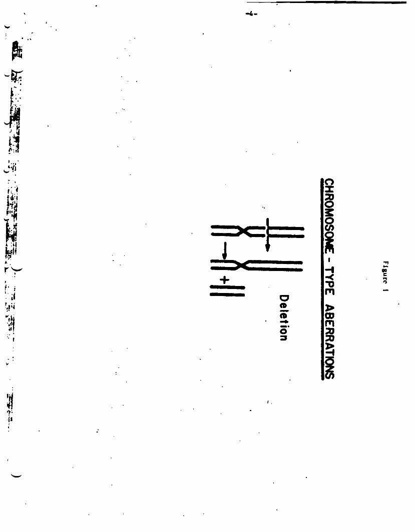
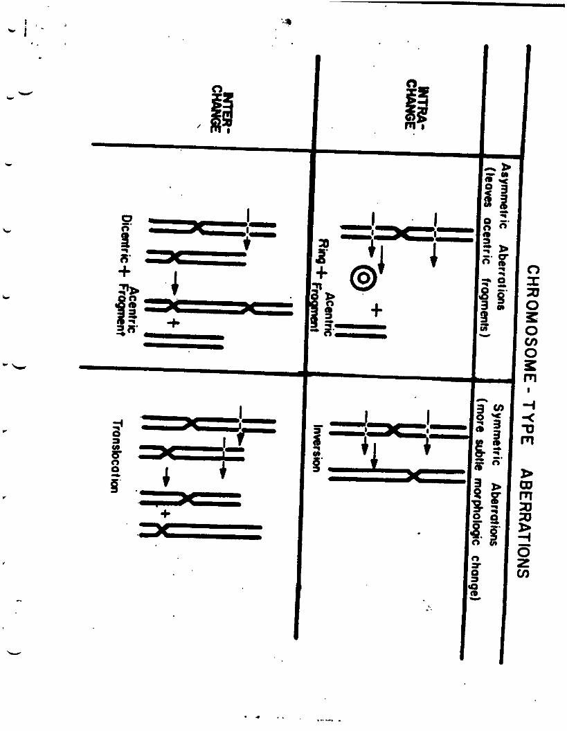
But which "rrsponse" should be studied? AB noted above. rad ia t ion u w e ~ ekrowmooe breakage i n nucleoprotein or poly- nucleotide chaixu (25) which may o r may not be followed by recombination betwean broken ends (1-3). An a l te rna t ive wch- anism ham been proposed by R e v e l l , the so-called "exchanp- first" hypothesis (26). end. do not r e jo in perfectly) are of two types. c h r m t i d and chrorosorc a b e r r a t i o ~ . whsn chromsoncs a r e in the C pre-DNA synthesis phase. a r e the types observed i n the l p b o c y t e rys t ao with which we are concerned. Breakagr and recolPbination occurring in C w i l l be duplicated during the 8 phase BO that both chromahd. of a chromoswc vi11 show the abnormalities when viewed i n metaphase. Therefore, abnormalities in jwt one of A pa i r of chromatids are unrelated t o radiat ion in the system. the CoIPYIn c h n o s ~ aberrations. Chromosomes.with more than two centrorcres have been observed. are far euier t o score than Che more s u b t l e tr8nBlocation and inversion typu wherr s l i g h t changes i n centromere posi t ion may be the only h i n t of preceding bredcage. Furthemore. In one study t h e s m of a l l abcr ra t iom apar t from dicentric. made up 60 t o 70% of the d icen t r i c y ie ld (27). With each d i cen t r i c one should a l s o ident i fy an acent r ic be l o s t a f t e r one division. one is indeed obsendng the f i r s t post-phytohemagglutinin induced mitosia. ~%UB one may w e f a r less experienced labor- a tory personnel fo r the chomooora analysis and it - be performed f a r -re rapidly than a search fo r synmoetric 8berrat ions would allow. containing the chromosome aberrat ion are not dividing it may be expected tha t even the unutablr d i cen t r i c and ring aberrationa may p e r s i s t for a COnBidBYlbh period i f the c e l l s containing them are not ac t iva ted by an endogenow mitogen (28). aad i f they are long-lived lymphocytes u opposed t o the lymphocyte population w i t h a br i e f l i f e span (29.36).
Th. aberrat ions produced ( i f broken
Incrementa i n the latter. which occur
Flgures 1 and 2 indicate
me dicentr ic and r ing f o m
fragment whidbwould Its presence k evidence tha t
Since v i r t u a l l y a l l of t h r lymphocytes
. .
c
I
I .
W L.
c
I . I
+ .. 4 0
8' 6
= '- + ,-
I I I I,
I
...--. . . I . . ,
-6-
b
c
'L
For the purpose of dosimetry however, 1y.phocyte cul tures shoyld be made (u 'aoon a8 poaaible. There d o u not appear t o be a difference t n aberrat ion yield over 24 hours pmt- i r radiat ion (5,?,31). 48 to 72 hours .the lylaphocyte count i n an i r rad ia tad human has usually f a l l e n so lat tha t finding enough mitoses to count (SO t o 200) I s more d i f f i c u l t . in peripheral leukocytes remained re la t ive ly constant for t h e f i r s t throe or fojrr weeks a f t e r radiation" (22). l og ica l dosimcter bear carefu l cornideration because of the current controversies surrounding t h e dose-response relationship. The rings and d lcen t r i c s we measure a r e each formed from two separate breaks i n one or two chromosomes respectively. (Figure 2). These aberrations. when induced by sparsely ionizing x-rays o r g- rays should therefore usually require tvo separate photons t o in t e rac t with the environment of t h e chromosome, and thus the aberrat ion frequency should increase w i t h the square of the rad ia t ion dose, while aberrations requiring b u t a s i n g l e interact ion should increase in l i n e a r re la t ionship t o the radi- a t i o n Jose. Only an occasional. well-placed single photon should lead t o two repara te chromasome breakr. high l i n e a r energy t r ans fe r (L.E.T.) such aa neutrons produce a much l a r g e r volume of ionizat ions per pa r t i c l e , and therefore for high L.E.T. rad ia t ion d icent r ics and rings incresse as a linear function of dose (32). I n other words, the neutron and other pa r t i c l e s with high l i n e a r energy t r ans fe r genera1ly.produce more than one chromosome break per neutron. Hcuever, for sparsely ionizing radiation. one m i g h t an t i c ipa t e t h e y le ld of rings and d icent r ics t o be represented by the quadratic equation Y = c + aD + bDn where Y ia the y ie ld of d icent r ics and rings a f t e r 48 hours of cul ture , c is the sponteneoua frequency fo r rings and d i cen t r i c s (approximately one in 5000 normal cellm). a the coef f ic ien t of aberrat ion for a s i n g l e photon inducing rings end d icent r ics , b t h e coe f f i c i en t for tvo h i t exchanges, D t h e dose of radiat ion i n rad8 and n e q u a l t o appmxlmately 2 (33). O r i f one believes t h a t two separata ionizing events are always neceasary fo r the production of d icent r ics 'and r ings. and s ince these abnormalities are esmantially absent i n a
After
Hwever, Bender haa r t a t e d tha t "aberration l eve l s measured
The k ine t ics of t h i s bio-
Radiation containing pa r t i c l e s w i t h
.
-7-
ir
c
"normal" population, one may try t o f i t the observed data t o Y - bDn assuming n w i l l be close t o 2. Tablee 1 and 2 s-rize avai lab le data f o r c e l l s analyzed a f t e r 44 t o 52 hours of cul ture tin for r i n g s plus d i cen t r i c s o r dicentr ica alone (since the r a t i o of dicentr ics t o ringa 1s usually About 5 to 1). Studies performed on c e l l s cultured f o r longer periods of time (for example, reference 14') a re excluded despi te t h e i r h i s t o r i c a l and, s c i e n t i f i c significance. (See paragraph 2 below).
explained? The discrepancies i n theme r e s u l t s a re s t r ik ing . How can they be
We may o f f e r several teaions fo r the var ia t ions i n data.
1. There remains uncertainty i f the chromosome response t o sparsely-ionizing radiat ion (x-ray and gama ray) follows l i nea r , quadratic, o r power-lnr (dose- squared) k ine t i c s , so t h a t there cannot ye t be agrec- ment on a ca l ibra t ion curve a f t e r 8 years of experimentation. The data may be f i t t e d by several axperiorntal models.
2. Technical differences do not appear to be a t the hear t of the matter i f lymphocytes are cultured for no more than 52 hours before f ixa t ion , a t a t i m e when they are i n the f i r s t mitotic d iv is ion (13). vi11 lead t o errora i n counting by l o s s of BOW d icen t r i c and r ing chroeoromes during second mitot ic divis ion and t h e formation of "artefactual" d icent r ics from chrourt id anmal i e s which s e l f rep l ica te pr ior t o the second mi to t ic division. theore t ica l ly could decrease the d e r of dicent r ics observed a t 50 rads, d o u not seem t o b e a problem & - v i t r o a t doses up t o 500 rad (13.36). Temperature variation8 of as l i t t l e as 1 degree centigrade may have marked e f f ec t s on influencing the r a t e of c e l l response t o phytohemagglutin (37). whose most recmt r e s u l t s diverge widely wed idan- t i c a l techniques. h k e v e r (17, 19).
Waiting longer
Mitotic delay, which
Wo laborator ies
One n u t obviously be
._ 4
::& '", . :, '!
+-L .. .. . . .
. .. ..
c
rrays a-
in vitro 100-200 1.9 nev -4 -aTTiZ
I rl- 1 . ~ 7
i200 xrays bv 14
-- xrays by 31 5 20
in v i t r o 250 W p
in vitro 1 2 I)LV xraym
1 2 aev xrays
- ---- ' 5 a* f i t t e d 1.17 17.5- 230.5
-- - -- 15
15
1-13 2 0.61
4 2 . 2 1-86 2 0.54 - -. whole body radistlon
whole body irradiation mame patientr an above, cells taken 24 brs . after
irradiation 1.2 metOurau rvm ( CO).
In vitro
rave ( CO) 250 kVp xrays
in vitro 1.5-1.9 mev
- - --- -----
17 5 7 2 36 .4 1-52 ,+ 0.10 0.04 - 0.55
1.24 + 0.06 - ---- 19 - 292 1.2 .cv6ganma - <6;63:+ 2 1 0
1170 '1 1540 . I.%?: 0.23 98 - 200 --- I ?
1 7-- -
----.*. ---- 8-.-3i, ------- . I* I .94
. .
k
L k
-10-
careful of observer bias i n analyzing slide. and so one should count only d i c m t r l u , In c e l l a which a l so contain an acentr ic fragment u noted above. indicat ing chat one is indeed observing the f i r s t poet-radiation mitosis. If one comts more sub t l e aberrations such as breaks. trsns- locations, and inversions. there is arch greater danger of erroneous subject ive in te rpre ta t ion . Data on gamna o r x- i r rad ia ted cul tures ( 5 , 17, 34) give lower pouer functions and higher coef f ic ien ts for rings and dicentr ics than data on i r rad ia ted whole blood ( 2 4 ) .
3. Lymphocytes a r e inhomogeneoua with respect t o function and l i f e span, only about 20% of t h e m having a l i f e span of 7 2 t o 96 hours, many l iv ing much longer (30 .39 .40) . mi&t hypothesize t h i s as a cause of a igni f icnnt var ia t ion between donor lymphocyte aberrations i n response to radiat ion Injury and to s t imulat ion by phytohemagglutinin. However, the var ia t ion i n response between individual blood donors does seem qui te small i n v i t r o (14.27) and -- i n vivo (31) when cul tures are begun within 24 hours of i r rad ia t ion . Thus. although there may be s ign i f i can t differences i n varieties of lymphocyte populations between individuals. these populations a l l seem t o respond s imi la r ly to radiat ion injury.
Dose r a t e s vary widely i n the experiments noted i n the table . Our own dose r a t e is ra ther low, 3.5 t o 6 rads per minute. aberration yield v u c lea r ly lower w i t h dose r a t e s of 0.04 to 0.55 rads per minute than with races of 17.5 to 230.5 reds per a inute i n a recent series of experiments ( 5 . 27) employing the same techniques i n a l l cul tures . The yie ld i n the range from 17.5 to 230.5 rads per minute WUI essen t i a l ly ident ica l
Thus. one
4. The
( 5 ) .
.. . -11-
5. The r e l a t ive biological e f f ec t (R.B.E.) variea because d i f fe ren t w d a l i t i e a of radiat ion t r m r f e r d i f fe ren t mounts of energy t o the c e l l a i r rad ia ted . Therefore, d i f f e ren t ca l ibra t ion curvea would seem neceesary fo r high and low mer# panma or r r q s end for neutron,. It is seneral ly agreed tha t neutroru giw a l i n e a r dose respome became of t h e i r high l i n u r energy t rensfer . The R.B.E. of 14.1 mill ion electron v u l t neutrona compared to 250 kVp x-rays hue been net a t 1.9 (41) while fo r 0.7 MeV neutron, an amrage value of 3 t o 3.S v u found by a d i f f e rea t laboratory (23,321. (17) when compared t o 250 kVp x-rays. The problem of chromosome biologic doa imtry w i t h d r r d radiat ion from reactom o r nuclear weaponry thm becomea increaeingly complex.
found in en hypoxic environment ( 4 2 ) . people wi th r e l a t ive ly low arterial oxygen aaturat ion might have mre radio-re- s f s t a n t chroaoclospca than those with ndrmol a r t e r i a l oxygen tension.
Another probles with the chromoame aystem is tha t of sa tura t ion of ava i l . b l a . s i t ea where breaka can occur a t higher rad ia t ion doaea (24,43.44). dose-reaponae k ine t i c s have indicated aaturat ion I n d icen t r i c and cen t r i c ring y i e l d . ( 2 4 ) .
b a t ' o f the ca l ibra t ion curvea published t o date have bean from -.__ i n v i t r o s tud ies with few exceptiom, and the in vivo radiat ion h a been with &rea of 50 rads or under (7.35). cu l tu re ti* which we ncw know is too long, tha t l a 72 t o 90 houra ( 4 5 ) . i r r ad ia t ion of 100 rads and three a t 200 rada are i l l u s t r a t e d i n the follawing Figure8 3 and 4. I n vivo w e find a much lower aberrat ion rerponre than h u been seen in v i t ro . a11 h a w wtu ta t ic , inoperable carcinou and are c l i n i c a l l y a rab le although they are hematologically noma1 excopt fo r
R.B.E. of Cobalt-60 g- rays WM found t o be 0.8
6. Because fewer chromosome aberrations a f t e r i r r ad ia t ion a r e
7.
Thus. a t doses over 500 rads in v i t r o the
8 .
Many use a
Our own preliminary data from one pat ient given whole body
(Our pa t ien ts
c
"
0-0
/ .
;I: /
IO
L /
1' /
/
. I 1 ..
50 100 DOSE (RAM)
500
. * *

-14-
' I
L 'r
c
c
frequent mild anemia. culture technique appear in Appendicu A and B. are all ambulatory and some of them are disease f ree , receiving prophylactic i r rad ia t ion) . In r l v n damped lymphocytu may be rapidly removed from the circulat ion. and since our radiat ion t i m e may extend over an hour, ve mi@t .ism seeing rmny c e l h con- taining chr-some aberrations. obtained from the pat ien t jwt before and i nwdia t a ly a f t e r i r rad ia t ion . Our dose rate, 3 t o 6 rads per minute of Cobalt-60 gama i r r ad ia t ion is lwcr than tha t employed i n the other i n vivo da ta (7,35). Perhape there may be grcatar res is tance t o radia- tion-induced chromoeome breakage in vivo than i n i n v i t r o or more rapid r epa i r of chromosome breaks i n vivo. In vivo-in- - v i t r o comparisons have b k n made largely from p a r t i a l body o r inhomogeneous i r r ad ia t ion which h a been extrapolated to whole body equivalents (22.40, 47) with mderately good agreement.
The radiat ion technique and chroloaow The pa t i en t s
However, our blood saaples are
9 . Even i f all the d i f f i c u l t f e e i n cal ibrat ion l i s t e d above can be over-, we are still l e f t with the problem of inhomo- pen- exposure which is always d i f f i c u l t t o quant i ta te exactly. yield fewer aberrations than the same dose given over t h e whole body. The n d e n of rings and d icent r ics found w i l l , be re la ted both t o the volume of tissue i r rad ia ted and to the nrmber of lymphocytee contained wi th in tha t t i a w e . For ex- ample, there are f a r more lymphocytu i n a 125 gram spleen than i n a 1400 gram hman brain. The man residence t ime of the h m m lymphocyte in the blood has been calculated at 4.7 t o 7.5 minutes by one estimate (45). culate very rapidly back i n t o the intravsacular couparcpant, then blood from a person with p a r t i a l body radiat ion delivered over so1y s ign i f i can t period of ti- ni&t shw the same in- cidence o t chromosome aberrationa aa i f i t had been delivered over the whole body. delivered homogeneowly wet a tvo hour period t o the lover ha l f of the body (31).
Other agent., such u viruses and drugr,mmy cause chrmsome aberrations. In addition to t h e non-specificity of the test system, there is always the poss ib i l i ty t ha t the individual has received p r i o r rad ia t ion of which there is no record.
A hetarogeneow dose of radiat ion w i l l obviously
I f .lymphocytes a l l c i r -
This doen not occur when 300 rads is
10.
c
c
The kinet ic8 of &oe-reopolue a n q u i t e important i n in te rpre t ing inhomoganeow expmuro. For r ing le broak aberration0 one night thoorire tha t 1.000 rad. diotr ibuted o w r 10% of the body r i g h t g i w the 0- c h r o w a aberration r aporue (after t h o f o r co.pleto d x h g of i r rad ia ted and mir rodia ted celh) u 100 rad. t o t he whole body, u a u i n g of ceuroo t h a t the dir t r ibu- t i o n of lymphocyteo t o the bo+ La everyuhera the rame. Xarever, f o r ringo and dieantriu th io logic doe0 not hold i f one u o m e o doae-oquarod kinet ics . For i f one d n e o the e f f ec t of 1.OOO rad0 0y.r 10% of the body i n term of the d icent r ic and ring yie ld , OM rhould find ten t h o M many rings and dicentrics u r ingle break abe r ra t i no. n i o i o becawe tbc r a t i o of ( 1 , ~ ) 0 ) 2
of radiated with unitradiated ce l lo a f t o r an a p o r u r e of 1,000 rads t o 10% of the body, there is di lu t ion of the ratio of dicentric-ring to oingle break &erratiOM from 100 t o 1 to 10 t o one. Thio log ic hao been employed t o confirm inho.ogenrour dia t r ibu t ion i n a radi-
d i a t r ibu t ion of dicentric. a t a given radiat ion done (24) t o construct h io t0gr .u t o pradict tho f r ac t ion of the body irradiated, although our preliminary data with ex- cellent do8iPwtry fo r p a r t i a l body red ia t ion & not confirm h i s theoret ical dircwoion (311, u tho aberration frequency appean to be uch lover.
Clearly much more i n pivo data are required vith good doaimtry. rad ia t ion dosea up to 250 rads with wen higher doors planned with the oupport of ~ r r w autotranafuolw and laminar flow *voterile" mar. body i r r a d i a t i o n i o aloo being perfotnvd t o learn mro
t o (100) s i o 100 t o 1 0 0 tha t ewo after t o t a l miring
a t ion accidUit (22). Dolphin (%) hM wed the POirsOn
We are purruing thia goal at whole body
Large voluw p a r t i a l
-16-
W c
L
c
about the efficacy of chronoaou mberrations u a ra- d ia t ion d o e i r t e r i n the more frequent a i tua t fon of inhollogeneow expoaure. we hope t o atudy the effecte of various dose r a t e s a v i r0 A. w e l l .
With e l i nea r accelerator
_c
E T I ~ O ~ O L O N E STUDIES OF -1: RESERVBS IN IBRADIATED PATIENTS.
The abeolute lymphocyte count of the peripheral blood drops U c h more rapidly than do the comts of the ce l l8 produced i n the erythroid, myeloid and megakaryocytic eerfcm. Similarly. am noted ebove. lymphocyte chrowearue may be analyzed immadfAtely a f t e r rad ia t ion for radiation-induced aberratfone. Chromoeome~ of t h e myelofd and erythroid ae r i e s pry be analyzed ~ l s o . the 6 i t u t i o n i e even mora complex than vf th the lymphocyte ae r i e r becawe boaa arrow cel l8 a re i n a l l etagee of the c e l l cycle (
tAtfOn of ohaerved abe=atfOM i e even more f r a q h t with error than i n the lymphocyte s y s t a et present. Bar thm can ve evaluate f a i l u r e of the myeloid eer ie6 soon after i r red ia t ion? Our Laboratory h u c h w m t o examine bone marrow granulocyte rememe f o r t h i e purpme.
adrenal And gonadal origin l e a potent 8tipU1m t o leukocytoeie i n man (49 , 50). The increment 18 eolaly in cell. of the g r n - ulocyt ic aerie., largely neutrophilic polywrphonuclear granulocytes (51). A t a doae of 0.10 milligram per kilogram of body weight given intr .murcularly the normal average wri- granulocyte incream within 24 hours is 5,850 2 770 par cubic millimeter i n e n end 6,700 2 1.400 pe r cubic d l l f m t a r in woman. The lover limit of the n o d granulocyte incramat f e 2.600 per cubic dll imter: .In a series of 151 in jec t ions of etiocholanolone we r e c e n t l y . d y z e d , the Uxiur increment occurred a t 16 hours ia 46%. 20 hOWA in 32%. and a t 24 houra i n 22% of tho injection.. We g f w the:etiocholanolone at 1600 houra and obtain blood couate
Here. however
,S.G2,M) w h i l e 99.9% of the lymphocyte a r e i n Gl(12). Since ra 9 i a t i o n e c a r i t f v i t y var ie r during the c e l l cycle, interprc-
Etiocholanolona. A naturally occurring s t e r o i d m t a b o l i t s of
..
, .* b0
-17-
at 0800, 1200, and 1600 houtl the foll@*ng day.
The increment in tho peripheral blood count a f t e r etio- cholanolone is the rosu l t of =b i l l r a t ion of granulocytea . from t he bone urrw reoervoa (531, and is na t ~ c r o l y the rmult of rediatr ibut lon of oxtramedullaxy cells from u r g i - d pools to the c i rcu la t ing granulocyte pool. Endotoxin hrr also been wed t o aval tuto bone marrow reserves (53, 54, 55). P y r a d , 8n endotoxin of Salmonella obortw , baa been withdrawn from tho b ' r i u n morket. but a Pseudownru product, Pixuun. i n avail- ab10 f o r t h i s p u r p a e (54) . However, this mote r id mst be in jec ted intravenously; o n d o t o x d a m y t r igger i n t r avucu la r coagulation, elthouph this r i d e effect hao not ye t beon reported with Piro-n. We a l so profor ot iocholanolon~ becarus ita only s i d e effects are local in f l ama t ion a t the in jec t ion si te and occa- sional fever never exceeding 2. Centigrade. Aloo. i n t r w c u l a r r a the r th8n intravenous in jec t ion allows a t rchnicirn to administer t h i s s t e ro id without resor t ing t o venepuncture. A. oppooad t o endotoxin, repeated d a e s of otiochol.nolone can be given without cawing reticuloendothelial blockade, an t igenic (49 ) .
Our pa t ien t population hem been described above. Whoa on8 is deal ing with pat ien ts vho are not m t i r e l y "noozp.l", the question arises UI t o whothor this pat ien t population c m be compared t o norm81 controls studied by o t h e a . tiwever, the . fact t h a t t h e average xi- granulocyte incrlmant pr ior to rad ia t ion in our pat ients WM 6,200 cubic nillimetea (normal average maxinu i n c r m n t 5,850-6,700) indicate. t ha t the N o groups are indeed canparable. d imin iehv i th ago. whole o r p a r t i a l body radiat ion doses (me Appendix B for technique).
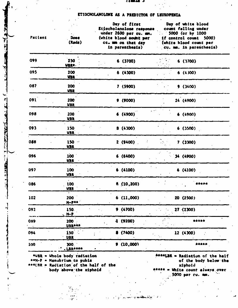
Fif teen pat ients have been studied (Table 3). In only 1 d i d the madmum granulocyte i n c r w n t become subnormal a f t e r the white blood c e l l count had f a l l an b o l w 5000 (by 2 days). In 10 of the 15 the f a l l in d m m granulocyte increment to eubnoraal level. pre- ceded any vhite blood c o l l count drop.
and tha latter i r a l s o non-
Thir rut- increment does not These pa t ieu ts recoived 100 t o 300 rad8
In 6 of these 10 whose white comts - t d l y d i d go under M O O per CU. P., tha a b n o r u l i t y in etiocholanolone response pre- codod the 10ukop.n~o by an averago of 12 days, suggesting tha t t h i r b io logica l .doshe tor may be of s i p i f i c m t r r s i a t m c e in giving varning of an irponding leukopenia. In 4 of the 15 the uximum
. ., .- ..,.:.
~ 1 o c H O ~ u m L As A ? l m x m or L m
D ~ Y or rirrt
undar 2600 per cu. m.
Day of uhlte blood ttiocholanolono trrporue c o a t fal l ing under
5000 (or by 1000 rattent w r (white blood oo\kc por i f control count 5000)
(kdr) CC- I O11 that day (whit. blood count per in paronthrri~) cu. m. i n patentheaia)
.. . .. . , * . 6 (3700) . . 6 (3700) . , .
099 250 ; UBI*- '
095 200 6 (b300) . . ' 6 (4100)
..i .i , .. . .
.. .
. . c
-a=- . .
W *
c
granulocyte increment dropped under 2600 per CU. I. on the same day t h a t the white count f s l l under MOO. cholanolone respolue generally occurred on days 6 t o 9 a f t e r radi- a t ion. One hrmdred rad. of whole body radiat ion v u ' a u f f i c i e n t t o c a u e the t e a t t o become a b n o d u w m 150 rad. to approximately 40% of the marrow wlw.
Ihe rllbnonul et io-
The etiochol.nolom reaponre v u leas helpful in predict ing
The etio- recowry. becoming w-1 before the white count had returned t o normal l eve la in ha l f the patienta who had leukopenia. cholanolone reaponre returned t o n o d in all patient# by 9 week# a t t h e latest (range 6-62 day.).
The m!aaur(PDant of the maximum granulocyte increment poat- r ad ia t ion al lova one t o predict vhether an i r rad ia ted individual w i l l experience leukopenia. H0Ipev.r. nei ther the timing, granu- locyte count nor granulocyte increment in the initial drop in the etiocholanolone reapocue ind ica tu how sevare the eventual leuko- penia w i l l be.
granulocyte r e a e r w s h u not y e t been determined and thi. i a under study noy. It l a s ign i f i can t ly lover t h ~ t ha t in a atudy employ- ing endotoxin where it v u fotmd t h a t a t l o u t 60% of the marrow mst be i r r ad ia t ed t o dosage levels o w r 1MO 1 before the endotoxin raapocua became abnormal ( 5 4 ) .
The lover l i m i t of the eensitivity of thia teat of M-
The timing of recovery of P=OY reaervea ruemblu t h a t noted in pat ien ts with Hodgkin's d i a e u e given a t c n s i v e frac- t ionated radiotherapy (56).
Deoxycytidine
Following the report of a radiation-indocad i n c r e u e in urinary excretion of deoxycytidine (Cdo) in the ra t . our laboratory has examined deoxycytidinutie in man u a biological doaimeter (5.9. 59, 60, 81). that t h e urinary and blood deoxycytidine levels of rats were elevated about ai. hours a f t e r X-irradiation, reaching a uxiwm in blood a t 9 hours, urine a t 12 houra.
Using 8 S e U 8 i t i ~ e C O l O f l m O t f l C techaiq- ( 5 9 ) W e found
Deoxycytidine excretion i n the rat w u proportional to the mount of r ad ia t ion up t o 200 R. Approximatelt. 8 eix-fold increase from the average p r c i r r a d i a t i o n value of 0.7 .(I per 24 hour urine followed 200 R.
.
c I I. .
-20-
W With tritiated CdR h b e l l e d on carbon-5 of cytonine, rat. i r rad ia ted v i t h 200 R excreted Z l f of the t o t a l radioact ivl ty while only 13% of rad ioac t iv i ty v u excreted by u n i r r d i a t e d rats (61, Pig.5). s p e c i f i c rad ioac t iv i ty of CdR inolated from the urine of i r rad ia ted rat. d e c r e a r d 2 t o 6 fold an eosparsd with t h a t of CdR in the urine of cmirradiated ram, the frw CdR pool nire v u i n c r e u e d in the i r r ad ia t ed rat (Table 4). Thin radiation-induced Cdlt excretion could be rupprenned w i t h rerotonin nnd I-cyntdna, the renpectiva dose re- duction facto- being 1.7 and 1.5 for thin variable . '
b
Since
c.
'r
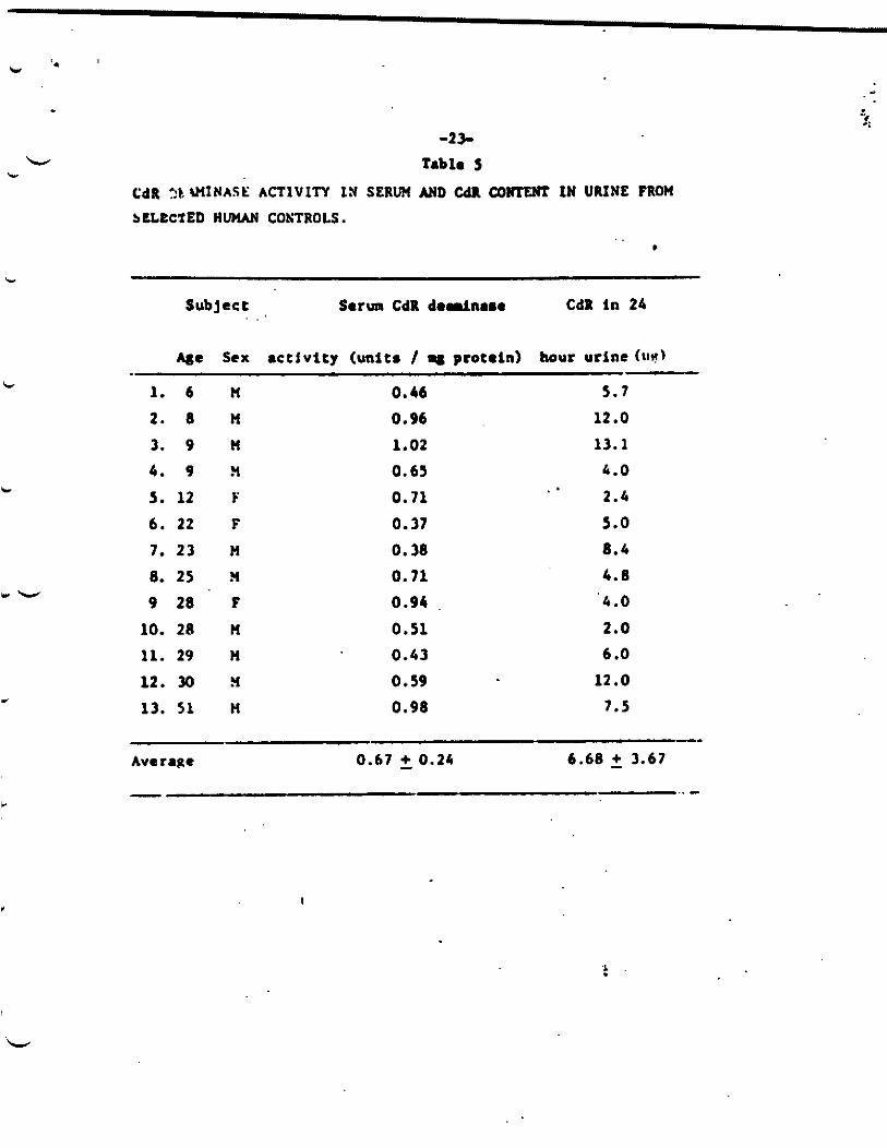
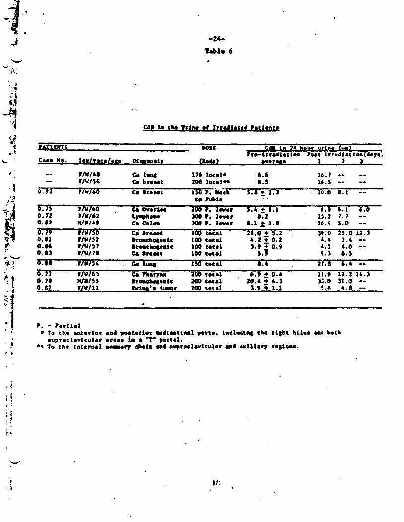
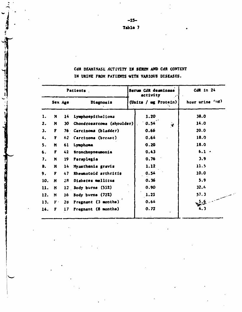
Man, however, excrete. a mch smaller quantity of CdR in h i n Urine than doen tha rat. SB of CdR per 24 hour urine v u found in m (Tnble 5) M coapared to 0.7 mg in t he r a t . The lw CdR sscretim, in am say be CAWLd by the a c t i v i t y of a CdR d e d n a n e in h w n plasma (31) and liver (82). Hwcver. the range in our cancer pqtientn w u 3.9 t o 26 microgram f o r 24 hours (Table 6). Pollotting whole o r pa r t i a l body rad ia t ion at dose of 150 rads o r above CdR excretion me. 50 t o 350% w day one port I r radiat ion. re-
c. turning t o normal an day two (Table 6). However, this is not a dosimeter npec i f ic t o radiation injuzy. A pat ient with lymphoepitheliomn and eewral burned pa t imtn alno rhowed abnormally h i & urinary CdR excretion in proportion t o the severity m d extant of the burn (Table 7). Thus, deoxycytidinuria appearn to be related t o general t i s n u e c8 tabol im from n e w r a l catmen, including radiation. Other problem in w i n g urinary CdR include variFtionn in excretion dum t o race (57) and age (92) .
An awra- pro-irradiation value of 0.007
Creatine
8.diation-induced crea t inur ia in rati (61) f o l l w r a f a i l u r e of m a c l e t i s n r u t o u t i l i z e creat ine nynthenired at A n o d r a t e by l i v e r and m n c l e (62 ) . a f t e r a s i n g l e whole body &eo in the rat, at lrut up t o 650 9. Bow- war, we have been tmabln t o d-trat. increased amount* of creat ine in the ur ine of our p a t i m t n pont- i r radht ion as r u u r e d by the in- c r e u c d c rea t ine t o creatininn r a t i o (83). Rune ntudien were done n t done8 of 100 rad. o r l u n of whole body rad ia t ion hwever, p.rhap. below the n m e i t i v l t y of thin donimnter in MU.
It is done-dapnndant, r u c h i n g a peak over 4 dayn
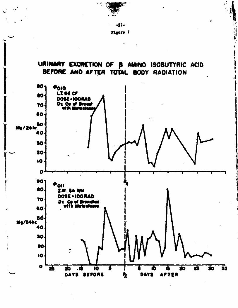
Beta-Aoi~iorobutyr ic Add (MM)
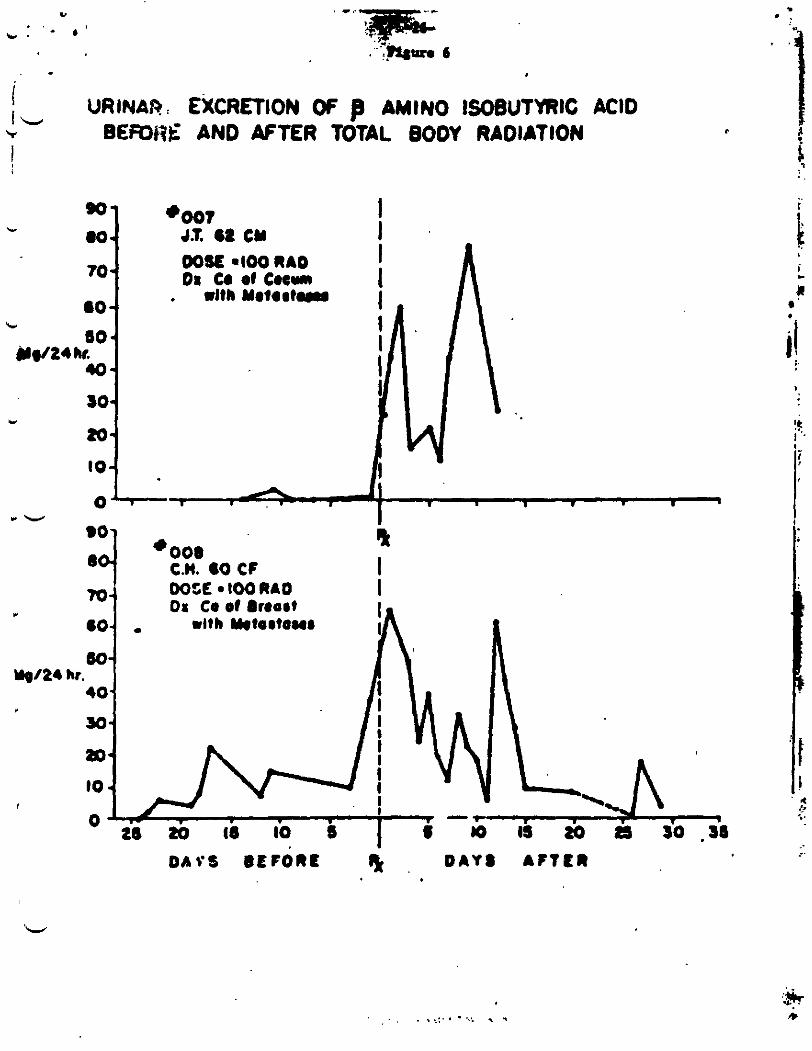
Another n u d d c acid r t a b o l i t e , be t a -dno inobu ty r i c acid, a product of thymidim a tabol iam has b o a focmd in i n c r e u e d m m t n in i r r i d i a t e d hrrunm, w i t h d i n a g M O Y l l t u t o the proportional increase i n BAIBA with &#orbed radiation &m (63, 80). Hwevar, the i n c r e u e han not been coruisteut in our patientn (Figurer 6.7).
L
. -21-
l i p u m 5
+--A unitradiated frradiuted (200R)
' TIME AFTER IWECTION (HOURS) Fffeci of lrrodi tior. ( OOA at 1) Excretion
of L t r t f Cdk in t o w
L ir
Y
c
lbt No.
1
2
3
-22- Tabla i
%-Cdl EXCRETED I N 22 HOUR URINE IN RATS
?re- t r r Ad iA t ion
192.000
179.000
lC1,WO
0.60
1.04
1.93
POW-irradiation
Specif ic Radioact iv i ty
.( cpdmg )
61,000
31,000
83,000
Totnl excreted
( m8ld.y)
5 . 1 3
7.16
6.50
c.
c
-23- Tabla S
C ~ R ? b r n I N ~ S t : ACTIVITY IN SERW AND cdn c o r n IW URINE FROM
bLLeC'fED HUMAN CONTROLS. . . .
c
ir
c
M e sex activity (units / q protein) hour urine (1111)
1. 6 M 2. 8 n 3. 9 n 4. 9 ?L
5. 12 F 6. 22 F 7. 23 M 8. 25 ?4
9 28 F 10. 20 n 11. 29 n 12. 30 n 13. 5 1 H
0.46 0.96 1.02 0.65 0.71 0.37 0.38 0.71 0.94 0.51 0.43 0.59 0.98
5.7 12 .o 13.1 4.0 2.4 5.0 8.4
4. 8 ' 4 . 0
2.0 6 .O
12.0 7.5
Average 0.67 5 0.24 6.68 2 3.67
t
-24-
Tablo 6 .
U l 1. ch. Urim of Irradhtod htfionck
C48 in 22 how urlne h - i r r d i . r t o o PO.( 1rra4tat:on + days.
?LtIPcfS DOIL
C u e no. Soxlraco1.m mumria ( W a 1 awraio 1 2
IN148 c. 1- I76 kcal* 6.6 16.7 -- -- 0.92 TlW60 Ca I rebat 130 ?. 'hit 5.8 2 1.1 10.0 8.1 --
-- -- -- T l U l 5 4 C. bront 200 loc.1H 8.5 16.5 -- --- . . t. -1.
b. 15 TlVI60 C. Ov.11r loo?. l a n r 5.4 2 1.1 6 .8 b . 1 6.0 0.72 Tlu161 L W - a t . Iwor 8. t 15.2 ? . 7 -- 0.81 U I ~ I L P C. &Ion #o ?. l a n r 8.1 2 1.1 16.4 1.0 --
h.b 3 .4 -- 0.81 T l U l 5 2 kmchqomlc 106 c01.1 4.2 0.2 0.8b F I Y I 5 7 8 t m e h q n l c 100 t.c.1 3.9 + 0.9 b.5 4.0 -- 0.83 T l U l 7 8 C. 8 t . Y t 100 t o w 1 5.5 9.3 6.5 b.88 t1w154 c. 1- 150 t o t r l 8.4 17.8 b.4 - 0.1? TlUlb3 & ?h.r)u 200 toc.1 . 6.9 2 0.b lJ.9 12.2 14.3 0.78 n1n155 8raebasaolc m tot.1 10.4 2 4.3 31.0 31.0 -- 0.87 tlulll M m ' a tsmor zoo t0t.l 3.9 + 1.1 5.8 4.6 --
a. 79 ~ I U I J O C. 8 r a n t 106 tot.1 21.0 2 5.2 39.0 25.0 11.1
1 ::
-25-
Tablo 7
Pat ients , .. Sex Ago Diagnosis
.
So- CdR d o m i n u ; Act iv i ty
@nitr / Protein) ..
CdR DUnINASE ALTIVITY I N SERLW AN0 CdR CON'TLXT
IU URINE FROM PATIEMTS WXfH VARIOUS DISEASES.
1. !4 14 LI.phoeptthelioaa 2. H 30 Chondrorsrcms (shoulder)
3. ? 76 Carcinoma (bladder) I . F 62 Carcinoma (brcnsc)
5. It 61 Lyaphou
6. F 42 Sronchopnouwnis 7 . x 19 ~ ~ r a p i 0 8 i a
8. I4 16 Hyuthonia 6 r J V i S
9. F 67 Rheuntoid a r t h r i t l r
10. J4 28 Diabetes mellitus 11. X 12 Body burna (55%)
12. X 16 Body burn8 (72%)
13. F. 28 Pregnant (3 m o t h s ) 16. F 1 7 Pregnant (8 months)
--
~ .~
1.20
' 0.5h5"' .+ 0.66
0.60
0.20 0.13 0.76 '
1.12 0 . s
0.36 0.90 1.21 0.64
0.72
I .
cdl i n 24
hour urine '111)
38.0 14.0
20.0 18.0
18.0 6.1 - 3.9
11.5
IO. 0
5.9 32.6
-*- " - * C I . .- . a&- 6 *
I URIN&? EXCRETION OF AWN0 lSoeUTYRlC ACID I L BEfOh'E AND AFTER TOTAL BODY RADIATION 'r
I
7 10
0 I. A I I 1 . 4
r w
'O1 I
L 100 RAO I _ -
6OCF
D A C S BEFORE I& , D A Y S A F T t R
. 9 . + '1 i ! i.
. . . ..:. . - ..: . . . .
.. I .
c
. .
v b
I I .
. .. ..
-27- P i lo r . I
URICIIIRY E%CR€TKWJ O f AMINO ISOSUTVRIC ACID 8EIcocK AN0 AFTER TWAL W O Y RAOIATION
W
70 -1
I
I 0' . I . . I I . I I I 1 I
-28-
W 1c.
Another p r o b l a in Interpret ing ur ine BAIBA data i a that ttere is an hereditary uymptolvt ic condition of high excretion
~ 1 0 % of people examined) ( 6 4 ) . This compound, too, is a non-specific sign of t ieaua breakdown.
Taurine and Other Amino Acid8
Another .&no ac id , taurine, is excreted by the r a t and dog. but not the guinea pig, in axcesa amounts a f t e r whole body radiat ion ( 6 5 , 66 . 67 , 68, 6 9 ) . A second radiat ion exposure in about a veek following the f i r s t do- not elicit a a imi la r excretion of taur ine ( 6 7 ) . Previow work h a sham that an a i r done grea te r than 450 R
required to ralae urinary taurine a f t e r hmmn whole body i r rad- i a t i o n (65). Then v u no s ignf f icant increase in the taurine ex- c re t ion amom' our pat ients up to a dose of 200 rads. Nor w.8 there any a lgn i f i can t change in excretion of any of the urinary &no acids examined (Table 8 ) . after surgery or in fec t ion and are therefore also a wn-specif ic s ign of t iaaue breakdown (84). evaluat ion of urimrp mino acid levels in general , and taurine s p e c i f i c a l l y , is t h a t the levels of most d n o acid. in the urine are grea t ly influenced by dietary protein intake. In s-ry, our laboratory h u been h l s t o confirm that there are any s igni f icant difference. behreao the mounts of there urinarp d n o acids ex- creted by cancer pa t ien ts before o r a f t e r whole body i r r ad ia t ion a t 1evela O f 50 t o 200 rad8 (81, 83).
Glycoprotein8 Including Traneferrin; Seem Iron
Urinary,taurine levels have been found in children
Another var iable d i f f i c u l t t o control in
Serrn glycoprotein leveh have been reported t o have prognostic s ign i f icance a f t e r rad ia t ion injury (70 ) . Uslng pre- i r radiat ion levels as controlr the concentration of t o t a l protein-bound carbo- hydrates. pa r t i cu la r ly t ranafer r in end haptoglobin (71) are found t o rise t o high levels in mice, rats and dogs who have died a f t e r rad i - a t i o n arposure, while s u r u i v o a ahowad little change. invaa t iga tor reported no s ign i f i can t difference between the man serum 8lycoprotein concentration in rats' blood 88 t o 90 hourr a f t e r 600 It of X-irradiation 8nd t h e i r unir tadiated control# (72) but t h i s my w e l l be due t o the expetiuntd d u i g n .Iployed (69) . The masurement of t o t a l iron binding capacity ( t ransfer r in) showed no s igni f icant rise a f t e r i r r ad ia t ion in our pat ien ts (Table?). p a t l e n t s aunr iwd their treatment this l a a t lwt consis tent with Evans' predictlonn t h a t lack of glycoprotein rise correlates with aur- Vivrl .
Harever. another
.Since dl our
c -29-
Table 8
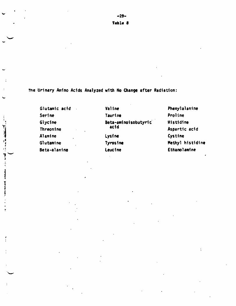
The Urinary Amino Adds Analyzed w i t h No Change af ter Radiation:
Glutamic acfd Serine
., Glycine 1 Threonine A1 ani ne
If 4 Glutamine Be ta-a1 ani ne
Y - 2 :
>
Valine Phenylalanine T a d ne Pro1 i ne Beta-aml noi tobutyrl c'
Lysine Cystine Tyrotfne Methyl h i t t l d i ne Leucine Ethanolani ne
H i s ti dine Aspartic acid acid
Y
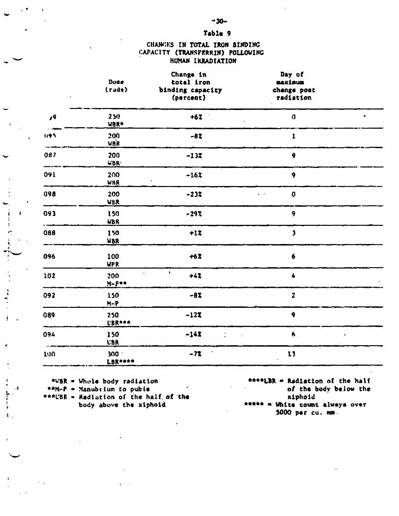
-30- Tabls 9
CIIANGES IN TOTAL IRON 8INDfNC CAPACITY (TRANSFERRIN) FOLLOUlNC
H W IKMDIATION
Change in
(percent)
Duae total iron (rilds) binding capacity
c.
% .
f "'L
i w.
Day of
change post radiation
M X i O U r
. -E% I
- UriR .--- OR 7 200 -1 32 9
09 1 200 -16X 9
0 098 200 -23% . .
09 3 150 -29% 9
086 150 +I% 3
-- wen. --- - UHR
- WBR
wait -
096 100 6% 6
102 ZOO +bt 4
092 1 SO -8Z 2
089 2 50 -122 9
09L 1 so -1bZ 6
UP R
?(-Pee
5-P
CBR***
-
.i L..
+WBR - Whole body radlrclon +*M-P - Xanubttum to pubts
***UBR - Nadiation of 'the hal f , of ,tho body above chr rlphoid
****LBR - Radiation nf the half of the body belou the xiphoid
****e - UWcc count always over WOO per CU. mm.
'W
I.
u -31-
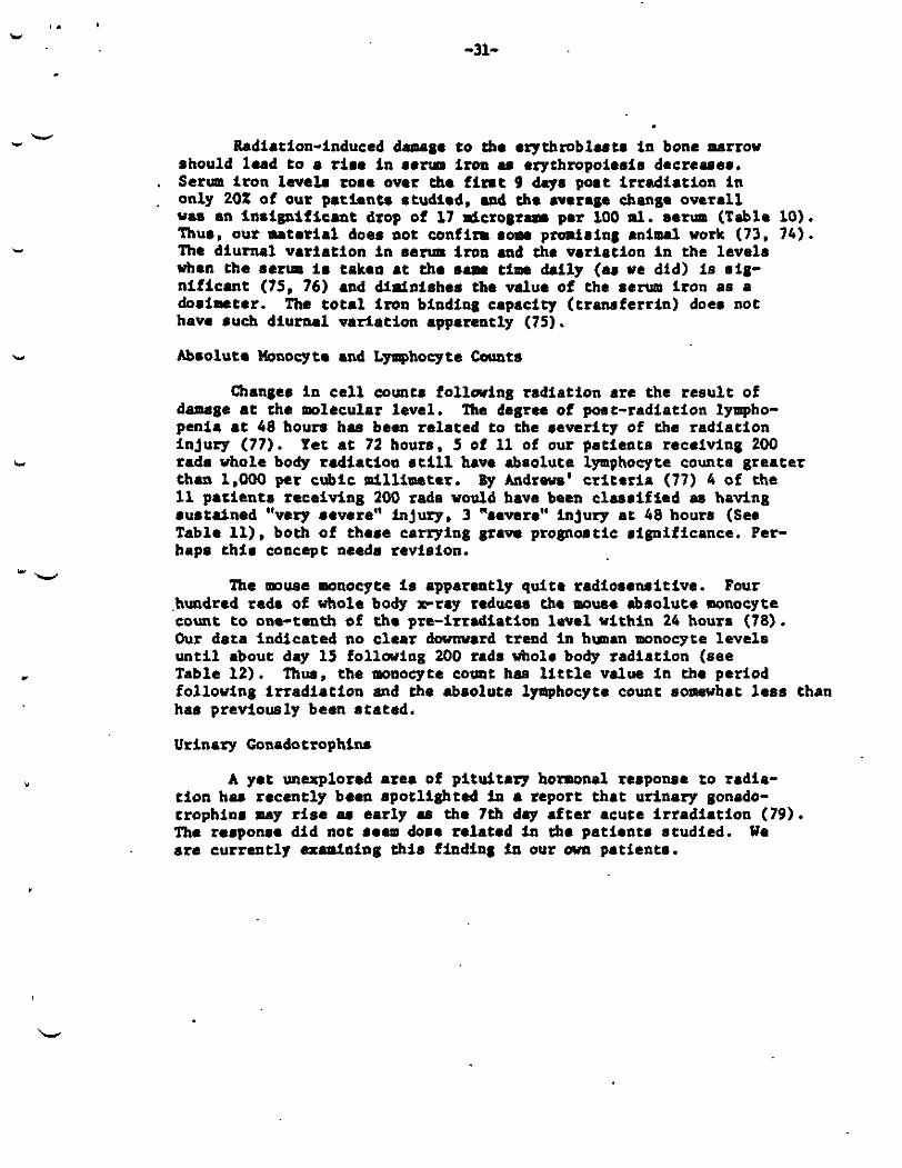
Radiation-induced damage t o the e ry th rob lu ta in bone marrow should lead t o a t ire in s e m iron am exythropoiesis decreaaee. Serlnr i r o n level. roe. over the f i r s t 9 days poat i r r ad ia t ion i n only 20% of our pa t i en t s studied, and che average change ove ra l l V.6 en i n s ign i f i can t drop of 17 dcrograzm per 100 ml. eerm (Table 10). Thus. our material does not confirm som promising animal work (73. 7 4 ) . The diurnal var ia t ion in rerum iron and the var ia t ion in the levels when the ec ru is taken at the s a m tin daily (er we did) is sig- n i f i can t (75, 76) and diminishes the value o f the serum i ron as a dosimeter. The total iron binding capacity ( t ransfer r in) docs not have such diurnal var ia t ion apparently (75).
Absolute Uonocyte and Lyrmphocyte Counts
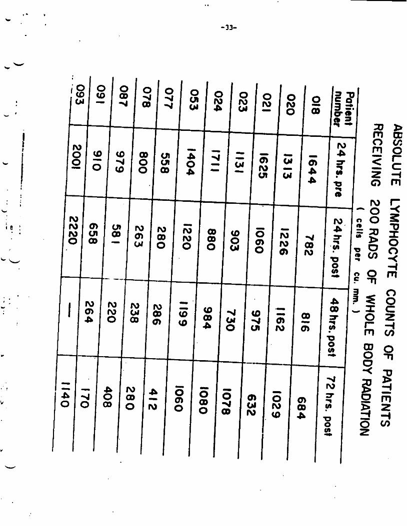
Changes i n c e l l counts fo l l a r ing radiat ion a re the r e s u l t of damage at the molecular level . penia a t 48 hours h a been re la ted to the sever i ty of the rad ia t ion in ju ry (77). rada whole body r ad ia t ioa s t i l l have absolute lymphocyte count8 grea te r than 1,000 per cubic millimeter. 11 pa t i en t s receiving 200 rads would have been c l a s s i f i ed rn having sus t r ined “very eevere“ injury, 3 “sewre“ injury a t 48 hours (See Table 11). both of these carrying grave p r o p a t i c rignificance. Per- haps t h i s concept needs revision.
The degree of port-radiation lympho-
Yet a t 72 hours. 5 of 11 of our pat ients receiving 200
By Andrews’ criteria (77) 4 of the
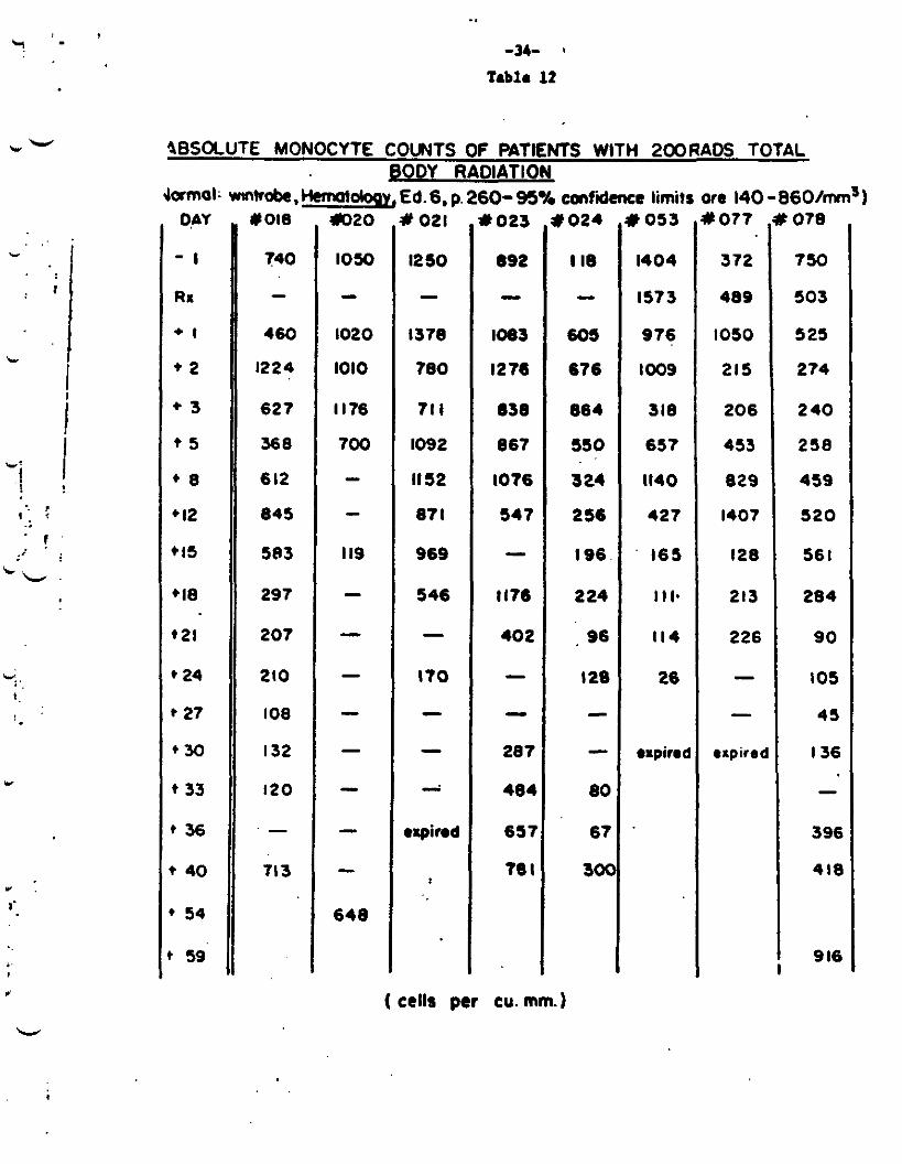
The muse monocyce is apparently qui te radiosensitive. Four .hundred rad. of whole body x-ray reducar the mouae abSOlUt8 monocyte count to ona-tenth of the pre-irradiation level v i t h i n 24 houra (78). Our data indicated no c l e a r downward trend in humn monocyte l eve l s u n t i l about day 15 folloving 200 rads whole body radiat ion (see Table 12). Thur, the monocyte count h a l i t t l e value in the period following i r r a d i a t i o n and the absolute lpphocyte count somewhat less than h a previously been s t a t ed .
Urinary Gonadotrophiru
A yet unexplored a rea of p i tu i t a rp hormonal response t o radia- cion ham recent ly been spotlighted in a report t ha t urinary gonado- t rophins may rise u ear ly u the 7th day a f t e r acute I r r ad ia t ion (79). The response did not 10.0 dare re lated in the pat ien ts rtudied. We are cur ren t ly axamlning t h i s finding i n our mm pat icntr .
.
W ”
i
W u”
-
v c
r
-32- Tabla 10
C W E S IN SERUM IRON ARLU HulM IRRMIATION
b x i a w charqa from prc-rndiation l evulu ,ent nose of serum i r o a v i t h i n nine days poor
(Rads1 radintion (percent) 250 -31% UBR*
r)r J 200 -592 VBR
VIR
WER
WllR
VllR
WBR
VBR
I---
c 081 --- 2 0 0 +782 - - 091 - 200 -332
o9n 200 +152X
ri9 3 150 -60%
088 150 -162
096 100 -62
102 200 -352
09 2 150 -562
089 2 0 0 -582
O K 150 +9t
100 300 - 7 s x
- ---- --
- -
-.- n-p** - .X-P
;m***
-- CBR
LBR****
- -. -
W B R - Whole body rAdiAcion ***L'BR - h d i a t t o n on t h e half of tha
body above the xiphoid
4W-P - Hanubrium t o publn ***4LBR - Radfation of the half of
the body b e l w the xiphoid
I -
t
I 1 I
..
-33-
* . I'
role
Y
l o 2 0 YO21
L
.. -34- '
Tablo 12
0,AY
- I
RI
+ I
* 2
+ 3
* 5
' 8
' 12 *I5
'18
21
* 24
* 27
e 3 0
t 33
r 3 6
* 40
* 54
1 59
740
- 460
1224
627
368
6 12
845
583
297
207
210
108
I32
I20
- 713
1250
- 1378
780
71 I
1092
1152
87 I
969
546
- I10
- - i
rxpind
P 023
692
- 1083
I276
6313
867
I076
547
- 1176
402
- - 287
484
657
10 I
P 024
I16
- 60s
876
664
550
3 24
2 56
I96
2 24
, 96
I28
-
80
67
sot
( cells per cu. m m )
t 053
1404
1573
976
loo9
318
657
1140
427
' 165
1 I I.
114
26
orpiroc
L 077
372
489
IO50
215
206
453
829
I407
128
213
226
- -
*wire(
078
750
503
5 25
274
2 40
258
459
5 2 0
56 I
284
90
105
45
I 3 6
- 396
418
9 I6
,. L.
I
w
. -35-
W " Anylane
F:lcvnted serm amylase levels (88) a f t e r p;~rorid rta1iV;iry
Tlie'amytnse rose t o B peak value at 2 4 t o VJ hours gliind r d i a t l u i i have heen noted w l t h doses as lou as 100 W (tmvr done) ( 8 5 ) . iiftcr I r rudla t ton , then declined t o normal over anorlier o w 1,r twu days. The response appears t o h e organ spec i f ic (31. 8 5 , 8 6 ) nnd liyperamylsvemia dnes not resu l t when the patient receives doses L O the pancreas (the usual source of sensa amylase) coaparabte'to those causing enzyme release from the salivary glands. h e mylase re- leased from the damaged (rlands is a t l e a s t grossly related t o the absorbed dose of radiat ion (85, 86).
Our data (Table 13, Figures 8,9) confirm these findings for whole and p a r t i a l body radiat ion in the hman. and ur ine leve ls i n pat ients receiving radiation to the sal ivary glands. The least s t r i k i n g of the three serum responses is i n J pat ien t whose parotid glands were not i n the radiat ion f i e l d . Onty the pa t ien t receiving the highest cumulative radiat ion dose shows elevated urine l eve l s of t he enzyme, however. suggesting chat t h i s is 4 less sensitive test than a e r m amylase. responses are prompt, however, and both dlminish as the cumulative rad ia t ion dose destroys glandular function. Table 11 indicates chat t h e s e n s i t i v i t y of t h i s biochemical dosimeter is ac l e a s t 100 rads. Radiation below the neck does not increase serum amylase . i n doses up t o 300 rads over the pancreas (not on char t ) . laboratory is current ly performing agar gel electrophoresis on the sefm of i r r ad ia t ed pat ients to separate the isoenzymes of amylase.
Other Wuman Urine and Serwa Protein Studies
The graphs show serum
Both urinary and serum
Our
A nmber of other hman senm and ur ine proteins have been examined f o r t h e i r response t o whole body radiation up t o 250 rads and half body radiat ion up to 300 rads. These a r e l i s t e d i n Table 1 4 . These tests have been found t o show absent or inconsistent changcs Eolloving i r rad ia t ion .
L c
Y
- 36-
Table 13
'W
- 3R-
L Y
c
I
W

-39- Table 14
-40-
. W
c
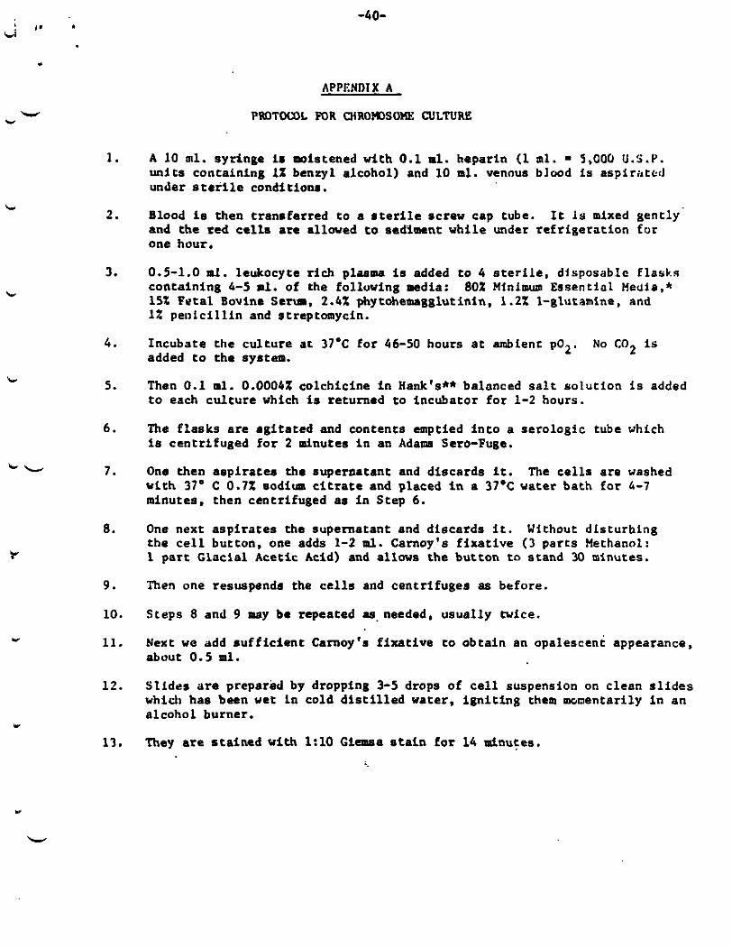
APPENDIX A
P I M T W L FOR MR0K)SOME CULTURE
L
1. A 10 m l . syringe is moistened with 0.1 ml. heparin (1 m l . = 5,000 U.S.P. uni t s containing 1% benzyl alcohol) and 10 ml. venous blood is aspirated under steri le conditions.
Blood is then transfarred t o a r te r l le screw cap tube . and the fed cells are allowed to sacliment while under refr igerat ion for one hour.
2. I t is mixed gently-
3. 0.5-1.0 ml. leukocyte r ich p l a n a is added t o 4 steri le, dlsposablc flasks containing 1-5 m l . of the folluving media: 15% Fata l Bovine S e w . 2.4% phytohemagglutinin, 1.2% 1-glutamine, and 1% pei i lc i l l in and streptomycin.
Incubate t h e cul ture at 37.C fo r 46-50 hours a t ambient p02. added to the system.
80% Minimum Essentiol Media.*
4. No C 0 2 i s
5 . Then 0.1 m l . 0.0004X colchicine i n Hank’s** balonced s a l t solut ion is added t o each cu l ture which is returned t o incubator f o r 1-2 hours.
The f lasks a r e agi ta ted and contents emptied i n t o a serologic t u b e which is centrifuged f o r 2 minutes i n an Adam Sero-Fuga.
6 .
b L 7 . One then asp i ra tes the supernatant and discards it. The cel ls a r e washed with 37. C 0.7% s o d i m citrare and placed i n a 37.C water bath fo r 1-7 minutes. then centrifuged as i n Step 6 .
8. One next asp i ra tes the supernatant and discards i t . the cel l button, one adds 1-2 m l . Carnoy’s f ixa t ive (3 par t s Methanol: 1 par t Glacial Acetic Acid) and allows the button t n stand 30 minutes.
Without dfsturhing
9. Then one resuspends the cells and centrifuges as before.
10. Steps 8 and 9 may be repeated as.needed, usually twice.
11. Next we add su f f i c i en t Carnoy’s f ixa t ive LO obtain an opalescent appearance, about 0.5 m l .
Sl ides are prepared by dropping 3-5 drops of cel l suspension on clean s l i d e s which has been wet In cold d i s t i l l e d water, ign i t ing them momentarily i n an alcohol burner.
They are s ta ined with 1 : l O Giemsa s t a fn f o r 14 Pinutes.
1 2 .
13.
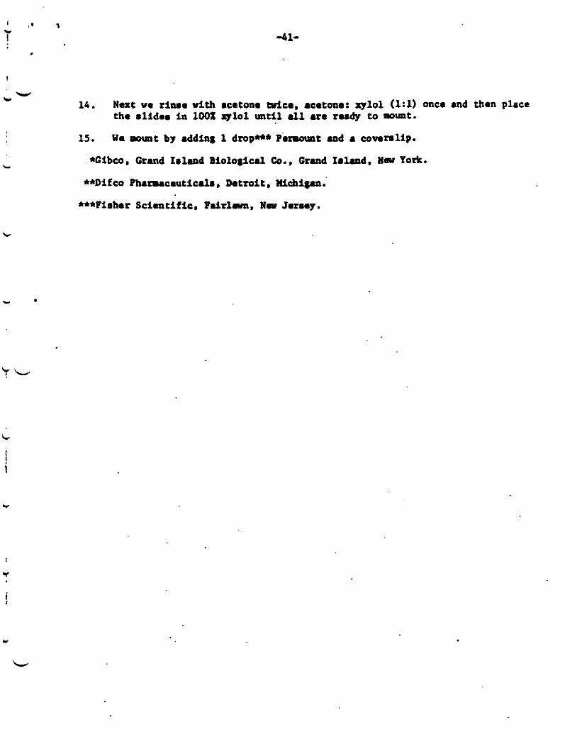
-41-
b-
c.
c
14. Next we rime w i t h acetone Mea, acetone: xylol (1:1) once and then place the mlidem in 100% ~ 1 0 1 until all are rudy to mount.
Wa -t by adding 1 drop*** Pm-t and 8 covers l ip . 15.
*Gibcos Grand I m l d Biological Co., Grand Imland. R.w yo*.
**Difco Pluruceuticalm, Datroit Wch1g.n.'
***Fisher Scimtif ic , P d r l u a , Ww Jarsay.
c
c
-4 2-
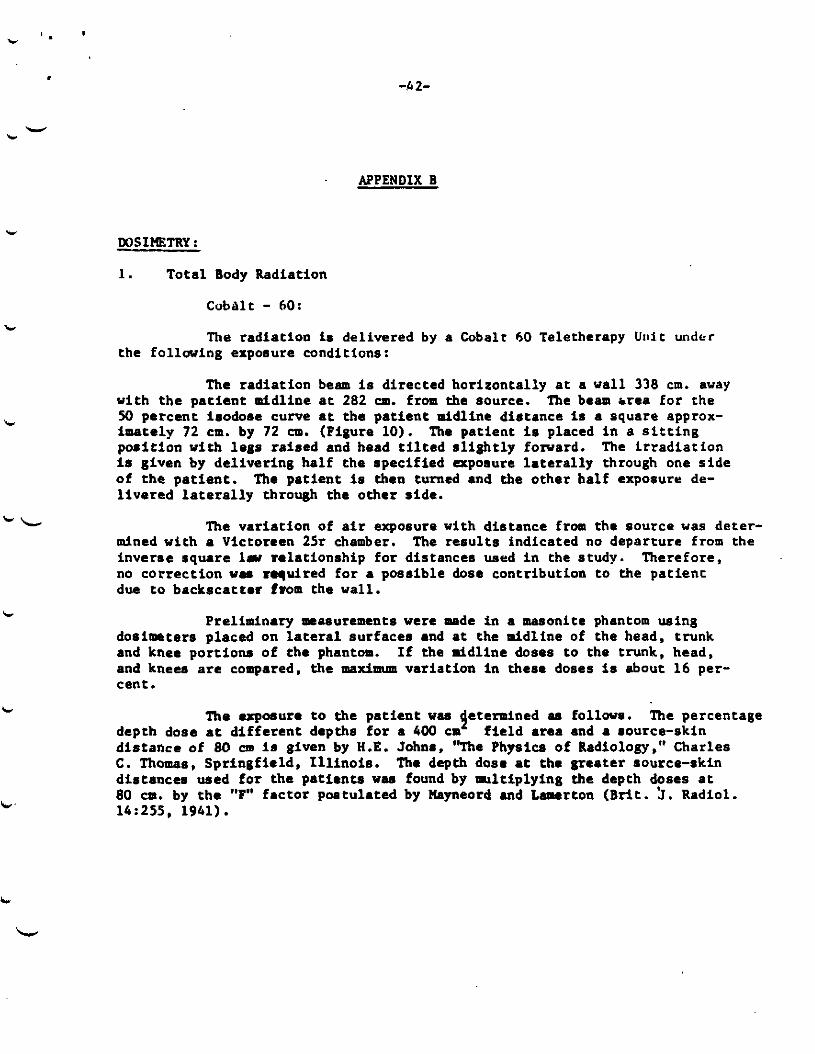
APPENDIX B
DOSIHETRY:
b
b
I-
C.
1. Total Body Radiation
CobAlt - 60:
?he radiation is delivered by a Cobalt 60 T e l e t h e r a p y U n i t under the following exposure conditions:
The radiat ion beam is directed horizontally at a wall 338 cm. away w i t h t h e pa t ien t mldline at 282 CP. from the source. 50 percent isodose curve a t the pat lent mldline distance is a square approx- imately 72 cm. by 72 cm. (Figure 10). posi t ion with legs raised and head t i l t e d s l i gh t ly f o n a r d . The i r r ad ia t ion is given by del iver ing half the specified apoaure l a t e r a l l y through one s i d e of the pa t ien t . l ivered l a t e r a l l y through the other side.
The beam &rea f o r t h e
The patient is placed i n a s i t t i n g
The pat ient is then turned and the other half exposure de-
The variat ion of air exposure with distance f r m the source was deter- mined with a Victoreen 25r chamber. The resu l t s indicated no departure from the inverse square l r r d a t i o n s h i p fo r distances used i n the study. no correct ion WY required fo r a possible dose contribution t o the pa t ien t due t o backscatto? I m m the w a l l .
Therefore,
Preliminary measurements were made in a masonite phantom using dosimeters placed on lateral surfaces and a t the d d l i n e of the head, trunk and knee portions of the phantom. If the d d l i n e doses t o the trunk, head. and knees are compared, the maximum variat ion in these doses is about 1 6 per- cent.
The expowre to the pat ient waa etermlned M follows. The percentage depth dose a t di f fe ren t depths f o r a 400 c 3 f i e l d area and a source-skin d is tance of 80 cm is given by H.E. Johns. "The Physics of Radiology." Charles C. Thomas, Springfield, I l l i n o i s . The depth dose at the greater source-skin distances used f o r t he pat ients WM found by multiplying the depth doses a t 80 cm. by t h e "F" fac tor postulated by Wyneord and tamerton ( B r i t . 5 . Radiol. 14:255, 1941).
C.
L
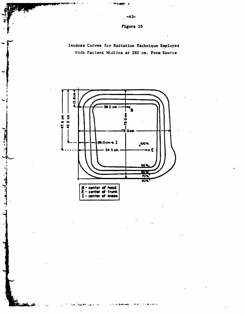
! f ' Figura 10
c mi ! I
Isodose Curves f o r Aadtation Technique Employed
141th Fatlent M l d l i n e at 282 c m . From Source
:. '" -44-
t
W b
F -
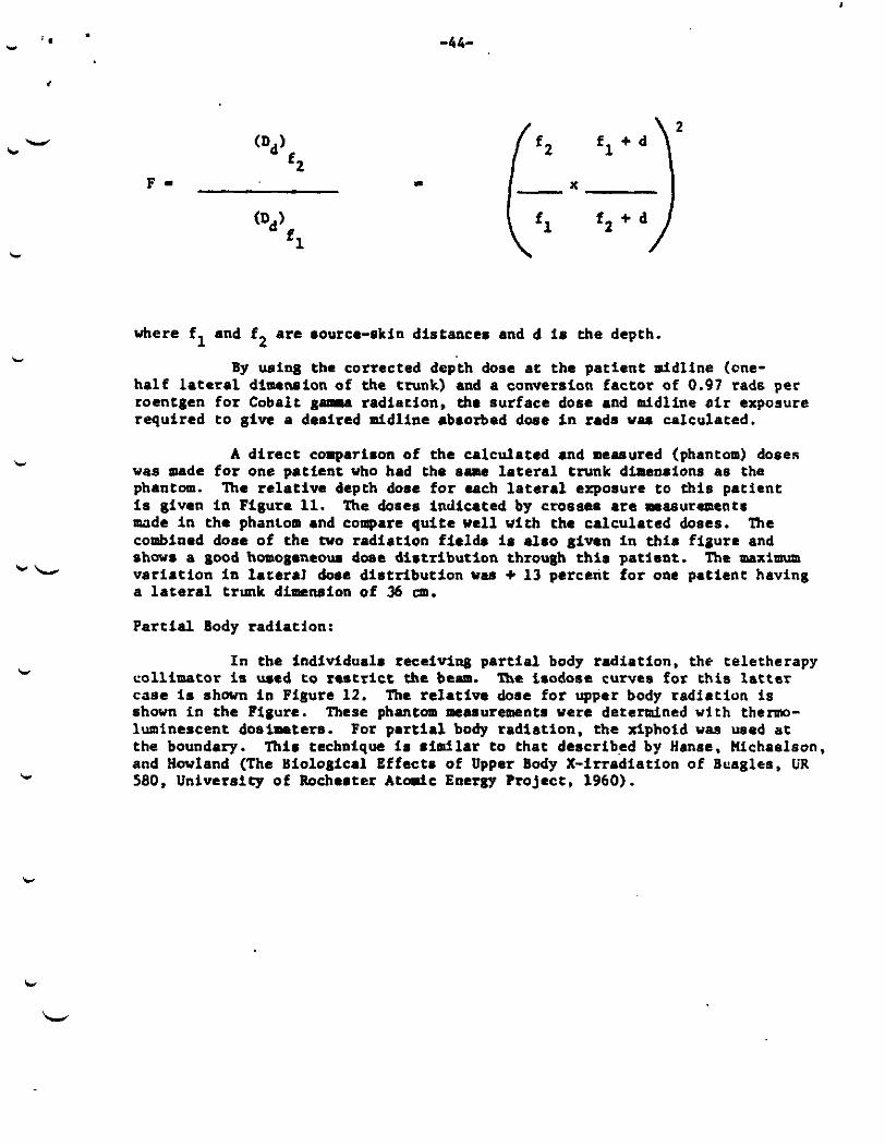
El + d f 2
c
where fl and f 2 are aource-skin distances and d l a the depth.
ha l f lateral dimension of the trunk) and a conversion factor of 0.97 rads per roentgen for Cobalt gama radiation. the surface dose and midline o i r exposure required t o give a desired midline absorbed d w e i n rads was calculated.
By w i n g the corrected depth dose a t t he pat ient midline (cne-
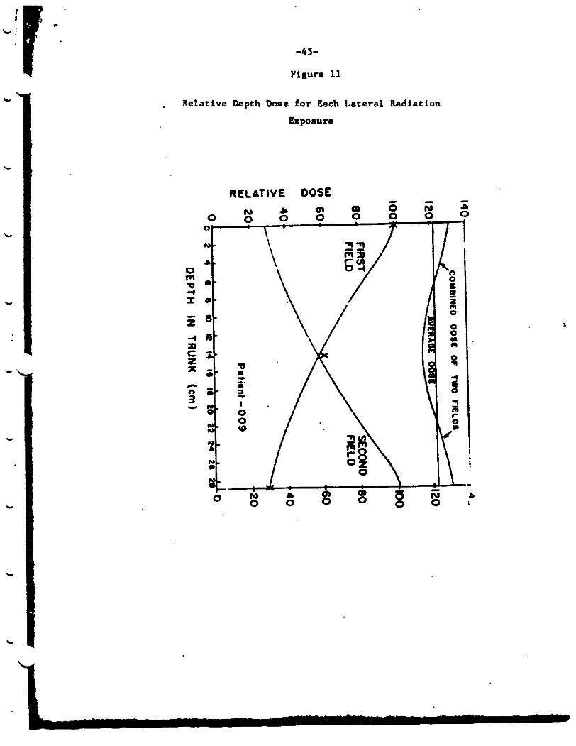
A d i r e c t comparison of the calculated and measured (phantom) doseR vas made f o r one pat ient who had the same l a t e r a l trunk dimenaions as the phantom. Is given in Figure 11. m d e in the phantom and compare qu i t e well with the calculated doses. The combined dose of the NO radiation f i e l d s l a ala0 given i n t h i s f iaurc and shows a good hoaogenew dose d is t r ibu t ion through t h i s pat5ent. va r i a t ion i n lateral h e dis t r ibu t ion was + 1 3 percent f o r one patient having a lateral trunk dimcnaion of 36 cm.
The r e l a t ive depth dose f o r each l a t e r a l exposure t o t h i s patient The doses indicated by crosses arc masurements
The maximum b L
c
c
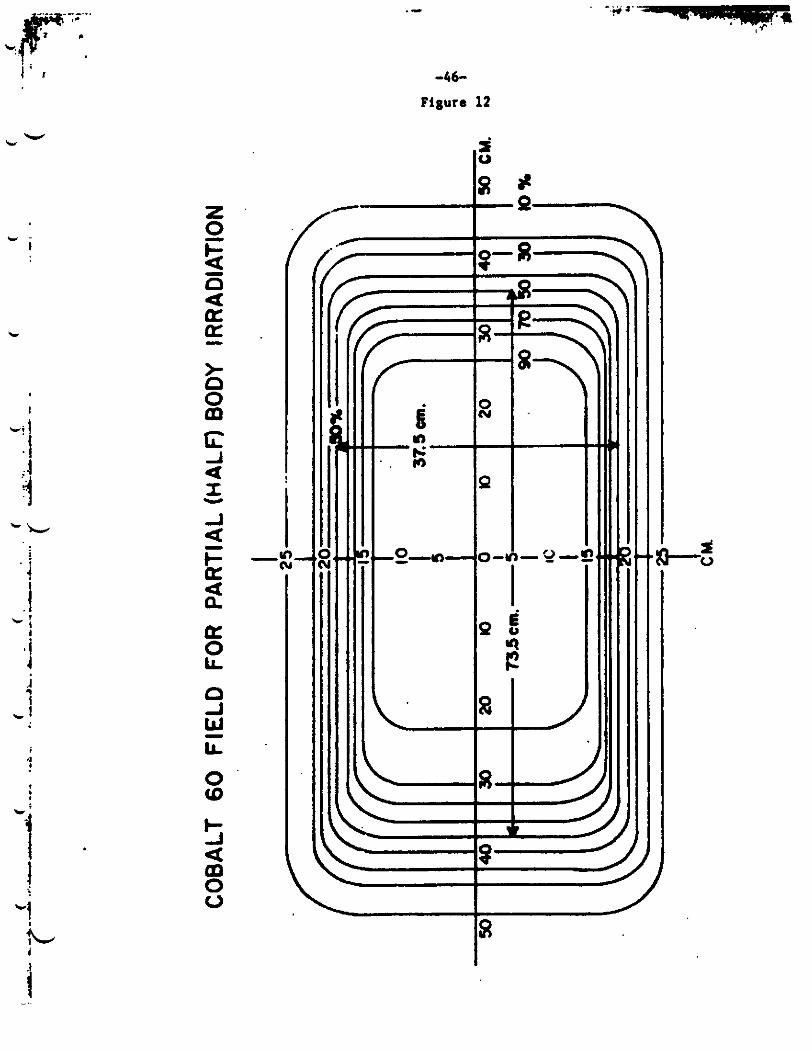
Partial Body radiation:
In the individuala receiving p a r t i a l body radiation, the teletherapy coll imator is rued to restrict the beam. 'he isodose curves for t h i s la t ter case is shown i n Figure 12. shown in the Figure. luminescent dosimeters. t h e boundary. and Hwland (The Miological Effects of Upper Body X-irradiation of Bcagles, VR 580, University of Rocheater A t d c Energy Project, 1960).
The relative dose f o r upper body radiation i s These phantom mcaauremcnta were determined with thermo-
For p a r t i a l body radiation. t he xiphoid w a s used a t Thir technique l a a i d l a r t o that described by Hanse. Michaelsoo,
. Relative Depth Dose for Each 1.ateral Radiation
Exposure
RELATIVE DOSf
* - I
'k
i I
-46-
Figure 12
-4 7-
REFERENCES
1. Sax. K.. Chromoaow aberrationa induced by x-rays, Genetics 2: 494, 1938.
2. Sax. K.. Ihe ti- factor in x-ray production of chrowsome aberrat ions, Proc. Nat. Acad. Sci.. Wash. 2: 397. 1939.
Sax. K. An analysis o f x-ray induced chromosome aberrationa i n Tradescantia. Genetics 2: 41. 1940.
Lea, D.E; 6 Cat&eside. D.C., The mechanism of induction by radiat ion of chromosome aberrations in Tradescantia. J. Genetics 9: 216. 1942.
3.
4.
v c
c
c 5.
6.
7.
a. 9.
10.
11.
12.
13.
c.
L
Evans. H.J. Dose response relat ion from i n vitro s tudies , Hman Radiation Cytogenetics. Ed. H. J. Evana, U.M. Court Brawn. A. S. McLean. North-Holland Publishing Co. h t e r d a m , p. 20, 1967.
Tjio. J.H. 6 Levan. A., The chromaom nmber of man. - 42: 1-6. 1956.
Buckton, K.E., Langlands. A.O.. Smith, P.C. 6 Mclelland, J. ChromtMome aberrations f o l l m i n g i r rad ia t ion i n psn. Hman Radiation Cytogenetics, Ed. H.J. Evans. U.M. Court B r m , A. S. UcLean. North-Holland Publishing Co, Amsterdam, p. 122, 1967.
Bender, U.A. Ann. Acad. Braaileira de Cieaciaa. 2: Prempree. T.S.. Hert. T. Radioactivfty and repa i r time: r epa i r t i m a of chromcwoma breaks produced during the d i f fe ren t s tages of the cell.
Weber, W.T. 6 Nwell. P.C., Studies on longlived small lymphocytes i n the Rhesua monkey and some other psrmals. J. Reticuloendothelial SOC. 2: 326-342. 1965.
Moorehead, P.S., Nwell, P.C.. Mllman, U . J . , Batt ips , D.M. 6 Hungerford, D.A. Chromoaolla preparations of leukocytu cultured from hman peripheral blood.
Rubini, J.B.. Bond, V.P. Kelltr. S., Fliedner, T.U., Cronkite, E.P.
Hereditas,
77, 1967.
t h e
Mutation Res. I: 441-451. 1969.
Experin. C e l l Rea. 20: 613-616, 1960.
ONA s nthesia in c i rcu la t ing blood leukocytes lebeled - v i t r o with ?i -thymidine. J. Lab. C l i n . Med. 58:751-762. 1961.
Heddle, J.A.. Evana. H.J.. Scott , D. Sampling time and the complexity of t h e htman leukocyte cul ture system. Radiation Cytogenetics, Ed. i3.J. Evans. W.H. Court Brown, A.S. McLean, North-Holland Publishing Co., Amsterdam, p.6, 1967.
Humn
-48-
L * 15.
16.
17.
18.
19.
20.
21.
22.
23.
24.
25.
26.
27.
28.
Bender, t4.A. h Coo&. P.C. &romosome aberratioru in hrpan blood i r r ad ia t ed i n v i t ro , Proc. Nat. Acad. Sci. 3: 522-532, 1962.
B e l l , A.C. 6 Baker, D.C. aberrat ions i n n o r ~ l h t n n a n leukocytes i n cu l ture , Canad. J. Can. Cytol. 6: 340-351. 1962.
de Grouchy, J . , Cytogenetic i t u d i e i i n i r r ad ia t ed morrow and blood c e l l s . Society of ilumatology, B a s i l : S. Urger, 1963. p. 52-62.
Scot t , D.. Sharpe, 8.. Batchelor, A.. Evans. H. J.. Papvorch, D.C. Radiation induced chromosome damage i n human peripheral ked lymphocytes in vi t ro . 11 RBE and dose rate s tudies vith g- and x-ray&. Mutation Research 9: 225-237. 1970.
ibid. Mutation Research 8: 367-381, 1969.
Bender. M.A. 6 Barcinski. M.A. Kin8ticS of two break aberration production by x-ray. in hman leukocytes.
NO-, A.. O t t o m a n , LE.. S ~ a k i . M. 6 Veomett. 8.' The frequency of dicentr ic8 i n hman leukocytes i r rad ia ted & - vivo and in vi t ro .
Kelly, S. h Brown. C. Chro~ome abarretionm M a biological QsiMter, An. J. Public ilealth 2: 1419-1428. 1965.
Bender. iI. Somatic chromoiomal aberrations. Arch. Environ.
Types and rates of x-ray induced
Irradiation-induced chromosome
Proceedings of tho Ninth Congress of the European
Co
Cytogenetics 5: 241-246, 1969.
Radiology E: 108-110, 1964.
H u l t h 2: 556-564. 1968.
Sco t t , D., Batchelor. A.. Sharpe, H. 6 Evans, H.J. RBE For fast neutrons and dose r a t e s tud ies using faat neutron i r r ad ia t ion . Cytogenetics. Ed. R.J. Eva-, W.M. Court Brown, A. S . McLean, North-Holland Publishing Co.. haterdam. 1967. p. 37-52.
N o m a , A. 6 S u a l t i . M.. Chromcuome exchange aberrations i n hmaa lymphocytes, In t . J . Rad. Biol. I& 321-8, 1966.
Kapp, D. h Smi th . K.. Qemlcal t u t u r e of chain breaks produced i n DNA by x-irradiation in Yitm, Rad. Res. 3: 34-49, 1970.
Revell. 8 . R.. 'Iho accurate estimation of chromatid aterra- t i ons induced by ionizing radiation. Proc. Royal SOC. London, S i r . B. 150: 562-589. 1959.
Sharpe. H., Scott. D., Dolphin. G. Chromosome aberrations induced in hlrun lymphocytes by x- i r radiat ion in v i t ro : effect of cul ture techniques and blood donora i n aberrat ion y ie ld .
Human Radiation
me
Mutation Rei. 1: 453-461, 1969.
Narell, P.C. &rowsome aberrations and imrmnological memory, Hlnan Uadiation Cytogenetics. Ed. H.J. Evans, W.M. Court Brown, A. S. McLean. North-Holland Publlshfng Co. 1967. p. 99.
! .f .I
t i ! ,
-49-
29. Fitrgerald, P. Hman Radiation Cytogenetics, Ed. H.J. Evans. W.H..Court Brown, A. S. HcLcan. North-Holland Publishing Co. Amrterdam, 1967. p. 94.
Buckcon. K., Smith. P and Court Brown. U.H. lymphocyte lifespan. ibid, p. 107.
The life-span and role of the small lymphocyte.
W k
.30. Estimatfon of
c
c
c
31. Si lbr rs te in . E.B. and Chen, I .W. unpubli~~hed observations.
32. Scott , D.. Sharpe, H., Batehelor, A.L., .Evans. H.J . . Popworth. D.G. Radiation-induced chromosome damage i n hman peripheral blood lymphocytes in vitro. f a s t neutrons.
1 RBE and dose-rate studies v l t h Nutation Res. 5: 367-381, 1969.
33. Catchaside, D.G., Lea, D.B. 6 Thoday. J.H. The production of chromosomal s t ruc tu ra l changes i n Tradescantia microspores In r e l a t ion t o dosage, i n t a u i t y , and temperature. J . Genet. - 47: 137, 1946.
Dolphin, C.. A review of r t h o d . of biological dosimetry v i t h pa r t i cu la r reference t o chro-some aberration analysis, in "Handling of Radiation Accidents Sympodm", IAEA-SH-119/4. STI/Pub/ 229, Vienna (Hay. 1969) p.215.
3 4 .
35. Langlands. A.O., Smith. P.C. Buckcon, K.E., Woodcock. G., HcLelland, J. Chromosou damage induced by radiation. Nature
+L - 218: 1133-1135. 1968.
c
36. . Sasaki, M. 6 Norman, A. Prol i ferat ion of human lymphocytes in cul ture . Nature 210:913-914, 1966.
37. Radiation-induced chromosaa abcr ra t iom in h-n cells, I n Report of the United Natioru Sc ien t i f i c C o r m i t t e e on the Effects of Atomic Radiation t o the General hsembly. Twenty Fourth Seasion. New York, 1969, p. 111.
38. Sasaki, N. ibid. p. 116.
39. Octensen, J. blood. Act. Phyaiol. Scan. 2: 75, 1954.
Hamilton. L. D.. Coned and functions of the l m h o c y t e . Ann. N.Y. Acad. Sc. 73:39, 1958.
Cooch, P.C.. Bender, M. 4 bndolph, N.C. i n The Biological. Effects of Neutron and Proton Irradiat ions, Vol. 1. I.A.E.A.. Vienna. 1964. p. 325.
Thoday. J.H. 6 Read, J . chromosome aberrations produced by x-rays, Nature 160: 608- 610. 1947.
On the age of the h-n v h i t e cells i n peripheral
40.
41.
42. Effect of o%yRen on t h e frequency of
L' . b
I
-50-
c
7 i
I
4
43.
44. Savage, J. t%rolro.eu-atchange a i t w in Traderc.ntb rmludoma
Uolff, S., Zhe k i a e t i u f o r two-bred chnnosou d a p p , J. Th&oret. B i o l . 2, #4, 1962.
microsporu, Iat. J. Rad. Biol. 0: 81, 1965.
ShArpe, El., Dolphin, G., Danon, K., Field, E. computinll l m h o c y t e kinet ic# in men d W m of ch ro roaou l aberrat iolu a ru tdnod during a t r r c o r p o r u l irradiatiim of the blood, C e l l Timsue Kinet. 1: 263-271, 1968.
norman. A., Ottoman, R., S u a k i , n., V . o y t t . 1. me frquency of d i c m t r i m . i n hmun lwkocytea i r radiated in vivo M d & - vi t ro . R~diology a: 108-110, 1964.
t iona in a radiat ion accident. .Rad. Rea. g: 61-17, 1969.
The "Recuplu" c r i t i c a l i t y
45. Xethodm f o r
46.
47. Schneider, G., Chon.. B. 6 Blonai8e8, T. &hnnoaorl sberra-
48. Bender, W. 6 Gooch, P. S m a t i c cnromeow aberrations induced . by h-n a l e body i r rad ia t ion . . accident. Bh. Rem. 29: 568-582, 1966.
49. Uolff , S., Kimball. E., Perry, 5.. b o t . P.. K8ppu. A. The bio logica l propertien of etiocholanolwe. Arm. Int . Ilcd. 2: 1268-1293, 1967.
50. Kappaa, A., E e l l a m , L., Fukuahiu, 0.. Gallagher, T. The 3. Clin. Bnd. Hetab. pyrogenic e f fec t of etiochol.nolone. - 17: 451-453, 1967.
51. Kimball. B.P.. Vogel, J.. Perry, S., Uolff, S. quant i ta t ive aapecta of pyrogenic and hemrtolugic rewponnm t o etiocholanolone la man. J. Lab. Clin. Xed. 69: 415-427, 1967.
Vog.1. J.. Y.nk.8. 1.. Kimball, B..,Uolff, S., Perqr, S. The e f fec t of etiocholmaloae on g r~au locy t8 kinet ics . Blood 0:
52.
474-484, 1967.
Craddock, C., Perry, S., V a t z l u , 1. 6 Laurence, J. 53. EValrU- cion of lyTroy granulocytic ? e r r w e i n 80-1 rqd d i a e u e a t a t w . Blood I: UO-855, 1960.
54. Bellman. S. 6 F h k , H. Granuloqte maem follow in^ rad ia t ion therapy u atudied by the reaponre to a 1irc:r:risl eadotmds. Blood 3: 310-324, 1965.
Korblt:. D., Tonr. F. , Davir, E., h i r e s , C., Arufield, F. The Pi- t ea t : A ruefu l of bow marrow granulocyte remervem. Current That. Rea. u: 491-505, 1969.
Vogal. J., Kidall , tl., Foley. T., Wolff, S., Parry. 9. Effect of exrenaive radiotherapy on the urmu grmtulocyte reaervea of patienw w i t h tlodgkin'a Dlswe. Cancer, 2:,798-804. 1968.
55.
56.
c
-51-
57. Parizek, J., Arlent , M.. Disnstbier, 2.. Sbda . J. Deoxycytidlne i n u r ine as an Indicator of changes a f t e r irradiation. - 182: 721-2. 1958.
Nature,
58. men, I.W., Kereiakrs, J.C., Friedman, B. I. and Saengor. E.L. Colorimetric analysis of deoxycytidine i n u r i n e after separation by ion-exchange column chromatography. 230-240. 1968.
A n a l y t i c a l Biochem. 2:
59. Lhn. I.W.. Kereiakes. J.C.. Friedman, B . I . and Saenger. E. L . Radiation-induced urtnary excretion of deoxcytidine by rata and humans. Radiology 91: 343-348. 1968.
60. Chen, I . W . , Wrede. D.E., Kereiakes, J.C. 6 Saenger. E. L. Radiation e f f e c t on the metdol iaa of isotopically-labelled deoxcytidine i n rats, Radiation Res. 2: 490-491, 1969.
61. Cerber, C. 8.. Gertler, P.. AltMn, K. 6 Heaipelmann. L. Dose dependency of radiation-induced creatine excretion i n rat urine. Rad. Rea. Is: 307-313. 1961.
Cerber, G. B., Cerber, G.. Altaan, K. induced crea t inur ia .
Gerber. C.B.. Cerber, G.. Kurohara, S., Altaan, K. 6 Herpelmann, L. Urinary excretion of several l a tabol i tea i n persons accidental ly exposed to ionizing radiation. Rad. Rea. g: 314-318, 1961.
HaKria. H., Family Studies on the urinary excretion of beta- aminoisobutyric acid, Ann. E ~ e n l c r s: 13-44. 1951.
excretion i n humans t reated by total body radiation. Med. 1: 186-190, 1960.
excretion of taur ine and urea by r a t a following x-irradiation. Rad. Res. - 6: 98-109, 1957.
and p a r t i a l body x-irradiation on urinazy taurine excretion by t h e rat.
62. The rchanism of radiation- Proc. Soc. Exp. Biol. Med. 110: 797-799. 1962.
63.
64.
65. Cavalieri, R., Van Metre, M., Chaubera. P . . King, E. Taurine J. Nuc.
66. Kay, R. E., Early. J .C. 6 Entenman, C. Increased urinary
67. Kay, R.E. 6 Entenman. C. The ef fec t of m l t i p l e exposures
Rad. Res. 2: 357-369, 1959.
68. Angel, C.R. 6 Noonan. T. Urinary taurine excretion and t he p8r t i t fon of s u l f u r i n four apeciea of mammals a f t e r whole body x-irradiation. Rad. R e 8 . 2: 298-306. 1961.
b
69. Mitsuyuki, A.. Takahashi U.. T.lrCuch1, K. 6 Fukado. U. Studies on t he significance of taurine In radiat ion Injury Rad. Res. 33: 563-573, 1968.
70. Evans, A., guinn, P., Brown. J.. Strike, T. Effect of c ionizing rad ia t ion on t o t a l protein bound neutral hexoses
i n the plasma of mice. Rad. Res. 3: 128-137, 1968. L
I ' I . ,
-5 2-
, . 1 71. E v m , A. Effecr of I w k l n g r a d l a t i o w on d i s t r ibu t ion of
. pl- protein-bound neutral hexose. in mice and dogs. ArmAd Forces Uadiobiolom Itmearch fna t i t u t e . Scimntffic Report 69-24. Dec. 1969.
k
72. O l e t s k l l , E. The e f fec t of t o t a l x-radiation on the glyco- protein content of blood #arm in n o m 1 4nd hypothyroid animala, Byul. Elupeti.. Blol. i. Ned. g: 54-56. 1965, quoted by . E v ~ M , A. in Ref. 70.
73. Halay. T., Flasher. A., Yapeau. N. Effect of x-irradiation on bound iron and unsaturated Iron-binding capacity i n rabbi t s . Ao. J . Physiol. 192: %, 1958.
7 4 . Hartwlg. A., mi~iii., G., Leffiagvell . T. 6 Young, R. ~ t a b o l l 8 m u UI index of erythropoiet ic damage and recovery in mnkeya upad t o n u c l u r radiation. 156, 1959.
Hamilton, L.D., Gt&lar, C.. Cartwright. G.. Wintrobe, l4. Diurnal va r i a t ion I n the p l u m iron level of mn. SOC. Exp. Bid. I(.d. 3: 65-68, 1950.
76. Bwie. E., Tame. W.. Sjoberg, W'., Y ~ ~ ~ g u c h i , li. D a i l y
Iron-59
Am. J. Physiol. 196:
75. Proc.
variationr in the concentration of i ron i n .arm. An. J . Clfn. Path. - 40: 491-4, 1963.
dosimetry t o the w d i u l mmag-nt of persona accidental ly exposed t o high levels of i r rad ia t ion . in Personnel Doslmetry f o r Rsdiation Accidents, I.A.E.A., Vienna, 1965, p. 13.
L 77. Andrew.. C,. Amier. J. 6 Luahbaugh, C. The importance of
78. Volkmn, A. 6 Collina, I. Recovery of delayed-type hypsr- s e n a i t i v i t p in aim fo l la r ing suppressive doma of x-radiation. J. Innunol. 101: 846-859, 1968.
total gonadotrophim excretion determination a f t e r acu te irradiations by penetrating r ip s .
of beta-dnoisobutygric acid i n i r rad ia ted hmano.
Berry. H.. Saenger, E., Perry, H.. Friedman, B. I Kereiaku. J . , Scheel. C. i r r a d i a t i o n . Science 242: 396-398, 1963.
79. Popescu. 8. . Kleprch, I., kncranfan , 1. U t i l i t y of urinary
Rad. R e 6 . - 40: 544-551, 1964.
80. R u b i n i , J., Cronkite. E., Bond, V., Fliedner, T. Excretion Pgoc.
Sot. Exp. Biol. Md. 1M): 130-133, 1959.
81. Deoxycytidine i n ur ine of hrmana after whole-body
Related Documents