10.1101/gr.2.4.293 Access the most recent version at doi: 1993 2: 293-300 Genome Res. B Andersson, J H Ying, D E Lewis, et al. sequencing of PCR products. denaturing gradient gel electrophoresis and direct automated DNA Rapid characterization of HIV-1 sequence diversity using References http://genome.cshlp.org/content/2/4/293.refs.html This article cites 21 articles, 14 of which can be accessed free at: service Email alerting click here top right corner of the article or Receive free email alerts when new articles cite this article - sign up in the box at the http://genome.cshlp.org/subscriptions go to: Genome Research To subscribe to Copyright © Cold Spring Harbor Laboratory Press Cold Spring Harbor Laboratory Press on July 13, 2011 - Published by genome.cshlp.org Downloaded from

Welcome message from author
This document is posted to help you gain knowledge. Please leave a comment to let me know what you think about it! Share it to your friends and learn new things together.
Transcript
10.1101/gr.2.4.293Access the most recent version at doi: 1993 2: 293-300Genome Res.
B Andersson, J H Ying, D E Lewis, et al. sequencing of PCR products.denaturing gradient gel electrophoresis and direct automated DNA Rapid characterization of HIV-1 sequence diversity using
References http://genome.cshlp.org/content/2/4/293.refs.html
This article cites 21 articles, 14 of which can be accessed free at:
serviceEmail alerting
click heretop right corner of the article orReceive free email alerts when new articles cite this article - sign up in the box at the
http://genome.cshlp.org/subscriptions go to: Genome ResearchTo subscribe to
Copyright © Cold Spring Harbor Laboratory Press
Cold Spring Harbor Laboratory Press on July 13, 2011 - Published by genome.cshlp.orgDownloaded from

Rapid Characterization of HIV-1 Sequence Diversity Using Denatur, ng Gradient Gel
Electrophoresis and Direct Automated DNA Sequencing of PCR Products
Bj6rn Andersson, Ying Jia-Hsu, Dorothy E. Lewis, 1 and Richard A. Gibbs
Institute for Molecular Genetics and ~ Depar tment of Microbiology and Immunology, Baylor College of Medicine, Houston, Texas 77030
A direct method for visualization and isolation of sequence variants of hu- man immunodeflciency virus type 1 (HIV-1) utilizing denaturing gradi- ent gel electrophoresis (DGGE) com- bined with automated direct DNA se- quencing was developed. Two fragments from the env gene and one from the nefgene of HIV-1, which to- gether constitute approximately 1.0 kb of sequence, were amplified by PCR and analyzed. HIV-1 variants from each region were resolved and excised from the gel; this was fol- lowed by direct sequencing of differ- ent viral variants. In 9 infected pa- tients, a limited number of dominant sequence variants could be seen in the three regions, together with a faint background of minor variants. The use of DGGE makes it possible to obtain a direct estimate of overall HIV-1 sequence diversity within pa- tient samples without an intermedi- ate DNA cloning step.
T h e nucleic acid sequence of the hu- man immunodeficiency virus type 1 (HIV-1) varies greatly between and within individual isolates/1~ The extent of variation and the distribution of dif- ferent HIV-1 forms has been extensively studied using PCR amplification fol- lowed by sequencing of individual sub- clones, to generate both a consensus se- quence and representatives of minor subspecies. ~2-8~ These studies suggest that the distribution of HIV-1 sequence variants can be very broad, with many different forms represented in single iso- lates, and relatively small differences in abundance between the major and mi- nor subspecies. Distributions have been found to range from a dominat ing form, constituting more than 50% of a sample together with several minor forms, to some cases where all of 20 sequences were found to be different. ~4~ Because of this extensive variation, it has been pre- dicted that all HIV-1 genomes may be different, and even that there is no such thing as a single HIV-1 sequence. ~s~
The method of sequencing cloned PCR products has the limitation, how- ever, that only a small number of clones can be analyzed, and the statistical rep- resentation of the actual distribution is therefore poor. Balfe et al. (9~ used an al- ternative method of limiting dilution of the patient DNA followed by PCR ampli- fication to isolate different HIV-1 forms that were analyzed via direct DNA se- quencing. In this way, the rate of evolu- tion of HIV-1 was estimated by compar- ing HIV-1 sequences from a group of individuals infected from the same
source. This method also results in a small number of sequences from each sample, which makes it difficult to draw conclusions about the distribution of different forms within an isolate.
Using direct DNA sequencing of un- diluted PCR-amplified HIV-1 sequences, it is possible to characterize directly the predominant form(s) in patient samples. Because this method does not involve cloning, it yields an overall view of the distribution of different sequences. ~1~ This method has the disadvantage, how- ever, that there is no physical separation of the different forms, thus making it impossible to determine the sequence of individual subspecies. By combining di- rect sequence analysis with a method to separate directly DNA molecules con- taining sequence differences, it would be possible to isolate the major forms and to obtain a direct estimate of the quan- titative distribution of different se- quences.
Several techniques to separate DNA fragments containing single-base differ- ences have been described. These meth- ods include single-strand conformat ion polymorphism (SSCP), ~12~ chemical mis- match cleavage, ~3~ and denaturing gra- dient gel electrophoresis (DGGE). ~4~ The method that has been found to be best suited for screening purposes and to have the broadest range of detectable changes in a sequence is DGGE with a GC-clamp. ~s'~6) Using DGGE, it is possi- ble to separate DNA fragments differing by a single-base pair, due to their differ- ent melt ing properties. ~ DGGE has been used in many studies and it has
2:293-300�9 by Cold Spring Harbor Laboraton/ Press ISSN 1054-9803/93 $3.00 I~.R Methods and Applications 293
Cold Spring Harbor Laboratory Press on July 13, 2011 - Published by genome.cshlp.orgDownloaded from

been shown that nearly all changes in a fragment result in separation by this method.
In the present study, the feasibility of using DGGE combined with direct se- quence analysis for the determinat ion of HIV-1 sequence variation with patient samples was investigated. DGGE was per- formed on PCR products from three dif- ferent variable regions of HIV-1. The re- sults show that it is possible to visualize directly the distribution of HIV-1 vari- ants using DGGE, and that the individ- ual variants can be isolated and their se- quence determined by direct sequencing from DNA amplified from the isolated bands.
MATERIALS AND METHODS
Source of HIV-1 Samples
PNL4 DNA and the H9 cell line were kindly supplied by Dr. L. Donehower. The patient samples were from a cohort of HIV-infected patients who had been followed for as long as 7 years. All pa- tients were examined following in- formed consent. Of the 9 patients in- cluded in this study, 3 were symptomatic (JH, MBM, and TH) and 6 were asymp- tomatic (RK, TW, DP, DH, RL and LB). All but RK, DP, and RL were taking AZT or ddI. All except JH had CD4 numbers greater than 200/mm ~.
Cocultivation
Peripheral blood was fractionated using Ficoll gradients and 5-10 x 106 mono- nuclear cells were taken for DNA extrac- tion. Co-cultures of patient lymphocytes with phytohemagglut in in (PHA)-stimu- lated recipient lymphocytes were per-
Env Nef
gpl20 gp41 i LTR ]
I v3,| II q PCR 1.
A C
[ 500 bp [
FIGURE 1 Schematic map of the distal portion of HIV-1, including the env and nef genes. To obtain DNA for DGGE analysis, PCR was performed in two steps. The approximate position of the different amplified fragments is shown beneath the map. In the first step (PCR 1.), one fragment was amplified from the env and nefgenes, respectively. One-hundredth of this PCR was used in PCR 2. where fragments A-F were generated. The diagonal tails indicate the position of a 40-bp GC-clamp in each fragment.
formed using a 1:1 ratio of donor to re- cipient cells in the presence of 10 units/ ml interleukin-2 (Amgen, Thousand Oaks, CA).
PCR
Nested PCR was used for the PCR ampli- fication of three different regions of HIV-1 from genomic DNA prepared as described. ~17) The primers used are listed in Table 1 and their corresponding posi- tions in the HIV-1 genome can be seen in Figure 1. Primer sequences were cho- sen to be complementary to conserved regions of the virus. All PCR reactions were performed using a GeneAmp PCR system 9600 thermocycler (Perkin-Elmer Cetus). The first-step PCR reactions were performed in 1.5 mM MgCI2, 50 mM KCI, 10 mM Tris-HC1 (pH 8.4), 200 ~M of each dNTP, 25 pmoles of each primer, 100- 300 ng of genomic DNA, and 2.5 units AmpliTaq DNA polymerase (Perkin- Elmer Cetus) in a total volume of 50 ~.l.
PCR was performed as follows: 30 sec at 94~ 30 sec at 60~ (with m i n i m u m ramp times) and after a ramp time of 30 sec, 72~ for 30 sec, repeated for 25 cy- cles. Second-step PCRs were performed with one internal primer using 0.5 ~l from the first-step reaction as template. In the second-step reactions, one of the primers contained a 40-base GC-rich tail (GC-clamp), to form a high melt ing tem- perature domain in one end of the frag- ment. The GC-clamp sequence was as de- scribed by Sheffield et al. (is) Reaction conditions were as above except for the addition of 10% DMSO, 400 ~M of each dNTP, 55~ anneal ing temperature, and a cycle number of 30. Oligonucleotides were synthesized using a 380B DNA syn- thesizer (Applied Biosystems). The GC- clamp primers were purified using HPLC.
Theoretical Melting Profiles
The theoretical melt ing profiles were ob-
TABLE 1 PCR Primers Used in This Study
Primer name Position Sequence 5' --~ 3'
R185 A5' 4077 A3' 1120 B5' R184 B3' R154 C5' 1118 C3' R183 D5' RIS3 D3' R171 E5' R186 E3' Ro132 3 ' o f n e f
CGCCCGCCGCGCCCCGCGCCCGTCCCGCCGCCCCCGCCCGCAGCACAGTACAATGTACACATGG TTACAGTAGAAAAATTCCCCTCCACAA CAGCACAGTACAATGTACACATGG GCGGGCGGCGCGGGGCGCGGGCAGGGCGGCGGGGGCGGGCTI'ACAGTAGAAAAATTCCCCTCCACAA CGCCCGCCGCGCCCCGCGCCCGTCCCGCCGCCCCCGCCCGCCTFGGAATGCTAGTTGGAG ATGGTGAATATCCCTGCCTAACTC CCTFGGAATGCTAGTTGGAG GCGGGCGGCGCGGGGCGCGGGCAGGGCGGCGGGGGCGGGCATGGTGAATATCCCTGCCTAACTC CGCCCGCCGCGCCCCGCGCCCGTCCCGCCGCCCCCGCCCGCGCCACATACCTAGAAGA TCAGGGAAGTAGCCTTGTGTGTGG TGCTAGAGATTTTCCACACTGAC
294 PCR Methods and Applications
Cold Spring Harbor Laboratory Press on July 13, 2011 - Published by genome.cshlp.orgDownloaded from

680 ~ tained using the Melt-program described by Lerman and Silverstein (18) compiled for use via UNIX. The plots were made using x__grtool on UNIX. A 50% probabil- ity of melting at each position was used for the profiles shown (Fig. 2).
Denaturing Gradient Gel Electrophoresis
DGGE was performed using 7.5%, 19:1 acrylamide/bisacrylamide, polyacrylamide gels containing l x TAE (40 mM Tris-ac- etate, 1 mM EDTA) buffer and a gradient of urea and formamide. Denaturant con- centrations ranged from 0 (0%) to 7 M urea and 40% (vol/vol) formamide (100%). For the two env gene fragments, a gradient of 0--50% denaturant was used, and for the two ne fgene fragments 0--75% denaturant was used. A gravita- tional gradient mixer (Hoefer Scientific Instruments) was used for casting the gradient gels. The gradients were either perpendicular or parallel to the direction of eletrophoresis. Electrophoresis was performed using a Mini-Protean II elec- trophoresis cell (Bio-Rad Laboratories) that had been modified to allow the buffer in both the inner and outer buffer chamber to be in contact with the gel glass plates as described by Smith- SOrensen et al. (19) The electrophoresis cell was submerged in l x TAE buffer heated to 59~ in a buffer tank supplied by Green Mountain Lab Supply, Inc. Electrophoresis times were between 1 hr, 15 min and 2 hr at 85 V. The gels were stained with ethidium bromide and the DNA was visualized using a UV transillu- minator.
Isolation and DNA Sequence Analysis of Individual Forms
Individual bands were excised from par- allel denaturing gradient gels. The gel slices were immersed in 40 ixl of H20 and frozen at - 80~ for 1 hr, thawed at room temperature, frozen as before, and thawed at + 4~ overnight. A small por- tion (0.25 ixl) of the gel slice solution was used to reamplify the fragment using one primer that was biotinylated at the 5' terminus and an opposing primer with a tail corresponding to the univer- sal M13 sequencing primer recognition sequence. Single-stranded sequencing templates were prepared using magnetic beads (Dynal) and sequence analysis was
~L E #
6 7 . 0
6 6 . 0
65.0
--,...._.
1000 L
90 0
V31 loop sequencel 1
6 4 0 t _ ~ _ _
0 0 1 O0 0 200 0 300 C 4.00 0 500.0 Base number
80 0
70.0
6O 0 O0
~000 I
I r
900
V3 loop sequence I I
'tO0 0 2C0 0 .300.0 4.000 500.0
i 1 i I
I
i
-4 I
i
! l i !
80.0 i
I V3 loop sequence :
i I 1 ' a
700 ~- S i
600 s ~ o o ~oo.o 2,::..: .3o0 o 4,00.0 500.0
FIGURE 2 Theoretical melting profiles, showing the temperature where there is 50% probability of melting in each position for a 432-bp fragment from the HIV-1 env gene. The melting tem- perature is on the y axis while the fragment base number is on the x axis. Melting profiles for the unmodified fragment, (top) the same fragment but with a 40-bp GC-clamp in the 5' end (frag- ment A, middle) and with a GC-clamp in the 3' end (fragment B, bottom) are shown.
performed using Sequenase (United States Biochemicals) and a DNA se- quencer (Applied Biosystems). The se- quencing procedure has been described in detail elsewhere. ~17)
RESULTS
Regions Analyzed
DGGE analyses were performed on four different PCR-amplified fragments from
PCR Methods and Applications 295
Cold Spring Harbor Laboratory Press on July 13, 2011 - Published by genome.cshlp.orgDownloaded from

HIV-1 (Fig. 1). Primers 1120 and 4077 amplify a 432-bp fragment of the gp120 reading frame of the env gene. Included in this fragment is the sequence coding for the principal neutralization domain of HIV-1, the V3-1oop. GC-clamp primers for each end of the fragment were syn- thesized (R185 and R184) to generate fragments A and B. A fragment spanning 319 bp of the gp41 reading frame of the env gene was amplif ied using primers R183 and 1118 and corresponding GC- clamp primers were synthesized (R154 and R153, which were used to amplify fragments C and D). A 420-bp fragment amplified using primers R171 (GC-clamp primer) and R186 covers the 5'-most part of the ne f gene. This fragment (E) has a GC-clamp in the 5' end. All fragment lengths are given for the HIV-1 reference strain HXB2. The exact lengths vary be- tween different isolates.
Melting Properties of the Fragments
Theoretical mel t ing profiles for the dif- ferent fragments from the reference se- quence HXB2 and for the predominant sequence from the patients described in Burger et al. (11) were obtained to predict which fragments were suitable for DGGE analysis. The overall pattern of mel t ing domains was similar for each fragment in the different HIV-1 genomes, despite some sequence differences. Theoretical melt ing profiles for the fragment con- taining the V3 loop sequence, unmodi- fied and with the GC-clamp attached to
either end (fragments A and B) for the HXB2 reference genome, are shown in Figure 2. In the absence of a GC-clamp, the fragment shows three major melt ing domains at approximately 68, 67.5, and 66~ respectively, with the highest melt- ing domain at the 5 ~ end and the lowest at the 3 ~ end. This results in a fragment A melt ing profile in which the same three melt ing domains can be seen, together with the high melt ing domain formed by the GC-clamp sequence, and where the lowest melt ing domain is located in the 3 ~ end of the fragment. In contrast, fragment B has a melt ing profile with a long 67~ melt ing domain in the 5 ' end of the fragment followed by a 65~ do- main, a 70~ domain, and the GC-clamp high-melt ing domain. This pattern was expected possibly to cause irregular melt ing of fragment B due to the posi- tion of the lowest melt ing domain with the fragment, which would make it un- suitable for DGGE analysis. On the other hand, fragment A was expected to be suitable for analysis. In a similar fashion, fragment C was expected to be more suit- able for DGGE than fragment D, and Fragment E was expected to be suitable for DGGE analysis.
The theoretical predictions were each confirmed using perpendicular DGGE of all fragments. Fragments A, C, and E all showed clear stable melt ing curves (frag- ment C from H9 and from patients RK and TH are shown in Fig. 3). Perpendic- ular DGGE of fragments B and D resulted in unclear, interrupted melt ing of some
domains (data not shown), which may cause problems in mutat ion detection. Subsequently, fragments A, C, and E were used for further analysis. It is likely, from the theoretical mel t ing curves and from the perpendicular DGGE, that it is possible to detect differences in a large part of the sequence of each fragment. The fragments used in this study are rel- atively long (319--434). Thus, in the higher melt ing domains closest to the GC-clamp that melt relatively late dur- ing DGGE, some changes may be unde- tectable because their effect on the mi- gration of the fragments will be small. This is also indicated by theoretical cal- culations using the program sqhtx. To detect these differences, narrow gradi- ents spanning a high denaturing range are necessary. The exact condit ions for these may be difficult to determine, however, due to the may sequence dif- ferences between different strains of HIV. Therefore, broad gradients have been used in this study, which means that mutat ions in some mel t ing do- mains are probably not as clearly sepa- rated as changes in the other domains.
DGGE of Control Samples
DGGE of fragments A, C, and E was per- formed on amplif ied samples from cloned HIV-1 genome (PNL4, courtesy of L. Donehower), which was expected to contain only one HIV-1 sequence. The analysis revealed a single dominan t band on DGGE for all fragments after 30
FIGURE 3 Perpendicular DGGE of amplified fragment C from cell line H9 (a), and from patients RK (b) and TH (c). Electrophoresis was performed at 60~ and 85 V for 2 hr, and the denaturing gradient range was 0-50%. For H9 and RK, only one dominating form can be detected together with one minor form in RK, whereas for TW four major homoduplex bands together with faint heteroduplexes can be seen.
296 PCR Methods and Applications
Cold Spring Harbor Laboratory Press on July 13, 2011 - Published by genome.cshlp.orgDownloaded from
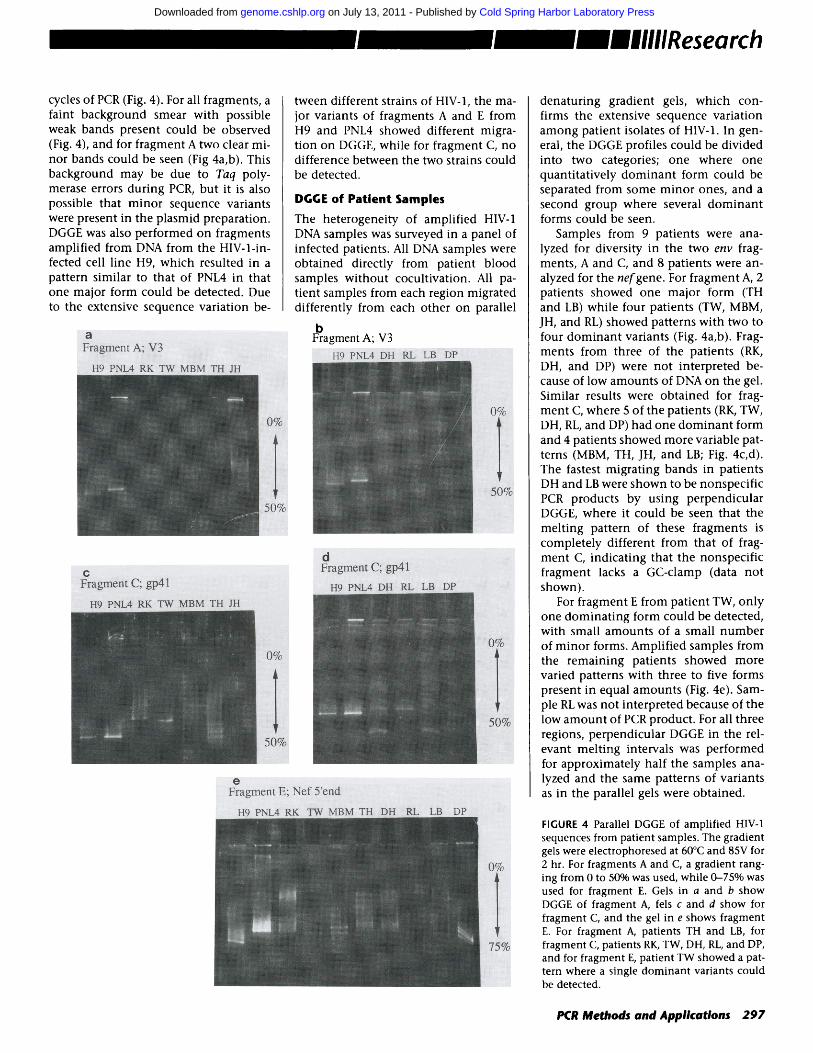
cycles of PCR (Fig. 4). For all fragments, a faint background smear with possible weak bands present could be observed (Fig. 4), and for f ragment A two clear mi- nor bands could be seen (Fig 4a,b). This background may be due to Taq poly- merase errors during PCR, but it is also possible that minor sequence variants were present in the plasmid preparation. DGGE was also performed on fragments amplified from DNA from the HIV-l-in- fected cell line H9, which resulted in a pattern similar to that of PNL4 in that one major form could be detected. Due to the extensive sequence variation be-
tween different strains of HIV-I, the ma- jor variants of fragments A and E from H9 and PNL4 showed different migra- tion on DGGE, while for fragment C, no difference between the two strains could be detected.
DGGE of Patient Samples
The heterogeneity of amplified HIV-1 DNA samples was surveyed in a panel of infected patients. All DNA samples were obtained directly from patient blood samples without cocultivation. All pa- tient samples from each region migrated differently from each other on parallel
denaturing gradient gels, which con- firms the extensive sequence variation among patient isolates of HIV-1. In gen- eral, the DGGE profiles could be divided into two categories; one where one quantitatively dominan t form could be separated from some minor ones, and a second group where several dominan t forms could be seen.
Samples from 9 patients were ana- lyzed for diversity in the two env frag- ments, A and C, and 8 patients were an- alyzed for the nefgene. For fragment A, 2 patients showed one major form (TH and LB) while four patients (TW, MBM, JH, and RL) showed patterns with two to four dominan t variants (Fig. 4a,b). Frag- ments from three of the patients (RK, DH, and DP) were not interpreted be- cause of low amounts of DNA on the gel. Similar results were obtained for frag- ment C, where 5 of the patients (RK, TW, DH, RL, and DP) had one dominan t form and 4 patients showed more variable pat- terns (MBM, TH, JH, and LB; Fig. 4c, d). The fastest migrating bands in patients DH and LB were shown to be nonspecific PCR products by using perpendicular DGGE, where it could be seen that the melting pattern of these fragments is completely different from that of frag- ment C, indicating that the nonspecific fragment lacks a GC-clamp (data not shown).
For f ragment E from patient TW, only one dominat ing form could be detected, with small amounts of a small number of minor forms. Amplified samples from the remaining patients showed more varied patterns with three to five forms present in equal amounts (Fig. 4e). Sam- ple RL was not interpreted because of the low amoun t of PCR product. For all three regions, perpendicular DGGE in the rel- evant melt ing intervals was performed for approximately half the samples ana- lyzed and the same patterns of variants as in the parallel gels were obtained.
FIGURE 4 Parallel DGGE of amplified HIV-1 sequences from patient samples. The gradient gels were electrophoresed at 60~ and 85V for 2 hr. For fragments A and C, a gradient rang- ing from 0 to 50% was used, while 0-75% was used for fragment E. Gels in a and b show DGGE of fragment A, fels c and d show for fragment C, and the gel in e shows fragment E. For fragment A, patients TH and LB, for fragment C, patients RK, TW, DH, RL, and DP, and for fragment E, patient TW showed a pat- tern where a single dominant variants could be detected.
PCR Methods and Applications 297
Cold Spring Harbor Laboratory Press on July 13, 2011 - Published by genome.cshlp.orgDownloaded from

Often, when there was a relatively high yield of PCR product, several forms melting at low denaturant concentra- tions could be detected. This can be seen for sample TH, fragment E, for example (Fig. 4e). These bands are characteristic of heteroduplexes formed in the later cy- cles of PCR. In addition, a background smear was often present, which was probably due to various sequence changes introduced by Taq polymerase during PCR (see above).
The results of the patient survey show that it is possible to separate different forms for all fragments tested, and that it is possible to resolve dominan t and mi- nor variants in single PCR-amplified pa- tient samples. The dominan t forms within each sample are limited to one or a few, which can be analyzed further (see below).
Effects of Cocultivation
Peripheral blood mononuclear cells (PBMC) from a patient infected with HIV-1 (patient JH) were cocultivated with PHA-activated PBMCs from an un- infected donor. Samples were taken at 11 and 15 days after initiation of culture, and DGGE analysis was performed on amplified fragments A and C (V3 and gp41) from both time points and from
FIGURE 5 Parallel DGGE of fragments A and C from patient JH, amplified either directly from a patient blood sample or after coculti- vation of patient cells with peripheral blood mononuclear cells from a healthy donor. DGGE was performed at 60~ and 85V for 2 hr, and the gradient range was 0-50%. (Lanes 1-3) Fragment C amplified, after 1S days of cocultivation, after 11 days, and directly, re- spectively; (lanes 4-6) fragment A amplified directly, after 11 days of cocultivation, and after 15 days, respectively. The arrows indi- cate the dominant variants after cocultiva- tion.
DNA prepared directly from a blood sam- ple from the patient. The results (Fig. 5) were similar for both fragments, and show that the pattern of variants changes during culture, so that a differ- ent major variant dominates after cul- ture (see arrows in Fig. S). This result is
a .
~. G 7 A G G A G G C 7 T G G T ~ G G 7 ~- - - = G - - - - G - - T
b. ~ G T a G G A G G C T T G a T a G G T - - : - G - - - ~ c
FIGURI: 6 Automated direct sequence analysis of variants of fragment C from patient TH. The different forms were separated by parallel DGGE (see Fig. 3c), excised from the gel, extracted by repeated freezing, and reamplified for sequencing. The two forms show a single base difference, which is indicated by arrows. Form a contains a G:C base pair at the varying position and therefore melted at a higher denaturant concentration.
in accordance with results obtained through cloning and sequencing PCR products.~ 2,4,s)
Isolation and Sequencing of Different Forms
The dominan t bands observed after DGGE of fragment C from patient TH (Fig. 3a) were excised from a parallel gra- dient gel and reamplified using the same primers as in the original amplification (R1S4-1118). When the reamplified sam- ple was analyzed using DGGE, a strong enr ichment for the desired band could be seen (the desired band consti tuting >90% of the total product; data not shown). Reamplification with primers for sequence analysis was performed as described in Materials and Methods, and high-quality sequence information was generated. Clear single-base differences between the different forms could be de- tected. For example, the difference be- tween the two fastest migrat ing forms in fragment C from fragment TH was found to be a G:C-A:T difference (Fig. 6) located approximately 70 bases from the 3' end of the fragment. The difference is lo- cated in the lowest melt ing domain and is therefore expected to be easily de- tected by DGGE. The form containing the AT base pair melts at a lower dena- turant concentration, and thus migrates a shorter distance in the parallel DGGE.
DISCUSSION
DGGE has been demonstra ted here to be a useful tool for the study of populations of HIV-1 DNA fragments amplified di- rectly from patient samples. Theoretical calculations and results from using this method in several different systems in- dicate that practically all sequence changes in a lower melt ing domain can be detected. r176 When multiple se- quence variants are present, the analysis is facilitated by the presence of only one band in the gel per unique sequence. The DNA fragments are not modified during the assay and the gels are easy to handle, so that individual variants can be isolated and reamplified for subse- quent sequence analysis. Together, these features make it possible to separate dif- ferent HIV-1 variants, and ult imately to determine their precise base differences.
Three different regions of HIV-1, spanning a total of approximately 1100
298 PCR Methods and Applications
Cold Spring Harbor Laboratory Press on July 13, 2011 - Published by genome.cshlp.orgDownloaded from
bases, have been analyzed here. The cal- culated melting properties suggest that the current GC-clamped fragments and DGGE conditions will allow detection of sequence changes in at least two-thirds of these fragments. The remaining bases are in higher melt ing domains close to the GC-clamps and may require DGGE with different conditions for reliable mutat ion detection. The exact amount of sequence available to DGGE analysis in these fragments will become more clear as additional fragments are isolated and sequenced.
Both perpendicular DGGE and paral- lel DGGE have been performed. Perpen- dicular gradient gels have the advantage that non specific PCR products, when present, do not confuse the analysis to the same extent as in parallel gels. How- ever, the resolution is superior in the parallel gradient gels, where the bands are concentrated when the migrat ion is retarded. Routinely, perpendicular gels are performed for a few samples to de- termine the overall melting properties of the region, and when it is needed to clar- ify the pattern of variants.
Using DGGE, it was possible to sepa- rate HIV-1 variants in all regions tested. Variation both between and within pa- tient samples could be detected, and both major and minor forms could be identified. It was confirmed that when lymphocytes from an HIV-l-infected pa- tient are concultivated with peripheral blood mononuclear cells (PBMCs) from a healthy donor, a minor variant in the patient sample can become the domi- nant variant after cocultivation.
The limit of detection of HIV-1 vari- ants that are present in low amounts is probably determined by the background caused by Taq DNA polymerase errors. It may be possible to increase the detection sensitivity by using Vent DNA poly- merase, which has proofreading activity and therefore a lower error rate. (23~ It is also possible that minor variants can be obscured through heteroduplex forma- tion with the major variants in the later PCR cycles. This can be avoided by lim- iting the number of PCR cycles. No more than 25 and 30 cycles were used for the first- and second-step PCR reactions, re- spectively, to minimize changes in the distribution of variants arising due to saturation of the PCR reaction. The pos- sibility that the pattern of different forms could be affected by Taq poly- merase errors occurring during PCR was
tested by repeated runs from samples with multiple forms. The patterns of se- quence variants were virtually indistin- guishable when samples were repeated in this way (data not shown).
DGGE is expected to resolve almost all variants in the suitable melting do- mains, and because there is no interme- diate cloning step the whole population of products is analyzed. Therefore, DGGE gives a direct representation of the distribution of different sequence variants in the amplified samples. Over- all, the diversity of different HIV-1 vari- ants within the patient samples analyzed here by DGGE appears different from that previously described using the method of cloning and sequencing of PCR products. DGGE suggests that the major variant(s) may be quantitatively dominant to a greater extent than what has been estimated using sequence anal- ysis of cloned PCR products. The overall distribution of HIV-1 variants within dif- ferent patient samples will be investi- gated.
Direct sequence analysis of DNA reamplified from single bands excised from denaturing gradient gels showed that it is possible to characterize differ- ent HIV-1 sequence variants unambigu- ously using this approach, despite the presence of multiple variants. Direct se- quence analysis was performed using magnetic beads for isolation of single- stranded templates combined with a universal priming site introduced via PCR and fluorescent sequence analysis. Using this method high-quality se- quence information could be reliably obtained (Fig. 6).
In conclusion, a technique using DGGE and direct automated sequence analysis for direct characterization of the distribution of sequence variants with HIV isolates is presented. Variants can be separated in PCR-amplified fragments from different regions of HIV-1, and in- dividual variants can be isolated from the gel and their sequence determined by direct automated sequence analysis. Using this method, variation within in- dividual patient samples was detected and it was confirmed that cocultivation in PBMCs leads to changes in HIV-1 pop- ulations. Using DGGE, it will be possible to study changes in HIV-1 populations during different stages of the infection, after transmission of HIV-1 between in- dividuals, in different cell types, and in proviral and free viral forms.
ACKNOWLEDGMENTS
The careful technical assistance of Derek Ng Tang, with the cocultivation, is grate- fully acknowledged. We thank Prof. L.S. Lerman for access to the software used for theoretical melt ing calculations. This work was supported by the W.M. Keck Foundation and National Institutes of Health grant 5U01AI 30243-03 and Genome Program Center grant 21- 272110105218.
REFERENCES
1. Myers, G., J.A. Berzofsky, B. Korber, and R.F. Smith. 1991. Human retroviruses and AIDS 1991. Los Alamos National Labora- tory, Los Alamos, New Mexico.
2. Meyerhans, A., R. Cheynier, J. Albert, M. Seth, S. Kwok, J. Sninsky, L. Morfeldt- M~nson, B. ~sj6, and S. Wain-Hobson. 1989. Temporal fluctuations in HIV qua- sispecies in vivo are not reflected by se- quential H1V isolations. Cell 58: 901-910.
3. Wolfs, T.F., J-J. deJong, H. van den Berg, J.M.G.H. Tunagel, W.J.A. Krone, and J. Goudsmit. 1990. Evolution of sequences encoding the principal neutralization epitope of human immunodeficiency vi- rus 1 is host dependent, rapid, and conti- nous. Proc. Natl. Acad. Sci. 87: 9938-9942.
4. Vartanian, J-P., A. Meyerhans, B./~sj6, and S. Wain-Hobson. 1991. Selection, recom- bination, and G-A hypermutation of hu- man immunodeficiency virus type 1 ge- nomes. J. Virol. 65" 1779-1788.
5. Delassus, S., R. Cheynier, and S. Wain- Hobson. 1991. Evolution of human im- munodeficiency virus type 1 nef and long terminal repeat sequences over 4 years in vivo and in vitro. J. Virol. 65: 225--231.
6. Holmes, E.C., L-Q. Zhang, P. Simmonds, C.A. Ludlam, and A.J. Leigh Brown. 1992. Convergent and divergent evolution in the surface envelope glycoprotein of hu- man immunodeficiency virus type 1 within a single infected patient. Proc. Natl. Acad. Sci. 89" 4835-4839.
7. Wolinsky, S.M., C.M. Wike, B.T.M. Korber, C. Hutto, W.P. Parks, L.L. Rosen- blum, K.J. Kunstman, M.R. Furtado, and J.L. Munoz. 1992. Selective transmission of human immunodeficiency virus type-1 variants from mothers to infants. Science 255: 1134-1137.
8. Ou, C-Y., C.A. Ciesielsky, G. Myers, C.I. Bandea, C-C. Luo, B.T.M. Korber, J.I. Mul- lins, G. Schochetman, R.L. Berkelman, A.N. Economou, J.J. WiRe, L.J. Furman, G.A. Satten, K.A. Maclnnes, J.W. Curran, H.W. Jaffe, Laboratory Investigation group, and Epidemiologic Investigation group. 1992. Molecular epidemiology of HIV transmission in a dental practice. Sci- ence 256: 1165-1171.
PCR Methods and Applications 299
Cold Spring Harbor Laboratory Press on July 13, 2011 - Published by genome.cshlp.orgDownloaded from

9. Balfe, P., P. Simmonds, C.A. Ludlam, J.O. Bishop, and A.J. Leigh Brown. 1990. Con- current evolution of human immunode- ficiency virus type 1 in patients infected from the same source: rate of sequence change and low frequency of inactivating mutations. J. Virol. 64: 6221-6233.
10. Wahlberg, J., J. Albert, J. Lundeberg, A. con Gegerfelt, K. Broliden, G. Utter, E-M. Feny6, and M. Uhl~n. 1991. Analysis of the V3 loop in neutralization-resistant human immunodeficiency virus type 1 variants by direct solid-phase DNA se- quencing. AIDS Res . Hum. Retrovir. 7: 983-989.
11. Burger, H., B. Weiser, K. Flaherty, J. Gulla, P-N. Nguyen, and R.A. Gibbs. 1991. Evo- lution of human immunodeficiency virus type 1 nucleotide sequence diversity among close contacts. Proc. Natl. Acad. Sci. 88: 11236-11240.
12. Orita, M., Y. Suzuki, T. Sekiya, and K. Ha- yashi. 1989. Rapid and sensitive detection of point mutations and DNA polymor- phisms using the polymerase chain reac- tion. Genomics 5: 874-879.
13. Cotton, R.G.H., N.R. Rodrigues, and R.D. Campbell. 1988. Reactivity of cytosine and thymine in single-base-pair mis- matches with hydroxylamine and os- mium tetroxide and its application to the study of mutations. Proc. Natl. Acad. Sci. 85: 4397-4401.
14. Fisher, S. and L.S. Lerman. 1983. DNA fragments differing by single base-pair substitutions are separated in denaturing gradient gels: Correspondence with melt- ing theory. Proc. Natl. Acad. Sci. 80: 1579- 1583.
15. Sheffield, V.C., D.R. Cox, L.S. Lerman, and R.M. Myers. 1989. Attachment of a 40-base pair G+C-rich sequence (GC- clamp) to genomic DNA fragments by the polymerase chain reaction results in im- proved detection of single-base changes. Proc. Natl. Acad. Sci 86: 232-236.
16. Theophilus, B.D.M., T. Latham, G.A. Grabowski, and F.I. Smith. 1989. Compar- ison of RNase A, a chemical cleavage and GC-clamped denaturing gradient gel elec- trophoresis for the detection of mutations in exon 9 of the human 13-glucosidase gene. Nucleic Acids Res. 17: 7707-7722.
17. Gibbs, R.A., P-N. Nguyen, A. Edwards, A.B. Civitello, and C.T. Caskey. 1990. Multi- plex DNA deletion detection and exon se- quencing of the hypoxanthine phospho- ribosyl transferase gene in Lesch-Nyhan families. Genomics 7: 235-244.
18. Lerman, L.S. and K. Silverstein. 1987. Computational simulation of DNA melt- ing and its application to denaturing gra- dient gel electrophoresis. Methods Enzy- tool. 155: 482-501.
19. Smith-SCrensen, B., E. Hovig, B. Anders- son, and A-L. BodCrresen. 1992. Screening for mutations in human hprt cDNA using
the polymerase chain reaction (PCR) in combination with constant denaturant gel electrophoresis (CDGE). Mutat. Res. (in press).
20. Cariello, N.F., P. Keohavong, A.G. Kat, and W.G. Thilly. 1990. Molecular analysis of complex cell populations: Mutational spectra of MNNG and ICR-191. Murat. Res. 231:165-176.
21. Myers, R.M., S.G. Fischer, T. Maniatis, and L.S. Lerman. 1985. Modification of the metling properties of duplex DNA by at- tachment of a GC-rich DNA sequence as determined by denaturing gradient gel electrophoresis. Nucleic Acids Res. 13: 3111-3129.
22. Myers, R.M., S.G. Fischer, L.S. Lerman, and T. Maniatis. 1985. Nearly all single base substitutions in DNA fragments joined to a GC-clamp can be detected by denaturing gradient gel electrophoresis. Nucleic Acids Res. 13: 3131-3145.
23. Ling, L.L., P. Keohavong, C. Dias, and W.G. Thilly. 1991. Optimization of the polymerase chain reaction with regard to fidelity: Modified T7, Taq, and Vent DNA polymerases. PCR Methods Applic. 1: 63- 69.
Received November 25, 1992; accepted in revised form February 1, 1993.
300 PCR Methods and Applications
Cold Spring Harbor Laboratory Press on July 13, 2011 - Published by genome.cshlp.orgDownloaded from
Related Documents