Quantification of the Vitamin D Receptor - Coregulator Interaction Arnaud Teichert ‡,§ , Leggy A. Arnold ‡,∥ , Steve Otieno ǂ , Yuko Oda § , Indre Augustinaite ∥ , Tim R. Geistlinger ⊥ , Richard W. Kriwacki ǂ , R. Kiplin Guy ∥ , and Daniel D. Bikle §,* §Endocrine Unit, University of California, San Francisco, CA 94121 ∥The Department of Chemical Biology and Therapeutics, St Jude Children's Research Hospital, 162 Danny Thomas Place, Memphis, TN 38105 ⊥Amyris Biotechnologies, Emeryville, CA 94608 ǂThe Department of Structural Biology, St. Jude Children's Research Hospital, 162 Danny Thomas Place, Memphis, Tennessee 38105, USA. Abstract The vitamin D receptor (VDR) regulates a diverse set of genes that control processes including bone mineral homeostasis, immune function and hair follicle cycling. Upon binding to its natural ligand, 1α,25(OH) 2 D 3 , the VDR undergoes a conformational change that allows the release of corepressor proteins and the binding of coactivator proteins necessary for gene transcription. We report the first comprehensive evaluation of the interaction of the VDR with a library of coregulator binding motifs in the presence of two ligands, the natural ligand 1α,25(OH) 2 D 3 and a synthetic, non-secosteroidal agonist LG190178. We show that the VDR has relatively high affinity for the second and third LxxLL motifs of SRC1, SRC2 and SRC3 and second LxxLL motif of DRIP205. This pattern is distinct in comparison to other nuclear receptors. The pattern of VDR-coregulator binding affinities was very similar for the two agonists investigated, suggesting that the biologic functions of LG190178 and 1α,25(OH) 2 D 3 are similar. Hairless binds the VDR in the presence of ligand through a LxxLL motif (Hr-1), repressing transcription in the presence and absence of ligand. The VDR binding patterns identified in this study may be used to predict functional differences among different tissues expressing different sets of coregulators, thus facilitating the goal of developing tissue and gene specific vitamin D response modulators. The vitamin D receptor (VDR), which binds 1α,25-dihydroxyvitamin D 3 (1α,25(OH) 2 D 3 ), contains several functional domains, including a ligand-binding domain (LBD), that mediates ligand-dependent gene regulation (1). A critical step in 1α,25(OH) 2 D 3 action is the induction of a LBD conformational change to form activation function 2 (AF-2)(2), a hydrophobic cleft formed by three helices and a short COOH-terminal amphipathic alpha helix (H12) (3), which serves as a binding surface for coactivators (4). Unliganded nuclear receptor (NR) heterodimers associate with corepressors such as the nuclear receptor corepressor (NCoR) and the silencing mediator of retinoic acid and thyroid hormone receptor (SMRT) (5,6) and associated histone deacetylases (7,8). These proteins function as adaptors to convey a repressive signal to the transcriptional apparatus by maintaining a closed chromatin structure with the histone N- terminal ‘tails’ in a charged state tightly associated with DNA (9). Ligand binding promotes the release of corepressors and the binding of coactivators, enhancing the transcription of ‡ These authors contributed equally to this work. * Corresponding author: Tel: 415-750-2089, Fax: 415-750-6929, e-mail: [email protected] Supporting Information Available FP isotherms for each Texas Red labeled peptide in the presence and absence 1α,25(OH) 2 D 3 and ITC isotherms of unlabeled and Texas Red labeled SRC2-3 peptide. This material is available free of charge via the Internet at http://pubs.acs.org”. NIH Public Access Author Manuscript Biochemistry. Author manuscript; available in PMC 2010 February 24. Published in final edited form as: Biochemistry. 2009 February 24; 48(7): 1454–1461. doi:10.1021/bi801874n. NIH-PA Author Manuscript NIH-PA Author Manuscript NIH-PA Author Manuscript

Welcome message from author
This document is posted to help you gain knowledge. Please leave a comment to let me know what you think about it! Share it to your friends and learn new things together.
Transcript
Quantification of the Vitamin D Receptor - Coregulator Interaction
Arnaud Teichert‡,§, Leggy A. Arnold‡,∥, Steve Otienoǂ, Yuko Oda§, Indre Augustinaite∥, TimR. Geistlinger⊥, Richard W. Kriwackiǂ, R. Kiplin Guy∥, and Daniel D. Bikle§,*
§Endocrine Unit, University of California, San Francisco, CA 94121
∥The Department of Chemical Biology and Therapeutics, St Jude Children's Research Hospital, 162 DannyThomas Place, Memphis, TN 38105
⊥Amyris Biotechnologies, Emeryville, CA 94608
ǂThe Department of Structural Biology, St. Jude Children's Research Hospital, 162 Danny Thomas Place,Memphis, Tennessee 38105, USA.
AbstractThe vitamin D receptor (VDR) regulates a diverse set of genes that control processes including bonemineral homeostasis, immune function and hair follicle cycling. Upon binding to its natural ligand,1α,25(OH)2D3, the VDR undergoes a conformational change that allows the release of corepressorproteins and the binding of coactivator proteins necessary for gene transcription. We report the firstcomprehensive evaluation of the interaction of the VDR with a library of coregulator binding motifsin the presence of two ligands, the natural ligand 1α,25(OH)2D3 and a synthetic, non-secosteroidalagonist LG190178. We show that the VDR has relatively high affinity for the second and third LxxLLmotifs of SRC1, SRC2 and SRC3 and second LxxLL motif of DRIP205. This pattern is distinct incomparison to other nuclear receptors. The pattern of VDR-coregulator binding affinities was verysimilar for the two agonists investigated, suggesting that the biologic functions of LG190178 and1α,25(OH)2D3 are similar. Hairless binds the VDR in the presence of ligand through a LxxLL motif(Hr-1), repressing transcription in the presence and absence of ligand. The VDR binding patternsidentified in this study may be used to predict functional differences among different tissuesexpressing different sets of coregulators, thus facilitating the goal of developing tissue and genespecific vitamin D response modulators.
The vitamin D receptor (VDR), which binds 1α,25-dihydroxyvitamin D3 (1α,25(OH)2D3),contains several functional domains, including a ligand-binding domain (LBD), that mediatesligand-dependent gene regulation (1). A critical step in 1α,25(OH)2D3 action is the inductionof a LBD conformational change to form activation function 2 (AF-2)(2), a hydrophobic cleftformed by three helices and a short COOH-terminal amphipathic alpha helix (H12) (3), whichserves as a binding surface for coactivators (4). Unliganded nuclear receptor (NR) heterodimersassociate with corepressors such as the nuclear receptor corepressor (NCoR) and the silencingmediator of retinoic acid and thyroid hormone receptor (SMRT) (5,6) and associated histonedeacetylases (7,8). These proteins function as adaptors to convey a repressive signal to thetranscriptional apparatus by maintaining a closed chromatin structure with the histone N-terminal ‘tails’ in a charged state tightly associated with DNA (9). Ligand binding promotesthe release of corepressors and the binding of coactivators, enhancing the transcription of
‡These authors contributed equally to this work.*Corresponding author: Tel: 415-750-2089, Fax: 415-750-6929, e-mail: [email protected] Information AvailableFP isotherms for each Texas Red labeled peptide in the presence and absence 1α,25(OH)2D3 and ITC isotherms of unlabeled and TexasRed labeled SRC2-3 peptide. This material is available free of charge via the Internet at http://pubs.acs.org”.
NIH Public AccessAuthor ManuscriptBiochemistry. Author manuscript; available in PMC 2010 February 24.
Published in final edited form as:Biochemistry. 2009 February 24; 48(7): 1454–1461. doi:10.1021/bi801874n.
NIH
-PA Author Manuscript
NIH
-PA Author Manuscript
NIH
-PA Author Manuscript
specific genes (10). Some coactivators, such as the SRC family (11-13), recruit othercoregulators with histone acetylase activity and remodel chromatin structure. Othercoactivators, such as the DRIP factors (14,15), interact with the basal transcriptionalmachinery. In the unliganded state, helix 12 projects away from the globular core of the LBD,while in the liganded state this helix contacts the LBD globular core domain to create an AF-2surface through which coactivator proteins can dock (16,17). Upon 1α,25(OH)2D3 binding,VDR localizes at the vitamin D response elements of target genes, recruits coactivators, whichrecruit histone acetyltransferase to modify histone or bridge the gap between the VDR and thetranscription machinery (18). Both coactivators and corepressors can interact with overlappingsurfaces on the LBD (19). It has been proposed that ligand-dependent exchange betweencorepressors and coactivators is caused by a difference in the length of the interacting motifsthat can be accommodated by the two conformations of the binding pocket (20). Therefore, aligand that displaces the AF2 helix from its active position would be expected to facilitate theinteraction with corepressor proteins and to repress transcriptional activity (21). Coactivatorproteins that bind to AF-2 contain one or more of the consensus sequence LxxLL (L is leucineand x is any amino acid) which forms an amphipathic alpha-helix (Figure 1, A) (22). This helixfits into the hydrophobic cleft of the liganded receptor (17). Receptor-specific binding ofcoactivators containing the LxxLL motif is governed by amino acid residues flanking thebinding site, including in human VDR the conserved residues E420 and K246 (23). Mutationof these residues and adjacent hydrophobic amino acids abolishes both 1α,25(OH)2D3-activated transcription and coactivator interactions with the VDR (24,25).
NR corepressors NCoR and SMRT encode multiple, short receptor interaction domainscomposed of the sequence ΦxxΦΦ (Φ is leucine or isoleucine and x is any amino acid) (26,27). This motif is predicted to form an alpha helix, that is one turn longer than that formed bythe LxxLL motif. In a manner analogous to the LxxLL-containing motifs, it has been suggestedthat this helix also binds the AF-2 surface without requiring a docked helix 12 (27). Indeed,deletion of helix 12 enhances corepressor binding (5), suggesting that this helix does not playan active role in nuclear receptor-corepressor recognition.
Another corepressor, Hr, is expressed primarily in brain, epidermis and hair follicles (28) andis known to repress VDR mediated transcription (29,30). The Hr-VDR interaction is of greatinterest because null mutations in either protein induces alopecia, in both the mouse or human(31-33). Despite a low sequence identity with other corepressors, Hr functions in a similarmanner. For example, it mediates moderate transcriptional repression of liganded TR but strongrepression in case of unliganded TR (34). In contrast to TR, Hr strongly represses VDRmediated transcription in the presence of ligand but moderately in the absence of ligand (30).Hr encodes two LxxLL motifs required for Hr/RORα interaction (35) and two ΦxxΦΦ motifsrequired for Hr/TR interaction (Figure 1, B) (30). In contrast to TR, VDR only interactsphysically and functionally with Hr through a region of Hr containing a LSELL motif at position778-782 (Hr1) and a LDSII motif at position 816-830 (Hr2)(30). As we show in this report onlythe LSELL motif (Hr1) mediates a ligand dependent binding. Some VDR mediated functions,like its control of hair follicle cycling, are ligand independent, but require Hr suggesting a rolefor Hr in this VDR regulated function (36,37).
Ligand selectivity of VDR may affect coregulator recruitment. Published data show that VDRbound to LG190178 is able to recruit coactivator SRC2 like VDR/1α,25(OH)2D3 (38). Unlike1α,25(OH)2D3, LG190178 does not bind to serum vitamin D binding protein and exhibits noinfluence on calcium serum levels in mice (39). Both ligands induce HL-60 differentiation intomacrophages and inhibit the growth of SK-BR-3 and LNCaP cells. Clearly the interplaybetween ligand, receptor, and pools of potentially interacting coregulators is important indefining the physiological activity of VDR.
Teichert et al. Page 2
Biochemistry. Author manuscript; available in PMC 2010 February 24.
NIH
-PA Author Manuscript
NIH
-PA Author Manuscript
NIH
-PA Author Manuscript
Herein we report the first comprehensive evaluation of the interaction of VDR and coregulatorNR boxes using two different biochemical assays: 1) a fluorescence polarization binding assaymeasuring the equilibrium binding of VDR to a library of fluorescent coregulator peptides, amethod that has previously been used to profile other nuclear receptors-coregulator interactions(40-42); and 2) a competition pull-down assay measuring the ability of unlabeled, but otherwiseidentical, coregulator peptides to inhibit the interaction between VDR and full length SRC2.Additionally, we confirm the binding affinities of one labeled and unlabeled coregulatorpeptide SRC2-3 using isothermal titration calorimetry (ITC). Because of the difficultiesencountered by others in working with a full-length VDR-LBD, we used a mutated form thathas no major differences in ligand binding, transactivation, or dimerization with RXRα (43).The binding constants for VDR to a wide range of coregulator NR boxes including LxxLL andLxxII motifs were determined in the presence of 1α,25(OH)2D3 and LG190178.
Experimental ProceduresReagents
1α,25(OH)2D3 (calcitriol) was purchased from WAKO CHEMICALS USA INC; LG190178was synthesized using a published procedure (39).
Protein Expression and PurificationThe VDR-LBDmt, provided by D. Moras (43), was cloned by PCR (primers 5′-CGCGGATCCAGATCTGACAGTCTGCGGCCCAAG-3′ and 5′-CGCGGATCCAGATCTGACAGTCTGCGGCCCAAG-3′) at the BamHI site of the pMAL-c2X vector (New England Biolabs), in fusion with the maltose binding protein (MBP). Theplasmid was expressed in Escherichia coli grown at ambient temperature in 2x LB with 2%glucose. MBP-VDR-LBDmt protein expression was induced with 0.2 mM isopropyl 1-thio-β-D-galactopyranoside for 16 h at 25°C before harvest and cell lysis by freeze-thawing andsonication. Proteins were purified in the absence or presence of ligand 2 μM (1α,25(OH)2D3)or 20 μM (LG190178) using an amylose resin column.
Peptide Library SynthesisCoregulator peptides were synthesized by the Hartwell Center (St. Jude Children's ResearchHospital), purified by RP-HPLC, and analyzed by LC/MS. The peptides containing an N-terminal cysteine were labeled using the thiol-reactive fluorophore, Texas Red maleimide(Molecular Probes): 3 mg of peptide was combined with 1.6 mg of fluorophore in 5 ml of 50%water/DMF. After stirring for 3 h in the dark the reaction mixture was purified by HPLC andanalyzed by LC/MS. All fluorescent peptides possessed purities greater than 95%.
Peptide Binding AssayMBP-VDR-LBDmt protein binding affinities, in presence of 1α,25(OH)2D3 (2 μM) orLG190178 (20 μM) using fluorescently labeled coregulator peptides were measured using apreviously described assay (40). The relative doses of 1α,25(OH)2D3 and LG190178 have beendescribed previously (39). Two different protein batches were used for two independentexperiments, with each carried out in triplet. Reported values reflect the mean value withassociated total error across all experiments.
Competitive Binding AssayFluorescently labeled SRC2 was produced by in vitro transcription–translation (TNT kit &FluoroTect™, Promega) from the plasmid pSGT-SRC2. In a 96 well polypropylene plate(Costar 3365) 8 peptides at a time were diluted from 10000-1.0 μM in DMSO. 2 μl of thediluted peptides were added to 91 μl of buffer (25 mM HEPES, 100 mM NaCl, 1 mM DTT,
Teichert et al. Page 3
Biochemistry. Author manuscript; available in PMC 2010 February 24.
NIH
-PA Author Manuscript
NIH
-PA Author Manuscript
NIH
-PA Author Manuscript
0.01% NP40, 0.1% BSA [added fresh], 0.5 μM VDRmt on amylose beads, and 1 μM 1α,25dihydroxyvitamin D3) in a 96 well filter plate (Millipore, Multiscreen HTS BV). After agitationfor 2 h (IKA microtiterplate shaker) at room temperature (rt), 7 μl of TNT solution was addedto each reaction followed by 2 h of agitation at rt. The filter plate was attached to 96-well plate(Costar 3365) and centrifuged at 50g for 3 minutes followed by the addition of 100 μl of buffer(25 mM HEPES, 100 mM NaCl, 1 mM DTT, 0.01% NP40, 0.1% BSA (added fresh). The filterplate was assembled with another 96-well plate (Costar 3365) and centrifuged at 50g for 3minutes. For elution of the SRC2-VDR complex 10 μl of a 10 mM maltose solution was added.After a 10 minute incubation period the filter plate was assembled with another 96-well plate(Costar 3365) and centrifuged at 50g for 3 minutes. The elutions were treated with 3 μl of 4xSDS-page loading buffer (invitrogen), sealed and incubated for 30 minutes in an oven at 70°C. After separation using SDS-page the fluorescent bands of SRC2 were visualized using afluorescence scanner (Typhoon, GE). The bands were integrated using ImageQuant (MolecularDynamics) and analyzed using Prism (GraphPad). Three independent experiments were carriedout for each state. The IC50 values were obtained by fitting data to the following equation (y= min + (max − min)/1 + (x/Kd) Hill slope.
Calorimetric studiesThe thermodynamics of the interaction between the SRC2-3 peptides and VDR-LBD weredetermined using a VP-ITC instrument (Microcal, Northampton, MA). Therefore, a solutionof either unlabeled or Texas Red-labeled SRC2-3 peptide (0.42 μM) in the syringe was titratedinto a solution of VDR (25 μM) in the cell. 2 μL of the titrant was first injected followed by25 injections of 10 μL each, with a 300 second delay between injections. The experiments wereperformed at 25 °C in a buffer containing 20 mM HEPES pH 7.5 and 150 mM NaCl. Thebinding data were fit to a 1:1 binding model using Origin software (Origin lab, Northampton,MA).
ResultsCoregulator Peptide Library
To evaluate VDR coregulator recruitment, the binding between the VDR-LBD and a libraryof known coregulator peptides consisting of a central LxxLL or ΦxxΦΦ sequence plus up to8 additional flanking residues at each terminus was measured (Figure 2). To allow attachmentof the fluorescent label a non-native cysteine was introduced at the N-terminus of each peptide.Additionally, a cysteine-serine exchange was employed in the case of SRC3-1 and Hr1 toprevent double labeling. Hr1 contains a second cysteine residue at the −3 position of the LxxLLmotif which was used for labeling. All peptide probes were synthesized in parallel using Fmoc(9-fluorenylmethyloxycarbonyl) protective group and purified by RP-HPLC. Identity andpurity were confirmed using HPLC and MALDI-TOF or LCMS and was greater than 95%.
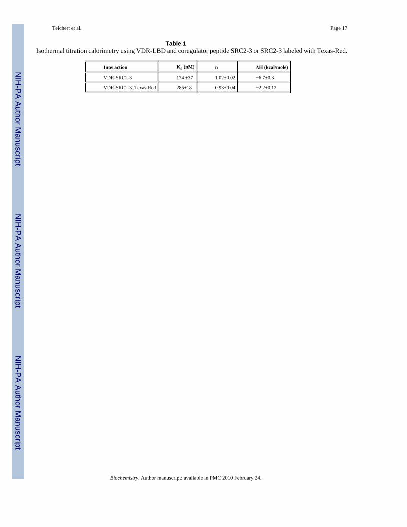
Initial validation of coactivator peptides binding assayInitial peptide binding studies were carried out with SRC2-2 in the presence of 1α,25(OH)2D3 with both MBP-VDR-LBDwt and MBP-VDR-LBDmt, from the construct developedby D. Moras (43). Control experiments using a MBP protein alone showed no interaction withSRC2-2 (data not shown). SRC2-2 binds to both MBP-VDR-LBDwt and MBP-VDR-LBDmtin a similar saturable dose-dependent manner, with somewhat higher affinity for MBP-VDR-LBDmt observed than for MBP-VDR-LBDwt (supplementary material). We used MBP-VDR-LBDmt for further studies because it was soluble at higher concentrations, which was essentialto determine the binding constants (Kd) using fluorescence polarization. To exclude potentialperturbing interactions between the label attached to the peptide and MBP-VDR-LBDmt wemeasured the binding affinity Kd values for labeled and unlabeled coregulator peptide SRC2-3using isothermal titration calorimetry (ITC) (Table 1 and supplementary material). The Kd
Teichert et al. Page 4
Biochemistry. Author manuscript; available in PMC 2010 February 24.
NIH
-PA Author Manuscript
NIH
-PA Author Manuscript
NIH
-PA Author Manuscript
values observed for the Texas-Red labeled and unlabeled peptide SRC2-3 were 285 nM and174 nM, respectively. To demonstrate the ability of unlabeled peptides to inhibit the interactionbetween VDR-LBD and full length SRC2 exhibiting three LxxLL motifs, we performed pull-down competition assays. In these assays the native peptides (LxxLL) blocked this interaction,whereas altered peptides (LxxAA) did not affect this binding (Figure 3, D). To verify thatbinding of Hr-1 is not compromised by Cystein/Serine (C/S) exchange, a competitivefluorescence polarization experiment was carried out showing that Hr-1wt and Hr-1C/S equallyinhibited the binding between SRC2-3 Texas-Red and MBP-VDR-LBDmt (supplementarymaterial).
Coactivator binding assaysThe fluorescence polarization assay was executed by maintaining a constant concentration offluorescently labeled coactivator peptide (10 nM) and a variable concentration of VDR-LBDmtfrom 0.002—199 μM in the absence and presence of 1α,25(OH)2D3 or LG190178. The datawere fit to a sigmoidal dose-response curve and Kd values with 95% confidence intervals aresummarized in Figure 4. The competition pull down assay was carried out with labeled SRC2,solid - supported VDR-LBDmt, and unlabeled coregulator peptides. Displaced fluorescentSRC2 was removed by washes and residual SRC2 was quantified after separation by SDS-page (Figure 3, D). The data were fit to a sigmoidal dose-response curve and Kd values with95% confidence intervals are summarized in Figure 4 and supplementary material.
Coregulator peptides bind VDR-LBDmt in the absence or presence of ligand in three differentmodes
Binding isotherms for coregulator peptides exhibited three behaviors: saturable binding witha clear high dosage plateau, clear interaction without saturation, and no interaction. The higheraffinity group of peptides bound in a ligand-dependent, dose-dependent, and saturable mannerwhere a plateau was reached within the protein concentration range studied (Figure 3, A ■).The Kd values for this class in the presence of ligand were lower than 20 μM as determined byfitting the data to a sigmoidal dose-response curve. Nine coactivator peptides exhibited thismode: SRC1-2, SRC1-3, SRC2-2, SRC2-3, SRC3-2, SRC3-3, DRIP205-2, RIP140-5, and Hr1(Figure 4, green and dark green color code). The binding affinities of theses peptides in theabsence of ligand were dose dependent but exhibited no plateaus at higher proteinconcentrations (Figure 3, A ▲). Because of the missing isotherm saturation we labeled theseinteractions as weak or no binding (Figure 4, grey color code and supplementary material).The IC50 values determined by an independent competition pull down assay were similar toKd values in the presence of ligand measured by FP.
The lower affinity group included peptides exhibiting dose-dependent binding in the presenceof ligand but did not display a plateau at the highest protein concentrations used. In comparisonwith the binding isotherm in the absence of ligand we observed a clear shift indicating ligand-dependent binding (Figure 3, B). For this group of peptides we suggest a Kd value higher than20 μM, and the group includes SRC3-1, DRIP-1, and P300. (Figure 4, light green color code).The binding constants in the absence of ligand exhibited high values indicative of very weakbinding (Figure 4, grey color code and supplementary material). Inhibition of binding betweenSRC2 and VDR was observed at concentrations of these peptides greater than 50 μM.
The third group of peptides showed identical dose-dependent binding in the presence andabsence of ligand and did not display saturation at higher concentrations of peptide. This groupincluded SRC1-1 and SRC2-1 bearing an LxxLL motif and corepressor peptides NCoR, SMRTand Hr2 with LxxIIxxxL/V motifs (Figure 3, C). The mode of binding of these peptides wasindicated as weak binding or no binding (Figure 4, grey color code and supplementarymaterial).
Teichert et al. Page 5
Biochemistry. Author manuscript; available in PMC 2010 February 24.
NIH
-PA Author Manuscript
NIH
-PA Author Manuscript
NIH
-PA Author Manuscript

DiscussionIn this study, we examined the binding of VDR-LBD to a range of target motifs within potentialVDR coregulators. Because of the difficulties encountered by others in working with wild-type VDR-LBD, we used a mutated form, which lacks a domain with no apparent function andlittle homology to other NRs (43-46). Native VDR-LBD and VDR-LBDmt have been shownto function similarly with respect to ligand binding, transactivation, or dimerization withRXRα LBD (43). We confirmed these results by showing that SRC2-2 bound to VDR-LBDmtwith high affinity in a ligand dependent manner. We observed that VDR-LBDmt exhibitedhigher affinity for SRC2-2 than did native VDR-LBD, possibly due to the higher solubility ofthe former construct under our assay conditions.
Our results verify that VDR binds LxxLL motifs from numerous coactivators in a selectivefashion. The Kd values measured for the absolute binding of coregulator motifs to the VDRare similar to those observed for binding to other nuclear receptors (40,47). In contrast to theestrogen receptor and thyroid receptor, which have the highest affinity for the second NR boxof each SRC, VDR binds to the second and third NR box with higher affinities than to the firstNR box. Considering all NR boxes of each SRC, we observed the same affinity between VDR/SRC1 and VDR/SRC2. In contrast, SRC3 exhibited a stronger interaction between SRC3-1and VDR in comparison with SRC1-1 and SRC2-1, whereas SRC3-2 has a weaker interactionwith VDR in comparison with SRC1-2 and SRC2-2. This may suggest that SRC3 dominatesbinding to VDR relative to SRC1 or SRC2. SRC3 has been previously described as a more“general” coactivator, but our results may reveal selectivity for VDR for this protein (48).
It has been reported that amino acid residues N-terminal to the LxxLL motif have a majorimpact on the binding affinities of coregulator peptides (22,49,50). In particular, coregulatorpeptides bearing hydrophobic amino acids at the −1 position of the LxxLL motif exhibitedhigh affinities for ER, PPAR, TR and VDR (22,40,51,52). In the case of the peptidesinvestigated in this study we observed the same correlation. In contrast, we showed thatcoregulator peptides missing a hydrophobic amino acid residue at the −1 position like SRC1-1,SRC2-1, SRC3-1, P300, NCoR, and SMRT exhibited a low binding affinity towards VDR withthe exception of Hr1. The crystal structure of SRC1-2 bound to VDR-LBD illustrates theimportance of the isoleucine residue in contact with the hydrophobic surface of AF-2 (Figure5, A) (53). In overlay, the SRC1-1 structure exhibits a protonated lysine residue destabilizingthe interaction between coregulator and VDR. Zella et al. (52) reported that VDR agonistderived LxxLL peptides, originated from a phage display screening, have the proposedsequence L × E/H × H/F P L/M/I. Because of the sequence similarity with DRIP peptides weobserved a binding constant of 1.6 μM (Figure 4) for DRIP-2 following the proposed paradigmof a HPMLxxLL sequence. In contrast DRIP-1, bearing a NPILxxLL sequence has a weakinteraction with VDR. DRIP205 is known to interact directly with ligand-activated VDR/RXRheterodimers mostly through the second motif, although both motifs are equally required forVDR mediated transcription in cells (54).
Hr1 is part of the coregulator Hairless which represses VDR mediated transcription in thepresence and absence of 1α,25(OH)2D3 (30). Hairless interacts with VDR predominantlythrough the 750-864 domain, exhibiting a coactivator LxxLL motif (Hr1) and a corepressorLxxIIxxxV motif (Hr2) (Figure 1, B). Our investigation of these motifs separately showed thatHr2 exhibits a weak affinity towards VDR in the presence and absence of ligand in contrast toHr1 which exhibited a 1α,25(OH)2D3-dependent VDR-LBD binding of 5.9 μM (Figure 4).Because Hr1 is bearing a serine residue at the −1 N-terminal position of LxxLL (Figure 5, B)this is the first reported coregulator peptide binding to VDR and bearing a polar residue at thisparticular position. Peptides with a similar sequence to Hr1 were identified by Zella at al.(52) in the presence of the VDR antagonist ZK15922, but binding of these peptides could not
Teichert et al. Page 6
Biochemistry. Author manuscript; available in PMC 2010 February 24.
NIH
-PA Author Manuscript
NIH
-PA Author Manuscript
NIH
-PA Author Manuscript
be confirmed by a two-hybrid assay (52). The binding affinities for Hr1 measured in thepresence of agonist 1α,25(OH)2D3 and LG190178 were similar for VDR. We can concludethat Hr1 is supporting the interaction between VDR and Hr in the presence of ligand. This wasa surprise. Our previous findings in keratinocytes with full length molecules showed that VDRand Hairless bind to each other in the absence of ligand and that this binding is partiallydisplaced by addition of 1α,25(OH)2D3 (29). This suggests that results at least for Hairlessobtained with peptides recapitulates only partially the binding properties of full length proteins.
Like Hr2, corepressor SMRT and NCoR peptides exhibited weak interactions with VDR inthe presence and absence of ligand, although stronger interactions have been reported for fulllength proteins (55,56). The length of a polypeptide has a strong influence on its threedimensional structure which determines binding affinity and solubility. Lower affinities ofsmall peptide mimics in comparison with full-length proteins are not uncommon. The natureof the FP assay does not allow reliable Kd measurements of lower affinity probes due to lightscattering caused by protein aggregation at high concentrations. In contrast, high affinity probesexhibiting a saturated signal at higher protein concentrations do not suffer from thisinterference. Moreover, a recent study showed that NCoR and SMRT dependant repression of1α,25(OH)2D3 mediated transcription by VDR/RXR heterodimer is achieved through theirrecruitment by RXR but not by VDR (56), suggesting that further studies involving VDR/RXRheterodimers are needed to determine the affinity of these co-repressors for these nuclearreceptors.
In the presence of synthetic agonist LG190178, VDR adopts an agonistic conformation,recruiting all investigated coregulators with the same affinities as VDR/1α,25(OH)2D3. Thisresult is supported by the fact that in the case of VDR, crystal structures with different vitaminD3 analogs in the presence of coregulator peptide exhibit an unaltered AF-2 domain (23,53,57,58). We can conclude that the signal transduction mediated by VDR is similar regardlessof which of the two ligands is bound.
The IC50 values determined by a competition pull-down assay confirmed the Kd valuesdetermined by the FP measurements. We reported a similar behavior for TR (59) suggestingthat although full length SRC2 exhibits multiple NR boxes no assisted binding among themoccurs in this biochemical assay.
Presumably there are additional factors that influence NR recruitment of coregulators such aspost-translational modifications, structural determinants arising from specific DNA responseelements, cooperativity, cellular environment, and additional interaction surfaces on the NRand coregulator proteins, needing more complex models to fully dissect NR-coregulatorinteractions. These binding patterns draw a first picture of potential functional differences fordifferent ligands in tissues with different sets of coregulators. The use of more ligands shouldallow us to predict these functional differences, thus facilitating the goal of developing tissueand gene specific vitamin D response modulators.
Supplementary MaterialRefer to Web version on PubMed Central for supplementary material.
AcknowledgementWe thank Dr. Patrick Rodrigues and Bob Cassell for peptide synthesis at the Hartwell Center of Bioinformatics andBiotechnology, St. Jude Children's Research Hospital, Memphis, TN.
This work was supported by NIH grants AR050023 (D. D. B), AR39448 (D. D. B), DK58080 (R. K. G.), a CancerCenter Support Grant 2P30CA021765 (L. A. A., S. O., R. W. K. and R. K. G.), the American Lebanese and SyrianAssociated Charities (ALSAC) and St Jude Children's Research Hospital (SJCRH).
Teichert et al. Page 7
Biochemistry. Author manuscript; available in PMC 2010 February 24.
NIH
-PA Author Manuscript
NIH
-PA Author Manuscript
NIH
-PA Author Manuscript

Abbreviations1,25(OH)2D3, 1,25 dihydroxyvitamin D3 Calcitriol; DRIP, vitamin D receptor-interactingprotein; Hr, Hairless; MBP, Maltose binding protein; NCoR, Nuclear receptor corepressor;NR box, Nuclear receptor box; SMRT, silencing mediator for retinoic acid and thyroidhormone receptors; SRC, Steroid receptor coactivator; TR, thyroid hormone receptor; VDR,Vitamin D receptor.
References1. Carlberg C, Polly P. Gene regulation by vitamin D3. Crit Rev Eukaryot Gene Expr 1998;8:19–42.
[PubMed: 9673449]2. Moras D, Gronemeyer H. The nuclear receptor ligand-binding domain: structure and function. Curr
Opin Cell Biol 1998;10:384–391. [PubMed: 9640540]3. Danielian P, White R, Lees J, Parker M. Identification of a conserved region required for hormone
dependent transcriptional activation by steroid hormone receptors. Embo J 1992;11:1025–1033.[PubMed: 1372244]
4. Feng W, Ribeiro R, Wagner R, Nguyen H, Apriletti J, Fletterick R, Baxter J, Kushner P, West B.Hormone-dependent coactivator binding to a hydrophobic cleft on nuclear receptors. Science1998;280:1747–1749. [PubMed: 9624051]
5. Chen JD, Evans RM. A transcriptional co-repressor that interacts with nuclear hormone receptors.Nature 1995;377:454–457. [PubMed: 7566127]
6. Kurokawa R, Soderstrom M, Horlein A, Halachmi S, Brown M, Rosenfeld M, Glass C. Polarity-specific activities of retinoic acid receptors determined by a co-repressor. Nature 1995;377:451–454.[PubMed: 7566126]
7. Heinzel T, Lavinsky RM, Mullen TM, Soderstrom M, Laherty CD, Torchia J, Yang WM, Brard G,Ngo SD, Davie JR, Seto E, Eisenman RN, Rose DW, Glass CK, Rosenfeld MG. A complex containingN-CoR, mSin3 and histone deacetylase mediates transcriptional repression. Nature 1997;387:43–48.[PubMed: 9139820]
8. Nagy L, Kao HY, Chakravarti D, Lin RJ, Hassig CA, Ayer DE, Schreiber SL, Evans RM. Nuclearreceptor repression mediated by a complex containing SMRT, mSin3A, and histone deacetylase. Cell1997;89:373–380. [PubMed: 9150137]
9. Belandia B, Parker MG. Nuclear receptors: a rendezvous for chromatin remodeling factors. Cell2003;114:277–280. [PubMed: 12914692]
10. Robyr D, Wolffe A, Wahli W. Nuclear hormone receptor coregulators in action: diversity for sharedtasks. Mol Endocrinol 2000;14:329–347. [PubMed: 10707952]
11. Zhu Y, Qi C, Calandra C, Rao MS, Reddy JK. Cloning and identification of mouse steroid receptorcoactivator-1 (mSRC-1), as a coactivator of peroxisome proliferator-activated receptor gamma. GeneExpr 1996;6:185–195. [PubMed: 9041124]
12. Hong H, Kohli K, Trivedi A, Johnson D, Stallcup M. GRIP1, a novel mouse protein that serves as atranscriptional coactivator in yeast for the hormone binding domains of steroid receptors. Proc NatlAcad Sci U S A 1996;93:4948–4952. [PubMed: 8643509]
13. Voegel JJ, Heine MJ, Zechel C, Chambon P, Gronemeyer H. TIF2, a 160 kDa transcriptional mediatorfor the ligand-dependent activation function AF-2 of nuclear receptors. Embo J 1996;15:3667–3675.[PubMed: 8670870]
14. Yuan C, Ito M, Fondell J, Fu Z, Roeder R. The TRAP220 component of a thyroid hormone receptor-associated protein (TRAP) coactivator complex interacts directly with nuclear receptors in a ligand-dependent fashion. Proc Natl Acad Sci U S A 1998;95:7939–7944. [PubMed: 9653119]
15. Rachez C, Lemon B, Suldan Z, Bromleigh V, Gamble M, Naar A, Erdjument-Bromage H, TempstP, Freedman L. Ligand-dependent transcription activation by nuclear receptors requires the DRIPcomplex. Nature 1999;398:824–828. [PubMed: 10235266]
16. Renaud J, Rochel N, Ruff M, Vivat V, Chambon P, Gronemeyer H, Moras D. Crystal structure of theRAR-gamma ligand-binding domain bound to all-trans retinoic acid. Nature 1995;378:681–689.[PubMed: 7501014]
Teichert et al. Page 8
Biochemistry. Author manuscript; available in PMC 2010 February 24.
NIH
-PA Author Manuscript
NIH
-PA Author Manuscript
NIH
-PA Author Manuscript

17. Shiau A, Barstad D, Loria P, Cheng L, Kushner P, Agard D, Greene G. The structural basis of estrogenreceptor/coactivator recognition and the antagonism of this interaction by tamoxifen. Cell1998;95:927–937. [PubMed: 9875847]
18. Kim S, Shevde NK, Pike JW. 1,25-Dihydroxyvitamin D3 stimulates cyclic vitamin D receptor/retinoid X receptor DNA-binding, co-activator recruitment, and histone acetylation in intactosteoblasts. J Bone Miner Res 2005;20:305–317. [PubMed: 15647825]
19. Li Y, Lambert MH, Xu HE. Activation of nuclear receptors: a perspective from structural genomics.Structure 2003;11:741–746. [PubMed: 12842037]
20. Perissi V, Staszewski L, McInerney E, Kurokawa R, Krones A, Rose D, Lambert M, Milburn M,Glass C, Rosenfeld M. Molecular determinants of nuclear receptor-corepressor interaction. GenesDev 1999;13:3198–3208. [PubMed: 10617569]
21. Xu HE, Stanley TB, Montana VG, Lambert MH, Shearer BG, Cobb JE, McKee DD, Galardi CM,Plunket KD, Nolte RT, Parks DJ, Moore JT, Kliewer SA, Willson TM, Stimmel JB. Structural basisfor antagonist-mediated recruitment of nuclear co-repressors by PPARalpha. Nature 2002;415:813–817. [PubMed: 11845213]
22. Heery D, Kalkhoven E, Hoare S, Parker M. A signature motif in transcriptional co-activators mediatesbinding to nuclear receptors. Nature 1997;387:733–736. [PubMed: 9192902]
23. Vanhooke JL, Benning MM, Bauer CB, Pike JW, DeLuca HF. Molecular structure of the rat vitaminD receptor ligand binding domain complexed with 2-carbon-substituted vitamin D3 hormoneanalogues and a LXXLL-containing coactivator peptide. Biochemistry 2004;43:4101–4110.[PubMed: 15065852]
24. Jurutka PW, Hsieh JC, Remus LS, Whitfield GK, Thompson PD, Haussler CA, Blanco JC, Ozato K,Haussler MR. Mutations in the 1,25-dihydroxyvitamin D3 receptor identifying C-terminal aminoacids required for transcriptional activation that are functionally dissociated from hormone binding,heterodimeric DNA binding, and interaction with basal transcription factor IIB, in vitro. J Biol Chem1997;272:14592–14599. [PubMed: 9169418]
25. Jimenez-Lara AM, Aranda A. Lysine 246 of the vitamin D receptor is crucial for ligand-dependentinteraction with coactivators and transcriptional activity. J Biol Chem 1999;274:13503–13510.[PubMed: 10224118]
26. Hu X, Lazar M. The CoRNR motif controls the recruitment of corepressors by nuclear hormonereceptors. Nature 1999;402:93–96. [PubMed: 10573424]
27. Nagy L, Kao H, Love J, Li C, Banayo E, Gooch J, Krishna V, Chatterjee K, Evans R, Schwabe J.Mechanism of corepressor binding and release from nuclear hormone receptors. Genes Dev1999;13:3209–3216. [PubMed: 10617570]
28. Cachon-Gonzalez MB, Fenner S, Coffin JM, Moran C, Best S, Stoye JP. Structure and expression ofthe hairless gene of mice. Proc Natl Acad Sci U S A 1994;91:7717–7721. [PubMed: 8052649]
29. Xie Z, Chang S, Oda Y, Bikle DD. Hairless suppresses vitamin D receptor transactivation in humankeratinocytes. Endocrinology 2006;147:314–323. [PubMed: 16269453]
30. Hsieh JC, Sisk JM, Jurutka PW, Haussler CA, Slater SA, Haussler MR, Thompson CC. Physical andfunctional interaction between the vitamin D receptor and hairless corepressor, two proteins requiredfor hair cycling. J Biol Chem 2003;278:38665–38674. [PubMed: 12847098]
31. Cichon S, Anker M, Vogt IR, Rohleder H, Putzstuck M, Hillmer A, Farooq SA, Al-Dhafri KS, AhmadM, Haque S, Rietschel M, Propping P, Kruse R, Nothen MM. Cloning, genomic organization,alternative transcripts and mutational analysis of the gene responsible for autosomal recessiveuniversal congenital alopecia. Hum Mol Genet 1998;7:1671–1679. [PubMed: 9736769]
32. Li YC, Pirro AE, Amling M, Delling G, Baron R, Bronson R, Demay MB. Targeted ablation of thevitamin D receptor: an animal model of vitamin D-dependent rickets type II with alopecia.Proceedings of the National Academy of Sciences USA 1997;94:9831–9835.
33. Yoshizawa T, Handa Y, Uematsu Y, Takeda S, Sekine K, Yoshihara Y, Kawakami T, Arioka K, SatoH, Uchiyama Y, Masushige S, Fukamizu A, Matsumoto T, Kato S. Mice lacking the vitamin Dreceptor exhibit impaired bone formation, uterine hypoplasia and growth retardation after weaning.Nat Genet 1997;16:391–396. [PubMed: 9241280]
Teichert et al. Page 9
Biochemistry. Author manuscript; available in PMC 2010 February 24.
NIH
-PA Author Manuscript
NIH
-PA Author Manuscript
NIH
-PA Author Manuscript

34. Potter GB, Zarach JM, Sisk JM, Thompson CC. The thyroid hormone-regulated corepressor hairlessassociates with histone deacetylases in neonatal rat brain. Mol Endocrinol 2002;16:2547–2560.[PubMed: 12403844]
35. Moraitis AN, Giguere V, Thompson CC. Novel mechanism of nuclear receptor corepressor interactiondictated by activation function 2 helix determinants. Mol Cell Biol 2002;22:6831–6841. [PubMed:12215540]
36. Skorija K, Cox M, Sisk JM, Dowd DR, MacDonald PN, Thompson CC, Demay MB. Ligand-independent actions of the vitamin D receptor maintain hair follicle homeostasis. Mol Endocrinol2005;19:855–862. [PubMed: 15591533]
37. Panteleyev AA, Botchkareva NV, Sundberg JP, Christiano AM, Paus R. The role of the hairless (hr)gene in the regulation of hair follicle catagen transformation. Am J Pathol 1999;155:159–171.[PubMed: 10393848]
38. Perakyla M, Malinen M, Herzig KH, Carlberg C. Gene regulatory potential of nonsteroidal vitaminD receptor ligands. Mol Endocrinol 2005;19:2060–2073. [PubMed: 15860548]
39. Boehm MF, Fitzgerald P, Zou A, Elgort MG, Bischoff ED, Mere L, Mais DE, Bissonnette RP, HeymanRA, Nadzan AM, Reichman M, Allegretto EA. Novel nonsecosteroidal vitamin D mimics exert VDR-modulating activities with less calcium mobilization than 1,25-dihydroxyvitamin D3. Chem Biol1999;6:265–275. [PubMed: 10322128]
40. Moore JM, Galicia SJ, McReynolds AC, Nguyen NH, Scanlan TS, Guy RK. Quantitative proteomicsof the thyroid hormone receptor-coregulator interactions. J Biol Chem 2004;279:27584–27590.[PubMed: 15100213]
41. Estebanez-Perpina E, Moore JM, Mar E, Delgado-Rodrigues E, Nguyen P, Baxter JD, Buehrer BM,Webb P, Fletterick RJ, Guy RK. The molecular mechanisms of coactivator utilization in ligand-dependent transactivation by the androgen receptor. Journal of Biological Chemistry 2005;280:8060–8068. [PubMed: 15563469]
42. Krylova IN, Sablin EP, Moore J, Xu RX, Waitt GM, MacKay JA, Juzumiene D, Bynum JM, MadaussK, Montana V, Lebedeva L, Suzawa M, Williams JD, Williams SP, Guy RK, Thornton JW, FletterickRJ, Willson TM, Ingraham HA. Structural analyses reveal phosphatidyl inositols as ligands for theNR5 orphan receptors SF-1 and LRH-1. Cell 2005;120:343–355. [PubMed: 15707893]
43. Rochel N, Wurtz JM, Mitschler A, Klaholz B, Moras D. The crystal structure of the nuclear receptorfor vitamin D bound to its natural ligand. Mol Cell 2000;5:173–179. [PubMed: 10678179]
44. Zamir I, Harding HP, Atkins GB, Horlein A, Glass CK, Rosenfeld MG, Lazar MA. A nuclear hormonereceptor corepressor mediates transcriptional silencing by receptors with distinct repression domains.Mol Cell Biol 1996;16:5458–5465. [PubMed: 8816459]
45. McKenna N, Lanz R, O'Malley B. Nuclear receptor coregulators: cellular and molecular biology.Endocr Rev 1999;20:321–344. [PubMed: 10368774]
46. Jurutka PW, Hsieh JC, Nakajima S, Haussler CA, Whitfield GK, Haussler MR. Human vitamin Dreceptor phosphorylation by casein kinase II at Ser-208 potentiates transcriptional activation. ProcNatl Acad Sci U S A 1996;93:3519–3524. [PubMed: 8622969]
47. Bramlett KS, Wu Y, Burris TP. Ligands specify coactivator nuclear receptor (NR) box affinity forestrogen receptor subtypes. Mol Endocrinol 2001;15:909–922. [PubMed: 11376110]
48. Liao L, Kuang SQ, Yuan Y, Gonzalez SM, O'Malley BW, Xu J. Molecular structure and biologicalfunction of the cancer-amplified nuclear receptor coactivator SRC-3/AIB1. J Steroid Biochem MolBiol 2002;83:3–14. [PubMed: 12650696]
49. McInerney EM, Rose DW, Flynn SE, Westin S, Mullen TM, Krones A, Inostroza J, Torchia J, NolteRT, Assa-Munt N, Milburn MV, Glass CK, Rosenfeld MG. Determinants of coactivator LXXLLmotif specificity in nuclear receptor transcriptional activation. Genes & Development 1998;12:3357–3368. [PubMed: 9808623]
50. Chang C, Norris JD, Gron H, Paige LA, Hamilton PT, Kenan DJ, Fowlkes D, McDonnell DP.Dissection of the LXXLL nuclear receptor-coactivator interaction motif using combinatorial peptidelibraries: discovery of peptide antagonists of estrogen receptors alpha and beta. Mol Cell Biol1999;19:8226–8239. [PubMed: 10567548]
Teichert et al. Page 10
Biochemistry. Author manuscript; available in PMC 2010 February 24.
NIH
-PA Author Manuscript
NIH
-PA Author Manuscript
NIH
-PA Author Manuscript
51. Nolte RT, Wisely GB, Westin S, Cobb JE, Lambert MH, Kurokawa R, Rosenfeld MG, Willson TM,Glass CK, Milburn MV. Ligand binding and co-activator assembly of the peroxisome proliferator-activated receptor-gamma. Nature 1998;395:137–143. [PubMed: 9744270]
52. Zella LA, Chang CY, McDonnell DP, Wesley Pike J. The vitamin D receptor interacts preferentiallywith DRIP205-like LxxLL motifs. Arch Biochem Biophys 2007;460:206–212. [PubMed: 17254542]
53. Vanhooke JL, Tadi BP, Benning MM, Plum LA, DeLuca HF. New analogs of 2-methylene-19-nor-(20S)-1,25-dihydroxyvitamin D3 with conformationally restricted side chains: evaluation ofbiological activity and structural determination of VDR-bound conformations. Arch BiochemBiophys 2007;460:161–165. [PubMed: 17227670]
54. Rachez C, Gamble M, Chang C, Atkins G, Lazar M, Freedman L. The DRIP complex and SRC-1/p160 coactivators share similar nuclear receptor binding determinants but constitute functionallydistinct complexes. Molecular & Cellular Biology 2000;20:2718–2726. [PubMed: 10733574]
55. Tagami T, Lutz WH, Kumar R, Jameson JL. The interaction of the vitamin D receptor with nuclearreceptor corepressors and coactivators. Biochem Biophys Res Commun 1998;253:358–363.[PubMed: 9878542]
56. Sanchez-Martinez R, Zambrano A, Castillo AI, Aranda A. Vitamin D-dependent recruitment ofcorepressors to vitamin D/retinoid X receptor heterodimers. Mol Cell Biol 2008;28:3817–3829.[PubMed: 18362166]
57. Ciesielski F, Rochel N, Moras D. Adaptability of the Vitamin D nuclear receptor to the syntheticligand Gemini: remodelling the LBP with one side chain rotation. J Steroid Biochem Mol Biol2007;103:235–242. [PubMed: 17218092]
58. Shimizu M, Miyamoto Y, Takaku H, Matsuo M, Nakabayashi M, Masuno H, Udagawa N, DelucaHF, Ikura T, Ito N. 2-Substituted-16-ene-22-thia-1alpha,25-dihydroxy-26,27-dimethyl-19-norvitamin D(3) analogs: Synthesis, biological evaluation, and crystal structure. Bioorg Med Chem. 2008
59. Geistlinger TR, Guy RK. An inhibitor of the interaction of thyroid hormone receptor beta andglucocorticoid interacting protein 1. J Am Chem Soc 2001;123:1525–1526. [PubMed: 11456738]
Teichert et al. Page 11
Biochemistry. Author manuscript; available in PMC 2010 February 24.
NIH
-PA Author Manuscript
NIH
-PA Author Manuscript
NIH
-PA Author Manuscript

Figure 1. Structural Composition of Coregulator SRC1 and HrA, functional domains of SRC1 including the nuclear interaction domain (NID) and its threenuclear receptor interaction domains (NR boxes, SRC1-1, SRC1-2, SRC1-3). B, functionaldomains of Hairless including the three transcriptional repression domains (RDs). RD2,comprised of a LxxLL and a ΦxxΦΦ motif, is necessary and sufficient for Hr-VDR interaction.
Teichert et al. Page 12
Biochemistry. Author manuscript; available in PMC 2010 February 24.
NIH
-PA Author Manuscript
NIH
-PA Author Manuscript
NIH
-PA Author Manuscript
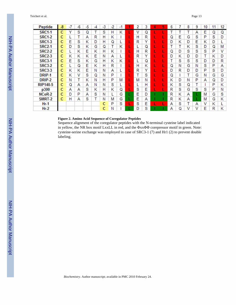
Figure 2. Amino Acid Sequence of Coregulator PeptidesSequence alignment of the coregulator peptides with the N-terminal cysteine label indicatedin yellow, the NR box motif LxxLL in red, and the ΦxxΦΦ corepressor motif in green. Note:cysteine-serine exchange was employed in case of SRC3-1 (7) and Hr1 (2) to prevent doublelabeling.
Teichert et al. Page 13
Biochemistry. Author manuscript; available in PMC 2010 February 24.
NIH
-PA Author Manuscript
NIH
-PA Author Manuscript
NIH
-PA Author Manuscript
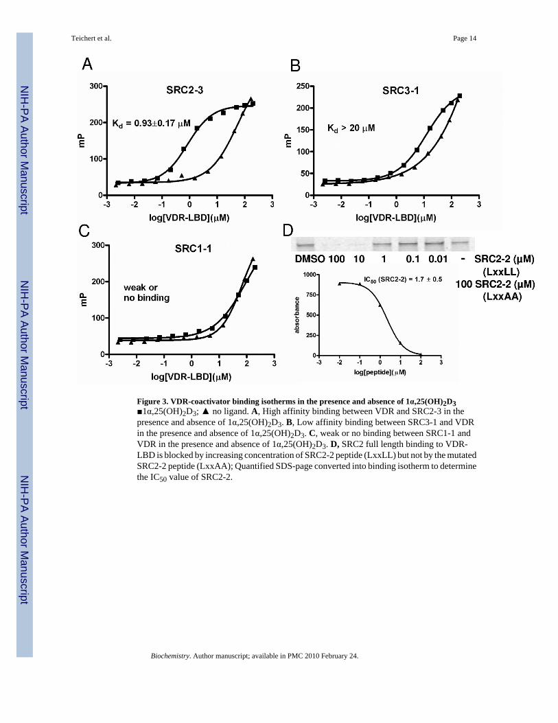
Figure 3. VDR-coactivator binding isotherms in the presence and absence of 1α,25(OH)2D3■1α,25(OH)2D3; ▲ no ligand. A, High affinity binding between VDR and SRC2-3 in thepresence and absence of 1α,25(OH)2D3. B, Low affinity binding between SRC3-1 and VDRin the presence and absence of 1α,25(OH)2D3. C, weak or no binding between SRC1-1 andVDR in the presence and absence of 1α,25(OH)2D3. D, SRC2 full length binding to VDR-LBD is blocked by increasing concentration of SRC2-2 peptide (LxxLL) but not by the mutatedSRC2-2 peptide (LxxAA); Quantified SDS-page converted into binding isotherm to determinethe IC50 value of SRC2-2.
Teichert et al. Page 14
Biochemistry. Author manuscript; available in PMC 2010 February 24.
NIH
-PA Author Manuscript
NIH
-PA Author Manuscript
NIH
-PA Author Manuscript
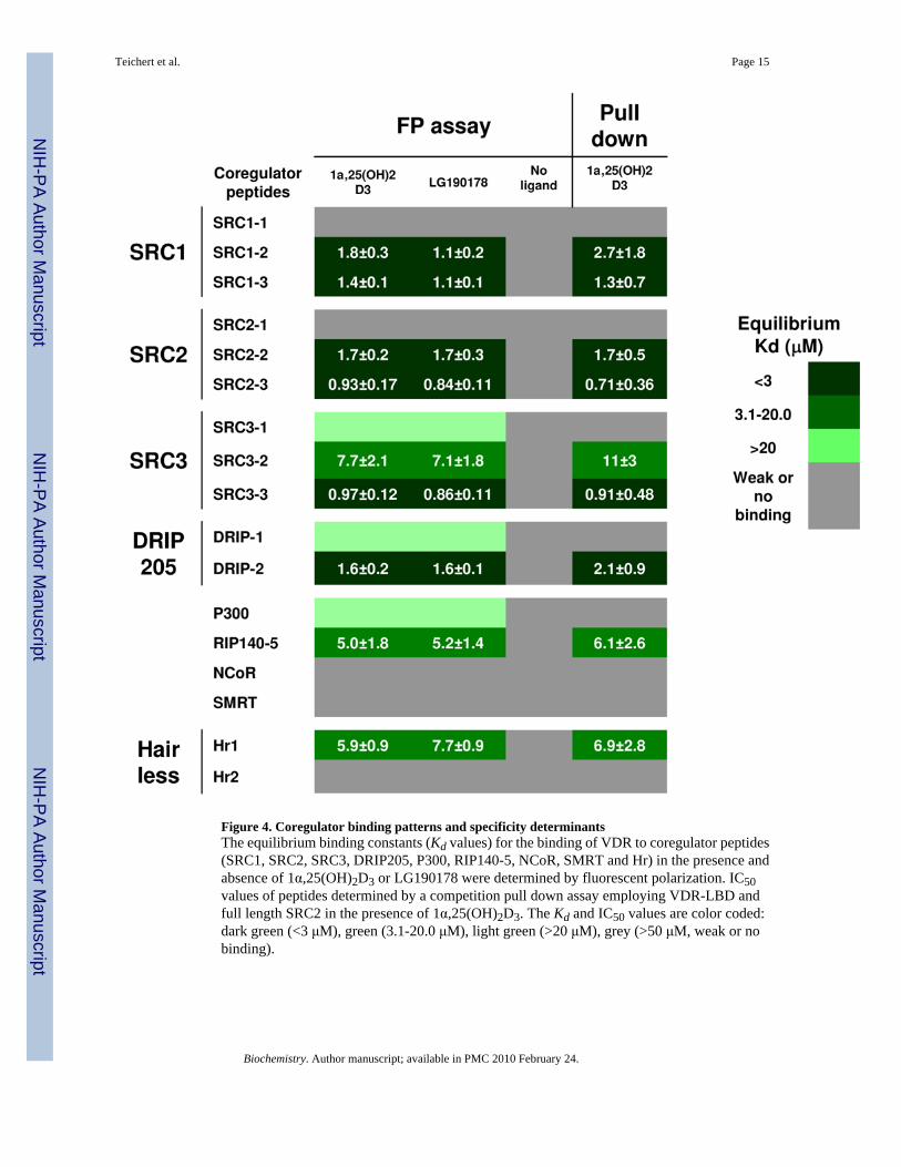
Figure 4. Coregulator binding patterns and specificity determinantsThe equilibrium binding constants (Kd values) for the binding of VDR to coregulator peptides(SRC1, SRC2, SRC3, DRIP205, P300, RIP140-5, NCoR, SMRT and Hr) in the presence andabsence of 1α,25(OH)2D3 or LG190178 were determined by fluorescent polarization. IC50values of peptides determined by a competition pull down assay employing VDR-LBD andfull length SRC2 in the presence of 1α,25(OH)2D3. The Kd and IC50 values are color coded:dark green (<3 μM), green (3.1-20.0 μM), light green (>20 μM), grey (>50 μM, weak or nobinding).
Teichert et al. Page 15
Biochemistry. Author manuscript; available in PMC 2010 February 24.
NIH
-PA Author Manuscript
NIH
-PA Author Manuscript
NIH
-PA Author Manuscript
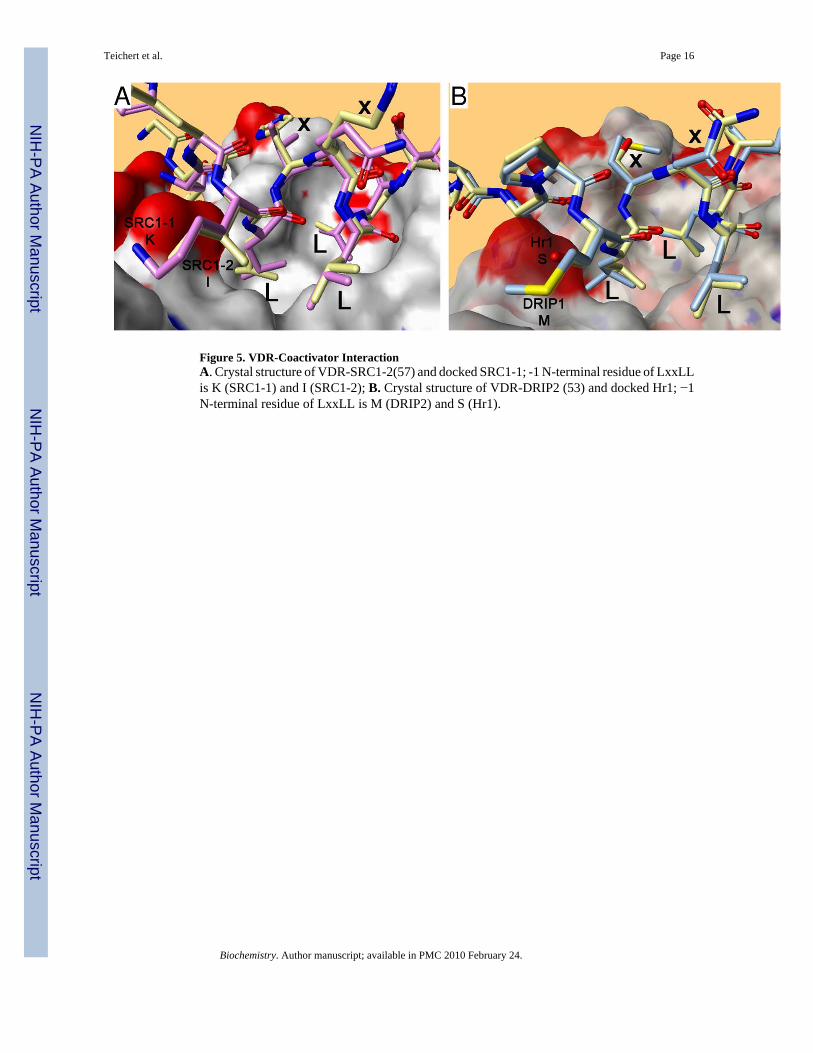
Figure 5. VDR-Coactivator InteractionA. Crystal structure of VDR-SRC1-2(57) and docked SRC1-1; -1 N-terminal residue of LxxLLis K (SRC1-1) and I (SRC1-2); B. Crystal structure of VDR-DRIP2 (53) and docked Hr1; −1N-terminal residue of LxxLL is M (DRIP2) and S (Hr1).
Teichert et al. Page 16
Biochemistry. Author manuscript; available in PMC 2010 February 24.
NIH
-PA Author Manuscript
NIH
-PA Author Manuscript
NIH
-PA Author Manuscript
NIH
-PA Author Manuscript
NIH
-PA Author Manuscript
NIH
-PA Author Manuscript
Teichert et al. Page 17
Table 1Isothermal titration calorimetry using VDR-LBD and coregulator peptide SRC2-3 or SRC2-3 labeled with Texas-Red.
Interaction Kd (nM) n ΔH (kcal/mole)
VDR-SRC2-3 174 ±37 1.02±0.02 −6.7±0.3
VDR-SRC2-3_Texas-Red 285±18 0.93±0.04 −2.2±0.12
Biochemistry. Author manuscript; available in PMC 2010 February 24.
Related Documents