CHARACTERIZATION AND EXPRESSION STUDY OF PREL2A . AND PREL2B ON EARLY VERTEBRATE DEVELOPMENT Jonathan Chan Chia Jui (36395) QL Bachelor of Science with Honours (Resource Biotechnology) Al 2015 C454 2015 605

Welcome message from author
This document is posted to help you gain knowledge. Please leave a comment to let me know what you think about it! Share it to your friends and learn new things together.
Transcript

CHARACTERIZATION AND EXPRESSION STUDY OF PREL2A AND PREL2B ON EARLY VERTEBRATE DEVELOPMENT
Jonathan Chan Chia Jui
(36395)
QL Bachelor of Science with Honours (Resource Biotechnology)
Al 2015 C454 2015
605
Pusar Khidmu MakJumnr k ldmiddotrrI UNIVE SIll MALAYSIA SARAAlshy
Characterization and Expression Study of Pre12a and Pre12b on Early Vertebrate
Development
JONATHAN CHAN CHIA JUI (36395)
A thesis submitted in partial fulfillment of the requirement for the degree of Bachelor of
Science with Honours (Resource Biotechnology)
Faculty of Resource Science and Technology
UNlVERSITI MALAYSIA SARA W AK
2015
I
if if
Acknowledgement
This project was conducted in partial fulfillment for the Degree of Bachelor of Science
with Honors First I would like to express my indebtness appreciation and veneration to
my supervisor Dr Lee Kui Soon for giving me the opportunity to work on project related
with molecular biology under his guidance full support and cherished advices I dedicate
my gratitude and special thanks to Pang Shek Li (Msc) candidate for her persistent advices
experiences well-regarded comments and indelible inspiration patiently guiding me and
teaching me various lab techniques throughout the project I would also like to thank AP
Dr Mohd Hasnain Md Hussain AP Dr Awang Ahmad Sallehin AP Dr Edmund Sim Ui
Hang AP Dr Hairul Roslan and Ng Kher Lee (PhD) for providing necessary equipments
and reagents for my experiments Furthermore I take this opportunity to thank the rest
postgraduates of Animal Biotech Lab and lab assistants including Mr Iskandar for their
assistances and advices on technical troubles as well as supportive atmosphere for learning
Finally I thank to my friends for their encouragement and to my parents for their absolute
moral supports with a wish for their good health
II
UNIVERSITI MALAYSIA SARAWAK
Grade
Please tick (J)
Final Year Project Report 0 ~asters 0 PhD 0
DECLARATION OF ORIGINAL WORK
This declaration is made on the day of 2012
Students Declaration
I ----- --- -- ----- ---- ----- --- -- -- ---- ------ ------ -- ---- -- -- -------- ------ ------ ---- -------- --- ------------- ---------- ---- -- ----- --shy------- (PLEASE INDICATE STUDENTS N~E ~ATRIC NO AND FACULTY) hereby declare that the work e n titled -------- -- -- --------------------------------------------------------------- -------------shy-------- is my original work I have not copied from any other students work or from any other sources except where due reference or acknowledgement is made explicitly in the text nor has any part been written for me by another person
Date submitted Name of the student (Matric No)
Supervisors Declaration
1------------------------------------------------------------- (SUPERVISORS NA~E) hereby certifies that the work entitled ------------------------------------------------------------- --------------(TITLE) was prepared by the above named student and was submitted to the FACULTY as a partialfull fulfillment for the conferment of ------------------------------------------------------------------------shy(PLEASE INDICATE THE DEGREE) and the aforementioned work to the best of my know ledge is the said students work
Received for examination by Date________
(Name of the supervisor)
III
I declare that ProjectThesis is classified as (Please tick C-J))
DCONFIDENTIAL (Contains confidential information under the Official Secret Act 1972)
DRESTRICTED (Contains restricted information as specified by the organisation where
research was done)
DOPEN ACCESS
Validation of ProjectThesis
I therefore duly affirm with free consent and willingly declare that this said ProjectThesis shall be placed officially in the Centre for Academic Information Services with the abiding interest and rights as follows
bull This ProjectThesis is the sole legal property of Universiti Malaysia Sarawak (UNIMAS)
bull The Centre for Academic Information Services has the lawful right to make copies for the purpose of academic and research only and not for other purpose
bull The Centre for Academic Information Services has the lawful right to digitalise the content for the Local Content Database
bull The Centre for Academic Information Services has the lawful right to make copies of the ProjectThesis for academic exchange between Higher Learning Institute
bull No dispute or any claim shall arise from the student itself neither third party on this ProjectThesis once it becomes thesole property of UNIMAS
bull This ProjectThesis or any material data and information related to it shall not be distributed published or disclosed to any party by the student except with UNIMAS permission
Student signature ___________ Supervisor signature _______
(Date) (Date)
Current Address
Notes If the ProjectThesis is CONFIDENTIAL or RESTRICTED please attach together as annexure a letter from the organisation with the period and reasons of confidentiality and restriction
[The instrument is duly prepared by The Centre for Academic Information Services]
IV
PusltKhidmru aklumH A middotudcr middot I J r 1 l ~ I Y L- S L WAt
Title and Front Cover
Acknowledgement
Declaration of original work
Table of Contents
List of Abbreviations
List of Tables
List of Figures
Abstract
10 Introduction 2-3
20 Literature Review 4-9
Table of Contents
I
n
III-IV
V-VI
VII
VIII
IX
21 Proline-rich EVH 1 ligand (PREL) family genes 4-5
22 Proline-rich EVH1 ligand 2 (PREL2) or Lamellipodin 5-6
23 EnaV ASP protein family 6-7
24 Zebrafish (Dania rerio) 7-8
25 Gene Duplication 8
26 Duplication -Degeneration- Complementary (DDC) Model 9
30 Methodology 10-15
31 Primer Design and Sequence 10
32 Recovery ofE coli Recombinant Culture from Glycerol Stock 10
33 Colony PCR of Plasmid DNA 10-11
34 Agarose-gel Electrophoresis (AGE) 11
35 Plasmid DNA Extraction using PureYieldtrade Plasmid Miniprep 11-12
36 Plasmid Purification using Wizardreg SV Gel and PCR Clean-Up System 12-13
37 Quantification ofDNA 13
38 Minipreparations of Plasmid DNA using Alkaline Lysis 13-14
39 DNA Sequencing 14
310 Preparation of Digoxigenin-Iabelled RNA in situ Probes 14-15
311 Zebrafish Embryo Fixation and Dehydration 15
312 Embryo Mounting and Microscope Techniques 15
40 Results 16-23
41 Recovery of E coli Recombinant Culture from Glycerol Stock 16
42 Colony PCR 16
43 Plasmid Minipreparations 17
44 Gel Purification of Plasmid DNA 17
45 Plasmid DNA quantification 18
46 Direct DNA Sequencing 18-21
47 Multiple sequence alignment between zebrafish Prel2 peptides from sequenced transcripts with other vertebrates Prel2 peptides 22-25
48 Preparation of Digoxigenin-Iabelled RNA in situ Probes 25-26
50 Discussion 27-35
51 Colony PCR and Subculture of Recombinant E coli 27
52 Plasmid Extraction and Purification 27-29
53 Plasmid DNA Quantification 29-30
54 Direct DNA Sequencing 31-32
55 Preparation of Digoxigenin-Iabelled RNA in situ Probes 32-34
56 Zebrafish Embryo Fixation and Dehydration 35
60 Conclusion 36
70 References 37-41
80 Appendices 42-45
VI
I
I
List of Abbreviations
AGE
CME
DDC
EVH
Hoxb
LAIX
LB
Lpd
MRL
PFA
PH
PBST
Prel2
PTU
RA
RE
RIAM
RT
RT-PCR
SDS
SH3
TE buffer
VASP
Agarose gel electrophoresis
Clathrin-mediated endocytosis
Duplication-Degeneration-Complementary
EnaIV ASP homology
Homeobox
LBIAmpicillinlIPTGX-Gal
Luria-BertaniLuria Broth
Lamellipodin
MiglORIAMlLpd
Parafonnaldehyde
Pleckstrin homology
Plrosphate Butfered Saline with Tween-20
Proline-rich EVHl ligand 2
I-Phenyl-2-thiourea
Ras association
Restriction enzyme
Rap I-GTP-interacting adapter molecule
Room temperature
Reverse Transcription-Polymerase Chain Reaction
Sodium dodecyl sulfate
SRC Homology 3
Tris-EDTA buffer
Vasodilator-stimulated phosphoprotein
V
List of Tables
Figure Description Page
Table 331 Setup conditions for colony peR and master mix 11
Table 451 Spectrophotometric analysis of plasmid samples 17
BLASTn significant alignments (~99) for construct Table 461 17-18 Prel2a (1381 letters)
Table 462 BLASTn significant alignments (~99) for construct 19
Prel2b (1171 letters)
I
I
1shy
VIllI
List of Figures
Figure Description Page
Figure 221 Topological features of (A) PH domain (B) RA domain 4
Figure 261 The three possible outcomes of duplicate gene pairs that
contains regulatory regions with distinct functions 8
represented by small boxes
Figure 411 Agar plates of recombinant culture Prel2a (1 amp 2) and 15 Prel2b (3 amp 4) after overnight incubation
Figure 421 Gel result for colony PCR for Prel2a 15
Figure 422 Gel result for colony PCR for Prel2b 15
Figure 431 Plasmid mini prep for Prel2a 16
Figure 432 Plasmid mini prep for Prel2b 16
Figure 441 Purified plasmid for Prel2a 16
Figure 442 Purified plasmid for Prel2b 16
Figure 461 Pairwise alignment bet~een extracted plasmid Prel2a
18and PREDICTED Dania reria sidkeyl h243
(sidkeylh243) transcript variant X6 mRNA
Figure 462 Pairwise alignment between extracted plasmid Prel2b
20and PREDICTED Dania reria Ras association
(RaIGDSAF-6) and pleckstrin homology domains 1
(raphl) transcript variant X6 mRNA
Figure 471 Multiple sequence alignment Prei2a and Pre12b peptide
sequences translated from the construct with Prel2 24
peptides of other vertebrates
Figure 481 Gel result of linearized pGEMT -Easy-PreI2a 25
Figure 482 Gel result oflinearized pGEMT-Easy-PreI2b 25
Figure 483 Gel result of purified linearized pGEMT-Easy-PreI2a 25
Figure 484 Prel2a RNA probes 25
I
Characterization and Expression Study of Prel2a and Prel2b on Early Vertebrate Development
Jonathan Chan Chia Jui
Resource Bioteclmology Faculty of Resource Science and Technology
University Malaysia Sarawak
Abstract
Proline-rich EVHlligand 1 (Prell) and Proline-rich EVHI ligand 2 (PreI2) belong to Prel gene family Both genes are homologous and participate in cellular integrin activation Pre12 interacts with GTPase called Ras in GTP-bound state and is essential in regulating cell adhesion and migration It has been identified to involve in mechanism assisting endocytosis of epidermal growth factor receptor (EGFR) that participates in development of arious diseases such as cancer However the comprehensive and mechanistic model of protein signaling involving cell migration is still unavailable Hence Prel2 is selected as candidate gene with zebrafish (Danio rerio) in this study to examine its function This tudy was done partly to verify the occurrence ofPrel2 gene duplication as well as to fulfill
our understanding on the importance of Prel2 during vertebrate development The methodology involves colony peR plasmid extraction DNA sequencing zebrafish cultivation RNA probe preparation in situ hybridization embryo mounting and microscopy Through these experiments Prel2 genes were characterized and plasmid with the gene construct was isolated purified and ready for RNA probe synthesis for in situ hybridization of zebrafish
Keywords Prel gene family EVH Danio rerio endocytosis gene duplication
Abstrak
Proline-rich EVHl ligand 1 (Prell) dan Proline-rich EVHl ligand 2 (PreI2) merupakan gen-gen di bawah kategori kumpulan Prel gen Kedua-dua gen tersebut adalah homolog dan menyertai aktivasi integrin sel Prel2 berinteraksi dengan satu GTPase dinamakan Ras yang berikat kepada GTP dan diperlukan dalam regulasi pelekatan dan migrasi sel Gen fer ebut juga dikaitkan menyertai mekanisme endocytosis epidermal growth factor receptor (EGFR) yang penting dalam perkembangan pelbagai penyakit termasuk kanser Walau bagaimanapun model yang komprehensiJ dan mekanistik tentang protein signaling melibati migrasi sel masih tiada Dengan ini Prel2 dipilih sebagai gen calon dan zebrajish (Danio rerio) sebagai model binatang dalpm kajian ini untuk menguji jimgsi gen tersebul Kajian ini dilaksana sebahagiannya menentukan kejadian duplikasi gen Prel2 dan juga menunaikan pemahaman Prel2 gen dalam perkembangan vertebrate Metodologi yang digunakan termasuk colony peR plasmid extraction DNA sequencing kultivasi zebraji h persediaan RNA probe in situ hybridization embryo mounting dan microscopy Sepanjangan eksperimen ini gen-gen Prel2 telah dicirikan dan plasmid yang membawa konstruk gen telah diasingkan ditapiskan dan bersedia untuk sintesis RNA probe untuk kegunaan in situ hybridization zebrajish
Kata kunci Gen Prel EVH Danio rerio endocytosis duplikasi gen
1
10 Introduction
Cell behavior is an exciting field of research in the past two decades They have made
significant progresses in understanding how cells migrate by examining the underlying
molecular mechanisms Dynamic reorganization of actin cytoskeleton which contributes to
celI motility cell shape via structural modifications and cell-cell adhesion via anchoring
ha been intensively studied Cell migration has also been implicated with an a~
heterodimer called integrin in multiple cellular processes such as migration of leukocytes
during immune surveillance tissue in guided regeneration and repair epithelial stem cells
to target regions as wen as migration of cellular sheets during embryonic morphogenesis
including gastrulation (Friedl amp Wolf 20 10 Krause et al 2003 Ridley et at 2003)
The MiglORIAMLpd (MRL) protein family is a group of adaptor molecules that
have known to interact with EnaVASP proteins and conbbute to diverse cellular and
phy iological functions For instance Proline-rich EVHl ligand 2 (PreI2) or known as
Lamellipodin (Lpd) has crucial roles in cell adhesion and cell migration including
lamellipodial protrusion via actin polymerization and Ras GTPase signaling Lofthouse
(2013) mentioned that decreased expression of MRL proteins will cause slowed cell
division increased monomeric (G) filamentous (F) actin ratio and impairs cell migration
Moreover EnaIV ASP protein family is critical for regulating actin assembly and
cell motility in diverse cell types of different orgaisms For instance Drosophila Enabled
(Ena) protein was found in a screen for dominant suppressors whereas vasodilator-
stimulated phosphoprotein (VASP) was detected in platelets in phosphorylated form due to
increased levels of secondary messengers (Krause et aI 2003) Despite huge efforts have
been done on characterizing these molecules there is yet no conc1usi ve evidence for their
effects on actin polymerization and filament elongation linked to profilin
2
Until now many aspects of cell motility such as invasive properties signaling
activities and regulation are very complex and still poorly understood The amount of data
on studying Prel2 genes in zebrafish and their co-relationship with Prel2 genes from other
vertebrates are still limited Hence this study will provide an insight regarding Prel2 as one
f the crucial regulators of cell motility It is also crucial to determine whether Prel2 is an
evolutionarily conserved gene duplicate of the Prel2 gene family
The aim of this project is to characterize and establish the expression profiles of
Pre12a and Prel2b of Prel gene family in selected embryonic stages of Danio rerio The
pres nce of two copies of Prel2 genes has helped to prove the occurrence of gene
duplication in zebrafish genome This duplicative event is a key mechanism of evolving
novel gene function during evolution Once characterized the data was helpful to
structurally and functionally distinguish Prel2a from other gene families The expression
patterns of Prel2a homologs in other mammals will also be compared to reveal functional
and expression relationships between them This will provide a deeper insight into how
Prel2a expression and the recruitment of EnaiV ASP proteins to cell leading edge are being
regulated
3
20 Literature Review
21 Proline-rich EVHl ligand (Prel) family genes
Members of Prel gene family include Prell and Prel2 Both molecules belong to the MRL
(Mig- 10RIAMILamellipodin) protein family Prell is also known as Rap 1-GTPshy
interacting adaptor molecule (RIAM) as it interacts with Ras-associated protein 1 (Rap 1)
and was identified from a yeast-two hybrid system while hunting for Rap I-interacting
molecules in Jurkat T-cell line (Lafuente 2004) It has a molecular weight of 74 kDa and
carries 665 amino acids It possesses binding motifs for Profilin EVH 1 SH3 and WW
domain-containing molecules (Holt amp Koffer 2001 Lafuente 2004) On the other hand
Prel2 or called Lamellipodin (Lpd) directly interacts with a small GTPase called Ras in
GTP-bound state Both RIAM and Lpd share about 36 of RA domain homology with
Grb proteins and a homology section of 300 amino acids called Grb7-MIG-IO homology
region is detected in both MRL and Grb proteins (Jenzora et at 2005 Manser
Roonprapunt amp Margolis 1997) However SH2 and BPS domains are absent in MRL
while baving several proline-rich motifs at C-terminus as well as differences in Ras
association (RA) and pleckstrin homology (PH) compared to Grb proteins (Lafuente 2004)
Prell and Prel2 are both involved in cellular integrin activation (Lee et at 2008)
The activation relies on the formation of scaffolds by the MRL proteins which link the Ras
GTPase membrane-targeting sequences to talin and recruit it to membrane integrins The
change in affinity of cellular integrins is crucial to physiological processes which include
association of extracellular matrix cell migration immune functions and homeostatic
regulation (Hynes 2002) RIAM acts as one of the Rap 1 effector molecules that possesses
two domains RA and PH (Hynes 2002) The PH domain allows RIAM to recruit proteins
onto membrane directing them to cellular compartments and participate in signal
transduction pathway (Hirata Kanematsu Takeuchi amp Yagisawa 1998) RIAM IS
actively expressed in hematopoietic cells whereas LpdPrel2 is particularly active as
4
Pus Khidm 1 a umat d UNI VERSITf MALAYSIA SARAVAj~
paralogue in fibroblasts and somatic cells (Krause et al 2004) Together with EnaIV ASP
proteins Prell directly localizes at lamellipodial and fillopodial tips as well as involving in
the direct binding to activated Ras via phosphoinositide pathway (Jenzora et al 2005)
Targeting ofPrell to focal adhesions and lamellipodia was proven through immunolabelling
experiment with green fluorescent protein (Jenzora et al 2005)
22 Proline-rich EVHl ligand 2 (Prell) or Lamellipodin (Lpd)
Prel2 or Lpd constitutes of 1250 amino acids much shorter than RIAM and contains eight
SH3 binding motifs 6 EVHI binding motifs and three Profilin binding motifs Lpd is a key
EnaIVASP binding protein in regulating cell migration It shares significant homology with
RIAM through the conservation of RA and PH domains and possess binding sites for
Profilin EVHI and SH3 domains Lpd and RIAM differ in amino acid identity at both C
and N termini with RIAM contains more EVHl binding regions than Lpd The N terminus
of Lpd is constituted of approximately 50 amino acids and is favorably polar followed by
RA and PH domains whereas the C terminJls is proline-rich The Prell PH domain binds at
great affinity to phosphatidylinositol monophosphates PI(3)P and PI(5)P
~ Figure 221 Topological features of (A) PH domain (B) RA domain (Images adapted from (A) httpllwwwcellsignalcoml Ferguson et al 1995 (B) httpwwwebiacukpdbe)
Conversely the PH domain of Lpd is phosphatidylinositol (34) bisphosphate
(PI(34)P2) -specific (Krause et aI 2004) In addition the interaction between Lpd and
EnaIV ASP molecule was determined by immunoprecipitation of mouse cortical neurons
5
with high expressivity of mena (Krause et a 2004) In vivo study of Lpd also proves the
need of ScarlW A VE complex for regulating cell migration (Law et al 2013)
In human the corresponding gene is situated on a region of chromosome 10 which
is linked to amyotrophic lateral sclerosis (ALS) or called Lou Gehrigs disease (Hadano et
at 2001) ALS is implicated as a common destructive progressive motor neuron disorder
Krause and his team have hypothesized that combined role of EVH1 and EVH2 are
essential for leading edge localization of EnaV ASP molecules on lamellipodia and
filopodia However EVH2-containing proteins were first discovered before EVH1shy
containing Lpd was eventually screened from databases using FPPPP sequence In genetic
knockdown of Lpd lamellipodial formation and velocity were reduced in Lpd knock-down
cell lines (Krause et al 2004) On the other hand along with the dorsal ruffling of
lamellipodia in Lpd-overexpressed c-Abl-coexpressed fibroblasts the results suggested
Lpd regulatory role in lamellipodial dynamics (Krause et at 2004 Michael et a 2010)
Study of Lpd knock-down human podocytes (LPDkd Hupo) revealed Lpd importance
in lamellipodia generation and focal adhesion which is mediated by the Nephrins ability to
recruit Lpd (Venkatareddy et a 2011) The role of Lpd is also associated in neuronal
development by fostering axonal morphogenesis via EnaIV ASP-Lpd interaction (Michael
et at 2010) In addition Lpd is vital for endophilin recruitment at clathrin-coated pits and
mediates endophihn downstream to regulate F-actio polymerization necessary for clathrinshy
mediated endocytosis (CME) (Vehlow et a 2013) In neuronal cells endophilin will
gather synaptojanin to assist in membrane fission during vesicle invagination and In
uncoating ofclathrin-bound vesicles (Schuske et a 2003 Verstreken et at 2003) I ~
23 EnaNASP protein family
EnaNASP homology (EVH) protein family comprises of Enabled proteins of Drosophila
(Ena) in Caenorrhabditis elegans (Unc-34) Dictyostelium (DdV ASP) and three proteins
6
(VASP Mena and EVL) in vertebrates (Krause et aI 2003) The domains shared among
these proteins include EVHl domain at N-terminus the proline-rich domain and EVH2
domain at C-terminus EVH 1 domain is required for successful focal adhesions by
interacting with FPPPP sequence motifs in Lpd vinculin and zyxin (Boeda et al 2007)
Based on Lafuente et al (2004) and Krause et al (2003) studies RIAM
independence over EnaVASP proteins suggested a positive regulatory effect as opposed to
the negative regulation of cell adhesion by Lpd When EnaV ASP proteins are reduced
lamellipodiaI protrusion is retarded tollowed by improved cell translocation (Bear 2002)
24 Zebrafish (Danio rerio)
Zebrafish (Danio rerio) is a member of Super-Order Teleost family native to the hurtling
streams of southeastern Himalayan region (Mayden et al 2007) It becomes an excellent
animal model for studying gene function in the embryogenic and physiological processes
of vertebrates and for temporal comparison of gene expression between different
vertebrates to determine evolutionary rates (Weinberg 1992) As a cold-water fish
zebrafish lives at growth conditions of 18degC to 24 degC and pH 65 to pH 70 The oviparous
fish can lay around 300 translucent eggs that hatch into larvae around 2 days after
fertilization and capable of swimming and feeding independently after Day 5 At third
month the feeds and growing temperature are critical in determining the fish maturity
It is an ideal model system in scientificresearch for its optical transparency of
embryos a relatively rapid growth and short generation time to facilitate genetic
experiments less difficulty encountered in breeding fish and producing gametes and the
effectiveness of inducing mutations (Kimmel 1989 Weinberg 1992) The transparent
embryos are oviparous to allow easy access and monitoring development and cell growth
Additionally the pattern of cell cleavage and their migration can be clearly observed and
characterized
7
As vertebrate zebrafish shares certain major biological characters with human
Also the developmental behaviors particularly neural crests are analogous to vertebrates
which can be clearly labelled and altered by ablation or heterotopic transplantation (Eisen amp
Weston 1993) Genetic manipulations including morpholino (modified anti-sense
oligonucleotides) knock-down have been extensively used to study individual gene
functions in vivo (Amores et at 1998 Corey amp Abrams 2001) Besides that one study on
the teleost genetic complexity implied that the fish genome has been duplicated over 100
million years ago giving rise to morphological diversity within such group of vertebrates
25 Gene Duplication
Gene duplication is crucial for the emergence of new gene functions that are essential to
allow organisms to adapt to their living environment (Hughes 1994 Lynch amp Conery
2000) It i well documented that genes in large portion of every species genome are
functionally constrained as nucleotide substitutions are deleterious to the organisms
fitness (Hurles 2004) In these protein coding genes a base change would most likely alter
the corresponding amino acid and subsequently changes the phenotype However gene
duplication provides a mean of making another copy of particular gene such that both
evolve separately without functionally interfering one another
Duplicated genes are believed to be naturally selected if they acquire early and late
functions or specificities or otherwise would be eliminated during evolution Several
mechanisms ofhow genes duplicate have been described (Hurles 2004) In retrotransposition
mRNA transcripts of individual genes transcribe reversely and the products integrate into
the genome randomly to become intron-free poly-A tail-containing retrogenes which are
functional in most genomes (Yu et al 2004) Moreover segmental duplication either due
to homologous or non-homologous recombination generate tandem fragments of genome
(Samonte amp Eichler 2002) In rare instances whole genome may also be duplicated via
polyploidization as seen in many plants to aid in diversification (Soltis et at 2008)
g
26 DupUcation-Degeneration-Complementary (nDC) Model
The functional divergence outcomes of duplicate gene pairs theorized under this model
include nonfunctionalization where one copy is silenced through degenerative mutations
neofunctionalization where one copy obtained a new beneficial function and is naturally
selected and subfunctionalization where functions of both copies are partially compromised
by neutral mutation producing paralogs with subfunctions different from the ancestral
function (Semon amp Wolfe 2008) In other words the ancestral genes of such model have
neutrally mutable regions or alleles that tend to loss portion of its functions For instance
the expres ion patterns of two copies of hoxb5 hoxb5a and hoxb5b indicate
subfunctionalization of ancestral hoxb5 gene (Jarinova et at 2008) Besides a conjecture
was given by Lee (2008) that Prel2 genes might have evolved due to subfunctionalization
producing Pre12a and Prel2b with early and late functions
Furthermore degenerative loss (nonfunctionalization) of gene sub functions from
duplicated genes due to mutatjons at gene regulatory elements can increase the chance of
preserving the duplicates (Force et at 1999) As a result of degenerative loss of duplicated
copies this has allowed the late vertebrates or mammals to evolve complex gene functions
such that each might be partitioned complementarily by two zebra fish homologues
- PuJl fwIcu ~ Dude n IW fllllilLQIl
---shy +COOCH
--shymiddotmiddotmiddotmiddotmiddotmiddotmiddot middotmiddot middotmiddotmiddotmiddotmiddotmiddotmiddotmiddotmiddotmiddottmiddotmiddotmiddotmiddotmiddotmiddotmiddotmiddotmiddotmiddotmiddotmiddot__middotmiddotmiddot -- ---~ + -
JIlIl i n
-shy -shyshyt Imiddotj middot t
c _ o
Figure 261 The three possible outcomes of duplicate gene pairs that contains regulatory regions
with distinct functions represented by small boxes
9
3 Methodology
31 Primer Design and Sequence
Prel 2a and Prel 2b primer pairs previously designed usmg Primer3
(httpbiot 1 umassmededuibioappsprimer3_ wwwcgi) were used for PCR amplification
Forward 5-TGG TGG ATG AAA GAC AGA CG-3 Prel2b
Reverse 5-GCT TCC CAT ACT TGG CAA TG-3
32 Recovery of E coli Recombinant Culture from Glycerol Stock
E coli strain JM109 cells containing the recombinant DNA for Prel2a and Prel2b were
recovered from -80degC storage An inoculum of each recombinant culture in the Eppendorf
tube was streaked on LB agar plate containing ampicillin (100 Ilglml) Next the plates were
sealed with parafllm tapes followed by overnight incubation at 37degC
33 Colony PCR of Plasmid DNA
Individual white colonies from each plate were gently swabbed using sterile 10 III pipette
tip and inoculated into the bottom of sterile 15 ml microcentrifuge tubes Next 25 III PCR
reaction was prepared from 100 IlM forward primer 100 IlM reverse primer 10 mM dNTP
mix 5 x PCR buffer 25 mM MgCz 05 U Taq DNA Polymerase (Invitrogen USA) and
ddH20 Mastercycler Gradient Thermal Cycler was set for PCR Then 10 agarose gel
electrophoresis was performed to check for positive clones which were then recultured
10
Table 331 Setup conditions for colony peR and master mix
Composition Prel2a Piel2b
1x (f1I) 3x (f1I) 1x (f1I) 3x (f1I)
5x PCR buffer-Mg 50 150 50 150
10 mM dNTP mix 05 15 05 15
25mMMgCh 15 45 15 45
100 jlM Forward primer of Pre12a 10 30 - -
100 jlM Rever primer of Pre12a I
10 30 - -100 J1M Forward primer of Pre12b - - 10 30
100 J1M Rever primer of Pre12b - - I 10 30
Template DNA 10 30 I I 10 30
(colony) (colonies) (colony) (colonies)
Go-Taq DNA polymerase 02 06 02 06
Double-distilled water (ddH20) 148 444 148 444
Final volume 250 750 250 750 I
34 Agarose-gel Electrophoresis (AGE)
1 agarose gel was prepared from 05 g of agarose powder added into 50 ml of 1 x T AE
buffer (0040 M Tris-acetate and 0001 M EDTA) in a 200 m1 bottle (Schott Duran) and
followed by heating in Sharp R-315JS Carousel 12 CF Microwave Oven for 40 seconds
with rotating ring activated and left cooled to about 55degC 1 jll of ethidium bromide (EtBr)
was added by using 05 to 100 jll BIOHIT pipettor The solution was poured into 50 ml
gel tray cooled and then loading dye (6x) was mixed into each sample on a clean parafilm
before loading to wells The electrophoresis tank was connected to Enduro Power Supply
(max at 300 V) and electrophoresed at 90 V for 45 minutes
35 Plasmid DNA Extraction using PureYieldtrade Plasmid Miniprep
Plasmid minipreparation was performed at 20 reactions for each gene 15 ml of bacterial
culture grown in LB medium was transferred to a 15 ml microcentrifuge tube The tube
as centrifuged at RT for 2 minutes followed by discarding the broth Another 15ml of
culture was added and centrifugation was repeated After pouring off supernatant the
11
pellet was resuspended in 600 )ll ultrapure water Then 100 )ll of Cell Lysis Buffer was
added and the content was mixed by inverting the tube 6 to 10 times until the solution
turned into clear blue entirely After that 350 )ll of chilled (4degC) Neutralization Solution
was added and mixed thoroughly by inverting the tube 10 to 12 times Once the content
turned yellow and precipitate formed the tube was centrifuged at 13000 rpm for 3 minutes
Approximately 900 )ll of the supernatant was transferred to a PureYieldtrade
minicolumn as embled on a collection tube and centrifuged for 30 seconds The
flowthrough wa discarded and the minicolumn was placed back into the collection tube
Next 200 )ll of Endotoxin Removal Wash was added to the mini column followed by
centrifugation at 13000 rpm for 30 seconds After that 400 )ll of Column Wash Solution
(diluted with EtOH) was added to the mini column and spun for another 1 min The
minicolumn was transferred to a clean 15 ml microcentrifuge tube followed by adding 30
Jl1 of Elution Buffer directly to the minicolumn matrix The assembly was incubated for 2
minutes at RT before spinning at max speed for 30 seconds followed by keeping at -20degC
36 Plasmid Purification using Wizardreg SV Gel and PCR Clean-Up System
Prior to purification 07 agarose gel with two wells taped was set up 100 to 200 Jl1 of
plasmid was then loaded into the well and electrophoresed After electrophoresis DNA
band from the gel was excised and the gel slice was placed in a pre-weighed 15 ml microcentrifuge tube After weighing on a balance the weight of gel slice is determined
(100 to 200 mg) 10 ~ll of Membrane Binding Solution pet 10 mg of gel slice was then
added to the tube The content was briefly vortexed and incubated at 55degC until gel slice is
completely dissolved Next the melted gel was transferred to SV Minicolumn assembled
n a collection tube followed by adding 700 Jl1 of Membrane Wash Solution (diluted with
tOR) The assembly was centrifuged at 13000 rpm for 1 minute The flowthrough was
12
discarded and the minicolumn was reinserted into collection tube Washing was repeated
with 500 J11 Membrane Wash Solution and centrifuged at 13000 rpm for 5 minutes In
elution minicolumn was transferred to new microcentrifuge tube and 30 J11 of ultrapure
H20 was added to mini column followed by incubating for 2 to 3 minutes Next the
assembly was spun at 13000 rpm for 1 minute Similar elution step was repeated twice
using 20 J11 ultrapure H20 The purified plasmids were then stored at -20degC
37 Quantification of DNA
Unpurified and purified plasmid DNAs were recovered from -20oe storage Each aliquot (1
J1l of plasmid + 999 J11 of dH20 at dilution 1 1 000) was quantified using UV -VIS-
Spectrophotometer at 260 nm wavelength (Aranda et aI 2009) Purity can be determined
from A260-320A280-320 ratio and concentration was calculated as follow
DNA (J1g1J11) = [A260 x (40 )lg RNAml)(1 A 260 unit) x (dilution factor)]11 000
38 Minipreparations of Plasmid DNA using Alkaline Lysis
The step was performed as described by Sambrook et al (2001) Individual white colonies
were picked up and grown in 15 ml Eppendorf tubes for 6 hours Next 200 )ll of each
culture were inoculated into 50 ml Falcon tubes containing ampicillin and incubated
overnight at 37degC in shaking incubator at 160 rpm In next day 3 ml bacterial cells were
harvested by centrifugation in each 15 ml tube removing liquid traces resuspending pellet
in 100 )ll resuspension buffer Lysis buffer was added and the tubes were gently inverted
until turning into clear viscous liquid before chilled for 5 minutes 150 )ll Neutralisation
solution (3M NaOAc pH 48) was added followed by inverting and chilling the mixture for
3 minutes before centrifugation for 5 minutes Next equal volume of PCI (2524 1) was
added to supernatant followed by spinning for 3 minutes The upper layer was transferred into
new tube followed by adding 2x volumes of 95 ethanol to precipitate plasmid for 3
minutes The content was spun at max speed for 5 minutes and the supernatant was
discarded The pellet was washed with 70 ethanol by spinning for 3 minutes The ethanol
was removed and the pellet was air-dried The plasmid was then redissolved in 30 III
ultrapure H20 before storing at storage at -20dege
39 DNA Sequencing
Samples (zPrel2a amp zPrel2b) were sequenced using single pass DNA sequencing method
at the 1 st BASE DNA Sequencing Facility (Selangor Malaysia) with Applied Biosystem
3700 DNA Analyzer and BigDyereg Terminator v31 cycle sequencing kit M13 primer pairs
(F 20 and R -20) were used in forward and reverse reactions
310 Preparation of Digoxigenin-Iabelled RNA in situ Probes
Once insert orientation was checked 5 Ilg of plasmid DNA incorporated with the gene of
interest was linearized with IOU of appro~riate RE to a total of 50 Ill An aliquot was
loaded on gel to check for complete digestion The remaining sample was purified using
Wizardreg SV Gel and PCR Clean-Up Kit (Promega USA) and concentration was estimated
by comparing with a quantitative ladder Probes were synthesized using Digoxigenin (Dig)
RNA Labelling Kit (Roche GmbH Mannheim Germany)
1 Ilg of linearized plasmid was then combined with 2 III 10 x transcription buffer 2
III 10 x Dig-NTP Ipbelling mixture 2 III RNA polymerase and 1 111 RNase inhibitor
followed by adding dH20 to make up a volume of20 111 The mixture was incubated at 37dege
for 2 hours 2 111 aliquot of content was kept at -200 e and 1 III DNase was added to remaining
reaction During RNA cleaning 5 111 of 4M LiCl and 140 111 of 100 ethanol were added
the sample and incubated overnight The sample was spun at 14000 rpm for 15 minutes
followed by washing with 300 111 of 70 ethanol The sample was pelleted by spinning at
14000 rpm and air-dried for 5 minutes After that 15 111 of ultrapure water was added to
t 14
I
List of Abbreviations
AGE
CME
DDC
EVH
Hoxb
LAIX
LB
Lpd
MRL
PFA
PH
PBST
Prel2
PTU
RA
RE
RIAM
RT
RT-PCR
SDS
SH3
TE buffer
VASP
Agarose gel electrophoresis
Clathrin-mediated endocytosis
Duplication-Degeneration-Complementary
EnaIV ASP homology
Homeobox
LBIAmpicillinlIPTGX-Gal
Luria-BertaniLuria Broth
Lamellipodin
MiglORIAMlLpd
Parafonnaldehyde
Pleckstrin homology
Plrosphate Butfered Saline with Tween-20
Proline-rich EVHl ligand 2
I-Phenyl-2-thiourea
Ras association
Restriction enzyme
Rap I-GTP-interacting adapter molecule
Room temperature
Reverse Transcription-Polymerase Chain Reaction
Sodium dodecyl sulfate
SRC Homology 3
Tris-EDTA buffer
Vasodilator-stimulated phosphoprotein
V
List of Tables
Figure Description Page
Table 331 Setup conditions for colony peR and master mix 11
Table 451 Spectrophotometric analysis of plasmid samples 17
BLASTn significant alignments (~99) for construct Table 461 17-18 Prel2a (1381 letters)
Table 462 BLASTn significant alignments (~99) for construct 19
Prel2b (1171 letters)
I
I
1shy
VIllI
List of Figures
Figure Description Page
Figure 221 Topological features of (A) PH domain (B) RA domain 4
Figure 261 The three possible outcomes of duplicate gene pairs that
contains regulatory regions with distinct functions 8
represented by small boxes
Figure 411 Agar plates of recombinant culture Prel2a (1 amp 2) and 15 Prel2b (3 amp 4) after overnight incubation
Figure 421 Gel result for colony PCR for Prel2a 15
Figure 422 Gel result for colony PCR for Prel2b 15
Figure 431 Plasmid mini prep for Prel2a 16
Figure 432 Plasmid mini prep for Prel2b 16
Figure 441 Purified plasmid for Prel2a 16
Figure 442 Purified plasmid for Prel2b 16
Figure 461 Pairwise alignment bet~een extracted plasmid Prel2a
18and PREDICTED Dania reria sidkeyl h243
(sidkeylh243) transcript variant X6 mRNA
Figure 462 Pairwise alignment between extracted plasmid Prel2b
20and PREDICTED Dania reria Ras association
(RaIGDSAF-6) and pleckstrin homology domains 1
(raphl) transcript variant X6 mRNA
Figure 471 Multiple sequence alignment Prei2a and Pre12b peptide
sequences translated from the construct with Prel2 24
peptides of other vertebrates
Figure 481 Gel result of linearized pGEMT -Easy-PreI2a 25
Figure 482 Gel result oflinearized pGEMT-Easy-PreI2b 25
Figure 483 Gel result of purified linearized pGEMT-Easy-PreI2a 25
Figure 484 Prel2a RNA probes 25
I
Characterization and Expression Study of Prel2a and Prel2b on Early Vertebrate Development
Jonathan Chan Chia Jui
Resource Bioteclmology Faculty of Resource Science and Technology
University Malaysia Sarawak
Abstract
Proline-rich EVHlligand 1 (Prell) and Proline-rich EVHI ligand 2 (PreI2) belong to Prel gene family Both genes are homologous and participate in cellular integrin activation Pre12 interacts with GTPase called Ras in GTP-bound state and is essential in regulating cell adhesion and migration It has been identified to involve in mechanism assisting endocytosis of epidermal growth factor receptor (EGFR) that participates in development of arious diseases such as cancer However the comprehensive and mechanistic model of protein signaling involving cell migration is still unavailable Hence Prel2 is selected as candidate gene with zebrafish (Danio rerio) in this study to examine its function This tudy was done partly to verify the occurrence ofPrel2 gene duplication as well as to fulfill
our understanding on the importance of Prel2 during vertebrate development The methodology involves colony peR plasmid extraction DNA sequencing zebrafish cultivation RNA probe preparation in situ hybridization embryo mounting and microscopy Through these experiments Prel2 genes were characterized and plasmid with the gene construct was isolated purified and ready for RNA probe synthesis for in situ hybridization of zebrafish
Keywords Prel gene family EVH Danio rerio endocytosis gene duplication
Abstrak
Proline-rich EVHl ligand 1 (Prell) dan Proline-rich EVHl ligand 2 (PreI2) merupakan gen-gen di bawah kategori kumpulan Prel gen Kedua-dua gen tersebut adalah homolog dan menyertai aktivasi integrin sel Prel2 berinteraksi dengan satu GTPase dinamakan Ras yang berikat kepada GTP dan diperlukan dalam regulasi pelekatan dan migrasi sel Gen fer ebut juga dikaitkan menyertai mekanisme endocytosis epidermal growth factor receptor (EGFR) yang penting dalam perkembangan pelbagai penyakit termasuk kanser Walau bagaimanapun model yang komprehensiJ dan mekanistik tentang protein signaling melibati migrasi sel masih tiada Dengan ini Prel2 dipilih sebagai gen calon dan zebrajish (Danio rerio) sebagai model binatang dalpm kajian ini untuk menguji jimgsi gen tersebul Kajian ini dilaksana sebahagiannya menentukan kejadian duplikasi gen Prel2 dan juga menunaikan pemahaman Prel2 gen dalam perkembangan vertebrate Metodologi yang digunakan termasuk colony peR plasmid extraction DNA sequencing kultivasi zebraji h persediaan RNA probe in situ hybridization embryo mounting dan microscopy Sepanjangan eksperimen ini gen-gen Prel2 telah dicirikan dan plasmid yang membawa konstruk gen telah diasingkan ditapiskan dan bersedia untuk sintesis RNA probe untuk kegunaan in situ hybridization zebrajish
Kata kunci Gen Prel EVH Danio rerio endocytosis duplikasi gen
1
10 Introduction
Cell behavior is an exciting field of research in the past two decades They have made
significant progresses in understanding how cells migrate by examining the underlying
molecular mechanisms Dynamic reorganization of actin cytoskeleton which contributes to
celI motility cell shape via structural modifications and cell-cell adhesion via anchoring
ha been intensively studied Cell migration has also been implicated with an a~
heterodimer called integrin in multiple cellular processes such as migration of leukocytes
during immune surveillance tissue in guided regeneration and repair epithelial stem cells
to target regions as wen as migration of cellular sheets during embryonic morphogenesis
including gastrulation (Friedl amp Wolf 20 10 Krause et al 2003 Ridley et at 2003)
The MiglORIAMLpd (MRL) protein family is a group of adaptor molecules that
have known to interact with EnaVASP proteins and conbbute to diverse cellular and
phy iological functions For instance Proline-rich EVHl ligand 2 (PreI2) or known as
Lamellipodin (Lpd) has crucial roles in cell adhesion and cell migration including
lamellipodial protrusion via actin polymerization and Ras GTPase signaling Lofthouse
(2013) mentioned that decreased expression of MRL proteins will cause slowed cell
division increased monomeric (G) filamentous (F) actin ratio and impairs cell migration
Moreover EnaIV ASP protein family is critical for regulating actin assembly and
cell motility in diverse cell types of different orgaisms For instance Drosophila Enabled
(Ena) protein was found in a screen for dominant suppressors whereas vasodilator-
stimulated phosphoprotein (VASP) was detected in platelets in phosphorylated form due to
increased levels of secondary messengers (Krause et aI 2003) Despite huge efforts have
been done on characterizing these molecules there is yet no conc1usi ve evidence for their
effects on actin polymerization and filament elongation linked to profilin
2
Until now many aspects of cell motility such as invasive properties signaling
activities and regulation are very complex and still poorly understood The amount of data
on studying Prel2 genes in zebrafish and their co-relationship with Prel2 genes from other
vertebrates are still limited Hence this study will provide an insight regarding Prel2 as one
f the crucial regulators of cell motility It is also crucial to determine whether Prel2 is an
evolutionarily conserved gene duplicate of the Prel2 gene family
The aim of this project is to characterize and establish the expression profiles of
Pre12a and Prel2b of Prel gene family in selected embryonic stages of Danio rerio The
pres nce of two copies of Prel2 genes has helped to prove the occurrence of gene
duplication in zebrafish genome This duplicative event is a key mechanism of evolving
novel gene function during evolution Once characterized the data was helpful to
structurally and functionally distinguish Prel2a from other gene families The expression
patterns of Prel2a homologs in other mammals will also be compared to reveal functional
and expression relationships between them This will provide a deeper insight into how
Prel2a expression and the recruitment of EnaiV ASP proteins to cell leading edge are being
regulated
3
20 Literature Review
21 Proline-rich EVHl ligand (Prel) family genes
Members of Prel gene family include Prell and Prel2 Both molecules belong to the MRL
(Mig- 10RIAMILamellipodin) protein family Prell is also known as Rap 1-GTPshy
interacting adaptor molecule (RIAM) as it interacts with Ras-associated protein 1 (Rap 1)
and was identified from a yeast-two hybrid system while hunting for Rap I-interacting
molecules in Jurkat T-cell line (Lafuente 2004) It has a molecular weight of 74 kDa and
carries 665 amino acids It possesses binding motifs for Profilin EVH 1 SH3 and WW
domain-containing molecules (Holt amp Koffer 2001 Lafuente 2004) On the other hand
Prel2 or called Lamellipodin (Lpd) directly interacts with a small GTPase called Ras in
GTP-bound state Both RIAM and Lpd share about 36 of RA domain homology with
Grb proteins and a homology section of 300 amino acids called Grb7-MIG-IO homology
region is detected in both MRL and Grb proteins (Jenzora et at 2005 Manser
Roonprapunt amp Margolis 1997) However SH2 and BPS domains are absent in MRL
while baving several proline-rich motifs at C-terminus as well as differences in Ras
association (RA) and pleckstrin homology (PH) compared to Grb proteins (Lafuente 2004)
Prell and Prel2 are both involved in cellular integrin activation (Lee et at 2008)
The activation relies on the formation of scaffolds by the MRL proteins which link the Ras
GTPase membrane-targeting sequences to talin and recruit it to membrane integrins The
change in affinity of cellular integrins is crucial to physiological processes which include
association of extracellular matrix cell migration immune functions and homeostatic
regulation (Hynes 2002) RIAM acts as one of the Rap 1 effector molecules that possesses
two domains RA and PH (Hynes 2002) The PH domain allows RIAM to recruit proteins
onto membrane directing them to cellular compartments and participate in signal
transduction pathway (Hirata Kanematsu Takeuchi amp Yagisawa 1998) RIAM IS
actively expressed in hematopoietic cells whereas LpdPrel2 is particularly active as
4
Pus Khidm 1 a umat d UNI VERSITf MALAYSIA SARAVAj~
paralogue in fibroblasts and somatic cells (Krause et al 2004) Together with EnaIV ASP
proteins Prell directly localizes at lamellipodial and fillopodial tips as well as involving in
the direct binding to activated Ras via phosphoinositide pathway (Jenzora et al 2005)
Targeting ofPrell to focal adhesions and lamellipodia was proven through immunolabelling
experiment with green fluorescent protein (Jenzora et al 2005)
22 Proline-rich EVHl ligand 2 (Prell) or Lamellipodin (Lpd)
Prel2 or Lpd constitutes of 1250 amino acids much shorter than RIAM and contains eight
SH3 binding motifs 6 EVHI binding motifs and three Profilin binding motifs Lpd is a key
EnaIVASP binding protein in regulating cell migration It shares significant homology with
RIAM through the conservation of RA and PH domains and possess binding sites for
Profilin EVHI and SH3 domains Lpd and RIAM differ in amino acid identity at both C
and N termini with RIAM contains more EVHl binding regions than Lpd The N terminus
of Lpd is constituted of approximately 50 amino acids and is favorably polar followed by
RA and PH domains whereas the C terminJls is proline-rich The Prell PH domain binds at
great affinity to phosphatidylinositol monophosphates PI(3)P and PI(5)P
~ Figure 221 Topological features of (A) PH domain (B) RA domain (Images adapted from (A) httpllwwwcellsignalcoml Ferguson et al 1995 (B) httpwwwebiacukpdbe)
Conversely the PH domain of Lpd is phosphatidylinositol (34) bisphosphate
(PI(34)P2) -specific (Krause et aI 2004) In addition the interaction between Lpd and
EnaIV ASP molecule was determined by immunoprecipitation of mouse cortical neurons
5
with high expressivity of mena (Krause et a 2004) In vivo study of Lpd also proves the
need of ScarlW A VE complex for regulating cell migration (Law et al 2013)
In human the corresponding gene is situated on a region of chromosome 10 which
is linked to amyotrophic lateral sclerosis (ALS) or called Lou Gehrigs disease (Hadano et
at 2001) ALS is implicated as a common destructive progressive motor neuron disorder
Krause and his team have hypothesized that combined role of EVH1 and EVH2 are
essential for leading edge localization of EnaV ASP molecules on lamellipodia and
filopodia However EVH2-containing proteins were first discovered before EVH1shy
containing Lpd was eventually screened from databases using FPPPP sequence In genetic
knockdown of Lpd lamellipodial formation and velocity were reduced in Lpd knock-down
cell lines (Krause et al 2004) On the other hand along with the dorsal ruffling of
lamellipodia in Lpd-overexpressed c-Abl-coexpressed fibroblasts the results suggested
Lpd regulatory role in lamellipodial dynamics (Krause et at 2004 Michael et a 2010)
Study of Lpd knock-down human podocytes (LPDkd Hupo) revealed Lpd importance
in lamellipodia generation and focal adhesion which is mediated by the Nephrins ability to
recruit Lpd (Venkatareddy et a 2011) The role of Lpd is also associated in neuronal
development by fostering axonal morphogenesis via EnaIV ASP-Lpd interaction (Michael
et at 2010) In addition Lpd is vital for endophilin recruitment at clathrin-coated pits and
mediates endophihn downstream to regulate F-actio polymerization necessary for clathrinshy
mediated endocytosis (CME) (Vehlow et a 2013) In neuronal cells endophilin will
gather synaptojanin to assist in membrane fission during vesicle invagination and In
uncoating ofclathrin-bound vesicles (Schuske et a 2003 Verstreken et at 2003) I ~
23 EnaNASP protein family
EnaNASP homology (EVH) protein family comprises of Enabled proteins of Drosophila
(Ena) in Caenorrhabditis elegans (Unc-34) Dictyostelium (DdV ASP) and three proteins
6
(VASP Mena and EVL) in vertebrates (Krause et aI 2003) The domains shared among
these proteins include EVHl domain at N-terminus the proline-rich domain and EVH2
domain at C-terminus EVH 1 domain is required for successful focal adhesions by
interacting with FPPPP sequence motifs in Lpd vinculin and zyxin (Boeda et al 2007)
Based on Lafuente et al (2004) and Krause et al (2003) studies RIAM
independence over EnaVASP proteins suggested a positive regulatory effect as opposed to
the negative regulation of cell adhesion by Lpd When EnaV ASP proteins are reduced
lamellipodiaI protrusion is retarded tollowed by improved cell translocation (Bear 2002)
24 Zebrafish (Danio rerio)
Zebrafish (Danio rerio) is a member of Super-Order Teleost family native to the hurtling
streams of southeastern Himalayan region (Mayden et al 2007) It becomes an excellent
animal model for studying gene function in the embryogenic and physiological processes
of vertebrates and for temporal comparison of gene expression between different
vertebrates to determine evolutionary rates (Weinberg 1992) As a cold-water fish
zebrafish lives at growth conditions of 18degC to 24 degC and pH 65 to pH 70 The oviparous
fish can lay around 300 translucent eggs that hatch into larvae around 2 days after
fertilization and capable of swimming and feeding independently after Day 5 At third
month the feeds and growing temperature are critical in determining the fish maturity
It is an ideal model system in scientificresearch for its optical transparency of
embryos a relatively rapid growth and short generation time to facilitate genetic
experiments less difficulty encountered in breeding fish and producing gametes and the
effectiveness of inducing mutations (Kimmel 1989 Weinberg 1992) The transparent
embryos are oviparous to allow easy access and monitoring development and cell growth
Additionally the pattern of cell cleavage and their migration can be clearly observed and
characterized
7
As vertebrate zebrafish shares certain major biological characters with human
Also the developmental behaviors particularly neural crests are analogous to vertebrates
which can be clearly labelled and altered by ablation or heterotopic transplantation (Eisen amp
Weston 1993) Genetic manipulations including morpholino (modified anti-sense
oligonucleotides) knock-down have been extensively used to study individual gene
functions in vivo (Amores et at 1998 Corey amp Abrams 2001) Besides that one study on
the teleost genetic complexity implied that the fish genome has been duplicated over 100
million years ago giving rise to morphological diversity within such group of vertebrates
25 Gene Duplication
Gene duplication is crucial for the emergence of new gene functions that are essential to
allow organisms to adapt to their living environment (Hughes 1994 Lynch amp Conery
2000) It i well documented that genes in large portion of every species genome are
functionally constrained as nucleotide substitutions are deleterious to the organisms
fitness (Hurles 2004) In these protein coding genes a base change would most likely alter
the corresponding amino acid and subsequently changes the phenotype However gene
duplication provides a mean of making another copy of particular gene such that both
evolve separately without functionally interfering one another
Duplicated genes are believed to be naturally selected if they acquire early and late
functions or specificities or otherwise would be eliminated during evolution Several
mechanisms ofhow genes duplicate have been described (Hurles 2004) In retrotransposition
mRNA transcripts of individual genes transcribe reversely and the products integrate into
the genome randomly to become intron-free poly-A tail-containing retrogenes which are
functional in most genomes (Yu et al 2004) Moreover segmental duplication either due
to homologous or non-homologous recombination generate tandem fragments of genome
(Samonte amp Eichler 2002) In rare instances whole genome may also be duplicated via
polyploidization as seen in many plants to aid in diversification (Soltis et at 2008)
g
26 DupUcation-Degeneration-Complementary (nDC) Model
The functional divergence outcomes of duplicate gene pairs theorized under this model
include nonfunctionalization where one copy is silenced through degenerative mutations
neofunctionalization where one copy obtained a new beneficial function and is naturally
selected and subfunctionalization where functions of both copies are partially compromised
by neutral mutation producing paralogs with subfunctions different from the ancestral
function (Semon amp Wolfe 2008) In other words the ancestral genes of such model have
neutrally mutable regions or alleles that tend to loss portion of its functions For instance
the expres ion patterns of two copies of hoxb5 hoxb5a and hoxb5b indicate
subfunctionalization of ancestral hoxb5 gene (Jarinova et at 2008) Besides a conjecture
was given by Lee (2008) that Prel2 genes might have evolved due to subfunctionalization
producing Pre12a and Prel2b with early and late functions
Furthermore degenerative loss (nonfunctionalization) of gene sub functions from
duplicated genes due to mutatjons at gene regulatory elements can increase the chance of
preserving the duplicates (Force et at 1999) As a result of degenerative loss of duplicated
copies this has allowed the late vertebrates or mammals to evolve complex gene functions
such that each might be partitioned complementarily by two zebra fish homologues
- PuJl fwIcu ~ Dude n IW fllllilLQIl
---shy +COOCH
--shymiddotmiddotmiddotmiddotmiddotmiddotmiddot middotmiddot middotmiddotmiddotmiddotmiddotmiddotmiddotmiddotmiddotmiddottmiddotmiddotmiddotmiddotmiddotmiddotmiddotmiddotmiddotmiddotmiddotmiddot__middotmiddotmiddot -- ---~ + -
JIlIl i n
-shy -shyshyt Imiddotj middot t
c _ o
Figure 261 The three possible outcomes of duplicate gene pairs that contains regulatory regions
with distinct functions represented by small boxes
9
3 Methodology
31 Primer Design and Sequence
Prel 2a and Prel 2b primer pairs previously designed usmg Primer3
(httpbiot 1 umassmededuibioappsprimer3_ wwwcgi) were used for PCR amplification
Forward 5-TGG TGG ATG AAA GAC AGA CG-3 Prel2b
Reverse 5-GCT TCC CAT ACT TGG CAA TG-3
32 Recovery of E coli Recombinant Culture from Glycerol Stock
E coli strain JM109 cells containing the recombinant DNA for Prel2a and Prel2b were
recovered from -80degC storage An inoculum of each recombinant culture in the Eppendorf
tube was streaked on LB agar plate containing ampicillin (100 Ilglml) Next the plates were
sealed with parafllm tapes followed by overnight incubation at 37degC
33 Colony PCR of Plasmid DNA
Individual white colonies from each plate were gently swabbed using sterile 10 III pipette
tip and inoculated into the bottom of sterile 15 ml microcentrifuge tubes Next 25 III PCR
reaction was prepared from 100 IlM forward primer 100 IlM reverse primer 10 mM dNTP
mix 5 x PCR buffer 25 mM MgCz 05 U Taq DNA Polymerase (Invitrogen USA) and
ddH20 Mastercycler Gradient Thermal Cycler was set for PCR Then 10 agarose gel
electrophoresis was performed to check for positive clones which were then recultured
10
Table 331 Setup conditions for colony peR and master mix
Composition Prel2a Piel2b
1x (f1I) 3x (f1I) 1x (f1I) 3x (f1I)
5x PCR buffer-Mg 50 150 50 150
10 mM dNTP mix 05 15 05 15
25mMMgCh 15 45 15 45
100 jlM Forward primer of Pre12a 10 30 - -
100 jlM Rever primer of Pre12a I
10 30 - -100 J1M Forward primer of Pre12b - - 10 30
100 J1M Rever primer of Pre12b - - I 10 30
Template DNA 10 30 I I 10 30
(colony) (colonies) (colony) (colonies)
Go-Taq DNA polymerase 02 06 02 06
Double-distilled water (ddH20) 148 444 148 444
Final volume 250 750 250 750 I
34 Agarose-gel Electrophoresis (AGE)
1 agarose gel was prepared from 05 g of agarose powder added into 50 ml of 1 x T AE
buffer (0040 M Tris-acetate and 0001 M EDTA) in a 200 m1 bottle (Schott Duran) and
followed by heating in Sharp R-315JS Carousel 12 CF Microwave Oven for 40 seconds
with rotating ring activated and left cooled to about 55degC 1 jll of ethidium bromide (EtBr)
was added by using 05 to 100 jll BIOHIT pipettor The solution was poured into 50 ml
gel tray cooled and then loading dye (6x) was mixed into each sample on a clean parafilm
before loading to wells The electrophoresis tank was connected to Enduro Power Supply
(max at 300 V) and electrophoresed at 90 V for 45 minutes
35 Plasmid DNA Extraction using PureYieldtrade Plasmid Miniprep
Plasmid minipreparation was performed at 20 reactions for each gene 15 ml of bacterial
culture grown in LB medium was transferred to a 15 ml microcentrifuge tube The tube
as centrifuged at RT for 2 minutes followed by discarding the broth Another 15ml of
culture was added and centrifugation was repeated After pouring off supernatant the
11
pellet was resuspended in 600 )ll ultrapure water Then 100 )ll of Cell Lysis Buffer was
added and the content was mixed by inverting the tube 6 to 10 times until the solution
turned into clear blue entirely After that 350 )ll of chilled (4degC) Neutralization Solution
was added and mixed thoroughly by inverting the tube 10 to 12 times Once the content
turned yellow and precipitate formed the tube was centrifuged at 13000 rpm for 3 minutes
Approximately 900 )ll of the supernatant was transferred to a PureYieldtrade
minicolumn as embled on a collection tube and centrifuged for 30 seconds The
flowthrough wa discarded and the minicolumn was placed back into the collection tube
Next 200 )ll of Endotoxin Removal Wash was added to the mini column followed by
centrifugation at 13000 rpm for 30 seconds After that 400 )ll of Column Wash Solution
(diluted with EtOH) was added to the mini column and spun for another 1 min The
minicolumn was transferred to a clean 15 ml microcentrifuge tube followed by adding 30
Jl1 of Elution Buffer directly to the minicolumn matrix The assembly was incubated for 2
minutes at RT before spinning at max speed for 30 seconds followed by keeping at -20degC
36 Plasmid Purification using Wizardreg SV Gel and PCR Clean-Up System
Prior to purification 07 agarose gel with two wells taped was set up 100 to 200 Jl1 of
plasmid was then loaded into the well and electrophoresed After electrophoresis DNA
band from the gel was excised and the gel slice was placed in a pre-weighed 15 ml microcentrifuge tube After weighing on a balance the weight of gel slice is determined
(100 to 200 mg) 10 ~ll of Membrane Binding Solution pet 10 mg of gel slice was then
added to the tube The content was briefly vortexed and incubated at 55degC until gel slice is
completely dissolved Next the melted gel was transferred to SV Minicolumn assembled
n a collection tube followed by adding 700 Jl1 of Membrane Wash Solution (diluted with
tOR) The assembly was centrifuged at 13000 rpm for 1 minute The flowthrough was
12
discarded and the minicolumn was reinserted into collection tube Washing was repeated
with 500 J11 Membrane Wash Solution and centrifuged at 13000 rpm for 5 minutes In
elution minicolumn was transferred to new microcentrifuge tube and 30 J11 of ultrapure
H20 was added to mini column followed by incubating for 2 to 3 minutes Next the
assembly was spun at 13000 rpm for 1 minute Similar elution step was repeated twice
using 20 J11 ultrapure H20 The purified plasmids were then stored at -20degC
37 Quantification of DNA
Unpurified and purified plasmid DNAs were recovered from -20oe storage Each aliquot (1
J1l of plasmid + 999 J11 of dH20 at dilution 1 1 000) was quantified using UV -VIS-
Spectrophotometer at 260 nm wavelength (Aranda et aI 2009) Purity can be determined
from A260-320A280-320 ratio and concentration was calculated as follow
DNA (J1g1J11) = [A260 x (40 )lg RNAml)(1 A 260 unit) x (dilution factor)]11 000
38 Minipreparations of Plasmid DNA using Alkaline Lysis
The step was performed as described by Sambrook et al (2001) Individual white colonies
were picked up and grown in 15 ml Eppendorf tubes for 6 hours Next 200 )ll of each
culture were inoculated into 50 ml Falcon tubes containing ampicillin and incubated
overnight at 37degC in shaking incubator at 160 rpm In next day 3 ml bacterial cells were
harvested by centrifugation in each 15 ml tube removing liquid traces resuspending pellet
in 100 )ll resuspension buffer Lysis buffer was added and the tubes were gently inverted
until turning into clear viscous liquid before chilled for 5 minutes 150 )ll Neutralisation
solution (3M NaOAc pH 48) was added followed by inverting and chilling the mixture for
3 minutes before centrifugation for 5 minutes Next equal volume of PCI (2524 1) was
added to supernatant followed by spinning for 3 minutes The upper layer was transferred into
new tube followed by adding 2x volumes of 95 ethanol to precipitate plasmid for 3
minutes The content was spun at max speed for 5 minutes and the supernatant was
discarded The pellet was washed with 70 ethanol by spinning for 3 minutes The ethanol
was removed and the pellet was air-dried The plasmid was then redissolved in 30 III
ultrapure H20 before storing at storage at -20dege
39 DNA Sequencing
Samples (zPrel2a amp zPrel2b) were sequenced using single pass DNA sequencing method
at the 1 st BASE DNA Sequencing Facility (Selangor Malaysia) with Applied Biosystem
3700 DNA Analyzer and BigDyereg Terminator v31 cycle sequencing kit M13 primer pairs
(F 20 and R -20) were used in forward and reverse reactions
310 Preparation of Digoxigenin-Iabelled RNA in situ Probes
Once insert orientation was checked 5 Ilg of plasmid DNA incorporated with the gene of
interest was linearized with IOU of appro~riate RE to a total of 50 Ill An aliquot was
loaded on gel to check for complete digestion The remaining sample was purified using
Wizardreg SV Gel and PCR Clean-Up Kit (Promega USA) and concentration was estimated
by comparing with a quantitative ladder Probes were synthesized using Digoxigenin (Dig)
RNA Labelling Kit (Roche GmbH Mannheim Germany)
1 Ilg of linearized plasmid was then combined with 2 III 10 x transcription buffer 2
III 10 x Dig-NTP Ipbelling mixture 2 III RNA polymerase and 1 111 RNase inhibitor
followed by adding dH20 to make up a volume of20 111 The mixture was incubated at 37dege
for 2 hours 2 111 aliquot of content was kept at -200 e and 1 III DNase was added to remaining
reaction During RNA cleaning 5 111 of 4M LiCl and 140 111 of 100 ethanol were added
the sample and incubated overnight The sample was spun at 14000 rpm for 15 minutes
followed by washing with 300 111 of 70 ethanol The sample was pelleted by spinning at
14000 rpm and air-dried for 5 minutes After that 15 111 of ultrapure water was added to
t 14
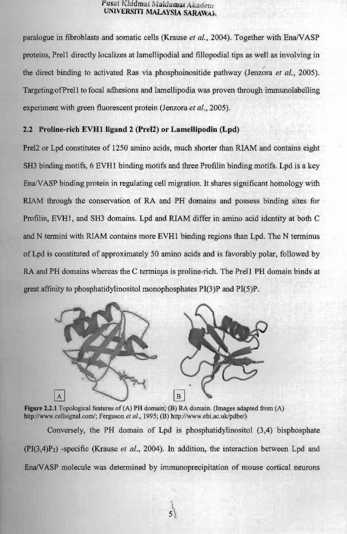
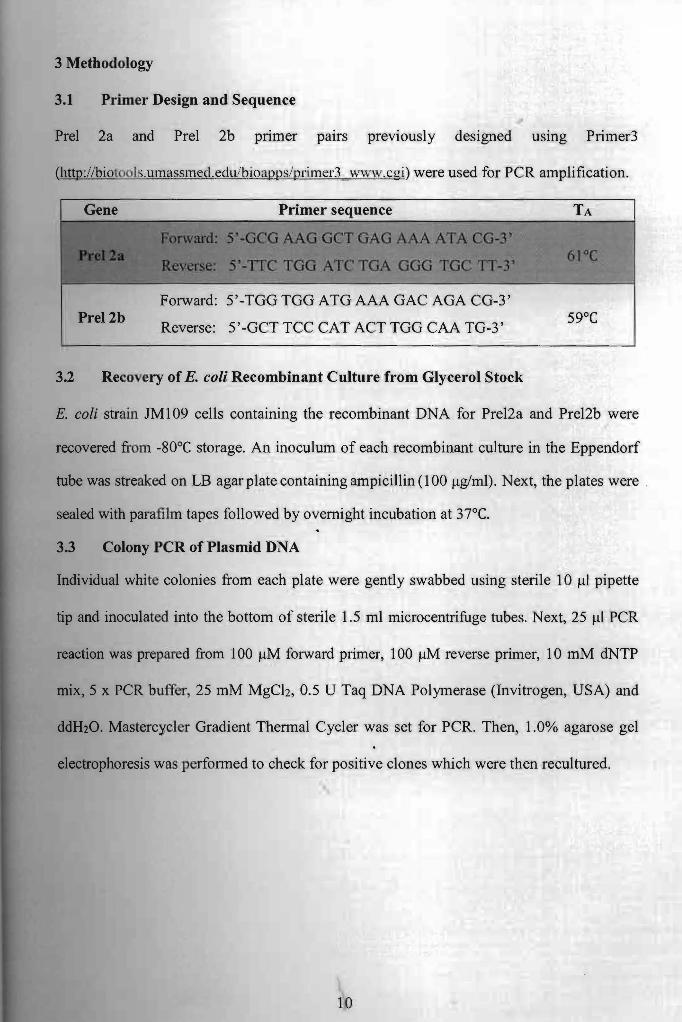
Related Documents


