Original Article Pulsed laser deposition and in vitro characteristics of triphasic – HASi composition on titanium Rajesh Palangadan 1 , Anil Sukumaran 2 , Francis B Fernandez 3 , Annie John 3 and Harikrishna Varma 1 Abstract Pulsed laser deposition was used to deposit bioactive triphasic glass-ceramic composition (HASi) over titanium substrate using dense HASi target. Bioactive glass compositions are considered the most useful synthetic materials for immediate bone attachment because of its bioresorption, osteoconduction and osteointegration characteristics under in vivo conditions. The disadvantage of its brittleness associated with bioactive glass-ceramics has prompted its coating over metallic implants for the combination of duo mechanical and bioactive properties. The hard HASi target was able to undergo laser ablation under ambient gas pressure without bulk erosion of the target. Laser deposition was found to be efficient in depositing triphasic composition for immediate bone integration. The target and deposits were analyzed for the phase, composition and microstructural characteristics by means of X-ray diffraction, Fourier transform infrared spectroscopy, energy-dispersive X-ray analysis and scanning electron microscopy. Simultaneously, the adherent nature and mechanical behaviour of deposits were confirmed by scratch test and micro-indentation methods. Further, the in vitro dissolution and bioactivity were assessed by soaking in simulated body fluid followed by elemental analysis using inductively coupled plasma spectroscopy. The deposits were found to be cell-friendly, which was indicated by the phenomenology of stem cells under in vitro conditions. Keywords Calcium phosphate, bioactive coating, triphasic, bioactive glass, laser deposition, titanium implant Introduction Ceramics and glasses have been used for a long time in the health-care industry. Among these, bioceramics were specially designed for the repair and reconstruc- tion of diseased or damaged parts of the body. Calcium phosphate ceramics and bioactive glass compositions are extensively used as bone substitutes because of their better and faster osseointegration capacity under in vivo conditions. 1 Among the various bioactive materials, bioactive glass compositions starting from the original bioglass Õ , a series of bioactive glasses have been proposed and a few of them have been found to be bone bonding. 1–3 The limited mechanical strength and low toughness of bioactive glasses has pre- vented their use as load-bearing devices. Combining the mechanical properties of metals or polymers with a bio- active phase with optimised properties has long been a goal in biomedical applications. 4 Some bioactive mater- ials, such as hydroxyapatite (HA) and bioactive glasses are increasingly used as hard tissue replacements and to improve the bonding between implants and host tissues. These bioactive materials can bond to living bone by creating a bone-like apatite layer on their surface after implantation by preventing the formation of fibrous tissues. Apatite formation is currently believed to be Journal of Biomaterials Applications 0(0) 1–10 ! The Author(s) 2013 Reprints and permissions: sagepub.co.uk/journalsPermissions.nav DOI: 10.1177/0885328213484545 jba.sagepub.com 1 Bioceramic Laboratory, Biomedical Technology Wing, Sree Chitra Tirunal Institute for Medical Sciences and Technology, Trivandrum, Kerala, India 2 Department of Periodontics and Community Dentistry, College of Dentistry, King Saud University, Riyadh, Saudi Arabia 3 Division of Implant Biology, Biomedical Technology Wing, Sree Chitra Tirunal Institute for Medical Sciences and Technology, Trivandrum, Kerala, India Corresponding author: Harikrishna Varma, Bioceramic Laboratory, Biomedical Technology Wing, Sree Chitra Tirunal Institute for Medical Sciences and Technology, Trivandrum 695012, Kerala, India. Email: [email protected]; [email protected]

Welcome message from author
This document is posted to help you gain knowledge. Please leave a comment to let me know what you think about it! Share it to your friends and learn new things together.
Transcript

Original Article
Pulsed laser deposition and in vitrocharacteristics of triphasic – HASicomposition on titanium
Rajesh Palangadan1, Anil Sukumaran2, Francis B Fernandez3, Annie John3 andHarikrishna Varma1
Abstract
Pulsed laser deposition was used to deposit bioactive triphasic glass-ceramic composition (HASi) over titanium substrate
using dense HASi target. Bioactive glass compositions are considered the most useful synthetic materials for immediate
bone attachment because of its bioresorption, osteoconduction and osteointegration characteristics under in vivo
conditions. The disadvantage of its brittleness associated with bioactive glass-ceramics has prompted its coating over
metallic implants for the combination of duo mechanical and bioactive properties. The hard HASi target was able to
undergo laser ablation under ambient gas pressure without bulk erosion of the target. Laser deposition was found to be
efficient in depositing triphasic composition for immediate bone integration. The target and deposits were analyzed for
the phase, composition and microstructural characteristics by means of X-ray diffraction, Fourier transform infrared
spectroscopy, energy-dispersive X-ray analysis and scanning electron microscopy. Simultaneously, the adherent nature
and mechanical behaviour of deposits were confirmed by scratch test and micro-indentation methods. Further, the
in vitro dissolution and bioactivity were assessed by soaking in simulated body fluid followed by elemental analysis using
inductively coupled plasma spectroscopy. The deposits were found to be cell-friendly, which was indicated by the
phenomenology of stem cells under in vitro conditions.
Keywords
Calcium phosphate, bioactive coating, triphasic, bioactive glass, laser deposition, titanium implant
Introduction
Ceramics and glasses have been used for a long time inthe health-care industry. Among these, bioceramicswere specially designed for the repair and reconstruc-tion of diseased or damaged parts of the body. Calciumphosphate ceramics and bioactive glass compositionsare extensively used as bone substitutes because oftheir better and faster osseointegration capacity underin vivo conditions.1 Among the various bioactivematerials, bioactive glass compositions starting fromthe original bioglass�, a series of bioactive glasseshave been proposed and a few of them have beenfound to be bone bonding.1–3 The limited mechanicalstrength and low toughness of bioactive glasses has pre-vented their use as load-bearing devices. Combining themechanical properties of metals or polymers with a bio-active phase with optimised properties has long been agoal in biomedical applications.4 Some bioactive mater-ials, such as hydroxyapatite (HA) and bioactive glasses
are increasingly used as hard tissue replacements and toimprove the bonding between implants and host tissues.These bioactive materials can bond to living bone bycreating a bone-like apatite layer on their surface afterimplantation by preventing the formation of fibroustissues. Apatite formation is currently believed to be
Journal of Biomaterials Applications
0(0) 1–10
! The Author(s) 2013
Reprints and permissions:
sagepub.co.uk/journalsPermissions.nav
DOI: 10.1177/0885328213484545
jba.sagepub.com
1Bioceramic Laboratory, Biomedical Technology Wing, Sree Chitra
Tirunal Institute for Medical Sciences and Technology, Trivandrum,
Kerala, India2Department of Periodontics and Community Dentistry, College of
Dentistry, King Saud University, Riyadh, Saudi Arabia3Division of Implant Biology, Biomedical Technology Wing, Sree Chitra
Tirunal Institute for Medical Sciences and Technology, Trivandrum, Kerala,
India
Corresponding author:
Harikrishna Varma, Bioceramic Laboratory, Biomedical Technology Wing,
Sree Chitra Tirunal Institute for Medical Sciences and Technology,
Trivandrum 695012, Kerala, India.
Email: [email protected]; [email protected]
the prime requirement for the bone-bonding ability ofmaterials.5
The coating of fast resorbing materials with suffi-cient strength characteristics may be advantageous formetallic implants, which require fast fixation as indental prosthesis. Bioactive coatings can improveimplant performance, but we are still far from thedevelopment of ideal materials and fabrication technol-ogies. Ideally, the coatings should be tailored to exhibitprescribed biological attributes for each specific appli-cation. They should have strong adhesion to theimplant and good fixation to bone. Their compositionand microstructure should be controlled such that theirdissolution rates match the in vivo healing process.6
The most predominant fast resorbing materials arederived from silicate-based glasses. These system of bio-active glasses were originally developed by Hench basedon the SiO2-P2O5-CaO-Na2O composition.7 Over theyears, the preparation and use of Hench’s glass is wellestablished in biomedical field specially in orthopedicand dental care.4 Bioactive glasses are typically silicateglasses with silica content below 60 wt%. This is animportant requirement since glasses with silica contentsgreater than 60 wt% are no longer bioactive. Most ofthese glasses are hygroscopic and crystallize readily atthe firing temperatures required for enamelling. Thesematerials when immersed in simulated body fluid (SBF)for 30 days have shown the presence of HA crystalsprecipitating on the surface. The thermal expansioncoefficients of bioactive glasses are typically muchlarger than those of Ti alloys. The simplest way toreduce thermal expansion is to increase the SiO2 con-tent of the glass; unfortunately, this is at the expense ofbioactivity, which is significantly reduced. The rate ofdissolution and remodelling of these bioactive glassesdepend on the composition, mainly the Si percentage.1,8
The use of bioactive glass in the bulk, granules orpowder form is well known, but as coating on metallicimplants such as titanium is still challenging because ofthe high melting point, amorphous characteristics andlarge difference in thermal expansion coefficientsbetween the metallic base and the bioactive surface.The resulting high residual stress is thought to beresponsible for the low bonding strength between thetwo materials.9,10 Most of the technique employed tilldate use a high temperature process such as plasmaspray or enamelling to obtain coating over metallic sub-strate. This may cause failure of coating at the interfacedue to the non-homogenous nature of deposit interms of composition, thickness, etc. or by formingmicro-cracks developed during the process.8,11,12
Maintaining the bioactive property and to improvethe interface strength, many attempts lead to the devel-opment of various glass compositions other than theoriginal 45S5 glass. Also, various techniques attempted
to suit the coating of bioactive glasses include sol-gel,electrophoresis, ion sputtering, plasma spraying andpulsed laser deposition.3,13,14
Pulsed laser deposition (PLD) is unique as it pro-vides the growth of meta-stable materials, chemicallycomplex (multi element) materials, stoichiometrictransfer of composite materials, reactive deposition inambient gases and the capability towards the growth ofmultilayered structures. Films deposited by PLD atappropriate laser energy densities will have the samecompositions as the target due to high heating ratesand non-thermal ablation of the target by the lasergenerated plasma. In PLD, a high power laser isfocused onto a target. When the laser radiation isabsorbed by the target, electromagnetic energy is usedto form a plume of energetic atoms, electrons, ions andmolecules. The plume expands from the target withhydrodynamic flow, and propagates in a forwardpeaked manner towards the substrate. The average kin-etic energy of the laser-ablated species is between 100and 1000 kT. This kinetic energy cause the particles tostick to the surface and be compressed, forming a densecontinuous film.15 PLD can be used to deposit highlybioactive and hygroscopic glasses, or glasses with veryhigh softening points that cannot be applied by othermethods. By controlling the substrate temperature orannealing after deposition, coating adhesion and sur-face roughness can be manipulated.6,15 The laser depos-itions of bioactive glasses were not well documentedcompared to bioceramic depositions, probably becauseof the difficulty in fabricating a dense target. Further,being a glass-ceramic system, the vaporisation bylaser or other means will not be as efficient as ceramiclike HA.
Large area bioceramic coatings with slow resorption,usually of HA, relatively thicker plasma spray coatingsare preferred for implants used in orthopedics such asin total hip replacement.16,17 But, when the applicationis confined to small area insertion, such as in dentalimplants, adherent thin bioactive layer with fast resorp-tion characteristic is preferred. In this context, bioactiveglass composition, based on HASi, would be a promis-ing candidate. HASi, a triphasic glass ceramic compos-ition has dual benefits of HA and Si–O in one productand is composed of HA, tricalcium phosphates and cal-cium silicate.18,19 This HASi, in its bulk form wasshown previously as a scaffold in bone tissue engineer-ing.19,20 This triphasic bioactive composition can alsoattain high strength level at optimum heating process tobe used as target for PLD process. It was also shownthat an appropriate Si ion concentration source couldinduce angiogenesis through increasing gene expressionof proangiogenic cytokine receptors and up-regulateddownstream signalling which contributes to their abilityto enhance bone regeneration.21 These substituted
2 Journal of Biomaterials Applications 0(0)

silicates have influence on both the very early events atthe scaffold interface via variation in surface chemistryand subsequently on the activity of both bone-formingand bone-resorbing cells, presumably by providingtrace levels of bioavailable Si, which affects equilibriumlevels of bone ingrowth within the scaffold.22 In thisstudy, we propose the development of a novel bioactivecoating by pulsed laser deposition using dense HASitarget. This bioactive composition, if made as coatingonto the titanium implants, may have excellent boneremodelling and resorption capacity for fast implantfixation.
The bioactivity was examined in vitro with respect tothe ability of HASi coating to grow or transform to HAonto the surface as a result of contact with SBF.23,24
The percentage of weight gain or loss was estimated inaddition to the elemental analysis of SBF to understandthe growth or depletion of the bioactive coatings.Preliminary observations with mesenchymal stem cellsunder in vitro cell culture conditions had shown theproliferating efficiency of cells over HASi deposits.
Materials and methods
Synthesis of HASi powder and fabrication of target
Bioactive composition prepared indigenously havingthe composition in weight percent SiO2 (34.2); CaO(44.9); P2O5 (16.3); CaF2 (0.5); MgO (4.6) was used inthis study. The preparation of HASi composition isbased on the sol–gel synthesis, which involves thehydrolysis of tetraethylorthosilicate (TEOS) in alcohol.Table 1 shows the composition of reagent grade chem-icals used to prepare HASi powder.
HASi powder was prepared in batches of 100 g.14.8mL of phosphoric acid (H3PO4; specific grav-ity¼ 1.75) was diluted to 200mL with distilled water.Ca(NO3)2.4H2O and Mg(NO3)2.6H2O were added tothe phosphoric acid with stirring. Mean time, TEOSwas refluxed with ethanol at 60�C for 20min in around bottom flask fitted with a condenser. To this,
the prepared phosphoric acid solution was added fol-lowed by the addition of CaF2 dispersed in minimumquantity of water. Stirring and heating was continuedfor 30min and the resulting sol was transferred into aglass petri dish. The sol transforms to gel at about36–48 h, and which was dried in an oven at 100�C for24 h. Calcination of the glassy composition was thenperformed at 600�C for 2 h. The calcined lumps wereground in planetary ball mill and sieved to have�125mm size powder.
HASi discs were prepared by uniaxial pressing (at7 ton with 2min holding) followed by heat treatment.Unlike dense crystalline ceramics formed using the con-ventional sintering process, bioactive glasses were notable to strengthen to use as target for laser ablation, soan optimisation was performed with regard to the firingtemperature and duration for HASi. In this way, highstrength target was obtained by applying a temperatureclose tomelting. The heating process was done in normalair inside furnace in steps: 3.5�C/min from room tem-perature to 600�C, then holding at this temperature for1 h, heating at 2.5�C/min till 1280�C followed by holdingat this temperature for 2 h and subsequent cooling innormal air inside the furnace. Hard target obtained atthis temperature, is suitable for effective ablation pro-cess by the focussed laser beam. The discs were smooth-ened by mechanical polishing and then cleanedultrasonically in distilled water and acetone.
Preparation of titanium-based substrate
Commercially available Ti6Al4V was cut into dimen-sions of 15� 10� 2mm3. Titanium-based substrateswere mechanically polished with graded SiC papersand were finely smoothened with 0.05 mm alumina sus-pension. These substrates were cleaned ultrasonically indistilled water, acetone and finally dried in hot air.
Laser deposition
The pulsed laser deposition of HASi was performed in acustom-designed vacuum chamber as previouslydetailed.25 A Nd-YAG laser source delivering 355 nmlaser (Quanta systems, Italy) was used to produceplume of HASi under controlled oxygen atmosphere(of the order of 10�3mbar). The plume was directedtowards the titanium substrate placed inside a cylin-drical heater at a distance of 35mm from the target.The deposition was done at a substrate temperature of400�C. The HASi target was allowed to have rotationalmotion so as to prevent the surface erosion due laser at asingle point and helps to produce a uniform fresh sur-face for each incoming laser pulse. HASi was depositedfor 1 h using laser of power 2W (10Hz) focussed to2mm spot onto the target at an angle of 45�.
Table 1. Reagents and quantity used for preparing
100 g of HASi.
Reagent Quantity
TEOS 129.56 mL
C2H5OH 129.56 mL
H3PO4 14.8 mL
Ca(NO3)2 � 4H2O 193.3 g
Mg(NO3)2 � 6H2O 29.56 g
CaF2 0.52 g
Water 200 mL
Palangadan et al. 3

Characterization of HASi target and deposit
The HASi target and deposits were analyzed usingX-ray diffractometer (Brucker D8 advance, Germany)to identify the phase and to examine the crystallinenature. The samples were scanned at 2y¼ 10�–70�
using Cu-Ka (�¼ 1�54 A) radiation at a voltage of40 kV and 30mA current.
Coating thickness was quantified by means of a non-contact surface profilometry Talysurf CLI 1000 (TaylorHobson, UK). A portion of the substrate is maskedprior to coating to get a height difference and ‘stepheight measurement’ mode available with the instru-ment is used to measure height difference and thickness.The surface micromorphology of deposits were exam-ined using an environmental scanning electron micro-scope (ESEM, FEI Quanta 200, The Netherlands)equipped with an energy-dispersive X-ray analysis(EDS) for the elemental composition. SEM was alsoused to examine the growth morphology of apatite asa result of immersion in SBF. The functional groupspresent in the target and deposits were identified byFTIR spectroscopy (Thermo Nicolet 5700, USA). Thespectra were collected in the diffuse reflectance (DRIFT)mode. Neat KBr powder was used as the backgroundand sample was prepared by diluting with KBr. Thespectra were recorded as an average of 64 scans between400 and 4000 cm�1, at a resolution of 4 cm�1.
The mechanical properties of the deposit wereassured by means of microindentation hardness andscratch test. Microindentation hardness of the depos-ited HASi were analyzed using a Vickers indenter(Shimadzu-HMV-WIN, Ver 1.03, Japan) under con-stant test load of 1.961N with 14 S duration. Vickershardness value was calculated as an average of sixindentations per sample. The adhesion of HASi thincoating was evaluated using a microscratch tester(Micro-combi tester; CSM Instruments, Switzerland).A diamond tip of 100 mm was used for the analysis. Ascratch length of 10mm with a progressive loading rateof 5.98N/min was performed at a minimum load of0.03N at the beginning and a maximum of 10N atthe end. The coating failure was assessed by the criticalnormal forces of the selected failure events occurredduring the scratch process. Measurement of acousticemission (AE) and frictional force (FF) were simultan-eously performed along with microscopic observationof the failure event.
In vitro bioactivity and dissolution study insimulated body fluid
Laser-deposited HASi were subjected to in vitro dissol-ution and bioactivity studies with regard to its behaviorwhile in contact with SBF, a solution which is similar in
ionic concentration and pH to that of human bloodplasma.26 The SBF solution was prepared with refer-ence to Kokubo’s recipe as detailed elsewhere.24,27 Thedissolution behavior was estimated as the changes inionic concentration of SBF, while the bioactivity wasidentified as the transformation of the deposited film toapatite phase. The HASi-coated titanium substrateswere soaked in 30mL of SBF taken in polypropylenevials and kept at 37� 1�C. After specific time periods of3 and 7 days the samples were washed with deionizedwater and dried in a desiccator. The influence of SBFonto the deposit is assessed as the variation in weightwith respect to the initial deposits. Weight differencebefore and after SBF soaking was estimated to obtainweight gain/loss of coating after SBF soaking. The per-centage weight gain or loss was assessed with respect toinitial deposit using equation (1)27
Weight gain or loss after soaking in SBF
Initial weight of coating
�� 100%
�
ð1Þ
The analysis for determining the elemental compositionof SBF was done by inductively coupled plasma-opticalemission spectroscopy (ICP-OES, Perkin Elmer5300DV, USA). Sample solutions were prepared in5% HNO3. The SBF were analyzed for calcium, phos-phorous, magnesium ions, etc. to understand dissol-ution of coating and leaching of ions. The ionconcentration of fresh SBF was compared with theSBF after carrying samples for 3 and 7 days. Theexperiments were done in triplicate with a calibrationplot of known standards.
In vitro cell culture studies
In order to assess material’s response under in vitro; themorphology, proliferation, and attachment of mesen-chymal stem cells (MSCs) towards the deposited bio-active coatings under in vitro cell culture conditionswere observed. Current evaluation has been undertakenusing rabbit adipose derived mesenchymal stem cells(RADMSC). Cell culture reagents were purchasedfrom Invitrogen, USA unless otherwise mentioned.Scaffolds for evaluation were pre-conditioned inDMEM-HG (Dulbecco’s Modified Eagle’s Medium)10% Fetal Bovine Serum, 1% anti–anti mix for 4 h.Approximately 1� 104 cells were seeded on the scaf-folds and scaffolds were incubated at 37�C, 5% CO2for 24 h (Heraeus BB 15). The samples were rinsed withsterile PBS (Sigma) and fixed in 3% glutaraldehydesolution. Then the samples were dehydrated in ascend-ing grades of alcohol (70%, 80%, 95% and 100%),washed with isoamyl alcohol and subjected to criticalpoint drying. The dried samples were gold coated for
4 Journal of Biomaterials Applications 0(0)
making the surface conducting and observed underSEM (ESEM, FEI Quanta 200, The Netherlands).
Results and discussion
X-ray diffraction analysis of HASi target and deposit
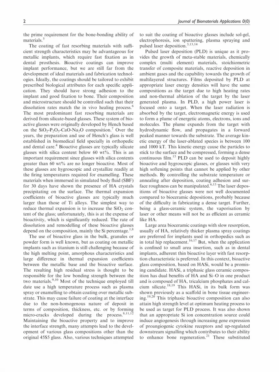
The X-ray diffraction analysis shows the glass-ceramicnature of HASi target prepared by the sol–gel method.The pattern (Figure 1(a)) indicates the prepared targetcomposed of phosphates and silicates of calcium. Thecomposition consists of wollastonite (PDF# 00-002-0689), whitlockite (PDF# 01-076-8364) and hydroxy-apatite (PDF# 00-009-0432) as major constituentphases. Wollastonite is having major peaks at 2yvalues of 23, 25.3, 26.9, 30.06 and 39.6. The whitlockitepeaks appear at 2y values of 13.5, 17, 28.8, 31.2 and34.6. The presence of HA phase is indicated by thepeaks at 2y values 25.8, 31.7, 32.1, 32.9, and 49.4.Minor content of SiF4 (PDF# 01-078-1988) was indi-cated by the less intense peaks at 2y values of 23.2, 40.8,and 47.4. The amorphous content in the pattern isderived from the glassy silicates and the crystalline con-tent because of the calcium phosphates. Thus the sin-tered material can be regarded as a glass-ceramicsystem.
All the constituent phases present in the target canbe clearly distinguished in the XRD pattern of coatingproduced over titanium substrate kept at 400�C(Figure 1(b)). However, the spectra seems less resolvedpeaks of HASi, than the target due to the lower thick-ness of deposit (�2 mm) else due to the intense peaks ofcrystalline titanium substrate.
FTIR analysis of HASi target and deposit
The FTIR spectrum of sintered HASi composition(Figure 2(a)) and the deposits formed by PLD of thiscomposition onto the titanium substrate (Figure 2(b))confirms the presence of functional groups. The spectraof HASi target and the deposits were similar with thepresence of all characteristic vibrational peaks. Thespectra of deposits after exposed to SBF (Figure 2(c)and (d)) indicates introduction of new –OH functionalgroup characteristic of apatite phase.
FTIR spectrum indicates IR active functionalgroups present in phosphate and silicate phases intarget as well as in the deposits. The major vibrationsare due to silicate, phosphate, and hydroxyl groups.The Si–O, –PO4 groups and the –OH present as free –OH and H-bonded –OH are IR active groups present inbioactive glasses. The broad nature of the spectrum atregion 3700–3000 cm�1 is due to the combined effect ofSi–OH which is usually present at �3740 cm�1, theH-bonded hydroxyl groups located in the 3700–2200 cm�1 region and also due to the P–OH stretchingat� 3670 cm�1.
Zones� 600–475 cm�1 correspond to calcium phos-phates. The intensity of bands at� 930 cm�1 related tothe presence of SiO bonds. Because of the presence ofHA in the prepared HASi composition, it was expectedto find the hydroxyl peak� 3570 cm�1, but the –OHgroup cannot be clearly detected in the spectrum(Figure 2(a) and (b)). Even though the XRD patternshows the presence of HA phase, the absence of –OH isrelated to decomposition caused by the addition of thesilicate glass, which is highly reactive at high
010 20 30 40
2 Theta
Whitlockite
TitaniumSilicon fluorideWollastonite
Apatite
50 60 70
2000
4000
(a)
(b)
6000
Cou
nts
8000
Figure 1. X-ray diffraction pattern of: (a) HASi target heat treated at 1280�C and (b) HASi coating obtained by PLD on titanium
substrate kept at 400�C.
PLD: pulsed laser deposition.
Palangadan et al. 5

temperature and forces major chemical changes asso-ciated with the hydroxyl site.28 It was also due todecomposition of HA by the presence of glass, whichenters the HA structure and causes the hydroxyl groupsto be driven off. This may be due to the formation ofoxyapatite at elevated temperature. The charge imbal-ance due to the entry of silicon group was balanced byleaving the hydroxyl group from the structure andcause the absence of hydroxyl peaks around3570 cm�1.29
FTIR spectra of HASi coating after soaking in SBFhave significant difference as a result of transformationand growth of the deposits to apatite (Figure 2(c)and (d)). The HASi deposit, which originally have a
silicon-rich bioactive layer of calcium phosphate, wastransformed to calcium phosphate as a result of reac-tion with ion-rich SBF. The spectra correspond to car-bonated calcium phosphate. This is evident by theincreased concentration of –OH and �CO2
3� groupsas a result of immersion in SBF and further ionexchanges from the saturated solution.
Micro-structural observation by SEM and elementalcomposition by EDS
Typical surface morphology of the HASi coatingsdeposited at a substrate temperature of 400�C isdepicted (Figure 3(a) and (b)). They consist mostly of
0.00
4000
(a)
(b)
(c)
(d)
SiO-H (Stret.)
Si-O-Si (Stret.)
PO43−(Bend.)
PO43−(Stret.)
CO32−(Stret.)
-OH (Stret.)
3500 3000 2500 2000
Wavenumbers (cm−1)
1500 1000 500
0.00.2
0.4
0.6
0.0
Kub
elka
-Mun
k
0.1
0.2
0.3
0.00.1
0.2
0.3
0.05
0.10
Figure 2. FTIR spectrum of: (a) HASi target heat treated at 1280�C, (b) as deposited HASi, (c) SBF soaked HASi coating after 3 days
and (d) after 7 days.
FTIR: Fourier transform infrared; SBF: simulated body fluid.
C
0.60 1.80 3.00 4.20 5.40Energy - KeV
Ca
Ca
Mg
O
P
Si(c)
Ti Ti
Figure 3. Scanning electron micrographs of laser-deposited HASi coating over titanium substrate: (a)� 1000, (b)� 6000 and (c) EDS
of laser-deposited HASi coating over titanium substrate at 400�C.
EDS: energy-dispersive X-ray spectroscopy.
6 Journal of Biomaterials Applications 0(0)
micron-size spheroids. The spheres appear to be welladhered to titanium as well as to each other with aglassy-molten appearance. The EDS analysis of thecoating (Figure 3(c)) shows the presence of calcium,silicon, phosphorous and magnesium in the coatingwhose intensity corresponds to the weight percentageof each element present in the deposit.
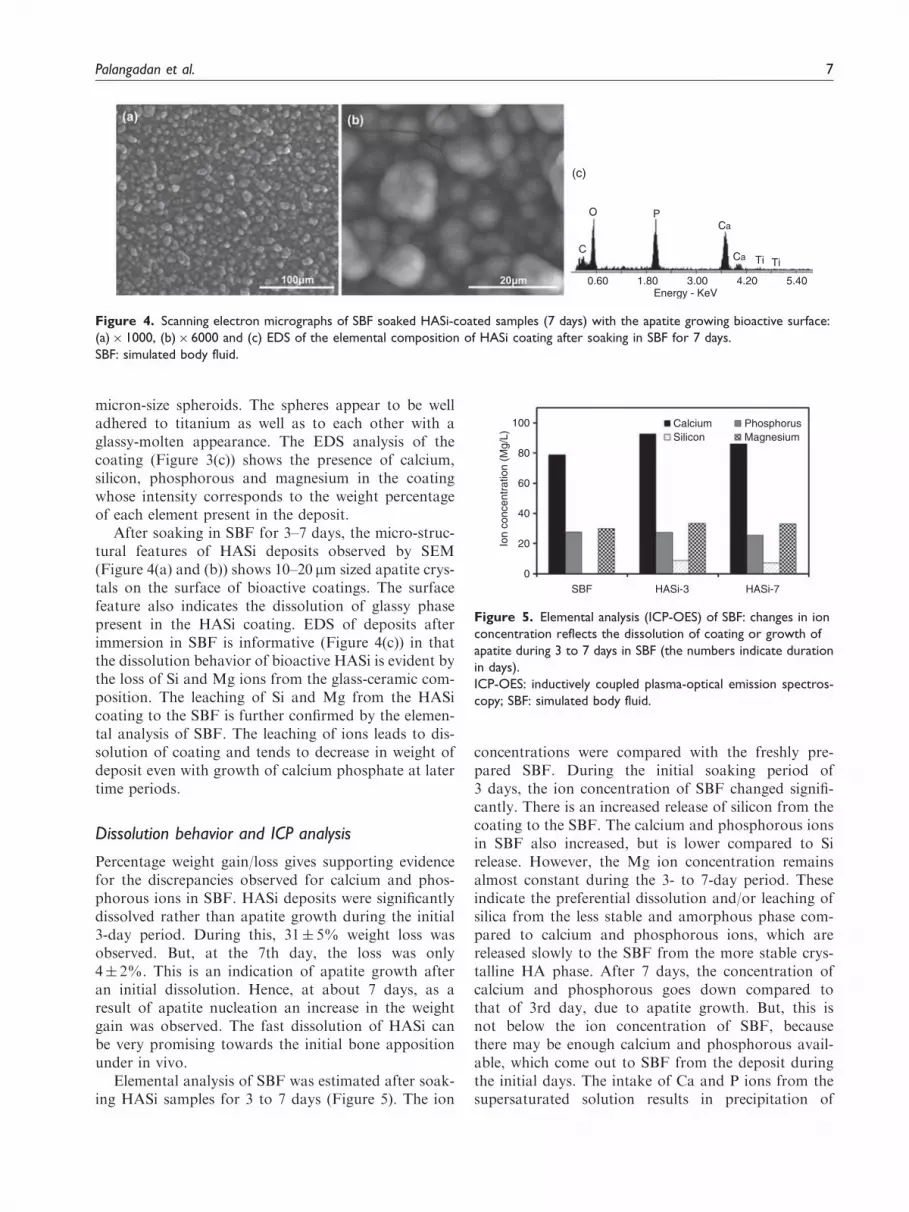
After soaking in SBF for 3–7 days, the micro-struc-tural features of HASi deposits observed by SEM(Figure 4(a) and (b)) shows 10–20 mm sized apatite crys-tals on the surface of bioactive coatings. The surfacefeature also indicates the dissolution of glassy phasepresent in the HASi coating. EDS of deposits afterimmersion in SBF is informative (Figure 4(c)) in thatthe dissolution behavior of bioactive HASi is evident bythe loss of Si and Mg ions from the glass-ceramic com-position. The leaching of Si and Mg from the HASicoating to the SBF is further confirmed by the elemen-tal analysis of SBF. The leaching of ions leads to dis-solution of coating and tends to decrease in weight ofdeposit even with growth of calcium phosphate at latertime periods.
Dissolution behavior and ICP analysis
Percentage weight gain/loss gives supporting evidencefor the discrepancies observed for calcium and phos-phorous ions in SBF. HASi deposits were significantlydissolved rather than apatite growth during the initial3-day period. During this, 31� 5% weight loss wasobserved. But, at the 7th day, the loss was only4� 2%. This is an indication of apatite growth afteran initial dissolution. Hence, at about 7 days, as aresult of apatite nucleation an increase in the weightgain was observed. The fast dissolution of HASi canbe very promising towards the initial bone appositionunder in vivo.
Elemental analysis of SBF was estimated after soak-ing HASi samples for 3 to 7 days (Figure 5). The ion
concentrations were compared with the freshly pre-pared SBF. During the initial soaking period of3 days, the ion concentration of SBF changed signifi-cantly. There is an increased release of silicon from thecoating to the SBF. The calcium and phosphorous ionsin SBF also increased, but is lower compared to Sirelease. However, the Mg ion concentration remainsalmost constant during the 3- to 7-day period. Theseindicate the preferential dissolution and/or leaching ofsilica from the less stable and amorphous phase com-pared to calcium and phosphorous ions, which arereleased slowly to the SBF from the more stable crys-talline HA phase. After 7 days, the concentration ofcalcium and phosphorous goes down compared tothat of 3rd day, due to apatite growth. But, this isnot below the ion concentration of SBF, becausethere may be enough calcium and phosphorous avail-able, which come out to SBF from the deposit duringthe initial days. The intake of Ca and P ions from thesupersaturated solution results in precipitation of
C
0.60 1.80 3.00 4.20 5.40Energy - KeV
Ca
Ca
O P
(c)
Ti Ti
Figure 4. Scanning electron micrographs of SBF soaked HASi-coated samples (7 days) with the apatite growing bioactive surface:
(a)� 1000, (b)� 6000 and (c) EDS of the elemental composition of HASi coating after soaking in SBF for 7 days.
SBF: simulated body fluid.
0SBF HASi-3
CalciumSilicon
PhosphorusMagnesium
HASi-7
20
40Io
n co
ncen
trat
ion
(Mg/
L)
60
80
100
Figure 5. Elemental analysis (ICP-OES) of SBF: changes in ion
concentration reflects the dissolution of coating or growth of
apatite during 3 to 7 days in SBF (the numbers indicate duration
in days).
ICP-OES: inductively coupled plasma-optical emission spectros-
copy; SBF: simulated body fluid.
Palangadan et al. 7

apatitic calcium phosphates on the HASi-coated sam-ples. The released amount of silicon and the ion con-centration of Mg remained same towards the 7th day, asthese ions remain in SBF without forming part of apa-tite growth.
Behavior of cells to HASi coating under in vitrocondition
The behavior of a bioactive surface is of utmost import-ance while looking for an implantable substitute. HASicompositions obtained in the form of coating overtitanium substrate were subjected for preliminaryin vitro cell culture using RADMSC. Mesenchymalstem cells were chosen as it is known that primary heal-ing is undertaken by the recruitment and differentiationof mesenchymal stem cells to the site of injury.30 Thesurface structure of HASi deposit after 24 h in the cellculture medium (Figure 6(a)) with little surface
morphological changes as a result of reaction withmedium, indicates the stability of deposited bioactivecoating. The polished titanium surface in direct contactwith cells after 24 h (Figure 6(b)) did not show muchproliferation compared to the bioactive coating(Figure 7). The HASi bioactive coating was found tobe cell-friendly as indicated by the phenomenology ofthe cells within 24 h.31 The whole surface was coveredby the cells in 24 h, which depicted strong attraction ofcells towards the micro-roughness and channels formedby the granular deposit of HASi (Figure 7(a) and (b)).
Micro hardness and scratch test
Hardness tests have been found to be very useful forevaluation of materials, quality control of manufactur-ing processes and research and development efforts.Hardness, although empirical in nature, can be corre-lated to tensile strength for many metals, and is an
Figure 6. (a) Scanning electron micrograph of surface morphology of HASi deposit after exposure to cell culture medium (24 h) and
(b) scanning electron micrograph of RADMSC on polished titanium substrate (24 h).
RADMSC: rabbit adipose derived mesenchymal stem cells.
Figure 7. Scanning electron micrographs of: (a) RADMSC adhered on HASi-deposited titanium substrate (24 h) and (b) the cell layer
indicated alongside the HASi coating.
RADMSC: rabbit adipose derived mesenchymal stem cells.
8 Journal of Biomaterials Applications 0(0)

indicator of wear resistance and ductility. The Vickershardness value of HASi coating was 343.22� 14.62while that of titanium substrate was 247.54� 5.19.The value is an indication of the mode of attachmentbetween the HASi species and to the Ti substrate. If thedeposit is less adhered to the substrate and poorlypacked, the hardness value may tend to that of sub-strate. The higher value of deposit even with the under-lying metallic substrate, indicate the dense deposit ofHASi obtained by PLD at 400�C.
The resistance of HASi bioactive coating towardsthe diamond stylus during scratch test indicate the sta-bility of deposit obtained by PLD. The scratch profileconsists of an initial crack in the deposit indicated bythe first variation in acoustic emission (Lc1). After Lc1,the deposit starts peel of at certain intervals. The failurepoints were designated as first crack (Lc1), first delam-ination (Lc2) and complete delamination (Lc3) in thescratch profile (Figure 8). Lc3 is the maximum load theHASi deposit withstands. The average values of Lc1,Lc2 and Lc3 respectively obtained at 1.6, 2.56 and5.19N for the deposit on polished substrate. Sincethese values are for deposit on polished substrate, theadhesion can be further improved on real texturedimplant surfaces.
Conclusions
Stable adherent bioactive coatings based on HASi weredeveloped onto the titanium substrate by pulsed laser
deposition technique. The target prepared by high tem-perature firing of HASi composition efficiently under-goes laser deposition under controlled atmosphere andsubstrate temperature. The deposit composed of wol-lastonite, whitlockite and apatite as major phases asevident by XRD and FTIR analysis. The glassy(amorphous) silicate content in the deposits underwentearly dissolution characteristics in SBF which indicatedthe possible early integration to the host tissue. TheHASi deposit was adhered well onto the titanium-based substrate and is able to form apatite layer inSBF. The HASi bioactive coating was stable undercell culture conditions and found to be cell-friendlytowards MSCs in 24 h. The phenomenology of MSCsand in vitro behavior of HASi deposit on titanium dem-onstrates future possibility of adherent bioactive metal-lic implants.
Acknowledgment
The authors acknowledge the Director SCTIMST, HeadBMT for the facilities provided to carry out this work.
Conflict of interest
None declared.
Funding
This research received no specific grant from anyfunding agency in the public, commercial, or not-for-profit
sectors.
0.00 0%
10
20
30
40
50
60
70
80
90
100
0.10
0.20
0.30
0.40
0.50
Nor
mal
load
and
fric
tion
Aco
ustic
Em
issi
on
0.60
0.70
0.80
0.00
1.00
0.00 1.00 2.00 3.00 4.00 5.00
Scratch length and applied force
6.00 7.00 8.00 9.00 10.00
1.03 2.02 3.02 4.02 5.01 6.01 7.01 8.00
Acoustic Emission
Frictional force
Friction Coefficient
Normal force
Lc1 Lc2 Lc3
9.00 10.00
mm
0.03 N
Figure 8. Representative profile of scratch test: acoustic emission profile combined with frictional force and friction coefficient
performed on the HASi coating deposited at a substrate temperature of 400�C.
Palangadan et al. 9

References
1. Hench LL. Bioceramics – from concept to clinic. J Am
Ceram Soc 1991; 74(7): 1487–1510.2. Kokubo T, Kim HM and Kawashita M. Novel bioactive
materials with different mechanical properties.Biomaterials 2003; 24(13): 2161–2175.
3. Joanni E, Carmona Ferr M, Cela Mardare C, et al.Pulsed laser deposition of SiO2 – P2O5 – CaO – MgOglass coatings on titanium substrates. Mater Res 2004; 7:
431–436.4. Hench LL. The story of bioglass. J Mater Sci Mater Med
2006; 17: 967–978.
5. Liu XY, Chu PK and Ding CX. Surface modification oftitanium, titanium alloys, and related materials for bio-medical applications. Mater Sci Eng R 2004; 47(3–4):49–121.
6. Tomsia AP, Saiz E, Song J, et al. Biomimetic bonelikecomposites and novel bioactive glass coatings. Adv EngMater 2005; 7(11): 999–1004.
7. Hench LL, Splinter RJ, Allen WC, et al. Bonding mech-anisms at the interface of ceramic prosthetic materials.J Biomed Mater Res 1971; 5(6): 117–141.
8. Lopez-Esteban S, Saiz E, Fujino S, et al. Bioactive glasscoatings for orthopedic metallic implants. J Eur CeramSoc 2003; 23(15): 2921–2930.
9. Ruseska G, Fidancevska E and Bossert J. Mechanicaland thermal-expansion characteristics of Ca-10(PO4)(6)(OH)(2)-Ca-3(PO4)(2) composites. Sci Sinter2006; 38(3): 245–253.
10. Fidancevska E, Pavlovski B, Ruseska G, et al.Thermal expansion and mechanical properties of theCa10(PO4)6(OH)2-TiO2 composite. Sci Sinter 2002;
34(3): 241–246.11. Navarro M, Michiardi A, Castano O, et al.
Biomaterials in orthopaedics. J R Soc Interface 2008;
5(27): 1137–1158.12. Yan LL, Leng Y and Weng LT. Characterization of
chemical inhomogeneity in plasma-sprayed hydroxyapa-
tite coatings. Biomaterials 2003; 24(15): 2585–2592.13. Lee TM, Chang E, Wang BC, et al. Characteristics of
plasma-sprayed bioactive glass coatings on Ti-6Al-4Valloy: an in vitro study. Surf Coat Technol 1996; 79(1–
3): 170–177.14. Oliveira GM, Ferraz MP, Gonzalez PG, et al. PLD bio-
active ceramic films: the influence of CaO-P2O5 glass
additions to hydroxyapatite on the proliferation andmorphology of osteblastic like-cells. J Mater Sci MaterMed 2008; 19(4): 1775–1785.
15. Narayan RJ, Jin C, Doraiswamy A, et al. Laser process-ing of advanced bioceramics. Adv Eng Mater 2005; 7(12):1083–1098.
16. Landor I, Vavrik P, Sosna A, et al. Hydroxyapatite
porous coating and the osteointegration of the total hipreplacement. Arch Orthopaed Trauma Surg 2007; 127(2):81–89.
17. Jaffe WL and Scott DF. Total hip arthroplasty withhydroxyapatite-coated prostheses. J Bone Joint SurgAm 1996; 78(12): 1918–1934.
18. Nair MB, Varma HK, Menon KV, et al. Reconstructionof goat femur segmental defects using triphasic ceramic-coated hydroxyapatite in combination with autologouscells and platelet-rich plasma. Acta Biomater 2009; 5(5):
1742–1755.19. Nair MB, Suresh Babu S, Varma HK, et al. A triphasic
ceramic-coated porous hydroxyapatite for tissue engin-
eering application. Acta Biomater 2008; 4(1): 173–181.20. Nair MB, Varma HK, Shenoy SJ, et al. Treatment of
goat femur segmental defects with silica-coated hydro-
xyapatite-one-year follow-up. Tissue Eng Part A 2010;16(2): 385–391.
21. Zhai WY, Lu HX, Chen L, et al. Silicate bioceramics
induce angiogenesis during bone regeneration. ActaBiomater 2012; 8(1): 341–349.
22. Hing KA, Revell PA, Smith N, et al. Effect of silicon levelon rate, quality and progression of bone healing within
silicate-substituted porous hydroxyapatite scaffolds.Biomaterials 2006; 27(29): 5014–5026.
23. Kokubo T, Kushitani H, Sakka S, et al. Solutions able to
reproduce in vivo surface-structure changes in bioactiveglass-ceramic A-W. J Biomed Mater Res 1990; 24(6):721–734.
24. Kokubo T and Takadama H. How useful is SBF in pre-dicting in vivo bone bioactivity? Biomaterials 2006;27(15): 2907–2915.
25. Rajesh P, Muraleedharan CV, Komath M, et al. Laser
surface modification of titanium substrate for pulsedlaser deposition of highly adherent hydroxyapatite.J Mater Sci Mater Med 2011; 22(7): 1671–1679.
26. Zeng HT and Lacefield WR. The study of surface trans-formation of pulsed laser deposited hydroxyapatite coat-ings. J Biomed Mater Res 2000; 50(2): 239–247.
27. Rajesh P, Muraleedharan CV, Sureshbabu S, et al.Preparation and analysis of chemically gradient func-tional bioceramic coating formed by pulsed laser depos-
ition. J Mater Sci Mater Med 2012; 23(2): 339–348.28. Ravarian R, Moztarzadeh F, Solati Hashjin M, et al.
Synthesis, characterization and bioactivity investigationof bioglass/hydroxyapatite composite. Ceram Int 2010;
36(1): 291–297.29. Lopes MA, Santos JD, Monteiro FJ, et al. Glass-rein-
forced hydroxyapatite: a comprehensive study of the
effect of glass composition on the crystallography of thecomposite. J Biomed Mater Res 1998; 39(2): 244–251.
30. Ren Y, Wu H, Zhou X, et al. Isolation, expansion, and
differentiation of goat adipose-derived stem cells. Res VetSci 2012; 93(1): 404–411.
31. Hunter A, Archer CW, Walker PS, et al. Attachment andproliferation of osteoblasts and fibroblasts on biomater-
ials for orthopaedic use. Biomaterials 1995; 16(4):287–295.
10 Journal of Biomaterials Applications 0(0)
Related Documents