Proton Pump Inhibitors Partially Restore Mucosal Integrity in Patients With Proton Pump Inhibitor–Responsive Esophageal Eosinophilia but Not Eosinophilic Esophagitis Bram D. van Rhijn, * ,‡ Pim W. Weijenborg, * ,‡ Joanne Verheij, § Marius A. van den Bergh Weerman, § Caroline Verseijden, ‡ René M. J. G. J. van den Wijngaard, * ,‡ Wouter J. de Jonge, * ,‡ Andreas J. P. M. Smout, * and Albert J. Bredenoord * *Department of Gastroenterology and Hepatology, ‡ Tytgat Institute for Liver and Intestinal Research, § Department of Pathology, Academic Medical Center, Amsterdam, The Netherlands BACKGROUND & AIMS: Histologic analysis is used to distinguish patients with proton pump inhibitor–responsive eosinophilia (PPI-REE) from those with eosinophilic esophagitis (EoE). It is not clear whether these entities have different etiologies. Exposure to acid reflux can impair the integrity of the esophageal mucosal. We proposed that patients with EoE and PPI-REE might have reflux- induced esophageal mucosal damage that promotes transepithelial flux of allergens. We therefore assessed the integrity of the esophageal mucosal in these patients at baseline and after PPI. METHODS: We performed a prospective study of 16 patients with suspected EoE and 11 controls. Patients had dysphagia, endoscopic signs of EoE, and esophageal eosinophilia (>15 eosinophils/high- power field [eos/hpf]). All subjects underwent endoscopy at baseline; endoscopy was per- formed again on patients after 8 weeks of treatment with high-dose esomeprazole. After PPI treatment, patients were diagnosed with EoE (>10 eos/hpf; n [ 8) or PPI-REE (£10 eos/hpf; n [ 8). We evaluated the structure (intercellular spaces) and function (electrical tissue impedance, transepithelial electrical resistance, transepithelial molecule flux) of the esopha- geal mucosal barrier. RESULTS: Compared with controls, electrical tissue impedance and transepithelial electrical resistance were reduced in patients with EoE (P < .001 and P < .001, respectively) and PPI-REE (P [ .01 and P [ .06, respectively), enabling transepithelial small-molecule flux. PPI therapy partially restored these changes in integrity and inflammation in patients with PPI-REE, but not in those with EoE. CONCLUSIONS: The integrity of the esophageal mucosa is impaired in patients with EoE and PPI-REE, allowing transepithelial transport of small molecules. PPI therapy partially restores mucosal integrity in patients with PPI-REE, but not in those with EoE. Acid reflux might contribute to transepithelial allergen flux in patients with PPI-REE. Trialregister.nl number: NTR3480. Keywords: Epithelial Barrier; Pathophysiology; Transepithelial Resistance; Treatment. E osinophilic esophagitis (EoE) is a rapidly emerging disorder clinically characterized by dysphagia and food impaction. 1 The pathophysiology is largely unknown, although genetic and allergic compo- nents seem to play a role. 2 More recently, gastroesoph- ageal reflux disease (GERD) also has been suggested to play a role. 3–5 A proton pump inhibitor (PPI) trial may differentiate between EoE and GERD, however, response to PPIs also has been observed in patients with typical symptoms and endoscopic and histopathologic signs of EoE; these patients now are considered to have PPI-responsive eosinophilia (PPI-REE). 4 Little is known about the differences between PPI-REE and EoE. 6 PPI-REE patients may have GERD instead of EoE, Abbreviations used in this paper: CCL26, eotaxin-3; EoE, eosinophilic esophagitis; eos/hpf, eosinophils/high-power field; ETIS, electrical tissue impedance spectroscopy; FLG, filaggrin; GERD, gastroesophageal reflux disease; IL, interleukin; IQR, interquartile range; mcs/hpf, mast cells/high- power field; PCR, polymerase chain reaction; POSTN, periostin; PPI, proton pump inhibitor; PPI-REE, proton pump inhibitor–responsive eosinophilia; TER, transepithelial electrical resistance. © 2014 by the AGA Institute 1542-3565/$36.00 http://dx.doi.org/10.1016/j.cgh.2014.02.037 Clinical Gastroenterology and Hepatology 2014;-:-–- FLA 5.2.0 DTD ĸ YJCGH53748_proof ĸ 21 May 2014 ĸ 3:12 am ĸ ce All studies published in Clinical Gastroenterology and Hepatology are embargoed until 3PM ET of the day they are published as corrected proofs on-line. Studies cannot be publicized as accepted manuscripts or uncorrected proofs.

Welcome message from author
This document is posted to help you gain knowledge. Please leave a comment to let me know what you think about it! Share it to your friends and learn new things together.
Transcript
Clinical Gastroenterology and Hepatology 2014;-:-–-
All studies published in Clinical Gastroenterology and Hepatology are embargoed until 3PM ET of the day they are published as correctedproofs on-line. Studies cannot be publicized as accepted manuscripts or uncorrected proofs.
Proton Pump Inhibitors Partially Restore Mucosal Integrity inPatients With Proton Pump Inhibitor–Responsive EsophagealEosinophilia but Not Eosinophilic Esophagitis
Bram D. van Rhijn,*,‡ PimW. Weijenborg,*,‡ Joanne Verheij,§ Marius A. van den Bergh Weerman,§
Caroline Verseijden,‡ René M. J. G. J. van den Wijngaard,*,‡ Wouter J. de Jonge,*,‡
Andreas J. P. M. Smout,* and Albert J. Bredenoord*
*Department of Gastroenterology and Hepatology, ‡Tytgat Institute for Liver and Intestinal Research, §Department ofPathology, Academic Medical Center, Amsterdam, The Netherlands
BACKGROUND & AIMS:
Histologic analysis is used to distinguish patients with proton pump inhibitor–responsiveeosinophilia (PPI-REE) from those with eosinophilic esophagitis (EoE). It is not clear whetherthese entities have different etiologies. Exposure to acid reflux can impair the integrity of theesophageal mucosal. We proposed that patients with EoE and PPI-REE might have reflux-induced esophageal mucosal damage that promotes transepithelial flux of allergens. Wetherefore assessed the integrity of the esophageal mucosal in these patients at baseline andafter PPI.METHODS:
We performed a prospective study of 16 patients with suspected EoE and 11 controls. Patientshad dysphagia, endoscopic signs of EoE, and esophageal eosinophilia (>15 eosinophils/high-power field [eos/hpf]). All subjects underwent endoscopy at baseline; endoscopy was per-formed again on patients after 8 weeks of treatment with high-dose esomeprazole. After PPItreatment, patients were diagnosed with EoE (>10 eos/hpf; n [ 8) or PPI-REE (£10 eos/hpf;n [ 8). We evaluated the structure (intercellular spaces) and function (electrical tissueimpedance, transepithelial electrical resistance, transepithelial molecule flux) of the esopha-geal mucosal barrier.RESULTS:
Compared with controls, electrical tissue impedance and transepithelial electrical resistancewere reduced in patients with EoE (P < .001 and P < .001, respectively) and PPI-REE (P [ .01and P [ .06, respectively), enabling transepithelial small-molecule flux. PPI therapy partiallyrestored these changes in integrity and inflammation in patients with PPI-REE, but not in thosewith EoE.CONCLUSIONS:
The integrity of the esophageal mucosa is impaired in patients with EoE and PPI-REE, allowingtransepithelial transport of small molecules. PPI therapy partially restores mucosal integrity inpatients with PPI-REE, but not in those with EoE. Acid reflux might contribute to transepithelialallergen flux in patients with PPI-REE. Trialregister.nl number: NTR3480.Keywords: Epithelial Barrier; Pathophysiology; Transepithelial Resistance; Treatment.
Abbreviations used in this paper: CCL26, eotaxin-3; EoE, eosinophilicesophagitis; eos/hpf, eosinophils/high-power field; ETIS, electrical tissueimpedance spectroscopy; FLG, filaggrin; GERD, gastroesophageal refluxdisease; IL, interleukin; IQR, interquartile range; mcs/hpf, mast cells/high-power field; PCR, polymerase chain reaction; POSTN, periostin; PPI, protonpump inhibitor; PPI-REE, proton pump inhibitor–responsive eosinophilia;TER, transepithelial electrical resistance.
© 2014 by the AGA Institute1542-3565/$36.00
http://dx.doi.org/10.1016/j.cgh.2014.02.037
Eosinophilic esophagitis (EoE) is a rapidlyemerging disorder clinically characterized by
dysphagia and food impaction.1 The pathophysiology islargely unknown, although genetic and allergic compo-nents seem to play a role.2 More recently, gastroesoph-ageal reflux disease (GERD) also has been suggested toplay a role.3–5 A proton pump inhibitor (PPI) trial maydifferentiate between EoE and GERD, however, responseto PPIs also has been observed in patients with typicalsymptoms and endoscopic and histopathologic signsof EoE; these patients now are considered to havePPI-responsive eosinophilia (PPI-REE).4 Little is known
FLA 5.2.0 DTD � YJCGH53748_proo
about the differences between PPI-REE and EoE.6
PPI-REE patients may have GERD instead of EoE,
f � 21 May 2014 � 3:12 am � ce
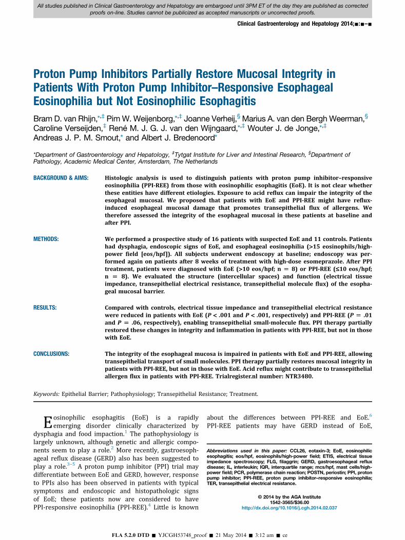
Figure 1. Study protocol and patient subgroup selection. Theesophageal mucosal integrity of patients and controls wasmeasured at baseline. In patients, the integrity and inflam-mation were measured after PPI treatment. Based on theeosinophil count after PPI treatment, patients were catego-rized as PPI-REE (�10 eos/hpf) or EoE (>10 eos/hpf).
2 van Rhijn et al Clinical Gastroenterology and Hepatology Vol. -, No. -
however, they cannot be distinguished from EoE patientsby clinical features, endoscopic signs, or histopathologicsigns.4,7
In patients with GERD, it has been well documentedthat acid reflux causes impaired esophageal mucosalintegrity, which has been suggested to decrease esoph-ageal intraluminal baseline impedance.8,9 Acid suppres-sion with PPIs restores the esophageal mucosal integrityin GERD.10 In EoE, esophageal baseline impedance valuesare decreased as well, and histologic studies also haveshown dilation of intercellular spaces—a morphologicfeature of epithelial permeability changes—in EoE.11,12
We hypothesized that acid-induced impairment ofesophageal mucosal integrity facilitates permeation offood allergens, thereby promoting immune activation inpatients with PPI-REE and EoE. If this hypothesis is valid,proton pump inhibition will restore the esophagealmucosal integrity and thereby reduce the passage of al-lergens into the epithelium. The aim of our study was toshow an impaired esophageal mucosal integrity in PPI-REE and EoE patients, and to study the effect of acidsuppression on the esophageal mucosal integrity.
Methods
Study Subjects
In this prospective study, we included 16 adult pa-tients with suspected EoE (>15 eosinophils/high-powerfield [eos/hpf], predominant symptoms of dysphagiaand/or food impaction, and endoscopic signs of EoE),and 11 healthy controls. Based on peak eosinophil countsafter PPI, patients were divided into 2 subgroups: re-sponders (PPI-REE, �10 eos/hpf) and nonresponders(EoE, >10 eos/hpf) (Figure 1). Patients were recruitedconsecutively from the outpatient clinic of our hospital.Healthy controls were recruited by advertisement in thehospital and had no dysphagia/reflux symptoms or othergastrointestinal complaints. None of the study subjectshad undergone surgery of the digestive tract. Each studysubject provided written informed consent and the studyprotocol was approved by the Medical Ethics Committeeof our institution. All authors had access to the studydata and reviewed and approved the final manuscript.
Study Protocol
In all study subjects, dietary, anti-inflammatory, andacid-suppressive treatments were discontinued 8 weeksbefore baseline upper endoscopy. In the 24 hours pre-ceding endoscopy, smoking and alcohol intake were notallowed. In patients, endoscopy was repeated after 8weeks of 40 mg esomeprazole twice-daily treatment, andthe frequency and severity of dysphagia for liquids andsolids was assessed using a 6-grade Likert scale, in which0 represents no dysphagia and 5 represents daily/severedysphagia, analogous to the reflux disease questionnaire.
FLA 5.2.0 DTD � YJCGH53748_proo
Upper Endoscopy
After routine inspection of the duodenum and stom-ach, pictures were taken of the esophagus for assessmentof endoscopic signs of EoE. After this, 5 electrical tissueimpedance spectroscopy (ETIS) measurements wereperformed in the distal esophagus 5 cm proximal to theZ-line. At the same level, 4 biopsy specimens wereobtained with a large biopsy forceps (diameter, 3.7 mm)for the assessment of functional mucosal integrity inUssing chambers. In addition, 2 large biopsy specimenswere taken to evaluate dilation of intercellular spaces bytransmission electron microscopy. For histopathologicanalysis in patients, 6 biopsy specimens were taken, and 1biopsy specimen was taken for gene expression profiling.
To assess endoscopic signs of EoE (white exudates,linear furrows, concentric rings, solitary ring, crepe pa-per mucosa, pallor, and narrow-caliber esophagus), aphysician blinded to the patient’s status scored theendoscopic pictures.
Esophageal Mucosal Integrity AssessmentIn Vivo
As a functionalmeasure of esophagealmucosal integrity,we performed ETIS measurements to measure the extra-cellular esophageal impedance during endoscopy, as pre-viously described.13 The extracellular impedance wascorrelated with structural and functional in vitro parame-ters of esophageal mucosal integrity; ETIS is therefore ameasure of esophageal mucosal integrity in vivo.13
f � 21 May 2014 � 3:12 am � ce
- 2014 Impaired Esophageal Mucosal Integrity in EoE 3
Esophageal Mucosal Integrity AssessmentIn Vitro
Esophageal mucosal integrity was assessed accordingto previously described methods.13 In short, 4 esopha-geal biopsy specimens were mounted in Ussing cham-bers immediately after endoscopy. Electrodes were usedto measure the transepithelial electrical resistance (TER).Simultaneously, the transepithelial flux of fluorescentmolecules of 2 different sizes (fluorescein, 332 daltons;rhodamine, 40,000 daltons; size similar to common foodallergens) was measured. After 15 minutes of acclimati-zation in Meyler buffer, we sampled the serosal bath andsubsequently replaced the luminal buffer with a modifiedMeyler buffer containing these fluorescent molecules at aconcentration of 0.5 mg/mL. We sampled the serosalbath every 15 minutes during 1 hour. A fluorescenceplate reader (BioTek Synergy; BioTek, Winooski, VT)measured the concentration of fluorescein and rhoda-mine molecules using excitation wavelengths of 485 and530 nm and emission wavelengths of 528 and 590 nm,respectively. Rhodamine signal strength was correctedfor the presence of fluorescein.
Transmission Electron Microscopy
As a structural marker of esophageal mucosal integ-rity related to esophageal permeability changes,8,10 thespace between individual esophageal epithelial cells wasmeasured, as previously described.13 The laboratorytechnician was blinded to the status of the biopsy andselected 10 random photographs of each biopsy at thebasal prickle layer (magnification, 4600�). Image pro-cessing and analysis was performed using Qwin (LeicaMicrosystems, Wetzlar, Germany).
Histopathologic Analysis
For histopathologic analysis, 2 biopsy specimenswere taken at the distal esophagus, midesophagus (5 cmand 10–15 cm above the gastroesophageal junction), andproximal esophagus (5 cm below the upper esophagealsphincter). Specimens were stained with H&E and tryp-tase to determine the eosinophil and mast cell counts. Anexperienced gastrointestinal pathologist blinded to bi-opsy location and the patient’s treatment status analyzedthe specimens in random order, using an Olympus BX41microscope (Olympus Europe, Hamburg, Germany). Ineach biopsy specimen, the area of greatest eosinophildensity was detected with a low-power view. By using amagnification of 400� (1 hpf), the peak eosinophil countwas determined. Mast cells were counted identically.
Furthermore, the presence of eosinophilic micro-abscesses (defined as clusters of �4 eosinophils) wereanalyzed at high-power, and basal hyperplasia andspongiosis were analyzed at low-power (for both: 0 ¼absent, 1 ¼ extending to lower third of total epithelial
FLA 5.2.0 DTD � YJCGH53748_proo
thickness [mild], 2 ¼ extending to middle third [mod-erate], 3 ¼ extending to upper third [severe]), accordingto the literature.11
Quantitative Real-Time PolymeraseChain Reaction
During endoscopy at baseline and after PPI, 1mucosal biopsy specimen from each patient wascollected and immersed in RNA stabilization reagent(RNAlater; Qiagen, Hilden, Germany). Samples werestored overnight at 4�C and subsequently were stored at-80�C until analysis. Biopsy specimens were homoge-nized and total RNA was extracted using the RNeasyMicro Kit (Qiagen) according to the manufacturer’s rec-ommendations. The RNA concentration was assessedusing the Nanodrop Spectrophotometer (NanodropTechnologies, Wilmington, DE). Complementary DNAwas synthesized using a reverse-transcriptase reactionperformed according to the MBI Fermentas comple-mentary DNA synthesis kit (Fermentas, Vilnius,Lithuania), using both the Oligo(dT)18 and the D(N)6primers. Quantitative real-time polymerase chain reac-tion (PCR) was performed on the LightCycler 480 (RocheDiagnostic, Almere, The Netherlands) using SYBR GreenPCR Master Mix (Roche Diagnostic) and primers fromInvitrogen (Life Technologies Corporation, Carlsbad, CA)(Supplementary Table 1). For quantitative real-time PCR,samples were normalized for the mean of the 3 moststable housekeeping genes (cyclophilin, glyceraldehyde-3-phosphate dehydrogenase, and b-actin) as determined byanalysis with geNorm method software (available at:http://medgen.ugent.be/wjvdesomp/genorm/). Tran-script levels of interleukin (IL)5, IL13, eotaxin-3 (CCL26),periostin (POSTN), filaggrin (FLG), and thymic stromallymphopoietin were determined in duplicate.
Statistical Analysis
Continuous data were expressed as medians (inter-quartile range [IQR]). Differences between 2 or moregroups were calculated with the Kruskal–Wallis test,with Bonferroni post hoc correction for multiple testing.Patient groups were compared using the Mann–WhitneyU test or the Wilcoxon signed rank test where appro-priate. Proportions were compared using the chi-squaretest, the Fisher exact test, or the McNemar test whereappropriate. Correlations were calculated using theSpearman correlation coefficient. We considered a Pvalue less than .05 to be significant.
Results
Patients vs Controls
Subject characteristics. We consecutively included 16patients (13 men) with suspected EoE, and 11 controls
f � 21 May 2014 � 3:12 am � ce
4 van Rhijn et al Clinical Gastroenterology and Hepatology Vol. -, No. -
(7 men). No patients were excluded or dropped out afterenrolment in the study. The median age at first endos-copy was 42 years (IQR, 32–46 y) for patients and 35years (IQR, 29–53 y) for controls (P ¼ .6). None of thecontrols had abnormalities on endoscopy. Based on peakeosinophil counts after PPI, 8 patients (6 men) wereclassified as PPI-REE (range, 0–10 eos/hpf), and 8 pa-tients (7 men) were classified as EoE patients (range,19–100 eos/hpf).
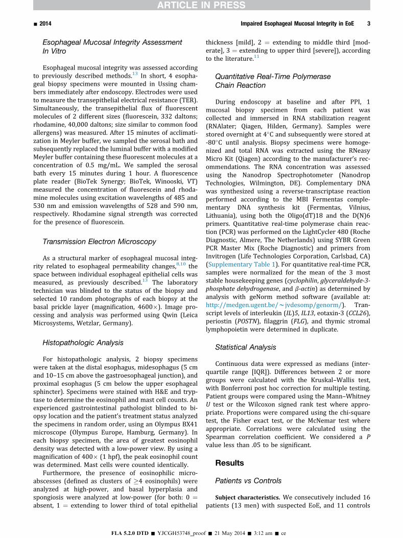
Esophageal mucosal integrity and intercellular spaces. Thedistributions of the extracellular impedance (P < .001),TER (P < .001), fluorescein flux (P ¼ .001), and inter-cellular spaces (P ¼ .003) were significantly differentbetween PPI-REE and EoE patients and healthy controls.Post hoc analysis showed that the extracellular imped-ance was significantly lower in EoE (2014 U�m;1288–3963 U�m; P < .001) and PPI-REE patients (3128U�m; 1869–5213 U�m; P ¼ .01) compared with controls(7707 U�m; 6146–10,488 U�m; (Figure 2A). TER valuesalso were lower in EoE (31.3 U�m; 25.4–41.8 U�cm2;P < .001) and borderline lower in PPI-REE patients (42.7U�m; 34.1–62.4 U�cm2; P ¼ .06) than in controls (116.7U�m; 90.3–126.4 U�cm2) (Figure 2B). Furthermore,compared with controls (345 nmol/cm2/h; 0–1451nmol/cm2/h), the transepithelial flux of fluoresceinmolecules in the Ussing chambers was significantlyincreased in EoE patients (2974 nmol/cm2/h;2680–3362 nmol/cm2/h; P < .001), but not in PPI-REEpatients (1640 nmol/cm2/h; 1–2424 nmol/cm2/h; P ¼.8) (Figure 2C).
At baseline, intercellular spaces were evaluated inall EoE patients, in 7 PPI-REE patients, and in 6 con-trols. Samples of 1 PPI-REE patient and 5 control
FLA 5.2.0 DTD � YJCGH53748_proo
subjects unfortunately were lost as a result of a tech-nical problem during sample processing. The intercel-lular space was significantly larger in EoE (0.34;0.32–0.45; P ¼ .008) and PPI-REE patients (0.37;0.33–0.42; P ¼ .007) than in controls (0.14; 0.11–0.23)(Figure 2D).
Effect of Proton Pump Inhibitor
Esophageal mucosal integrity and intercellular spaces. InPPI-REE patients (Figure 3), the extracellular impedanceincreased significantly from 3128 U�m (1869–5213U�m) to 6848 U�m (5761–9363 U�m) (P ¼ .01) afterPPI, and was no longer different from healthy controlvalues (P ¼ .6). The TER increased from 42.7 U�m(34.1–62.4 U�m) to 100.0 U�m (68.8–121.9 U�m; P ¼.004), and was not different from healthy control values(P ¼ 1.0). The transepithelial flux of the small fluoresceinmolecules decreased from 1640 nmol/cm2/h (1–2424nmol/cm2/h) to 255 nmol/cm2/h (39–628 nmol/cm2/h), although not significantly (P ¼ .06), and was notdifferent from healthy control values (P ¼ 1.0). More-over, the flux of the larger, food allergen–sized rhoda-mine molecules decreased significantly from 22 nmol/cm2/h (0–38 nmol/cm2/h) to 0 nmol/cm2/h (0–0 nmol/cm2/h; P ¼ .046). The intercellular space remained un-changed, being 0.37 (0.33–0.42) at baseline and 0.33(0.27–0.38) after PPI (P ¼ .1).
In EoE patients (Figure 4), the extracellular imped-ance increased significantly from 2014 U�m (1288–3963U�m) to 3404 U�m (2810–6579 U�m; P ¼ .01) after PPI,however, it was still significantly lower than in healthycontrols (P ¼ .04). The TER (from 31.3 U�m; 25.4–41.8
Figure 2. (A) Extracellularimpedance, (B) TER, (C)fluorescein flux, and (D)intercellular space showimpaired mucosal integrityin PPI-REE patients andEoE patients comparedwith controls. Black barsindicate medians.
f � 21 May 2014 � 3:12 am � ce
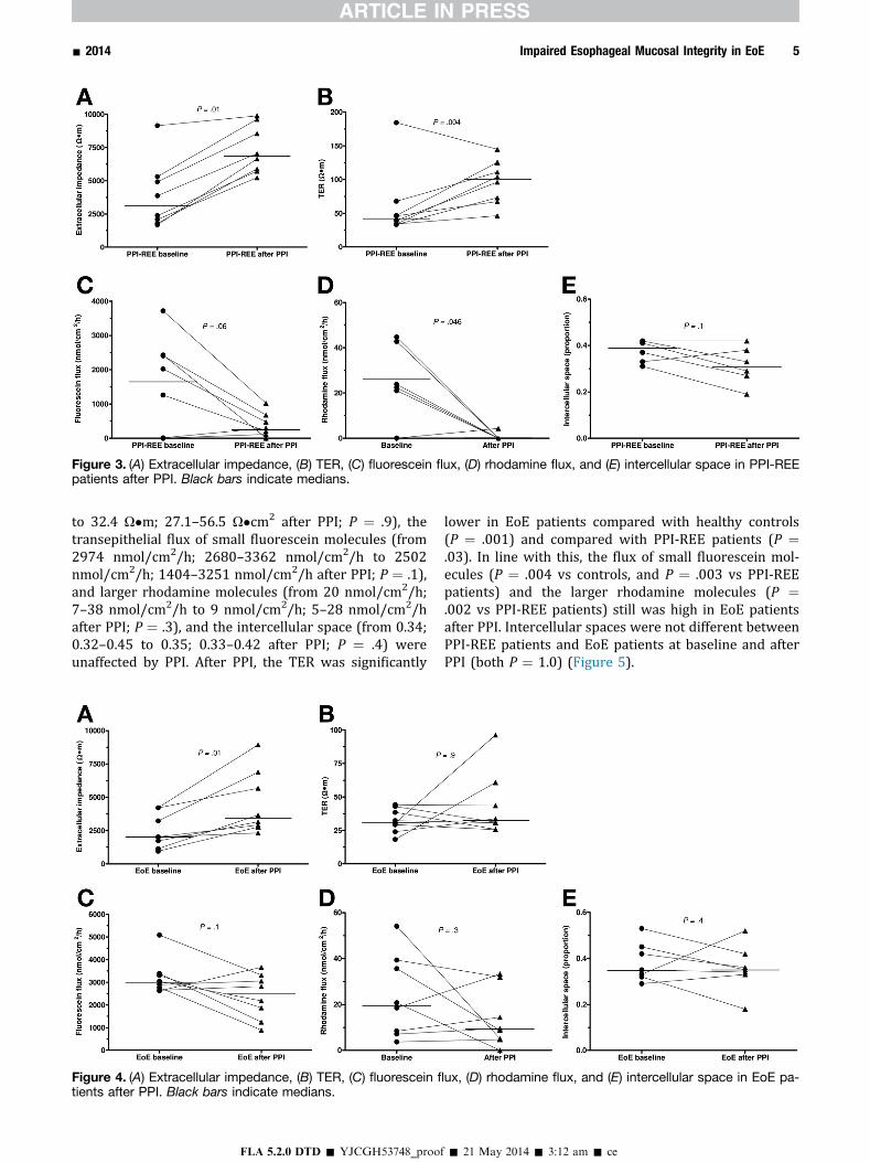
Figure 3. (A) Extracellular impedance, (B) TER, (C) fluorescein flux, (D) rhodamine flux, and (E) intercellular space in PPI-REEpatients after PPI. Black bars indicate medians.
- 2014 Impaired Esophageal Mucosal Integrity in EoE 5
to 32.4 U�m; 27.1–56.5 U�cm2 after PPI; P ¼ .9), thetransepithelial flux of small fluorescein molecules (from2974 nmol/cm2/h; 2680–3362 nmol/cm2/h to 2502nmol/cm2/h; 1404–3251 nmol/cm2/h after PPI; P ¼ .1),and larger rhodamine molecules (from 20 nmol/cm2/h;7–38 nmol/cm2/h to 9 nmol/cm2/h; 5–28 nmol/cm2/hafter PPI; P ¼ .3), and the intercellular space (from 0.34;0.32–0.45 to 0.35; 0.33–0.42 after PPI; P ¼ .4) wereunaffected by PPI. After PPI, the TER was significantly
Figure 4. (A) Extracellular impedance, (B) TER, (C) fluorescein fl
tients after PPI. Black bars indicate medians.
FLA 5.2.0 DTD � YJCGH53748_proo
lower in EoE patients compared with healthy controls(P ¼ .001) and compared with PPI-REE patients (P ¼.03). In line with this, the flux of small fluorescein mol-ecules (P ¼ .004 vs controls, and P ¼ .003 vs PPI-REEpatients) and the larger rhodamine molecules (P ¼.002 vs PPI-REE patients) still was high in EoE patientsafter PPI. Intercellular spaces were not different betweenPPI-REE patients and EoE patients at baseline and afterPPI (both P ¼ 1.0) (Figure 5).
ux, (D) rhodamine flux, and (E) intercellular space in EoE pa-
f � 21 May 2014 � 3:12 am � ce
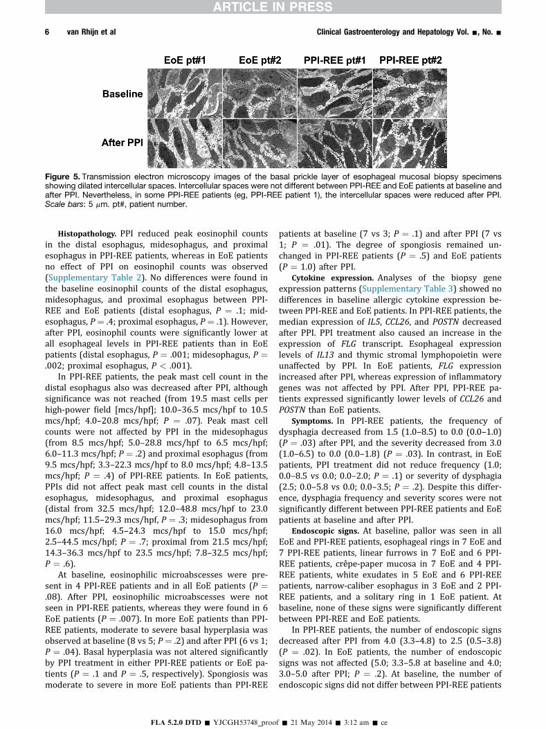
Figure 5. Transmission electron microscopy images of the basal prickle layer of esophageal mucosal biopsy specimensshowing dilated intercellular spaces. Intercellular spaces were not different between PPI-REE and EoE patients at baseline andafter PPI. Nevertheless, in some PPI-REE patients (eg, PPI-REE patient 1), the intercellular spaces were reduced after PPI.Scale bars: 5 mm. pt#, patient number.
6 van Rhijn et al Clinical Gastroenterology and Hepatology Vol. -, No. -
Histopathology. PPI reduced peak eosinophil countsin the distal esophagus, midesophagus, and proximalesophagus in PPI-REE patients, whereas in EoE patientsno effect of PPI on eosinophil counts was observed(Supplementary Table 2). No differences were found inthe baseline eosinophil counts of the distal esophagus,midesophagus, and proximal esophagus between PPI-REE and EoE patients (distal esophagus, P ¼ .1; mid-esophagus, P ¼ .4; proximal esophagus, P ¼ .1). However,after PPI, eosinophil counts were significantly lower atall esophageal levels in PPI-REE patients than in EoEpatients (distal esophagus, P ¼ .001; midesophagus, P ¼.002; proximal esophagus, P < .001).
In PPI-REE patients, the peak mast cell count in thedistal esophagus also was decreased after PPI, althoughsignificance was not reached (from 19.5 mast cells perhigh-power field [mcs/hpf]; 10.0–36.5 mcs/hpf to 10.5mcs/hpf; 4.0–20.8 mcs/hpf; P ¼ .07). Peak mast cellcounts were not affected by PPI in the midesophagus(from 8.5 mcs/hpf; 5.0–28.8 mcs/hpf to 6.5 mcs/hpf;6.0–11.3 mcs/hpf; P ¼ .2) and proximal esophagus (from9.5 mcs/hpf; 3.3–22.3 mcs/hpf to 8.0 mcs/hpf; 4.8–13.5mcs/hpf; P ¼ .4) of PPI-REE patients. In EoE patients,PPIs did not affect peak mast cell counts in the distalesophagus, midesophagus, and proximal esophagus(distal from 32.5 mcs/hpf; 12.0–48.8 mcs/hpf to 23.0mcs/hpf; 11.5–29.3 mcs/hpf, P ¼ .3; midesophagus from16.0 mcs/hpf; 4.5–24.3 mcs/hpf to 15.0 mcs/hpf;2.5–44.5 mcs/hpf; P ¼ .7; proximal from 21.5 mcs/hpf;14.3–36.3 mcs/hpf to 23.5 mcs/hpf; 7.8–32.5 mcs/hpf;P ¼ .6).
At baseline, eosinophilic microabscesses were pre-sent in 4 PPI-REE patients and in all EoE patients (P ¼.08). After PPI, eosinophilic microabscesses were notseen in PPI-REE patients, whereas they were found in 6EoE patients (P ¼ .007). In more EoE patients than PPI-REE patients, moderate to severe basal hyperplasia wasobserved at baseline (8 vs 5; P¼ .2) and after PPI (6 vs 1;P ¼ .04). Basal hyperplasia was not altered significantlyby PPI treatment in either PPI-REE patients or EoE pa-tients (P ¼ .1 and P ¼ .5, respectively). Spongiosis wasmoderate to severe in more EoE patients than PPI-REE
FLA 5.2.0 DTD � YJCGH53748_proo
patients at baseline (7 vs 3; P ¼ .1) and after PPI (7 vs1; P ¼ .01). The degree of spongiosis remained un-changed in PPI-REE patients (P ¼ .5) and EoE patients(P ¼ 1.0) after PPI.
Cytokine expression. Analyses of the biopsy geneexpression patterns (Supplementary Table 3) showed nodifferences in baseline allergic cytokine expression be-tween PPI-REE and EoE patients. In PPI-REE patients, themedian expression of IL5, CCL26, and POSTN decreasedafter PPI. PPI treatment also caused an increase in theexpression of FLG transcript. Esophageal expressionlevels of IL13 and thymic stromal lymphopoietin wereunaffected by PPI. In EoE patients, FLG expressionincreased after PPI, whereas expression of inflammatorygenes was not affected by PPI. After PPI, PPI-REE pa-tients expressed significantly lower levels of CCL26 andPOSTN than EoE patients.
Symptoms. In PPI-REE patients, the frequency ofdysphagia decreased from 1.5 (1.0–8.5) to 0.0 (0.0–1.0)(P ¼ .03) after PPI, and the severity decreased from 3.0(1.0–6.5) to 0.0 (0.0–1.8) (P ¼ .03). In contrast, in EoEpatients, PPI treatment did not reduce frequency (1.0;0.0–8.5 vs 0.0; 0.0–2.0; P ¼ .1) or severity of dysphagia(2.5; 0.0–5.8 vs 0.0; 0.0–3.5; P ¼ .2). Despite this differ-ence, dysphagia frequency and severity scores were notsignificantly different between PPI-REE patients and EoEpatients at baseline and after PPI.
Endoscopic signs. At baseline, pallor was seen in allEoE and PPI-REE patients, esophageal rings in 7 EoE and7 PPI-REE patients, linear furrows in 7 EoE and 6 PPI-REE patients, crêpe-paper mucosa in 7 EoE and 4 PPI-REE patients, white exudates in 5 EoE and 6 PPI-REEpatients, narrow-caliber esophagus in 3 EoE and 2 PPI-REE patients, and a solitary ring in 1 EoE patient. Atbaseline, none of these signs were significantly differentbetween PPI-REE and EoE patients.
In PPI-REE patients, the number of endoscopic signsdecreased after PPI from 4.0 (3.3–4.8) to 2.5 (0.5–3.8)(P ¼ .02). In EoE patients, the number of endoscopicsigns was not affected (5.0; 3.3–5.8 at baseline and 4.0;3.0–5.0 after PPI; P ¼ .2). At baseline, the number ofendoscopic signs did not differ between PPI-REE patients
f � 21 May 2014 � 3:12 am � ce
print&
web4C=FPO
- 2014 Impaired Esophageal Mucosal Integrity in EoE 7
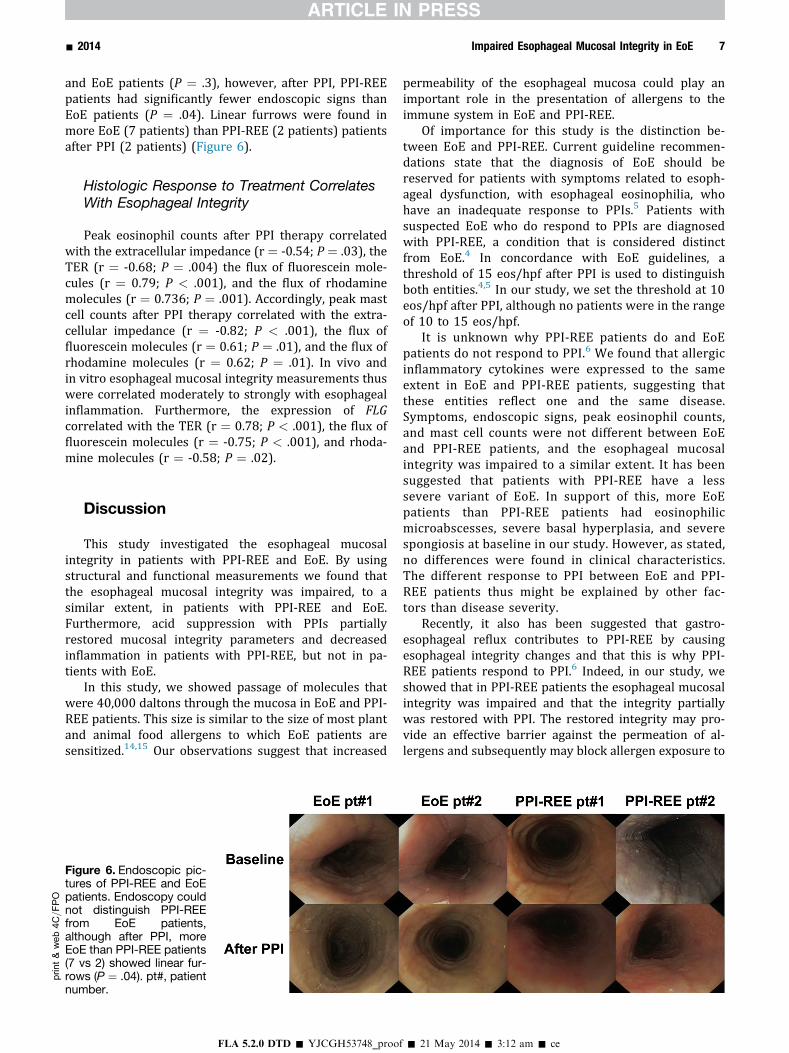
and EoE patients (P ¼ .3), however, after PPI, PPI-REEpatients had significantly fewer endoscopic signs thanEoE patients (P ¼ .04). Linear furrows were found inmore EoE (7 patients) than PPI-REE (2 patients) patientsafter PPI (2 patients) (Figure 6).
Histologic Response to Treatment CorrelatesWith Esophageal Integrity
Peak eosinophil counts after PPI therapy correlatedwith the extracellular impedance (r ¼ -0.54; P ¼ .03), theTER (r ¼ -0.68; P ¼ .004) the flux of fluorescein mole-cules (r ¼ 0.79; P < .001), and the flux of rhodaminemolecules (r ¼ 0.736; P ¼ .001). Accordingly, peak mastcell counts after PPI therapy correlated with the extra-cellular impedance (r ¼ -0.82; P < .001), the flux offluorescein molecules (r ¼ 0.61; P ¼ .01), and the flux ofrhodamine molecules (r ¼ 0.62; P ¼ .01). In vivo andin vitro esophageal mucosal integrity measurements thuswere correlated moderately to strongly with esophagealinflammation. Furthermore, the expression of FLGcorrelated with the TER (r ¼ 0.78; P < .001), the flux offluorescein molecules (r ¼ -0.75; P < .001), and rhoda-mine molecules (r ¼ -0.58; P ¼ .02).
Discussion
This study investigated the esophageal mucosalintegrity in patients with PPI-REE and EoE. By usingstructural and functional measurements we found thatthe esophageal mucosal integrity was impaired, to asimilar extent, in patients with PPI-REE and EoE.Furthermore, acid suppression with PPIs partiallyrestored mucosal integrity parameters and decreasedinflammation in patients with PPI-REE, but not in pa-tients with EoE.
In this study, we showed passage of molecules thatwere 40,000 daltons through the mucosa in EoE and PPI-REE patients. This size is similar to the size of most plantand animal food allergens to which EoE patients aresensitized.14,15 Our observations suggest that increased
Figure 6. Endoscopic pic-tures of PPI-REE and EoEpatients. Endoscopy couldnot distinguish PPI-REEfrom EoE patients,although after PPI, moreEoE than PPI-REE patients(7 vs 2) showed linear fur-rows (P ¼ .04). pt#, patientnumber.
FLA 5.2.0 DTD � YJCGH53748_proo
permeability of the esophageal mucosa could play animportant role in the presentation of allergens to theimmune system in EoE and PPI-REE.
Of importance for this study is the distinction be-tween EoE and PPI-REE. Current guideline recommen-dations state that the diagnosis of EoE should bereserved for patients with symptoms related to esoph-ageal dysfunction, with esophageal eosinophilia, whohave an inadequate response to PPIs.5 Patients withsuspected EoE who do respond to PPIs are diagnosedwith PPI-REE, a condition that is considered distinctfrom EoE.4 In concordance with EoE guidelines, athreshold of 15 eos/hpf after PPI is used to distinguishboth entities.4,5 In our study, we set the threshold at 10eos/hpf after PPI, although no patients were in the rangeof 10 to 15 eos/hpf.
It is unknown why PPI-REE patients do and EoEpatients do not respond to PPI.6 We found that allergicinflammatory cytokines were expressed to the sameextent in EoE and PPI-REE patients, suggesting thatthese entities reflect one and the same disease.Symptoms, endoscopic signs, peak eosinophil counts,and mast cell counts were not different between EoEand PPI-REE patients, and the esophageal mucosalintegrity was impaired to a similar extent. It has beensuggested that patients with PPI-REE have a lesssevere variant of EoE. In support of this, more EoEpatients than PPI-REE patients had eosinophilicmicroabscesses, severe basal hyperplasia, and severespongiosis at baseline in our study. However, as stated,no differences were found in clinical characteristics.The different response to PPI between EoE and PPI-REE patients thus might be explained by other fac-tors than disease severity.
Recently, it also has been suggested that gastro-esophageal reflux contributes to PPI-REE by causingesophageal integrity changes and that this is why PPI-REE patients respond to PPI.6 Indeed, in our study, weshowed that in PPI-REE patients the esophageal mucosalintegrity was impaired and that the integrity partiallywas restored with PPI. The restored integrity may pro-vide an effective barrier against the permeation of al-lergens and subsequently may block allergen exposure to
f � 21 May 2014 � 3:12 am � ce
8 van Rhijn et al Clinical Gastroenterology and Hepatology Vol. -, No. -
antigen-presenting cells, thereby prohibiting immuneactivation. The observed effect of acid suppression withhigh-dose PPIs on the mucosal integrity in PPI-REEpatients supports the hypothesis that acid reflux con-tributes to the impaired epithelial barrier function inPPI-REE. Alternatively, it also is possible that restorationof the mucosal integrity is the result of a direct anti-inflammatory effect of PPIs on the esophageal epithe-lium. Unfortunately, pretreatment predictive factors forPPI response have not been identified yet, either inin vitro studies or in pH-monitoring studies.
An alternative cause of impaired mucosal integritycould be that the intrinsic inflammatory cytokines andcytotoxic eosinophil and mast cell secretory productsthat are present in the esophagus of EoE and PPI-REEpatients impair the mucosal integrity, similar to mecha-nisms in asthma and atopic dermatitis.16,17 The loss ofdesmoglein-1, an intercellular adhesion molecule that isaltered in various skin disorders, may potentiate thisallergic inflammation through the induction of proin-flammatory mediators such as POSTN.18 The observationthat EoE patients did not respond to a PPI suggests thatgastroesophageal reflux does not play a role in EoE pa-tients, and favors this second explanation for theimpaired mucosal integrity in EoE. Furthermore, thestrongly decreased expression of FLG, previouslydescribed in EoE, increased after PPI in PPI-REE but notin EoE patients.19 The finding that FLG expression alsocorrelated inversely with the permeability to allergen-sized molecules suggests that down-regulation of FLGalso may contribute to the observed epithelial barrierdysfunction.
In conclusion, the esophageal mucosal integrity isseverely impaired in patients with PPI-REE and in pa-tients with EoE. This finding could be of pathophysio-logical importance because increased permeability mayfacilitate transepithelial food allergen flux. Furthermore,the finding that histologic response correlates withimproved esophageal barrier integrity suggests thatbarrier integrity is a potential therapeutic target. Theobservation that PPI partially improves the esophagealmucosal integrity in PPI-REE patients but not in EoEpatients may indicate a role for acid reflux in PPI-REEbut not in EoE.
Supplementary Material
Note: To access the supplementary material accom-panying this article, visit the online version of ClinicalGastroenterology and Hepatology at www.cghjournal.org,and at http://dx.doi.org/10.1016/j.cgh.2014.02.037.
References
1. van Rhijn BD, Verheij J, Smout AJ, et al. Rapidly increasingincidence of eosinophilic esophagitis in a large cohort. Neuro-gastroenterol Motil 2013;25:47–52.
FLA 5.2.0 DTD � YJCGH53748_proo
2. Rothenberg ME. Biology and treatment of eosinophilic esoph-agitis. Gastroenterology 2009;137:1238–1249.
3. Spechler SJ, Genta RM, Souza RF. Thoughts on the complexrelationship between gastroesophageal reflux disease andeosinophilic esophagitis. Am J Gastroenterol 2007;102:1301–1306.
4. Molina-Infante J, Ferrando-Lamana L, Ripoll C, et al. Esoph-ageal eosinophilic infiltration responds to proton pump inhi-bition in most adults. Clin Gastroenterol Hepatol 2011;9:110–117.
5. Liacouras CA, Furuta GT, Hirano I, et al. Eosinophilic esopha-gitis: updated consensus recommendations for children andadults. J Allergy Clin Immunol 2011;128:3–20.
6. Dellon ES, Gonsalves N, Hirano I, et al. ACG clinical guideline:evidenced based approach to the diagnosis and managementof esophageal eosinophilia and eosinophilic esophagitis (EoE).Am J Gastroenterol 2013;108:679–692.
7. Dranove JE, Horn DS, Davis MA, et al. Predictors ofresponse to proton pump inhibitor therapy among childrenwith significant esophageal eosinophilia. J Pediatr 2009;154:96–100.
8. Tobey NA, Carson JL, Alkiek RA, et al. Dilated intercellularspaces: a morphological feature of acid reflux–damagedhuman esophageal epithelium. Gastroenterology 1996;111:1200–1205.
9. Kessing BF, Bredenoord AJ, Weijenborg PW, et al. Esophagealacid exposure decreases intraluminal baseline impedancelevels. Am J Gastroenterol 2011;106:2093–2097.
10. Calabrese C, Bortolotti M, Fabbri A, et al. Reversibility ofGERD ultrastructural alterations and relief of symptoms afteromeprazole treatment. Am J Gastroenterol 2005;100:537–542.
11. Mueller S, Neureiter D, Aigner T, et al. Comparison of histo-logical parameters for the diagnosis of eosinophilic oesophagitisversus gastro-oesophageal reflux disease on oesophageal bi-opsy material. Histopathology 2008;53:676–684.
12. van Rhijn BD, Kessing BF, Smout AJ, et al. Oesophagealbaseline impedance values are decreased in patients witheosinophilic oesophagitis. United Eur Gastroenterol J 2013;1:242–248.
13. Weijenborg PW, Rohof WO, Akkermans LM, et al. Electricaltissue impedance spectroscopy: a novel device to measureesophageal mucosal integrity changes during endoscopy.Neurogastroenterol Motil 2013;25:574–e458.
14. Hoffmann-Sommergruber K, Mills EN. Food allergen proteinfamilies and their structural characteristics and application incomponent-resolved diagnosis: new data from the EuroPrevallproject. Anal Bioanal Chem 2009;395:25–35.
15. van Rhijn BD, van Ree R, Versteeg SA, et al. Birch pollensensitization with cross-reactivity to food allergens pre-dominates in adults with eosinophilic esophagitis. Allergy 2013;68:1475–1481.
16. Holgate ST. The sentinel role of the airway epithelium in asthmapathogenesis. Immunol Rev 2011;242:205–219.
17. De Benedetto A, Rafaels NM, McGirt LY, et al. Tight junctiondefects in patients with atopic dermatitis. J Allergy Clin Immunol2011;127:773–786.
18. Sherrill JD, Kc K, Wu D, et al. Desmoglein-1 regulates esopha-geal epithelial barrier function and immune responses in eosin-ophilic esophagitis. Mucosal Immunol 2013 Nov 13. Epub aheadof print.
f � 21 May 2014 � 3:12 am � ce
- 2014 Impaired Esophageal Mucosal Integrity in EoE 9
19. Blanchard C, Wang N, Stringer KF, et al. Eotaxin-3 and auniquely conserved gene-expression profile in eosinophilicesophagitis. J Clin Invest 2006;116:536–547.
Reprint requestsAddress requests for reprints to: Bram D. van Rhijn, MD, Department ofGastroenterology and Hepatology, Academic Medical Center, Amsterdam,PO Box 22660, 1100 DD Amsterdam, The Netherlands. e-mail: [email protected]; fax: (31) 20-691-70-33.
FLA 5.2.0 DTD � YJCGH53748_proo
Conflicts of interestThese authors disclose the following: Albert Bredenoord has received researchfunding from AstraZeneca, Endostim, Medical Measurement Systems, andShire, and has received speaker fees from Medical Measurement Systems andShire; and Wouter de Jonge has received funding from Glaxo Smith Kline. Theremaining authors disclose no conflicts.
FundingA.J.B. and W.J.d.J. are supported by The Netherlands Organization forScientific Research (Nederlandse Organisatie voor WetenschappelijkOnderzoek).
f � 21 May 2014 � 3:13 am � ce
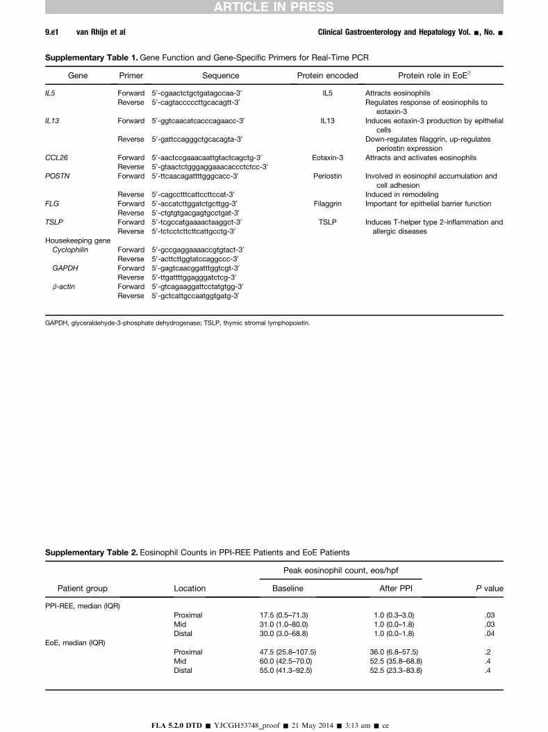
Supplementary Table 1.Gene Function and Gene-Specific Primers for Real-Time PCR
Gene Primer Sequence Protein encoded Protein role in EoE2
IL5 Forward 50-cgaactctgctgatagccaa-30 IL5 Attracts eosinophilsReverse 50-cagtacccccttgcacagtt-30 Regulates response of eosinophils to
eotaxin-3IL13 Forward 50-ggtcaacatcacccagaacc-30 IL13 Induces eotaxin-3 production by epithelial
cellsReverse 50-gattccagggctgcacagta-30 Down-regulates filaggrin, up-regulates
periostin expressionCCL26 Forward 50-aactccgaaacaattgtactcagctg-30 Eotaxin-3 Attracts and activates eosinophils
Reverse 50-gtaactctgggaggaaacaccctctcc-30
POSTN Forward 50-ttcaacagattttgggcacc-30 Periostin Involved in eosinophil accumulation andcell adhesion
Reverse 50-cagcctttcattccttccat-30 Induced in remodelingFLG Forward 50-accatcttggatctgcttgg-30 Filaggrin Important for epithelial barrier function
Reverse 50-ctgtgtgacgagtgcctgat-30
TSLP Forward 50-tcgccatgaaaactaaggct-30 TSLP Induces T-helper type 2-inflammation andallergic diseasesReverse 50-tctcctcttcttcattgcctg-30
Housekeeping geneCyclophilin Forward 50-gccgaggaaaaccgtgtact-30
Reverse 50-acttcttggtatccaggccc-30
GAPDH Forward 50-gagtcaacggatttggtcgt-30
Reverse 50-ttgattttggagggatctcg-30
b-actin Forward 50-gtcagaaggattcctatgtgg-30
Reverse 50-gctcattgccaatggtgatg-30
GAPDH, glyceraldehyde-3-phosphate dehydrogenase; TSLP, thymic stromal lymphopoietin.
Supplementary Table 2. Eosinophil Counts in PPI-REE Patients and EoE Patients
Patient group Location
Peak eosinophil count, eos/hpf
P valueBaseline After PPI
PPI-REE, median (IQR)Proximal 17.5 (0.5–71.3) 1.0 (0.3–3.0) .03Mid 31.0 (1.0–80.0) 1.0 (0.0–1.8) .03Distal 30.0 (3.0–68.8) 1.0 (0.0–1.8) .04
EoE, median (IQR)Proximal 47.5 (25.8–107.5) 36.0 (6.8–57.5) .2Mid 60.0 (42.5–70.0) 52.5 (35.8–68.8) .4Distal 55.0 (41.3–92.5) 52.5 (23.3–83.8) .4
FLA 5.2.0 DTD � YJCGH53748_proof � 21 May 2014 � 3:13 am � ce
9.e1 van Rhijn et al Clinical Gastroenterology and Hepatology Vol. -, No. -
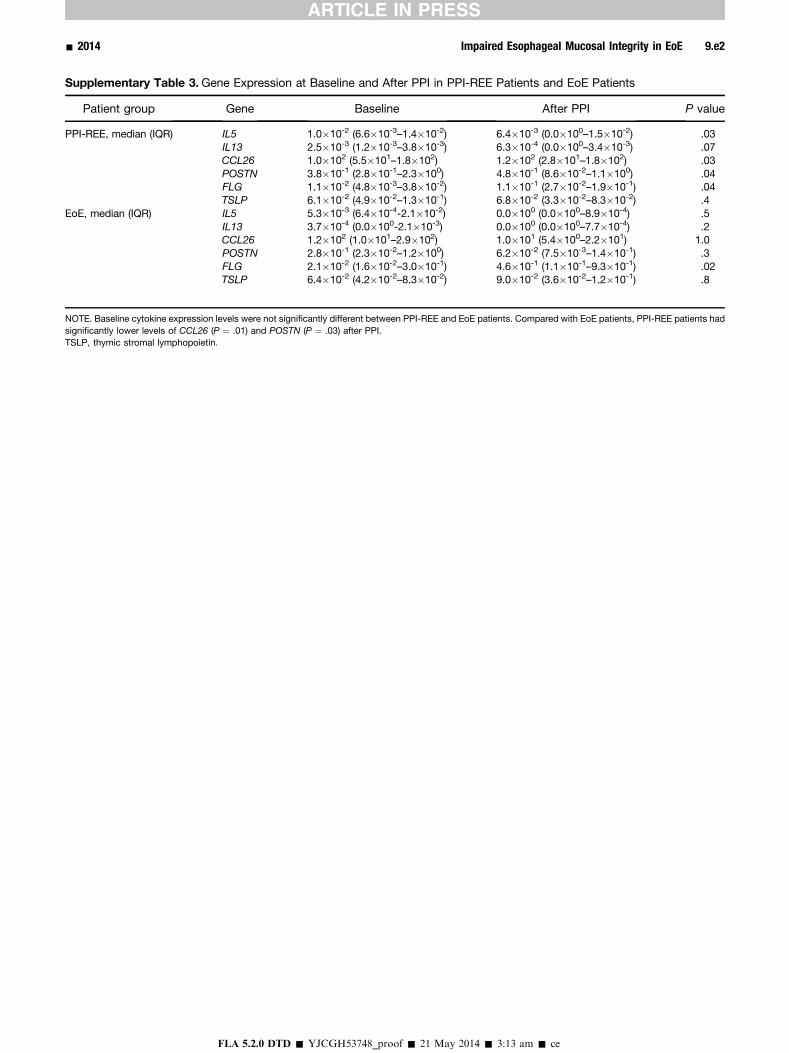
Supplementary Table 3.Gene Expression at Baseline and After PPI in PPI-REE Patients and EoE Patients
Patient group Gene Baseline After PPI P value
PPI-REE, median (IQR) IL5 1.0�10-2 (6.6�10-3–1.4�10-2) 6.4�10-3 (0.0�100–1.5�10-2) .03IL13 2.5�10-3 (1.2�10-3–3.8�10-3) 6.3�10-4 (0.0�100–3.4�10-3) .07CCL26 1.0�102 (5.5�101–1.8�102) 1.2�102 (2.8�101–1.8�102) .03POSTN 3.8�10-1 (2.8�10-1–2.3�100) 4.8�10-1 (8.6�10-2–1.1�100) .04FLG 1.1�10-2 (4.8�10-3–3.8�10-2) 1.1�10-1 (2.7�10-2–1.9�10-1) .04TSLP 6.1�10-2 (4.9�10-2–1.3�10-1) 6.8�10-2 (3.3�10-2–8.3�10-2) .4
EoE, median (IQR) IL5 5.3�10-3 (6.4�10-4-2.1�10-2) 0.0�100 (0.0�100–8.9�10-4) .5IL13 3.7�10-4 (0.0�100-2.1�10-3) 0.0�100 (0.0�100–7.7�10-4) .2CCL26 1.2�102 (1.0�101–2.9�102) 1.0�101 (5.4�100–2.2�101) 1.0POSTN 2.8�10-1 (2.3�10-2–1.2�100) 6.2�10-2 (7.5�10-3–1.4�10-1) .3FLG 2.1�10-2 (1.6�10-2–3.0�10-1) 4.6�10-1 (1.1�10-1–9.3�10-1) .02TSLP 6.4�10-2 (4.2�10-2–8.3�10-2) 9.0�10-2 (3.6�10-2–1.2�10-1) .8
NOTE. Baseline cytokine expression levels were not significantly different between PPI-REE and EoE patients. Compared with EoE patients, PPI-REE patients hadsignificantly lower levels of CCL26 (P ¼ .01) and POSTN (P ¼ .03) after PPI.TSLP, thymic stromal lymphopoietin.
FLA 5.2.0 DTD � YJCGH53748_proof � 21 May 2014 � 3:13 am � ce
- 2014 Impaired Esophageal Mucosal Integrity in EoE 9.e2
Related Documents