REVIEW Open Access Pediatric eosinophilic esophagitis: a review for the clinician Simona Barni 1 , Stefania Arasi 2 , Carla Mastrorilli 3,4 , Luca Pecoraro 5,6 , Mattia Giovannini 1* , Francesca Mori 1 , Lucia Liotti 7 , Francesca Saretta 8 , Riccardo Castagnoli 9 , Lucia Caminiti 10 , Antonella Cianferoni 11,12† and Elio Novembre 1† Abstract Eosinophilic esophagitis (EoE) is a chronic clinical-pathologic disease characterized by eosinophilic infiltration of the esophageal epithelium with esophageal dysfunction symptoms. EoE can occur at any age and has different clinical manifestations depending on the age onset. To date, esophago-gastroduodenal endoscopy (EGD) with biopsy is the gold-standard for EoE diagnosis. According to the recent consensus guidelines, proton pump inhibitors, corticosteroids and elimination diets could be a first-line therapy option. The aim of the treatment is clinical and histological remission for preventing long- lasting untreatable fibrosis. A multidisciplinary approach (allergist, gastroenterology, dietitian, and pathologist) is recommended for managing patients affected by EoE, given the complexity of its treatment. This review will provide a practical guide to assist pediatricians treating children with EoE. Moreover, it highlights the unmet needs in diagnosis and treatment that require urgent attention from the scientific community in the aim of improving the management of patients with EoE. Introduction Eosinophilic esophagitis (EoE) is a chronic immune- mediated disease characterized by clinical manifestations related to esophageal dysfunction and, histologically, by esophageal eosinophilic inflammation [1]. In recent decades, the incidence and prevalence of EoE has increased [2–4], being the leading cause of dysphagia in children and young adults [5]. Currently, esophageal bi- opsy through upper gastrointestinal endoscopy still re- mains the gold standard diagnostic test to perform when EoE is suspected [1]. Since upper endoscopy is an invasive and expensive procedure, less invasive tools are being de- veloped to evaluate esophagus inflammation, however, lar- ger studies are needed to validate these tests [6–12]. According to the last consensus guidelines, proton pump inhibitors (PPIs) are considered first-line thera- peutic options on the same level as steroids and elimin- ation diet [1]. The treatment must be individualized according to each patient’s lifestyle and family situation and could be interchangeable over time [13]. The aim of this review is to update the current evidence on EoE in children, moreover, it is intended as a practical guide for clinicians treating patients with EoE in order to avoid delayed diagnosis which can lead to inflammatory progression and ultimately, fibrostenosis [14]. Research strategies and literature analysis We reviewed the most relevant studies on “Pediatric Eo- sinophilic Esophagitis” present in databases including PubMed (https://www.ncbi.nlm.nih.gov/pubmed/) and the Cochrane Library up until 2020. Manuscripts were selected from randomized controlled trials, case reports, © The Author(s). 2021 Open Access This article is licensed under a Creative Commons Attribution 4.0 International License, which permits use, sharing, adaptation, distribution and reproduction in any medium or format, as long as you give appropriate credit to the original author(s) and the source, provide a link to the Creative Commons licence, and indicate if changes were made. The images or other third party material in this article are included in the article's Creative Commons licence, unless indicated otherwise in a credit line to the material. If material is not included in the article's Creative Commons licence and your intended use is not permitted by statutory regulation or exceeds the permitted use, you will need to obtain permission directly from the copyright holder. To view a copy of this licence, visit http://creativecommons.org/licenses/by/4.0/. The Creative Commons Public Domain Dedication waiver (http://creativecommons.org/publicdomain/zero/1.0/) applies to the data made available in this article, unless otherwise stated in a credit line to the data. * Correspondence: [email protected] † Antonella Cianferoni and Elio Novembre are joint last co-authors. 1 Allergy Unit, Department of Pediatrics, Meyer Children’s University Hospital, Florence, Italy Full list of author information is available at the end of the article Barni et al. Italian Journal of Pediatrics (2021) 47:230 https://doi.org/10.1186/s13052-021-01178-2

Welcome message from author
This document is posted to help you gain knowledge. Please leave a comment to let me know what you think about it! Share it to your friends and learn new things together.
Transcript
REVIEW Open Access
Pediatric eosinophilic esophagitis: a reviewfor the clinicianSimona Barni1, Stefania Arasi2, Carla Mastrorilli3,4, Luca Pecoraro5,6, Mattia Giovannini1* , Francesca Mori1,Lucia Liotti7, Francesca Saretta8, Riccardo Castagnoli9, Lucia Caminiti10, Antonella Cianferoni11,12† andElio Novembre1†
Abstract
Eosinophilic esophagitis (EoE) is a chronic clinical-pathologic disease characterized by eosinophilic infiltration of theesophageal epithelium with esophageal dysfunction symptoms.EoE can occur at any age and has different clinical manifestations depending on the age onset.To date, esophago-gastroduodenal endoscopy (EGD) with biopsy is the gold-standard for EoE diagnosis.According to the recent consensus guidelines, proton pump inhibitors, corticosteroids and elimination diets couldbe a first-line therapy option. The aim of the treatment is clinical and histological remission for preventing long-lasting untreatable fibrosis.A multidisciplinary approach (allergist, gastroenterology, dietitian, and pathologist) is recommended for managingpatients affected by EoE, given the complexity of its treatment.This review will provide a practical guide to assist pediatricians treating children with EoE.Moreover, it highlights the unmet needs in diagnosis and treatment that require urgent attention from thescientific community in the aim of improving the management of patients with EoE.
IntroductionEosinophilic esophagitis (EoE) is a chronic immune-mediated disease characterized by clinical manifestationsrelated to esophageal dysfunction and, histologically, byesophageal eosinophilic inflammation [1].In recent decades, the incidence and prevalence of EoE
has increased [2–4], being the leading cause of dysphagiain children and young adults [5]. Currently, esophageal bi-opsy through upper gastrointestinal endoscopy still re-mains the gold standard diagnostic test to perform whenEoE is suspected [1]. Since upper endoscopy is an invasiveand expensive procedure, less invasive tools are being de-veloped to evaluate esophagus inflammation, however, lar-ger studies are needed to validate these tests [6–12].
According to the last consensus guidelines, protonpump inhibitors (PPIs) are considered first-line thera-peutic options on the same level as steroids and elimin-ation diet [1]. The treatment must be individualizedaccording to each patient’s lifestyle and family situationand could be interchangeable over time [13].The aim of this review is to update the current evidence
on EoE in children, moreover, it is intended as a practicalguide for clinicians treating patients with EoE in order toavoid delayed diagnosis which can lead to inflammatoryprogression and ultimately, fibrostenosis [14].
Research strategies and literature analysisWe reviewed the most relevant studies on “Pediatric Eo-sinophilic Esophagitis” present in databases includingPubMed (https://www.ncbi.nlm.nih.gov/pubmed/) andthe Cochrane Library up until 2020. Manuscripts wereselected from randomized controlled trials, case reports,
© The Author(s). 2021 Open Access This article is licensed under a Creative Commons Attribution 4.0 International License,which permits use, sharing, adaptation, distribution and reproduction in any medium or format, as long as you giveappropriate credit to the original author(s) and the source, provide a link to the Creative Commons licence, and indicate ifchanges were made. The images or other third party material in this article are included in the article's Creative Commonslicence, unless indicated otherwise in a credit line to the material. If material is not included in the article's Creative Commonslicence and your intended use is not permitted by statutory regulation or exceeds the permitted use, you will need to obtainpermission directly from the copyright holder. To view a copy of this licence, visit http://creativecommons.org/licenses/by/4.0/.The Creative Commons Public Domain Dedication waiver (http://creativecommons.org/publicdomain/zero/1.0/) applies to thedata made available in this article, unless otherwise stated in a credit line to the data.
* Correspondence: [email protected]†Antonella Cianferoni and Elio Novembre are joint last co-authors.1Allergy Unit, Department of Pediatrics, Meyer Children’s University Hospital,Florence, ItalyFull list of author information is available at the end of the article
Barni et al. Italian Journal of Pediatrics (2021) 47:230 https://doi.org/10.1186/s13052-021-01178-2
reviews, systematic reviews, cohort and case–controlstudies, and observational studies. Single-case reports,abstracts/posters without sufficient detail, articles withduplicated data, and articles in non-English languagewere all excluded. The terms searched for were“pediatric eosinophilic esophagitis” [all fields]; “eosino-philic esophagitis” and “children” [all fields]; “pediatriceosinophilic esophagitis” and “management” [all fields];“pediatric eosinophilic esophagitis” and “treatment” [allfields]; “pediatric eosinophilic esophagitis” and “therapy”[all fields]; “pediatric eosinophilic esophagitis” and “diet”[all fields]; “esophageal eosinophilia” [all fields];“pediatric GERD” [all fields]; “gastroesophageal reflux”and “children” [all fields].
EpidemiologyEoE has been characterized as a disease relatively re-cently, with the first description by Landres et al. in1978 [15]. Over time, EoE has gained more and moreinterest and, in parallel, the epidemiology of EoE hasalso rapidly increased, in particular after the publicationof the consensus recommendations for diagnosis andmanagement of the disease in 2007 [16]. However, theincrease in recognition and knowledge of EoE would notseem to be the only explanation for such a rise in fre-quency [1]. This observation might entail implicationsfor the knowledge of the etiology of EoE which hastransformed from a rare case-reportable condition into amajor cause of food impaction and the leading cause ofdysphagia in children and young adults [5].Most data come from population-based studies,
conducted primarily in North America and Europe [2,3, 17–21]. Incidence rates range from 2.1/100,000/year in the Netherlands [4] to 12.8/100,000/year inOhio in the United States [22]. In 2016, a meta-analysis evaluated the epidemiology of EoE reportedby various countries and calculated an overall pooledincidence of 3.7/100,000/year (95% confidence interval[CI], 1.7–6.5) which was higher in adults (7/100,000/year; 95% CI:1–18.3) than in children (5/100,000/year;95% CI: 1.5–10.9). The same study assessed an overallprevalence of 22.7 cases per 100,000 inhabitants, be-ing the highest in North America with a prevalenceof 30.7 cases per 100,000 inhabitants, despite substan-tial heterogeneity [23].However, when interpreting the published epidemi-
ology data, it is important to recognize heterogeneitiesamong studies performed at different centers and duringdifferent periods. The reported frequency varied widely,probably due to differences in multiple factors such asclinical and laboratory criteria, patients’ ages, duration ofillness, and geographic variations in dietary habits. Fur-ther data from well-designed homogeneous studies arenecessary.
PathogenesisEoE is characterized by a multifactorial esophageal in-flammation occurring in individuals with genetic predis-position, which combines an esophageal epitheliadysfunction and an abnormal T-helper cell type 2 (Th2)-mediated immune response to environmental allergens,and leads to esophageal lesion and dysmotility, second-ary remodeling and fibrosis [24–26].
Genetic factorsThe risk of developing EoE is higher among first-degreefamily members, in particular, being a father or abrother of a patients affected by EoE, increases the riskof developing the disease 43-fold and 64-fold respect-ively, whereas the EoE frequency exhibited in monozy-gotic and dizygotic twins is 41 and 22% respectively [7].Multiple genome-wide association studies (GWAS)
have been conducted to identify genetic loci associatedwith EoE [27, 28]. Some of the genes have known func-tions, while others are still unknown [28]. Most of thegenetic loci identified affect epithelial barrier functionsand Th2-mediated immune responses [29, 30].The thymic stromal lymphopoietin (TSLP) gene has
been associated with EoE in multiple independent stud-ies. TSLP is an epithelial cytokine that favors a pro-Th2environment [27, 31, 32]. However other genes haveemerged in the EoE pathogenesis that are located in theepidermal differentiation complex (1q21), several ofwhich are dysregulated and involved in epithelial cell dif-ferentiation [33].Desmoglein-1 (DSG1), downregulated by interleukin
(IL)-13, gives rise to an epithelial cell barrier disfunc-tion [31].Calpain-14 (CALPN14), an esophagus-specific proteo-
lytic enzyme induced by IL-13, leads to a loss of DSG1expression and impaired epithelial barrier function [34].Moreover, filaggrin (FLG), a fundamental epithelial pro-tein, is downregulated in EoE [30].Lastly, the serine peptidase inhibitor Kazal type 5 and
7 (SPINK5 and SPINK7) participates in EoE pathogen-esis because the activity of serine proteases is not undercontrol [30].As a result, increased esophageal tissue permeability
and antigen uptake could give rise to an abnormal Th2-immune response. This involves several cytokines suchas IL-4, IL-5 and IL-13 [24, 25]. IL-4, secreted by Th2cells, natural killer (NK) cells, and basophils, which in-duce the differentiation of T cell into Th2 and active Bcells [25, 35]. IL-5, secreted by Th2 cells, mast cells, andeosinophils, promotes eosinophil proliferation, survival,activation, and chemotaxis [36–38].In addition to the effects mentioned above, IL-13, se-
creted by Th2 cells, upregulates eotaxin-3 (CCL26) thatcauses chemotaxis of the eosinophils [33]. Moreover, IL-
Barni et al. Italian Journal of Pediatrics (2021) 47:230 Page 2 of 14
13 upregulates periostin (POSTN) and transforms thegrowth factor (TGF)-β, increasing the adhesion of theeosinophils to fibronectin [39]. Furthermore, TGF-β dir-ectly induces the expression of profibrotic genes, such asfibronectin, collagen I, periostin, and smooth muscleactin, causing esophageal epithelial fibrosis and decreas-ing esophageal smooth-muscle contraction [40, 41].Ultimately, IL-13 induces tissue remodeling by pro-
moting collagen deposition, angiogenesis, and epithelialhyperplasia [37, 42].A close relationship between DSG1 and POSTN high-
lights the interaction between epithelial barrier dysfunc-tion and the Th2-mediated immune response: whenDSG1 is downregulated, POSTN increases, triggeringthe production of TSLP by the esophageal epithelium.TSLP induces a Th2-immune response and is implicatedin eosinophil survival, and mast cell and basophil activa-tion [40, 43–45].STAT6 (12q13), which is associated with the patho-
genesis of EoE. is activated by IL-4 and IL-13 and en-codes for a transcription factor activating many EoEgenes [29, 30].
Environmental factorsThe rapid increase in the incidence and prevalence of EoEdemonstrates the paramount role of environmental factorsin its pathogenesis [24, 26, 46]. The exposure to environ-mental allergens can also play a critical role in developingthe disease and its exacerbation, such as the pollen season,Fall, or living in a cold or dry climate [24].Moreover, living in a rural environment increases the
risk of developing EoE because of increased exposure toaeroallergens [47]. Furthermore, animal models supporta link between antigen exposure and EoE [48]. Specific-ally, a mice model of experimental esophagitis inducedby exposing to a respiratory allergen (Aspergillus fumi-gatus and dust mite mixture) was developed by Mishraet al. [49]. Mice exposed to the allergen revealed an in-crease in esophageal eosinophils, free eosinophil gran-ules, and epithelial cell hyperplasia [49].Atopy is present in 75% of patients with EoE [50]; in
particular, it has been demonstrated that a history ofatopic dermatitis (AD), IgE-mediated food allergy, andasthma are independently and cumulatively associatedwith the diagnosis of EoE [51]. In addition, children af-fected by IgE-mediated food allergy, especially those withmultiple food allergies, can develop EoE at 9 times the rateof children without this disorder [52]. Similarly, the risk ofEoE development in patients suffering from IgE-mediatedfood allergy who are on oral immunotherapy increases to2.7–5.3% [53]. Additionally, an association between EoE,asthma and airway hyperresponsiveness (AHR) has beenhighlighted [54]: a cross-sectional prospective study dem-onstrated that AHR is more frequent in subjects affected
by EoE than in the control group (33% vs. 11%) and a highlevel of s-IgE in these patients is associated with a greaterrisk of AHR.Early-life environmental exposures play a role too: ma-
ternal fever, pre-and postnatal antibiotics, proton pompinhibitor (PPI) therapy, and neonatal intensive care unitadmission in early life are associated with an increasedrisk of EoE [26, 55]. The theory is that early exposurecould alter gut microbiota, which in turn may alter thedevelopment of the normal immune system [56].On the other hand, Helicobacter pylori (H. pylori)
seems to have a protective effect on esophageal eosino-philia [57]. This evidence could be explained by the factthat H. pylori may drive a Th1 mediated allergic re-sponse, shifting the Th1–Th2 balance and protectingfrom an allergic Th2 response [58]. An overview of EoEpathogenesis is shown in Fig. 1.
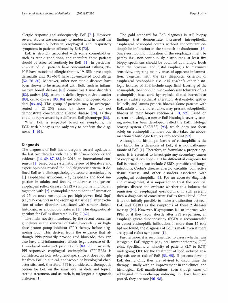
Clinical manifestationsEoE is characterized by different clinical manifestationsaccording to different ages of onset [60, 61]. Infantspresent with food refusal, failure to thrive and gastro-esophageal reflux; school-aged children present withsymptoms of gastroesophageal reflux and finally, adoles-cents and adults presents with dysphagia and food im-paction. Clinical manifestation of EoE in infancy,childhood and adolescence/adulthood is indicated inFig. 2.These differences in clinical onset is postulated to de-
rive from different causes: first of all, infants and chil-dren fail to express their symptoms in the same manneras adolescents and adults, secondly, the esophageal tis-sue remodeling is progressive and develops over timefrom an inflammatory phenotype in childhood to afibrostenotic phenotype in adulthood [14, 63–65].Moreover, the symptoms could be masked, because the
patients may have adapted compensatory feeding habits(eating slowly, excessive mastication, cutting food intosmall pieces) and dietary changes (preference for liquidsand soft food, and avoidance of specific textures like meatand bread) to prevent the development of symptoms (dys-phagia and food impaction) [58, 60, 62, 66, 67].Therefore, it is of paramount importance to include
the following questions in the medical history: “Do youchew your food a lot?” “Are you the last to get up fromthe table?” “Do you drink plenty of water during themeal to help you swallow?” “Do you avoid eating certainfoods such as bread, rice, or meat?” “Do you cut the foodinto very small pieces?” [60, 68].Since EoE symptoms are unspecific, especially in in-
fants and children, diagnosis is reached with an averagedelay of 3–5 years [69, 70], with an increasing risk of de-veloping fibrosis [70]. Indeed, the risk of fibrostenosis
Barni et al. Italian Journal of Pediatrics (2021) 47:230 Page 3 of 14
increases by 5% with each year of symptoms before diag-nosis [70].Extraesophageal symptoms could be associated with
EoE as reported for the first time by Orenstein et al.[71]. It is unclear whether the respiratory symptomswere caused by EoE or whether the relationship was dueto allergic conditions coexisting in a cohort of childrenhaving a common atopic background [72]. There are alot of theories on this subject: first of all, the hypothesis
that explains the onset of respiratory symptoms due tothe generating of proinflammatory cytokines by the eo-sinophils living in the upper esophagus [73–75]. Alterna-tively, other authors have hypothesized the role ofmicro-aspiration, resulting in food antigens gaining dir-ect access to the immune system via the lungs while cir-cumventing the gastrointestinal tract [71]. According tothis hypothesis, the interaction of food antigens directlywith the immune system causes the development of an
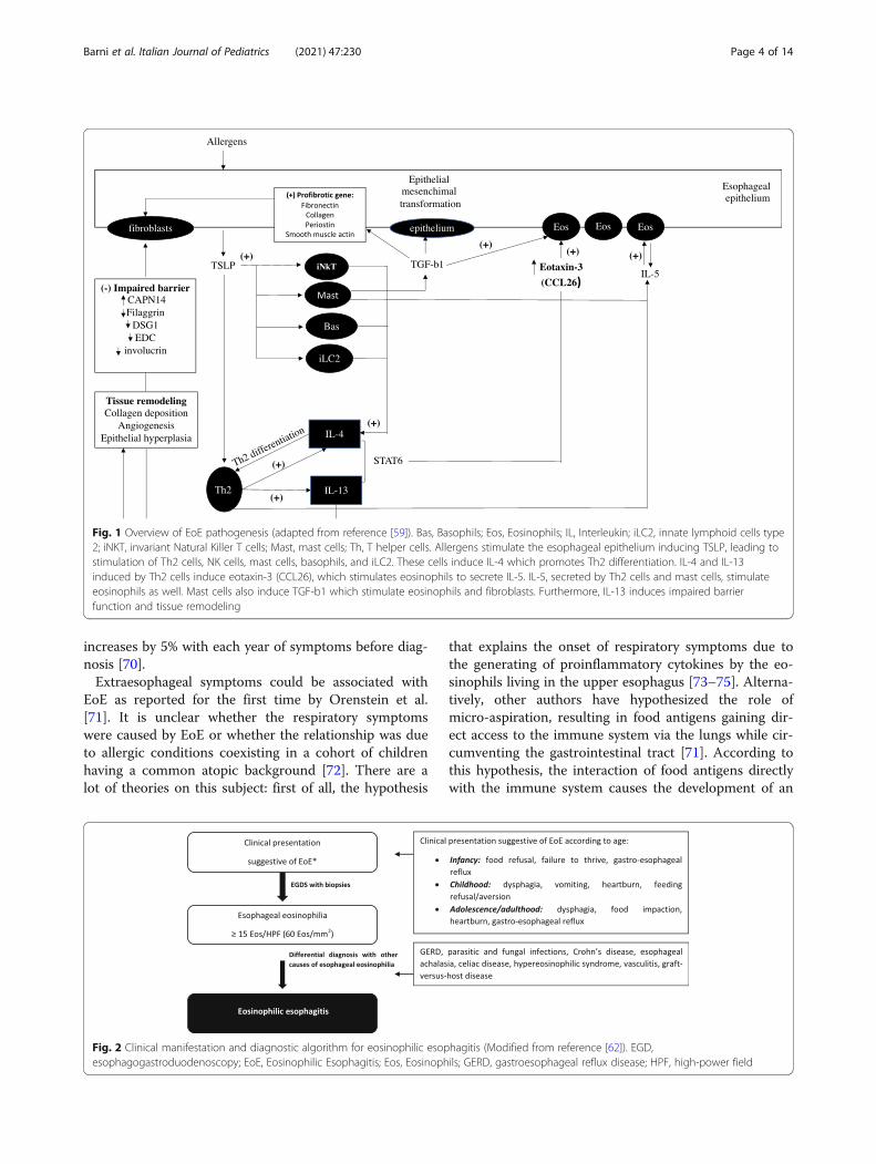
Fig. 1 Overview of EoE pathogenesis (adapted from reference [59]). Bas, Basophils; Eos, Eosinophils; IL, Interleukin; iLC2, innate lymphoid cells type2; iNKT, invariant Natural Killer T cells; Mast, mast cells; Th, T helper cells. Allergens stimulate the esophageal epithelium inducing TSLP, leading tostimulation of Th2 cells, NK cells, mast cells, basophils, and iLC2. These cells induce IL-4 which promotes Th2 differentiation. IL-4 and IL-13induced by Th2 cells induce eotaxin-3 (CCL26), which stimulates eosinophils to secrete IL-5. IL-5, secreted by Th2 cells and mast cells, stimulateeosinophils as well. Mast cells also induce TGF-b1 which stimulate eosinophils and fibroblasts. Furthermore, IL-13 induces impaired barrierfunction and tissue remodeling
Fig. 2 Clinical manifestation and diagnostic algorithm for eosinophilic esophagitis (Modified from reference [62]). EGD,esophagogastroduodenoscopy; EoE, Eosinophilic Esophagitis; Eos, Eosinophils; GERD, gastroesophageal reflux disease; HPF, high-power field
Barni et al. Italian Journal of Pediatrics (2021) 47:230 Page 4 of 14
allergic response and subsequently, EoE [71]. However,several studies are necessary to understand in detail theinterrelationship between esophageal and respiratorysymptoms in patients affected by EoE [72].EoE is strongly associated with some comorbidities,
such as atopic conditions, and therefore these patientsshould be screened routinely for EoE [51]. In particular,26–50% of EoE patients have concomitant asthma, 30–90% have associated allergic rhinitis, 19–55% have atopicdermatitis and, 9.8–68% have IgE-mediated food allergy[52, 76–80]. Moreover, other non-atopic diseases havebeen shown to be associated with EoE, such as inflam-matory bowel disease [81] connective tissue disorders[82], autism [83], attention deficit hyperactivity disorder[83], celiac disease [83, 84] and other monogenic disor-ders [83, 85]. This group of patients may be overrepre-sented in 21–25% cases by those who do notdemonstrate concomitant allergic disease [78], as theycould be represented by a different EoE-phenotype [86].When EoE is suspected based on symptoms, the
EGD with biopsy is the only way to confirm the diag-nosis [1, 61].
DiagnosisThe diagnosis of EoE has undergone several updates inthe last two decades with the birth of new concepts andevidence [16, 69, 87, 88]. In 2018, an international con-sensus [1] based on a systematic review of literature andexpert opinions revised the EoE diagnostic criteria. It de-fined EoE as a clinicopathologic disease characterized by[1] esophageal symptoms, e.g., dysphagia and food im-paction in adults, and feeding intolerance and gastro-esophageal reflux disease (GERD) symptoms in children,together with [2] eosinophil-predominant inflammationof 15 or more eosinophils per high-power field (hpf)(i.e., ≥15 eos/hpf) in the esophageal tissue [3] after exclu-sion of other disorders associated with similar clinical,histologic, or endoscopic features [1]. The diagnostic al-gorithm for EoE is illustrated in Fig. 2 [62].The main novelty introduced by the recent consensus
guidelines is the removal of failed twice-daily or high-dose proton pump inhibitor (PPI) therapy before diag-nosing EoE. This derives from the evidence that al-though PPIs primarily provide acid blockade, they canalso have anti-inflammatory effects (e.g., decrease of IL-13–induced eotaxin-3 production) [89, 90]. Currently,PPI-responsive esophageal eosinophilia (PPI-REE) isconsidered an EoE sub-phenotype, since it does not dif-fer from EoE in clinical, endoscopic or histological char-acteristics and, therefore, PPI is considered a therapeuticoption for EoE on the same level as diets and topicalsteroid treatment, and as such, is no longer a diagnosticcriterion [1].
The gold standard for EoE diagnosis is still biopsyfindings that demonstrate increased intraepithelialesophageal eosinophil counts without concomitant eo-sinophilic infiltration in the stomach or duodenum [16].Since eosinophilic infiltration of the esophagus might bepatchy (i.e., non-continuously distributed), at least fivebiopsy specimens should be obtained at multiple levelsfrom the proximal and distal esophagus to maximizesensitivity, targeting mainly areas of apparent inflamma-tion. Together with the key diagnostic criterion ofesophageal eosinophilia (i.e., ≥15 eos/hpf), other histo-logic features of EoE include superficial layering of theeosinophils, eosinophilic micro-abscesses (clusters of > 4eosinophils), basal zone hyperplasia, dilated intercellularspaces, surface epithelial alteration, dyskeratotic epithe-lial cells, and lamina propria fibrosis. Some patients withEoE, adults and children alike, may present subepithelialfibrosis in their biopsy specimens [91, 92]. Based oncurrent knowledge, a newer EoE histologic severity scor-ing index has been developed, called the EoE histologicscoring system (EoEHSS) [93], which does not focussolely on eosinophil numbers but also takes the above-mentioned histologic features into account [93].Although the histologic feature of eosinophilia is the
key factor for a diagnosis of EoE, it is not pathogno-monic of EoE [1]. Therefore, to formulate a proper diag-nosis, it is essential to investigate any secondary causesof esophageal eosinophilia. The differential diagnosis forEoE is broad and can include GERD, parasitic and fungalinfections, Crohn’s disease, allergic vasculitis, connectivetissue disease, and other disorders associated withesophageal eosinophilia [1]. For an accurate diagnosisand management, it is important to treat the potentialprimary disease and evaluate whether this induces theremission of esophageal eosinophilia. If still present,then a diagnosis of concurrent EoE is formulated. Oftenit is not initially possible to make a distinction betweenEoE and GERD as the symptoms of these 2 diseasesoverlap [94]. However, if symptoms fail to improve withPPIs or if they recur shortly after PPI suspension, anesophago-gastro-duodenoscopy (EGD) is recommendedto detect eosinophilic infiltration. If more than 15 eos/hpf are found, the diagnosis of EoE is made even if thereare typical reflux symptoms [1].Furthermore, it is recommended to assess whether any
iatrogenic EoE triggers (e.g., oral immunotherapy, OIT)exist. Specifically, a minority of patients (2.7 to 5.7%)undergoing OIT for the treatment of food induced ana-phylaxis are at risk of EoE [53, 95]. If patients developEoE during OIT, they are advised to discontinue thetherapy, usually with an improvement in the clinical andhistological EoE manifestations. Even though cases ofsublingual immunotherapy inducing EoE have been re-ported, they are rare [96–98].
Barni et al. Italian Journal of Pediatrics (2021) 47:230 Page 5 of 14
Less invasive techniques to evaluate the esophagusResearchers have focused on the development of less in-vasive tools to evaluate esophagus inflammation, sinceupper endoscopy is an invasive and expensive procedure,even requiring general anesthesia in children and con-scious sedation in most adult patients. Moreover, in2016, the Food and Drug Administration issued a safetywarning states that “repeated or lengthy use of generalanesthetic and sedation drugs during surgeries or proce-dures in children younger than 3 years or in pregnantwomen during their third trimester may affect the devel-opment of children’s brains”, caused increase concernsover anesthetic exposure at a young age leading toanesthetic-induced neurotoxicity [99, 100]. Since then,more caution has been used to do repeated EGD underanesthesia.Un-sedated transnasal endoscopy has been used in
children and adults to assess esophageal mucosal inflam-mation through biopsy [6].Although radiography is not useful in identifying the
inflammatory findings of edema and exudates, it can behelpful in evaluating fibrostenotic disease [7, 8]. Bariumesophagography is more sensitive than endoscopy fordetecting esophageal stricture and diffuse small-caliberesophagus [9].An instrumental examination currently being validated
is the esophageal string-test (EST). Consisting of a nylonthread the distal end of which is attached to a gelatincapsule that captures eosinophil-associated proteinsfrom the esophageal lumen, it has shown good correl-ation with eosinophilic infiltration in esophageal biopsyspecimens in both children and adults [10]. Anothercapsule-based technology is the Cytosponge (Medtronic,Minneapolis, Minn); originally designed for assessmentof the esophageal mucosa in patients with Barrett’sesophagus, it has recently been used to assess inflamma-tion in adult patients with EoE [11].A tethered confocal microscopy capsule has been
piloted in adults with EoE, with preliminary results sug-gesting that comprehensive cellular data can be gatheredfor assessing tissue inflammation [12]. All these methodsare promising for assessing inflammation without theuse of standard endoscopy. Although they are unlikelyto replace the diagnostic or therapeutic benefits of en-doscopy when a diagnosis or dilation is needed, theycould play an important role in replacing repeated en-doscopies for disease surveillance after treatmentinterventions.Other non-invasive biomarkers are also under investi-
gation for reducing the need for repeated endoscopiesand biopsies which entail significant costs, risks, and dis-comfort for patients, [101]. Several candidates have beenstudied, mainly in the blood/serum, but also in the urine,stools and breath testing [101]. The composition of the
salivary microbiome community structure has beenshown to be altered in children with EoE [102, 103].Fractionated exhaled nitric oxide testing (FeNO) is a
standardized non-invasive test with proven utility in theevaluation of asthma. The use of elevated FeNO has alsobeen reported in other eosinophilic inflammatory condi-tions. However, in eosinophilic esophagitis, FeNO haslimited clinical efficacy in predicting severity of esopha-geal eosinophilia. A study conducted in 50 patients withEoE has shown that in patients with FeNO levels > 40ppb, specificity of testing was high, but very few patientsreached this FeNO level [104].
Emerging diagnostic toolsThickening of the deeper layers of the esophagus hasbeen demonstrated via use of endoscopic ultrasonog-raphy [105, 106]. The endoscopic functional luminal im-aging probe (endoFLIP) is a new technique for assessingmucosal and submucosal fibrosis and smooth-musclehypertrophy which are likely to cause decreased esopha-geal compliance, contributing to dysphagia symptoms inthe absence of an identifiable stricture [107]. A balloonmucosal impedance (MI) catheter system that instantlydetects changes in esophageal mucosal integrity duringendoscopy over a long segment of the esophagus duringendoscopy has been found to be safe and reliable inidentifying patients with GERD, EoE, or non-GERD[108]. Furthermore, endoscopic mucosal impedancemeasurements correlate with eosinophilia and dilation ofintercellular spaces in patients with EoE, adults and chil-dren alike [11, 109].While histologic assessment is the gold standard in
diagnosing EoE, in those patients with a high pre-testprobability of EoE and eosinophil count < 15 hpf, tissuestaining for eosinophil products, such as eosinophil per-oxidase (EPX), might be useful, even though EPX stain-ing remains a research tool [110].Furthermore, the eosinophilic esophagitis diagnostic
panel (EDP) is a molecular tool which assesses the expres-sion of 96 genes that are dysregulated in patients with EoEand has high sensitivity and specificity for diagnosis andmolecular phenotyping in EoE [111]. In this regard, a mul-ticenter cross-sectional study analyzed 185 esophageal bi-opsy specimens from pediatric and adult patients withEoE, using EDP. Histological and endoscopic featureswere assessed by quantification of esophageal eosinophilsand via use of the eosinophilic esophagitis histology scor-ing system (EoEHSS) and the eosinophilic esophagitisendoscopic reference score (EREFS). In a validation cohortconsisting of 100 specimens, the EDP identified three clus-ters significantly associated with distinct endotypes(termed EoEe1–3) despite similar eosinophil levels. EoEe1was associated with a normal-appearing esophagus (riskratio [RR] 3.7, 95% CI 1.04–10.27; p = 0.0443), an inverse
Barni et al. Italian Journal of Pediatrics (2021) 47:230 Page 6 of 14
association with a history of esophageal dilation (0.27,0.09–0.82; p = 0.0105), and relatively mild histological,endoscopic, and molecular changes. EoEe2 showed an in-flammatory and steroid-refractory phenotype (RR 2.77,95% CI 1.11–6·95; p = 0.0376) and had the highest expres-sion of inflammatory cytokines and steroid-respondinggenes. EoEe3, which was associated with a narrow-caliberesophagus (RR 7.98, 95% CI 1.84–34.64; p = 0.0013) andadult onset (2.22, 1.19–4.12; p = 0.0155), showed the high-est degree of endoscopic and histological severity and thelowest expression of epithelial differentiation genes. It hasbeen speculated that this classification might have poten-tial clinical and therapeutic significance in the perspectiveof a precision-medicine approach to EoE [31].
TherapyThe aims of EoE therapy include the accomplishment ofclinical and histological remission in order to preventlong-lasting untreatable fibrosis [63–65].As the therapy is likely to be lifelong, the prevention
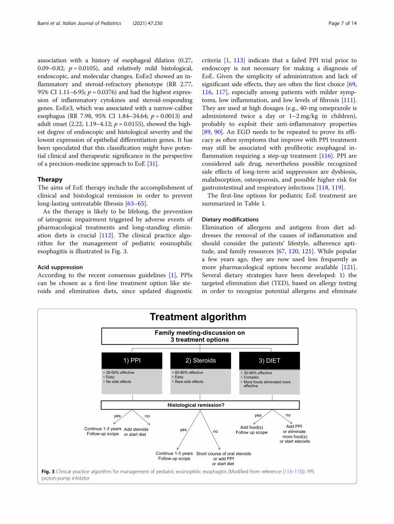
of iatrogenic impairment triggered by adverse events ofpharmacological treatments and long-standing elimin-ation diets is crucial [112]. The clinical practice algo-rithm for the management of pediatric eosinophilicesophagitis is illustrated in Fig. 3.
Acid suppressionAccording to the recent consensus guidelines [1], PPIscan be chosen as a first-line treatment option like ste-roids and elimination diets, since updated diagnostic
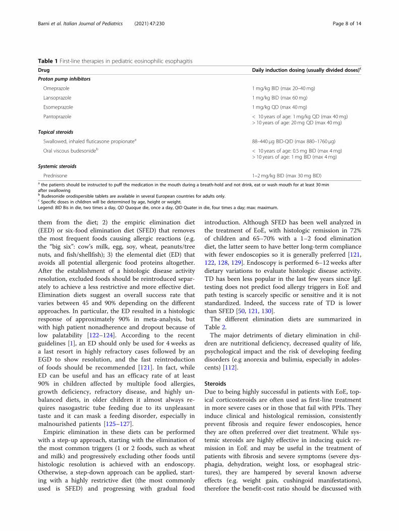
criteria [1, 113] indicate that a failed PPI trial prior toendoscopy is not necessary for making a diagnosis ofEoE. Given the simplicity of administration and lack ofsignificant side effects, they are often the first choice [69,116, 117], especially among patients with milder symp-toms, low inflammation, and low levels of fibrosis [111].They are used at high dosages (e.g., 40-mg omeprazole isadministered twice a day or 1–2 mg/kg in children),probably to exploit their anti-inflammatory properties[89, 90]. An EGD needs to be repeated to prove its effi-cacy as often symptoms that improve with PPI treatmentmay still be associated with profibrotic esophageal in-flammation requiring a step-up treatment [116]. PPI areconsidered safe drug, nevertheless possible recognizedside effects of long-term acid suppression are dysbiosis,malabsorption, osteoporosis, and possible higher risk forgastrointestinal and respiratory infections [118, 119].The first-line options for pediatric EoE treatment are
summarized in Table 1.
Dietary modificationsElimination of allergens and antigens from diet ad-dresses the removal of the causes of inflammation andshould consider the patients’ lifestyle, adherence apti-tude, and family resources [67, 120, 121]. While populara few years ago, they are now used less frequently asmore pharmacological options become available [121].Several dietary strategies have been developed: 1) thetargeted elimination diet (TED), based on allergy testingin order to recognize potential allergens and eliminate
Fig. 3 Clinical practice algorithm for management of pediatric eosinophilic esophagitis (Modified from reference [113–115]). PPI,proton-pump inhibitor
Barni et al. Italian Journal of Pediatrics (2021) 47:230 Page 7 of 14
them from the diet; 2) the empiric elimination diet(EED) or six-food elimination diet (SFED) that removesthe most frequent foods causing allergic reactions (e.g.the “big six”: cow’s milk, egg, soy, wheat, peanuts/treenuts, and fish/shellfish); 3) the elemental diet (ED) thatavoids all potential allergenic food proteins altogether.After the establishment of a histologic disease activityresolution, excluded foods should be reintroduced separ-ately to achieve a less restrictive and more effective diet.Elimination diets suggest an overall success rate thatvaries between 45 and 90% depending on the differentapproaches. In particular, the ED resulted in a histologicresponse of approximately 90% in meta-analysis, butwith high patient nonadherence and dropout because oflow palatability [122–124]. According to the recentguidelines [1], an ED should only be used for 4 weeks asa last resort in highly refractory cases followed by anEGD to show resolution, and the fast reintroductionof foods should be recommended [121]. In fact, whileED can be useful and has an efficacy rate of at least90% in children affected by multiple food allergies,growth deficiency, refractory disease, and highly un-balanced diets, in older children it almost always re-quires nasogastric tube feeding due to its unpleasanttaste and it can mask a feeding disorder, especially inmalnourished patients [125–127].Empiric elimination in these diets can be performed
with a step-up approach, starting with the elimination ofthe most common triggers (1 or 2 foods, such as wheatand milk) and progressively excluding other foods untilhistologic resolution is achieved with an endoscopy.Otherwise, a step-down approach can be applied, start-ing with a highly restrictive diet (the most commonlyused is SFED) and progressing with gradual food
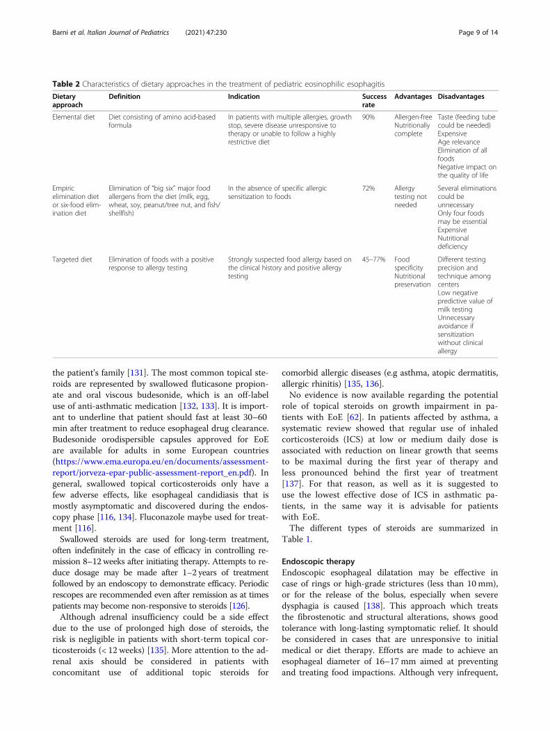
introduction. Although SFED has been well analyzed inthe treatment of EoE, with histologic remission in 72%of children and 65–70% with a 1–2 food eliminationdiet, the latter seem to have better long-term compliancewith fewer endoscopies so it is generally preferred [121,122, 128, 129]. Endoscopy is performed 6–12 weeks afterdietary variations to evaluate histologic disease activity.TD has been less popular in the last few years since IgEtesting does not predict food allergy triggers in EoE andpath testing is scarcely specific or sensitive and it is notstandardized. Indeed, the success rate of TD is lowerthan SFED [50, 121, 130].The different elimination diets are summarized in
Table 2.The major detriments of dietary elimination in chil-
dren are nutritional deficiency, decreased quality of life,psychological impact and the risk of developing feedingdisorders (e.g anorexia and bulimia, especially in adoles-cents) [112].
SteroidsDue to being highly successful in patients with EoE, top-ical corticosteroids are often used as first-line treatmentin more severe cases or in those that fail with PPIs. Theyinduce clinical and histological remission, consistentlyprevent fibrosis and require fewer endoscopies, hencethey are often preferred over diet treatment. While sys-temic steroids are highly effective in inducing quick re-mission in EoE and may be useful in the treatment ofpatients with fibrosis and severe symptoms (severe dys-phagia, dehydration, weight loss, or esophageal stric-tures), they are hampered by several known adverseeffects (e.g. weight gain, cushingoid manifestations),therefore the benefit-cost ratio should be discussed with
Table 1 First-line therapies in pediatric eosinophilic esophagitis
Drug Daily induction dosing (usually divided doses)c
Proton pump inhibitors
Omeprazole 1 mg/kg BID (max 20–40 mg)
Lansoprazole 1 mg/kg BID (max 60 mg)
Esomeprazole 1 mg/kg QD (max 40 mg)
Pantoprazole < 10 years of age: 1 mg/kg QD (max 40 mg)> 10 years of age: 20 mg QD (max 40 mg)
Topical steroids
Swallowed, inhaled fluticasone propionatea 88–440 μg BID-QID (max 880–1760 μg)
Oral viscous budesonideb < 10 years of age: 0.5 mg BID (max 4 mg)> 10 years of age: 1 mg BID (max 4 mg)
Systemic steroids
Prednisone 1–2 mg/kg BID (max 30 mg BID)a the patients should be instructed to puff the medication in the mouth during a breath-hold and not drink, eat or wash mouth for at least 30minafter swallowingb Budesonide orodispersible tablets are available in several European countries for adults only.c Specific doses in children will be determined by age, height or weight.Legend: BID Bis in die, two times a day, QD Quoque die, once a day, QID Quater in die, four times a day; max: maximum.
Barni et al. Italian Journal of Pediatrics (2021) 47:230 Page 8 of 14
the patient’s family [131]. The most common topical ste-roids are represented by swallowed fluticasone propion-ate and oral viscous budesonide, which is an off-labeluse of anti-asthmatic medication [132, 133]. It is import-ant to underline that patient should fast at least 30–60min after treatment to reduce esophageal drug clearance.Budesonide orodispersible capsules approved for EoEare available for adults in some European countries(https://www.ema.europa.eu/en/documents/assessment-report/jorveza-epar-public-assessment-report_en.pdf). Ingeneral, swallowed topical corticosteroids only have afew adverse effects, like esophageal candidiasis that ismostly asymptomatic and discovered during the endos-copy phase [116, 134]. Fluconazole maybe used for treat-ment [116].Swallowed steroids are used for long-term treatment,
often indefinitely in the case of efficacy in controlling re-mission 8–12 weeks after initiating therapy. Attempts to re-duce dosage may be made after 1–2 years of treatmentfollowed by an endoscopy to demonstrate efficacy. Periodicrescopes are recommended even after remission as at timespatients may become non-responsive to steroids [126].Although adrenal insufficiency could be a side effect
due to the use of prolonged high dose of steroids, therisk is negligible in patients with short-term topical cor-ticosteroids (< 12 weeks) [135]. More attention to the ad-renal axis should be considered in patients withconcomitant use of additional topic steroids for
comorbid allergic diseases (e.g asthma, atopic dermatitis,allergic rhinitis) [135, 136].No evidence is now available regarding the potential
role of topical steroids on growth impairment in pa-tients with EoE [62]. In patients affected by asthma, asystematic review showed that regular use of inhaledcorticosteroids (ICS) at low or medium daily dose isassociated with reduction on linear growth that seemsto be maximal during the first year of therapy andless pronounced behind the first year of treatment[137]. For that reason, as well as it is suggested touse the lowest effective dose of ICS in asthmatic pa-tients, in the same way it is advisable for patientswith EoE.The different types of steroids are summarized in
Table 1.
Endoscopic therapyEndoscopic esophageal dilatation may be effective incase of rings or high-grade strictures (less than 10mm),or for the release of the bolus, especially when severedysphagia is caused [138]. This approach which treatsthe fibrostenotic and structural alterations, shows goodtolerance with long-lasting symptomatic relief. It shouldbe considered in cases that are unresponsive to initialmedical or diet therapy. Efforts are made to achieve anesophageal diameter of 16–17 mm aimed at preventingand treating food impactions. Although very infrequent,
Table 2 Characteristics of dietary approaches in the treatment of pediatric eosinophilic esophagitis
Dietaryapproach
Definition Indication Successrate
Advantages Disadvantages
Elemental diet Diet consisting of amino acid-basedformula
In patients with multiple allergies, growthstop, severe disease unresponsive totherapy or unable to follow a highlyrestrictive diet
90% Allergen-freeNutritionallycomplete
Taste (feeding tubecould be needed)ExpensiveAge relevanceElimination of allfoodsNegative impact onthe quality of life
Empiricelimination dietor six-food elim-ination diet
Elimination of “big six” major foodallergens from the diet (milk, egg,wheat, soy, peanut/tree nut, and fish/shellfish)
In the absence of specific allergicsensitization to foods
72% Allergytesting notneeded
Several eliminationscould beunnecessaryOnly four foodsmay be essentialExpensiveNutritionaldeficiency
Targeted diet Elimination of foods with a positiveresponse to allergy testing
Strongly suspected food allergy based onthe clinical history and positive allergytesting
45–77% FoodspecificityNutritionalpreservation
Different testingprecision andtechnique amongcentersLow negativepredictive value ofmilk testingUnnecessaryavoidance ifsensitizationwithout clinicalallergy
Barni et al. Italian Journal of Pediatrics (2021) 47:230 Page 9 of 14
surgical esophagectomy is the extreme treatment optionwhen complications occur.
Other therapiesOther drug treatments have been studied, such as leuko-triene receptor antagonists (montelukast) [139], biologicagents (e.g., omalizumab, infliximab, mepolizumab, resli-zumab) [140–144], immunomodulators (e.g., azathio-prine, 6-mercaptopurine) [145] and oral viscous sodiumcromoglicate [146], but without showing efficacy.More promising are the data on biologics like dupilu-
mab in treating EoE. Data from the first phase III trial ofdupilumab, an antibody that inhibits the signaling of Il-4and IL-13 proteins, highlighted how this drug changesthe structural and histologic characters of EoE and de-creases the patients’ symptoms, such as the ability toswallow [147]. Hence, in September 2020, the FDA ap-proved breakthrough investigational therapy with dupi-lumab for treating EoE in patients aged 12 years andolder (https://www.globenewswire.com/news-release/2020/09/14/2092666/0/en/FDA-grants -Dupixent -dupilumab-Breakthrough-Therapy-designation-for-eosinophilic-esophagitis.html), although it use in clinicalpractice has not yet been approved. Many other bio-logics are currently being studied for this disease [62].
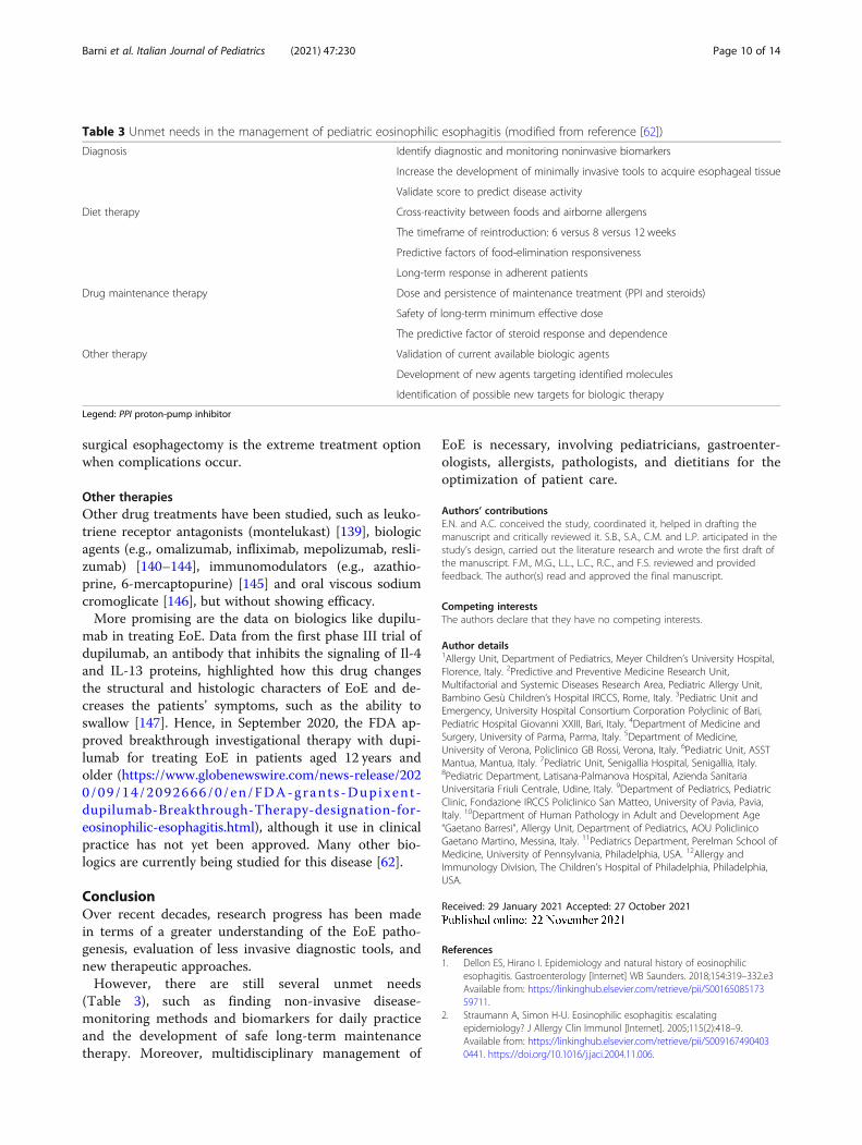
ConclusionOver recent decades, research progress has been madein terms of a greater understanding of the EoE patho-genesis, evaluation of less invasive diagnostic tools, andnew therapeutic approaches.However, there are still several unmet needs
(Table 3), such as finding non-invasive disease-monitoring methods and biomarkers for daily practiceand the development of safe long-term maintenancetherapy. Moreover, multidisciplinary management of
EoE is necessary, involving pediatricians, gastroenter-ologists, allergists, pathologists, and dietitians for theoptimization of patient care.
Authors’ contributionsE.N. and A.C. conceived the study, coordinated it, helped in drafting themanuscript and critically reviewed it. S.B., S.A., C.M. and L.P. articipated in thestudy’s design, carried out the literature research and wrote the first draft ofthe manuscript. F.M., M.G., L.L., L.C., R.C., and F.S. reviewed and providedfeedback. The author(s) read and approved the final manuscript.
Competing interestsThe authors declare that they have no competing interests.
Author details1Allergy Unit, Department of Pediatrics, Meyer Children’s University Hospital,Florence, Italy. 2Predictive and Preventive Medicine Research Unit,Multifactorial and Systemic Diseases Research Area, Pediatric Allergy Unit,Bambino Gesù Children’s Hospital IRCCS, Rome, Italy. 3Pediatric Unit andEmergency, University Hospital Consortium Corporation Polyclinic of Bari,Pediatric Hospital Giovanni XXIII, Bari, Italy. 4Department of Medicine andSurgery, University of Parma, Parma, Italy. 5Department of Medicine,University of Verona, Policlinico GB Rossi, Verona, Italy. 6Pediatric Unit, ASSTMantua, Mantua, Italy. 7Pediatric Unit, Senigallia Hospital, Senigallia, Italy.8Pediatric Department, Latisana-Palmanova Hospital, Azienda SanitariaUniversitaria Friuli Centrale, Udine, Italy. 9Department of Pediatrics, PediatricClinic, Fondazione IRCCS Policlinico San Matteo, University of Pavia, Pavia,Italy. 10Department of Human Pathology in Adult and Development Age“Gaetano Barresi”, Allergy Unit, Department of Pediatrics, AOU PoliclinicoGaetano Martino, Messina, Italy. 11Pediatrics Department, Perelman School ofMedicine, University of Pennsylvania, Philadelphia, USA. 12Allergy andImmunology Division, The Children’s Hospital of Philadelphia, Philadelphia,USA.
Received: 29 January 2021 Accepted: 27 October 2021
References1. Dellon ES, Hirano I. Epidemiology and natural history of eosinophilic
esophagitis. Gastroenterology [Internet] WB Saunders. 2018;154:319–332.e3Available from: https://linkinghub.elsevier.com/retrieve/pii/S0016508517359711.
2. Straumann A, Simon H-U. Eosinophilic esophagitis: escalatingepidemiology? J Allergy Clin Immunol [Internet]. 2005;115(2):418–9.Available from: https://linkinghub.elsevier.com/retrieve/pii/S0091674904030441. https://doi.org/10.1016/j.jaci.2004.11.006.
Table 3 Unmet needs in the management of pediatric eosinophilic esophagitis (modified from reference [62])
Diagnosis Identify diagnostic and monitoring noninvasive biomarkers
Increase the development of minimally invasive tools to acquire esophageal tissue
Validate score to predict disease activity
Diet therapy Cross-reactivity between foods and airborne allergens
The timeframe of reintroduction: 6 versus 8 versus 12 weeks
Predictive factors of food-elimination responsiveness
Long-term response in adherent patients
Drug maintenance therapy Dose and persistence of maintenance treatment (PPI and steroids)
Safety of long-term minimum effective dose
The predictive factor of steroid response and dependence
Other therapy Validation of current available biologic agents
Development of new agents targeting identified molecules
Identification of possible new targets for biologic therapy
Legend: PPI proton-pump inhibitor
Barni et al. Italian Journal of Pediatrics (2021) 47:230 Page 10 of 14
3. Prasad GA, Alexander JA, Schleck CD, Zinsmeister AR, Smyrk TC, Elias RM,et al. Epidemiology of eosinophilic esophagitis over three decades inolmsted county, minnesota. Clin Gastroenterol Hepatol [Internet] ElsevierInc. 2009;7:1055–61. https://doi.org/10.1016/j.cgh.2009.06.023.
4. van Rhijn BD, Verheij J, Smout AJPM, Bredenoord AJ. Rapidly increasingincidence of eosinophilic esophagitis in a large cohort. NeurogastroenterolMotil [Internet]. 2013;25(1):47–e5. https://doi.org/10.1111/nmo.12009.
5. Straumann A, Schoepfer A. Update on basic and clinical aspects ofeosinophilic oesophagitis. Gut [Internet]. 2014;63(8):1355–63. https://doi.org/10.1136/gutjnl-2013-306414.
6. Friedlander JA, Deboer EM, Soden JS, Furuta GT, Menard-Katcher CD, AtkinsD, et al. Unsedated transnasal esophagoscopy for monitoring therapy inpediatric eosinophilic esophagitis. Gastrointest Endosc [Internet] Elsevier, Inc.2016;83:299–306.e1. https://doi.org/10.1016/j.gie.2015.05.044.
7. Alexander ES, Martin LJ, Collins MH, Kottyan LC, Sucharew H, He H, et al.Twin and family studies reveal strong environmental and weaker geneticcues explaining heritability of eosinophilic esophagitis. 2012;100:130–4.
8. Menard-Katcher C, Swerdlow MP, Mehta P, Furuta GT, Fenton LZ.Contribution of esophagram to the evaluation of complicated pediatriceosinophilic esophagitis. J Pediatr Gastroenterol Nutr. 2015;61(5):541–6.https://doi.org/10.1097/MPG.0000000000000849.
9. Gentile N, Katzka D, Ravi K, Trenkner S, Enders F, Killian J, et al. Oesophagealnarrowing is common and frequently under-appreciated at endoscopy inpatients with oesophageal eosinophilia. Aliment Pharmacol Ther. 2014;40(11-12):1333–40. https://doi.org/10.1111/apt.12977.
10. Furuta GT, Kagalwalla AF, Lee JJ, Alumkal P, Maybruck BT, Fillon S, et al. Theoesophageal string test: a novel, minimally invasive method measuresmucosal inflammation in eosinophilic oesophagitis. Gut. 2013;62(10):1395–405. https://doi.org/10.1136/gutjnl-2012-303171.
11. Katzka DA, Geno DM, Ravi A, Smyrk TC, Lao-Sirieix P, Miramedi A, et al. Accuracy,safety, and tolerability of tissue collection by cytosponge vs endoscopy forevaluation of eosinophilic esophagitis. Clin Gastroenterol Hepatol [Internet]Elsevier, Inc. 2015;13:77–83.e2. https://doi.org/10.1016/j.cgh.2014.06.026.
12. Tabatabaei N, Kang D, Wu T, Kim M, Carruth RW, Leung J, et al. Tetheredconfocal endomicroscopy capsule for diagnosis and monitoring ofeosinophilic esophagitis. Biomed Opt Express [Internet]. 2014;5:197 Availablefrom: https://www.osapublishing.org/boe/abstract.cfm?uri=boe-5-1-197.
13. González-Cervera J, Lucendo AJ. Eosinophilic esophagitis: an evidence-based approach to therapy. J Investig Allergol Clin Immunol. 2016;26(1):8–18. https://doi.org/10.18176/jiaci.0002.
14. Gonsalves N. Distinct features in the clinical presentations of eosinophilicesophagitis in children and adults: is this the same disease? Dig Dis. 2014;32(1-2):89–92. https://doi.org/10.1159/000357078.
15. Landres RT, Kuster GGR, Strum WB. Eosinophilic esophagitis in a patientwith vigorous achalasia. Gastroenterology [internet]. Elsevier Masson SAS.1978;74(6):1298–301. https://doi.org/10.1016/0016-5085(78)90710-2.
16. Furuta GT, Liacouras CA, Collins MH, Gupta SK, Justinich C, Putnam PE, et al.Eosinophilic esophagitis in children and adults: a systematic review andconsensus recommendations for diagnosis and treatment. Gastroenterol Int.2007;133(4):1342–63. Available from: https://linkinghub.elsevier.com/retrieve/pii/S0016508507014746. https://doi.org/10.1053/j.gastro.2007.08.017.
17. Dellon ES, Erichsen R, Baron JA, Shaheen NJ, Vyberg M, Sorensen HT, et al.The increasing incidence and prevalence of eosinophilic oesophagitisoutpaces changes in endoscopic and biopsy practice: national population-based estimates from Denmark. Aliment Pharmacol Ther. 2015;41(7):662–70.https://doi.org/10.1111/apt.13129.
18. Giriens B, Yan P, Safroneeva E, Zwahlen M, Reinhard A, Nydegger A, et al.Escalating incidence of eosinophilic esophagitis in Canton of Vaud,Switzerland, 1993–2013: a population-based study. Allergy [Internet]. 2015;70:1633–9. https://doi.org/10.1111/all.12733.
19. Hruz P, Straumann A, Bussmann C, Heer P, Simon HU, Zwahlen M, et al.Escalating incidence of eosinophilic esophagitis: a 20-year prospective,population-based study in Olten County, Switzerland. J Allergy ClinImmunol [Internet] American Academy of Allergy, Asthma & Immunology.2011;128:1349–1350.e5. https://doi.org/10.1016/j.jaci.2011.09.013.
20. Dalby K, Nielsen RG, Kruse-Andersen S, Fenger C, Bindslev-Jensen C,Ljungberg S, et al. Eosinophilic Oesophagitis in infants and children in theregion of southern Denmark: a prospective study of prevalence and clinicalpresentation. J Pediatr Gastroenterol Nutr [Internet]. 2010;51(3):280–2.Available from: https://journals.lww.com/00005176-201009000-00006.https://doi.org/10.1097/MPG.0b013e3181d1b107.
21. Licari A, Votto M, Scudeller L, De Silvestri A, Rebuffi C, Cianferoni A, et al.Epidemiology of nonesophageal eosinophilic gastrointestinal diseases insymptomatic patients: a systematic review and Meta-analysis. J allergy ClinImmunol Pract [internet] Elsevier Inc. 2020;8:1994–2003.e2. https://doi.org/10.1016/j.jaip.2020.01.060.
22. Falk GW. Eosinophilic Esophagitis. Gastroenterol Clin North Am [Internet].2014;43:xiii Available from: https://linkinghub.elsevier.com/retrieve/pii/S0889855314000387.
23. Arias A, Pérez-Martínez I, Tenías JM, Lucendo AJ. Systematic review withmeta-analysis: the incidence and prevalence of eosinophilic oesophagitis inchildren and adults in population-based studies. Aliment Pharmacol Ther.2016;43(1):3–15. https://doi.org/10.1111/apt.13441.
24. Lyles J, Rothenberg M. Role of genetics, environment, and their interactions inthe pathogenesis of eosinophilic esophagitis. Curr Opin Immunol [internet]Elsevier ltd. 2019;60:46–53. https://doi.org/10.1016/j.coi.2019.04.004.
25. Vinit C, Dieme A, Courbage S, Dehaine C, Dufeu CM, Jacquemot S, et al.Eosinophilic esophagitis: pathophysiology, diagnosis, and management.Arch Pédiatrie [internet] Elsevier Masson SAS. 2019;26:182–90. https://doi.org/10.1016/j.arcped.2019.02.005.
26. Votto M, Marseglia GL, De Filippo M, Brambilla I, Caimmi SME, Licari A. Earlylife risk factors in pediatric EoE: could we prevent this modern disease?Front Pediatr [Internet]. 2020a;8:1–10. https://doi.org/10.3389/fped.2020.00263/full.
27. Sleiman PMA, Wang ML, Cianferoni A, Aceves S, Gonsalves N, Nadeau K,et al. GWAS identifies four novel eosinophilic esophagitis loci. Nat Commun[internet]. Nat Publ Group. 2014;5(1):1–5. https://doi.org/10.1038/ncomms6593.
28. Kottyan LC, Parameswaran S, Weirauch MT, Rothenberg ME, Martin LJ. Thegenetic etiology of eosinophilic esophagitis. J allergy Clin Immunol[internet] Elsevier Inc. 2020;145:9–15. https://doi.org/10.1016/j.jaci.2019.11.013.
29. O’Shea KM, Aceves SS, Dellon ES, Gupta SK, Spergel JM, Furuta GT, et al.Pathophysiology of eosinophilic esophagitis. Gastroenterology [internet]Elsevier, Inc. 2018;154:333–45. https://doi.org/10.1053/j.gastro.2017.06.065.
30. Kottyan LC, Rothenberg ME. Genetics of eosinophilic esophagitis. MucosalImmunol [internet]. Nat Publ Group. 2017;10(3):580–8. https://doi.org/10.1038/mi.2017.4.
31. Sherrill JD, Gao PS, Stucke EM, Blanchard C, Collins MH, Putnam PE, et al.Variants of thymic stromal lymphopoietin and its receptor associate witheosinophilic esophagitis. J Allergy Clin Immunol [Internet] Elsevier Ltd. 2010;126:160–165.e3. https://doi.org/10.1016/j.jaci.2010.04.037.
32. Dunn JLM, Shoda T, Caldwell JM, Wen T, Aceves SS, Collins MH, et al.Esophageal type 2 cytokine expression heterogeneity in eosinophilicesophagitis in a multisite cohort. J allergy Clin Immunol [internet] ElsevierInc. 2020;145:1629–1640.e4. https://doi.org/10.1016/j.jaci.2020.01.051.
33. Blanchard C, Wang N, Stringer KF, Mishra A, Fulkerson PC, Abonia JP, et al.Eotaxin-3 and a uniquely conserved gene-expression profile in eosinophilicesophagitis. J Clin Invest. 2006;116(2):536–47. https://doi.org/10.1172/JCI26679.
34. Litosh VA, Rochman M, Rymer JK, Porollo A, Kottyan LC, Rothenberg ME.Calpain-14 and its association with eosinophilic esophagitis. J Allergy ClinImmunol [Internet] Elsevier Inc. 2017;139:1762–1771.e7. https://doi.org/10.1016/j.jaci.2016.09.027.
35. Rothenberg ME. Molecular, genetic, and cellular bases for treatingeosinophilic esophagitis. Gastroenterology [internet] Elsevier Ltd. 2015;148(6):1143–57. https://doi.org/10.1053/j.gastro.2015.02.002.
36. Clayton F, Peterson K. Eosinophilic esophagitis: pathophysiology anddefinition. Gastrointest Endosc Clin N am [internet] Elsevier Inc. 2018;28:1–14. https://doi.org/10.1016/j.giec.2017.07.011.
37. Davis BP, Rothenberg ME. Mechanisms of disease of eosinophilicesophagitis. Annu Rev Pathol Mech Dis. 2016;11(1):365–93. https://doi.org/10.1146/annurev-pathol-012615-044241.
38. O’Byrne PM, Inman MD, Parameswaran K. The trials and tribulations of IL-5,eosinophils, and allergic asthma. J Allergy Clin Immunol [Internet]. 2001;108(4):503–8. Available from: https://linkinghub.elsevier.com/retrieve/pii/S0091674901188020. https://doi.org/10.1067/mai.2001.119149.
39. Blanchard C, Mingler MK, McBride M, Putnam PE, Collins MH, Chang G, et al.Periostin facilitates eosinophil tissue infiltration in allergic lung andesophageal responses. Mucosal Immunol [Internet]. 2008;1(4):289–96.Available from: http://www.nature.com/articles/mi200815. https://doi.org/10.1038/mi.2008.15.
Barni et al. Italian Journal of Pediatrics (2021) 47:230 Page 11 of 14
40. Wright BL, Nguyen N, Shim KP, Masterson JC, Jacobsen EA, Ochkur SI, LeeJJ, Furuta GT. Increased GATA-3 and T-bet expression in eosinophilicesophagitis versus gastroesophageal reflux disease. J Allergy Clin Immunol.2018;141(5):1919-21.e5. https://doi.org/10.1016/j.jaci.2017.12.993.
41. Beppu LY, Anilkumar AA, Newbury RO, Dohil R, Broide DH, Aceves SS. TGF-β1-induced phospholamban expression alters esophageal smooth musclecell contraction in patients with eosinophilic esophagitis. J Allergy ClinImmunol [Internet] Elsevier Ltd. 2014;134:1100–1107.e4. https://doi.org/10.1016/j.jaci.2014.04.004.
42. Zuo L, Fulkerson PC, Finkelman FD, Mingler M, Fischetti CA, Blanchard C,et al. IL-13 induces esophageal remodeling and gene expression by aneosinophil-independent, IL-13Rα2–inhibited pathway. J Immunol. 2010;185(1):660–9. https://doi.org/10.4049/jimmunol.1000471.
43. Wong CK, Hu S, Cheung PFY, Lam CWK. Thymic stromal lymphopoietininduces chemotactic and prosurvival effects in eosinophils: implications inallergic inflammation. Am J Respir Cell Mol Biol. 2010;43(3):305–15. https://doi.org/10.1165/rcmb.2009-0168OC.
44. Allakhverdi Z, Comeau MR, Jessup HK, Yoon BRP, Brewer A, Chartier S, et al.Thymic stromal lymphopoietin is released by human epithelial cells inresponse to microbes, trauma, or inflammation and potently activates mastcells. J Exp Med. 2007;204(2):253–8. https://doi.org/10.1084/jem.20062211.
45. Noti M, Wojno EDT, Kim BS, Siracusa MC, Giacomin PR, Nair MG, et al.Thymic stromal lymphopoietin–elicited basophil responses promoteeosinophilic esophagitis. Nat med [internet]. Nat Publ Group. 2013;19(8):1005–13. https://doi.org/10.1038/nm.3281.
46. Jensen ET, Shah ND, Hoffman K, Sonnenberg A, Genta RM, Dellon ES.Seasonal variation in detection of oesophageal eosinophilia andeosinophilic oesophagitis. Aliment Pharmacol Ther [Internet]. 2015;42(4):461–9. https://doi.org/10.1111/apt.13273.
47. Rayapudi M, Mavi P, Zhu X, Pandey AK, Abonia JP, Rothenberg ME, et al.Indoor insect allergens are potent inducers of experimental eosinophilicesophagitis in mice. J Leukoc Biol [Internet]. 2010;88(2):337–46. https://doi.org/10.1189/jlb.0110025.
48. Caldwell JM, Paul M, Rothenberg ME. Novel immunologic mechanisms ineosinophilic esophagitis. Curr Opin Immunol [internet]. Elsevier Ltd. 2017;48:114–21. https://doi.org/10.1016/j.coi.2017.08.006.
49. Mishra A, Hogan SP, Brandt EB, Rothenberg ME. An etiological role foraeroallergens and eosinophils in experimental esophagitis. J Clin Invest.2001;107(1):83–90. https://doi.org/10.1172/JCI10224.
50. Spergel JM, Beausoleil JL, Mascarenhas M, Liacouras CA. The use of skinprick tests and patch tests to identify causative foods in eosinophilicesophagitis. J Allergy Clin Immunol [Internet]. 2002;109(2):363–8. Availablefrom: https://linkinghub.elsevier.com/retrieve/pii/S0091674902794670.https://doi.org/10.1067/mai.2002.121458.
51. Ruffner MA, Capucilli P, Hill DA, Spergel JM. Screening children foreosinophilic esophagitis: allergic and other risk factors. Expert rev ClinImmunol [internet] Taylor & Francis. 2019;15:315–8. https://doi.org/10.1080/1744666X.2019.1579643.
52. Hill DA, Dudley JW, Spergel JM. The prevalence of eosinophilic esophagitisin pediatric patients with IgE-mediated food allergy. J Allergy Clin ImmunolPract. 2017;5(2):369–75. https://doi.org/10.1016/j.jaip.2016.11.020.
53. Cianferoni A. Eosinophilic esophagitis as a side effect of food oralimmunotherapy. Med. 2020;56:1–12.
54. Krupp NL, Sehra S, Slaven JE, Kaplan MH, Gupta S, Tepper RS. Increasedprevalence of airway reactivity in children with eosinophilic esophagitis.Pediatr Pulmonol. 2016;51(5):478–83. https://doi.org/10.1002/ppul.23327.
55. Jensen ET, Kuhl JT, Martin LJ, Langefeld CD, Dellon ES, Rothenberg ME.Early-life environmental exposures interact with genetic susceptibilityvariants in pediatric patients with eosinophilic esophagitis. J Allergy ClinImmunol [Internet] Elsevier Inc. 2018;141:632–637.e5. https://doi.org/10.1016/j.jaci.2017.07.010.
56. Jensen ET, Bertelsen RJ. Assessing early life factors for eosinophilicesophagitis: lessons from other allergic diseases. Curr Treat OptionsGastroenterol. 2016;14(1):39–50. https://doi.org/10.1007/s11938-016-0083-1.
57. Dellon ES, Peery AF, Shaheen NJ, Morgan DR, Hurrell JM, Lash RH, et al.Inverse association of esophageal eosinophilia with Helicobacter pylori basedon analysis of a US pathology database. Gastroenterology [Internet] ElsevierInc. 2011;141:1586–92. https://doi.org/10.1053/j.gastro.2011.06.081.
58. Chen JW, Kao JY. Eosinophilic esophagitis: update on management andcontroversies. BMJ [Internet]. 2017;359:j4482. https://doi.org/10.1136/bmj.j4482.
59. Ryu S, Lee KH, Tizaoui K, Terrazzino S, Cargnin S, Effenberger M, et al.Pathogenesis of eosinophilic esophagitis: a comprehensive review of thegenetic and molecular aspects. Int J Mol Sci. 2020;21(19):1–20. https://doi.org/10.3390/ijms21197253.
60. Furuta GT, Katzka DA. Eosinophilic Esophagitis. Ingelfinger JR, editor. N Engl JMed [Internet]. 2015;373:1640–1648. https://doi.org/10.1056/NEJMra1502863
61. Spergel JM, Brown-Whitehorn TA, Muir A, Liacouras CA. Medical algorithm:diagnosis and treatment of eosinophilic esophagitis in children. Allergy[Internet]. 2020;75(6):1522–4. https://doi.org/10.1111/all.14188.
62. Rossetti D, Isoldi S, Oliva S. Eosinophilic esophagitis: update on diagnosisand treatment in pediatric patients. Pediatr drugs [internet] Springerinternational publishing. 2020;22:343–56. https://doi.org/10.1007/s40272-020-00398-z.
63. Muir AB, Brown-Whitehorn T, Godwin B, Cianferoni A. Eosinophilicesophagitis: early diagnosis is the key. Clin Exp Gastroenterol [Internet].2019;12:391–9. Available from: https://www.dovepress.com/eosinophilic-esophagitis-early-diagnosis-is-the-key-peer-reviewed-article-CEG. https://doi.org/10.2147/CEG.S175061.
64. Whelan KA, Godwin BC, Wilkins B, Elci OU, Benitez A, DeMarshall M, et al.Persistent basal cell hyperplasia is associated with clinical and endoscopicfindings in patients with histologically inactive eosinophilic esophagitis. ClinGastroenterol Hepatol [Internet]. 2020;18:1475–1482.e1 Available from:https://linkinghub.elsevier.com/retrieve/pii/S1542356519309772.
65. Kasagi Y, Dods K, Wang JX, Chandramouleeswaran PM, Benitez AJ,Gambanga F, et al. Fibrostenotic eosinophilic esophagitis might reflectepithelial lysyl oxidase induction by fibroblast-derived TNF-α. J allergy ClinImmunol [internet] Elsevier Inc. 2019;144:171–82. https://doi.org/10.1016/j.jaci.2018.10.067.
66. Gomez Torrijos E, Gonzalez-Mendiola R, Alvarado M, Avila R, Prieto-Garcia A, Valbuena T, et al. Eosinophilic Esophagitis: Review andUpdate. Front Med [Internet] Frontiers Media SA. 2018;5. https://doi.org/10.3389/fmed.2018.00247/full.
67. Votto M, Castagnoli R, De Filippo M, Brambilla I, Cuppari C, Marseglia GL,Licari A. Behavioral issues and quality of life in children with eosinophilicesophagitis. Minerva Pediatr. 2020;72(5):424-32. https://doi.org/10.23736/S0026-4946.20.05913-7.
68. Lucendo AJ, Sánchez-Cazalilla M. Adult versus pediatric eosinophilicesophagitis: important differences and similarities for the clinician tounderstand. Expert Rev Clin Immunol. 2012;8(8):733–45. https://doi.org/10.1586/eci.12.68.
69. Liacouras CA, Furuta GT, Hirano I, Atkins D, Attwood SE, Bonis PA, et al.Eosinophilic esophagitis: Updated consensus recommendations forchildren and adults. J Allergy Clin Immunol [Internet] Mosby Inc. 2011;128:3–20.e6 Available from: https://linkinghub.elsevier.com/retrieve/pii/S0091674911003733.
70. Schoepfer AM, Safroneeva E, Bussmann C, Kuchen T, Portmann S,Simon H, et al. Delay in diagnosis of eosinophilic esophagitis increasesrisk for stricture formation in a time-dependent manner.Gastroenterology [Internet] Elsevier Ltd. 2013;145:1230–1236.e2. https://doi.org/10.1053/j.gastro.2013.08.015.
71. Orenstein SR, Shalaby TM, Di Lorenzo C, Putnam PE, Sigurdsson L, KocoshisSA. The spectrum of pediatric eosinophilic esophagitis beyond infancy: aclinical series of 30 children. Am J Gastroenterol. 2000;95(6):1422–30. https://doi.org/10.1111/j.1572-0241.2000.02073.x.
72. Rubinstein E, Rosen RL. Respiratory symptoms associated with eosinophilicesophagitis. Pediatr Pulmonol. 2018;53(11):1587–91. https://doi.org/10.1002/ppul.24168.
73. Kelly EA, Linn D, Keppel KL, Noel RJ, Chun RH. Otolaryngologic surgeries arefrequent in children with eosinophilic esophagitis. Ann Otol Rhinol Laryngol[Internet]. 2015;124(5):355–60. https://doi.org/10.1177/0003489414558108.
74. Dauer EH, Ponikau JU, Smyrk TC, Murray JA, Thompson DM. Airwaymanifestations of pediatric eosinophilic esophagitis: a clinical andhistopathologic report of an emerging association. Ann Otol RhinolLaryngol [Internet]. 2006;115(7):507–17. https://doi.org/10.1177/000348940611500705.
75. Orizio P, Cinquini M, Minetti S, Alberti D, Di Paolo C, Villanacci V, et al. Chroniccough and eosinophilic esophagitis: an uncommon association. Case RepGastroenterol. 2011;5(2):497–501. https://doi.org/10.1159/000331510.
76. Dellon ES, Jensen ET, Martin CF, Shaheen NJ, Kappelman MD. Prevalence ofEosinophilic Esophagitis in the United States. Clin Gastroenterol Hepatol
Barni et al. Italian Journal of Pediatrics (2021) 47:230 Page 12 of 14
[Internet] Elsevier, Inc. 2014;12:589–596.e1. https://doi.org/10.1016/j.cgh.2013.09.008.
77. Waibel KH. 14 years of eosinophilic esophagitis: clinical features andprognosis. Pediatrics. 2009;124(Supplement 2):30–6. https://doi.org/10.1542/peds.2009-1870DD.
78. Assa’ad AH, Putnam PE, Collins MH, Akers RM, Jameson SC, Kirby CL, et al.Pediatric patients with eosinophilic esophagitis: an 8-year follow-up. JAllergy Clin Immunol [Internet]. 2007;119(3):731–8. Available from: https://linkinghub.elsevier.com/retrieve/pii/S0091674906037924. https://doi.org/10.1016/j.jaci.2006.10.044.
79. Guajardo JR, Plotnick LM, Fende JM, Collins MH, Putnam PE, RothenbergME. Eosinophil-associated gastrointestinal disorders: A world-wide-webbased registry. J Pediatr [Internet]. 2002;141:576–81 Available from: https://linkinghub.elsevier.com/retrieve/pii/S0022347602001452.
80. Sugnanam KKN, Collins JT, Smith PK, Connor F, Lewindon P, Cleghorn G,et al. Dichotomy of food and inhalant allergen sensitization in eosinophilicesophagitis. Allergy Eur J Allergy Clin Immunol. 2007;62(11):1257–60. https://doi.org/10.1111/j.1398-9995.2007.01454.x.
81. Talathi S, Knight T, Dimmitt R, Mestre J, Jester T. Concurrent eosinophilicesophagitis in pediatric patients with inflammatory bowel disease. AnnAllergy, Asthma Immunol [Internet]. 2019;123:313–6 Available from: https://linkinghub.elsevier.com/retrieve/pii/S1081120619304545.
82. Abonia JP, Wen T, Stucke EM, Grotjan T, Griffith MS, Kemme KA, et al. Highprevalence of eosinophilic esophagitis in patients with inherited connectivetissue disorders. J Allergy Clin Immunol [internet] Elsevier Ltd. 2013;132(2):378–86. https://doi.org/10.1016/j.jaci.2013.02.030.
83. Capucilli P, Cianferoni A, Grundmeier RW, Spergel JM. Comparison ofcomorbid diagnoses in children with and without eosinophilic esophagitisin a large population. Ann Allergy Asthma Immunol [internet] Elsevier Inc.2018;121:711–6. https://doi.org/10.1016/j.anai.2018.08.022.
84. Peterson K, Firszt R, Fang J, Wong J, Smith KR, Brady KA. Risk ofautoimmunity in EoE and families: a population-based cohort study. Am JGastroenterol [internet]. Nat Publ Group. 2016;111(7):926–32. https://doi.org/10.1038/ajg.2016.185.
85. Kottyan L, Spergel JM, Cianferoni A. Immunology of the ancestraldifferences in eosinophilic esophagitis. Ann Allergy Asthma Immunol[internet]. American College of Allergy, Asthma & Immunology. 2019;122:443–4. https://doi.org/10.1016/j.anai.2018.10.026.
86. Ruffner MA, Cianferoni A. Phenotypes and endotypes in eosinophilicesophagitis. Ann Allergy Asthma Immunol [internet] American College ofAllergy, Asthma & Immunology. 2020;124:233–9. https://doi.org/10.1016/j.anai.2019.12.011.
87. Dellon ES, Liacouras CA, Molina-Infante J, Furuta GT, Spergel JM, Zevit N,et al. Updated international consensus diagnostic criteria for eosinophilicesophagitis: proceedings of the AGREE conference. Gastroenterology[internet] Elsevier, Inc. 2018;155:1022–1033.e10. https://doi.org/10.1053/j.gastro.2018.07.009.
88. Dellon ES, Gonsalves N, Hirano I, Furuta GT, Liacouras CA, Katzka DA. ACGclinical guideline: evidenced based approach to the diagnosis andManagement of Esophageal Eosinophilia and Eosinophilic Esophagitis (EoE).Am J Gastroenterol [internet] Nat Publ Group. 2013;108(5):679–92. https://doi.org/10.1038/ajg.2013.71.
89. Cheng E, Zhang X, Huo X, Yu C, Zhang Q, Wang DH, et al. Omeprazoleblocks eotaxin-3 expression by oesophageal squamous cells from patientswith eosinophilic oesophagitis and GORD. Gut [Internet]. 2013;62(6):824–32.https://doi.org/10.1136/gutjnl-2012-302250.
90. Zhang X, Cheng E, Huo X, Yu C, Zhang Q, Pham TH, et al. Omeprazoleblocks STAT6 binding to the Eotaxin-3 promoter in eosinophilicesophagitis cells. PLoS One. 2012;7(11). https://doi.org/10.1371/journal.pone.0050037.
91. Kim HP, Vance RB, Shaheen NJ, Dellon ES. The prevalence and diagnosticutility of endoscopic features of eosinophilic esophagitis: a meta-analysis.Clin Gastroenterol Hepatol [Internet] Elsevier Inc. 2012;10:988–996.e5.https://doi.org/10.1016/j.cgh.2012.04.019.
92. Hirano I, Moy N, Heckman MG, Thomas CS, Gonsalves N, Achem SR.Endoscopic assessment of the oesophageal features of eosinophilicoesophagitis: validation of a novel classification and grading system.Gut [Internet]. 2013;62(4):489–95. https://doi.org/10.1136/gutjnl-2011-301817.
93. Collins MH, Martin LJ, Alexander ES, Todd Boyd J, Sheridan R, He H, et al.Newly developed and validated eosinophilic esophagitis histology scoring
system and evidence that it outperforms peak eosinophil count for diseasediagnosis and monitoring. Dis Esophagus. 2017;30(3):1–8. https://doi.org/10.1111/dote.12470.
94. Dellon ES, Gibbs WB, Fritchie KJ, Rubinas TC, Wilson LA, Woosley JT, et al.Clinical, endoscopic, and histologic findings distinguish eosinophilicesophagitis from gastroesophageal reflux disease. Clin GastroenterolHepatol [Internet] Elsevier Inc. 2009;7:1305–13. https://doi.org/10.1016/j.cgh.2009.08.030.
95. Votto M, De Filippo M, Caminiti L, Carella F, de Castro G, Landi M, et al.Eosinophilic gastrointestinal disorders and allergen immunotherapy: lightsand shadows. Pediatr Allergy Immunol Pediatr Allergy Immunol. 2021;32(5):814–23. https://doi.org/10.1111/pai.13458.
96. Miehlke S, Alpan O, Schröder S, Straumann A. Induction of eosinophilicesophagitis by sublingual pollen immunotherapy. Case Rep Gastroenterol.2013;7(3):363–8. https://doi.org/10.1159/000355161.
97. Egan M, Atkins D. What Is the Relationship Between Eosinophilic Esophagitis(EoE) and Aeroallergens? Implications for Allergen Immunotherapy. CurrAllergy Asthma Rep [Internet] Curr Allergy Asthma Rep. 2018;18(8):43.https://doi.org/10.1007/s11882-018-0798-2.
98. Rokosz M, Bauer C, Schroeder S. Eosinophilic esophagitis induced byaeroallergen sublingual immunotherapy in an enteral feeding tube–dependent pediatric patient. Ann Allergy, Asthma Immunol [Internet]American College of Allergy, Asthma & Immunology. 2017;119:88–9. https://doi.org/10.1016/j.anai.2017.05.004.
99. Derderian CA, Szmuk P, Derderian CK. Behind the black box. Plast ReconstrSurg [Internet]. 2017;140(4):787–92. Available from: http://journals.lww.com/00006534-201710000-00028. https://doi.org/10.1097/PRS.0000000000003691.
100. Soriano SG, McCann ME. Is anesthesia bad for the brain? Current knowledgeon the impact of anesthetics on the developing brain. Anesthesiol Clin.2020;38(3):477–92. https://doi.org/10.1016/j.anclin.2020.05.007.
101. Hines BT, Rank MA, Wright BL, Marks LA, Hagan JB, Straumann A, et al.Minimally invasive biomarker studies in eosinophilic esophagitis: asystematic review. Ann Allergy Asthma Immunol [internet] Elsevier Inc. 2018;121:218–28. https://doi.org/10.1016/j.anai.2018.05.005.
102. Benitez AJ, Hoffmann C, Muir AB, Dods KK, Spergel JM, Bushman FD, et al.Inflammation-associated microbiota in pediatric eosinophilic esophagitis.Microbiome [Internet]. 2015;3:23 Available from: ???
103. Hiremath G, Shilts MH, Boone HH, Correa H, Acra S, Tovchigrechko A, et al.The salivary microbiome is altered in children with eosinophilic esophagitisand correlates with disease activity. Clin Transl Gastroenterol. 2019;10(6):1–9.https://doi.org/10.14309/ctg.0000000000000039.
104. Johnson K, Iyer V, Katzka D, Ravi K, Lennon R, Pendegraft R, et al. Poorrelationship between fractionated exhaled nitric oxide and disease activityin eosinophilic esophagitis. Dysphagia [internet] Springer US. 2019;34:138–44. https://doi.org/10.1007/s00455-018-9925-4.
105. Fox VL, Nurko S, Teitelbaum JE, Badizadegan K, Furuta GT. High-resolutionEUS in children with eosinophilic “allergic” esophagitis. Gastrointest Endosc.2003;57(1):30–6. https://doi.org/10.1067/mge.2003.33.
106. Straumann A, Conus S, Degen L, Frei C, Bussmann C, Beglinger C, et al.Long-term budesonide maintenance treatment is partially effective forpatients with eosinophilic esophagitis. Clin Gastroenterol Hepatol [Internet]Elsevier Inc. 2011;9:400–409.e1. https://doi.org/10.1016/j.cgh.2011.01.017.
107. Carlson DA, Hirano I, Zalewski A, Gonsalves N, Lin Z, Pandolfino JE.Improvement in esophageal Distensibility in response to medical and diettherapy in eosinophilic esophagitis. Clin Transl Gastroenterol [internet] NatPubl Group. 2017;8(10):e119–8. https://doi.org/10.1038/ctg.2017.47.
108. Patel DA, Higginbotham T, Slaughter JC, Aslam M, Yuksel E, Katzka D, et al.Development and Validation of a Mucosal Impedance Contour AnalysisSystem to Distinguish Esophageal Disorders. Gastroenterology. 2019;156:1617–1626.e1.
109. Han ES. Goleman, Daniel; boyatzis, Richard; Mckee a. 済無no title No title. JChem Inf Model. 2019;53:1689–99.
110. Protheroe C, Woodruff SA, de Petris G, Mukkada V, Ochkur SI, Janarthanan S,et al. A novel histologic scoring system to evaluate mucosal biopsies frompatients with eosinophilic esophagitis. Clin Gastroenterol Hepatol [Internet]AGA Institute. 2009;7:749–755.e11. https://doi.org/10.1016/j.cgh.2009.03.022.
111. Shoda T, Wen T, Aceves SS, Abonia JP, Atkins D, Bonis PA, et al. Eosinophilicoesophagitis endotype classification by molecular, clinical, andhistopathological analyses: a cross-sectional study. Lancet GastroenterolHepatol [internet] Elsevier Ltd. 2018;3(7):477–88. https://doi.org/10.1016/S2468-1253(18)30096-7.
Barni et al. Italian Journal of Pediatrics (2021) 47:230 Page 13 of 14
112. Cianferoni A, Shuker M, Brown-Whitehorn T, Hunter H, Venter C, Spergel JM.Food avoidance strategies in eosinophilic oesophagitis. Clin Exp Allergy.2019a;49(3):269–84. https://doi.org/10.1111/cea.13360.
113. Munoz-Persy M, Lucendo AJ. Treatment of eosinophilic esophagitis in thepediatric patient: an evidence-based approach. Eur J Pediatr [Internet]. 2018;177(5):649–63. https://doi.org/10.1007/s00431-018-3129-7.
114. Gonsalves NP, Aceves SS. Diagnosis and treatment of eosinophilicesophagitis. J allergy Clin Immunol [internet] Elsevier Inc. 2020;145:1–7.https://doi.org/10.1016/j.jaci.2019.11.011.
115. Greuter T, Straumann A. Medical algorithm: diagnosis and treatment ofeosinophilic esophagitis in adults. Allergy [Internet]. 2020;75(3):727–30.https://doi.org/10.1111/all.14112.
116. Lucendo AJ, Molina-Infante J, Arias Á, von Arnim U, Bredenoord AJ,Bussmann C, et al. Guidelines on eosinophilic esophagitis: evidence-basedstatements and recommendations for diagnosis and management inchildren and adults. United European Gastroenterol J. 2017;5(3):335–58.https://doi.org/10.1177/2050640616689525.
117. Ruffner MA, Spergel JM. Eosinophilic Esophagitis in Children. Curr AllergyAsthma Rep [Internet] Current Medicine Group LLC 1. 2017a;17:54. https://doi.org/10.1007/s11882-017-0721-2.
118. Eusebi LH, Rabitti S, Artesiani ML, Gelli D, Montagnani M, Zagari RM, et al.Proton pump inhibitors: risks of long-term use. J Gastroenterol Hepatol.2017;32(7):1295–302. https://doi.org/10.1111/jgh.13737.
119. De Bruyne P, Ito S. Toxicity of long-term use of proton pump inhibitors inchildren. Arch Dis Child. 2018;103(1):78–82. https://doi.org/10.1136/archdischild-2017-314026.
120. Bashaw H, Schwartz S, Kagalwalla AF, Wechsler JB. Tutorial: nutrition therapyin eosinophilic esophagitis—outcomes and deficiencies. J Parenter EnterNutr [Internet]. 2020;44(4):600–9. https://doi.org/10.1002/jpen.1738.
121. Cianferoni A, Shuker M, Brown-Whitehorn T, Hunter H, Venter C, Spergel JM.Food avoidance strategies in eosinophilic oesophagitis. Clin Exp Allergy[Internet]. 2019b;49(3):269–84. https://doi.org/10.1111/cea.13360.
122. Arias Á, González-Cervera J, Tenias JM, Lucendo AJ. Efficacy of dietaryinterventions for inducing histologic remission in patients with eosinophilicesophagitis: a systematic review and Meta-analysis. Gastroenterology [internet]Elsevier, Inc. 2014;146(7):1639–48. https://doi.org/10.1053/j.gastro.2014.02.006.
123. Kelly KJ, Lazenby AJ, Rowe PC, Yardley JH, Perman JA, Sampson HA.Eosinophilic esophagitis attributed to gastroesophageal reflux: improvementwith an amino acid-based formula. Gastroenterology. 1995;109(5):1503–12.https://doi.org/10.1016/0016-5085(95)90637-1.
124. Liacouras CA, Spergel JM, Ruchelli E, Verma R, Mascarenhas M, Semeao E,et al. Eosinophilic esophagitis: a 10-year experience in 381 children. 2005.
125. Markowitz JE, Clayton SB. Eosinophilic esophagitis in children and adults.Gastrointest Endosc Clin N am [internet] Elsevier Inc. 2018;28:59–75. https://doi.org/10.1016/j.giec.2017.07.004.
126. Papadopoulou A, Koletzko S, Heuschkel R, Dias JA, Allen KJ, Murch SH, et al.Management guidelines of eosinophilic esophagitis in childhood. J PediatrGastroenterol Nutr [Internet]. 2014;58(1):107–18. Available from: https://journals.lww.com/00005176-201401000-00027. https://doi.org/10.1097/MPG.0b013e3182a80be1.
127. Votto M, De Filippo M, Olivero F, Raffaele A, Cereda E, De Amici M, et al.Malnutrition in eosinophilic gastrointestinal disorders. Nutrients [Internet].2020b;13:128 Available from: https://www.mdpi.com/2072-6643/13/1/128.
128. Kagalwalla AF, Sentongo TA, Ritz S, Hess T, Nelson SP, Emerick KM, et al.Effect of six-food elimination diet on clinical and histologic outcomes ineosinophilic esophagitis. Clin Gastroenterol Hepatol [Internet]. 2006;4(9):1097–102. Available from: https://linkinghub.elsevier.com/retrieve/pii/S1542356506004952. https://doi.org/10.1016/j.cgh.2006.05.026.
129. Rank MA, Sharaf RN, Furuta GT, Aceves SS, Greenhawt M, Spergel JM, et al.Technical review on the management of eosinophilic esophagitis: a reportfrom the AGA institute and the joint task force on allergy-immunologypractice parameters. Ann Allergy, Asthma Immunol [Internet] Elsevier. 2020;124:424–440.e17. https://doi.org/10.1016/j.anai.2020.03.021.
130. Spergel JM, Brown-Whitehorn TF, Cianferoni A, Shuker M, Wang M-L, VermaR, et al. Identification of causative foods in children with eosinophilicesophagitis treated with an elimination diet. J Allergy Clin Immunol[Internet] Mosby Inc. 2012a;130:461–467.e5 Available from: https://linkinghub.elsevier.com/retrieve/pii/S0091674912008597.
131. Ruffner MA, Spergel JM. Eosinophilic Esophagitis in Children. Curr AllergyAsthma Rep [Internet]. 2017b;17(8):54. https://doi.org/10.1007/s11882-017-0721-2.
132. Aceves SS, Bastian JF, Newbury RO, Dohil R. Oral viscous budesonide: apotential new therapy for eosinophilic esophagitis in children. Am JGastroenterol. 2007;102(10):2271–9. https://doi.org/10.1111/j.1572-0241.2007.01379.x.
133. Dohil R, Newbury R, Fox L, Bastian J, Aceves S. Oral viscous budesonide iseffective in children with eosinophilic esophagitis in a randomized, placebo-controlled trial. Gastroenterology [Internet] Elsevier Inc. 2010;139:418–429.e1.https://doi.org/10.1053/j.gastro.2010.05.001.
134. Benitez AJ, Tanes C, Mattei L, Hofstaedter CE, Kim DK, Gross J, et al. Effect oftopical swallowed steroids on the bacterial and fungal esophagealmicrobiota in eosinophilic esophagitis. Allergy [Internet]. 2021;76:1549–52.https://doi.org/10.1111/all.14602.
135. Hsu S, Wood C, Pan Z, Rahat H, Zeitler P, Fleischer D, et al. Adrenalinsufficiency in pediatric eosinophilic esophagitis patients treated withswallowed topical steroids. Pediatr Allergy Immunol Pulmonol. 2017;30(3):135–40. https://doi.org/10.1089/ped.2017.0779.
136. Philpott H, Dougherty MK, Reed CC, Caldwell M, Kirk D, Torpy DJ, et al.Systematic review: adrenal insufficiency secondary to swallowed topicalcorticosteroids in eosinophilic oesophagitis. Aliment Pharmacol Ther[Internet]. 2018;47:1071–8. https://doi.org/10.1111/apt.14573.
137. MacEdo MS. The effects on growth of inhaled corticosteroids in childrenwith persistent asthma. Am J Nurs. 2015;115(6):21. https://doi.org/10.1097/01.NAJ.0000466309.16014.9e.
138. Akhondi H. Diagnostic approaches and treatment of eosinophilicesophagitis. A review article. Ann med Surg [internet] Elsevier Ltd. 2017;20:69–73. https://doi.org/10.1016/j.amsu.2017.06.022.
139. Alexander JA, Ravi K, Enders FT, Geno DM, Kryzer LA, Mara KC, et al.Montelukast does not maintain symptom remission after topical steroidtherapy for eosinophilic esophagitis. Clin Gastroenterol Hepatol [Internet]Elsevier Ltd. 2017;15:214–221.e2. https://doi.org/10.1016/j.cgh.2016.09.013.
140. Clayton F, Fang JC, Gleich GJ, Lucendo AJ, Olalla JM, Vinson LA, et al.Eosinophilic esophagitis in adults is associated with IgG4 and not mediatedby IgE. Gastroenterology [internet] Elsevier Ltd. 2014;147(3):602–9. https://doi.org/10.1053/j.gastro.2014.05.036.
141. Assa’ad AH, Gupta SK, Collins MH, Thomson M, Heath AT, Smith DA, et al.An antibody against IL-5 reduces numbers of esophageal intraepithelialeosinophils in children with eosinophilic esophagitis. Gastroenterology[Internet] Elsevier Inc. 2011;141:1593–604. https://doi.org/10.1053/j.gastro.2011.07.044.
142. Straumann A, Conus S, Grzonka P, Kita H, Kephart G, Bussmann C, et al.Anti-interleukin-5 antibody treatment (mepolizumab) in active eosinophilicoesophagitis: a randomised, placebo-controlled, double-blind trial. Gut.2010;59(01):21–30. https://doi.org/10.1136/gut.2009.178558.
143. Markowitz JE, Jobe L, Miller M, Frost C, Laney Z, Eke R. Safety and efficacy ofReslizumab for children and adolescents with eosinophilic esophagitistreated for 9 years. J Pediatr Gastroenterol Nutr. 2018;66(6):893–7. https://doi.org/10.1097/MPG.0000000000001840.
144. Spergel JM, Rothenberg ME, Collins MH, Furuta GT, Markowitz JE, Fuchs G,et al. Reslizumab in children and adolescents with eosinophilic esophagitis:Results of a double-blind, randomized, placebo-controlled trial. J Allergy ClinImmunol [Internet] Elsevier Ltd. 2012b;129:456–463.e3. https://doi.org/10.1016/j.jaci.2011.11.044.
145. Netzer P, Gschossmann JM, Straumann A, Sendensky A, Weimann R,Schoepfer AM. Corticosteroid-dependent eosinophilic oesophagitis:azathioprine and 6-mercaptopurine can induce and maintain long-termremission. Eur J Gastroenterol Hepatol. 2007;19(10):865–9. https://doi.org/10.1097/MEG.0b013e32825a6ab4.
146. Lieberman JA, Zhang J, Whitworth J, Cavender C. A randomized, double-blinded, placebo-controlled study of the use of viscous oral cromolynsodium for the treatment of eosinophilic esophagitis. Ann Allergy AsthmaImmunol [internet] Elsevier Inc. 2018;120:527–31. https://doi.org/10.1016/j.anai.2018.03.006.
147. Wechsler JB, Hirano I. Biological therapies for eosinophilic gastrointestinaldiseases. J allergy Clin Immunol [internet] Elsevier Inc. 2018;142:24–31.e2.https://doi.org/10.1016/j.jaci.2018.05.018.
Publisher’s NoteSpringer Nature remains neutral with regard to jurisdictional claims inpublished maps and institutional affiliations.
Barni et al. Italian Journal of Pediatrics (2021) 47:230 Page 14 of 14
Related Documents