Proteomic profile of the skin mucus of farmed gilthead seabream (Sparus aurata) Juan Jurado a , Carlos A. Fuentes-Almagro b , Francisco Antonio Guardiola c , Alberto Cuesta c , M a . Ángeles Esteban c , María-José Prieto-Álamo a, ⁎ a Department of Biochemistry and Molecular Biology, Agrifood Campus of International Excellence (ceiA3), University of Córdoba, Severo Ochoa Building, Rabanales Campus, 14071 Córdoba, Spain b Proteomics Facility, SCAI, University of Córdoba, Ramón y Cajal Building, Rabanales Campus, 14071 Córdoba, Spain c Fish Innate Immune System Group, Department of Cell Biology and Histology, Faculty of Biology, Campus Regional de Excelencia Internacional “Campus Mare Nostrum”, University of Murcia, 30100 Murcia, Spain ARTICLE INFO ABSTRACT Article history: Received 18 July 2014 Accepted 20 February 2015 Fish skin mucus is the first line of defense against infections and it discriminates between pathogenic and commensal bacterial strains. Mucus composition varies amongst fish species and is influenced by endogenous and exogenous factors. This study describes the first proteome map of the epidermal mucus of farmed gilthead seabream (Sparus aurata). We used an integrative proteomic approach by combining a label-free procedure (LC-MS/ MS) with the classical 2-DE-PMF-MS/MS methodology. The identified mucosal proteins were clustered in four groups according to their biological functions. Structural proteins (actins, keratins, tubulins, tropomyosin, cofilin-2 and filamin-A) and metabolic proteins (ribosomal proteins, proteasomal subunits, NACA, VCP, histones, NDPK, transferrin, glycolytic enzymes, ATP synthase components, beta-globin, Apo-A1 and FABP7) were the best represented functional categories. We also found proteins involved in stress response (WAP65, HSPC70, Cu,Zn-SOD, and PRDX1 and PRDX2) and signal transduction (PP2A 65 kDa regulatory subunit, 14-3-3 protein beta/alpha, tyrosine 3-monooxygenase/tryptophan 5-monooxygenase activation protein, RhoGDI and PEBP1). Most of the identified proteins address different aspects of the innate immune response. Additionally, we analyzed bacterial peptides identified in the skin mucus of healthy S. aurata. These results revealed that genera belonging to the Lactobacillales order constitute the most abundant microorganism populations in this habitat. Biological significance This work shows that proteomic methods can be used to characterize fish skin mucus. Using a coupled approach of LC-MS/MS and a 2-DE-PMF-MS/MS, we have obtained the first Keywords: Skin mucus Gilthead seabream (Sparus aurata L.) Teleost Proteome Microbiota JOURNAL OF PROTEOMICS 120 (2015) 21 – 34 Abbreviations: Apo-A1, apolipoprotein-A1; ENOA, enolase; FABP7, brain-type fatty acid binding protein; FBPA, fructose-bisphosphate aldolase; GAPDH, glyceraldehyde-3-phosphate dehydrogenase; HSC70, heat-shock cognate 70 kDa protein; HBB, beta globin; LPS, lipopolysaccharide; NACA, nascent polypeptide-associated complex subunit alpha; NDPK, nucleotide diphosphate kinase; PEBP1, phosphatidylethanolamine-binding protein 1; PP2A, serine/threonine-protein phosphatase 2A; PRDX1, peroxiredoxin 1; PRDX2, peroxiredoxin 2; PRDX6, , peroxiredoxin 6; RhoGDI, Rho GDP dissociation inhibitor alpha; SOD, superoxide dismutase; TF, transferrin; TPIS, triose phosphate isomerase; VCP, valosin-containing protein; WAP65, warm temperature acclimation protein 65 kDa. ⁎ Corresponding author. Tel.: +34 957 218082; fax: +34 957 218856. E-mail address: [email protected] (M.-J. Prieto-Álamo). http://dx.doi.org/10.1016/j.jprot.2015.02.019 1874-3919/© 2015 Elsevier B.V. All rights reserved. Available online at www.sciencedirect.com ScienceDirect www.elsevier.com/locate/jprot

Welcome message from author
This document is posted to help you gain knowledge. Please leave a comment to let me know what you think about it! Share it to your friends and learn new things together.
Transcript
J O U R N A L O F P R O T E O M I C S 1 2 0 ( 2 0 1 5 ) 2 1 – 3 4
Ava i l ab l e on l i ne a t www.sc i enced i r ec t . com
ScienceDirectwww.e l sev i e r . com/ loca te / j p ro t
Proteomic profile of the skin mucus of farmed
gilthead seabream (Sparus aurata)Juan Juradoa, Carlos A. Fuentes-Almagrob, Francisco Antonio Guardiolac, Alberto Cuestac,Ma. Ángeles Estebanc, María-José Prieto-Álamoa,⁎aDepartment of Biochemistry and Molecular Biology, Agrifood Campus of International Excellence (ceiA3), University of Córdoba, Severo OchoaBuilding, Rabanales Campus, 14071 Córdoba, SpainbProteomics Facility, SCAI, University of Córdoba, Ramón y Cajal Building, Rabanales Campus, 14071 Córdoba, SpaincFish Innate Immune System Group, Department of Cell Biology and Histology, Faculty of Biology, Campus Regional de Excelencia Internacional“Campus Mare Nostrum”, University of Murcia, 30100 Murcia, Spain
A R T I C L E I N F O
Abbreviations: Apo-A1, apolipoprotein-A1;aldolase; GAPDH, glyceraldehyde-3-phosphlipopolysaccharide; NACA, nascent polypephosphatidylethanolamine-binding proteinperoxiredoxin 2; PRDX6, , peroxiredoxin 6; RTPIS, triose phosphate isomerase; VCP, valos⁎ Corresponding author. Tel.: +34 957 218082;E-mail address: [email protected] (M.-J. P
http://dx.doi.org/10.1016/j.jprot.2015.02.0191874-3919/© 2015 Elsevier B.V. All rights rese
A B S T R A C T
Article history:Received 18 July 2014Accepted 20 February 2015
Fish skin mucus is the first line of defense against infections and it discriminates betweenpathogenic and commensal bacterial strains. Mucus composition varies amongst fishspecies and is influenced by endogenous and exogenous factors. This study describes thefirst proteome map of the epidermal mucus of farmed gilthead seabream (Sparus aurata).We used an integrative proteomic approach by combining a label-free procedure (LC-MS/MS) with the classical 2-DE-PMF-MS/MSmethodology. The identifiedmucosal proteins wereclustered in four groups according to their biological functions. Structural proteins (actins,keratins, tubulins, tropomyosin, cofilin-2 and filamin-A) and metabolic proteins (ribosomalproteins, proteasomal subunits, NACA, VCP, histones, NDPK, transferrin, glycolyticenzymes, ATP synthase components, beta-globin, Apo-A1 and FABP7) were the bestrepresented functional categories. We also found proteins involved in stress response(WAP65, HSPC70, Cu,Zn-SOD, and PRDX1 and PRDX2) and signal transduction (PP2A 65 kDaregulatory subunit, 14-3-3 protein beta/alpha, tyrosine 3-monooxygenase/tryptophan5-monooxygenase activation protein, RhoGDI and PEBP1). Most of the identified proteinsaddress different aspects of the innate immune response. Additionally, we analyzedbacterial peptides identified in the skin mucus of healthy S. aurata. These results revealedthat genera belonging to the Lactobacillales order constitute the most abundantmicroorganism populations in this habitat.
Biological significanceThis work shows that proteomic methods can be used to characterize fish skin mucus.Using a coupled approach of LC-MS/MS and a 2-DE-PMF-MS/MS, we have obtained the first
Keywords:Skin mucusGilthead seabream (Sparus aurata L.)TeleostProteomeMicrobiota
ENOA, enolase; FABP7, brain-type fatty acid binding protein; FBPA, fructose-bisphosphateate dehydrogenase; HSC70, heat-shock cognate 70 kDa protein; HBB, beta globin; LPS,ptide-associated complex subunit alpha; NDPK, nucleotide diphosphate kinase; PEBP1,1; PP2A, serine/threonine-protein phosphatase 2A; PRDX1, peroxiredoxin 1; PRDX2,hoGDI, Rho GDP dissociation inhibitor alpha; SOD, superoxide dismutase; TF, transferrin;in-containing protein; WAP65, warm temperature acclimation protein 65 kDa.fax: +34 957 218856.rieto-Álamo).
rved.

22 J O U R N A L O F P R O T E O M I C S 1 2 0 ( 2 0 1 5 ) 2 1 – 3 4
comprehensive view of the skin mucosal proteome of S. aurata, a fish species that iseconomically relevant for Mediterranean aquaculture. We identified a panel of proteinsinvolved in a variety of biological functions, particularly in the innate immune response.Furthermore, to our knowledge, this is the first time a proteomic approach has been used toexamine the microbiota in the skin mucus of a fish species. Overall, these results supportfurther immunological researches in S. aurata and are relevant for the culture of thisimportant fish species.
© 2015 Elsevier B.V. All rights reserved.
1. Introduction
The fish skin mucus is mainly produced by goblet cells, whichgenerate mucous granules that release their contents at thecell surface, but other skin cell types also contribute to themucus layer [1]. The main structural proteins of mucus arehigh molecular mass (∼106 kDa) glycoproteins called mucins[2]. Mucin fibers are long flexible strings that are denselycoated with short and negatively charged (carboxyl or sulfategroups) glycans. These glycosylated and highly hydrophilicregions are separated by globular and hydrophobic zones thatare stabilized by multiple internal disulfide bonds (Cys-richdomains). The interactions of mucins with each other andwith water explain the most apparent properties of this layer,that is, its slipperiness and stickiness [3].
Several vital biological functions have been attributed tothe fish mucus, including mechanical and disease protection,respiration, communication, nest building, and particle trap-ping. Fish skin mucus is the first line of defense againstpathogens. Given that it is continuously secreted and shed,pathogens must move ‘upstream’ through the unstirredlayers of mucus on the epithelial surface, so in most cases,sticky mucus prevents the stable colonization of potentialinfectious microorganisms as well as invasion by metazoanparasites [1,3]. Mucus also constitutes a biochemical barriercontaining enzymes, such as proteases and other antimicro-bial proteins, that contribute to the fish innate immunity [4].Immune molecules in fish mucus include lysozyme [5],immunoglobulin [6], lectins [7], calmodulin [8], interferon [9],galectin [10], histones and ribosomal proteins [11], comple-ment, C-reactive protein, proteolytic enzymes, antimicrobialpeptides, and vitellogenin, (reviewed in [12]). Moreover,environmental changes affect the expression of certaingenes and proteins in fish skin cells [13].
The role of the mucus layer in fish health is particularlyrelevant in farmed fish due to the diverse infectious diseasesthat hinder the development of modern aquaculture. Conse-quently, the characterization of the mucus from fish skin hasbeen approached from different perspectives, and has focusedon fish species of economic interest to aquaculture. Despitethe unquestionable potential of having a global vision on theprotein composition of fish mucus, only a few studies haveaddressed this problem by using high throughput techniques.These include a comparison of the mucus and venom ofmarine catfish (Cathorops spixii) [14], the proteome referencemap of the skin mucus of Atlantic cod (Gadus morhua) [15] andthe study of changes observed in this proteome followinginfection with the bacteria Vibrio anguillarum [16]. Similarly,the epidermal mucus protein composition in Atlantic salmon
(Salmo salar) after infection with sea lice has been examined[13]. Additionally, the proteome of a cichlid (Symphysodonaequifasciata) has been explored to demonstrate parental care[17,18], and recently, the protein composition of the epidermalmucus of turbot (Scophthalmus maximus) living in differenttemperatures has been analyzed [19]. With the exception ofthe marine catfish study [14], these studies constructed theproteome maps by 2-DE followed by MS/MS analysis. Thistechnique limits the identification of proteins to those thatreach a relative abundance in samples; the relative abun-dance required depends on the detection limit of the dye usedin gel staining, for example, silver [13,19], Coomassie brilliantblue [15,17,18] or SYPRO Ruby [16]. 2-DE resolution is alsohindered by the high mucin content in mucus. The large sizeof mucin fibers and their high level of glycosylation requirespecific methodologies to be analyzed [20].
The identification of proteins in non-model organismstypically relies on similarity (rather than identity) between thefragmented peptide sequences and homologous proteinsfrom phylogenetically related species that are available inpublic databases (reviewed in [21]). As such, homology-drivenproteomics is a major tool for characterizing the proteomes oforganisms without sequenced genomes [22]. However, thefish skin mucus is not an ordinary tissue in that it would notbe expected to contain only proteins synthesized by the fish.Mucus is directly exposed to marine water, which includessuspended particles and a wide variety of microorganisms.Numerous studies indicate that healthy fish possess bacterialpopulations living on mucus and that the number andtaxonomic composition of these bacterial communities gen-erally reflect those of the surrounding water [23]. Therefore, ahomology-driven proteomics approach in fish skin mucuscould identify a number of proteins from different origins,unless precautions are taken.
In this paper, the proteome of gilthead seabream skinmucus was mapped using two different proteomic method-ologies: (i) a gel-free approach based on LC-MS/MS analysis, awell-established technique for mapping the proteomes ofcomplex samples that overcomes the limitations of gel-based proteomics [24], and (ii) a conventional 2-DE experi-ment followed by PMF and MS/MS coupled identification.Interestingly, the gel-free approach allowed the unambigu-ous identification of proteins from bacteria living in themucus samples supporting the potential of proteomictechniques for these studies. The results obtained fromboth methodologies were integrated to establish the firstproteome map for the skin mucus of gilthead seabreamwhich is a major product of marine aquaculture in theMediterranean area.
23J O U R N A L O F P R O T E O M I C S 1 2 0 ( 2 0 1 5 ) 2 1 – 3 4
2. Material and methods
2.1. Animals and skin mucus collection
Ten juvenile specimens (125 ± 25 g body weight) of thehermaphroditic protandrous seawater teleost giltheadseabream (Sparus aurata L.) were obtained from a local farm(Murcia, Spain) and were kept in seawater aquaria (250 l) inthe Marine Fish Facilities at the University of Murcia. Thewater was maintained at 20 ± 2 °C with a flow rate of 900 l/hand 28‰ salinity. The photoperiod was 12 h light:12 h darkand fish were fed with a commercial pellet diet (Skretting,Spain) at a rate of 2% body weight per day. Fish were allowedto acclimatize for 15 days before sampling. All experimentalprotocols were approved by the Bioethical Committee of theUniversity of Murcia.
Fish were anesthetized prior to sampling with 100 mg/lMS222 (Sandoz). Skin mucus samples were collected fromnaïve specimens using the method described by Guardiola etal. [25] with slight modifications. Briefly, skin mucus wascollected by gently scraping the dorso-lateral surface of naïveseabream specimens using a cell scraper while avoidingcontamination with blood and urino-genital or intestinalexcretions. To obtain a sufficient amount of mucus, equalsamples of mucus were pooled (10 fish per pool). The mucuswas centrifuged (12,000 g, 4 °C, 10 min) and immediatelystored at −80 °C until further analysis.
2.2. Sample preparation
The mucus samples were solubilized by adding stock solutionsof DTT, SDS and Tris–HCl, pH 7.6, to reach a final concentrationof 40 mM Tris–HCl, 60 mM DTT and 2% SDS. Samples wereprepared using a 2-D Clean-UpKit (GE Healthcare) following theprotocol recommended by the manufacturer. Proteins wereresuspended in 6 M urea and 200 mM ammonium bicarbonateif the samples were analyzed by LC-MS/MS or in rehydrationbuffer (8 M urea, 2% CHAPS, 12 μl/ml DeStreak reagent, 2%Pharmalyte 3–10, 0.004%bromophenol blue) if the sampleswereanalyzed by 2-DE.
2.3. LC-MS/MS analysis
All LC-MS/MS analyses were performed at the ProteomicFacility of the University of Córdoba (SCAI, Proteomic Unit).After clean up, the samples were reduced, alkylated anddigested with trypsin using standard protocols. All analyseswere performed with an Ultimate Plus HPLC System intandem with a Finnigan LTQ Orbitrap XL spectrometer(Thermo Fisher Scientific, San Jose, USA) equipped with ananoelectrospray ionization interface (nESI). For trapping, aZorbax 300 SB-C18 column (Agilent), 5 μm particle size, 300 Åpore size, and 300 μm i.d. × 50 mm length was used at a flowrate of 10 μl/min for 10 min. The trapping column wasswitched on-line with the separation column, a ProteoPep2C18 (New Objective), 5 μm particle size, 300 Å pore size, and150 μm i.d. × 150 mm length. Elution was performed using a60 min gradient of 5–40% acetonitrile/0.1% formic acid solu-tion at a flow rate of 300 nl/min. MS data (Full Scan) were
acquired in the positive ion mode over the 400–1500 m/zrange. MS/MS data were acquired in a data-dependent scanmode, automatically selecting the five most intense ionsfor fragmentation. The Orbitrap resolution was set at 30,000,and dynamic exclusion was applied during 30-second inter-vals. Tandem mass spectra were extracted using ThermoProteome-Discoverer 1.0 software (Thermo Fisher Scientific)and the data were analyzed using the SEQUEST algorithm(Thermo) applying the following search parameters: peptidetolerance, 10 ppm; tolerance for fragment ions, 0.8 Da; b-and y-ion series; fixed modification, carbamidomethyla-tion of cysteine; variable modifications, oxidation of methi-onine; andmaximum trypsinmissed cleavage sites, 2. Firstly,peptides were searched against the UniProtKB databasewithout taxonomic restriction. Peptide identifications wereaccepted if they exceeded the filter parameter Xcorr score vscharge state with SequestNode Probability Score (+1 = 1.5,+2 = 2.0, +3 = 2.25, +4 = 2.5). In order to avoid false positivedeterminations derived from natural exposure of mucus toenvironment, accepted individual peptides were used in aBLAST search against the NCBI non-redundant protein sequencedatabase, which was restricted to the Actinopterygii class. Onlyproteins that contained the exact full-length sequence of thepeptides were considered. Two different skin mucus sampleswere independently processed.
2.4. Two dimensional gel electrophoresis and MS analysis
To complement this study, the most abundant proteins in theskin mucus of the gilthead seabream were identified byseparating proteins by 2-DE. Proteins (200 μg) from skinmucus in rehydration buffer (340 μl) were first separated byisoelectric point in 18 cm, pH 3–10 IPG strips and then bySDS-PAGE as previously described [26]. After electrophoresis,the gels were stainedwith SYPRO Ruby dye and scanned usinga Molecular Imager FX (Bio-Rad). All subsequent procedureswere performed at the SCAI, Proteomic Unit, University ofCórdoba. A total of 36 of the most abundant spots in 2-DE gelswere automatically excised using an Investigator™ ProPicstation (Genomic Solutions). Immediately after excision, thegel was rescanned to confirm that the desired protein spotshad been accurately obtained. The excised spots were thendestained and digested with trypsin using the Investigator™Progest apparatus (Genomic Solution), and digested pep-tides were placed onto an Opti-TOF® MALDI plate (AB SCIEX)using the Investigator™ProMS apparatus (Genomic Solution).Peptide mixtures were analyzed using a 4700 ProteomicsAnalyzer (Applied Biosystems) mass spectrometer. Spectrawere obtained using the reflector acquisition mode in themass range of 700 to 3500 Da, and the eight strongestprecursors from the MS scan were isolated and fragmentedby CID. Combined MS-MS/MS data were used in Mascot(Version 2.1, Matrix Science, London, UK) to search the NCBInrdatabase, subset Actinopterigyii with the following parame-ters: parent ion mass tolerance at 100 ppm, MS/MS masstolerance of 0.2 Da, carbamidomethylation of cysteine select-ed as fixed modification, and methionine oxidation asvariable modification. The probability score (95% confidencelevel) was calculated by the software and used as criteria forprotein identification.

24 J O U R N A L O F P R O T E O M I C S 1 2 0 ( 2 0 1 5 ) 2 1 – 3 4
2.5. Western blot
Protein samples were separated by SDS-PAGE with Mini-PROTEAN TGX Stain-Free Precast Gels (Bio-Rad, Hercules, CA).Ten micrograms of protein sample prepared as describedabove for 2-DE were loaded onto the gel. After electrophoresis,the gel was activated resulting in UV-induced protein fluores-cence. The separated proteins were transferred to a PVDFmembrane using the Trans-Blot® Turbo™ Transfer System(Bio-Rad). The membrane was observed with a ChemiDoc MPimaging system (Bio-Rad) to verify protein transference.The blot was processed using the iBind™ Western System(Life Technology, Carlsbad, CA) according to the manufacturer'sprotocol. Rabbit anti-actin polyclonal antibody at a 1:200 dilution(CSA-400, Stressgene Biotechnology, Victoria, Canada), andgoat anti-rabbit IgG-Peroxidase antibody at a 1:2000 dilution(A9169, Sigma-Aldrich) were used as the primary and second-ary antibodies, respectively. Blots were developed using theECL-Plus kit (AmershamBiosciences) following themanufacturer'sinstructions, and the membrane was imaged at the end of theprocedure.
3. Results and discussion
3.1. Two complementary strategies for mucus proteomeanalysis
LC-MS/MS yield a total of 99 and 96 different peptidesmatching proteins from mucus samples 1 and 2, respective-ly. Both samples shared a total of 50 peptides (Supplemen-tary file 1). As indicated in the Material and methods section,individual peptides were used to perform a BLAST searchrestricted to the Actinopterygii class, the results correspond-ing to one peptide per identified protein are in Supplemen-tary file 2. The 52 proteins identified and the majorparameters of BLAST searches are summarized in Table 1.
A representative gel of 2-DE analysis of mucus samples isshown in Fig. 1, a 2-DE from liver was also included forcomparison. Remarkably, most of the S. aurata proteins frommucus samples were in the pI 4–6.5 range, while in liver mostproteinswere in the 4–8.5 range. To exclude a deficient focusingof proteins in the alkaline region of the IPG strips, theexperiment was repeated using 3-11NL strips (instead of the 3–10 pH range in Fig. 1) with identical results (data not shown).Moreover, this observation was consistent with several previ-ous studies on different fish species showing that most of thespots in the 2-DE gelswere into the area corresponding to pH 4–7 range [13,15,16,19]; thus, we reasoned that themajority of skinmucus proteins were acidic. Thirty of the most intense spotswere excised and identified by PMFandMS/MScoupled analysissearching with the Actinopterygii entries in the UniProtdatabase. The identified proteins are shown in Table 2.
Comparison of the proteins identified by the two approachesrevealed that few proteins were present in both lists (Tables 1and 2), indicating that the LC-MS/MS and 2-DE-PMF-MS/MSmethodologies are complementary, and together they provide amore comprehensive view of the skin mucosal proteome.
Interestingly, the mucus map proteome was very differentfrom the proteomic profiles of other tissues in S. aurata
[27–32]. However, many of the proteins identified herein havebeen described as components of the mucus of other fishspecies. Altogether, these data suggest that these proteins arespecific to the mucus proteome of the gilthead seabream andthat they are most likely responsible, at least in part, for thebiological functions of the skin mucus.
3.2. The S. aurata skin mucus contains immune-relatedproteins
The proteins identified using both proteomic approaches weregrouped into four clusters based on their general biologicalfunctions (Fig. 2). Two major groups, structural proteins: cytoskel-eton and extracellular and metabolism, collectively represented92% and 74% of the proteins identified by LC-MS/MS and2-DE-PMF-MS/MS, respectively. In contrast, the stress responseand signal transduction functional categories were more equita-bly represented by each methodology (4% with LC-MS/MS and10%and 17%, respectively,with 2-DE-PMF-MS/MS). As expected,most proteins in these four groups address different aspects ofthe immune response. As discussed below, some of them havea recognized function in immunity, and others are indirectlyinvolved or have potential immune roles inferred from thefunctions of their orthologs in other species.
3.2.1. Structural proteinsThis group includes several isoforms of actins, keratins andtubulins and also tropomyosin4-2, cofilin-2 and filamin-A-likeprotein. We found β-actin using both, the LC-MS/MS (Table 1)and 2-DE-PMF-MS/MS (Table 2) approaches, and the 2-DE spotintensities (Fig. 1) indicated that this is one of the mostabundant proteins in the gilthead seabream skin mucus.β-actin is a significant component of Atlantic salmon mucusand its high level suggests that its presence is not simply due tocontamination by ruptured cells but rather has a discrete role inmucus structure [13]. It has been previously reported thatincreased fragmentation of mucus actin correlates to stresssituations, and the protein fragments generated by proteaseactivity could trigger or prime an immune response [33] similarto the nitric oxide response in goldfish (Carassius auratus)macrophages, which is induced by transferrin cleavage prod-ucts [34]. Comparison of the expected and observed molecularweights from the matched ortholog proteins in Table 2 (ap-proximately 42 kDa) suggested that β-actin remained mostlyintact in agreementwith thehealthy and non-stressed status ofthe fish used in this study.Westernblot analysis confirmed thatβ-actin was not fragmented in the mucus samples (Fig. 3).
We identified keratins type I and type II by LC-MS/MS(Table 1) and 2-DE-PMF-MS/MS (Table 2), and these proteinswere some of the most abundant proteins in the S. auratamucus proteome (Fig. 1). Although human keratin is afrequent contaminant in proteomic analyses, our data clearlyshowed the presence of fish keratin (Supplementary file 3).Only three out of fourteen peptides showed a perfect match tohuman sequences, indicating that S. aurata skin mucussamples were not contaminated. The presence of keratins ortheir fragments in fish skin mucus is not surprising[13,15,16,19], according to the well known structural role ofthese proteins but different functions of keratins in skinmucus have been suggested. Interestingly, a pore-forming
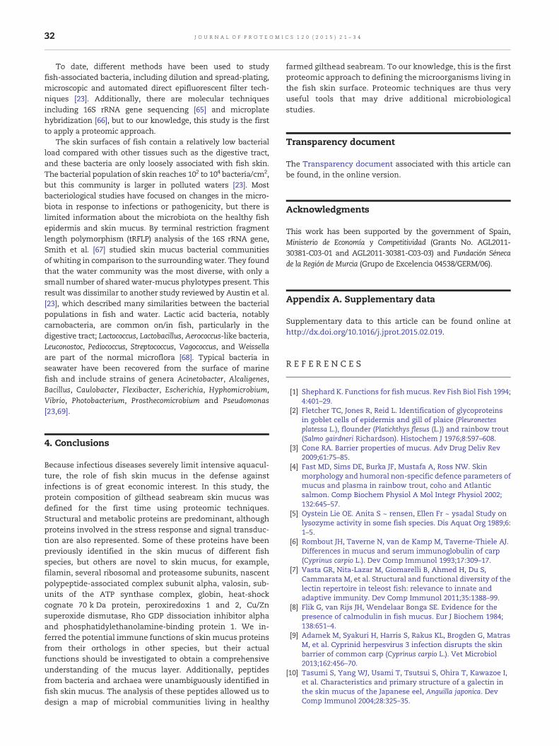
Table 1 – Proteins in the skinmucus of S. aurata identified by tandemMS. MS/MS-derived peptide sequence data were usedfor a BLAST analysis in which the search was restricted to the class Actinopterygii. Proteins shown match completely withthe sequence peptide.
Proteina Sequence IDa UniProt a PMb Sample c Scored Expect e
Structural proteins: cytoskeleton and extracellularAlpha-actin, partial [Deltistes luxatus] gb|AEO79977.1| H6DA56 1, 1 1, 2 75.3 2.0E−16Skeletal alpha-actin [Sparus aurata] gb|AAF22646.1| Q9PTJ5 1, 1 1, 2 60.4 2.0E−11Actin-related protein 3 [Perca flavescens] gb|ADX97138.1| F1C778 1 1 48.1 4.0E−08Beta-actin [Sparus aurata] gb|AAK63074.1| Q90Z11 7, 7 1, 2 99.0 7.0E−16Beta actin isoform 2a, partial [Sparus aurata] gb|AFA25665.1| H6UWY4 1, 1 1, 2 96.5 2.0E−23B-actin [Pagrus major] dbj|BAA89429.1| Q9PTU4 1 1 84.6 7.0E−20Beta-actin, partial [Oreochromis niloticus] gb|ABK20357.1| A0FKD6 1 2 33.3 3.0E−03Keratin, type I cytoskeletal 13 [Oncorhynchus mykiss] ref|NP_001117848.1| Q8JFQ6 1 2 42.2 2.0E−05Keratin, type I cytoskeletal 13-like [Oreochromis niloticus] ref|XP_003442600.1| N.E. 1 1 39.2 2.0E−04Type I keratin-like protein [Sparus aurata] gb|ACN62548.1| C0LMQ3 1, 1 1, 2 53.7 4.0E−09Type I keratin isoform 1 [Solea senegalensis] dbj|BAF56913.1| A4UYK3 2, 3 1, 2 43.9 5.0E−06PREDICTED: keratin, type I cytoskeletal 13-like
[Maylandia zebra]ref|XP_004556558.1| N.E. 1 2 37.1 7.0E−04
Keratin, type II cytoskeletal 8 [Epinephelus coioides] gb|AEG78360.1| F6KMI6 1, 1 1, 2 43.5 7.0E−06Keratin type II [Epinephelus coioides] gb|AEG78338.1| F6KMG4 1, 1 1, 2 52.8 6.0E−09Type II keratin E3, partial [Gillichthys seta] gb|ACO57583.1| C1J0K3 1, 1 1, 2 51.5 2.0E−08Type II keratin E3-like protein [Sparus aurata] gb|AAT44423.1| Q4QY72 1 2 34.6 4.0E−03Keratin [Poecilia reticulata] gb|AAD47884.1|AF172645_1 Q9PW53 1 1 30.8 6.9E−02Keratin 18 [Epinephelus coioides] gb|ACE06742.1| B3GPH2 1 2 52.4 1.0E−10Alpha-tubulin [Sparus aurata] gb|AAP89018.1| Q7T1F8 2 1 59.2 7.0E−11Beta tubulin [Chionodraco rastrospinosus] gb|AAG15329.1|AF255955_1 Q9DFS7 1, 2 1, 2 57.1.5 3.0E−10Uncharacterized protein LOC767806 [Danio rerio] ref|NP_001070241.1| Q08CC8 1 1 57.1 3.0E−10PREDICTED: collagen alpha-1(I) chain-like isoform
X1 [Maylandia zebra]ref|XP_004572575.1| N.E. 1 1 73.6 2.0E−15
Filamin-A-like [Oreochromis niloticus] ref|XP_003454305.1| N.E. 1 1 60.9 2.0E−11MetabolismProtein metabolismRibosomal protein L8 [Sander lucioperca] gb|AEE81293.1| F6KH17 1, 1 1, 2 52.8 7.0E−09Ribosomal protein L11 [Perca flavescens] gb|ABW06869.1| A8HTH7 1 1 46.4 7.0E−0740S ribosomal protein Sa-like protein [Sparus aurata] gb|AAT44424.1| Q4QY71 1,1 1, 2 56.2 6.0E−1040S ribosomal protein S7 [Oncorhynchus mykiss] ref|NP_001117902.1| Q2YHL9 1 1 71.5 6.0E−1540S ribosomal protein S3 [Salmo salar] gb|ACI67536.1| B5X9L6 1 2 44.8 3.0E−0660S ribosomal protein L7A [Siniperca chuatsi] gb|AAY79207.1| Q2KL19 1, 1 1, 2 51.1 2.0E−0860S ribosomal protein L24 [Gillichthys mirabilis] gb|AAG13295.1|AF266175_1 Q9DFQ7 1 2 42.2 2.0E−0560S ribosomal protein L19 [Epinephelus coioides] gb|ADG29150.1| D6PVQ5 1 2 59.2 5.0E−1120S proteasome beta 6 subunit [Pagrus major] gb|AAP20145.1| Q6Y267 1 1 64.3 1.0E−12NAC alpha, partial [Oryzias melastigma] gb|AEB71553.1| I1SSG5 1 2 48.6 2.0E−07Valosin containing protein [Oncorhynchus mykiss] ref|NP_001117982.1| Q1M179 1, 1 1, 2 57.1 4.0E−10
DNA metabolismHistone H2A-like [Oreochromis niloticus] ref|XP_003451178.1| N.E. 2, 2 1, 2 71.9 9.0E−16Histone h2a.x [Perca flavescens] gb|ADX97213.1| F1C7F3 2, 1 1, 2 90.1 2.0E−21Histone H4-like [Oreochromis niloticus] ref|XP_003460383.1| P62796 2, 1 1, 2 41.4 3.0E−05Nucleoside diphosphate kinase [Sparus aurata] gb|ACF75416.1| B5APB7 1, 1 1, 2 56.6 3.0E−10
Carbohydrate metabolismAlpha-1 enolase-1 [Salmo trutta] gb|AAG16310.1| N.E. 2, 1 1, 2 56.6 3.0E−12Enolase [Epinephelus bruneus] gb|AEB31337.1| F5BZS7 1 2 49.8 7.0E−08Fructose-bisphosphate aldolase [Epinephelus coioides] gb|ACL98138.1| B9V3W3 1 1 44.8 2.0E−08Glyceraldehyde 3-phosphate dehydrogenase
[Pagrus major]dbj|BAB62812.1| Q90WD9 3, 1 1, 2 81.2 4.0E−18
Triose phosphate isomerase [Polypterus ornatipinnis] dbj|BAD17930.1| Q76BC6 1 2 51.5 2.0E−08Triose phosphate isomerase [Amia calva] dbj|BAD17915.1| Q76BE1 1 2 43.1 1.0E−05
Energy metabolismATP synthase subunit alpha, mitochondrial precursor
[Psetta maxima]emb|CAY56619.1| C4QUY7 1 2 70.6 5.0E−15
ATP synthase beta-subunit [Pagrus major] dbj|BAF37105.1| A0PA13 1, 1 1, 2 61.7 7.0E−12Beta globin [Sparus aurata] gb|ABE28021.1| Q1PCB2 1, 1 1, 2 43.1 9.0E−06
Lipid metabolismApolipoprotein A-I [Sparus aurata] sp|O42175.1|APOA1_SPAAU O42175 3, 4 1, 2 74.4 6.0E−18
Stress responseHeat shock cognate 70 kDa [Carassius auratus] dbj|BAC67185.1| Q801X8 2, 2 1, 2 71.9 7.0E−15Superoxide dismutase [Cu-Zn] [Xiphias gladius] sp|P03946.2|SODC_XIPGL P03946 1 1 53.7 3.0E−11
(continued on next page)
25J O U R N A L O F P R O T E O M I C S 1 2 0 ( 2 0 1 5 ) 2 1 – 3 4

Table 1 (continued)
Proteina Sequence IDa UniProt a PMb Sample c Scored Expect e
Signal transductionPREDICTED: 14-3-3 protein beta/alpha-1-like
[Oryzias latipes]ref|XP_004070571.1| H2M383 1, 1 1, 2 46.0 1.0E−08
Serine/threonine-protein phosphatase 2A 65 kDaregulatory subunit A beta isoform [Salmo salar]
gb|ACN58639.1| C0PUA0 1 1 57.5 2.0E−10
a Protein name, sequence ID and UniProt database ID of the record with the highest score retrieved by BLAST. N.E.: No entry in UniProt.b PM: Number of MS/MS derived peptides from samples 1, and/or 2 that match exactly the protein sequence.c Sample(s) in which the protein has been identified.d Maximum score obtained in BLAST analysis by a peptide matching this protein.e Number of times we would expect to obtain an equal or higher score by chance.
26 J O U R N A L O F P R O T E O M I C S 1 2 0 ( 2 0 1 5 ) 2 1 – 3 4
glycoprotein with substantial homology to trout type IIcytokeratin is present in the skin mucus of rainbow trout(Oncorhynchus mykiss). The pore-forming properties of thisglycoprotein correlate well with strong antibacterial activi-ty [35]. This immune related function might explain the highlevels of keratins in S. aurata skin mucus (Fig. 1).
Both, α- and β-tubulins were also identified by LC-MS/MS(Table 1). In addition to their structural function, a role inphagocytic activity has been discussed to justify theincreased levels of β-tubulin protein in Atlantic cod skinmucus after infection with V. anguillarum [16]. This phe-nomenon was explained by the up-regulation of the tubb2gene in the mucosal cells associated with phagocyticprocesses.
The 2-DE-PMF-MS/MS experiment (Table 2) demonstratedthat tropomyosin is present in the S. aurata epithelial mucus,and the molecular weight and pI of the spot matched theexpected values for the full-length protein. Tropomyosin hasbeen found previously in the skin mucus of Atlantic cod [15]and Cathorops spixii (a common catfish on the Brazilian coast)[14]. To our knowledge tropomyosin has not been directly tiedto immunity, although it has been suggested that thedown-regulation of tropomyosin in patients with ulcerativecolitis may decrease immune functions [36]. Moreover, thisprotein may have antigenic properties [37,38].
Fig. 1 – Representative 2-DE gel of S. aurata skinmucus proteomeby PMF-MS/MS is in bold next to the spot number.
We identified filamin and cofilin in S. aurata skin mucusby LC-MS/MS (Table 1) and 2-DE PMF-MS/MS (Table 2),respectively. Cofilin has been previously reported in fishmucus [15], but to our knowledge, this is the first reportdescribing filamin in this specific extracellular location. Bothstructural proteins have been associated with immunity atleast in mammals and insects. Cofilins are components of aco-stimulatory signaling pathway in human T cells [39], andfilamins interact with the cytoplasmic domain of Tollreceptors [40], but their roles in fish immunity have notbeen investigated so far.
3.2.2. Metabolic proteinsThis group includes proteins involved in pathways related tothe central metabolism of biomolecules (proteins, carbohy-drates, lipids and nucleic acids) and also proteins related tometal and energy metabolism.
We identified eight different ribosomal proteins (L7A, L8,L11, L19, L24, S3, S7 and Sa-like) by LC-MS/MS (Table 1). Theseproteins have not been reported before in fish skin mucus, butmany other ribosomal proteins have been identified in theepithelial mucus of different fish species. For instance, apeptide derived from the S30 ribosomal protein is present inskin secretions from rainbow trout [41], and L40, L36A, L35 anda protein similar to ribosomal protein P2 are found in the
(left) and liver (right). The UniProt ID of the proteins identified

Table 2 – Proteins identified by coupled PMF and MS/MS.
UniProt SC Mass
SNa Proteinb Organismb IDb Symbol c Scored Expect e PMf PF f % f (kDa) pI
Structural proteins: cytoskeleton and extracellular201 Alpha-actin 4 Rachycentron canadum E9L834 ACTA 572 2.4E − 52 11 5 41 42.3 5.22203 Beta actin Acipenser
transmontanusB6E4I1 ACTB 279 4.9E − 23 10 5 37 42.1 5.30
105 Beta-actin Tetraodon nigroviridis Q4SMI4 ACTB 508 6.1E − 46 12 8 43 42.9 5.57106 Beta-actin (Fragment) Gobio gobio G8A4Z9 ACTB 397 7.7E − 35 7 5 45 30.0 5.33119 keratin, type I cytoskeletal 13-like Oreochromis niloticus I3JS53 CYT1L 229 4.9E − 18 7 3 11 49.0 5.68122 Type I cytokeratin, enveloping layer, like Oreochromis niloticus I3JS53 CYT1L 224 1.5E − 17 11 3 17 49.0 5.68107 Type II cytokeratin Danio rerio Q9PUB5 KRT5 462 2.4E − 41 18 4 32 58.5 5.34108 Type II keratin E3 (Fragment) Gillichthys mirabilis C1J0K KRT 52 2.4E − 00 3 2 11 34.1 4.83215 Coactosin-like protein Tetraodon nigroviridis Q4SKB8 COTL1 89 4.5E − 01 3 2 16 16.2 4.92125 Cofilin-2 Tetraodon nigroviridis Q4RP95 COF2 229 4.9E − 18 4 3 18 18.8 6.82110 Tropomyosin4-2 Takifugu rubripes Q805C2 TPM4-2 93 1.8E − 04 7 1 30 28.4 4.58
MetabolismProtein metabolism113 Proteasome subunit alpha type Oryzias latipes H2L6P7 PSMA 283 1.9E − 23 9 3 43 29.7 6.07115 Proteasome subunit alpha type Tetraodon nigroviridis Q4SRB7 PSMA5 407 7.7E − 36 9 5 41 26.5 4.74116 Proteasome subunit alpha type Gasterosteus aculeatus G3PZP3 PSMA4 353 1.9E − 30 7 4 32 29.5 5.34120 Proteasome subunit alpha type Oreochromis niloticus I3JJY5 PSMA 704 1.5E − 65 9 7 52 25.9 5.99123 Proteasome subunit beta type Danio rerio Q6DHI9 PMSB2 242 2.4E − 19 6 2 27 22.7 6.1
Other metabolism pathways218 Brain-type fatty acid binding protein Epinephelus coioides A8HG12 FABP 96 1.0E − 04 3 2 25 14.9 6.17219 Brain-type fatty acid binding protein Epinephelus coioides A8HG12 FABP 118 6.1E − 07 3 2 25 14.9 6.17109 Glyceraldehyde-3-phosphate
dehydrogenasePagrus major Q90WD9 GAPDH 114 1.5E − 06 12 1 40 36.4 6.36
128 Nucleoside diphosphate kinase Sparus aurata B5APB7 NDPK 236 9.7E − 19 4 3 26 17.1 6.42101 Transferrin Sparus aurata F2YLA1 TF 720 3.9E − 67 26 6 40 76.1 5.93102 Transferrin Sparus aurata F2YLA1 TF 491 3.1E − 44 14 6 30 76.1 5.93
Stress response211 Peroxiredoxin 1 Sparua aurata G0T332 PRDX1 119 4.9E − 07 5 2 29 22.1 6.30212 Peroxiredoxin 2 Sparus aurata G0T333 PRDX2 232 2.4E − 18 6 4 38 21.9 5.79104 Stress protein HSC70-1 Seriola quinqueradiata B6F133 HSC70-1 1020 3.9E − 97 25 7 37 71.4 5.23127 Superoxide dismutase [Cu-Zn]
(Fragment)Sparus aurata Q571Q7 Cu/Zn
SOD89 5.2E − 04 2 2 28 6970 5.41
103 Warm temperatureacclimation-related65 kDa protein
Sparus aurata C0L788 WAP65 394 1.5E − 34 20 6 41 49.7 5.41
Signal transduction124 Phosphatidylethanolamine-binding
protein 1Takifugu rubripes H2UXL0 PEBP1 172 2.4E − 12 3 2 13 21.1 5.65
118 Rho GDP dissociation inhibitor (GDI)alpha
Danio rerio Q6P3J2 ARHGDIA 268 6.1E − 22 3 2 20 23.1 5
112 Tyrosine 3-monooxygenase/tryptophan5-monooxygenase activation protein,beta polypeptide like
Gasterosteus aculeatus G3NHX0 YWHABL 69 5.4E − 02 6 1 21 29.6 4.65
a Spot number in reference 2-DE gel.b Protein name, organism and UniProt ID of the first hit returned by Mascot search, except for spots 105, 119, 122, 125, 112, 124, and 215 in whichthe first in the list was an unidentified protein. In these cases, the protein name that is shown is the first identified protein after a BLAST searchperformed in the UniProt page.c Protein symbol as UniProtKB/Swiss-Prot database.d MOWSE score based on MS data. Protein scores greater than 68 are significant (p < 0.05). SN 108 have a score below 68 but two fragmentedpeptides from this spot match with high score with this protein.e Number of times we would expect to obtain an equal or higher score by chance.f PM: Number of non redundant matching peptides. PF: Number of fragmented peptides matching the protein. SC: % of sequence coverage.
27J O U R N A L O F P R O T E O M I C S 1 2 0 ( 2 0 1 5 ) 2 1 – 3 4
epidermal mucus of Atlantic cod [11,16]. These ribosomalproteins or their fragments have been shown to haveantimicrobial properties, although the specific mechanismsof action are not known [42]. Similarly, ribosomal proteins in
the human colonic mucus seem to be involved in the hostdefense against microorganisms [42]. Hence, we speculatethat the ribosomal proteins identified in the giltheadseabream skin mucus have a similar role.

28 J O U R N A L O F P R O T E O M I C S 1 2 0 ( 2 0 1 5 ) 2 1 – 3 4
The 20S protease proteasome complex is well represented inthe S. aurata skinmucus. We found the beta 6 subunit by LC-MS/MS (Table 1), and five spots were identified as proteasomesubunits (four alpha and one beta type) with the conventional2-DE-PMF-MS/MS approach (Table 2). These results are inagreement with a previous study that identified proteasomesubunit alpha type 3 and 7 in Atlantic cod skin mucus [15].Furthermore, the proteasome 26S subunit levels are significantlyincreased in the skin mucus of this fish in response toV. anguillarum infection [16], and the proteasome subunit alphatype 4 is up-regulated in the oral mucus of mouthbroodingtilapia fish (Orechromis spp.) [43]. Although the role of theproteasome subunits in mucus-rich organs has not beenexperimentally determined, the authors of this last studyspeculated that proteasome-mediated degradation of phos-phorylated complexes might contribute to the release of theNF-κβ, a transcription factor that activates the expression ofseveral genes essential for mucus production ([43] and thereferences therein).
Nascent polypeptide-associated complex subunit alpha(NACA) and valosin-containing protein (VCP) are involvedin protein metabolism, and we identified them in S. aurataskin mucus by LC-MS/MS (Table 1). NACA and VCP have notbeen reported in fish skin mucus, and their functions in cellsecretions are unknown. In mammals, NACA is involved ininnate immune signaling; because NACA binds DNA, it maysense DNA in the cytoplasm and transduce signals to α-taxilin,which is required for HSV-induced type I interferon production[44]. VCPhas an important role in antiviral immunity based on itsenergy-dependent, ubiquitin-selective segregase and unfoldaseactivity. VCP is an essential cofactor in the antibody-dependentintracellular neutralization of virus, which is mediated bytripartite motif-containing 21, a cytosolic IgG receptor [45].
We identified proteins involved in DNAmetabolism, includ-ing histones and nucleotide diphosphate kinase (NDPK). Thepresence of H4 and A2A-like histones in skinmucus of S. aurata(Table 1) agreedwith previous studies that found these histonesin themucus of Atlantic salmon [46] andAtlantic cod [11]. Theseproteins could have implications in gilthead seabream immu-nity since it is known that some derived peptides from histoneshave antimicrobial activity. Furthermore, the N-terminal seg-ments of catfish H2A are induced in the epidermalmucus uponstimulation, while expression of this histone is suppressedduring the early stages of stress and reduced in the absence of
LC-MS/MS
44 %
4 %
48 %
4 %
Structural proteins
Metabolism
Fig. 2 – Functional distribution of the proteins identified by bothretrieved from UniProt were used to manually place the proteins
disease (reviewed in [12]). The identification of NDPK by both,the LC-MS/MS and 2-DE-PMF-MS/MS approaches in the skinmucus of S. aurata confirmed previous data in the mucus ofdiscus fish (Symphysodon spp.) [18] and Atlantic cod [15,16]. Toour knowledge NDPK has not been linked to immunity in fish.However, this association has been established in other speciessuch as Chlamys farreri, where NDPK transcripts increasein hemocytes after bacterial challenge, and the protein isup-regulated in hemolymph [47], Litopenaeus vannamei, whereNDPK acts as a defense-related enzyme involved in the anti-viralinnate immune response [48] or even in mammalian NDPK-B isrequired for the activation of human CD4 T lymphocytes [49].
Two spots in the 2-DE gels were identified as transferrin(TF). The difference in the pI of these two isoforms was lessthan 0.1. In mammals, this protein has sites for O- andN-glycosylation [50] and the diverse grade of glycosylation ofthese sites could explain the differences in pI. TF in fish skinmucus was first reported in Atlantic salmon [46]. TF is alsoobserved in salmon skin mucus, where TF is cleaved duringsea lice infection. As discussed above, during infection,increased levels of fragmented TF may activate the nitrousoxide response in salmonmacrophages, to counteract sea liceinfection [13]. In addition, TF is part of the innate defensesystem against bacteria due to its iron chelating affinity [51].Overall, these data could explain the presence of TF in theskin mucus of S. aurata.
Four glycolytic enzymes were found in the skin mucus ofgilthead seabream. Fructose-bisphosphate aldolase (FBPA), triosephosphate isomerase (TPIS), glyceraldehyde-3-phosphate dehy-drogenase (GAPDH), and enolase (ENOA). All of these proteinshave been found in the mucus of other fish [15,16,18]. Whileglycolysis isnot known tooccur inmucus, extracellular glycolysishas been proposed in other biological fluids, for example, theperitoneal lavage fluid [52] and insect spermatophore [53].Moreover, the intermediates and products of extracellularglycolysis may have intracellular signaling actions [54].
We also found the F1 complex alpha and beta subunits inS. aurata mucus (Table 1). To our knowledge, these subunitshave not been reported in skin fish secretions. While thefunction of these proteins in mucus is unknown, recentstudies show that cell surface ATP synthases are expressedon normal human cells and that these enzymes may beimplicated in different processes including innate immunityand intracellular pH regulation [55].
PMF + MS/MS
37 %10 %
37 %
17 %
Stress response
Signal transduction
LC-MS/MS and 2DE-PMF-MS/MS. Functional annotationsinto these four general groups.

20
25
50
75
15
100
37
mw(kDa)
150250
1 2 1 2
Fig. 3 – Western blot showing intact β-actin in S. aurata skinmucus. SDS-PAGE was performed as described in theMaterials and methods section. Lane 1: five microliters ofmolecular weight marker (Precision Plus Protein All BlueStandards, Bio-Rad). Lane 2: Ten micrograms of S. aurataskin mucus proteins. The left panel corresponds to the totalprotein in the transferred membrane. Molecular weightmarkers, which were prestained to direct monitoringelectrophoresis and transfer efficiency, are refractory to UVactivation and appear as a negative fluorescence signal. Theright panel shows the same membrane after antibodyhybridization and contains a unique band corresponding insize to β-actin. No fragmentation of this protein wasobserved. The actin immunoreactive band overlaps one ofthe two major bands observed in the left panel, according tothe abundance of this protein observed in 2-DE gels (Fig. 1).
29J O U R N A L O F P R O T E O M I C S 1 2 0 ( 2 0 1 5 ) 2 1 – 3 4
LC-MS/MS (Table 1) also revealed beta globin (HBB), asubunit of the α2β2 hemoglobin tetramer, in the mucus ofgilthead seabream. This protein was not previously reportedin fish mucus, and consequently, its function in this locationremains undisclosed. However, this protein could serve thefunction suggested for other metal-binding proteins in mucus(e.g., lactoferrin and transferring) to create low-iron environ-ments that limit microbial pathogenesis. It is known that thisactivity damages the lipopolysaccharide (LPS) of the Gram-negative outer membrane altering its permeability [51], andthere is also evidence for globin-LPS binding [56] supporting arole for mucosal globins in fish immunity.
We found two proteins related to lipids metabolism in skinsecretions of S. aurata, apolipoprotein-A1 (Apo-A1) in mucussamples analyzed by LC-MS/MS (Table 1) and brain-type fattyacid binding protein (FABP7) in samples analized by 2-DE-PMF-MS/MS (Table 2). Apo-A1 protein of S. aurata is mainlyexpressed in liver and to a lower extent in intestine [57] but ithas also been found in skin secretions of other fish species. Inthis line, Apo-A1 is overexpressed in the skin mucus ofinfected Atlantic cod [15] and Atlantic salmon [13,16]. In carp
(Cyprinus carpio) it has antibacterial effects in vitro [58]. Allthese findings support a role for mucosal Apo-A1 in fishimmunity, but the mechanism has not been elucidated. Toour knowledge, this is the first time that FABP7 has beenidentified in fish skin secretions, although a member of thefamily specific to adipocytes is present in S. salar mucus [15].
3.2.3. Stress response proteinsWe found five proteins related to stress response in themucus of S. aurata: warm temperature acclimation protein65 kDa (WAP65), heat-shock cognate 70 kDa protein (HSC70),peroxiredoxin 1 and 2 (PRDX1 and PRDX2) and Cu/Zn superoxidedismutase (Cu/Zn-SOD). HSC70 and SOD were detected by bothLC-MS/MS and 2-DE, while WAP65, PRDX1 and PRDX2 were onlydetected by 2-DE. With the exception of WAP65 [14], theseproteins have not been reported in fish mucus, but Rajan et al.identified a different peroxiredoxin, PRDX6, in Atlantic cod skinmucus [16]. In mammals, SOD and PRDX1 and PRDX2 aresecreted in fluids and are involved in inflammation, immunityand tissue repair [59,60]. Nevertheless, the role of these antiox-idant proteins in skin mucosal needs clarification.
WAP65 and HSC70 proteins are both involved in theresponse to temperature changes. WAP65 expression is anatural physiological adaptation to warm temperature inteleost fish, although WAP65 may also have other functionsincluding immune system modulation [61]. WAP65 proteinhas a high affinity for heme, and given that iron is a keyelement in bacterial infections, the role of this protein couldbe involved in mucus innate immunity. WAP65 is found in C.spixii skin mucus and has inflammatory action [14]. HSC70 isconstitutively expressed in non-stressed cells and only mildlyinduced during stress. This protein forms a stable complexwith HSP70 upon stress and plays specific roles in manycellular processes including innate immunity. HSC70 proteinis well characterized in mammals and may be secreted underspecific conditions [62]. In fish, the HSC70 mRNA is ubiqui-tously expressed in several species [63,64], but so far, theprotein has not been found in fish skin secretions.
3.2.4. Signal transduction proteinsFive proteins belonging to this group were identified, the betaisoform of the 65 kDa regulatory subunit A of serine/threonine-protein phosphatase 2A (PP2A), two members ofthe 14-3-3 family (14-3-3 protein beta/alpha-1-like and tyro-sine 3-monooxygenase/tryptophan 5-monooxygenase activa-tion protein, beta polypeptide like), Rho GDP dissociationinhibitor alpha (RhoGDI), and phosphatidylethanolamine-binding protein 1 (PEBP1). To our knowledge, only the 65 kDaregulatory subunit A of PP2A and 14-3-3 proteins have beenpreviously reported in fish skin mucus [15,16], but their role infish mucus is unknown. The spot intensity of the 65 kDaregulatory subunit A of PP2A increases 2.3-fold upon naturalinfection of Atlantic cod with V. anguillarum, suggesting a rolein the immunity [16].
3.3. A proteomic approach to the bacterial population ofS. aurata skin mucus
Thirty-six of the peptides derived from LC-MS/MS did notcompletely match any database sequence when the search

Table 3 – LC-MS/MS-derived peptide sequences from skin mucus of S. aurata matching exclusively bacterial or archaeal sequences.
Peptide sequencea Proteinb Organismc Score b E valueb Accessionb
1 DDLLSEYDFPGDDLPVIQGSALK Elongation factor Tu Streptococcus (144) 72.3 2E − 13 ACX81418.12 DLLSEYDFPGDDIPVIQGSALK Elongation factor Tu Streptococcus (33) 72.7 3E − 13 ABW24197.13 AVVELAGVADITSK 30S ribosomal protein S5 Streptococcus (12) 43.9 4E − 04 WP_019319452.14 MLADLAVNDAVAFTALADAAK 50S ribosomal protein L20 Streptococcus (3) 66.4 1E − 11 WP_001841176.15 ELADAAVSTIEIER 30S ribosomal protein S3, partial Streptococcus (35) 46.0 7E − 05 WP_008291857.16 AAAELELISGQKPLITK 50S ribosomal protein L5 Streptococcus (5) 54.1 2E − 07 WP_018367195.17 YPEFAQLEGQLK 6-Phosphofructokinase Streptococcus (89) 42.2 1E − 03 WP_001831447.18 ITDFLSANAEVIAR Adenylosuccinate synthetase Streptococcus (115) 46.4 2E − 05 WP_006150535.19 VVFGENIGTTVSNNIEEKE Uridylate kinase Streptococcus (23) 63.0 2E − 10 WP_008275990.110 LVVLYDSNDINLDGETK Transketolase, partial Streptococcus (162) 57.1 2E − 08 AGG36758.111 LSQETSVYVTGIVK Asparaginyl-tRNA synthetase Streptococcus (86) 46.0 9E − 05 YP_003879782.112 GGAVGDQYVTVNVVTPTGLNDR Chaperone protein DnaJ Streptococcus (98) 70.6 6E − 13 WP_001808904.113 GVYLNEDGSVNLSK Pyruvate formate lyase, partial Streptococcus (180) 46.0 8E − 05 AFN66486.114 ISQALEVAEPGVTNR Selenide, water dikinase Streptococcus (96) 49.0 7E − 06 WP_001852559.115 LFLEEEGLQSR pyruvate oxidase Streptococcus (105) 38.0 2E − 02 ETJ01702.116 TGAQVAGPIPLPTER 30S ribosomal protein S10 Streptococcus and Aerococcus (9) 49.0 7E − 06 WP_000649303.117 FDATVEVAYNLNIDVK 50S ribosomal protein L1, partial Streptococcus and Lactococcus (64) 54.5 2E − 07 WP_019785915.118 IEDQLGEVAEYR Alpha-enolase Streptococcus and Catellicoccus marimammalium (96) 42.2 8E − 04 ADQ38428.119 LADAAVSTIEIER 30S ribosomal protein S3, partial Streptococcus, Enterococcus, and Melissococcus (53) 42.6 1E − 03 WP_008291857.120 ADIDYAWEEADTTYGK 30S ribosomal protein S3, partial Streptococcus, Listeria, Bacillus, Lactococcus,
Enterococcus, Tetragenococcus, Carnobacterium, andGranulicatella (126)
56.6 3E − 09 WP_008288328.1
21 VLLGLSGGVDSSVVGVLLQK GMP synthase, partial Streptococcus, Enterococcus,Weissella, Granulicatella,Carnobacterium,Melissococcus, and Aerococcus (286)
60.4 2E − 09 ETJ01807.1
22 SGETEDSTIADIAVATNAGQIK Alpha-enolase Streptococcus, Listeria, Bacillus, Geobacillus,Exiguobacterium, Singulisphaera, Leptothrix, Brevibacillus,Caldibacillus, Catellicoccus, Anoxybacillus, Facklamia,Carnobacterium, Melissococcus, Granulicatella,Enterococcus, Acetivibrio, Eremococcus, Micavibrio,Rhodomicrobium, and Alistipes (244)
68.9 6E − 13 ADQ38428.1
23 IWPTEDALKR Phosphoribosylaminoimidazolecarboxamideformyltransferase
Clostridium (6) 37.1 6E − 02 WP_017210864.1
24 TDLVHAVYDEIPDVLRDLGVSEVHGVLMDLGVSSLQLDERER S-adenosyl-methyltransferase MraW Arthrobacter sp. (1) 85.5 7E − 19 YP_831056.125 NMITGASQADAAILVcAAPDGVMQQTK Elongation factor 1-alpha Methanocella arvoryzae (1) 88.0 3E − 18 YP_684657.126 EDDDIWDVLEDVIK DNA-directed RNA polymerase subunit beta Lactobacillus (26) 51.1 2E − 06 WP_021355686.127 FEQFVAEGAYLDALR Glycyl-tRNA ligase subunit beta Pelobacter carbinolicus (1) 51.1 2E − 06 YP_006716293.128 MLEAVLCDYQTSQILQLTDAMVR DNA polymerase III, delta subunit Mycoplasma (3) 79.5 2E − 15 WP_014574988.129 ADMLQQECEALLVDFPDQEKELR Lipoate-protein ligase A domain protein,
partialEscherichia coli and Shigella (78) 79.5 4E − 17 WP_001342437.1
30 STLIRCVNLLERPTEGSVQVGGQELTTLSESELTK Methionine ABC transporter ATP-bindingprotein
Salmonella enterica, Citrobacter youngae, Citrobacterfreundii, Shigella flexneri (Family Enterobacteriaceae) (31)
70.5 1E − 14 WP_001570617.1
31 ANVLIFPDLDAGNIAYK Phosphate acetyltransferase Geobacter, Caldicellulosiruptor and Deferribacterdesulfuricans, Calditerrivibrio nitroreducens, Slackiaheliotrinireducens and Clostridium thermocellum (21)
57.1 3E − 08 YP_003497026.1
30JO
UR
NA
LO
FPR
OT
EO
MIC
S120
(2015)
21–34

Fig. 4 – Phylogenetic tree of microorganisms matching peptides found in healthy S. aurata skin mucus. A filled circle on theright of the figure next to the genus name means that one peptide exclusively matched this genus. An open circle indicatesthat a peptide matched this genus, but other genus sequences had the same score.
31J O U R N A L O F P R O T E O M I C S 1 2 0 ( 2 0 1 5 ) 2 1 – 3 4
was restricted to the Actinopterygii class. Five peptidescontain sequences from more or less specific taxonomicgroup, i.e., the genus Caenorhabditis (EEVSAVDEIIKDK) orkingdom Viridiplantae (HVVFGQVVEGLDVVK). Organisms inthese taxa could live in the tanks where gilthead seabream isfarmed and thus, become incorporated into fish the skinmucus. The most remarkable finding was that thirty-one ofthe peptides exclusively matched bacterial or archaeal se-quences (Table 3). When these peptides were used to performa BLAST search against the Actinopterygii database, the scorewas low, and the E-value did not indicate a statisticallysignificant match. For comparison, the result of a similarunrestricted search is shown in Supplementary file 4. Becausewe used healthy animals, we hypothesize that certain speciesof bacteria, or closely related ones, live commensally in thegilthead seabream epidermal mucus. The peptides, the mostclosely related organisms, and the database search parame-
Notes to Table 3:aPeptide sequences derived from MS/MS data of S. aurata skin mucus (samples 1 abFirst hit retrieved by BLAST search of the peptide sequence against non-redundanaccession number of the first hit are shown.cOrganism having sequences matching 100% with peptide sequence (in parenthreported, it indicates that several species of the genus satisfy the condition.
ters are summarized in Table 3. The represented taxonomicgroups are organized on a phylogenetic tree in Fig. 4.
Some peptides matched only one genus, indicating thatspecies in these genera are most likely living in the fishmucus.We found peptides from the genera Streptococcus, Lactobacillus,Mycoplasma, Arthobacter, Clostridium, and Pelobacter, and onepeptidematching only theAchaea speciesMetanocella arvoryzae.Notably, 15 peptides matched only Streptococcus species andeight more matched Streptococcus and other Lactobacillalesspecies. In general, the Lactobacillales order was the mostabundant population of bacteria in this particular habitat. Twodifferent peptides matched only Enterobacteriaceae familystrains, signifying that this group of bacteria is also present inthe skin mucus of healthy S. aurata. The remaining threepeptideswere less informative because theymatched species ofseveral bacterial phyla; thesematches most likely occurred in aconserved region of the protein.
nd/or 2, see Supplementary file 1).t protein sequences database (100% identity). Protein name, score, E value, and
eses is the number of hits showing the highest score). When only genus is
32 J O U R N A L O F P R O T E O M I C S 1 2 0 ( 2 0 1 5 ) 2 1 – 3 4
To date, different methods have been used to studyfish-associated bacteria, including dilution and spread-plating,microscopic and automated direct epifluorescent filter tech-niques [23]. Additionally, there are molecular techniquesincluding 16S rRNA gene sequencing [65] and microplatehybridization [66], but to our knowledge, this study is the firstto apply a proteomic approach.
The skin surfaces of fish contain a relatively low bacterialload compared with other tissues such as the digestive tract,and these bacteria are only loosely associated with fish skin.The bacterial population of skin reaches 102 to 104 bacteria/cm2,but this community is larger in polluted waters [23]. Mostbacteriological studies have focused on changes in the micro-biota in response to infections or pathogenicity, but there islimited information about the microbiota on the healthy fishepidermis and skin mucus. By terminal restriction fragmentlength polymorphism (tRFLP) analysis of the 16S rRNA gene,Smith et al. [67] studied skin mucus bacterial communitiesof whiting in comparison to the surroundingwater. They foundthat the water community was the most diverse, with only asmall number of shared water-mucus phylotypes present. Thisresult was dissimilar to another study reviewed by Austin et al.[23], which described many similarities between the bacterialpopulations in fish and water. Lactic acid bacteria, notablycarnobacteria, are common on/in fish, particularly in thedigestive tract; Lactococcus, Lactobacillus, Aerococcus-like bacteria,Leuconostoc, Pediococcus, Streptococcus, Vagococcus, and Weissellaare part of the normal microflora [68]. Typical bacteria inseawater have been recovered from the surface of marinefish and include strains of genera Acinetobacter, Alcaligenes,Bacillus, Caulobacter, Flexibacter, Escherichia, Hyphomicrobium,Vibrio, Photobacterium, Prosthecomicrobium and Pseudomonas[23,69].
4. Conclusions
Because infectious diseases severely limit intensive aquacul-ture, the role of fish skin mucus in the defense againstinfections is of great economic interest. In this study, theprotein composition of gilthead seabream skin mucus wasdefined for the first time using proteomic techniques.Structural and metabolic proteins are predominant, althoughproteins involved in the stress response and signal transduc-tion are also represented. Some of these proteins have beenpreviously identified in the skin mucus of different fishspecies, but others are novel to skin mucus, for example,filamin, several ribosomal and proteasome subunits, nascentpolypeptide-associated complex subunit alpha, valosin, sub-units of the ATP synthase complex, globin, heat-shockcognate 70 k Da protein, peroxiredoxins 1 and 2, Cu/Znsuperoxide dismutase, Rho GDP dissociation inhibitor alphaand phosphatidylethanolamine-binding protein 1. We in-ferred the potential immune functions of skin mucus proteinsfrom their orthologs in other species, but their actualfunctions should be investigated to obtain a comprehensiveunderstanding of the mucus layer. Additionally, peptidesfrom bacteria and archaea were unambiguously identified infish skin mucus. The analysis of these peptides allowed us todesign a map of microbial communities living in healthy
farmed gilthead seabream. To our knowledge, this is the firstproteomic approach to defining the microorganisms living inthe fish skin surface. Proteomic techniques are thus veryuseful tools that may drive additional microbiologicalstudies.
Transparency document
The Transparency document associated with this article canbe found, in the online version.
Acknowledgments
This work has been supported by the government of Spain,Ministerio de Economía y Competitividad (Grants No. AGL2011-30381-C03-01 and AGL2011-30381-C03-03) and Fundación Sénecade la Región de Murcia (Grupo de Excelencia 04538/GERM/06).
Appendix A. Supplementary data
Supplementary data to this article can be found online athttp://dx.doi.org/10.1016/j.jprot.2015.02.019.
R E F E R E N C E S
[1] Shephard K. Functions for fishmucus. Rev Fish Biol Fish 1994;4:401–29.
[2] Fletcher TC, Jones R, Reid L. Identification of glycoproteinsin goblet cells of epidermis and gill of plaice (Pleuronectesplatessa L.), flounder (Platichthys flesus (L.)) and rainbow trout(Salmo gairdneri Richardson). Histochem J 1976;8:597–608.
[3] Cone RA. Barrier properties of mucus. Adv Drug Deliv Rev2009;61:75–85.
[4] Fast MD, Sims DE, Burka JF, Mustafa A, Ross NW. Skinmorphology and humoral non-specific defence parameters ofmucus and plasma in rainbow trout, coho and Atlanticsalmon. Comp Biochem Physiol A Mol Integr Physiol 2002;132:645–57.
[5] Oystein Lie OE. Anita S ~ rensen, Ellen Fr ~ ysadal Study onlysozyme activity in some fish species. Dis Aquat Org 1989;6:1–5.
[6] Rombout JH, Taverne N, van de Kamp M, Taverne-Thiele AJ.Differences in mucus and serum immunoglobulin of carp(Cyprinus carpio L.). Dev Comp Immunol 1993;17:309–17.
[7] Vasta GR, Nita-Lazar M, Giomarelli B, Ahmed H, Du S,Cammarata M, et al. Structural and functional diversity of thelectin repertoire in teleost fish: relevance to innate andadaptive immunity. Dev Comp Immunol 2011;35:1388–99.
[8] Flik G, van Rijs JH, Wendelaar Bonga SE. Evidence for thepresence of calmodulin in fish mucus. Eur J Biochem 1984;138:651–4.
[9] Adamek M, Syakuri H, Harris S, Rakus KL, Brogden G, MatrasM, et al. Cyprinid herpesvirus 3 infection disrupts the skinbarrier of common carp (Cyprinus carpio L.). Vet Microbiol2013;162:456–70.
[10] Tasumi S, Yang WJ, Usami T, Tsutsui S, Ohira T, Kawazoe I,et al. Characteristics and primary structure of a galectin inthe skin mucus of the Japanese eel, Anguilla japonica. DevComp Immunol 2004;28:325–35.

33J O U R N A L O F P R O T E O M I C S 1 2 0 ( 2 0 1 5 ) 2 1 – 3 4
[11] Bergsson G, Agerberth B, Jornvall H, Gudmundsson GH.Isolation and identification of antimicrobial componentsfrom the epidermal mucus of Atlantic cod (Gadus morhua).FEBS J 2005;272:4960–9.
[12] Esteban MA. An overview of the immunological defenses infish skin. ISRN Immunol 2012;2012:1–29.
[13] Easy RH, Ross NW. Changes in Atlantic salmon (Salmo salar)epidermal mucus protein composition profiles followinginfection with sea lice (Lepeophtheirus salmonis).Comp Biochem Physiol Part D Genomics Proteomics 2009;4:159–67.
[14] Ramos AD, Conceicao K, Silva Jr PI, Richardson M, Lima C,Lopes-Ferreira M. Specialization of the sting venom and skinmucus of Cathorops spixii reveals functional diversification ofthe toxins. Toxicon 2012;59:651–65.
[15] Rajan B, Fernandes JM, Caipang CM, Kiron V, Rombout JH,Brinchmann MF. Proteome reference map of the skin mucusof Atlantic cod (Gadus morhua) revealing immune competentmolecules. Fish Shellfish Immunol 2011;31:224–31.
[16] Rajan B, Lokesh J, Kiron V, Brinchmann MF. Differentiallyexpressed proteins in the skin mucus of Atlantic cod (Gadusmorhua) upon natural infection with Vibrio anguillarum. BMCVet Res 2013;9:103.
[17] Chong K, Ying T, Foo J, Jin L, Chong A. Characterisation ofproteins in epidermal mucus of discus fish (Symphysodonspp.) during parental phase. Aquaculture 2005;249:469–76.
[18] Chong K, Joshi S, Jin LT, Shu-Chien AC. Proteomics profilingof epidermal mucus secretion of a cichlid (Symphysodonaequifasciata) demonstrating parental care behavior.Proteomics 2005;5:2251–8.
[19] Ai-Jun M, Zhi-Hui H, Xin-An W. Changes in proteincomposition of epidermal mucus in turbot Scophthalmusmaximus (L.) under high water temperature. Fish PhysiolBiochem 2013;39:1411–8.
[20] Issa SM, Schulz BL, Packer NH, Karlsson NG. Analysis ofmucosal mucins separated by SDS-urea agarosepolyacrylamide composite gel electrophoresis.Electrophoresis 2011;32:3554–63.
[21] Liska AJ, Shevchenko A. Expanding the organismal scope ofproteomics: cross-species protein identification by massspectrometry and its implications. Proteomics 2003;3:19–28.
[22] Junqueira M, Spirin V, Balbuena TS, Thomas H, Adzhubei I,Sunyaev S, et al. Protein identification pipeline for thehomology-driven proteomics. J Proteomics 2008;71:346–56.
[23] Austin B. The bacterial microflora of fish, revised.ScientificWorldJournal 2006;6:931–45.
[24] Fernandez-Puente P, Mateos J, Blanco FJ, Ruiz-Romero C.LC-MALDI-TOF/TOF for shotgun proteomics. Methods MolBiol 2014;1156:27–38.
[25] Guardiola FA, Cuesta A, Arizcun M, Meseguer J, Esteban MA.Comparative skin mucus and serum humoral defencemechanisms in the teleost gilthead seabream(Sparus aurata). Fish Shellfish Immunol 2014;36:545–51.
[26] Montes-Nieto R, Fuentes-Almagro CA, Bonilla-Valverde D,Prieto-Alamo MJ, Jurado J, Carrascal M, et al. Proteomics infree-living Mus spretus to monitor terrestrial ecosystems.Proteomics 2007;7:4376–87.
[27] Addis MF, Cappuccinelli R, Tedde V, Pagnozzi D, Viale I,Meloni M, et al. Influence of Moraxella sp. colonizationon the kidney proteome of farmed gilthead sea breams(Sparus aurata, L.). Proteome Sci 2010;8:50.
[28] Ibarz A, Martin-Perez M, Blasco J, Bellido D, de Oliveira E,Fernandez-Borras J. Gilthead sea bream liver proteomealtered at low temperatures by oxidative stress. Proteomics2010;10:963–75.
[29] Isani G, Andreani G, Carpene E, Di Molfetta S, Eletto D, SpisniE. Effects of waterborne Cu exposure in gilthead sea bream(Sparus aurata): a proteomic approach. Fish ShellfishImmunol 2011;31:1051–8.
[30] Martin-Perez M, Fernandez-Borras J, Ibarz A, Millan-Cubillo A,Felip O, de Oliveira E, et al. New insights into fish swimming:a proteomic and isotopic approach in gilthead sea bream. JProteome Res 2012;11:3533–47.
[31] Rufino-Palomares E, Reyes-Zurita FJ, Fuentes-Almagro CA, dela Higuera M, Lupianez JA, Peragon J. Proteomics in the liverof gilthead sea bream (Sparus aurata) to elucidate the cellularresponse induced by the intake of maslinic acid. Proteomics2011;11:3312–25.
[32] Silva TS, Cordeiro OD, Matos ED, Wulff T, Dias JP, Jessen F,et al. Effects of preslaughter stress levels on the post-mortemsarcoplasmic proteomic profile of gilthead seabream muscle.J Agric Food Chem 2012;60:9443–53.
[33] Easy RH, Ross NW. Changes in Atlantic salmon Salmo salarmucus components following short- and long-term handlingstress. J Fish Biol 2010;77:1616–31.
[34] Stafford JL, Belosevic M. Transferrin and the innate immuneresponse of fish: identification of a novel mechanism ofmacrophage activation. Dev Comp Immunol 2003;27:539–54.
[35] Molle V, Campagna S, Bessin Y, Ebran N, Saint N, Molle G.First evidence of the pore-forming properties of a keratinfrom skin mucus of rainbow trout (Oncorhynchus mykiss,formerly Salmo gairdneri). Biochem J 2008;411:33–40.
[36] Li N, Wang X, Zhang Y, Zhai J, Zhang T, Wei K. Comparativeproteomics analysis of serum proteins in ulcerative colitispatients. Mol Biol Rep 2012;39:5659–67.
[37] Jenkins RE, Taylor MJ, Gilvary NJ, Bianco AE. Tropomyosinimplicated in host protective responses to microfilariae inonchocerciasis. Proc Natl Acad Sci U S A 1998;95:7550–5.
[38] Mor F, Weinberger A, Cohen IR. Identification of alpha-tropomyosin as a target self-antigen in Behcet's syndrome.Eur J Immunol 2002;32:356–65.
[39] Lee KH, Meuer SC, Samstag Y. Cofilin: a missing link betweenT cell co-stimulation and rearrangement of the actincytoskeleton. Eur J Immunol 2000;30:892–9.
[40] Silverman N, Maniatis T. NF-kappaB signaling pathways inmammalian and insect innate immunity. Genes Dev 2001;15:2321–42.
[41] Fernandes JM, Smith VJ. A novel antimicrobial function for aribosomal peptide from rainbow trout skin. Biochem BiophysRes Commun 2002;296:167–71.
[42] Antoni L, Nuding S, Weller D, Gersemann M, Ott G, WehkampJ, et al. Human colonic mucus is a reservoir for antimicrobialpeptides. J Crohns Colitis 2013;7:e652–64.
[43] Iq KC, Shu-Chien AC. Proteomics of buccal cavity mucus infemale tilapia fish (Oreochromis spp.): a comparison betweenparental and non-parental fish. PLoS One 2011;6:e18555.
[44] Li S, Wang L, Berman M, Kong YY, Dorf ME. Mapping adynamic innate immunity protein interaction networkregulating type I interferon production. Immunity 2011;35:426–40.
[45] Hauler F, Mallery DL, McEwan WA, Bidgood SR, James LC.AAA ATPase p97/VCP is essential for TRIM21-mediated virusneutralization. Proc Natl Acad Sci U S A 2012;109:19733–8.
[46] Raeder IL, Paulsen SM, Smalas AO, Willassen NP. Effect of fishskin mucus on the soluble proteome of Vibrio salmonicidaanalysed by 2-D gel electrophoresis and tandem massspectrometry. Microb Pathog 2007;42:36–45.
[47] Shi XZ, Zhao XF, Wang JX. Molecular cloning and analysis offunction of nucleoside diphosphate kinase (NDPK) from thescallop Chlamys farreri. Biochemistry 2008;73:686–92.
[48] Zhao ZY, Yin ZX, Weng SP, Guan HJ, Li SD, Xing K, et al.Profiling of differentially expressed genes in hepatopancreasof white spot syndrome virus-resistant shrimp (Litopenaeusvannamei) by suppression subtractive hybridisation. FishShellfish Immunol 2007;22:520–34.
[49] Srivastava S, Li Z, Ko K, Choudhury P, Albaqumi M, JohnsonAK, et al. Histidine phosphorylation of the potassiumchannel KCa3.1 by nucleoside diphosphate kinase B is

34 J O U R N A L O F P R O T E O M I C S 1 2 0 ( 2 0 1 5 ) 2 1 – 3 4
required for activation of KCa3.1 and CD4 T cells. Mol Cell2006;24:665–75.
[50] Satomi Y, Shimonishi Y, Hase T, Takao T. Site-specificcarbohydrate profiling of human transferrin by nano-flowliquid chromatography/electrospray ionization massspectrometry. Rapid Commun Mass Spectrom 2004;18:2983–8.
[51] Ellison III RT, Giehl TJ, LaForce FM. Damage of the outermembrane of enteric gram-negative bacteria by lactoferrinand transferrin. Infect Immun 1988;56:2774–81.
[52] Bansal S. Carbohydrate metabolism in the rat peritonealmacrophages. J Biosci 1987;12:415–20.
[53] Osanai M, Kunitomo Y, Nagaoka S. Glycogen phosphorylasein the male reproductive system and spermatophore of thesilkmoth, Bombyx mori. J Seric Sci Jpn 1995;64:523–33.
[54] Henderson B, Martin A. Bacterial moonlighting proteins andbacterial virulence. Curr Top Microbiol Immunol 2013;358:155–213.
[55] Fu Y, Zhu Y. Ectopic ATP synthase in endothelial cells: a novelcardiovascular therapeutic target. Curr Pharm Des 2010;16:4074–9.
[56] Yang H, Wang H, Bernik TR, Ivanova S, Wang H, Ulloa L, et al.Globin attenuates the innate immune response to endotoxin.Shock 2002;17:485–90.
[57] Llewellyn L, Ramsurn VP, Wigham T, Sweeney GE, Power DM.Cloning, characterisation and expression of theapolipoprotein A-I gene in the sea bream (Sparus aurata).Biochim Biophys Acta 1998;1442:399–404.
[58] Concha MI, Smith VJ, Castro K, Bastias A, Romero A,Amthauer RJ. Apolipoproteins A-I and A-II are potentiallyimportant effectors of innate immunity in the teleost fishCyprinus carpio. Eur J Biochem 2004;271:2984–90.
[59] Terrazzano G, Rubino V, Damiano S, Sasso A, Petrozziello T,Ucci V, et al. T cell activation induces CuZn superoxidedismutase (SOD)-1 intracellular re-localization, productionand secretion. Biochim Biophys Acta 1843;2014:265–74.
[60] Ishii T, Warabi E, Yanagawa T. Novel roles of peroxiredoxinsin inflammation, cancer and innate immunity. J Clin BiochemNutr 2012;50:91–105.
[61] Picard DJ, Schulte PM. Variation in gene expression inresponse to stress in two populations of Fundulus heteroclitus.Comp Biochem Physiol A Mol Integr Physiol 2004;137:205–16.
[62] Liu T, Daniels CK, Cao S. Comprehensive review on the HSC70functions, interactions with related molecules andinvolvement in clinical diseases and therapeutic potential.Pharmacol Ther 2013;136:354–74.
[63] Zafarullah M, Wisniewski J, Shworak NW, Schieman S, MisraS, Gedamu L. Molecular cloning and characterization of aconstitutively expressed heat-shock-cognate hsc71 genefrom rainbow trout. Eur J Biochem 1992;204:893–900.
[64] Wang TT, Wang N, Liao XL, Meng L, Liu Y, Chen SL. Cloning,molecular characterization and expression analysis of heatshock cognate 70 (Hsc70) cDNA from turbot (Scophthalmusmaximus). Fish Physiol Biochem 2013;39:1377–86.
[65] Kim DH, Brunt J, Austin B. Microbial diversity of intestinalcontents and mucus in rainbow trout (Oncorhynchus mykiss). JAppl Microbiol 2007;102:1654–64.
[66] Sugita H, Nakamura T, Tanaka K, Deguchi Y. Identification ofAeromonas species isolated from freshwater fish with themicroplate hybridization method. Appl Environ Microbiol1994;60:3036–8.
[67] Smith CJ, Danilowicz BS, Meijer WG. Characterization of thebacterial community associated with the surface and mucuslayer of whiting (Merlangius merlangus). FEMS Microbiol Ecol2007;62:90–7.
[68] Ringø E, Gatesoupe FJ. Lactic acid bacteria in fish: a review.Aquaculture 1998;134:339–49.
[69] Montes M, Perez MJ, Nieto TP. Numerical taxonomy ofgram-negative, facultative anaerobic bacteria isolated fromskin of turbot (Scophthalmus maximus) and surrounding water.Syst Appl Microbiol 1999;22:604–18.
Related Documents