INVITED REVIEW Proteomic Analysis of HIV-Infected Macrophages Loyda M. Meléndez & Krystal Colon & Linda Rivera & Eillen Rodriguez-Franco & Dianedis Toro-Nieves Received: 2 July 2010 / Accepted: 23 November 2010 / Published online: 14 December 2010 # The Author(s) 2010. This article is published with open access at Springerlink.com Abstract Mononuclear phagocytes (monocytes, macro- phages, and microglia) play an important role in innate immunity against pathogens including HIV. These cells are also important viral reservoirs in the central nervous system and secrete inflammatory mediators and toxins that affect the tissue environment and function of surrounding cells. In the era of antiretroviral therapy, there are fewer of these inflammatory mediators. Proteomic approaches including surface enhancement laser desorption ionization, one- and two-dimensional difference in gel electrophoresis, and liquid chromatography tandem mass spectrometry have been used to uncover the proteins produced by in vitro HIV-infected monocytes, macrophages, and microglia. These approaches have advanced the understanding of novel mechanisms for HIV replication and neuronal damage. They have also been used in tissue macrophages that restrict HIV replication to understand the mechanisms of restriction for future therapies. In this review, we summarize the proteomic studies on HIV-infected mono- nuclear phagocytes and discuss other recent proteomic approaches that are starting to be applied to this field. As proteomic instruments and methods evolve to become more sensitive and quantitative, future studies are likely to identify more proteins that can be targeted for diagnosis or therapy and to uncover novel disease mechanisms. Keywords Monocytes . Macrophages . HIV . SELDI-TOF . 2D DIGE . Tandem mass spectrometry . Proteomics Abbreviations 2D DIGE Two-dimensional difference in gel electrophoresis AIDS Acquired immunodeficiency syndrome BBB Blood–brain barrier CI Cognitive impairment CNS Central nervous system CSF Cerebrospinal fluid GEE Generalized estimating equation HAD HIV-1 associated dementia HAND HIV-associated neurological disorders HBMEC Human brain microvascular endothelial cell HIV ADA A macrophage tropic R5 virus isolated from blood of a patient with HIV dementia HPLC High-performance liquid chromatography ICAT Isotope coded affinity tags IFN Interferon IL-1β Interleukin-1β ITRAQ Isobaric tag for relative and absolute quantitation LC/ESI-FTICR MS Liquid chromatography coupled with electrospray ionization and a hybrid quadrupole linear ion-trap and Fourier-transform ion-cyclotron-resonance mass spectrometry L. M. Meléndez (*) : K. Colon : L. Rivera : E. Rodriguez-Franco Department of Microbiology and Medical Zoology, School of Medicine, University of Puerto Rico, Medical Sciences Campus, San Juan 00935, Puerto Rico e-mail: [email protected] D. Toro-Nieves NeuroAIDS Program, University of Puerto Rico, Medical Sciences Campus, San Juan, Puerto Rico J Neuroimmune Pharmacol (2011) 6:89–106 DOI 10.1007/s11481-010-9253-4

Welcome message from author
This document is posted to help you gain knowledge. Please leave a comment to let me know what you think about it! Share it to your friends and learn new things together.
Transcript

INVITED REVIEW
Proteomic Analysis of HIV-Infected Macrophages
Loyda M. Meléndez & Krystal Colon & Linda Rivera &
Eillen Rodriguez-Franco & Dianedis Toro-Nieves
Received: 2 July 2010 /Accepted: 23 November 2010 /Published online: 14 December 2010# The Author(s) 2010. This article is published with open access at Springerlink.com
Abstract Mononuclear phagocytes (monocytes, macro-phages, and microglia) play an important role in innateimmunity against pathogens including HIV. These cells arealso important viral reservoirs in the central nervous systemand secrete inflammatory mediators and toxins that affectthe tissue environment and function of surrounding cells. Inthe era of antiretroviral therapy, there are fewer of theseinflammatory mediators. Proteomic approaches includingsurface enhancement laser desorption ionization, one- andtwo-dimensional difference in gel electrophoresis, andliquid chromatography tandem mass spectrometry havebeen used to uncover the proteins produced by in vitroHIV-infected monocytes, macrophages, and microglia.These approaches have advanced the understanding ofnovel mechanisms for HIV replication and neuronaldamage. They have also been used in tissue macrophagesthat restrict HIV replication to understand the mechanismsof restriction for future therapies. In this review, wesummarize the proteomic studies on HIV-infected mono-nuclear phagocytes and discuss other recent proteomicapproaches that are starting to be applied to this field. Asproteomic instruments and methods evolve to become moresensitive and quantitative, future studies are likely toidentify more proteins that can be targeted for diagnosisor therapy and to uncover novel disease mechanisms.
Keywords Monocytes . Macrophages . HIV.
SELDI-TOF. 2D DIGE . Tandem mass spectrometry .
Proteomics
Abbreviations2D DIGE Two-dimensional difference in gel
electrophoresisAIDS Acquired immunodeficiency
syndromeBBB Blood–brain barrierCI Cognitive impairmentCNS Central nervous systemCSF Cerebrospinal fluidGEE Generalized estimating equationHAD HIV-1 associated dementiaHAND HIV-associated neurological
disordersHBMEC Human brain microvascular
endothelial cellHIV ADA A macrophage tropic R5 virus
isolated from blood of a patientwith HIV dementia
HPLC High-performance liquidchromatography
ICAT Isotope coded affinity tagsIFN InterferonIL-1β Interleukin-1βITRAQ Isobaric tag for relative and
absolute quantitationLC/ESI-FTICR MS Liquid chromatography coupled
with electrospray ionizationand a hybrid quadrupole linearion-trap and Fourier-transformion-cyclotron-resonance massspectrometry
L. M. Meléndez (*) :K. Colon : L. Rivera : E. Rodriguez-FrancoDepartment of Microbiology and Medical Zoology, School ofMedicine, University of Puerto Rico,Medical Sciences Campus,San Juan 00935, Puerto Ricoe-mail: [email protected]
D. Toro-NievesNeuroAIDS Program, University of Puerto Rico,Medical Sciences Campus,San Juan, Puerto Rico
J Neuroimmune Pharmacol (2011) 6:89–106DOI 10.1007/s11481-010-9253-4

LC/MS/MS Liquid chromatography/massspectrometry/mass spectrometry
LTQ-FT Linear ion trap Fourier transforminstrument
M-tropic Macrophage tropicM-tropism Macrophage-tropismMALDI-MS Matrix assisted laser desorption/
ionization-mass spectrometryMALDI-TOF Matrix-assisted laser desorption
ionization/time of flightMCMD Minor cognitive motor disorderMDM Monocyte derived macrophageMS Mass spectrometryNC Normal cognitionPM Placental macrophagesPTMs Post-translational modificationsPY Tyrosine phosphorylationQ-TOF Quadrupole time-of-flightSDS PAGE Sodium dodecyl sulfate
polyacrylamide gel electrophoresisSELDI Surface enhancement laser
desorption/ionizationSELDI-TOF Surface-enhanced laser desorption/
ionization time-of-flightSILAC Stable isotope labeling of cells
in cultureSIV Simian immunodeficiency virusSOD Superoxide dismutaseSTAT-1 Signal transducer and activator of
transcription-1THP-1 A monocyte cell lineTNF Tumor necrosis factorVSV Vesicular stomatitis virus
Macrophages and HIV infection: early evidence
When HIV infection was first discovered in 1981, multiplestudies pointed to CD4+ Tcells as the only targets of infection(Maddon et al. 1986). A few years later, new evidencerevealed that different HIV isolates could productively infectother CD4+ cells including monocytes and macrophages(Cheng-Mayer et al. 1988; Gendelman et al. 1988; Collmanet al. 1989). Macrophages are mononuclear phagocytesinvolved in both innate and adaptive immune responses.These cells act as sentinels of the immune system because oftheir phagocytic and inflammatory functions. Those HIVisolates that preferentially infect macrophages are termedmacrophage tropic (M-tropic) or non-syncytia inducing,whereas those that productively infect CD4+ T cells aretermed T cell tropic or syncytia-inducing based on thephenotype developed after infection. HIV uses the CCR5
(R5), and the CXCR4 (X4), as co-receptors for entry(Alkhatib et al. 1996; Deng et al. 1996). In addition, severalgroups have provided evidence of the dual usage ofchemokine co-receptors by some viral isolates (Doranz etal. 1996). A classification system was developed whereinviral isolates were designated as R5, X4, or R5X4 viruses,depending on co-receptor usage (Berger et al. 1998). Shortlyafter identification of co-receptors for HIV entry, it wasdemonstrated that co-receptor use cannot be assumed to be asurrogate for tropism, owing to the presence of dual-tropicviral strains that are X4 but not R5 or vice versa (Goodenowand Collman 2006). CD4(+) T cells and macrophages can beinfected by R5-using viruses, as they both bear R5 co-receptors (Duenas-Decamp et al. 2010; Hladik et al. 1999),but R5-using viruses vary in their capacity to infect macro-phages (Duenas-Decamp et al. 2010; Tuttle et al. 2002). Thepresence of X4 viruses correlates with risk of diseaseprogression (Raymond et al. 2010; Tuttle et al. 2002). Buthighly macrophage-tropic R5 viruses have been related toneurological complications (Peters et al. 2007). HIV-1disease progression is also associated with an increasedcapacity of the virus to replicate in macrophages, indicatingthat M-tropism of HIV-1 is an important determinant(Gendelman et al. 1990; Li et al. 1999; Tuttle et al. 2002;as reviewed by Gorry et al. 2005). During initial viraltransmission in vivo, preferential infection is via M-tropic,R5 viruses (Bieniasz and Cullen 1998; Bachis et al. 2010;Raymond et al. 2010). X4 viruses in primary infections arenot usual (Raymond et al. 2010). Macrophages are to beamong the first cells infected with HIV-1 following sexualtransmission (Zhu et al. 1993) although cervical mucosaCD4(+) T cells can also be infected as they possess CXCR4and CCR5 co-receptors (Hladik et al. 1999), and R5 virusesfrom a few patients have been shown to replicate in T cellsand not in macrophages (Li et al. 2010).
Macrophages as HIV reservoirs
HIV persists in the host system despite antiretroviraltreatment. There are essentially two theories of persistentinfection: ongoing replication, which is a consequence ofdrug resistance, and latency, which involves the presence ofHIV in reservoir cells such as resting memory CD4+ Tcells, and in mononuclear phagocytes including peripheralblood monocytes, macrophages, microglia, and dendriticcells (Le Douce et al. 2010; Embretson et al. 1993; Finzi et al.1997; Chun et al. 1997; Wong et al. 1997; Bailey et al. 2006;Keele et al. 2008; Zhu 2002). Mononuclear phagocytes areimportant sites of viral persistence (Popovic et al. 1988;Gendelman et al. 1988; Le Douce et al. 2010). Sincemonocytes and tissue macrophages live for a long time,they can act as reservoirs and vehicles for viral dissemination
90 J Neuroimmune Pharmacol (2011) 6:89–106

(Crowe et al. 2003). Infectious virus is present not only indifferentiated macrophages but also in circulating monocytes(Zhu et al. 2002; Lambotte et al. 2000) affecting cellularreceptors (Melendez-Guerrero et al. 1990) and antigenpresentation (Melendez-Guerrero et al. 1991). It has beenshown that the CD16+ subset of monocytes is morepermissive to HIV infection than CD16- cells (Ellery et al.2007). These CD16+ monocytes are recruited to sites ofinfection or inflammation (Crowe et al. 2003; Alexaki andWigdahl 2008), and represent an intermediate state ofactivation between monocytes and macrophages (Ancuta etal. 2009). Upon subsequent differentiation, they becomeresting cells and viral reservoirs in different tissues includingthe brain (Gartner 2000; Fischer-Smith et al. 2001).
Macrophages and HIV-associated neurologicaldisorders
HIV-associated neurological disorders (HAND) is a repertoireof several central nervous system (CNS) disease manifes-tations caused by HIV infection in advanced stages of thedisease that range from mild forgetfulness to frank dementia.CNS disease results as a consequence of the neuronal deathcaused by the secretion of soluble viral and cellularneurotoxins from activated and/or infected perivascularmacrophages and microglia (Gonzalez-Scarano andMartin-Garcia 2005; Kaul et al. 2001). These neurotoxinsinclude the cytokines [tumor necrosis factor-α (TNF-α) orinterleukin-1β (IL-1β)] (Fischer-Smith and Rappaport2005), excitatory amino acids (Dreyer and Lipton 1995;Yeh et al. 2000), chemokines that induce cellular inflamma-tory cascades, and viral proteins (Gonzalez-Scarano andBaltuch 1999; Kaul et al. 2001). The secretion of thesefactors, together with a severe dysregulation of the normalfunctions of macrophages, can lead to neuronal dysfunctionand apoptosis (Adle-Biassette et al. 1995; Shi et al. 1998),resulting in the development of severe dementia commonlycalled HIV-1 associated dementia (HAD; Gonzalez-Scaranoand Martin-Garcia 2005; Kaul et al. 2001).
Two theories of viral entry into the CNS currentlyprevail: cell-free entrance and the Trojan horse model. HIVcan enter the brain as cell-free virus at an early stage of thedisease (Banks et al. 2001). Once in the brain, HIVestablishes productive infection mostly in the mononuclearphagocytes of the CNS. These include perivascular macro-phages derived from blood monocytes, meningeal macro-phages, macrophages of the choroid plexus, and themicroglia. The perivascular macrophages and the micro-glia are the major HIV-producing cells in the CNS(Fischer-Smith et al. 2001; Williams and Hickey 2002;Kim et al. 2003, 2005, 2006). Mononuclear phagocytes areless susceptible to the cytopathic effects of HIV infection
compared to lymphocytes (Collman et al. 1989). In contrast tolymphocytes, viral replication is more persistent in mononu-clear phagocytes, which leads to a continuous low-level virusproduction for the lifespan of the cells.
The Trojan horse model establishes that HIV-infectedblood monocytes traffic from the periphery to the brain andhence are the primary source of virus in the brain (Narayan etal. 1982; Peluso et al. 1985; Meltzer et al. 1990; Davis et al.1992; Fischer-Smith et al. 2001; Williams and Blakemore1990; Kim et al. 2003, 2005, 2006). Monocyte trafficking tothe CNS is part of the normal turnover and is augmentedwith inflammation and viral infection (Hickey and Kimura1988). HIV-infected monocytes have been shown to crossthe blood–brain barrier (BBB) more efficiently than non-infected monocytes (Persidsky et al. 1999). More impor-tantly, at late stages of the disease, when the immune systemis highly compromised and the BBB is deteriorated, there isan increased accumulation of monocyte-derived macro-phages (MDM) from the circulation in the brain. Most ofmacrophages accumulating in the perivascular space appearto be an activated CD14+/CD16+ blood monocyte subpop-ulation (Fischer-Smith et al. 2001; Williams et al. 2001),which is more permissive to HIV infection (Ellery et al.2007; Jaworski et al. 2007) and is increased in patients withHAD (Pulliam et al. 1997). The activated CD14+/CD16+population is elevated in the blood of Hispanic womentaking antiretroviral therapy (unpublished) as well as in theircerebrospinal fluid (CSF; Agasalda et al. 2010). Altogether,current data support the influence of the peripheral compart-ment in the development HIV-induced CNS disease, asreviewed by Fischer-Smith et al. (2008).
HIV alterations of cellular functions of macrophages:evidence before the proteomics era
Perivascular macrophages and resident microglia are theprincipal targets for HIV in the CNS. As described above,these cells play an important role in HIV pathogenesis andpersistence in the CNS due to the secretion of toxic factorsthat affect neuronal function and survival (Giulian et al.1990). Understanding how HIV infection modulates thenormal physiology of these cells and contributes toneuronal injury has been of great interest. A key questionis how specific proteins are modulated during HIV infectionand how such modulation affects neuronal survival. Theinitial evidence of modulation of host cellular proteins wasgathered via the study of individual proteins as smallcomponents of the whole cellular system. The most widelyused methods were one dimensional (1D) gel electrophoresis,high-performance liquid chromatography (HPLC), Westernblot, peptide sequencing by mass spectrometry, enzyme-linked immunosorbent assay (ELISA), flow cytometry, and
J Neuroimmune Pharmacol (2011) 6:89–106 91
electron microscopy. For example, in an early study Jiang etal. (2001) using several time-consuming and complexmethods, which included supernatant fractionation,HPLC, and immunostaining, showed that glutamate is amediator of neurotoxicity. The role of secreted arach-idonic acid in neuronal injury was demonstrated by usingHPLC, ELISA, and Southern blot (Genis et al. 1992).The group of Talley (1995) demonstrated the expressionand activity of TNF- α, after fractionation, HPLC, andimmunostaining, that this protein promotes loss of viabilityin neurons treated with HIV-infected MDM conditionedmedia. Although valuable information was found by themethods available before the 1990s, investigations had torely on known proteins and pathways and thus were limitedin a way that proteomic analysis is not.
Monocytes, macrophages, and microgliain the proteomics era
Before the proteomics era fully developed, researchersaccumulated vast databases from genome sequences, but itbecame clear that sequences were not enough to elucidatethe cell’s proteome or biological functions as theirinformation is restricted to DNA and RNA. An organism’sgenome is quite stable, whereas its proteome is highlydynamic. Proteins are very difficult to predict fromgenomic data. The existence of an open-reading framedoes not necessarily indicate the presence of a functionalprotein; one gene can give rise to more than one functionalprotein. Also, protein post-translational modifications(PTMs) are not marked on the genome, and moreimportantly mRNA concentration does not necessarilycorrelate with protein concentration (Pandey and Mann2000). Protein modifications such as PTMs, alternativeRNA splicing, RNA editing, and proteolytic processinggive rise to a very diverse proteome (Link and LaBaer2009). Generally, most diseases are the consequence of theabsence or dysfunction of a protein. Proteomics is a rapidlyevolving field that gives a comprehensive view of thecharacteristics and activity of all cellular proteins expressedat a given time under specific conditions. By studying thecell proteome and secretome, we can examine PTMs,interactions, localization, conformation, stability, and traceof their functions in the cell (Morrison et al. 2002). Theseproteomic-based techniques have replaced in part tradition-al identification methods that failed to provide answers toimportant questions in research.
Proteomic analyses vary according to the goal of theexperiment and may include methods to enhance solubili-zation or reduce the complexity of the protein samplebefore protein separation. Classical methods of proteinseparation prior to identification include the 1D and two-
dimensional (2D) gel electrophoresis analyses (Table 1).The disadvantages of 1D gel electrophoresis are its poorsensitivity and specificity for comparison of bands betweenexperimental and control samples and the alteration inprotein chemistry after denaturation prior to electrophoresisanalysis. 2D gel electrophoresis is an effective startingpoint for protein purification because it gives an overviewof the sample proteome. This method separates proteins onthe basis of molecular weight and isoelectric focusing(O’Farrell 1975). In the past, 2D gel electrophoresisprovided considerable variability between control andexperimental samples due to technical difficulties with thedifferent gels for comparison. The 2D difference in gelelectrophoresis (2D DIGE) has recently evolved into aquantitative method because the sample is labeled withfluorescent dyes, thus enabling separation of up to threedifferent samples within the same 2D gel, with anautomated analysis program included for statistical analysisof differential expression (Decyder, GE). It is less time-consuming than the classical 2D electrophoresis, as fewergels are used, and the internal standard consisting of a poolfrom all samples, reduces variability (Table 1).
The surface-enhanced laser desorption/ionization time-of-flight (SELDI-TOF) is a mass spectrometry method thatrequires a minimal sample amount for analysis and involvesthe use of special chips with different types of surfaces fordiscovery and characterization of hydrophobic, cationic, oranionic proteins from biological samples (Table 1). It hasbeen used to uncover signature profiles for possiblebiomarkers of HIV associated dementia in MDM andmonocytes from HIV seropositive patients (Table 2), andfrom in vitro infected MDM and other tissue macrophages(Table 3). Of great importance in the HIV field, SELDI-TOF was used in the identification of the antiviral activityof CD8 T cell-derived anti-HIV factor (Zhang et al. 2002;Table 3).
Matrix-assisted laser desorption ionization/time of flight(MALDI-TOF) functions similarly to SELDI-TOF as bothare mass spectrometry based and are used for analysis ofcomplex peptide mixtures (Table 1). The obtained peptidemass fingerprint is subsequently compared to virtualfingerprints of protein sequences in databases, and thetop-scoring proteins are retrieved as possible candidateproteins (Gevaert and Vandekerckhove 2000). UsingMALDI-TOF and liquid chromatography/mass spectrome-try/mass spectrometry (LC/MS/MS), Chertova et al. (2006)characterized proteins in HIV-1 virions produced from MDMinfected with HIV clone NLAD8, an R5 virus (Table 3). Theirfindings revealed proteins related to cell signaling, intracel-lular trafficking, cytoskeleton, and activation of immuneresponse systems that contributed to understanding the viralassembly process in macrophages (Chertova et al. 2006).Other studies integrating MALDI-TOF and 2D electropho-
92 J Neuroimmune Pharmacol (2011) 6:89–106
resis have shown macrophage dysfunction related to inflam-matory and infectious diseases (Dupont et al. 2004). Thesestudies showed the first two-dimensional maps of the humanmacrophage and secretome with a tremendous utility forunderstanding the biological functions of this immune cell.
LC/MS/MS or tandem mass spectrometry is the mostsensitive technique used to identify proteins present in abiological sample (Table 1). This method works by ionizingmolecules and sorting and identifying them according totheir mass-to-charge (m/z) ratios (Jemal and Xia 2006). Theimportance of protein identification lies in the fact that eachprotein has a unique cell function that may be altered when,for example, an obligate parasite such as HIV-1 interactswith the cell causing a proteome unbalance that maycorrelate with disease pathogenesis.
Current in vitro and ex vivo studies of HIV-infectedmacrophages were done mostly with non-quantitative orsemi-quantitative proteomic methods (Tables 2 and 3). Onelimitation of the proteomics approaches described in studiesof patient MDM has been the amount of protein required bygel-based studies (Luo et al. 2003; Wojna et al. 2004; Table 2).Another limitation has been the challenge in verification ofproteins in finding antibodies that recognize the same epitopesdetected by MS/MS (Kraft-Terry et al. 2010; Table 2).
Recent advances in chromatography, MS, and bioinfor-matics demonstrate that proteomics studies are becoming
quantitative and more functional as in the case of proteinPTMs assessment. Some of the techniques currently of greatinterest are stable isotope labeling of cells in culture (SILAC),isobaric tag for relative and absolute quantitation (ITRAQ),isotope coded affinity tags (ICAT), and protein arrays(Table 4). Gel-free quantitative proteomics have been devel-oped and offer the advantage of increased sensitivity foridentification of low abundant, hydrophobic, and low molec-ular weight proteins. In general, these methods requireadvanced MS/MS and rely on highly sophisticated computa-tional analyses programs. They include stable isotope labelingmethods: SILAC, ITRAQ, and ICAT. They offer theadvantages of high throughput proteomics analyses (Table 4).
In the SILAC method, experimental and control cells arelabeled in cultures containing different types of media, onewith a “heavy” arginine and lysine containing carbon (13C)isotope and nitrogen isotope (15N) and “light” and mediacontaining L-lysine and L-arginine. Subsequently, cellextracts are combined and analyzed with a high-resolutionMS/MS or a linear ion trap Fourier transform instrument(LTQ-FT) or LTQ-Orbitrap, recently developed by Thermo-Fisher (Table 4). Originally, the SILAC approach wasrecommended for dividing cells that can easily incorporatethese amino acids in culture while undergoing several celldivisions (Mann 2006). However, recent studies havedemonstrated that even primary can incorporate these
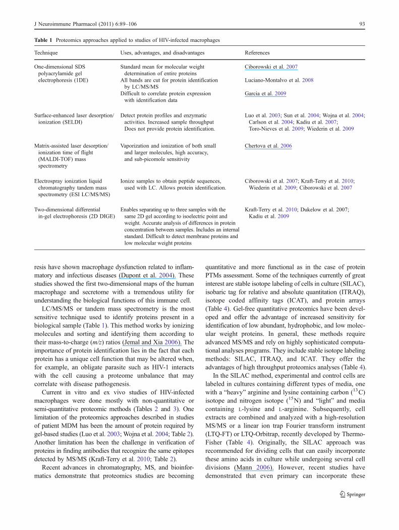
Table 1 Proteomics approaches applied to studies of HIV-infected macrophages
Technique Uses, advantages, and disadvantages References
One-dimensional SDSpolyacrylamide gelelectrophoresis (1DE)
Standard mean for molecular weightdetermination of entire proteins
Ciborowski et al. 2007
All bands are cut for protein identificationby LC/MS/MS
Luciano-Montalvo et al. 2008
Difficult to correlate protein expressionwith identification data
Garcia et al. 2009
Surface-enhanced laser desorption/ionization (SELDI)
Detect protein profiles and enzymaticactivities. Increased sample throughputDoes not provide protein identification.
Luo et al. 2003; Sun et al. 2004; Wojna et al. 2004;Carlson et al. 2004; Kadiu et al. 2007;Toro-Nieves et al. 2009; Wiederin et al. 2009
Matrix-assisted laser desorption/ionization time of flight(MALDI-TOF) massspectrometry
Vaporization and ionization of both smalland larger molecules, high accuracy,and sub-picomole sensitivity
Chertova et al. 2006
Electrospray ionization liquidchromatography tandem massspectrometry (ESI LC/MS/MS)
Ionize samples to obtain peptide sequences,used with LC. Allows protein identification.
Ciborowski et al. 2007; Kraft-Terry et al. 2010;Wiederin et al. 2009; Ciborowski et al. 2007
Two-dimensional differentialin-gel electrophoresis (2D DIGE)
Enables separating up to three samples with thesame 2D gel according to isoelectric point andweight. Accurate analysis of differences in proteinconcentration between samples. Includes an internalstandard. Difficult to detect membrane proteins andlow molecular weight proteins
Kraft-Terry et al. 2010; Dukelow et al. 2007;Kadiu et al. 2009
J Neuroimmune Pharmacol (2011) 6:89–106 93
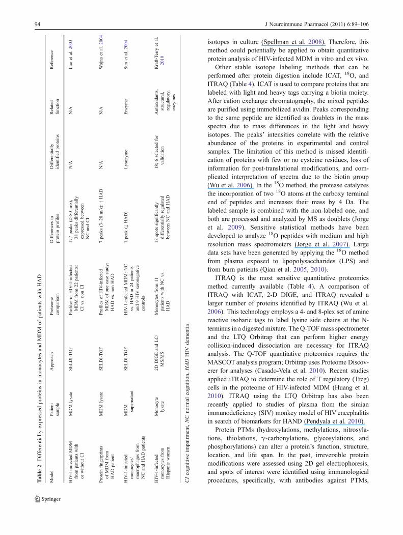
isotopes in culture (Spellman et al. 2008). Therefore, thismethod could potentially be applied to obtain quantitativeprotein analysis of HIV-infected MDM in vitro and ex vivo.
Other stable isotope labeling methods that can beperformed after protein digestion include ICAT, 18O, andITRAQ (Table 4). ICAT is used to compare proteins that arelabeled with light and heavy tags carrying a biotin moiety.After cation exchange chromatography, the mixed peptidesare purified using immobilized avidin. Peaks correspondingto the same peptide are identified as doublets in the massspectra due to mass differences in the light and heavyisotopes. The peaks’ intensities correlate with the relativeabundance of the proteins in experimental and controlsamples. The limitation of this method is missed identifi-cation of proteins with few or no cysteine residues, loss ofinformation for post-translational modifications, and com-plicated interpretation of spectra due to the biotin group(Wu et al. 2006). In the 18O method, the protease catalyzesthe incorporation of two 18O atoms at the carboxy terminalend of peptides and increases their mass by 4 Da. Thelabeled sample is combined with the non-labeled one, andboth are processed and analyzed by MS as doublets (Jorgeet al. 2009). Sensitive statistical methods have beendeveloped to analyze 18O peptides with medium and highresolution mass spectrometers (Jorge et al. 2007). Largedata sets have been generated by applying the 18O methodfrom plasma exposed to lipopolysaccharides (LPS) andfrom burn patients (Qian et al. 2005, 2010).
ITRAQ is the most sensitive quantitative proteomicsmethod currently available (Table 4). A comparison ofITRAQ with ICAT, 2-D DIGE, and ITRAQ revealed alarger number of proteins identified by ITRAQ (Wu et al.2006). This technology employs a 4- and 8-plex set of aminereactive isobaric tags to label lysine side chains at the N-terminus in a digestedmixture. The Q-TOFmass spectrometerand the LTQ Orbitrap that can perform higher energycollision-induced dissociation are necessary for ITRAQanalysis. The Q-TOF quantitative proteomics requires theMASCOT analysis program; Orbitrap uses Proteome Discov-erer for analyses (Casado-Vela et al. 2010). Recent studiesapplied iTRAQ to determine the role of T regulatory (Treg)cells in the proteome of HIV-infected MDM (Huang et al.2010). ITRAQ using the LTQ Orbitrap has also beenrecently applied to studies of plasma from the simianimmunodeficiency (SIV) monkey model of HIV encephalitisin search of biomarkers for HAND (Pendyala et al. 2010).
Protein PTMs (hydroxylations, methylations, nitrosyla-tions, thiolations, γ-carbonylations, glycosylations, andphosphorylations) can alter a protein’s function, structure,location, and life span. In the past, irreversible proteinmodifications were assessed using 2D gel electrophoresis,and spots of interest were identified using immunologicalprocedures, specifically, with antibodies against PTMs,T
able
2Differentially
expressedproteins
inmonocytes
andMDM
ofpatientswith
HAD
Model
Patient
sample
Approach
Proteom
ecomparison
Differences
inproteinprofiles
Differentially
identifiedproteins
Related
functio
nReference
HIV-1-infectedMDM
from
patientswith
orwith
outCI
MDM
lysate
SELDI-TOF
Profilesof
HIV-1-infected
MDM
from
22patients:
CIvs.nonCI
177peaks(2–80m/z);
38peaksdifferentially
expressedbetween
NC
andCI
N/A
N/A
Luo
etal.2003
Protein
fingerprints
ofMDM
from
HAD
patient
MDM
lysate
SELDI-TOF
Profilesof
HIV-infected
MDM
ofonecase
study:
HAD
vs.nonHAD
7peaks(3–20m/z):↑HAD
N/A
N/A
Wojna
etal.2004
HIV-1-infected
monocytes/
macrophages
from
NC
andHAD
patients
MDM
supernatant
SELDI-TOF
HIV-1-infectedMDM:NC
vs.HAD
in24
patients
and9HIV
seronegativ
econtrols
1peak
(↓HAD)
Lysozym
eEnzym
eSun
etal.2004
HIV-1-infected
monocytes
from
Hispanicwom
en
Monocyte
lysate
2DDIG
EandLC/
MS/M
SMonocytes
from
11patientswith
NC
vs.
HAD
18spotssignificantly
differentially
regulated
betweenNCandHAD
18;6selected
for
valid
ation
Antioxidants,
structural,
regulatory,
enzymes
Kraft-Terry
etal.
2010
CIcogn
itive
impairment,NCno
rmal
cogn
ition
,HAD
HIV
dementia
94 J Neuroimmune Pharmacol (2011) 6:89–106

Tab
le3
Differentially
expressedproteins
inHIV-1
“invitro”
infected
macroph
ages
Model
Sam
ple
Approach
Proteom
ecomparison
Differences
inproteinprofiles
Differentially
identifiedproteins
Related
functio
nReference
Proteom
eof
HIV-1
ADA-infectedMDM
Celllysate
SELDI
HIV-1
ADA-infected
MDM
vs.control
Up-regulatio
n2peaksin
HIV-1-infectedMDM
58Structural,regulatory,
enzymes,HIV
proteins
Carlson
etal.2004
LC/M
S/M
S
HIV-1
NLAD8-infected
MDM
Supernatant
MALDI
HIV-1
NLAD8virions
produced
byinfected
MDM
253unique
proteins
>280
Cytoskeleton,
adhesion,
signaling,
intracellular
trafficking,
metabolism,
immuneresponse
Chertovaet
al.2006
LC/M
S/M
S
Cytoskeletaltransform
ation
ofHIV-1
ADA-infectedMDM
Supernatant
SELDI
HIV-1
ADA-infected
MDM
vs.control
21peaksup-or
downregulated
15Structural/cytoskeletal
Kadiu
etal.2007
LC/M
S/M
S
HIV-1
ADA-infectedMDM
inHBMEC
proteins
Celllysate
2DDIG
EHBMEC
co-culturedwith
controlMDM
vs.with
HIV-1
ADA
infected
MDM
161spotsupregulated
78Structural/cytoskeletal,
regulatory,redox,
enzymes,HIV
proteins
Ricardo-D
ukelow
etal.2007
LC/M
S/M
S47
spotsdownregulated
HIV-1
ADA-infected
MDM
secretom
eSupernatant
1Delectro-phoresis
HIV-1
ADA-infected
MDM
secretom
e9differentially
expressedproteins
110
Cytoskeletal,enzymes,
redox,
immunoregulation
Ciborow
skiet
al.2007
LC/M
S/M
S
Microglia
astrocytecrosstalk
Celllysate
DIG
EHIV-1
ADA-infected
microglia/absence
ofastrocytes
39spotsupregulated
14Structural,regulatory,enzymes
Wanget
al.2008
LC/M
S/M
S24
spotsdownregulated
Effectsof
HIV-1
ADA
inmonocyteplasma
mem
braneproteome
Celllysate
2DDIG
EHIV-1-infectedmonocytes
vs.control
67spotsupregulated
986
Structural/cytoskeletal,regulatory,
enzymes,o
xidativestress
Kadiu
etal.2009
LC/M
S/M
S172spotsdownregulated
MDM:uninfected
vs.
infected
with
HIV-1
SF162andprim
ary
isolates
from
patients
with
NCor
CI
Celllysate
SELDI
Control
vs.NC
2downregulated
NC:6
Structural,replication,
metabolism,signaling
Toro-N
ieveset
al.2009
LC/M
S/M
SControl
vs.CI
1downregulated
CI:20
Replication,
chem
otaxis,
proteintrafficking,
apoptosis,redox
1upregulated
Control
vs.SF162
1upregulated
SF-162
:7
Metabolism,replication,
viralassembly,
structural
NCvs.CI
1upregulated
Com
mon
toSF162
andCI:
3Structural,signaling
SF-162
vs.NC
3downregulated)
Com
mon
toNC
andCI:
2Metabolism,viralproteins
Com
mon
toSF162,
NC,andCI:
3Metabolism
Rat
microglia
andastrocytes
infected
with
VSV
recombinant
HIV-A
DA
Celllysate
2DDIG
EHIV-1-infectedmicroglia
inpresence
ofastrocytes
149upregulated
68Structural,regulatory,
enzymes
Wanget
al.2008
LC/M
S/M
S360downregulated
Monocytecelllin
e(THP-1)
infected
with
HIV-1
HTLV
IIIB
Celllysate
SILAC
Protein
profile
ofHIV-1-
infected
undifferentiated
monocytic
THP-1
cells
26HIV
vs.uninfected;9downregulated
and17
up-regulated
inHIV-infected
THP-1cells
651
Structural,regulatory,
inflam
mation,
signaling,
antib
iotic
Pathaket
al.2009
MS/M
S
J Neuroimmune Pharmacol (2011) 6:89–106 95
such as nitrotyrosine (Dalle-Donne et al. 2006). Now PTMscan be analyzed using proteomic approaches, mostly MS/MS, but other techniques are also used (Table 4). Cysteineoxidation and thiolation can be directly and easily detectedby MS approaches (Dalle-Donne et al. 2006). Numerousprotein activities depend on phosphorylation or dephos-phorylation, that can occur in serine, threonine, tyrosine,and histidine residues. Using databases, investigators canpredict possible phosphosites, but not all phosphosites arephysically available to be phosphorylated or they simplyare not used. Some useful databases for prediction ofphosphosites are PHOSIDA, a PTM database that canpredict phosphorylations, acetylations, and N-glycosylations,and Phospho.ELM, a database designed exclusively forphosphosite prediction in eukaryotes. Ibarrola et al. (2003)
used SILAC to quantitate the extent of phosphosites aswell as to identify and quantitate novel phosphorylationsites. Other techniques such as immobilized metalaffinity chromatography, which uses Ga3+ to enrichphosphopeptides from cell lysates and phosphoproteinisotope-coded solid-phase tag, which labels and enrichesphosphopeptides from complex mixtures, have also beenused for the proteomic analysis of phosphopeptides (Kotaet al. 2009). More recently MS/MS/MS or MS3 method-ologies have been applied to phophopeptide PTMs (Ulintzet al. 2009).
Another important feature of proteins is glycosylation.Proteins can be N- and/or O-glycosylated. N-gycosylationattaches to the nitrogen of asparagines or arginines; O-glycosylation attaches to the oxygen of serine, threonine,
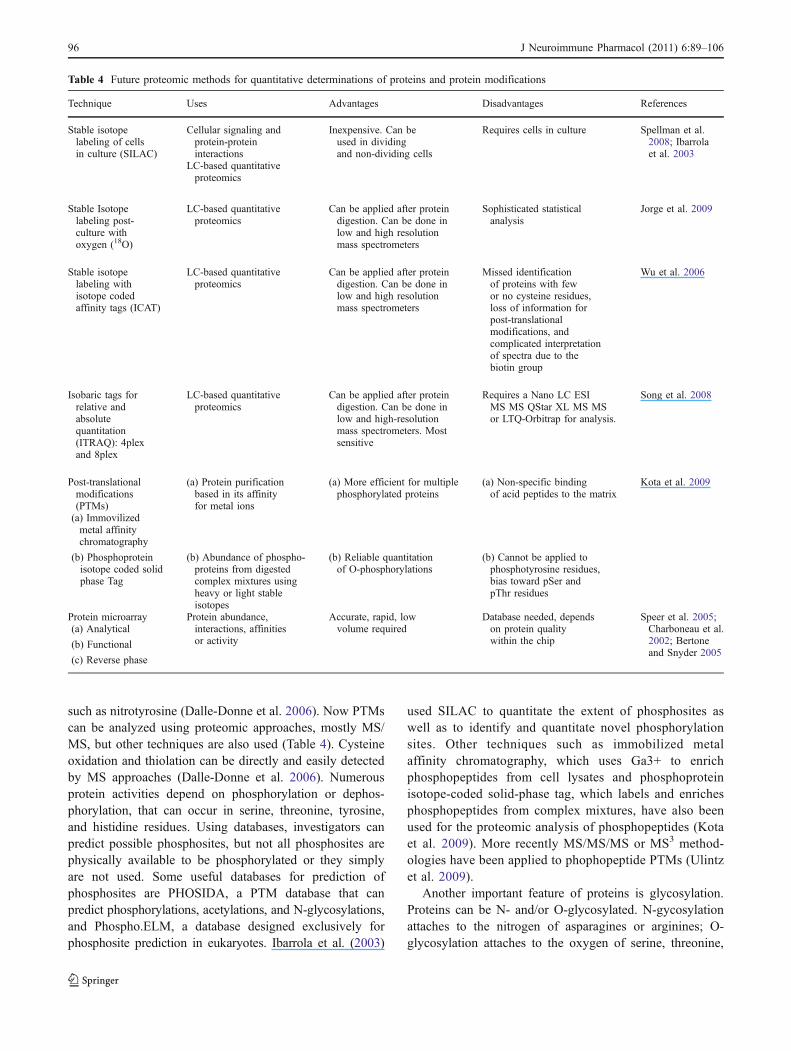
Table 4 Future proteomic methods for quantitative determinations of proteins and protein modifications
Technique Uses Advantages Disadvantages References
Stable isotopelabeling of cellsin culture (SILAC)
Cellular signaling andprotein-proteininteractions
Inexpensive. Can beused in dividingand non-dividing cells
Requires cells in culture Spellman et al.2008; Ibarrolaet al. 2003
LC-based quantitativeproteomics
Stable Isotopelabeling post-culture withoxygen (18O)
LC-based quantitativeproteomics
Can be applied after proteindigestion. Can be done inlow and high resolutionmass spectrometers
Sophisticated statisticalanalysis
Jorge et al. 2009
Stable isotopelabeling withisotope codedaffinity tags (ICAT)
LC-based quantitativeproteomics
Can be applied after proteindigestion. Can be done inlow and high resolutionmass spectrometers
Missed identificationof proteins with fewor no cysteine residues,loss of information forpost-translationalmodifications, andcomplicated interpretationof spectra due to thebiotin group
Wu et al. 2006
Isobaric tags forrelative andabsolutequantitation(ITRAQ): 4plexand 8plex
LC-based quantitativeproteomics
Can be applied after proteindigestion. Can be done inlow and high-resolutionmass spectrometers. Mostsensitive
Requires a Nano LC ESIMS MS QStar XL MS MSor LTQ-Orbitrap for analysis.
Song et al. 2008
Post-translationalmodifications(PTMs)
(a) Protein purificationbased in its affinityfor metal ions
(a) More efficient for multiplephosphorylated proteins
(a) Non-specific bindingof acid peptides to the matrix
Kota et al. 2009
(a) Immovilizedmetal affinitychromatography
(b) Phosphoproteinisotope coded solidphase Tag
(b) Abundance of phospho-proteins from digestedcomplex mixtures usingheavy or light stableisotopes
(b) Reliable quantitationof O-phosphorylations
(b) Cannot be applied tophosphotyrosine residues,bias toward pSer andpThr residues
Protein microarray Protein abundance,interactions, affinitiesor activity
Accurate, rapid, lowvolume required
Database needed, dependson protein qualitywithin the chip
Speer et al. 2005;Charboneau et al.2002; Bertoneand Snyder 2005
(a) Analytical
(b) Functional
(c) Reverse phase
96 J Neuroimmune Pharmacol (2011) 6:89–106
and tyrosine. Glycans can undergo changes from beingvery homogenous and simple to highly complex andheterogeneous structures. Because glycosylations areimmunogenic, making a glycosylation profile is potential-ly useful for HIV vaccine development. Glycosylation ofHIV proteins influences tropism, neutralization, viralinfectivity, and viral processing (Graham et al. 2008). Goet al. (2009) used a combination of MALDI-MS andLC/ESI-FTICR MS, in addition to reverse-phase HPLC, toseparate glycopeptides and to determine the glycosylationprofile of two clade C recombinant HIV envelopes.Graham et al. (2008) assessed the glycosylation in allHIVand SIV proteins using 2D DIGE and either ESI-MS/MSor LTQ-ion trap-MS/MS.
There are three different types of protein microarrays:analytical, functional, and reverse phase. Analytical micro-arrays are mostly used to detect affinity and expression(Table 4). Usually antibodies are attached to a microscopeslide to detect the protein or probe (Bertone and Snyder2005). Functional microarrays, which contain full-lengthfunctional proteins or domains, are used to study activity orinteractions: protein–protein, protein–RNA, protein–phos-pholipid, or protein–ligand (Bertone and Snyder 2005). Thereverse phase microarray uses denaturated protein lysatesfixed to the slide and then probed, whereas conventionalmicroarrays immobilize the antibodies or probes. Thereverse-phase technique has been used to detect eitherquantitative protein changes in healthy and diseased tissue(Charboneau et al. 2002) or PTMs (Speer et al. 2005).
Proteomics of monocytes, macrophages in HIVreplication
HIV-1 infection alters the proteome and secretome ofmacrophages (Tables 2 and 3). How HIV-1 affects theintracellular and secreted proteins is incompletely under-stood. Several studies have shown that shortly after theviral protein gp120 interacts with the CD4 and the seventransmembrane G protein-coupled receptors, CCR5- orCXCR4-specific signaling pathways such as the intracellularcalcium mobilization, PI-3K activation, phosphorylation ofmitogen-activated protein kinases (MAPKs), ERK1/2, JNK/SAPK, and p38 are engaged (Lee et al. 2005; Cicala et al.2002). This HIV-1 activation of MAPKs leads to up- anddownregulation of several genes and consequently influencesthe transcriptional and post-transcriptional events relatedto the formation of important cellular and viral proteins(Wahl et al. 2006). Consequently, the gp120-mediatedactivation of these protein kinases leads to the formationof pro-inflammatory cytokines and chemokines such asTNF-α and MCP-1, respectively, which can influenceHIV-1 replication and cell functions (Del Corno et al.
2001; Lee et al. 2003). In monocytes, one of the functionalconsequences of viral exposure is the facilitation ofprotein transformation from the cytosol to the plasmamembrane (Kadiu et al. 2009). The dysregulation ofmacrophage function is mediated by HIV envelopegp120 protein with cellular co-receptor activation thatresults in the inflammatory responses observed in HADpathogenesis (Gendelman et al. 2006). Some reports haveshown the involvement of CXCR4 in the gp120 macro-phage/microglia activation with the induction of pro-inflammatory cellular pathways that may contribute toAIDS dementia (Bezzi et al. 2001).
Microarray studies or MDM transcriptome have providedlarger data sets for pathway analysis than proteomics (Brownet al. 2008; Giri et al. 2009; Tsang et al. 2009; Ancuta etal. 2009; Van den Bergh et al. 2010). A macrophage pro-inflammatory M1 phenotype after HIV infection wasdemonstrated with increased mRNAs for calcium upregula-tion, cell cycle, apoptosis, MAPK, and cytokines/chemokines(Brown et al. 2008). Since many of the over-expressedmRNAs may not represent the final protein product owing todelays in translation, the authors were able to validate ∼18%of these proteins including calcium related and pro-apoptoticproteins (caspase 7). They concluded that HIV primesmacrophages to a pro-inflammatory M1 phenotype that isindependent of toll-like receptor activation (Brown et al.2008). However, subsequent studies on the HIV-infectedMDM transcriptome had the opposite result, an anti-inflammatory phenotype (Giri et al. 2009; Tsang et al.2009). Agreement with some aspects of these studies relatedto the mechanisms of evasion of innate immunity by HIVinteraction with MDM has been confirmed by proteomicsstudies, with the activation of oxidative stress associatedproteins, inflammation, and signaling cascades (Ciborowskiet al. 2007; Luciano-Montalvo and Meléndez 2009;Toro-Nieves et al. 2009). Moreover, some inflammatoryproteins identified from a large data set generated byquantitative proteomics from plasma exposed to LPS andfrom burn patients (Qian et al. 2005, 2010) are also presentin monocytes from HIV-infected women (Velazquez et al.2009; Kraft-Terry et al. 2010).
Proteomics of patient cells has spearhead researchtoward the diagnosis and treatment of HIV-related disease(Luo et al. 2003; Wojna et al. 2004; Kraft-Terry et al.2010). These studies include the discovery of proteins ascandidates for biomarkers for HIV associated cognitivedysfunction at different stages (Velazquez et al. 2009) aswell as other HIV-associated cellular changes and pathologies(Rasheed et al. 2009). The validation of the proteincandidates as biomarkers in different populations and thediscovery of novel biomarkers and possible targets fortherapies by more sensitive and quantitative proteomicsapproach remains to be studied.
J Neuroimmune Pharmacol (2011) 6:89–106 97

Proteomics of placental macrophages and MDM:a model of HIV persistence
The HIV-1 persistence in the monocyte–macrophagelineage has been explained by two theories: the lack ofproviral gene expression (latency) and continuous viralexpression without cytopathic effects (ongoing replication;Le Douce et al. 2010). Several traits of monocyte–macrophage lineage make it an important source of viralpersistence: the HIV-1 infection is generally non-lytic forthese cells; they can harbor viruses longer than CD4+ Tcells; and cells from monocyte–macrophage are moreresistant to cytopathic effects. For example, microglial cellsin the brain can produce viruses during their total lifespan(Williams et al. 2001). However, not all of the mononuclearphagocyte populations equally support viral growth. Theplacental macrophages (PM) are a target for HIV-1infection inside the placenta, but these cells are also barriersto primary infection by HIV-1. They can be productivelyinfected by laboratory strains and clinical isolates (Fear etal. 1998; Kesson et al. 1993, 1994; McGann et al. 1994;Meléndez-Guerrero et al. 1994; Meléndez et al. 2001;Plaud-Valentin et al. 1999), but they are less susceptible toHIV-1 than are MDM. Although PM express lower levelsof HIV-1 receptor CD4 and co-receptors CCR5 andCXCR4 than do MDM, the receptor expression is not thesole determinants of HIV replication in these macrophages(Luciano-Montalvo et al. 2008; Torres et al. 2001;Melendez et al. 2001). In contrast to the consistent levelsof replication exhibited by MDM, HIV replication in PMreaches a peak of viral replication around 3–7 days afterinfection and then decreases (Kesson et al. 1994). Further-more, levels of replication in PM measured by production ofHIV-1 viral capsid protein (p24) are ten times lower than inMDM (Plaud-Valentin et al. 1999), a finding that cannot beexplained solely by co-receptor expression. The HIV-1persistence in MDM and the low permissiveness exhibitedby PM make them good candidates for the study of cellularfactors involved in restriction of viral replication.
Proteomics platforms including SELDI-TOF, 1D SDSPAGE, and LC/MS/MS have been used as initial steps toidentify protein candidates associated with HIV-1 restrictionin PM and persistence in MDM. Twenty-seven protein peaksdifferentially expressed between uninfected and infected PMand MDM cell lysates were identified by SELDI-TOF, and 12were correlated with proteins identified by LC/MS/MS(Table 5; Luciano-Montalvo et al. 2008). Proteins indentifiedincluded: profilin, protein S-100 A9 (calgranulin B), SH3glutamic acid rich-like protein 1, SOD, and cystatin B(Table 5). Profilin and protein S-100 A9 (calgranulin B) havebeen reported to be associated with HIV-1 infection in bothmacrophages and T cells (Chertova et al. 2006; Kadiu et al.2007; Ryckman et al. 2002). Differences between PM and
MDM cystatin B levels detected byWestern blots and LC/MS/MS, correlated well and showed the most significant differencewith SELDI-TOF protein peaks in uninfected and HIV-1-infected PM, as compared with MDM (Luciano-Montalvo etal. 2008). Cystatin B is a cysteine protease inhibitor identifiedas an important protein related to HIV-1 replication inmacrophages. A low level of cystatin B has been associatedwith restriction of HIV in PM (Luciano-Montalvo et al. 2008)and most recently in microglia (Rodriguez-Franco et al.2010), whereas a high level of cystatin B in MDM isassociated with HIV persistence (Luciano-Montalvo et al.2008). Cystatin B was found upregulated at 12 days postinfection in MDM and PM (Luciano-Montalvo et al. 2008).Cystatin B was found over-expressed in the secretome ofHIV-1-infected MDM (Ciborowski et al. 2007; Garcia et al.2009), suggesting that this protein is linked to virus infection.This link was further demonstrated by showing decreasedHIV replication in MDM treated with siRNA against cystatinB (Luciano-Montalvo et al. 2008). The signaling mechanismsfor cystatin B in HIV replication are related to its interactionwith signal transducer and activator of transcription-1(STAT-1) as confirmed by co-immunoprecipitation assays(Luciano-Montalvo and Meléndez 2009). It is known thatSTAT-1 activates HIV-1 replication, but the high levels oftyrosine phosphorylation (PY) have been associated withHIV-1 inhibitory activity (Chang et al. 2002). Recent studiesat our laboratories show a greater expression of STAT-1 PYin PM than in MDM (Luciano-Montalvo and Meléndez2009). However, there are conflicting reports regardingSTAT-1 phosphorylation and HIV infection. It has beenreported that HIV infection in MDM induces an increase inSTAT-1PY that starts after 6 days of infection (Magnani et al.2003). However, recent data of our laboratory usingmonoclonal antibodies to increase specificity showed verylow levels of STAT-1PY in MDM until 12 days afterinfection and higher levels in HIV-infected PM than inHIV-infected MDM (Luciano-Montalvo and Meléndez2009). The low levels of cystatin B associated with highlevels of STAT-1PY in placental macrophages suggest amechanism for restriction of HIV replication with anactivation of a tyrosine kinase that promotes STAT-1PY.Since STAT-1PY701 induces interferon regulatory factor,this results in decreased replication of HIV-1 byinterfering with its long terminal repeat-driven replication(Luciano-Montalvo and Meléndez 2009). Modulation ofIFN response was also observed in microarray studies of 2-and 7-day infected MDM with a shift from elevated todecreased IFN α/β responses in HIV-infected MDMs (Brownet al. 2008). Recent immunoprecipitation studies in HIV-infected macrophages followed by LC/MS/MS studies andluciferase assays has revealed possible mechanisms ofcystatin B interacting proteins in the inhibition of IFNresponses (Rivera et al. 2010). Indeed, other IFN response
98 J Neuroimmune Pharmacol (2011) 6:89–106

inhibitors have also been reported in studies from SIV-infected macaques (Akhtar et al. 2010). However, upregula-tion of an interferon-stimulated gene 15, an ubiquitin-likeprotein involved in interferon-mediated antiviral immunitywas recently found in Treg-MDM co-cultures with novelproteomic approaches (ITRAQ) indicating a modulation ofIFN responses in MDM by T-regs (Huang et al. 2010).Taken together, these proteomics studies have deepened ourcurrent understanding of pathways in HIV-infected MDM,and thus could provide better targets for elimination of HIVreservoirs in the future.
Proteomics platforms were also used to identify secretedfactors that might be associated with HIV-1 restriction in PMand HIV persistence inMDM. The screening of the secretomeusing SELDI-TOF followed by LC/MS/MS confirmed thatPM and MDM were secreting different proteins (Garcia et al.2009). After sequencing and identification, several proteinswere validated for differential expression in PM and MDMby Western blot analysis. This study reported that peroxir-edoxin 5 is significantly more abundant in PM than in MDMsupernatants. This protein is important in the cellularantioxidant mechanisms, and other members of its familyhave shown antiviral activity. Furthermore, peroxiredoxin 5has been found to negatively regulate TNF-α signaling, andso it could also suppress NF-κB activity and HIV-1replication in PM (Garcia et al. 2009). Cystatin B was also
found to be significantly more abundant in MDM than inPM supernatants (Garcia et al. 2009). These data confirmedthe higher levels of cystatin B in MDM cell lysates reportedby Luciano-Montalvo et al. (2008). We suggested that highexpression of peroxiredoxin 5 could be one of the mechanismsby which HIV-1 replicates inefficiently in placental macro-phages, whereas low expression of cystatin B could impairtheir capacity to replicate HIV-1, but the mechanisms remainunder investigation (Luciano-Montalvo et al. 2008; Luciano-Montalvo and Meléndez 2009; Garcia et al. 2009). Interest-ingly, peroxiredoxin 5 was one of the proteins identified asdownregulated in recent proteomics studies of monocytesderived from women with HAND, a finding that suggestschronically activated monocytes act as a Trojan horse thatcarries HIV to the brain (Kraft-Terry et al. 2010).
Taken together, proteomics approaches have been usedto elucidate the proteome and secretome of tissue macro-phages in order to determine how these new players interactwith proteins related to HIV-1 replication.
Proteomics of monocytes, macrophages, and microgliain HIV dementia
After HIV-1 enters the CNS, either as a cell-free virus or ininfected macrophages, several signaling pathways are
Table 5 Intracellular and secreted proteins associated with HIV restriction in Placental Macrophages as compared with MDM
Protein name Total peptides detectedin sequencing
Peak intensity in SELDI-TOFcompared with MDM
Reference
MDM PM MDM HIV PM HIV PM PM HIV
From whole cells lysates:
Cytoskeletal 14-like protein ND 2 ND ND ↓ ↓ Luciano-Montalvo et al. 2008
SH3 glutamic acid rich like protein 3 2 ND ND ND ↓ Luciano-Montalvo et al. 2008
Protein S-100 A8 (calgranulin A) ND ND 2 ND ↓ ↓ Luciano-Montalvo et al. 2008
10 kDa heat shock protein 2 2 2 2 ↓ ↓ Luciano-Montalvo et al. 2008
Cystatin B 4 2 4 3 ↓ ↓ Luciano-Montalvo et al. 2008
Cytochrome C 2 2 2 2 ↓ ↓ Luciano-Montalvo et al. 2008
SH3 glutamic acid rich like protein 1 4 ND 4 ND ↑ ↑ Luciano-Montalvo et al. 2008
Myotrophin ND 2 2 ND ↑ ↑ Luciano-Montalvo et al. 2008
Protein S-100 A8 (calgranulin B) ND ND 2 2 ↑ ↑ Luciano-Montalvo et al. 2008
Galectin-1 ND ND 2 ND ↑ ↑ Luciano-Montalvo et al. 2008
Profilin 5 6 5 5 ↓ ↓ Luciano-Montalvo et al. 2008
Superoxide dismutase CuZn 2 ND ND 2 ↑ ↑ Luciano-Montalvo et al. 2008
From macrophage secretome:
Fatty acid-binding protein 3 2 1 NE NE ↑ NE Garcia et al. 2009
FKBP12 NE NE ↑ NE Garcia et al. 2009
Thioredoxin 2 2 NE NE ↑ NE Garcia et al. 2009
Peroxiredoxin 5 ND 3 NE NE ↑ NE Garcia et al. 2009
PM placental macrophages, ND not determined, NE not examined
J Neuroimmune Pharmacol (2011) 6:89–106 99

activated with the release of inflammatory products,chemokines, and neurotoxins (cellular and viral) that attractother cells to the site of infection, resulting in an increase ofintra neuronal calcium and subsequently producing neuronaldeath (Gendelman et al. 1997; Li et al. 2005; Persidsky et al.1997). This damage results in psychomotor slowing,memory impairment, and brain atrophy (Anderson et al.2002). In fact, apoptosis of neurons and astrocytes has beendetected in brain tissue autopsies from AIDS patients withHAD (Adle-Biassette et al. 1995).
The communication between the nervous and theperipheral immune systems is an important pathway foractivation of macrophages during HAD (Ballabh et al.2004). The response of the CNS to systemic immunechallenge results in brain inflammation caused by infectedmonocytes/macrophages trafficking into the brain from theperiphery, producing disruption of the BBB by oxidativeproteins (Gonzalez-Scarano and Martin-Garcia 2005).
Luo et al. (2003) identified several protein peak differ-ences between uninfected and HIV-1-infected MDM linkedto the pathogenesis of HAD. We identified 177 proteinpeaks in lysates of HIV-1-infected MDM from Hispanicpatients receiving highly active antiretroviral treatment,among which 38 peaks were related to cognitive impair-ment (Table 2). These data supported the hypothesis of theemergence of a monocyte subset in patients at the onset ofdementia. Afterwards, our collaborators examined theproteome of HIV-infected MDM (Carlson et al. 2004).They used SELDI-TOF to detect 58 differentially expressedproteins in MDM after in vitro infection with HIV-1 ADA,a macrophage tropic R5 virus (Gendelman et al. 1988)(Table 3). Microsequencing of these proteins by LC/MS/MS permitted identification of important cellular proteinssuch as ß-actin, annexin A5, vimentin, L-plastin, anddesmoyokin, in addition to viral proteins such as gp120and vif (Table 3). The fact that many of these proteins arerelated to changes in cellular structure and functionssupports the hypothesis of alterations in the cell after HIVinfection (Carlson et al. 2004). Sun et al. (2004) used thesame approach to compare profiles of secreted MDMproteins from HIV-seropositive individuals with normalcognition, HAD, and HIV-negative controls. This groupfound a decrease in secretion of lysozyme in MDMsupernatants from individuals with HAD, thus demonstrat-ing alterations in cell function as a consequence of HAD(Table 2).
The SELDI-TOF approach was used initially to profilethe defining characteristics of disease progression (Luo etal. 2003; Wojna et al. 2004). However, the spectradeveloped with this approach were not comparable amonginstruments at various institutions and did not allow us toidentify the proteins that were differentially expressed.Therefore, identification of the proteome was the next
approach to be used, first by 1D gel electrophoresis andsequencing by LC/MS/MS. Using this approach, studiesaimed at deciphering the effect of HIV variants frompatients with cognitive impairment on the macrophageproteome, our group identified 20 proteins related toapoptosis, chemotaxis, inflammation, and redox metabo-lism (Toro-Nieves et al. 2009; Table 3). Some of theidentified proteins in HIV-infected MDM, including ferri-tin, ubiquitin, and apoptosis-related proteins, have beenidentified in macrophage-induced inflammation (Xue et al.2008). These studies showed that the macrophage’sproteome can change depending on the infecting viralstrain by stimulating an inflammatory and pro-apoptoticphenotype. The mechanisms by which these viruses affect themacrophage proteome are currently being investigated.
After HIV-1 infects macrophages, several signals areactivated causing morphological and functional changes tothe cell (Chertova et al. 2006; Ciborowski et al. 2007;Kadiu et al. 2007; Lee et al. 2003). By using SELDI-TOF,LC/MS/MS, and Western blots, culture fluids derived fromin vitro HIV-1-infected MDM identified 15 differentiallyexpressed proteins, including cytoskeletal proteins (Kadiuet al. 2007; Table 3). Their findings demonstrated HIV-1can drive the cell to a permissive state for viral replicationor can enhance phagocytosis and intracellular microbialkilling. This group subsequently demonstrated how HIV-1transforms the monocyte plasma membrane proteome bytheir use of cell surface labeling with fluorescent dyesfollowed by 2D DIGE and LC/MS/MS analysis (Kadiu etal. 2009). They found that 53% of HIV-1-induced proteinswere associated with the plasma membrane, cellularactivation, and oxidative stress, which are processes relatedto HAD neuropathogenesis (Table 3). Using a similarproteomics platform, Ricardo-Dukelow et al. (2007) estab-lished how HIV-1-infected MDM induced upregulation of161 human brain microvascular endothelial cell proteinsthat are related to important cellular processes such asmetabolism, transport, structural changes, and regulation.Their findings support the role of HIV-1-infected MDM inBBB dysfunction, which contribute to HAD (Table 3).
Despite all the confirmed information about the interactionsbetween HIV-1 and macrophages, the exact pathways ofmacrophage activation after HIV-1 infection remain incom-pletely understood. Using proteomics, other studies haveshown that the macrophage secretome is affected by HIV-1infection (Ciborowski et al. 2007) where cystatins B andC, L-plastin, superoxide dismutase, and α-enolase wereidentified preferentially from HIV-infected cells (Table 3).
Taken together, proteomics studies clearly demonstratethe effects of HIV-1 on the macrophage activation,structure, and function. These results are not distant frommicroarray studies where an inflammatory M1 HIVmacrophage predominates following HIV infection with
100 J Neuroimmune Pharmacol (2011) 6:89–106
activation of apoptotic signaling that is modulated aftertime in culture (Brown et al. 2008).
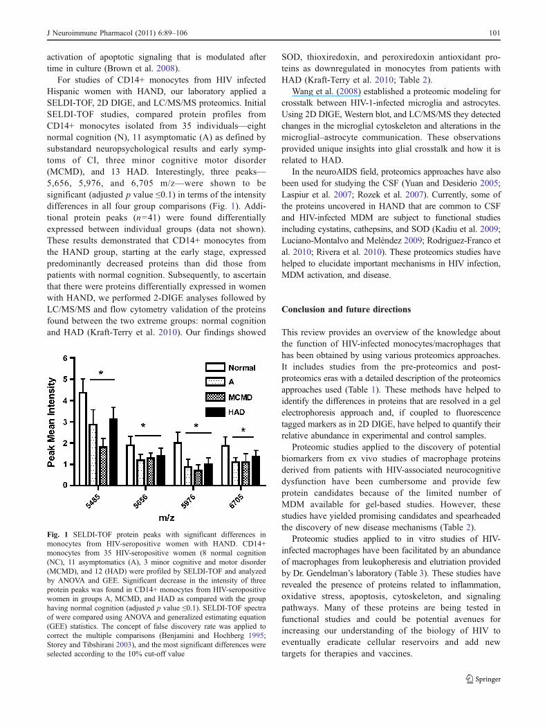
For studies of CD14+ monocytes from HIV infectedHispanic women with HAND, our laboratory applied aSELDI-TOF, 2D DIGE, and LC/MS/MS proteomics. InitialSELDI-TOF studies, compared protein profiles fromCD14+ monocytes isolated from 35 individuals—eightnormal cognition (N), 11 asymptomatic (A) as defined bysubstandard neuropsychological results and early symp-toms of CI, three minor cognitive motor disorder(MCMD), and 13 HAD. Interestingly, three peaks—5,656, 5,976, and 6,705 m/z—were shown to besignificant (adjusted p value ≤0.1) in terms of the intensitydifferences in all four group comparisons (Fig. 1). Addi-tional protein peaks (n=41) were found differentiallyexpressed between individual groups (data not shown).These results demonstrated that CD14+ monocytes fromthe HAND group, starting at the early stage, expressedpredominantly decreased proteins than did those frompatients with normal cognition. Subsequently, to ascertainthat there were proteins differentially expressed in womenwith HAND, we performed 2-DIGE analyses followed byLC/MS/MS and flow cytometry validation of the proteinsfound between the two extreme groups: normal cognitionand HAD (Kraft-Terry et al. 2010). Our findings showed
SOD, thioxiredoxin, and peroxiredoxin antioxidant pro-teins as downregulated in monocytes from patients withHAD (Kraft-Terry et al. 2010; Table 2).
Wang et al. (2008) established a proteomic modeling forcrosstalk between HIV-1-infected microglia and astrocytes.Using 2D DIGE, Western blot, and LC/MS/MS they detectedchanges in the microglial cytoskeleton and alterations in themicroglial–astrocyte communication. These observationsprovided unique insights into glial crosstalk and how it isrelated to HAD.
In the neuroAIDS field, proteomics approaches have alsobeen used for studying the CSF (Yuan and Desiderio 2005;Laspiur et al. 2007; Rozek et al. 2007). Currently, some ofthe proteins uncovered in HAND that are common to CSFand HIV-infected MDM are subject to functional studiesincluding cystatins, cathepsins, and SOD (Kadiu et al. 2009;Luciano-Montalvo and Meléndez 2009; Rodriguez-Franco etal. 2010; Rivera et al. 2010). These proteomics studies havehelped to elucidate important mechanisms in HIV infection,MDM activation, and disease.
Conclusion and future directions
This review provides an overview of the knowledge aboutthe function of HIV-infected monocytes/macrophages thathas been obtained by using various proteomics approaches.It includes studies from the pre-proteomics and post-proteomics eras with a detailed description of the proteomicsapproaches used (Table 1). These methods have helped toidentify the differences in proteins that are resolved in a gelelectrophoresis approach and, if coupled to fluorescencetagged markers as in 2D DIGE, have helped to quantify theirrelative abundance in experimental and control samples.
Proteomic studies applied to the discovery of potentialbiomarkers from ex vivo studies of macrophage proteinsderived from patients with HIV-associated neurocognitivedysfunction have been cumbersome and provide fewprotein candidates because of the limited number ofMDM available for gel-based studies. However, thesestudies have yielded promising candidates and spearheadedthe discovery of new disease mechanisms (Table 2).
Proteomic studies applied to in vitro studies of HIV-infected macrophages have been facilitated by an abundanceof macrophages from leukopheresis and elutriation providedby Dr. Gendelman’s laboratory (Table 3). These studies haverevealed the presence of proteins related to inflammation,oxidative stress, apoptosis, cytoskeleton, and signalingpathways. Many of these proteins are being tested infunctional studies and could be potential avenues forincreasing our understanding of the biology of HIV toeventually eradicate cellular reservoirs and add newtargets for therapies and vaccines.
Fig. 1 SELDI-TOF protein peaks with significant differences inmonocytes from HIV-seropositive women with HAND. CD14+monocytes from 35 HIV-seropositive women (8 normal cognition(NC), 11 asymptomatics (A), 3 minor cognitive and motor disorder(MCMD), and 12 (HAD) were profiled by SELDI-TOF and analyzedby ANOVA and GEE. Significant decrease in the intensity of threeprotein peaks was found in CD14+ monocytes from HIV-seropositivewomen in groups A, MCMD, and HAD as compared with the grouphaving normal cognition (adjusted p value ≤0.1). SELDI-TOF spectraof were compared using ANOVA and generalized estimating equation(GEE) statistics. The concept of false discovery rate was applied tocorrect the multiple comparisons (Benjamini and Hochberg 1995;Storey and Tibshirani 2003), and the most significant differences wereselected according to the 10% cut-off value
J Neuroimmune Pharmacol (2011) 6:89–106 101
Novel system biology and proteomics approaches forquantitative proteomics have been reviewed (Table 4).These methods will likely lead to more complex proteomesof HIV-infected MDM to facilitate the discovery ofincreased number of biomarkers for diagnosis and therapiesagainst HAND and other infectious and neuroinflammatorydiseases, which will further the understanding of the role ofmacrophages in different diseases.
Acknowledgments This work was supported in part by grants fromthe National Institutes of Health to Dr. Melendez (R01MH083516,U54NS4301, GM08224, G12RR03051) and to Krystal Colon andEillen Rodriguez-Franco (GM061838) and by institutional funds. Wethank Alexandra Cabán for her help in manuscript writing, sponsoredby Melendez R01MH083516.
Conflict of interest disclosure The authors report no conflicts ofinterest.
Open Access This article is distributed under the terms of theCreative Commons Attribution Noncommercial License which per-mits any noncommercial use, distribution, and reproduction in anymedium, provided the original author(s) and source are credited.
References
Adle-Biassette H, Levy Y, Colombel M, Poron F, Natchev S, KeohaneC, Gray F (1995) Neuronal apoptosis in HIV infection in adults.Neuropathol Appl Neurobiol 21:218–227
Agasalda MA, Wojna V, Melendez L, Shiramizu B (2010) CSFmonocyte reservoirs in HAND development and the possible roleof insertional mutagenesis. J Neurovirol 16S1:2
Akhtar LN, Qin H, Muldowney MT, Yanagisawa LL, Kutsch O,Clements JE, Benveniste EN (2010) Suppressor of cytokinesignaling 3 inhibits antiviral IFN-beta signaling to enhance HIV-1replication in macrophages. J Immunol 185:2393–2404
Alexaki A, Wigdahl B (2008) HIV-1 infection of bone marrowhematopoietic progenitor cells and their role in trafficking andviral dissemination. PLoS Pathog 4:e1000215
Alkhatib G, Combadiere C, Broder CC, Feng Y, Kennedy PE, MurphyPM, Berger EA (1996) CC CKR5: a RANTES, MIP-1alpha,MIP-1beta receptor as a fusion cofactor for macrophage-tropicHIV-1. Science 272:1955–1958
Ancuta P, Liu KY, Misra V, Wacleche VS, Gosselin A, Zhou X,Gabuzda D (2009) Transcriptional profiling reveals developmentalrelationship and distinct biological functions of CD16+ and CD16-monocyte subsets. BMC Genomics 10:403
Anderson E, Zink W, Xiong H, Gendelman HE (2002) HIV-1-associated dementia: a metabolic encephalopathy perpetrated byvirus-infected and immune-competent mononuclear phagocytes.J Acquir Immune Defic Syndr 31(Suppl 2):S43–S54
Bachis A, Cruz MI, Mocchetti I (2010) M-tropic HIVenvelope proteingp120 exhibits a different neuropathological profile than T-tropicgp120 in rat striatum. Eur J Neurosci 32:570–578
Bailey JR, Sedaghat AR, Kieffer T, Brennan T, Lee PK, Wind-RotoloM, Haggerty CM, Kamireddi AR, Liu Y, Lee J, Persaud D,Gallant JE, Cofrancesco J Jr, Quinn TC, Wilke CO, Ray SC,Siliciano JD, Nettles RE, Siliciano RF (2006) Residual humanimmunodeficiency virus type 1 viremia in some patients onantiretroviral therapy is dominated by a small number of
invariant clones rarely found in circulating CD4+ T cells. J Virol80:6441–6457
Ballabh P, Braun A, Nedergaard M (2004) The blood-brain barrier: anoverview: structure, regulation, and clinical implications. Neuro-biol Dis 16:1–13
Banks WA, Freed EO, Wolf KM, Robinson SM, Franko M, KumarVB (2001) Transport of human immunodeficiency virus type 1pseudoviruses across the blood-brain barrier: role of envelopeproteins and adsorptive endocytosis. J Virol 75:4681–4691
Benjamini Y and Hochberg Y (1995) Controlling the false discoveryrate: A practical and powerful approach to multiple testing. JRoyal Stat Soc. 57:289–300.
Berger EA, Doms RW, Fenyo EM, Krober BT, Littman DR, Moore JP,Sattentau QJ, Schuitemaker H, Sodroski J, Weiss RA (1998) Anew classififcation for HIV-1. Nature 391(6664):240
Bertone P, Snyder M (2005) Advances in functional protein micro-array technology. FEBS J 272:5400–5411.
Bezzi P, Domercq M, Brambilla L, Galli R, Schols D, De Clercq E,Vescovi A, Bagetta G, Kollias G, Meldolesi J, Volterra A (2001)CXCR4-activated astrocyte glutamate release via TNFalpha:amplification by microglia triggers neurotoxicity. Nat Neurosci4:702–710
Bieniasz PD, Cullen BR (1998) Chemokine receptors and humanimmunodeficiency virus infection. Front Biosci 3:d44–d58
Brown JN, Kohler JJ, Coberley CR, Sleasman JW, Goodenow MM(2008) HIV-1 activates macrophages independent of Toll-likereceptors. PLoS ONE 3:e3664
Casado-Vela J, Martínez-Esteso MJ, Rodriguez E, Borrás E, Elortza F,Bru-Martínez R (2010) iTRAQ-based quantitative analysis ofprotein mixtures with large fold change and dynamic range.Proteomics 10:343–347
Carlson KA, Ciborowski P, Schellpeper CN, Biskup TM, Shen RF, LuoX, Destache CJ, Gendelman HE (2004) Proteomic fingerprinting ofHIV-1-infected human monocyte-derived macrophages: a prelimi-nary report. J Neuroimmunol 147:35–42
Chang TL, Mosoian A, Pine R, Klotman ME, Moore JP (2002) Asoluble factor(s) secreted from CD8(+) T lymphocytes inhibitshuman immunodeficiency virus type 1 replication throughSTAT1 activation. J Virol 76:569–581
Charboneau L, Tory H, Chen T, Winters M, Petricoin EF 3rd, LiottaLA, Paweletz CP (2002) Utility of reverse phase protein arrays:applications to signalling pathways and human body arrays. BriefFunct Genomic Proteomic 1:305–315
Cheng-Mayer C, Homsy J, Evans LA, Levy JA (1988) Identificationof human immunodeficiency virus subtypes with distinct patternsof sensitivity to serum neutralization. Proc Natl Acad Sci USA85:2815–2819
Chertova E, Chertov O, Coren LV, Roser JD, Trubey CM, Bess JW Jr,Sowder RC 2nd, Barsov E, Hood BL, Fisher RJ, Nagashima K,Conrads TP, Veenstra TD, Lifson JD, Ott DE (2006) Proteomicand biochemical analysis of purified human immunodeficiencyvirus type 1 produced from infected monocyte-derived macro-phages. J Virol 80:9039–9052
Chun TW, Chadwick K, Margolick J, Siliciano RF (1997) Differentialsusceptibility of naive and memory CD4+ T cells to thecytopathic effects of infection with human immunodeficiencyvirus type 1 strain LAI. J Virol 71:4436–4444
Cicala C, Arthos J, Selig SM, Dennis G Jr, Hosack DA, Van Ryk D,SpanglerML, Steenbeke TD,Khazanie P, Gupta N, Yang J, DaucherM, Lempicki RA, Fauci AS (2002) HIVenvelope induces a cascadeof cell signals in non-proliferating target cells that favor virusreplication. Proc Natl Acad Sci USA 99:9380–9385
Ciborowski P, Kadiu I, Rozek W, Smith L, Bernhardt K, Fladseth M,Ricardo-Dukelow M, Gendelman HE (2007) Investigating thehuman immunodeficiency virus type 1-infected monocyte-derived macrophage secretome. Virology 363:198–209
102 J Neuroimmune Pharmacol (2011) 6:89–106

Collman R, Hassan NF, Walker R, Godfrey B, Cutilli J, Hasting JC,Friedman H, Douglas SD, Nathanson N (1989) Infection ofmonocyte-derived macrophages with human immunodeficiencyvirus type 1 (HIV-1). Monocyte-tropic and lymphocyte-tropicstrains of HIV-1 show distinctive patterns of replication in apanel of cell types. J Exp Med 170:1149–1163
Crowe S, Zhu T, Muller WA (2003) The contribution of monocyteinfection and trafficking to viral persistence, and maintenance ofthe viral reservoir in HIV infection. J Leukoc Biol 74:635–641
Davis LE, Hjelle BL, Miller VE, Palmer DL, Llewellyn AL, MerlinTL, Young SA, Mills RG, Wachsman W, Wiley CA (1992) Earlyviral brain invasion in iatrogenic human immunodeficiency virusinfection. Neurology 42:1736–1739
Dalle-Donne I, Aldini G, Carini M, Colombo R, Rossi R, Milzani A(2006) Protein carbonylation, cellular dysfunction, and diseaseprogression. J Cell Mol Med 10:389–406
Del Corno M, Liu QH, Schols D, de Clercq E, Gessani S, FreedmanBD, Collman RG (2001) HIV-1 gp120 and chemokine activationof Pyk2 and mitogen-activated protein kinases in primarymacrophages mediated by calcium-dependent, pertussis toxin-insensitive chemokine receptor signaling. Blood 98:2909–2916
Deng H, Liu R, Ellmeier W, Choe S, Unutmaz D, Burkhart M, Di MarzioP, Marmon S, Sutton RE, Hill CM, Davis CB, Peiper SC, Schall TJ,Littman TR, LandauNR (1996) Identification of a major co-receptorfor primary isolates of HIV-1. Nature 381:661–666
Doranz BJ, Rucker J, Yi Y, Smyth RJ, Samson M, Peiper SC,Parmentier M, Collman RG, Doms RW (1996) A dual-tropicprimary HIV-1 isolate that uses fusin and the beta-chemokinereceptors CKR-5, CKR-3, and CKR-2b as fusion cofactors. Cell85:1149–1158
Dreyer EB, Lipton SA (1995) The coat protein gp120 of HIV-1inhibits astrocyte uptake of excitatory amino acids via macrophagearachidonic acid. Eur J Neurosci 7:2502–2507
Duenas-Decamp MJ, Peters PJ, Repik A, Musich T, Gonzalez-PerezMP, Caron C, Brown R, Ball J, Clapham PR (2010) Variation inthe biological properties of the HIV-1 R5 envelope: implicationsof envelope structure, transmission and pathogenesis. FutureVirol 5(4):435–451
Dupont A, Tokarski C, Dekeyzer O, Guihot AL, Amouyel P, RolandoC, Pinet F (2004) Two-dimensional maps and databases of thehuman macrophage proteome and secretome. Proteomics4:1761–1778
Ellery PJ, Tippett E, Chiu YL, Paukovics G, Cameron PU, SolomonA, Lewin SR, Gorry PR, Jaworowski A, Greene WC, Sonza S,Crowe SM (2007) The CD16+ monocyte subset is morepermissive to infection and preferentially harbors HIV-1 in vivo.J Immunol 178:6581–6589
Embretson J, Zupancic M, Beneke J, Till M, Wolinsky S, Ribas JL,Burke A, Haase AT (1993) Analysis of human immunodeficiencyvirus-infected tissues by amplification and in situ hybridizationreveals latent and permissive infections at single-cell resolution.Nature 362:359–362
Fear WR, Kesson AM, Naif H, Lynch GW, Cunningham AL (1998)Differential tropism and chemokine receptor expression ofhuman immunodeficiency virus type 1 in neonatal monocytes,monocyte-derived macrophages, and placental macrophages. JVirol 72:1334–1344
Finzi D, Hermankova M, Pierson T, Carruth LM, Buck C, ChaissonRE, Quinn TC, Chadwick K, Margolick J, Brookmeyer R,Gallant J, Markowitz M, Ho DD, Richman DD, Siliciano RF(1997) Identification of a reservoir for HIV-1 in patients onhighly active antiretroviral therapy. Science 278:1295–1300
Fischer-Smith T, Bell C, Croul S, Lewis M, Rappaport J (2008)Monocyte/macrophages trafficking in acquired immunodeficiencysyndrome encephalitis: lessons from human and nonhuman primatestudies. J Neurovirol 14(4):318–326
Fischer-Smith T, Croul S, Sverstiuk AE, Capini C, L’Heureux D,Régulier EG, Richardson MW, Amini S, Morgello S, Khalili K,Rappaport J (2001) CNS invasion by CD14+/CD16+ peripheralblood-derived monocytes in HIV dementia: perivascular accu-mulation and reservoir of HIV infection. J Neurovirol 7:528–541
Fischer-Smith T, Rappaport J (2005) Evolving paradigms in thepathogenesis of HIV-1-associated dementia. Expert Rev MolMed 7:1–26
Garcia K, Garcia V, Perez Laspiur J, Duan F, Melendez LM (2009)Characterization of the placental macrophage secretome: impli-cations for antiviral activity. Placenta 30:149–155
Gartner S (2000) HIV infection and dementia. Science 287(5453):602–604
Gendelman H, Anderson E, Melendez L, Zheng J (2006) Chemokinesand their receptors in HIV-1 neuropathogenesis, In Vivo Modelsof HIV Disease and Control. Eds H. Friedman, S. Specter, andM. Bendinelli. Springer. pp 45–80
Gendelman HE, Baca LM, Husayni H, Turpin JA, Skillman D, KatlerDC, Burke DS, Tramont EC, Meltzer MS (1990) Macrophage–HIV interaction: viral isolation and target cell tropism. AIDS4:221–228
Gendelman HE, Orenstein JM, Martin MA, Ferrua C, Mitra R et al(1988) Efficient isolation and propagation of human immunode-ficiency virus on recombinant colony-stimulating factor 1-treatedmonocytes. J Exp Med 167:1428–1441
Gendelman HE, Persidsky Y, Ghorpade A, Limoges J, Stins M et al(1997) The neuropathogenesis of the AIDS dementia complex.AIDS 11(Suppl A):S35–S45
Genis P, Jett M, Bernton EW, Boyle T, Gelbard HA, Dzenko K, KeaneRW, Resnick L, Mizrachi Y, Volsky DJ (1992) Cytokines andarachidonic metabolites produced during human immunodefi-ciency virus (HIV)-infected macrophage–astroglia interactions:implications for the neuropathogenesis of HIV disease. J ExpMed 176:1703–1718
Gevaert K, Vandekerckhove J (2000) Protein identification methods inproteomics. Electrophoresis 21(6):1145–1154
Giulian D, Vaca K, Noonan CA (1990) Secretion of neurotoxins bymononuclear phagocytes infected with HIV-1. Science250:1593–1596
Giri MS, Nebozyhn M, Raymond A, Gekonge B, Hancock A, Creer S,Nicols C, Yousef M, Foulkes AS, Mounzer K, Shull J, SilvestriG, Kostman J, Collman RG, Showe L, Montaner LJ (2009)Circulating monocytes in HIV-1-infected viremic subjects exhibitan antiapoptosis gene signature and virus- and host-mediatedapoptosis resistance. J Immunol 182:4459–4470
Go EP, Chang Q, Liao HX, Sutherland LL, Alam SM, Haynes BF,Desaire H (2009) Glycosylation site-specific analysis of clade CHIV-1 envelope proteins. J Proteome Res 8:4231–4242
Gonzalez-Scarano F, Baltuch G (1999) Microglia as mediators ofinflammatory and degenerative diseases. Annu Rev Neurosci22:219–240
Gonzalez-Scarano F, Martin-Garcia J (2005) The neuropathogenesisof AIDS. Nat Rev Immunol 5:69–81
Goodenow MM, Collman RG (2006) HIV-1 co receptor preference isdistinct from target cell tropism: a dual-parameter nomenclatureto define viral phenotypes. J Leukoc Biol 80:965–972
Gorry PR, Churchill M, Crowe SM, Cunningham AL, Gabuzda D(2005) Pathogenesis of macrophage tropic HIV-1. Curr HIV Res3(1):53–60
Graham DR, Mitsak MJ, Elliott ST, Chen D, Whelan SA, Hart GW, VanEyk JE (2008) Two-dimensional gel-based approaches for theassessment of N-Linked and O-GlcNAc glycosylation in human andsimian immunodeficiency viruses. Proteomics 8:4919–4930
Hickey WF, Kimura H (1988) Perivascular microglial cells of theCNS are bone marrow-derived and present antigen in vivo.Science 239:290–292
J Neuroimmune Pharmacol (2011) 6:89–106 103

Hladik F, Lentz G, Akridge RE, Peterson G, Kelley H, McElroy A,McElrath MJ (1999) Dendritic cell–T cell interactions supportcoreceptor-independent human immonudeficiency virus type 1transmission in the human genital tract. J Virol 73(7):5833–5842
Huang X, Stone DK, Yu F, Zeng Y, Gendelman HE (2010) Functionalproteomic analysis for regulatory T cell surveillance of the HIV-1-infected macrophage. J Proteome Res (in press)
Ibarrola N, Kalume DE, Gronborg M, Iwahori A, Pandey A (2003) Aproteomic approach for quantitation of phosphorylation usingstable isotope labeling in cell culture. Anal Chem 75:6043–6049
Jaworski A, Smith CL, Burden SJ (2007) GA-binding protein isdispensable for neuromuscular synapse formation and synapse-specific gene expression. Mol Cell Biol 27:5040–5046
Jiang ZG, Piggee C, Heyes MP, Murphy C, Quearry B, Bauer M,Zheng J, Gendelman HE, Markey SP (2001) Glutamate is amediator of neurotoxicity in secretions of activated HIV-1-infected macrophages. J Neuroimmunol 117:97–107
Jemal M, Xia YQ (2006) LC-MS development strategies forquantitative bioanalysis. Curr Drug Metab 7:491–502
Jorge I, Casas EM, Villar M, Ortega-Pérez I, López-Ferrer D,Martínez-Ruiz A, Carrera M, Marina A, Martínez P, Serrano H,Cañas B, Were F, Gallardo JM, Lamas S, Redondo JM, García-Dorado D, Vázquez J (2007) High-sensitivity analysis of specificpeptides in complex samples by selected MS/MS ion monitoringand linear ion trap mass spectrometry: application to biologicalstudies. J Mass Spectrom 42:1391–1403
Jorge I, Navarro P, Martínez-Acedo P, Núñez E, Serrano H, AlfrancaA, Redondo JM, Vázquez J (2009) Statistical model to analyzequantitative proteomics data obtained by 18O/16O labeling andlinear ion trap mass spectrometry: application to the study ofvascular endothelial growth factor-induced angiogenesis inendothelial cells. Mol Cell Proteomics 8:1130–1149
Kadiu I, Ricardo-Dukelow M, Ciborowski P, Gendelman HE (2007)Cytoskeletal protein transformation in HIV-1-infected macro-phage giant cells. J Immunol 178:6404–6415
Kadiu I, Wang T, Schlautman JD, Dubrovsky L, Ciborowski P et al(2009) HIV-1 transforms the monocyte plasma membraneproteome. Cell Immunol 258:44–58
Kaul M, Garden GA, Lipton SA (2001) Pathways to neuronal injuryand apoptosis in HIV-associated dementia. Nature 410:988–994
Keele BF, Giorgi EE, Salazar-Gonzalez JF, Decker JM, Pham KT,Salazar MG, Sun C, Grayson T, Wang S, Li H, Wei X, Jiang C,Kirchherr JL, Gao F, Anderson JA, Ping LH, Swanstrom R,Tomaras GD, Blattner WA, Goepfert PA, Kilby JM, Saag MS,Delwart EL, Busch MP, Cohen MS, Montefiori DC, Haynes BF,Gaschen B, Athreya GS, Lee HY, Wood N, Seoighe C, PerelsonAS, Bhattacharya T, Korber BT, Hahn BH, Shaw GM (2008)Identification and characterization of transmitted and earlyfounder virus envelopes in primary HIV-1 infection. Proc NatlAcad Sci USA 105:7552–7557
Kesson AM, Fear WR, Kazazi F, Mathijs JM, Chang J et al (1993)Human immunodeficiency virus type 1 infection of humanplacental macrophages in vitro. J Infect Dis 168:571–579
Kesson AM, Fear WR, Williams L, Chang J, King NJ, CunninghamAL (1994) HIV infection of placental macrophages: theirpotential role in vertical transmission. J Leukoc Biol 56:241–246
Kim WK, Corey S, Alvarez X, Williams K (2003) Monocyte/macrophages traffic in HIV and SIV encephalitis. J Leukoc Biol74(5):650–656
Kim WK, Alvarez X, Fisher J, Bronfin B, Westmoreland S,McLaurin J, Williams K (2006) CD163 identifies perivascularmacrophages in normal and viral encephalitic brains andpotential precursors to perivascular macrophages in blood. AmJ Pathol 168:822–834
Kim WK, Avarez X, Williams K (2005) The role of monocytes andperivascular macrophages in HIV and SIV neuropathogenesis:
information from non-human primate models. Neurotox Res 8(1–2):107–115
Kota U, Chien KY, Goshe MB (2009) Isotope-labeling and affinityenrichment of phosphopeptides for proteomic analysis usingliquid chromatography-tandem mass spectrometry. Meth MolBiol 564:303–321
Kraft-Terry SD, Stothert AR, Buch S, Gendelman HE (2010) HIV-1neuroimmunity in the era of antiretroviral therapy. Neurobiol Dis37:542–548
Lambotte O, Taoufik Y, de Goer MG, Wallon C, Goujard C,Delfraissy JF (2000) Detection of infectious HIV in circulatingmonocytes from patients on prolonged highly active antiretroviraltherapy. J Acquir Immune Defic Syndr 23:114–119
Laspiur JP, Anderson ER, Ciborowski P, Wojna V, Rozek W, Duan F,Mayo R, Rodríguez E, Plaud-Valentín M, Rodríguez-Orengo J,Gendelman HE, Meléndez LM (2007) CSF proteomic finger-prints for HIV-associated cognitive impairment. J Neuroimmunol192:157–170
Lee C, Tomkowicz B, Freedman BD, Collman RG (2005) HIV-1gp120-induced TNF-{alpha} production by primary humanmacrophages is mediated by phosphatidylinositol-3 (PI-3) kinaseand mitogen-activated protein (MAP) kinase pathways. J LeukocBiol 78:1016–1023
Lee C, Liu QH, Tomkowicz B, Yi Y, Freedman BD, Collman RG(2003) Macrophage activation through CCR5- and CXCR4-mediated gp120-elicited signaling pathways. J Leukoc Biol 74(5):676–682
Le Douce V, Herbein G, Rohr O, Schwartz C (2010) Molecularmechanisms of HIV-1 persistence in the monocyte–macrophagelineage. Retrovirology 7:32
Li H, Bar KJ, Wang S, Decker JM, Chen Y, Sun C, Salazar-Gonzalez JF, Salazar MG, Learn GH, Morgan CJ, SchumacherJE, Hraber P, Giorgi EE, Bhattacharya T, Korber BT, PerelsonAS, Eron JJ, Cohen MS, Hicks CB, Haynes BF, MarkowitzM, Keele BF, Hahn BH, Shaw GM (2010) High multiplicityinfection by HIV-1 in men who have sex with men. PLoSPathog 13:e1000890
Li S, Juarez J, Alali M, Dwyer D, Collman R, Cunningham A, NaifHM (1999) Persistent CCR5 utilization and enhanced macro-phage tropism by primary blood human immunodeficiency virustype 1 isolates from advanced stages of disease and comparisonto tissue-derived isolates. J Virol 73:9741–9755
Li W, Galey D, Mattson MP, Nath A (2005) Molecular and cellularmechanisms of neuronal cell death in HIV dementia. NeurotoxRes 8:119–134
Link AJ, Labaer J (2009) In-gel trypsin digest of gel-fractionatedproteins. Cold Spring Harb Protoc. doi:10.1101/pdb.prot5110
Luciano-Montalvo C, Ciborowski P, Duan F, Gendelman HE,Melendez LM (2008) Proteomic analyses associate cystatin Bwith restricted HIV-1 replication in placental macrophages.Placenta 29:1016–1023
Luciano-Montalvo C, Meléndez LM (2009) Cystatin B associateswith signal transducer and activator of transcription 1 inmonocyte-derived and placental macrophages. Placenta30:464–467
Luo X, Carlson KA, Wojna V, Mayo R, Biskup TM, Stoner J,Anderson J, Gendelman HE, Meléndez LM (2003) Macrophageproteomic fingerprinting predicts HIV-1-associated cognitiveimpairment. Neurology 60:1931–1937
Maddon PJ, Dalgleish AG, McDougal JS, Clapham PR, Weiss RA, AxelR (1986) The T4 gene encodes the AIDS virus receptor and isexpressed in the immune system and the brain. Cell 47:333–348
Magnani M, Balestra E, Fraternale A, Aquaro S, Paiardini M et al(2003) Drug-loaded red blood cell-mediated clearance of HIV-1macrophage reservoir by selective inhibition of STAT1 expression. JLeukoc Biol 74:764–771
104 J Neuroimmune Pharmacol (2011) 6:89–106

Mann M (2006) Functional and quantitative proteomics using SILAC.Nat Rev Mol Cell Biol 7:952–958
McGann KA, Collman R, Kolson DL, Gonzalez-Scarano F, Coukos Get al (1994) Human immunodeficiency virus type 1 causesproductive infection of macrophages in primary placental cellcultures. J Infect Dis 169:746–753
Melendez-Guerrero LM, Nicholson JK, McDougal JS (1990) In vitroinfection of monocytes with HIVBa-L. Effect on cell surfaceexpression of CD4, CD14, HLA-DR, and HLA-DQ. AIDS ResHum Retroviruses 6:731–741
Melendez-Guerrero LM, Nicholson JK,McDougal JS (1991) Infection ofhuman monocytes with HIV-1Ba-L. Effect on accessory cellfunction for T-cell proliferation in vitro. AIDS Res HumRetroviruses 7(5):465–474
Meléndez-Guerrero L, Holmes R, Backé E, Polliotti B, Ibegbu C, LeeF, Panigel M, Schwartz D, Huddleston J, Nahmias A (1994) Invitro infection of Hofbauer cells with a monocyte-tropic strain ofHIV-1. Placenta 15(Supplement 1):33–45
Melendez J, Garcia V, Sanchez E, Delgado R, Torres G, Melendez-Guerrero LM. 2001. Is decreased HIV-1 infectivity of placentalmacrophages caused by high levels of beta-chemokines? CellMol Biol (Noisy-le-grand) 47 Online Pub:OL51-9.
Meltzer MS, Nakamura M, Hansen BD, Turpin JA, Kalter DC,Gendelman HE (1990) Macrophages as susceptible targets forHIV infection, persistent viral reservoirs in tissue, and keyimmunoregulatory cells that control levels of virus replicationand extent of disease. AIDS Res Hum Retroviruses 6:967–971
Morrison RS, Kinoshita JMD, Uo T, Ho JT, McBee JK, Conrads TP,Veenstra TD (2002) Proteomic analysis in the neurosciences. MolCell Proteomics 1:553–560
Narayan O, Wolinsky JS, Clements JE, Strandberg JD, Griffin DE,Cork LC (1982) Slow virus replication: the role of macrophagesin the persistence and expression of visna viruses of sheep andgoats. J Gen Virol 59:345–356
O’ Farrell PH (1975) High resolution two-dimensional electrophoresisof proteins. J Biol Chem 250:4007–4021
Pandey A, Mann M (2000) Proteomics to study genes and genomes.Nature 405:837–846
Pathak S, De Souza GA, Salte T, Wiker HG, Asjö B (2009) HIVinduces both a down-regulation of IRAK-4 that impairs TLRsignalling and an up-regulation of the antibiotic peptide dermci-din in monocytic cells. Scand J Immunol. 70:264–276.
Peluso R, Haase A, Stowring L, Edwards M, Ventura P (1985) ATrojan horse mechanism for the spread of visna virus inmonocytes. Virology 147:231–236
Persidsky Y, Buttini M, Limoges J, Bock P, Gendelman HE (1997) Ananalysis of HIV-1-associated inflammatory products in braintissue of humans and SCID mice with HIV-1 encephalitis. JNeurovirol 3:401–416
Persidsky Y, Ghorpade A, Rasmussen J, Limoges J, Liu XJ et al(1999) Microglial and astrocyte chemokines regulate mono-cyte migration through the blood–brain barrier in humanimmunodeficiency virus-1 encephalitis. Am J Pathol155:1599–1611
Peters PJ, Duenas-Decamp MJ, Sullivan WM, Clapham PR (2007)Variation of macrophage tropism among HIV-1 R5 envelopesin brain and other tissues. J Neuroimmune Pharmacol 2(1):32–41
Pendyala G, Trauger SA, Siuzdak G, Fox HS (2010) Quantitativeplasma proteomic profiling identifies the vitamin E bindingprotein afamin as a potential pathogenic factor in SIV inducedCNS disease. J Proteome Res 9:352–358
Plaud-Valentin M, Delgado R, Garcia V, Zorrilla C, Gandia J,Melendez-Guerrero LM (1999) HIV infection of placentalmacrophages: effect on the secretion of HIV stimulatorycytokines. Cell Mol Biol Noisy (Noisy-le-grand) 45:423–431
Popovic M, Mellert W, Erfle V, Gartner S (1988) Role of mononuclearphagocytes and accessory cells in human immunodeficiency virustype I infection of the brain. Ann Neurol 23(Suppl):S74–S77
Pulliam L, Gascon R, Stubblebine M, McGuire D, McGrath MS(1997) Unique monocyte subset in patients with AIDS dementia.Lancet 349:692–695
Qian WJ, Petritis BO, Kaushal A, Finnerty CC, Jeschke MG, MonroeME,Moore RJ, Schepmoes AA, XiaoW, Moldawer LL, Davis RW,Tompkins RG, Herndon DN, Camp DG 2nd, Smith RD, Inflam-mation and the Host Response to Injury Large Scale CollaborativeResearch Program (2010) Plasma proteome response to severe burninjury revealed by 18O-labeled “universal” reference-based quanti-tative proteomics. J Proteome Res 3:4779–4789
Qian WJ, Monroe ME, Liu T, Jacobs JM, Anderson GA, Shen Y,Moore RJ, Anderson DJ, Zhang R, Calvano SE, Lowry SF, XiaoW, Moldawer LL, Davis RW, Tompkins RG, Camp DG 2nd,Smith RD, Inflammation and the Host Response to Injury LargeScale Collaborative Research Program (2005) Quantitativeproteome analysis of human plasma following in vivo lipopoly-saccharide administration using 16O/18O labeling and theaccurate mass and time tag approach. Mol Cell Proteomics4:700–709
Rasheed S, Yan JS, Hussain A, Lai B (2009) Proteomic characteriza-tion of HIV-modulated membrane receptors, kinases and signal-ing proteins involved in novel angiogenic pathways. J TranslMed 7:75
Raymond S, Delobel P, Mavigner M, Cazabat M, Encinas S, SouyrisC, Bruel P, Sandres-Sauné K, Marchou B, Massip P, Izopet J(2010) CXCR4-using viruses in plasma and peripheral bloodmononuclear cells during primary HIV-1 infection and impact ondisease progression. AIDS 24:2305–2312
Ricardo-Dukelow M, Kadiu I, Rozek W, Schlautman J, Persidsky Y,Ciborowski P, Kanmogne GD, Gendelman HE (2007) HIV-1infected monocyte-derived macrophages affect the human brainmicrovascular endothelial cell proteome: new insights into blood-brain barrier dysfunction for HIV-1-associated dementia. JNeuroimmunol 185:37–46
Rivera LE, Perez Laspiur J, Medina F, Munoz-Jordan J, Melendez LM(2010) Inhibition of interferon response by Cystatin B: aproposed mechanism for HIV persistence in macropahge reser-voirs. J Neurovirol 16S1:71
Rodriguez-Franco E, Plaud M, Rome R, Skolasky R, Melendez L(2010) Late HIV infection modulates the expression and activityof Cathepsin B, and its inhibitors in macrophages: implications inneuropathogenesis. Retrovirology 7:10
Rozek W, Ricardo-Dukelow M, Holloway S, Gendelman HE, WojnaV, Melendez LM, Ciboroski P (2007) Cerebrospinal fluidproteomic profiling of HIV-1-infected patients with cognitiveimpairment. J Proteome Res 6(11):4189–4199
Ryckman C, Robichaud GA, Roy J, Cantin R, Tremblay MJ,Tessier PA (2002) HIV-1 transcription and virus productionare both accentuated by the proinflammatory myeloid-relatedproteins in human CD4+ T lymphocytes. J Immunol169:3307–3313
Shi B, Raina J, Lorenzo A, Busciglio J, Gabuzda D (1998) Neuronalapoptosis induced by HIV-1 tat protein and TNF-α: potentiationof neurotoxicity mediated by oxidative stress and implication forHIV-1 dementia. J Neurovirol 4:281–290
Song X, Bandow J, Sherman J, Baker JD, Brown PW, McDowell MT,Molloy MP (2008) iTRAQ experimental design for plasmabiomarker discovery. J Proteome Res 7(7):2952–2958
Spellman DS, Deinhardt K, Darie CC, Chao MV, Neubert TA (2008)Stable isotopic labeling by amino acids in cultured primaryneurons: application to brain-derived neurotrophic factor-dependent phosphotyrosine-associated signaling. Mol CellProteomics 7:1067–1076
J Neuroimmune Pharmacol (2011) 6:89–106 105

Speer R, Wulfkuhle JD, Liotta LA, Petricoin EF 3rd (2005) Reverse-phase protein microarrays for tissue-based analysis. Curr OpinMol Ther 7:240–245
Storey JD, Tibshirani R (2003) Statistical significance for genomewidestudies. Proc Natl Acad Sci USA 100:9440–9445
Sun B, Rempel HC, Pulliam L (2004) Loss of macrophage-secretedlysozyme in HIV-1-associated dementia detected by SELDI-TOFmass spectrometry. AIDS 18:1009–1012
Talley AK, Dewhurst S, Perry SW, Dollard SC, Gummuluru S, FineSM, New D, Epstein LG, Gendelman HE, Gelbard HA (1995)Tumor necrosis factor alpha-induced apoptosis in human neuro-nal cells: protection by the antioxidant N-acetylcysteine and thegenes bcl-2 and crmA. Mol Cell Biol 15:2359–2366
Toro-Nieves DM, Rodriguez Y, Plaud M, Ciborowski P, Duan F, PérezLaspiur J, Wojna V, Meléndez LM (2009) Proteomic analyses ofmonocyte-derived macrophages infected with human immuno-deficiency virus type 1 primary isolates from Hispanic womenwith and without cognitive impairment. J Neurovirol 15:36–50
Torres G, Garcia V, Sanchez E, Segarra A, Patterson BK, Melendez-Guerrero LM (2001) Expression of the HIV-1 co-receptors CCR5and CXCR4 on placental macrophages and the effect of IL-10 ontheir expression. Placenta 22(Suppl A):S29–S33
Tuttle DL, Anders CB, Aquino-De Jesus MJ, Poole PP, Lamers SL,Briggs DR, Pomeroy SM, Alexander L, Peden KW, AndimanWA, Sleasman JW, Goodenow MM (2002) Increased replica-tion of non-syncytium-inducing HIV type 1 isolates inmonocyte-derived macrophages is linked to advanced diseasein infected children. AIDS Res Hum Retroviruses 20(18):353–362
Tsang J, Chain BM, Miller RF, Webb BL, Barclay W, Towers GJ,Katz DR, Noursadeghi M (2009) HIV-1 infection of macro-phages is dependent on evasion of innate immune cellularactivation. AIDS 23:2255–2263
Ulintz PJ, Yocum AK, Bodenmiller B, Aebersold R, Andrews PC,Nesvizhskii AI (2009) Comparison of MS(2)-only, MSA, andMS(2)/MS(3) methodologies for phosphopeptide identification. JProteome Res 8:887–899
Van den Bergh R, Florence E, Vlieghe E, Boonefaes T, Grooten J,Houthuys E, Tran HT, Gali Y, De Baetselier P, Vanham G, RaesG (2010) Transcriptome analysis of monocyte–HIV interactions.Retrovirology 7:53
Velazquez I, Plaud M, Wojna V, Skolasky R, Laspiur JP, MelendezLM (2009) Antioxidant enzyme dysfunction in monocytes andCSF of Hispanic womenwith HIV-associated cognitive impairment.J Neuroimmunol 206:106–111
Wahl SM, Greenwell-Wild T, Vazquez N (2006) HIV accomplices andadversaries in macrophage infection. J Leukoc Biol 80:973–983
Wang T, Gong N, Liu J, Kadiu I, Kraft-Terry SD, Schlautman JD,Ciborowski P, Volsky DJ, Gendelman HE (2008) HIV-1-infectedastrocytes and the microglial proteome. J Neuroimmune Pharmacol3(3):173–186
Wiederin J, Rozek W, Duan F, Ciborowski P (2009) Biomarkers ofHIV-1 associated dementia: proteomic investigation of sera.Proteome Sci 7:8
Williams AE, Blakemore WF (1990) Monocyte-mediated entry ofpathogens into the central nervous system. Neuropathol ApplNeurobiol 16:377–392
Williams KC, Corey S, Westmoreland SV, Pauley D, Knight H et al(2001) Perivascular macrophages are the primary cell typeproductively infected by simian immunodeficiency virus in thebrains of macaques: implications for the neuropathogenesis ofAIDS. J Exp Med 193:905–915
Williams KC, Hickey WF (2002) Central nervous system damage,monocytes and macrophages, and neurological disorders inAIDS. Annu Rev Neurosci 25:537–562
Wojna V, Carlson KA, Luo X, Mayo R, Melendez LM, Kraiselburd E,Gendelman HE (2004) Proteomic fingerprinting of humanimmunodeficiency virus type 1-associated dementia from patientmonocyte-derived macrophages: a case study. J Neurovirol 10(Suppl 1):74–81
Wong JK, Gunthard HF, Havlir DV, Zhang ZQ, Haase AT, IgnacioCC, Kwok S, Emini E, Richman DD (1997) Reduction of HIV-1in blood and lymph nodes following potent antiretroviral therapyand the virologic correlates of treatment failure. Proc Natl AcadSci USA 94:12574–12579
WuWW, Wang G, Baek SJ, Shen RF (2006) Comparative study of threeproteomic quantitative methods, DIGE, cICAT, and iTRAQ, using2D gel- or LC-MALDI TOF/TOF. J Proteome Res 5:651–658.
Xue Y, Yun D, Esmon A, Zou P, Zou S, Yu Y, He F, Yang P, Chen X(2008) Proteomic dissection of agonist-specific TLR-mediatedinflammatory responses on macrophages at subcellular resolu-tion. J Proteome Res 7(8):3180–3193
Yeh MW, Kaul M, Zheng J, Nottet HS, Thylin M, Gendelman HE,Lipton SA (2000) Cytokine-stimulated, but not HIV-infected,human monocyte-derived macrophages produce neurotoxic levelsof L-cysteine. J Immunol 164:4265–4270
Yuan X, Desiderio DM (2005) Human cerebrospinal fluid peptidomics. JMass Spectrom 40(2):176–181
Zhang L, Yu W, He T, Yu J, Caffrey RE, Dalmasso EA, Fu S, Pham T,Mei J, Ho JJ, Zhang W, Lopez P, Ho DD (2002) Contribution ofhuman alpha-defensin 1, 2, and 3 to the anti-HIV-1 activity ofCD8 antiviral factor. Science 298:995–1000
Zhu T (2002) HIV-1 in peripheral blood monocytes: an underratedviral source. J Antimicrob Chemother 50:309–311
Zhu T, Mo H, Wang N, Nam DS, Cao Y et al (1993) Genotypic andphenotypic characterization of HIV-1 patients with primaryinfection. Science 261:1179–1181
Zhu T, Muthui D, Holte S, Nickle D, Feng F, Brodie S, Hwangbo Y,Mullins JI, Corey L (2002) Evidence for human immunodefi-ciency virus type 1 replication in vivo in CD14+ monocytes andits potential role as a source of virus in patients on highly activeantiretroviral therapy. J Virol 76(2):707–716
106 J Neuroimmune Pharmacol (2011) 6:89–106
Related Documents


![Mycobacterial Diseases Garcia Contreras et al., J Mycobac ... · infected macrophages that are taken up by the bystander non-infected APCs, including DCs (Figure 1) [31-33]. Phagocytosis](https://static.cupdf.com/doc/110x72/5f252b758b813e4ed1735125/mycobacterial-diseases-garcia-contreras-et-al-j-mycobac-infected-macrophages.jpg)