High-Throughput Quantitative Proteomic Analysis of Dengue Virus Type 2 Infected A549 Cells Han-Chen Chiu 1 , Holger Hannemann 1 , Kate J. Heesom 2 , David A. Matthews 1 , Andrew D. Davidson 1 * 1 School of Cellular and Molecular Medicine Faculty of Medical and Veterinary Sciences, University of Bristol, Bristol, United Kingdom, 2 Proteomics Facility, Faculty of Medical and Veterinary Sciences, University of Bristol, Bristol, United Kingdom Abstract Disease caused by dengue virus is a global health concern with up to 390 million individuals infected annually worldwide. There are no vaccines or antiviral compounds available to either prevent or treat dengue disease which may be fatal. To increase our understanding of the interaction of dengue virus with the host cell, we analyzed changes in the proteome of human A549 cells in response to dengue virus type 2 infection using stable isotope labelling in cell culture (SILAC) in combination with high-throughput mass spectrometry (MS). Mock and infected A549 cells were fractionated into nuclear and cytoplasmic extracts before analysis to identify proteins that redistribute between cellular compartments during infection and reduce the complexity of the analysis. We identified and quantified 3098 and 2115 proteins in the cytoplasmic and nuclear fractions respectively. Proteins that showed a significant alteration in amount during infection were examined using gene enrichment, pathway and network analysis tools. The analyses revealed that dengue virus infection modulated the amounts of proteins involved in the interferon and unfolded protein responses, lipid metabolism and the cell cycle. The SILAC-MS results were validated for a select number of proteins over a time course of infection by Western blotting and immunofluorescence microscopy. Our study demonstrates for the first time the power of SILAC-MS for identifying and quantifying novel changes in cellular protein amounts in response to dengue virus infection. Citation: Chiu H-C, Hannemann H, Heesom KJ, Matthews DA, Davidson AD (2014) High-Throughput Quantitative Proteomic Analysis of Dengue Virus Type 2 Infected A549 Cells. PLoS ONE 9(3): e93305. doi:10.1371/journal.pone.0093305 Editor: Volker Thiel, Kantonal Hospital St. Gallen, Switzerland Received January 29, 2014; Accepted March 1, 2014; Published March 26, 2014 Copyright: ß 2014 Chiu et al. This is an open-access article distributed under the terms of the Creative Commons Attribution License, which permits unrestricted use, distribution, and reproduction in any medium, provided the original author and source are credited. Funding: This work was supported by Medical Research Council, UK (www.mrc.ac.uk) Grant G0801973 to ADD. The funders had no role in study design, data collection and analysis, decision to publish, or preparation of the manuscript. Competing Interests: The authors have declared that no competing interests exist. * E-mail: [email protected] Introduction The four serotypes of dengue virus (DENV types 1–4) cause the most important arthropod-borne viral disease of humans. DENV infection results in a range of clinical outcomes ranging from the milder dengue fever to the potentially life threatening dengue haemorrhagic fever/dengue shock syndrome [1]. A recent study estimates that up to 390 million people are infected with DENV annually [2], making dengue a serious global public-health problem. Despite much effort, there are neither vaccines nor antiviral therapies in clinical use to prevent or treat dengue, and our understanding of dengue pathogenesis is still limited. DENV is a member of the Flavivirus genus of the Flaviviridae family and has a RNA genome of ,11 kb in size. Translation of the genome results in the production of a single large polyprotein that is subsequently processed by a combination of cellular and the viral NS2B/3 proteinase to yield the three structural proteins capsid (C), pre-membrane (prM) and envelope (E) and the non- structural (NS) proteins, NS1, NS2A, NS2B, NS3, NS4A, 2K, NS4B and NS5 [3]. Replication of the DENV genome occurs in intimate association with perinuclear ER membranes which are modified to form characteristic structures during virus infection [4]. High-throughput RNA interference studies have shown that DENV depends heavily on the cellular machinery for replication [5,6]. However the mechanisms by which DENV interacts with cellular pathways and the viral and cellular proteins involved, largely remain to be determined. Comparative analysis of the gene expression profiles of a range of cell types infected with DENV in vitro [7–13] and cells isolated from the blood of DENV infected individuals [14–19] has identified a number of genes and cellular signaling pathways that are specifically dysregulated in DENV infection and may be involved in pathogenesis. In addition, high-throughput interaction studies [20–22] have identified a number of interactions between DENV and cellular proteins that may play a role in replication or avoiding host defense mechanisms. By contrast to gene expression studies, the analysis of the host response to either DENV or flavivirus infection at the proteomic level is more limited. The standard approach of two-dimensional (2D) PAGE combined with the identification of specific proteins by mass spectrometry (MS) has been used to detect proteins that are altered in amount in DENV infected mammalian cells [23–27], insect cells [28,29] and in sera from DENV infected patients [30,31] and resulted in the identification of a number of cellular proteins potentially relevant to pathogenesis. However this type of analysis is limited by the resolution and sensitivity of 2D-PAGE. In recent years, advances in the sensitivity of MS, coupled with high-throughput protein identification has made it feasible to quantify global changes in cellular protein levels in response to viral infection. The use of stable isotope labeling techniques to distinguish proteins derived from different cell populations, either by metabolic labeling of proteins (stable isotope labeling by amino acids in cell culture; SILAC) or chemical modification of peptides PLOS ONE | www.plosone.org 1 March 2014 | Volume 9 | Issue 3 | e93305

Welcome message from author
This document is posted to help you gain knowledge. Please leave a comment to let me know what you think about it! Share it to your friends and learn new things together.
Transcript

High-Throughput Quantitative Proteomic Analysis ofDengue Virus Type 2 Infected A549 CellsHan-Chen Chiu1, Holger Hannemann1, Kate J. Heesom2, David A. Matthews1, Andrew D. Davidson1*
1 School of Cellular and Molecular Medicine Faculty of Medical and Veterinary Sciences, University of Bristol, Bristol, United Kingdom, 2 Proteomics Facility, Faculty of
Medical and Veterinary Sciences, University of Bristol, Bristol, United Kingdom
Abstract
Disease caused by dengue virus is a global health concern with up to 390 million individuals infected annually worldwide.There are no vaccines or antiviral compounds available to either prevent or treat dengue disease which may be fatal. Toincrease our understanding of the interaction of dengue virus with the host cell, we analyzed changes in the proteome ofhuman A549 cells in response to dengue virus type 2 infection using stable isotope labelling in cell culture (SILAC) incombination with high-throughput mass spectrometry (MS). Mock and infected A549 cells were fractionated into nuclearand cytoplasmic extracts before analysis to identify proteins that redistribute between cellular compartments duringinfection and reduce the complexity of the analysis. We identified and quantified 3098 and 2115 proteins in the cytoplasmicand nuclear fractions respectively. Proteins that showed a significant alteration in amount during infection were examinedusing gene enrichment, pathway and network analysis tools. The analyses revealed that dengue virus infection modulatedthe amounts of proteins involved in the interferon and unfolded protein responses, lipid metabolism and the cell cycle. TheSILAC-MS results were validated for a select number of proteins over a time course of infection by Western blotting andimmunofluorescence microscopy. Our study demonstrates for the first time the power of SILAC-MS for identifying andquantifying novel changes in cellular protein amounts in response to dengue virus infection.
Citation: Chiu H-C, Hannemann H, Heesom KJ, Matthews DA, Davidson AD (2014) High-Throughput Quantitative Proteomic Analysis of Dengue Virus Type 2Infected A549 Cells. PLoS ONE 9(3): e93305. doi:10.1371/journal.pone.0093305
Editor: Volker Thiel, Kantonal Hospital St. Gallen, Switzerland
Received January 29, 2014; Accepted March 1, 2014; Published March 26, 2014
Copyright: � 2014 Chiu et al. This is an open-access article distributed under the terms of the Creative Commons Attribution License, which permits unrestricteduse, distribution, and reproduction in any medium, provided the original author and source are credited.
Funding: This work was supported by Medical Research Council, UK (www.mrc.ac.uk) Grant G0801973 to ADD. The funders had no role in study design, datacollection and analysis, decision to publish, or preparation of the manuscript.
Competing Interests: The authors have declared that no competing interests exist.
* E-mail: [email protected]
Introduction
The four serotypes of dengue virus (DENV types 1–4) cause the
most important arthropod-borne viral disease of humans. DENV
infection results in a range of clinical outcomes ranging from the
milder dengue fever to the potentially life threatening dengue
haemorrhagic fever/dengue shock syndrome [1]. A recent study
estimates that up to 390 million people are infected with DENV
annually [2], making dengue a serious global public-health
problem. Despite much effort, there are neither vaccines nor
antiviral therapies in clinical use to prevent or treat dengue, and
our understanding of dengue pathogenesis is still limited.
DENV is a member of the Flavivirus genus of the Flaviviridae
family and has a RNA genome of ,11 kb in size. Translation of
the genome results in the production of a single large polyprotein
that is subsequently processed by a combination of cellular and the
viral NS2B/3 proteinase to yield the three structural proteins
capsid (C), pre-membrane (prM) and envelope (E) and the non-
structural (NS) proteins, NS1, NS2A, NS2B, NS3, NS4A, 2K,
NS4B and NS5 [3]. Replication of the DENV genome occurs in
intimate association with perinuclear ER membranes which are
modified to form characteristic structures during virus infection
[4]. High-throughput RNA interference studies have shown that
DENV depends heavily on the cellular machinery for replication
[5,6]. However the mechanisms by which DENV interacts with
cellular pathways and the viral and cellular proteins involved,
largely remain to be determined.
Comparative analysis of the gene expression profiles of a range
of cell types infected with DENV in vitro [7–13] and cells isolated
from the blood of DENV infected individuals [14–19] has
identified a number of genes and cellular signaling pathways that
are specifically dysregulated in DENV infection and may be
involved in pathogenesis. In addition, high-throughput interaction
studies [20–22] have identified a number of interactions between
DENV and cellular proteins that may play a role in replication or
avoiding host defense mechanisms.
By contrast to gene expression studies, the analysis of the host
response to either DENV or flavivirus infection at the proteomic
level is more limited. The standard approach of two-dimensional
(2D) PAGE combined with the identification of specific proteins by
mass spectrometry (MS) has been used to detect proteins that are
altered in amount in DENV infected mammalian cells [23–27],
insect cells [28,29] and in sera from DENV infected patients
[30,31] and resulted in the identification of a number of cellular
proteins potentially relevant to pathogenesis. However this type of
analysis is limited by the resolution and sensitivity of 2D-PAGE.
In recent years, advances in the sensitivity of MS, coupled with
high-throughput protein identification has made it feasible to
quantify global changes in cellular protein levels in response to
viral infection. The use of stable isotope labeling techniques to
distinguish proteins derived from different cell populations, either
by metabolic labeling of proteins (stable isotope labeling by amino
acids in cell culture; SILAC) or chemical modification of peptides
PLOS ONE | www.plosone.org 1 March 2014 | Volume 9 | Issue 3 | e93305
(ie tandem mass tagging), in combination with quantitative MS,
provides the most sensitive means of accurately analyzing the
proteome of a cell currently available. By combining differential
labeling techniques with subcellular fractionation and quantitative
MS, it is possible not only to measure changes in the amounts of
proteins, but also to study changes in the cellular distribution of
proteins, even if the total protein levels have not altered
significantly [32,33]. This approach is well suited to the
comparative analysis of cell populations such as control and virus
infected cells, but surprisingly there are very few reports of the
application of these techniques to study viral pathogenesis [34] and
none for DENV.
In this study we investigated the effects of DENV-2 infection on
the host cell proteome of human A549 cells using SILAC in
combination with high throughput liquid chromatography (LC)-
MS/MS. The mock and infected A549 cells were fractionated into
nuclear and cytoplasmic extracts before analysis to identify
proteins that redistribute between cellular compartments during
infection and reduce the complexity of the analysis. We identified
proteins that both increased and decreased in response to DENV-
2 infection, including many novel proteins not previously identified
to be effected by DENV infection. Bioinformatic analysis was used
to identify a number of processes affected by DENV-2 infection.
The results of the SILAC-MS analysis was validated for seven
selected proteins by Western blotting and immunofluorescence
microscopy. This is the first report of the application of SILAC-
MS to study changes in the host cell proteome in response to
DENV infection and demonstrates the power of this technique for
identifying and quantifying changes in cellular protein amounts in
response to DENV infection.
Materials and Methods
Cell lines and virusesHuman lung carcinoma (A549; ATCC CCL-185) and human
epithelial kidney cells (HEK293; ATCC CCL-1573) were cultured
in Dulbecco’s modified Eagle’s medium (DMEM) with glutamax
(Invitrogen) supplemented with 0.1 nM non-essential amino acids
and 10% foetal bovine serum (FBS) (Invitrogen). All cells were
maintained at 37 uC and 5% CO2 in a humidified atmosphere.
DENV-2 strain New Guinea C (GenBank accession number:
AF038403) was propagated and titered, and cells infected with
DENV-2 as described previously [35,36].
SILAC labeling of cells, DENV-2 infection and cellularfractionation
For the SILAC analysis, A549 cells were grown in DMEM
containing either unlabeled arginine and lysine amino acids
(R0K0) or 13C labeled arginine and lysine amino acids (R6K6)
(Dundee Cell Products, UK) supplemented with 10% SILAC
dialyzed FBS (MWCO 10,000 Da, Dundee Cell Products). After 8
population doublings, 46107 A549 cells grown in the R6K6 media
were infected with DENV-2 at a multiplicity of infection (m.o.i.) of
5 whilst 46107 A549 cells grown in the R0K0 media were mock
infected using previously described infection conditions [36]. At
28 hours post infection (p.i.) the culture supernatants were
removed, the cells were washed twice with ice cold phosphate
buffered saline (PBS), detached and harvested by centrifugation.
The cell pellets were resuspended in swelling buffer (10 mM
Hepes, pH 7.9, 10 mM KCl, 1.5 mM MgCl2, 0.5 mM DTT) and
incubated on ice for 10 min. The cell membrane was then
disrupted using a dounce homogenizer. The cell lysates were
centrifuged at 250 g for 5 min at 4 uC. The cytoplasmic fractions
were removed, added to an equal volume of 2X SDS-PAGE
sample buffer and heated at 95 uC for 10 min. The nuclear pellets
were resuspended in 3 ml of buffer S1 (0.25 M sucrose, 10 mM
MgCl2), layered over a 3 ml cushion of buffer S2 (0.35 M sucrose,
0.5 mM MgCl2) and centrifuged at 1500 g for 5 min at 4uC. The
supernatant was removed and the nuclear pellet resuspended in
200 ml of buffer S2 followed by disruption of the nuclei by
sonication (3620 sec) using a Bioruptor (Diagenode, Belgium).
The protein concentration in each fraction was determined using a
BCA Protein Assay kit (Pierce - Thermo Scientific). Twenty mg of
protein from the cytoplasmic fraction prepared from the DENV-2
infected and mock infected cells were mixed and the process
repeated for the nuclear fractions. The proteins in the two samples
were then separated by one-dimensional SDS-PAGE and stained
using Coomassie blue. Each of the lanes was excised and used for
LC-MS/MS analysis.
LC-MS/MS analysisEach gel lane was cut into 10 slices and each slice subjected to
in-gel tryptic digestion using a ProGest automated digestion unit
(Digilab, UK). The resulting peptides were fractionated using a
Dionex Ultimate 3000 nanoHPLC system in line with an LTQ-
Orbitrap Velos mass spectrometer (Thermo Scientific). In brief,
peptides in 1% (v/v) formic acid were injected onto an Acclaim
PepMap C18 nano-trap column (Dionex). After washing with
0.5% (v/v) acetonitrile 0.1% (v/v) formic acid, peptides were
resolved on a 250 mm675 mm Acclaim PepMap C18 reverse
phase analytical column (Dionex) over a 150 min organic
gradient, using 7 gradient segments (1–6% solvent B over 1 min,
6–15% B over 58 min, 15–32% B over 58 min, 32–40% B over
3 min, 40–90% B over 1 min, held at 90% B for 6 min and then
reduced to 1% B over 1 min) with a flow rate of 300 nl min21.
Solvent A was 0.1% formic acid and Solvent B was aqueous 80%
acetonitrile in 0.1% formic acid. Peptides were ionized by nano-
electrospray ionization at 2.3 kV using a stainless steel emitter with
an internal diameter of 30 mm (Thermo Scientific) and a capillary
temperature of 250 uC. Tandem mass spectra were acquired using
an LTQ-Orbitrap Velos mass spectrometer controlled by Xcalibur
2.1 software (Thermo Scientific) and operated in data-dependent
acquisition mode. The Orbitrap was set to analyze the survey
scans at 60,000 resolution (at m/z 400) in the mass range m/z 300
to 2000 and the top six multiply charged ions in each duty cycle
selected for MS/MS in the LTQ linear ion trap. Charge state
filtering, where unassigned precursor ions were not selected for
fragmentation, and dynamic exclusion (repeat count, 1; repeat
duration, 30 sec; exclusion list size, 500) were used. Fragmentation
conditions in the LTQ were as follows: normalized collision
energy, 40%; activation q, 0.25; activation time 10 msec; and
minimum ion selection intensity, 500 counts.
Quantification and bioinformatic analysisThe raw data files were processed and quantified using
MaxQuant (version 1.2.2.5) [37]. The Andromeda search engine
[38] was used to search the MS/MS spectra against the UniProt/
SwissProt human database release version 57.3 (20326 entries) and
a FASTA file containing the DENV-2 New Guinea C strain
(GenBank accession number: AF038403) polyprotein and individ-
ual processed proteins. Cysteine carbamidomethylation was set as
a fixed modification and methionine oxidation, N-terminal
acetylation and the SILAC labels (13C-lysine, 13C-arginine) as
variable modifications in the search. Searches were performed
with full tryptic digestion, a MS tolerance of 6 ppm, a maximum
number of 5 modifications per peptide and a minimum peptide
length of 6, a maximum of 2 missed cleavages and a maximum
charge of 7. Reverse database search options were enabled and
Proteomic Analysis of Dengue Virus Infected Cells
PLOS ONE | www.plosone.org 2 March 2014 | Volume 9 | Issue 3 | e93305
contaminants included. The MS/MS tolerance was set at 0.5 Da
and the false discovery rate (FDR) for peptides and proteins was set
to 0.01. Protein quantification was done using razor and unique
peptides and protein ratios were calculated as the median of the
raw measured peptide ratios for each protein. Only proteins with
two or more peptide quantification ratios were used for
bioinformatic analysis. A posterior error probability (PEP) score
was generated for each protein. Only proteins with a PEP of less
than 0.1 were considered in the analysis. Proteins that were found
to be increased or decreased by $1.5 fold in DENV-2 infected
cells were analyzed using the Software Tool for Researching
Annotations of Proteins (STRAP) [39], the Database for Anno-
tation, Visualization and Integrated Discovery (DAVID) v 6.7
[40,41] and the Search Tool for the Retrieval of Interacting
Genes/Proteins (STRING) 9.1 database [42] to identify classes of
proteins belonging to specific processes and pathways that were
over-represented in DENV infected cells.
Western blotting and antibodiesThe protein concentration in cell lysates was measured using a
BCA Protein Assay kit (Pierce - Thermo Scientific), separated by
10% SDS-PAGE and then transferred from the gel to a
polyvinylidene difluoride membrane (GE Healthcare Life Scienc-
es) using a Trans-Blot Semi-Dry Transfer Cell (Bio-Rad). The
membrane was blocked in TBST (50 mM Tris-HCl pH 7.6,
150 mM NaCl, 0.05% (v/v) Tween) containing 5% w/v skim milk
powder for 1 hour before incubation with an appropriate primary
antibody diluted in the blocking solution for 1 hour at room
temperature. The following primary antibodies were used anti-
PRAF2, anti-HYOU1, anti-ERC1 (ab53113, ab124884 and
ab50312 from Abcam), anti-b-tubulin (2146S, New England
Biolabs), anti-lamin A/C, anti-KPNA2, anti-UBE2S, anti-CTSL1,
anti-GAPDH (SAB4200236, I1784, SAB2102626, SAB4500559
and G8795 from Sigma-Aldrich) and anti-MFN1 (sc-50330 from
Santa Cruz). After washing with TBST, the membrane was
incubated with appropriate dilutions of anti-mouse IgG (12–349,
Millipore) or anti-rabbit IgG (sc-2054, Santa Cruz) secondary
antibodies conjugated to horseradish peroxidise in blocking
solution for 1 hour at room temperature. Following further
washes in TBST, proteins were detected by enhanced chemilu-
minescence using a LumiGLO Chemiluminescent Substrate
(Kirkegaard & Perry Laboratories) followed by exposure to X-
ray film (Amersham Hyperfilm ECL, GE Healthcare Limited).
Immunofluorescence assay and confocal microscopyCells grown on glass cover slips in 24 well trays were either
infected with DENV-2 or mock infected. The cells were fixed with
4% formaldehyde in PBS for 5 min followed by permeabilization
in 1% (v/v) Triton X-100 and then analyzed by immunofluores-
cence assay (IFA) as previously described using a mixture of anti-
DENV E or NS5 protein antibodies [43] to detect DENV-2
infected cells and primary antibodies against the ERC1 and
PRAF2 proteins and secondary antibodies coupled to Alexa-Fluor
568 or Alexa-Fluor 488 (Molecular Probes, Invitrogen). Cells were
mounted in Vectashield containing DAPI (Vecta Laboratories).
Confocal laser scanning microscopy was done using a Leica
confocal microscope (TCS-SP2). The images were processed using
the Volocity 6.0 software package (Perkin-Elmer). The images
shown are a typical result from at least two independent
experiments.
Results and Discussion
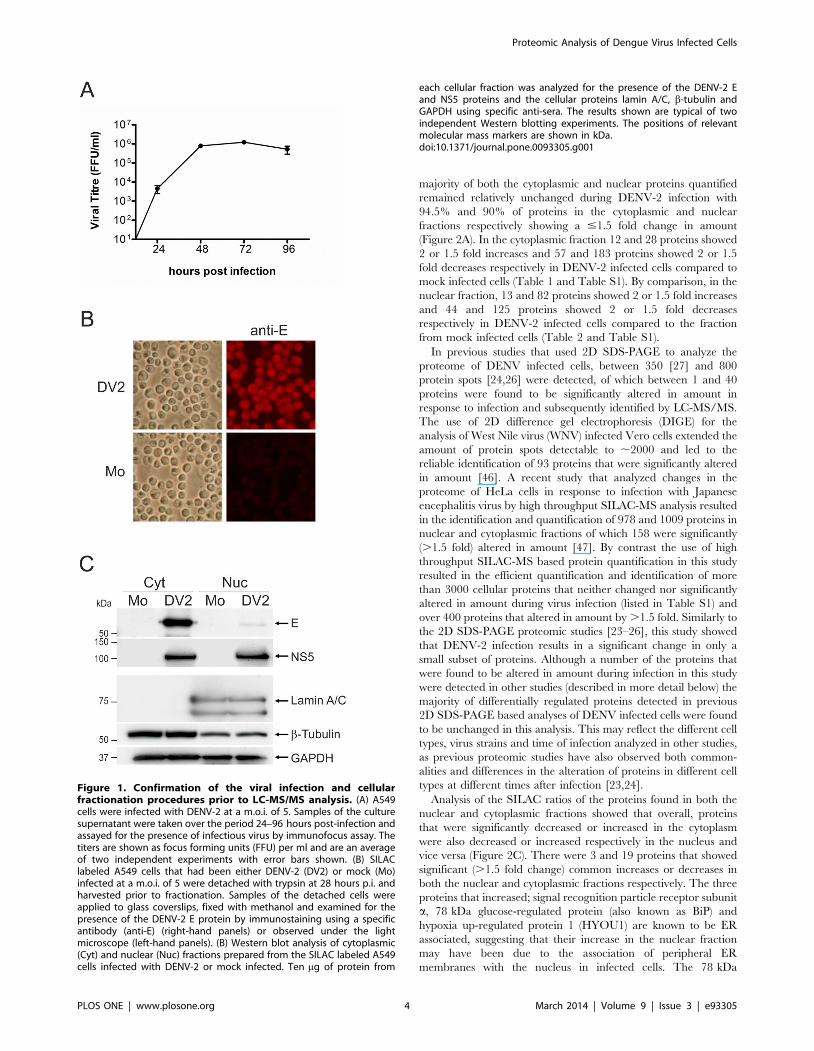
Infection and fractionation of DENV infected cellsIn order to conduct the proteomic analysis, human lung
carcinoma A549 cells were grown for eight cell doublings in either
light (R0K0) or heavy (R6K6) labeled media before being mock
infected or infected with DENV-2 respectively. Although not
believed to represent a target cell for DENV in vivo, A549 cells are
highly permissive for DENV infection and have been used in
previous studies examining the effect of DENV on the cellular
innate immune response [44,45] and the host cell transcriptome
[8]. At 28 hours p.i., a time point determined by growth curve
analysis (Figure 1A) to lie in exponential phase of DENV
replication, the cells were harvested and fractionated into nuclear
and cytoplasmic extracts. At this time there was no obvious
difference in the morphology of the mock and DENV infected
cells, suggesting that there was little cell death (data not shown).
IFA analysis of a sample of the mock and infected A549 cells
revealed that 100% of the cells had been infected (Figure 1B). The
fractionation was done to reduce the overall complexity of the
sample and to identify proteins that were altered in amount or
redistributed between the cytoplasm and nucleus during DENV
infection. DENV is known to replicate in tight association with
perinuclear membranes [4] therefore the cells were fractionated
using a procedure that removed as much of the perinuclear
membrane as possible without disrupting the nuclei. The presence
of protein markers in the cellular fractions specific to the nucleus,
cytoplasm and DENV infection were analyzed by Western blotting
to validate the infection and fractionation procedures (Figure 1C).
The analysis showed that whilst the nuclear and soluble
cytoplasmic protein fractions were distinct, the lack of detergent
in the lysis buffer led to the presence of some membraneous/
cytoskeletal proteins in the nuclear fractions. It appeared that the
perinuclear membrane and the associated viral replication
structures were not totally removed from the nuclei, as evidenced
by a minor amount of the virus E protein, which is cytoplasmically
localized, in the nuclear fraction. By contrast, the NS5 protein is
known to be found in both the nucleus and cytoplasm of infected
cells [43].
Quantitative LC-MS/MS analysisEqual protein amounts from the cytoplasmic fractions of the
mock and DENV-2 infected cells were pooled and the same
procedure repeated for the nuclear fractions. The proteins in the
cytoplasmic and nuclear fractions were separated by 1D SDS-
PAGE, subject to in-gel tryptic digestion and the peptides analyzed
by quantitative LC-MS/MS to determine the relative amounts of
proteins in the nuclear and cytoplasmic fractions from mock and
DENV-2 infected cells. This procedure was done once and
resulted in the identification of 4053 and 2881 cellular proteins in
the cytoplasmic and nuclear fractions of which 3098 and 2115
respectively, were reliably quantified (based on the determination
of a SILAC ratio for two or more peptides corresponding to each
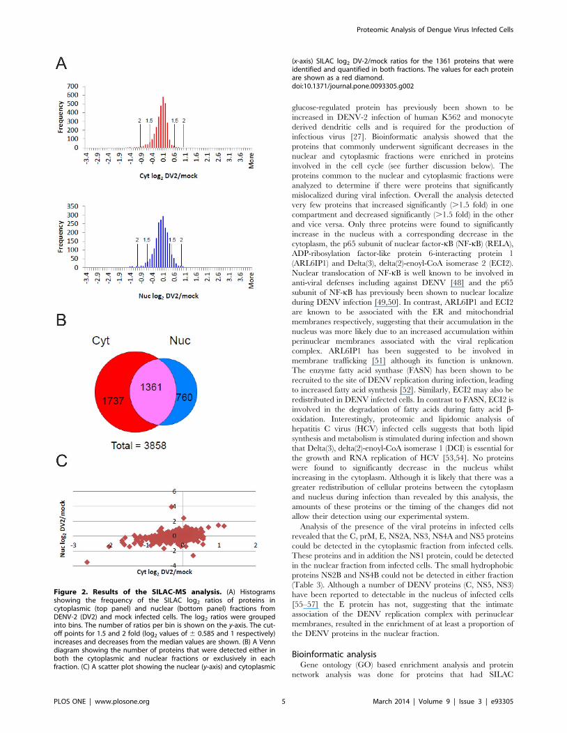
protein). Analysis of the distribution of log2 transformed SILAC
ratios for the proteins in the cytoplasmic and nuclear extracts
showed a symmetrical distribution around the normalized median
value of 1 (log2 = 0) for both sets of proteins (Figure 2A) suggesting
that there was no bias in the experimental approach used. There
were 3858 proteins reliably quantified in total in both the nuclear
and cytoplasmic fractions, of which 1361 proteins were common
(Figure 2B). Typically, an alteration in protein amount between
1.3–2 fold has been considered significant [32]. In this analysis we
used protein ratio cut-off values of 1.5 and 2 fold to select proteins
for further bioinformatic analysis (as described below). The
Proteomic Analysis of Dengue Virus Infected Cells
PLOS ONE | www.plosone.org 3 March 2014 | Volume 9 | Issue 3 | e93305
majority of both the cytoplasmic and nuclear proteins quantified
remained relatively unchanged during DENV-2 infection with
94.5% and 90% of proteins in the cytoplasmic and nuclear
fractions respectively showing a #1.5 fold change in amount
(Figure 2A). In the cytoplasmic fraction 12 and 28 proteins showed
2 or 1.5 fold increases and 57 and 183 proteins showed 2 or 1.5
fold decreases respectively in DENV-2 infected cells compared to
mock infected cells (Table 1 and Table S1). By comparison, in the
nuclear fraction, 13 and 82 proteins showed 2 or 1.5 fold increases
and 44 and 125 proteins showed 2 or 1.5 fold decreases
respectively in DENV-2 infected cells compared to the fraction
from mock infected cells (Table 2 and Table S1).
In previous studies that used 2D SDS-PAGE to analyze the
proteome of DENV infected cells, between 350 [27] and 800
protein spots [24,26] were detected, of which between 1 and 40
proteins were found to be significantly altered in amount in
response to infection and subsequently identified by LC-MS/MS.
The use of 2D difference gel electrophoresis (DIGE) for the
analysis of West Nile virus (WNV) infected Vero cells extended the
amount of protein spots detectable to ,2000 and led to the
reliable identification of 93 proteins that were significantly altered
in amount [46]. A recent study that analyzed changes in the
proteome of HeLa cells in response to infection with Japanese
encephalitis virus by high throughput SILAC-MS analysis resulted
in the identification and quantification of 978 and 1009 proteins in
nuclear and cytoplasmic fractions of which 158 were significantly
(.1.5 fold) altered in amount [47]. By contrast the use of high
throughput SILAC-MS based protein quantification in this study
resulted in the efficient quantification and identification of more
than 3000 cellular proteins that neither changed nor significantly
altered in amount during virus infection (listed in Table S1) and
over 400 proteins that altered in amount by .1.5 fold. Similarly to
the 2D SDS-PAGE proteomic studies [23–26], this study showed
that DENV-2 infection results in a significant change in only a
small subset of proteins. Although a number of the proteins that
were found to be altered in amount during infection in this study
were detected in other studies (described in more detail below) the
majority of differentially regulated proteins detected in previous
2D SDS-PAGE based analyses of DENV infected cells were found
to be unchanged in this analysis. This may reflect the different cell
types, virus strains and time of infection analyzed in other studies,
as previous proteomic studies have also observed both common-
alities and differences in the alteration of proteins in different cell
types at different times after infection [23,24].
Analysis of the SILAC ratios of the proteins found in both the
nuclear and cytoplasmic fractions showed that overall, proteins
that were significantly decreased or increased in the cytoplasm
were also decreased or increased respectively in the nucleus and
vice versa (Figure 2C). There were 3 and 19 proteins that showed
significant (.1.5 fold change) common increases or decreases in
both the nuclear and cytoplasmic fractions respectively. The three
proteins that increased; signal recognition particle receptor subunit
a, 78 kDa glucose-regulated protein (also known as BiP) and
hypoxia up-regulated protein 1 (HYOU1) are known to be ER
associated, suggesting that their increase in the nuclear fraction
may have been due to the association of peripheral ER
membranes with the nucleus in infected cells. The 78 kDa
Figure 1. Confirmation of the viral infection and cellularfractionation procedures prior to LC-MS/MS analysis. (A) A549cells were infected with DENV-2 at a m.o.i. of 5. Samples of the culturesupernatant were taken over the period 24–96 hours post-infection andassayed for the presence of infectious virus by immunofocus assay. Thetiters are shown as focus forming units (FFU) per ml and are an averageof two independent experiments with error bars shown. (B) SILAClabeled A549 cells that had been either DENV-2 (DV2) or mock (Mo)infected at a m.o.i. of 5 were detached with trypsin at 28 hours p.i. andharvested prior to fractionation. Samples of the detached cells wereapplied to glass coverslips, fixed with methanol and examined for thepresence of the DENV-2 E protein by immunostaining using a specificantibody (anti-E) (right-hand panels) or observed under the lightmicroscope (left-hand panels). (B) Western blot analysis of cytoplasmic(Cyt) and nuclear (Nuc) fractions prepared from the SILAC labeled A549cells infected with DENV-2 or mock infected. Ten mg of protein from
each cellular fraction was analyzed for the presence of the DENV-2 Eand NS5 proteins and the cellular proteins lamin A/C, b-tubulin andGAPDH using specific anti-sera. The results shown are typical of twoindependent Western blotting experiments. The positions of relevantmolecular mass markers are shown in kDa.doi:10.1371/journal.pone.0093305.g001
Proteomic Analysis of Dengue Virus Infected Cells
PLOS ONE | www.plosone.org 4 March 2014 | Volume 9 | Issue 3 | e93305
glucose-regulated protein has previously been shown to be
increased in DENV-2 infection of human K562 and monocyte
derived dendritic cells and is required for the production of
infectious virus [27]. Bioinformatic analysis showed that the
proteins that commonly underwent significant decreases in the
nuclear and cytoplasmic fractions were enriched in proteins
involved in the cell cycle (see further discussion below). The
proteins common to the nuclear and cytoplasmic fractions were
analyzed to determine if there were proteins that significantly
mislocalized during viral infection. Overall the analysis detected
very few proteins that increased significantly (.1.5 fold) in one
compartment and decreased significantly (.1.5 fold) in the other
and vice versa. Only three proteins were found to significantly
increase in the nucleus with a corresponding decrease in the
cytoplasm, the p65 subunit of nuclear factor-kB (NF-kB) (RELA),
ADP-ribosylation factor-like protein 6-interacting protein 1
(ARL6IP1) and Delta(3), delta(2)-enoyl-CoA isomerase 2 (ECI2).
Nuclear translocation of NF-kB is well known to be involved in
anti-viral defenses including against DENV [48] and the p65
subunit of NF-kB has previously been shown to nuclear localize
during DENV infection [49,50]. In contrast, ARL6IP1 and ECI2
are known to be associated with the ER and mitochondrial
membranes respectively, suggesting that their accumulation in the
nucleus was more likely due to an increased accumulation within
perinuclear membranes associated with the viral replication
complex. ARL6IP1 has been suggested to be involved in
membrane trafficking [51] although its function is unknown.
The enzyme fatty acid synthase (FASN) has been shown to be
recruited to the site of DENV replication during infection, leading
to increased fatty acid synthesis [52]. Similarly, ECI2 may also be
redistributed in DENV infected cells. In contrast to FASN, ECI2 is
involved in the degradation of fatty acids during fatty acid b-
oxidation. Interestingly, proteomic and lipidomic analysis of
hepatitis C virus (HCV) infected cells suggests that both lipid
synthesis and metabolism is stimulated during infection and shown
that Delta(3), delta(2)-enoyl-CoA isomerase 1 (DCI) is essential for
the growth and RNA replication of HCV [53,54]. No proteins
were found to significantly decrease in the nucleus whilst
increasing in the cytoplasm. Although it is likely that there was a
greater redistribution of cellular proteins between the cytoplasm
and nucleus during infection than revealed by this analysis, the
amounts of these proteins or the timing of the changes did not
allow their detection using our experimental system.
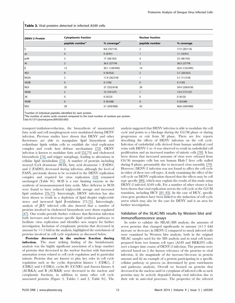
Analysis of the presence of the viral proteins in infected cells
revealed that the C, prM, E, NS2A, NS3, NS4A and NS5 proteins
could be detected in the cytoplasmic fraction from infected cells.
These proteins and in addition the NS1 protein, could be detected
in the nuclear fraction from infected cells. The small hydrophobic
proteins NS2B and NS4B could not be detected in either fraction
(Table 3). Although a number of DENV proteins (C, NS5, NS3)
have been reported to detectable in the nucleus of infected cells
[55–57] the E protein has not, suggesting that the intimate
association of the DENV replication complex with perinuclear
membranes, resulted in the enrichment of at least a proportion of
the DENV proteins in the nuclear fraction.
Bioinformatic analysisGene ontology (GO) based enrichment analysis and protein
network analysis was done for proteins that had SILAC
Figure 2. Results of the SILAC-MS analysis. (A) Histogramsshowing the frequency of the SILAC log2 ratios of proteins incytoplasmic (top panel) and nuclear (bottom panel) fractions fromDENV-2 (DV2) and mock infected cells. The log2 ratios were groupedinto bins. The number of ratios per bin is shown on the y-axis. The cut-off points for 1.5 and 2 fold (log2 values of 6 0.585 and 1 respectively)increases and decreases from the median values are shown. (B) A Venndiagram showing the number of proteins that were detected either inboth the cytoplasmic and nuclear fractions or exclusively in eachfraction. (C) A scatter plot showing the nuclear (y-axis) and cytoplasmic
(x-axis) SILAC log2 DV-2/mock ratios for the 1361 proteins that wereidentified and quantified in both fractions. The values for each proteinare shown as a red diamond.doi:10.1371/journal.pone.0093305.g002
Proteomic Analysis of Dengue Virus Infected Cells
PLOS ONE | www.plosone.org 5 March 2014 | Volume 9 | Issue 3 | e93305
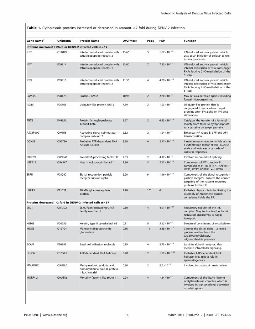
Table 1. Cytoplasmic proteins increased or decreased in amount .2 fold during DENV-2 infection.
Gene Namea UniprotID Protein Name DV2/Mock Peps PEP Function
Proteins increased .2fold in DENV-2 infected cells n = 12
IFIT3 O14879 Interferon-induced protein withtetratricopeptide repeats 3
13.66 5 1.62610242 IFN-induced antiviral protein whichacts as an inhibitor of cellular as wellas viral processes.
IFIT1 P09914 Interferon-induced protein withtetratricopeptide repeats 1
13.00 7 7.23610231 IFN-induced antiviral protein whichinhibits expression of viral messengerRNAs lacking 29-O-methylation of the59 cap
IFIT2 P09913 Interferon-induced protein withtetratricopeptide repeats 2
11.55 4 4.09610223 IFN-induced antiviral protein whichinhibits expression of viral messengerRNAs lacking 29-O-methylation of the59 cap
FAM3A P98173 Protein FAM3A 10.96 2 2.7061022 May act as a defensin against invadingfungal microorganisms.
ISG15 P05161 Ubiquitin-like protein ISG15 7.59 2 1.0361027 Ubiquitin-like protein that isconjugated to intracellular targetproteins after IFN-alpha or IFN-betastimulation.
FNTB P49356 Protein farnesyltransferasesubunit beta
2.67 2 6.33610225 Catalyzes the transfer of a farnesylmoiety from farnesyl pyrophosphateto a cysteine on target proteins.
ASC1P100 Q9H1I8 Activating signal cointegrator 1complex subunit 2
2.52 2 1.2461024 Enhances NF-kappa-B, SRF and AP1transactivation.
DDX58 O95786 Probable ATP-dependent RNAhelicase DDX58
2.50 4 2.97610225 Innate immune receptor which acts asa cytoplasmic sensor of viral nucleicacids and activates a cascade ofantiviral responses.
PRPF39 Q86UA1 Pre-mRNA-processing factor 39 2.33 2 4.1761023 Involved in pre-mRNA splicing
HSPB11 Q9Y547 Heat shock protein beta-11 2.24 3 2.51610210 Component of IFT complex Bcomposed of IFT88, IFT57, TRAF3IP1,IFT52, IFT27, HSPB11 and IFT20.
SRPR P08240 Signal recognition particlereceptor subunit alpha
2.20 9 1.16610210 Component of the signal recognitionparticle receptor. Ensures the correcttargeting of the nascent secretoryproteins to the ER.
HSPA5 P11021 78 kDa glucose-regulatedprotein
1.98 101 0 Probably plays a role in facilitating theassembly of multimeric proteincomplexes inside the ER.
Proteins decreased .2 fold in DENV-2 infected cells n = 57
ERC1 Q8IUD2 ELKS/Rab6-interacting/CASTfamily member 1
0.10 4 4.01610220 Regulatory subunit of the IKKcomplex. May be involved in Rab-6regulated endosomes to Golgitransport.
KRT6B P04259 Keratin, type II cytoskeletal 6B 0.11 8 5.1261024 Structural constituent of cytoskeleton
MOGS Q13724 Mannosyl-oligosaccharideglucosidase
0.16 11 2.38610217 Cleaves the distal alpha 1,2-linkedglucose residue from theGlc(3)Man(9)GlcNAc(2)oligosaccharide precursor.
BCAM P50895 Basal cell adhesion molecule 0.19 4 2.75610214 Laminin alpha-5 receptor. Maymediate intracellular signaling.
DDX3Y O15523 ATP-dependent RNA helicase 0.20 2 1.526102206 Probable ATP-dependent RNAhelicase. May play a role inspermatogenesis.
MMADHC Q9H3L0 Methylmalonic aciduria andhomocystinuria type D protein,mitochondrial
0.20 2 2.061023 Involved in cobalamin metabolism.
MORF4L1 Q9UBU8 Mortality factor 4-like protein 1 0.24 4 1.6461024 Component of the NuA4 histoneacetyltransferase complex which isinvolved in transcriptional activationof select genes.
Proteomic Analysis of Dengue Virus Infected Cells
PLOS ONE | www.plosone.org 6 March 2014 | Volume 9 | Issue 3 | e93305
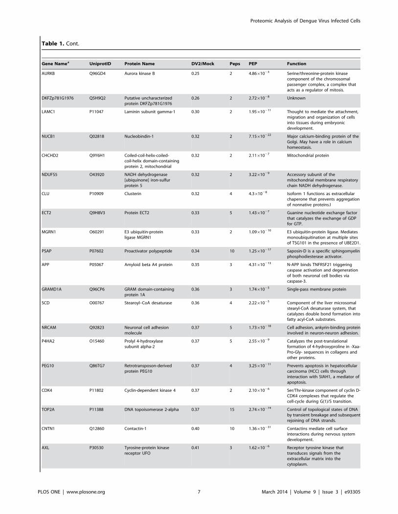
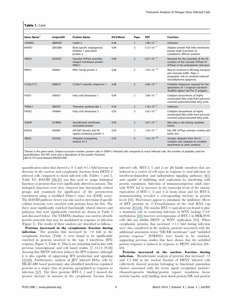
Table 1. Cont.
Gene Namea UniprotID Protein Name DV2/Mock Peps PEP Function
AURKB Q96GD4 Aurora kinase B 0.25 2 4.8661023 Serine/threonine-protein kinasecomponent of the chromosomalpassenger complex, a complex thatacts as a regulator of mitosis.
DKFZp781G1976 Q5H9Q2 Putative uncharacterizedprotein DKFZp781G1976
0.26 2 2.7261028 Unknown
LAMC1 P11047 Laminin subunit gamma-1 0.30 2 1.95610211 Thought to mediate the attachment,migration and organization of cellsinto tissues during embryonicdevelopment.
NUCB1 Q02818 Nucleobindin-1 0.32 2 7.15610222 Major calcium-binding protein of theGolgi. May have a role in calciumhomeostasis.
CHCHD2 Q9Y6H1 Coiled-coil-helix-coiled-coil-helix domain-containingprotein 2, mitochondrial
0.32 2 2.1161027 Mitochondrial protein
NDUFS5 O43920 NADH dehydrogenase[ubiquinone] iron-sulfurprotein 5
0.32 2 3.2261029 Accessory subunit of themitochondrial membrane respiratorychain NADH dehydrogenase.
CLU P10909 Clusterin 0.32 4 4.361028 Isoform 1 functions as extracellularchaperone that prevents aggregationof nonnative proteins.I
ECT2 Q9H8V3 Protein ECT2 0.33 5 1.4361027 Guanine nucleotide exchange factorthat catalyzes the exchange of GDPfor GTP.
MGRN1 O60291 E3 ubiquitin-proteinligase MGRN1
0.33 2 1.09610210 E3 ubiquitin-protein ligase. Mediatesmonoubiquitination at multiple sitesof TSG101 in the presence of UBE2D1.
PSAP P07602 Proactivator polypeptide 0.34 10 1.25610217 Saposin-D is a specific sphingomyelinphosphodiesterase activator.
APP P05067 Amyloid beta A4 protein 0.35 3 4.31610213 N-APP binds TNFRSF21 triggeringcaspase activation and degenerationof both neuronal cell bodies viacaspase-3.
GRAMD1A Q96CP6 GRAM domain-containingprotein 1A
0.36 3 1.7461025 Single-pass membrane protein
SCD O00767 Stearoyl–CoA desaturase 0.36 4 2.2261025 Component of the liver microsomalstearyl-CoA desaturase system, thatcatalyzes double bond formation intofatty acyl-CoA substrates.
NRCAM Q92823 Neuronal cell adhesionmolecule
0.37 5 1.73610218 Cell adhesion, ankyrin-binding proteininvolved in neuron-neuron adhesion.
P4HA2 O15460 Prolyl 4-hydroxylasesubunit alpha-2
0.37 5 2.5561029 Catalyzes the post-translationalformation of 4-hydroxyproline in -Xaa-Pro-Gly- sequences in collagens andother proteins.
PEG10 Q86TG7 Retrotransposon-derivedprotein PEG10
0.37 4 3.25610211 Prevents apoptosis in hepatocellularcarcinoma (HCC) cells throughinteraction with SIAH1, a mediator ofapoptosis.
CDK4 P11802 Cyclin-dependent kinase 4 0.37 2 2.1061026 Ser/Thr-kinase component of cyclin D-CDK4 complexes that regulate thecell-cycle during G(1)/S transition.
TOP2A P11388 DNA topoisomerase 2-alpha 0.37 15 2.74610274 Control of topological states of DNAby transient breakage and subsequentrejoining of DNA strands.
CNTN1 Q12860 Contactin-1 0.40 10 1.36610231 Contactins mediate cell surfaceinteractions during nervous systemdevelopment.
AXL P30530 Tyrosine-protein kinasereceptor UFO
0.41 3 1.6261026 Receptor tyrosine kinase thattransduces signals from theextracellular matrix into thecytoplasm.
Proteomic Analysis of Dengue Virus Infected Cells
PLOS ONE | www.plosone.org 7 March 2014 | Volume 9 | Issue 3 | e93305
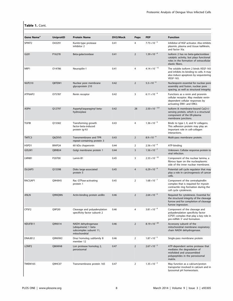
Table 1. Cont.
Gene Namea UniprotID Protein Name DV2/Mock Peps PEP Function
SPINT2 O43291 Kunitz-type proteaseinhibitor 2
0.41 4 7.7361025 Inhibitor of HGF activator. Also inhibitsplasmin, plasma and tissue kallikrein,and factor XIa.
GLB1 P16278 Beta-galactosidase 0.41 2 1.3961024 Isoform 2 has no beta-galactosidasecatalytic activity, but plays functionalroles in the formation of extracellularelastic fibers.
NRP1 O14786 Neuropilin-1 0.41 4 4.14610215 The soluble isoform 2 binds VEGF-165and inhibits its binding to cells. It mayalso induce apoptosis by sequesteringVEGF-165.
NUP210 Q8TEM1 Nuclear pore membraneglycoprotein 210
0.42 2 5.361024 Nucleoporin essential for nuclear poreassembly and fusion, nuclear porespacing, as well as structural integrity.
ATP6AP2 O75787 Renin receptor 0.42 3 6.1161024 Functions as a renin and prorenincellular receptor. May mediate renin-dependent cellular responses byactivating ERK1 and ERK2.
ASPH Q12797 Aspartyl/asparaginyl beta-hydroxylase
0.42 28 2.506102121 Isoform 8: membrane-bound Ca(2+)-sensing protein, which is a structuralcomponent of the ER-plasmamembrane junctions.
TGFBI Q15582 Transforming growthfactor-beta-inducedprotein ig-h3
0.43 4 1.3661029 Binds to type I, II, and IV collagens.This adhesion protein may play animportant role in cell-collageninteractions.
TMTC3 Q6ZXV5 Transmembrane and TPRrepeat-containing protein 3
0.43 2 8.961024 Multi-pass membrane protein.
HSPD1 B9VP24 60 kDa chaperonin 0.44 2 2.5661024 ATP-binding
GOLM1 Q8NBJ4 Golgi membrane protein 1 0.44 3 1.5661027 Unknown. Cellular response protein toviral infection.
LMNB1 P20700 Lamin-B1 0.45 3 2.33610232 Component of the nuclear lamina, afibrous layer on the nucleoplasmicside of the inner nuclear membrane.
DLGAP5 Q15398 Disks large-associatedprotein 5
0.45 4 6.2961029 Potential cell cycle regulator that mayplay a role in carcinogenesis of cancercells.
RACGAP1 Q9H0H5 Rac GTPase-activatingprotein 1
0.45 2 1.6861027 Component of the centralspindlincomplex that is required for myosincontractile ring formation during thecell cycle cytokinesis.
ANLN Q9NQW6 Actin-binding protein anillin 0.46 2 2.6461026 Required for cytokinesis. Essential forthe structural integrity of the cleavagefurrow and for completion of cleavagefurrow ingression.
CPSF2 Q9P2I0 Cleavage and polyadenylationspecificity factor subunit 2
0.46 4 3.8161024 Component of the cleavage andpolyadenylation specificity factor(CPSF) complex that play a key role inpre-mRNA 39-end formation.
NDUFB11 Q9NX14 NADH dehydrogenase[ubiquinone] 1 betasubcomplex subunit 11,mitochondrial
0.46 2 8.19610220 Accessory subunit of themitochondrial membrane respiratorychain NADH dehydrogenase.
DNAJB12 Q9NXW2 DnaJ homolog subfamily Bmember 12
0.46 2 1.8761025 Single-pass membrane protein
LONP2 Q86WA8 Lon protease homolog 2,peroxisomal
0.47 2 2.6761022 ATP-dependent serine protease thatmediates the degradation ofmisfolded and unassembledpolypeptides in the peroxisomalmatrix.
TMEM165 Q9HC07 Transmembrane protein 165 0.47 2 1.3561022 May function as a calcium/protontransporter involved in calcium and inlysosomal pH homeostasis.
Proteomic Analysis of Dengue Virus Infected Cells
PLOS ONE | www.plosone.org 8 March 2014 | Volume 9 | Issue 3 | e93305
quantification ratios that showed a $ 2 and $1.5 fold increase or
decrease in the nuclear and cytoplasmic fractions from DENV-2
infected cells compared to mock infected cells (Tables 1 and 2,
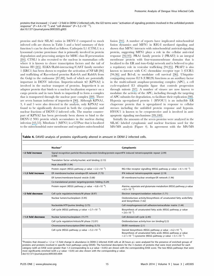
Table S1). DAVID [40,41] was first used to assign biological
functions to proteins that were significantly altered in amount. The
biological functions were then clustered into functionally related
groups and examined for significance of the protein-term
enrichment using a modified Fisher’s exact test (EASE score).
The DAVID pathway viewer was also used to determine if specific
cellular networks were enriched with proteins from the lists. The
three most significantly enriched functionally related clusters and
pathways that were significantly enriched are shown in Table 4
and discussed below. The STRING database was used to identify
protein networks that may be modulated in response to infection
(Figure 3). The results of these analyses are described as follows.
Proteins increased in the cytoplasmic fraction during
infection. The proteins that increased by .2 fold in the
cytoplasmic fraction (Table 1) were found to be significantly
enriched in proteins involved in the type I interferon (IFN)
response (Figure 3, Table 4). This is not surprising and in line with
previous transcriptomic and cell based studies [7–12,14–19,58]
showing that DENV infection induces the IFN response, although
it is also capable of suppressing IFN production and signaling
[59,60]. Furthermore, analysis of JEV infected HeLa cells by
SILAC-MS based proteomics also identified interferon regulated
proteins as a major class of proteins upregulated in response to
infection [47]. The three proteins IFIT-3, 1 and 2 showed the
greatest increase in amount in the cytoplasmic fraction from
infected cells. IFIT-3, 1 and 2 are Ifit family members that are
induced in a variety of cell types in response to viral infection via
interferon-dependent and independent signaling pathways [61]
and capable of inhibiting viral replication by interfering with
protein translation. Infection of immunocompetent adult mice
with WNV led to increases in the transcript levels of the murine
equivalents of IFIT-1, 2 and 3 in brain tissue and for IFIT-2,
immunostaining revealed a corresponding increase in protein
levels [62]. Flaviviruses appear to mimimize the inhibitory effects
of IFIT proteins by 29-O-methylation of the viral RNA cap
structure [63,64]. The murine IFIT-1 equivalent was found to play
a dominant role in restricting infection of WNV lacking 29-O-
methylation [63] however overexpression of IFIT-1 in HEK293T
cells did not inhibit DENV or WNV replication [65]. When
cytoplasmic proteins that increased .1.5 fold during infection
were also considered in the analysis, proteins associated with the
additional annotation terms ‘‘ER/ER membrane’’ and ‘‘unfolded
protein response’’ (STRING) were found to be enriched,
supporting previous studies that have shown that the unfolded
protein response is induced in response to DENV infection [66–
69].
Proteins increased in the nuclear fraction during
infection. Bioinformatic analysis of proteins that increased .2
and 1.5 fold in the nuclear fraction of DENV infected cells
collectively showed proteins belonging to functional annotation
clusters associated with the terms signal recognition particle/
ribonucleoprotein binding/protein export; translation factor
activity/nucleic acid binding; heat shock protein/ER and protein
Table 1. Cont.
Gene Namea UniprotID Protein Name DV2/Mock Peps PEP Function
CASKIN2 Q8WXE0 Caskin-2 0.48 3 1.9861027 Unknown
BAIAP2 Q9UQB8 Brain-specific angiogenesisinhibitor 1-associatedprotein 2
0.48 3 5.1261024 Adapter protein that links membrane-bound small G-proteins tocytoplasmic effector proteins.
VMA21 Q3ZAQ7 Vacuolar ATPase assemblyintegral membrane proteinVMA21
0.48 2 6.5761024 Required for the assembly of the V0complex of the vacuolar ATPase (V-ATPase) in the endoplasmic reticulum.
PRAF2 O60831 PRA1 family protein 2 0.48 3 7.02610214 May be involved in ER/Golgi transportand vesicular traffic. Plays aproapoptic role in cerulenin-inducedneuroblastoma apoptosis.
C1GALT1C1 Q96EU7 C1GALT1-specific chaperone 1 0.48 2 2.6861023 Probable chaperone required for thegeneration of 1 O-glycan Gal-beta1-3GalNAc-alpha1-Ser/Thr (T antigen).
FADS1 O60427 Fatty acid desaturase 1 0.49 2 7.0861027 Catalyzes biosynthesis of highlyunsaturated fatty acids from precursoressential polyunsaturated fatty acids.
THNSL1 Q8IYQ7 Threonine synthase-like 1 0.50 2 1.3661029 Unknown
FADS2 O95864 Fatty acid desaturase 2 0.50 3 5.4261025 Catalyzes biosynthesis of highlyunsaturated fatty acids from precursoressential polyunsaturated fatty acids.
SLMAP Q14BN4 Sarcolemmal membrane-associated protein
0.50 2 2.9761022 May play a role during myoblastfusion.
AGFG2 O95081 Arf-GAP domain and FGrepeat-containing protein 2
0.50 2 1.2061022 Has ARF GTPase activator activity andbinds zinc.
UBE2S Q16763 Ubiquitin-conjugatingenzyme E2 S
0.50 6 7.55610268 Accepts ubiquitin from the E1complex and catalyzes its covalentattachment to other proteins.
aShown is the gene name, Uniprot accession number, protein ratio in DENV-2 infected cells compared to mock infected cells, the number of peptides used forquantification, the PEP score and a description of the protein function).doi:10.1371/journal.pone.0093305.t001
Proteomic Analysis of Dengue Virus Infected Cells
PLOS ONE | www.plosone.org 9 March 2014 | Volume 9 | Issue 3 | e93305
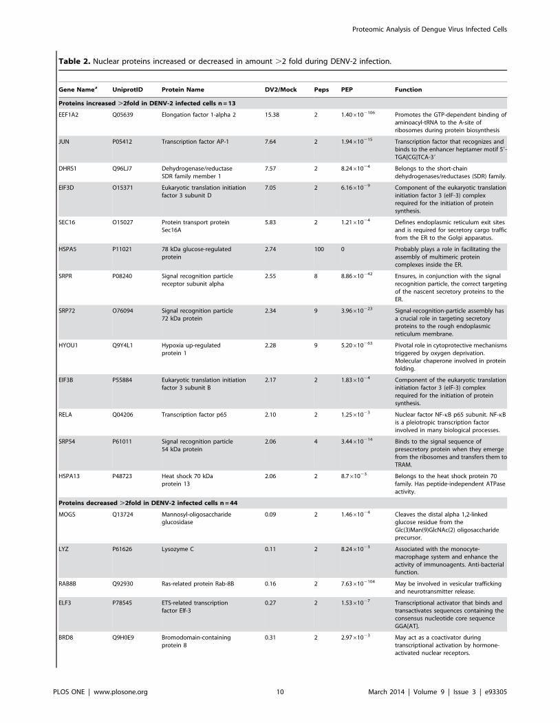
Table 2. Nuclear proteins increased or decreased in amount .2 fold during DENV-2 infection.
Gene Namea UniprotID Protein Name DV2/Mock Peps PEP Function
Proteins increased .2fold in DENV-2 infected cells n = 13
EEF1A2 Q05639 Elongation factor 1-alpha 2 15.38 2 1.406102106 Promotes the GTP-dependent binding ofaminoacyl-tRNA to the A-site ofribosomes during protein biosynthesis
JUN P05412 Transcription factor AP-1 7.64 2 1.94610215 Transcription factor that recognizes andbinds to the enhancer heptamer motif 59-TGA[CG]TCA-39
DHRS1 Q96LJ7 Dehydrogenase/reductaseSDR family member 1
7.57 2 8.2461024 Belongs to the short-chaindehydrogenases/reductases (SDR) family.
EIF3D O15371 Eukaryotic translation initiationfactor 3 subunit D
7.05 2 6.1661029 Component of the eukaryotic translationinitiation factor 3 (eIF-3) complexrequired for the initiation of proteinsynthesis.
SEC16 O15027 Protein transport proteinSec16A
5.83 2 1.2161024 Defines endoplasmic reticulum exit sitesand is required for secretory cargo trafficfrom the ER to the Golgi apparatus.
HSPA5 P11021 78 kDa glucose-regulatedprotein
2.74 100 0 Probably plays a role in facilitating theassembly of multimeric proteincomplexes inside the ER.
SRPR P08240 Signal recognition particlereceptor subunit alpha
2.55 8 8.86610242 Ensures, in conjunction with the signalrecognition particle, the correct targetingof the nascent secretory proteins to theER.
SRP72 O76094 Signal recognition particle72 kDa protein
2.34 9 3.96610223 Signal-recognition-particle assembly hasa crucial role in targeting secretoryproteins to the rough endoplasmicreticulum membrane.
HYOU1 Q9Y4L1 Hypoxia up-regulatedprotein 1
2.28 9 5.20610263 Pivotal role in cytoprotective mechanismstriggered by oxygen deprivation.Molecular chaperone involved in proteinfolding.
EIF3B P55884 Eukaryotic translation initiationfactor 3 subunit B
2.17 2 1.8361024 Component of the eukaryotic translationinitiation factor 3 (eIF-3) complexrequired for the initiation of proteinsynthesis.
RELA Q04206 Transcription factor p65 2.10 2 1.2561023 Nuclear factor NF-kB p65 subunit. NF-kBis a pleiotropic transcription factorinvolved in many biological processes.
SRP54 P61011 Signal recognition particle54 kDa protein
2.06 4 3.44610214 Binds to the signal sequence ofpresecretory protein when they emergefrom the ribosomes and transfers them toTRAM.
HSPA13 P48723 Heat shock 70 kDaprotein 13
2.06 2 8.761023 Belongs to the heat shock protein 70family. Has peptide-independent ATPaseactivity.
Proteins decreased .2fold in DENV-2 infected cells n = 44
MOGS Q13724 Mannosyl-oligosaccharideglucosidase
0.09 2 1.4661024 Cleaves the distal alpha 1,2-linkedglucose residue from theGlc(3)Man(9)GlcNAc(2) oligosaccharideprecursor.
LYZ P61626 Lysozyme C 0.11 2 8.2461023 Associated with the monocyte-macrophage system and enhance theactivity of immunoagents. Anti-bacterialfunction.
RAB8B Q92930 Ras-related protein Rab-8B 0.16 2 7.636102104 May be involved in vesicular traffickingand neurotransmitter release.
ELF3 P78545 ETS-related transcriptionfactor Elf-3
0.27 2 1.5361027 Transcriptional activator that binds andtransactivates sequences containing theconsensus nucleotide core sequenceGGA[AT].
BRD8 Q9H0E9 Bromodomain-containingprotein 8
0.31 2 2.9761023 May act as a coactivator duringtranscriptional activation by hormone-activated nuclear receptors.
Proteomic Analysis of Dengue Virus Infected Cells
PLOS ONE | www.plosone.org 10 March 2014 | Volume 9 | Issue 3 | e93305
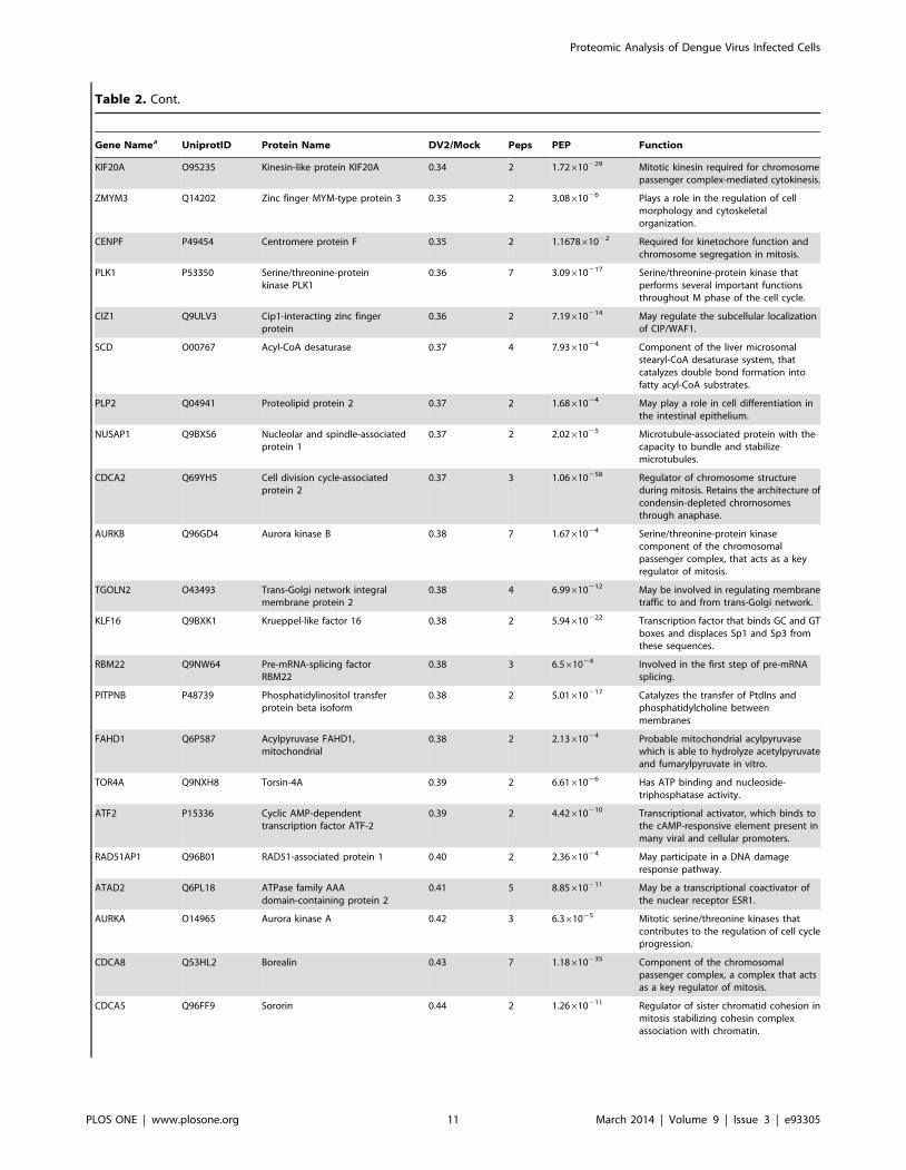
Table 2. Cont.
Gene Namea UniprotID Protein Name DV2/Mock Peps PEP Function
KIF20A O95235 Kinesin-like protein KIF20A 0.34 2 1.72610229 Mitotic kinesin required for chromosomepassenger complex-mediated cytokinesis.
ZMYM3 Q14202 Zinc finger MYM-type protein 3 0.35 2 3.0861026 Plays a role in the regulation of cellmorphology and cytoskeletalorganization.
CENPF P49454 Centromere protein F 0.35 2 1.167861022 Required for kinetochore function andchromosome segregation in mitosis.
PLK1 P53350 Serine/threonine-proteinkinase PLK1
0.36 7 3.09610217 Serine/threonine-protein kinase thatperforms several important functionsthroughout M phase of the cell cycle.
CIZ1 Q9ULV3 Cip1-interacting zinc fingerprotein
0.36 2 7.19610214 May regulate the subcellular localizationof CIP/WAF1.
SCD O00767 Acyl-CoA desaturase 0.37 4 7.9361024 Component of the liver microsomalstearyl-CoA desaturase system, thatcatalyzes double bond formation intofatty acyl-CoA substrates.
PLP2 Q04941 Proteolipid protein 2 0.37 2 1.6861024 May play a role in cell differentiation inthe intestinal epithelium.
NUSAP1 Q9BXS6 Nucleolar and spindle-associatedprotein 1
0.37 2 2.0261025 Microtubule-associated protein with thecapacity to bundle and stabilizemicrotubules.
CDCA2 Q69YH5 Cell division cycle-associatedprotein 2
0.37 3 1.06610258 Regulator of chromosome structureduring mitosis. Retains the architecture ofcondensin-depleted chromosomesthrough anaphase.
AURKB Q96GD4 Aurora kinase B 0.38 7 1.6761024 Serine/threonine-protein kinasecomponent of the chromosomalpassenger complex, that acts as a keyregulator of mitosis.
TGOLN2 O43493 Trans-Golgi network integralmembrane protein 2
0.38 4 6.99610212 May be involved in regulating membranetraffic to and from trans-Golgi network.
KLF16 Q9BXK1 Krueppel-like factor 16 0.38 2 5.94610222 Transcription factor that binds GC and GTboxes and displaces Sp1 and Sp3 fromthese sequences.
RBM22 Q9NW64 Pre-mRNA-splicing factorRBM22
0.38 3 6.561024 Involved in the first step of pre-mRNAsplicing.
PITPNB P48739 Phosphatidylinositol transferprotein beta isoform
0.38 2 5.01610217 Catalyzes the transfer of PtdIns andphosphatidylcholine betweenmembranes
FAHD1 Q6P587 Acylpyruvase FAHD1,mitochondrial
0.38 2 2.1361024 Probable mitochondrial acylpyruvasewhich is able to hydrolyze acetylpyruvateand fumarylpyruvate in vitro.
TOR4A Q9NXH8 Torsin-4A 0.39 2 6.6161026 Has ATP binding and nucleoside-triphosphatase activity.
ATF2 P15336 Cyclic AMP-dependenttranscription factor ATF-2
0.39 2 4.42610210 Transcriptional activator, which binds tothe cAMP-responsive element present inmany viral and cellular promoters.
RAD51AP1 Q96B01 RAD51-associated protein 1 0.40 2 2.3661024 May participate in a DNA damageresponse pathway.
ATAD2 Q6PL18 ATPase family AAAdomain-containing protein 2
0.41 5 8.85610211 May be a transcriptional coactivator ofthe nuclear receptor ESR1.
AURKA O14965 Aurora kinase A 0.42 3 6.361025 Mitotic serine/threonine kinases thatcontributes to the regulation of cell cycleprogression.
CDCA8 Q53HL2 Borealin 0.43 7 1.18610235 Component of the chromosomalpassenger complex, a complex that actsas a key regulator of mitosis.
CDCA5 Q96FF9 Sororin 0.44 2 1.26610211 Regulator of sister chromatid cohesion inmitosis stabilizing cohesin complexassociation with chromatin.
Proteomic Analysis of Dengue Virus Infected Cells
PLOS ONE | www.plosone.org 11 March 2014 | Volume 9 | Issue 3 | e93305
folding/the unfolded protein response. The strong association of
these terms with the ER once again suggested that the increased
amounts of proteins found in the nuclear fraction in response to
viral infection was due to the close association of viral induced
replication structures derived from the ER membranes with the
nuclear membrane [4] rather than increased localization of these
proteins into the nucleus. This is supported by the finding that
elongation factor 1-a2, the protein most increased in amount in
the nuclear fraction of infected cells, has previously been shown to
interact with the DENV 39 untranslated region and co-localize
with the DENV replication complex [70,71].
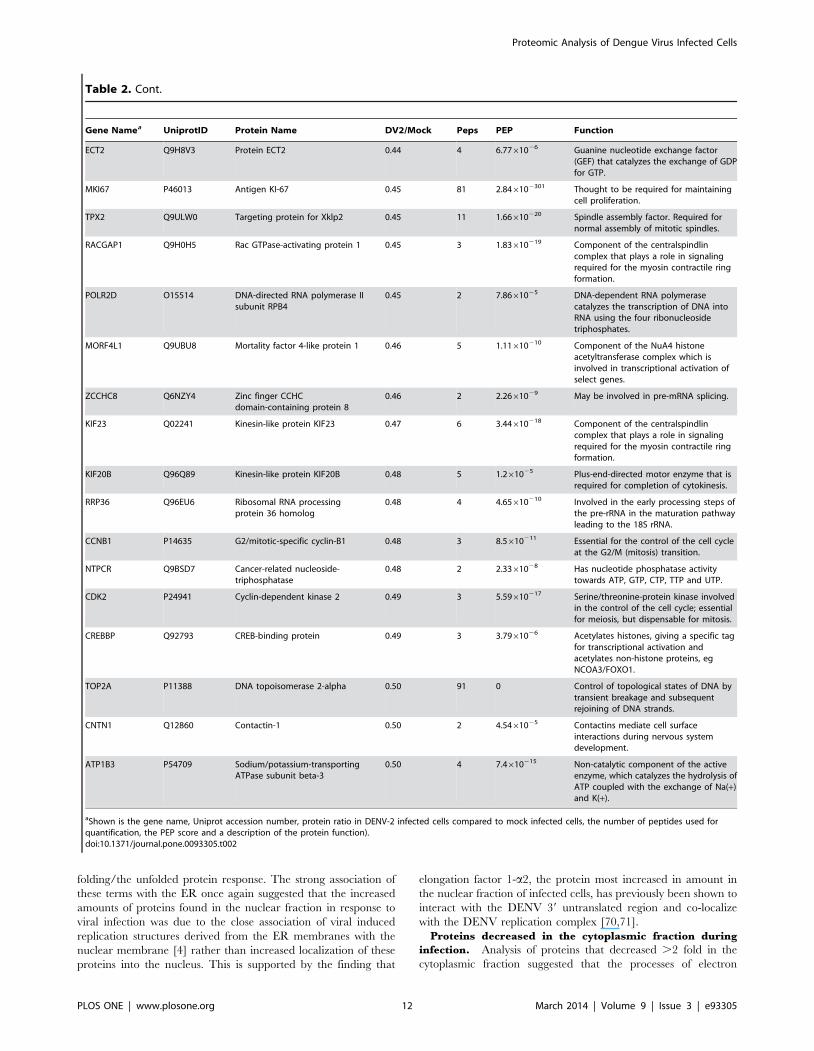
Proteins decreased in the cytoplasmic fraction during
infection. Analysis of proteins that decreased .2 fold in the
cytoplasmic fraction suggested that the processes of electron
Table 2. Cont.
Gene Namea UniprotID Protein Name DV2/Mock Peps PEP Function
ECT2 Q9H8V3 Protein ECT2 0.44 4 6.7761026 Guanine nucleotide exchange factor(GEF) that catalyzes the exchange of GDPfor GTP.
MKI67 P46013 Antigen KI-67 0.45 81 2.846102301 Thought to be required for maintainingcell proliferation.
TPX2 Q9ULW0 Targeting protein for Xklp2 0.45 11 1.66610220 Spindle assembly factor. Required fornormal assembly of mitotic spindles.
RACGAP1 Q9H0H5 Rac GTPase-activating protein 1 0.45 3 1.83610219 Component of the centralspindlincomplex that plays a role in signalingrequired for the myosin contractile ringformation.
POLR2D O15514 DNA-directed RNA polymerase IIsubunit RPB4
0.45 2 7.8661025 DNA-dependent RNA polymerasecatalyzes the transcription of DNA intoRNA using the four ribonucleosidetriphosphates.
MORF4L1 Q9UBU8 Mortality factor 4-like protein 1 0.46 5 1.11610210 Component of the NuA4 histoneacetyltransferase complex which isinvolved in transcriptional activation ofselect genes.
ZCCHC8 Q6NZY4 Zinc finger CCHCdomain-containing protein 8
0.46 2 2.2661029 May be involved in pre-mRNA splicing.
KIF23 Q02241 Kinesin-like protein KIF23 0.47 6 3.44610218 Component of the centralspindlincomplex that plays a role in signalingrequired for the myosin contractile ringformation.
KIF20B Q96Q89 Kinesin-like protein KIF20B 0.48 5 1.261025 Plus-end-directed motor enzyme that isrequired for completion of cytokinesis.
RRP36 Q96EU6 Ribosomal RNA processingprotein 36 homolog
0.48 4 4.65610210 Involved in the early processing steps ofthe pre-rRNA in the maturation pathwayleading to the 18S rRNA.
CCNB1 P14635 G2/mitotic-specific cyclin-B1 0.48 3 8.5610211 Essential for the control of the cell cycleat the G2/M (mitosis) transition.
NTPCR Q9BSD7 Cancer-related nucleoside-triphosphatase
0.48 2 2.3361028 Has nucleotide phosphatase activitytowards ATP, GTP, CTP, TTP and UTP.
CDK2 P24941 Cyclin-dependent kinase 2 0.49 3 5.59610217 Serine/threonine-protein kinase involvedin the control of the cell cycle; essentialfor meiosis, but dispensable for mitosis.
CREBBP Q92793 CREB-binding protein 0.49 3 3.7961026 Acetylates histones, giving a specific tagfor transcriptional activation andacetylates non-histone proteins, egNCOA3/FOXO1.
TOP2A P11388 DNA topoisomerase 2-alpha 0.50 91 0 Control of topological states of DNA bytransient breakage and subsequentrejoining of DNA strands.
CNTN1 Q12860 Contactin-1 0.50 2 4.5461025 Contactins mediate cell surfaceinteractions during nervous systemdevelopment.
ATP1B3 P54709 Sodium/potassium-transportingATPase subunit beta-3
0.50 4 7.4610215 Non-catalytic component of the activeenzyme, which catalyzes the hydrolysis ofATP coupled with the exchange of Na(+)and K(+).
aShown is the gene name, Uniprot accession number, protein ratio in DENV-2 infected cells compared to mock infected cells, the number of peptides used forquantification, the PEP score and a description of the protein function).doi:10.1371/journal.pone.0093305.t002
Proteomic Analysis of Dengue Virus Infected Cells
PLOS ONE | www.plosone.org 12 March 2014 | Volume 9 | Issue 3 | e93305
transport/oxidation-reduction, the biosynthesis of unsaturated
fatty acids and cell morphogenesis were modulated during DENV
infection. Previous studies have shown that DENV and other
flaviviruses are able to manipulate lipid biosynthesis and
redistribute lipids within cells to establish the viral replication
complex and evade host defense mechanisms [72]. DENV
infection is known to modulate fatty acid [52,73] and cholesterol
biosynthesis [74] and trigger autophagy, leading to alterations in
cellular lipid metabolism [75]. A number of proteins including
stearoyl–CoA desaturase (SCD), fatty acid desaturase 1 (FADS1)
and 2 (FADS2) decreased during infection, although the level of
FASN, previously shown to be recruited to the DENV replication
complex and required for virus replication [52] remained
unchanged (Table S1). SCD is a rate limiting enzyme in the
synthesis of monounsaturated fatty acids. Mice deficient in SCD
were found to have reduced triglyceride storage and increased
lipid oxidation [76,77]. Interestingly, DENV infection has also
been shown to result in a depletion of lipid droplet triglyceride
stores and increased lipid b-oxidation [73,75]. Interestingly,
analysis of JEV infected cells also showed that a number of
proteins involved in cholesterol biosynthesis were down regulated
[47]. Our results provide further evidence that flavivirus infection
both increases and decreases specific lipid synthesis pathways to
facilitate virus replication and identifies new targets for further
investigation. Inclusion of cytoplasmic proteins that decreased in
amount by .1.5 fold in the analysis, highlighted the enrichment of
proteins involved in cell cycle regulation as discussed below.
Proteins decreased in the nuclear fraction during
infection. The most striking finding of the bioinformatic
analysis was the highly significant association of a large number
of proteins that decreased in the nuclear fraction with functional
annotation terms related to cell cycle regulation and in particular
mitosis. Proteins that are known to play key roles in cell cycle
regulation such as the cyclin dependent kinases 1 (CDK1), 2
(CDK2) and 4 (CDK4), cyclin B1 (CCNB1) and Aurora kinases A
(AURKA) and B (AURKB) were decreased in the nuclear and
cytoplasmic fractions, in addition to many other cell cycle
associated proteins (Figure 3, Tables 1 and 3, Table S1). The
analysis suggested that DENV infection is able to modulate the cell
cycle and points to a blockage during the G2/M phase or during
progression or exit from M phase. There are few reports
describing the effects of DENV infection on the cell cycle.
Infection of endothelial cells derived from human umbilical cord
veins with DENV-1 to -4 was observed to result in endothelial cell
proliferation and an increased number of mitotic cells [78]. It has
been shown that increased amounts of virus were released from
C6/36 mosquito cells but not human Huh-7 liver cells stalled
during S phase, presumably due to increased virus assembly [79].
However, DENV-2 infection was not found to affect the cell cycle
in either of these two cell types. A study examining the effect of the
cell cycle on DENV replication showed that the effects may be cell
type specific [80], which may explain the results of this study using
DENV-2 infected A549 cells. For a number of other viruses it has
been shown that viral replication arrests the cell cycle at the G2/M
transition, including HCV [81–83]. In the case of HCV, specific
virus gene products have been linked to the induction of cell cycle
arrest which may also be the case for DENV and is an area for
further investigation.
Validation of the SILAC/MS results by Western blot andimmunofluorescence assays
In order to validate the SILAC-MS analysis, the amounts of
seven proteins that changed significantly in amount ($1.5 fold
increase or decrease) in DENV-2 compared to mock infected cells
were examined by Western blot analysis, both in the original
SILAC samples used for the MS analysis and in total cell lysates
prepared from two human cell types (A549 and HEK293 cells)
over a longer time course of DENV-2 infection. The proteins were
selected based on i) the known relevance of the proteins to viral
infection, ii) the magnitude of the increase/decrease in protein
amount and iii) an example of a protein participating in a specific
cellular pathway or process (as determined by gene enrichment
and pathways analysis). Overall we focused on proteins that
decreased in the nucleus and/or cytoplasm of infected cells as such
proteins may be actively degraded during viral infection due to
their role in anti-viral processes. The properties of the selected
Table 3. Viral proteins detected in infected A549 cells.
DENV-2 Protein Cytoplasmic fraction Nuclear fraction
peptide numbera % coverageb peptide number % coverage
C 2 8.8 (10/114) 2 17.5 (20/114)
pr 0 0 (0/91) 1 11 (10/91)
prM 3 17 (28/165) 4 23 (38/165)
M 2 36.5 (27/74) 2 36.5 (27/74)
E 13 30.1 (149/495) 14 26.9 (133/495)
NS1 0 0 (0/352) 2 5.7 (20/352)
NS2A 2 11.9 (26/218) 1 5.1 (11/218)
NS2B 0 0 (130) 0 0 (130)
NS3 35 57 (352/618) 39 54.4 (336/618)
NS4A 2 15 (19/127) 1 13.4 (17/127)
2K 0 0 (0/23) 0 0 (0/23)
NS4B 0 0 (0/248) 0 0 (0/248)
NS5 39 51 (459/900) 43 48.8 (439/900)
anumber of individual peptides identified for each protein.bthe number of amino acids covered compared to the total number of residues per protein.doi:10.1371/journal.pone.0093305.t003
Proteomic Analysis of Dengue Virus Infected Cells
PLOS ONE | www.plosone.org 13 March 2014 | Volume 9 | Issue 3 | e93305
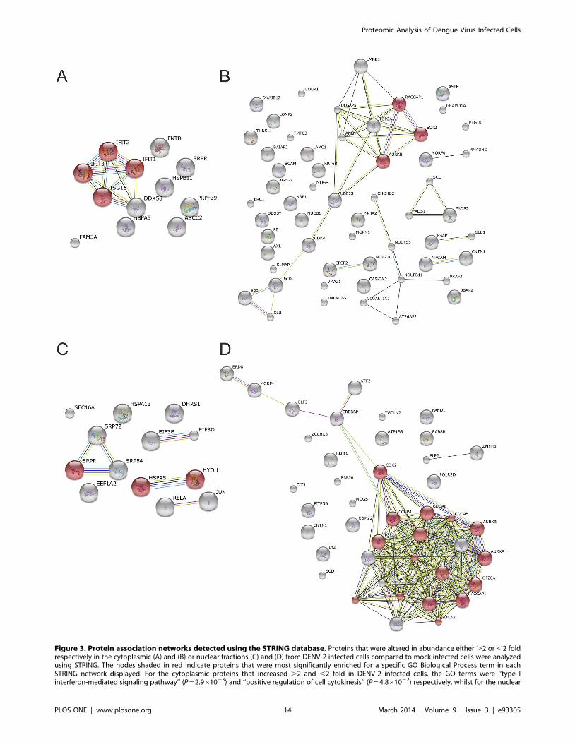
Figure 3. Protein association networks detected using the STRING database. Proteins that were altered in abundance either .2 or ,2 foldrespectively in the cytoplasmic (A) and (B) or nuclear fractions (C) and (D) from DENV-2 infected cells compared to mock infected cells were analyzedusing STRING. The nodes shaded in red indicate proteins that were most significantly enriched for a specific GO Biological Process term in eachSTRING network displayed. For the cytoplasmic proteins that increased .2 and ,2 fold in DENV-2 infected cells, the GO terms were ‘‘type Iinterferon-mediated signaling pathway’’ (P = 2.961023) and ‘‘positive regulation of cell cytokinesis’’ (P = 4.861022) respectively, whilst for the nuclear
Proteomic Analysis of Dengue Virus Infected Cells
PLOS ONE | www.plosone.org 14 March 2014 | Volume 9 | Issue 3 | e93305
proteins and their SILAC ratios in DENV-2 compared to mock
infected cells are shown in Table 5 and a brief summary of their
function/s can be described as follows. Cathepsin L1 (CTSL1) is a
lysosomal cysteine proteinase that is primarily involved in protein
degradation and processing antigen for MHC class II presentation
[84]. CTSL1 is also recruited to the nucleus in mammalian cells
where it is known to cleave transcription factors and the tail of
histone H3 [85]. ELKS/Rab6-interacting/CAST family member
1 (ERC1) has been shown to regulate the activation of NF-kB [86]
and trafficking of Ras-related proteins Rab-6A and Rab8A from
the Golgi to the endosome [87,88], both of which are potentially
important in DENV infection. Importin-subunit a2 (KPNA2) is
involved in the nuclear transport of proteins. Importin-a is an
adaptor protein that binds to a nuclear localization sequence on a
cargo protein and in turn binds to importin-b to form a complex
that is transported through the nuclear pore complex [89]. There
are seven human isoforms of importin-a [90]. Although KPNA1,
3, 4 and 5 were also detected in the analysis, only KPNA2 was
found to be significantly decreased in both the cytoplasmic and
nuclear fractions of DENV-2 infected cells. The murine counter-
part of KPNA2 has been previously been shown to bind to the
DENV-2 NS5 protein which accumulates in the nucleus during
infection [43,55]. Mitofusin 1 (MFN1) is a GTPase that is localized
to the mitochondrial outer membrane and regulates mitochondrial
fusion [91]. A number of reports have implicated mitochondrial
fusion dynamics and MFN1 in RIG-I mediated signaling and
shown that MFN1 interacts with mitochondrial antiviral-signaling
protein, suggesting MFN1 plays a role in the cellular anti-viral
response [92,93]. PRA1 family protein 2 (PRAF2) is an integral
membrane protein with four-transmembrane domains that is
localized to the ER and trans-Golgi network and is believed to play
a regulatory role in vesicular trafficking [94,95]. PRAF2 is also
known to interact with C-C chemokine receptor type 5 (CCR5)
[94,96] and Bcl-xL to modulate cell survival [96]. Ubiquitin-
conjugating enzyme E2 S (UBE2S) functions as an auxiliary factor
in the multi-subunit anaphase-promoting complex (APC), a cell
cycle-regulated E3 ubiquitin ligase that controls progression
through mitosis [97]. A number of viruses are now known to
modulate the activity of the APC, including through the targeting
of APC subunits for degradation, to facilitate their replication [98].
Hypoxia up-regulated protein 1 (HYOU1) is an inducible ER
chaperone protein that is upregulated in response to cellular
stresses including the unfolded protein response and hypoxia.
HYOU1 is known to be cytoprotective and is involved in anti-
apoptotic signaling mechanisms [99,100].
Initially the amounts of the seven proteins were analyzed in the
SILAC labeled cytoplasmic and nuclear fractions used for the
MS/MS analysis (Figure 4). In agreement with the MS/MS
proteins that increased .2 and ,2 fold in DENV-2 infected cells, the GO terms were ‘‘activation of signaling proteins involved in the unfolded proteinresponse’’ (P = 4.461022) and ‘‘cell division’’ (P = 5.2610212).doi:10.1371/journal.pone.0093305.g003
Table 4. DAVID analysis of proteins significantly altered in amount in DENV-2 infected cells.
Nucleara Cytoplasmic
.2 fold increase Signal recognition particle/ribonucleoprotein binding/protein export(3.44)
IFN induced tetratricopeptide repeat (2.99)
Translation factor activity/nucleic acid binding (2.13)
Heat shock/ER (1.94)
Protein export (KEGG pathway; p value = 2.261025) RIG-I-like receptor signalling (KEGG pathway; p value = 4.161022)
.1.5 fold increase ER membrane/nuclear envelope-ER network (7.73) IFN induced tetratricopeptide repeat (2.19)
ER lumen/membrane-bound vesicle (5.68) ER membrane/nuclear envelope-ER network (1.96)
Co-translational protein targeting/protein folding (5.29)
Protein export (KEGG pathway; p value = 6.861028) Alanine, aspartate and glutamate metabolism (KEGG pathway; p value= 6.561022)
.2 fold decrease Cell cycle regulation/mitosis/M phase (8.97) Electron transport/oxidation reduction (1.71)
Nuclear lumen/nucleoplasm (5.95) Oxidoreductase activity/biosynthesis of unsaturated fatty acids/fattyacid biosynthesis (1.62)
Nucleotide/ATP/purine binding (3.75) Cell morphogenesis/cell adhesion/extracellular matrix (1.44)
Cell cycle (KEGG pathway; p value = 2.761023) Biosynthesis of unsaturated fatty acids (KEGG pathway; p value= 2.661023)
.1.5 fold decrease Nuclear lumen/nucleoplasm (15.77) Cell division/cell cycle (2.45)
Cell cycle regulation/mitosis/M phase (12.01) Oxidoreductase activity/iron ion binding/(2.3)
Chromosome/transcription/DNA binding (5.73) ER/ER membrane (2.1)
Cell cycle (KEGG pathway; p value = 5.261024) Steroid biosynthesis (KEGG pathway; p value = 6.261024),Biosynthesis of unsaturated fatty acids (KEGG pathway; p value= 2.161022) Lysosome (KEGG pathway; p value = 3.161022)
aProteins that showed a .2 or 1.5 fold change in abundance in DENV-2 infected A549 cells at 28 hours p.i. were analyzed for the presence of enriched groups ofproteins and proteins involved in specific host pathways using DAVID. The functional descriptors for the 3 clusters of proteins that were most enriched for eachcategory (with an EASE score greater than 1.3 (corresponding to a p value ,0.05)) are shown with the corresponding EASE score. The two KEGG pathways that weremost significantly enriched (with a p value ,0.05) are also shown with the corresponding p value.doi:10.1371/journal.pone.0093305.t004
Proteomic Analysis of Dengue Virus Infected Cells
PLOS ONE | www.plosone.org 15 March 2014 | Volume 9 | Issue 3 | e93305
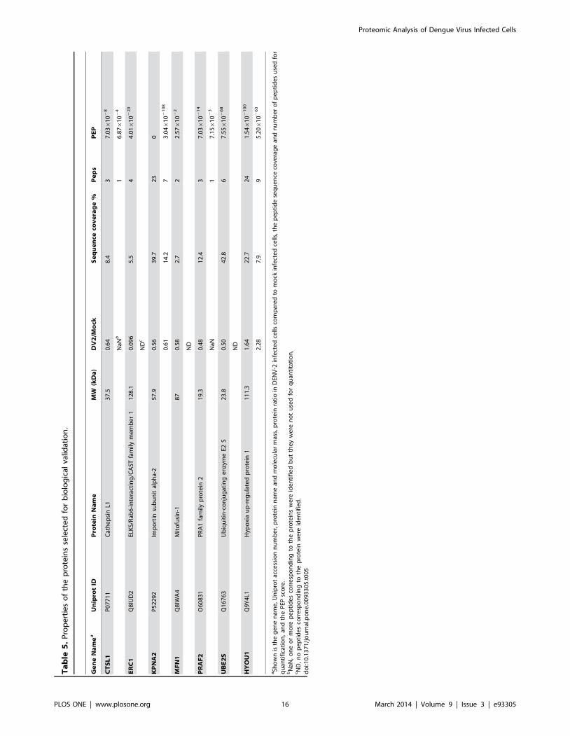
Ta
ble
5.
Pro
pe
rtie
so
fth
ep
rote
ins
sele
cte
dfo
rb
iolo
gic
alva
lidat
ion
.
Ge
ne
Na
me
aU
nip
rot
IDP
rote
inN
am
eM
W(k
Da
)D
V2
/Mo
ckS
eq
ue
nce
cov
era
ge
%P
ep
sP
EP
CT
SL
1P
07
71
1C
ath
ep
sin
L13
7.5
0.6
48
.43
7.0
361
02
8
NaN
b1
6.8
761
02
4
ER
C1
Q8
IUD
2EL
KS/
Rab
6-i
nte
ract
ing
/CA
STfa
mily
me
mb
er
11
28
.10
.09
65
.54
4.0
161
02
20
ND
c
KP
NA
2P
52
29
2Im
po
rtin
sub
un
ital
ph
a-2
57
.90
.56
39
.72
30
0.6
11
4.2
73
.046
10
21
08
MF
N1
Q8
IWA
4M
ito
fusi
n-1
87
0.5
82
.72
2.5
761
02
2
ND
PR
AF
2O
60
83
1P
RA
1fa
mily
pro
tein
21
9.3
0.4
81
2.4
37
.036
10
21
4
NaN
17
.156
10
23
UB
E2
SQ
16
76
3U
biq
uit
in-c
on
jug
atin
ge
nzy
me
E2S
23
.80
.50
42
.86
7.5
561
02
68
ND
HY
OU
1Q
9Y
4L1
Hyp
oxi
au
p-r
eg
ula
ted
pro
tein
11
11
.31
.64
22
.72
41
.546
10
21
00
2.2
87
.99
5.2
061
02
63
aSh
ow
nis
the
ge
ne
nam
e,U
nip
rot
acce
ssio
nn
um
be
r,p
rote
inn
ame
and
mo
lecu
lar
mas
s,p
rote
inra
tio
inD
ENV
-2in
fect
ed
cells
com
par
ed
tom
ock
infe
cte
dce
lls,t
he
pe
pti
de
seq
ue
nce
cove
rag
ean
dn
um
be
ro
fp
ep
tid
es
use
dfo
rq
uan
tifi
cati
on
,an
dth
eP
EPsc
ore
.b
NaN
,o
ne
or
mo
rep
ep
tid
es
corr
esp
on
din
gto
the
pro
tein
sw
ere
ide
nti
fie
db
ut
the
yw
ere
no
tu
sed
for
qu
anti
tati
on
,cN
D,
no
pe
pti
de
sco
rre
spo
nd
ing
toth
ep
rote
inw
ere
ide
nti
fie
d.
do
i:10
.13
71
/jo
urn
al.p
on
e.0
09
33
05
.t0
05
Proteomic Analysis of Dengue Virus Infected Cells
PLOS ONE | www.plosone.org 16 March 2014 | Volume 9 | Issue 3 | e93305
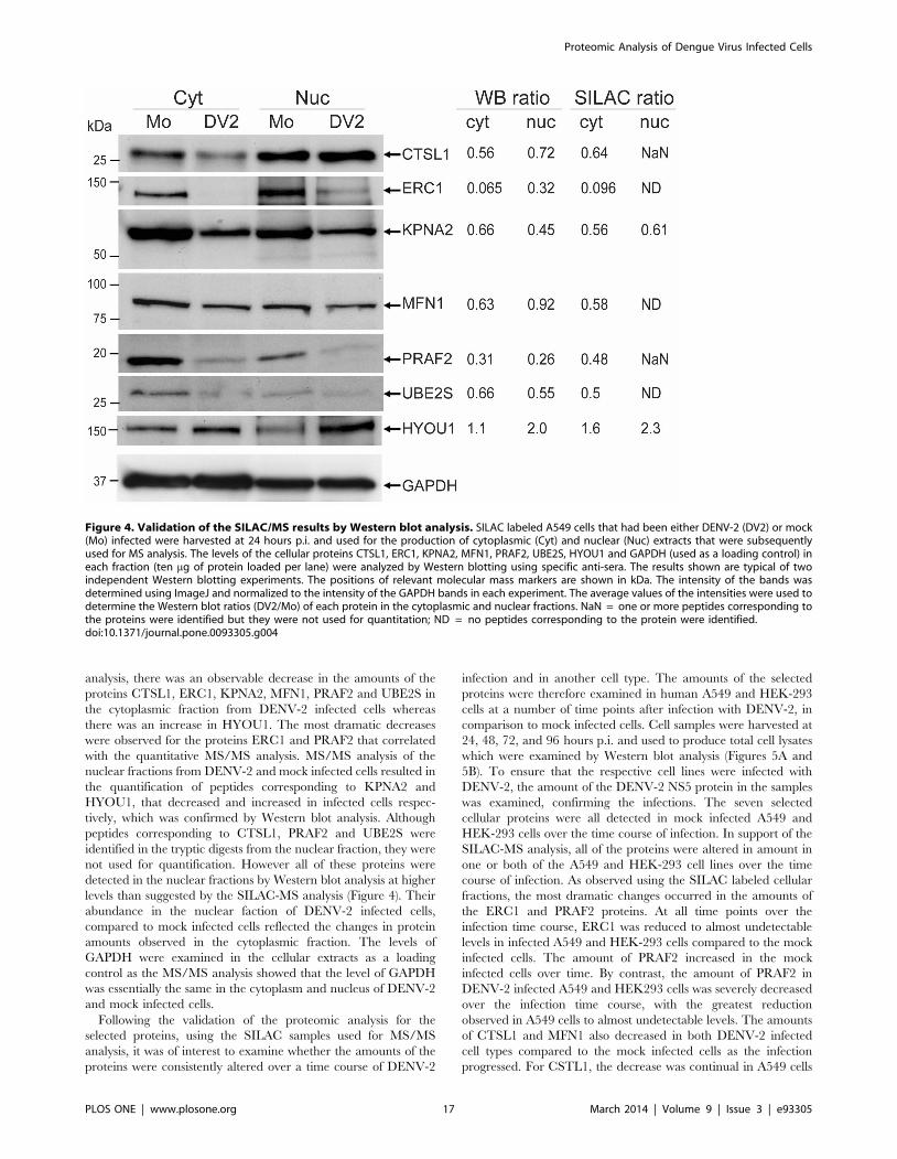
analysis, there was an observable decrease in the amounts of the
proteins CTSL1, ERC1, KPNA2, MFN1, PRAF2 and UBE2S in
the cytoplasmic fraction from DENV-2 infected cells whereas
there was an increase in HYOU1. The most dramatic decreases
were observed for the proteins ERC1 and PRAF2 that correlated
with the quantitative MS/MS analysis. MS/MS analysis of the
nuclear fractions from DENV-2 and mock infected cells resulted in
the quantification of peptides corresponding to KPNA2 and
HYOU1, that decreased and increased in infected cells respec-
tively, which was confirmed by Western blot analysis. Although
peptides corresponding to CTSL1, PRAF2 and UBE2S were
identified in the tryptic digests from the nuclear fraction, they were
not used for quantification. However all of these proteins were
detected in the nuclear fractions by Western blot analysis at higher
levels than suggested by the SILAC-MS analysis (Figure 4). Their
abundance in the nuclear faction of DENV-2 infected cells,
compared to mock infected cells reflected the changes in protein
amounts observed in the cytoplasmic fraction. The levels of
GAPDH were examined in the cellular extracts as a loading
control as the MS/MS analysis showed that the level of GAPDH
was essentially the same in the cytoplasm and nucleus of DENV-2
and mock infected cells.
Following the validation of the proteomic analysis for the
selected proteins, using the SILAC samples used for MS/MS
analysis, it was of interest to examine whether the amounts of the
proteins were consistently altered over a time course of DENV-2
infection and in another cell type. The amounts of the selected
proteins were therefore examined in human A549 and HEK-293
cells at a number of time points after infection with DENV-2, in
comparison to mock infected cells. Cell samples were harvested at
24, 48, 72, and 96 hours p.i. and used to produce total cell lysates
which were examined by Western blot analysis (Figures 5A and
5B). To ensure that the respective cell lines were infected with
DENV-2, the amount of the DENV-2 NS5 protein in the samples
was examined, confirming the infections. The seven selected
cellular proteins were all detected in mock infected A549 and
HEK-293 cells over the time course of infection. In support of the
SILAC-MS analysis, all of the proteins were altered in amount in
one or both of the A549 and HEK-293 cell lines over the time
course of infection. As observed using the SILAC labeled cellular
fractions, the most dramatic changes occurred in the amounts of
the ERC1 and PRAF2 proteins. At all time points over the
infection time course, ERC1 was reduced to almost undetectable
levels in infected A549 and HEK-293 cells compared to the mock
infected cells. The amount of PRAF2 increased in the mock
infected cells over time. By contrast, the amount of PRAF2 in
DENV-2 infected A549 and HEK293 cells was severely decreased
over the infection time course, with the greatest reduction
observed in A549 cells to almost undetectable levels. The amounts
of CTSL1 and MFN1 also decreased in both DENV-2 infected
cell types compared to the mock infected cells as the infection
progressed. For CSTL1, the decrease was continual in A549 cells
Figure 4. Validation of the SILAC/MS results by Western blot analysis. SILAC labeled A549 cells that had been either DENV-2 (DV2) or mock(Mo) infected were harvested at 24 hours p.i. and used for the production of cytoplasmic (Cyt) and nuclear (Nuc) extracts that were subsequentlyused for MS analysis. The levels of the cellular proteins CTSL1, ERC1, KPNA2, MFN1, PRAF2, UBE2S, HYOU1 and GAPDH (used as a loading control) ineach fraction (ten mg of protein loaded per lane) were analyzed by Western blotting using specific anti-sera. The results shown are typical of twoindependent Western blotting experiments. The positions of relevant molecular mass markers are shown in kDa. The intensity of the bands wasdetermined using ImageJ and normalized to the intensity of the GAPDH bands in each experiment. The average values of the intensities were used todetermine the Western blot ratios (DV2/Mo) of each protein in the cytoplasmic and nuclear fractions. NaN = one or more peptides corresponding tothe proteins were identified but they were not used for quantitation; ND = no peptides corresponding to the protein were identified.doi:10.1371/journal.pone.0093305.g004
Proteomic Analysis of Dengue Virus Infected Cells
PLOS ONE | www.plosone.org 17 March 2014 | Volume 9 | Issue 3 | e93305
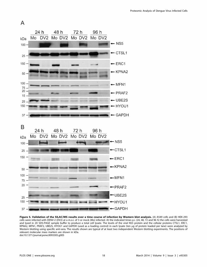
Figure 5. Validation of the SILAC/MS results over a time course of infection by Western blot analysis. (A) A549 cells and (B) HEK-293cells were infected with DENV-2 (DV2) at a m.o.i. of 3 or mock (Mo) infected. At the indicated times p.i. (24, 48, 72 and 96 h) the cells were harvestedand lysed in 2X SDS-PAGE sample buffer to produce a total cell lysate. The levels of the viral NS5 protein and the cellular proteins CTSL1, ERC1,KPNA2, MFN1, PRAF2, UBE2S, HYOU1 and GAPDH (used as a loading control) in each lysate (ten mg of protein loaded per lane) were analyzed byWestern blotting using specific anti-sera. The results shown are typical of at least two independent Western blotting experiments. The positions ofrelevant molecular mass markers are shown in kDa.doi:10.1371/journal.pone.0093305.g005
Proteomic Analysis of Dengue Virus Infected Cells
PLOS ONE | www.plosone.org 18 March 2014 | Volume 9 | Issue 3 | e93305
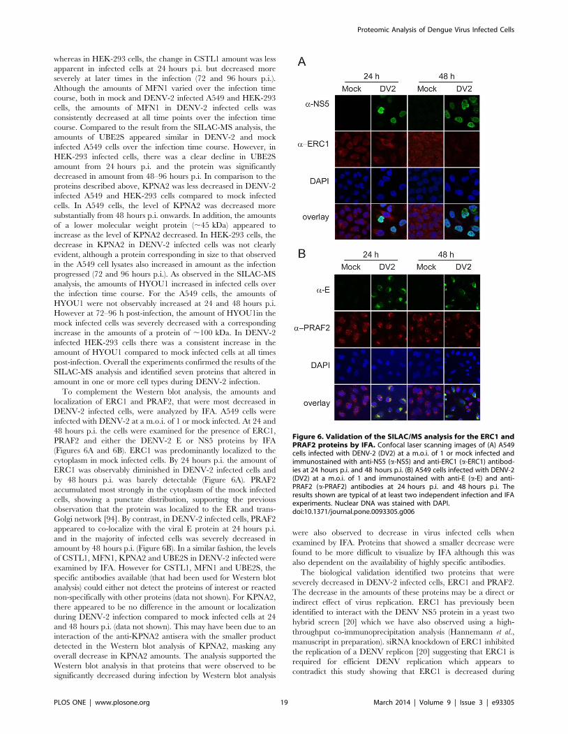
whereas in HEK-293 cells, the change in CSTL1 amount was less
apparent in infected cells at 24 hours p.i. but decreased more
severely at later times in the infection (72 and 96 hours p.i.).
Although the amounts of MFN1 varied over the infection time
course, both in mock and DENV-2 infected A549 and HEK-293
cells, the amounts of MFN1 in DENV-2 infected cells was
consistently decreased at all time points over the infection time
course. Compared to the result from the SILAC-MS analysis, the
amounts of UBE2S appeared similar in DENV-2 and mock
infected A549 cells over the infection time course. However, in
HEK-293 infected cells, there was a clear decline in UBE2S
amount from 24 hours p.i. and the protein was significantly
decreased in amount from 48–96 hours p.i. In comparison to the
proteins described above, KPNA2 was less decreased in DENV-2
infected A549 and HEK-293 cells compared to mock infected
cells. In A549 cells, the level of KPNA2 was decreased more
substantially from 48 hours p.i. onwards. In addition, the amounts
of a lower molecular weight protein (,45 kDa) appeared to
increase as the level of KPNA2 decreased. In HEK-293 cells, the
decrease in KPNA2 in DENV-2 infected cells was not clearly
evident, although a protein corresponding in size to that observed
in the A549 cell lysates also increased in amount as the infection
progressed (72 and 96 hours p.i.). As observed in the SILAC-MS
analysis, the amounts of HYOU1 increased in infected cells over
the infection time course. For the A549 cells, the amounts of
HYOU1 were not observably increased at 24 and 48 hours p.i.
However at 72–96 h post-infection, the amount of HYOU1in the
mock infected cells was severely decreased with a corresponding
increase in the amounts of a protein of ,100 kDa. In DENV-2
infected HEK-293 cells there was a consistent increase in the
amount of HYOU1 compared to mock infected cells at all times
post-infection. Overall the experiments confirmed the results of the
SILAC-MS analysis and identified seven proteins that altered in
amount in one or more cell types during DENV-2 infection.
To complement the Western blot analysis, the amounts and
localization of ERC1 and PRAF2, that were most decreased in
DENV-2 infected cells, were analyzed by IFA. A549 cells were
infected with DENV-2 at a m.o.i. of 1 or mock infected. At 24 and
48 hours p.i. the cells were examined for the presence of ERC1,
PRAF2 and either the DENV-2 E or NS5 proteins by IFA
(Figures 6A and 6B). ERC1 was predominantly localized to the
cytoplasm in mock infected cells. By 24 hours p.i. the amount of
ERC1 was observably diminished in DENV-2 infected cells and
by 48 hours p.i. was barely detectable (Figure 6A). PRAF2
accumulated most strongly in the cytoplasm of the mock infected
cells, showing a punctate distribution, supporting the previous
observation that the protein was localized to the ER and trans-
Golgi network [94]. By contrast, in DENV-2 infected cells, PRAF2
appeared to co-localize with the viral E protein at 24 hours p.i.
and in the majority of infected cells was severely decreased in
amount by 48 hours p.i. (Figure 6B). In a similar fashion, the levels
of CSTL1, MFN1, KPNA2 and UBE2S in DENV-2 infected were
examined by IFA. However for CSTL1, MFN1 and UBE2S, the
specific antibodies available (that had been used for Western blot
analysis) could either not detect the proteins of interest or reacted
non-specifically with other proteins (data not shown). For KPNA2,
there appeared to be no difference in the amount or localization
during DENV-2 infection compared to mock infected cells at 24
and 48 hours p.i. (data not shown). This may have been due to an
interaction of the anti-KPNA2 antisera with the smaller product
detected in the Western blot analysis of KPNA2, masking any
overall decrease in KPNA2 amounts. The analysis supported the
Western blot analysis in that proteins that were observed to be
significantly decreased during infection by Western blot analysis
were also observed to decrease in virus infected cells when
examined by IFA. Proteins that showed a smaller decrease were
found to be more difficult to visualize by IFA although this was
also dependent on the availability of highly specific antibodies.
The biological validation identified two proteins that were
severely decreased in DENV-2 infected cells, ERC1 and PRAF2.
The decrease in the amounts of these proteins may be a direct or
indirect effect of virus replication. ERC1 has previously been
identified to interact with the DENV NS5 protein in a yeast two
hybrid screen [20] which we have also observed using a high-
throughput co-immunoprecipitation analysis (Hannemann et al.,
manuscript in preparation). siRNA knockdown of ERC1 inhibited
the replication of a DENV replicon [20] suggesting that ERC1 is
required for efficient DENV replication which appears to
contradict this study showing that ERC1 is decreased during
Figure 6. Validation of the SILAC/MS analysis for the ERC1 andPRAF2 proteins by IFA. Confocal laser scanning images of (A) A549cells infected with DENV-2 (DV2) at a m.o.i. of 1 or mock infected andimmunostained with anti-NS5 (a-NS5) and anti-ERC1 (a-ERC1) antibod-ies at 24 hours p.i. and 48 hours p.i. (B) A549 cells infected with DENV-2(DV2) at a m.o.i. of 1 and immunostained with anti-E (a-E) and anti-PRAF2 (a-PRAF2) antibodies at 24 hours p.i. and 48 hours p.i. Theresults shown are typical of at least two independent infection and IFAexperiments. Nuclear DNA was stained with DAPI.doi:10.1371/journal.pone.0093305.g006
Proteomic Analysis of Dengue Virus Infected Cells
PLOS ONE | www.plosone.org 19 March 2014 | Volume 9 | Issue 3 | e93305
DENV infection. However it may be that ERC1 plays different
roles at various stages of the DENV lifecycle which illustrates how
the use of different high-throughput approaches can complement
one another to increase our understanding of the role of cellular
proteins in the DENV lifecycle.
Conclusions
The results of this study demonstrate for the first time the power
of quantitative LC-MS/MS for investigating the host cell response
to DENV infection. Compared to traditional 2D-PAGE based
analysis, thousands of proteins can be both identified and
quantified in response to DENV infection. Proteins that are either
significantly altered or remain unchanged in amount during
infection were identified, providing a resource for further
bioinformatic analyses and biological validation. Using this
approach we identified specific host proteins and cellular processes
that are modulated during DENV infection. A number of the
cellular processes identified have been associated with DENV
infection in previous transcriptomic and siRNA studies, validating
our results, however others were novel and require further
investigation. In addition, changes in the amounts of specific
proteins that were not associated with well studied pathways were
identified and validated, and these proteins can now be assessed
for their role in the DENV lifecycle. The use of high-throughput
quantitative proteomic analysis complements other high-through-
put approaches, and in combination offers the possibility to
identify cellular proteins that may be targets against which novel
anti-viral strategies can be developed which are urgently needed
against this important disease.
Supporting Information
Table S1 List of the proteins identified and quantifiedby SILAC LC-MS/MS in cytoplasmic and nuclearfractions from DENV-2 infected A549 cells.
(XLS)
Acknowledgments
Imaging was performed at the Wolfson Bioimaging Facility, University of
Bristol.
Author Contributions
Conceived and designed the experiments: H-HC HH KJM DAM ADD.
Performed the experiments: H-HC HH KJM DAM ADD. Analyzed the
data: H-HC HH KJM DAM ADD. Wrote the paper: H-HC HH KJM
DAM ADD.
References
1. WHO (2009) Dengue - Guidelines for diagnosis, Treatment, prevention and
control. WHO Press.
2. Bhatt S, Gething PW, Brady OJ, Messina JP, Farlow AW, et al. (2013) The
global distribution and burden of dengue. Nature 496: 504–507.
3. Lindenbach BD, Thiel HJ, Rice CM (2007) Flaviviridae: the viruses and their
replication. In: Knipe DM, Howley PM, editors. Fields Virology, 5th Edition.
Philadelphia: Lippincott-Raven Publishers. pp. 1101–1152.
4. Welsch S, Miller S, Romero-Brey I, Merz A, Bleck CK, et al. (2009)
Composition and three-dimensional architecture of the dengue virus replication
and assembly sites. Cell Host Microbe 5: 365–375.
5. Krishnan MN, Ng A, Sukumaran B, Gilfoy FD, Uchil PD, et al. (2008) RNA
interference screen for human genes associated with West Nile virus infection.
Nature 455: 242–245.
6. Sessions OM, Barrows NJ, Souza-Neto JA, Robinson TJ, Hershey CL, et al.
(2009) Discovery of insect and human dengue virus host factors. Nature 458:
1047–1050.
7. Balas C, Kennel A, Deauvieau F, Sodoyer R, Arnaud-Barbe N, et al. (2011)
Different innate signatures induced in human monocyte-derived dendritic cells
by wild-type dengue 3 virus, attenuated but reactogenic dengue 3 vaccine virus,
or attenuated nonreactogenic dengue 1–4 vaccine virus strains. J Infect Dis 203:
103–108.
8. Fink J, Gu F, Ling L, Tolfvenstam T, Olfat F, et al. (2007) Host gene expression
profiling of dengue virus infection in cell lines and patients. PLoS Negl Trop Dis
1: e86.
9. Sessions OM, Tan Y, Goh KC, Liu Y, Tan P, et al. (2013) Host cell
transcriptome profile during wild-type and attenuated dengue virus infection.
PLoS Negl Trop Dis 7: e2107.
10. Silveira GF, Meyer F, Delfraro A, Mosimann AL, Coluchi N, et al. (2011)
Dengue virus type 3 isolated from a fatal case with visceral complications induces
enhanced proinflammatory responses and apoptosis of human dendritic cells.
J Virol 85: 5374–5383.
11. Ubol S, Masrinoul P, Chaijaruwanich J, Kalayanarooj S, Charoensirisuthikul T,
et al. (2008) Differences in global gene expression in peripheral blood
mononuclear cells indicate a significant role of the innate responses in
progression of dengue fever but not dengue hemorrhagic fever. J Infect Dis
197: 1459–1467.
12. Warke RV, Martin KJ, Giaya K, Shaw SK, Rothman AL, et al. (2008) TRAIL
is a novel antiviral protein against dengue virus. J Virol 82: 555–564.
13. Warke RV, Xhaja K, Martin KJ, Fournier MF, Shaw SK, et al. (2003) Dengue
virus induces novel changes in gene expression of human umbilical vein
endothelial cells. J Virol 77: 11822–11832.
14. Devignot S, Sapet C, Duong V, Bergon A, Rihet P, et al. (2010) Genome-wide
expression profiling deciphers host responses altered during dengue shock
syndrome and reveals the role of innate immunity in severe dengue. PLoS One
5: e11671.
15. Hoang LT, Lynn DJ, Henn M, Birren BW, Lennon NJ, et al. (2010) The early
whole-blood transcriptional signature of dengue and features associated with
progression to dengue shock syndrome in Vietnamese children and young adults.
J Virol.
16. Loke P, Hammond SN, Leung JM, Kim CC, Batra S, et al. (2010) Gene
expression patterns of dengue virus-infected children from nicaragua reveal a
distinct signature of increased metabolism. PLoS Negl Trop Dis 4: e710.
17. Nascimento EJ, Braga-Neto U, Calzavara-Silva CE, Gomes AL, Abath FG, et
al. (2009) Gene expression profiling during early acute febrile stage of dengue
infection can predict the disease outcome. PLoS One 4: e7892.
18. Simmons CP, Popper S, Dolocek C, Chau TN, Griffiths M, et al. (2007) Patterns
of host genome-wide gene transcript abundance in the peripheral blood of
patients with acute dengue hemorrhagic fever. J Infect Dis 195: 1097–1107.
19. Tolfvenstam T, Lindblom A, Schreiber MJ, Ling L, Chow A, et al. (2011)
Characterization of early host responses in adults with dengue disease. BMC
Infect Dis 11: 209.
20. Khadka S, Vangeloff AD, Zhang C, Siddavatam P, Heaton NS, et al. (2011) A
physical interaction network of dengue virus and human proteins. Mol Cell
Proteomics.
21. Le Breton M, Meyniel-Schicklin L, Deloire A, Coutard B, Canard B, et al.
(2011) Flavivirus NS3 and NS5 proteins interaction network: a high-throughput
yeast two-hybrid screen. BMC Microbiol 11: 234.
22. Mairiang D, Zhang H, Sodja A, Murali T, Suriyaphol P, et al. (2013)
Identification of new protein interactions between dengue fever virus and its
hosts, human and mosquito. PLoS One 8: e53535.
23. Kanlaya R, Pattanakitsakul SN, Sinchaikul S, Chen ST, Thongboonkerd V
(2009) Alterations in actin cytoskeletal assembly and junctional protein
complexes in human endothelial cells induced by dengue virus infection and
mimicry of leukocyte transendothelial migration. J Proteome Res 8: 2551–2562.
24. Kanlaya R, Pattanakitsakul SN, Sinchaikul S, Chen ST, Thongboonkerd V
(2010) The Ubiquitin-Proteasome Pathway Is Important for Dengue Virus
Infection in Primary Human Endothelial Cells. J Proteome Res.
25. Pattanakitsakul SN, Poungsawai J, Kanlaya R, Sinchaikul S, Chen ST, et al.
(2010) Association of Alix with late endosomal lysobisphosphatidic acid is
important for dengue virus infection in human endothelial cells. J Proteome Res
9: 4640–4648.
26. Pattanakitsakul SN, Rungrojcharoenkit K, Kanlaya R, Sinchaikul S, Noisakran
S, et al. (2007) Proteomic analysis of host responses in HepG2 cells during
dengue virus infection. J Proteome Res 6: 4592–4600.
27. Wati S, Soo ML, Zilm P, Li P, Paton AW, et al. (2009) Dengue virus infection
induces upregulation of GRP78, which acts to chaperone viral antigen
production. J Virol 83: 12871–12880.
28. Patramool S, Surasombatpattana P, Luplertlop N, Seveno M, Choumet V, et al.
(2011) Proteomic analysis of an Aedes albopictus cell line infected with Dengue
serotypes 1 and 3 viruses. Parasit Vectors 4: 138.
29. Tchankouo-Nguetcheu S, Khun H, Pincet L, Roux P, Bahut M, et al. (2010)
Differential protein modulation in midguts of Aedes aegypti infected with
chikungunya and dengue 2 viruses. PLoS One 5.
30. Albuquerque LM, Trugilho MR, Chapeaurouge A, Jurgilas PB, Bozza PT, et al.
(2009) Two-dimensional difference gel electrophoresis (DiGE) analysis of
plasmas from dengue fever patients. J Proteome Res 8: 5431–5441.
31. Brasier AR, Garcia J, Wiktorowicz JE, Spratt HM, Comach G, et al. (2012)
Discovery Proteomics and Nonparametric Modeling Pipeline in the Development
Proteomic Analysis of Dengue Virus Infected Cells
PLOS ONE | www.plosone.org 20 March 2014 | Volume 9 | Issue 3 | e93305

of a Candidate Biomarker Panel for Dengue Hemorrhagic Fever. Clin Transl Sci5: 8–20.
32. Mann M (2006) Functional and quantitative proteomics using SILAC. Nat Rev
Mol Cell Biol 7: 952–958.
33. Walther TC, Mann M (2010) Mass spectrometry-based proteomics in cellbiology. J Cell Biol 190: 491–500.
34. Munday DC, Surtees R, Emmott E, Dove BK, Digard P, et al. (2012) Using
SILAC and quantitative proteomics to investigate the interactions between viraland host proteomes. Proteomics.
35. Gualano RC, Pryor MJ, Cauchi MR, Wright PJ, Davidson AD (1998)Identification of a major determinant of mouse neurovirulence of dengue virus
type 2 using stably cloned genomic-length cDNA. J Gen Virol 79: 437–446.
36. Kroschewski H, Lim SP, Butcher RE, Yap TL, Lescar J, et al. (2008)Mutagenesis of the dengue virus type 2 NS5 methyltransferase domain. J Biol
Chem 283: 19410–19421.
37. Cox J, Mann M (2008) MaxQuant enables high peptide identification rates,individualized p.p.b.-range mass accuracies and proteome-wide protein
quantification. Nat Biotechnol 26: 1367–1372.
38. Cox J, Neuhauser N, Michalski A, Scheltema RA, Olsen JV, et al. (2011)Andromeda: a peptide search engine integrated into the MaxQuant environ-
ment. J Proteome Res 10: 1794–1805.
39. Bhatia VN, Perlman DH, Costello CE, McComb ME (2009) Software tool forresearching annotations of proteins: open-source protein annotation software
with data visualization. Anal Chem 81: 9819–9823.
40. Huang da W, Sherman BT, Lempicki RA (2009) Systematic and integrativeanalysis of large gene lists using DAVID bioinformatics resources. Nat Protoc 4:
44–57.
41. Huang da W, Sherman BT, Lempicki RA (2009) Bioinformatics enrichmenttools: paths toward the comprehensive functional analysis of large gene lists.
Nucleic Acids Res 37: 1–13.
42. Franceschini A, Szklarczyk D, Frankild S, Kuhn M, Simonovic M, et al. (2013)
STRING v9.1: protein-protein interaction networks, with increased coverage
and integration. Nucleic Acids Res 41: D808–815.
43. Hannemann H, Sung PY, Chiu HC, Yousuf A, Bird J, et al. (2013) Serotype
Specific Differences in Dengue Virus Non-Structural Protein 5 Nuclear
Localization. J Biol Chem.
44. Umareddy I, Tang KF, Vasudevan SG, Devi S, Hibberd ML, et al. (2008)
Dengue virus regulates type I interferon signalling in a strain-dependent manner
in human cell lines. J Gen Virol 89: 3052–3062.
45. Yu CY, Chang TH, Liang JJ, Chiang RL, Lee YL, et al. (2012) Dengue virus
targets the adaptor protein MITA to subvert host innate immunity. PLoS Pathog8: e1002780.
46. Pastorino B, Boucomont-Chapeaublanc E, Peyrefitte CN, Belghazi M, Fusai T,
et al. (2009) Identification of cellular proteome modifications in response to WestNile virus infection. Mol Cell Proteomics.
47. Zhang LK, Chai F, Li HY, Xiao G, Guo L (2013) Identification of host proteins
involved in Japanese encephalitis virus infection by quantitative proteomicsanalysis. J Proteome Res 12: 2666–2678.
48. Munoz-Jordan JL, Bosch I (2010) Modulation of the antiviral response by
dengue virus. In: Hanley KA, Weaver SC, editors. Frontiers in Dengue VirusResearch: Caister Academic Press. pp. 121–140.
49. Chang TH, Liao CL, Lin YL (2006) Flavivirus induces interferon-beta gene
expression through a pathway involving RIG-I-dependent IRF-3 and PI3K-dependent NF-kappaB activation. Microbes Infect 8: 157–171.
50. Marianneau P, Cardona A, Edelman L, Deubel V, Despres P (1997) Dengue
virus replication in human hepatoma cells activates NF-kappaB which in turninduces apoptotic cell death. J Virol 71: 3244–3249.
51. Pettersson M, Bessonova M, Gu HF, Groop LC, Jonsson JI (2000)
Characterization, chromosomal localization, and expression during hematopoi-etic differentiation of the gene encoding Arl6ip, ADP-ribosylation-like factor-6
interacting protein (ARL6). Genomics 68: 351–354.
52. Heaton NS, Perera R, Berger KL, Khadka S, Lacount DJ, et al. (2010) Denguevirus nonstructural protein 3 redistributes fatty acid synthase to sites of viral
replication and increases cellular fatty acid synthesis. Proc Natl Acad Sci U S A107: 17345–17350.
53. Diamond DL, Syder AJ, Jacobs JM, Sorensen CM, Walters KA, et al. (2010)
Temporal proteome and lipidome profiles reveal hepatitis C virus-associatedreprogramming of hepatocellular metabolism and bioenergetics. PLoS Pathog 6:
e1000719.
54. Rasmussen AL, Diamond DL, McDermott JE, Gao X, Metz TO, et al. (2011)Systems virology identifies a mitochondrial fatty acid oxidation enzyme,
dodecenoyl coenzyme A delta isomerase, required for hepatitis C virusreplication and likely pathogenesis. J Virol 85: 11646–11654.
55. Pryor MJ, Rawlinson SM, Butcher RE, Barton CL, Waterhouse TA, et al.
(2007) Nuclear localization of dengue virus nonstructural protein 5 through itsimportin alpha/beta-recognized nuclear localization sequences is integral to
viral infection. Traffic 8: 795–807.
56. Sangiambut S, Keelapang P, Aaskov J, Puttikhunt C, Kasinrerk W, et al. (2008)Multiple regions in dengue virus capsid protein contribute to nuclear localization
during virus infection. J Gen Virol 89: 1254–1264.
57. Uchil PD, Kumar AV, Satchidanandam V (2006) Nuclear localization offlavivirus RNA synthesis in infected cells. J Virol 80: 5451–5464.
58. Long HT, Hibberd ML, Hien TT, Dung NM, Van Ngoc T, et al. (2009)
Patterns of gene transcript abundance in the blood of children with severe or
uncomplicated dengue highlight differences in disease evolution and hostresponse to dengue virus infection. J Infect Dis 199: 537–546.
59. Jones M, Davidson A, Hibbert L, Gruenwald P, Schlaak J, et al. (2005) Denguevirus inhibits alpha interferon signaling by reducing STAT2 expression. J Virol
79: 5414–5420.
60. Munoz-Jordan JL, Sanchez-Burgos GG, Laurent-Rolle M, Garcia-Sastre A
(2003) Inhibition of interferon signaling by dengue virus. Proc Natl AcadSci U S A 100: 14333–14338.
61. Diamond MS, Farzan M (2013) The broad-spectrum antiviral functions of IFITand IFITM proteins. Nat Rev Immunol 13: 46–57.
62. Wacher C, Muller M, Hofer MJ, Getts DR, Zabaras R, et al. (2007)Coordinated regulation and widespread cellular expression of interferon-
stimulated genes (ISG) ISG-49, ISG-54, and ISG-56 in the central nervoussystem after infection with distinct viruses. J Virol 81: 860–871.
63. Daffis S, Szretter KJ, Schriewer J, Li J, Youn S, et al. (2010) 29-O methylation ofthe viral mRNA cap evades host restriction by IFIT family members. Nature
468: 452–456.
64. Behera B, Chaudhry R, Pandey A, Mohan A, Dar L, et al. (2009) Co-infections
due to leptospira, dengue and hepatitis E: a diagnostic challenge. J Infect DevCtries 4: 48–50.
65. Jiang D, Weidner JM, Qing M, Pan XB, Guo H, et al. (2010) Identification offive interferon-induced cellular proteins that inhibit west nile virus and dengue
virus infections. J Virol 84: 8332–8341.
66. Fischl W, Bartenschlager R (2011) Exploitation of cellular pathways by Dengue
virus. Curr Opin Microbiol.
67. Paradkar PN, Ooi EE, Hanson BJ, Gubler DJ, Vasudevan SG (2010) Unfolded
protein response (UPR) gene expression during antibody-dependent enhancedinfection of cultured monocytes correlates with dengue disease severity. Biosci
Rep.
68. Umareddy I, Pluquet O, Wang QY, Vasudevan SG, Chevet E, et al. (2007)
Dengue virus serotype infection specifies the activation of the unfolded protein
response. Virol J 4: 91.
69. Yu CY, Hsu YW, Liao CL, Lin YL (2006) Flavivirus infection activates the
XBP1 pathway of the unfolded protein response to cope with endoplasmicreticulum stress. J Virol 80: 11868–11880.
70. Schussek S, Groves PL, Apte SH, Doolan DL (2013) Highly Sensitive
Quantitative Real-Time PCR for the Detection of Liver-Stage Parasite Burden
following Low-Dose Sporozoite Challenge. PLoS One 8: e77811.
71. De Nova-Ocampo M, Villegas-Sepulveda N, del Angel RM (2002) Translation
elongation factor-1alpha, La, and PTB interact with the 39 untranslated regionof dengue 4 virus RNA. Virology 295: 337–347.
72. Chukkapalli V, Heaton NS, Randall G (2012) Lipids at the interface of virus-host interactions. Curr Opin Microbiol 15: 512–518.
73. Perera R, Riley C, Isaac G, Hopf-Jannasch AS, Moore RJ, et al. (2012) Dengue
virus infection perturbs lipid homeostasis in infected mosquito cells. PLoS
Pathog 8: e1002584.
74. Rothwell C, Lebreton A, Young Ng C, Lim JY, Liu W, et al. (2009) Cholesterol
biosynthesis modulation regulates dengue viral replication. Virology.
75. Heaton NS, Randall G (2010) Dengue virus-induced autophagy regulates lipid
metabolism. Cell Host Microbe 8: 422–432.
76. Altindis E (2013) Antibacterial Vaccine Research in 21st Century: From
Inoculation to Genomics Approaches. Curr Top Med Chem.
77. Tanabe K, Zollner G, Vaughan JA, Sattabongkot J, Khuntirat B, et al. (2013)Plasmodium falciparum: Genetic diversity and complexity of infections in an
isolated village in western Thailand. Parasitol Int.
78. Bunyaratvej A, Butthep P, Yoksan S, Bhamarapravati N (1997) Dengue viruses
induce cell proliferation and morphological changes of endothelial cells.
Southeast Asian J Trop Med Public Health 28 Suppl 3: 32–37.
79. Helt AM, Harris E (2005) S-phase-dependent enhancement of dengue virus 2
replication in mosquito cells, but not in human cells. J Virol 79: 13218–13230.
80. Phoolcharoen W, Smith DR (2004) Internalization of the dengue virus is cellcycle modulated in HepG2, but not Vero cells. J Med Virol 74: 434–441.
81. Davy C, Doorbar J (2007) G2/M cell cycle arrest in the life cycle of viruses.Virology 368: 219–226.
82. Kannan RP, Hensley LL, Evers LE, Lemon SM, McGivern DR (2011) HepatitisC virus infection causes cell cycle arrest at the level of initiation of mitosis. J Virol
85: 7989–8001.
83. Munday DC, Emmott E, Surtees R, Lardeau CH, Wu W, et al. (2010)
Quantitative proteomic analysis of A549 cells infected with human respiratory
syncytial virus. Mol Cell Proteomics 9: 2438–2459.
84. Hsing LC, Rudensky AY (2005) The lysosomal cysteine proteases in MHC classII antigen presentation. Immunol Rev 207: 229–241.
85. Reiser J, Adair B, Reinheckel T (2010) Specialized roles for cysteine cathepsinsin health and disease. J Clin Invest 120: 3421–3431.
86. Ducut Sigala JL, Bottero V, Young DB, Shevchenko A, Mercurio F, et al. (2004)Activation of transcription factor NF-kappaB requires ELKS, an IkappaB kinase
regulatory subunit. Science 304: 1963–1967.
87. Grigoriev I, Splinter D, Keijzer N, Wulf PS, Demmers J, et al. (2007) Rab6
regulates transport and targeting of exocytotic carriers. Dev Cell 13: 305–314.
88. Grigoriev I, Yu KL, Martinez-Sanchez E, Serra-Marques A, Smal I, et al. (2011)
Rab6, Rab8, and MICAL3 cooperate in controlling docking and fusion ofexocytotic carriers. Curr Biol 21: 967–974.
89. Stewart M (2007) Molecular mechanism of the nuclear protein import cycle. NatRev Mol Cell Biol 8: 195–208.
Proteomic Analysis of Dengue Virus Infected Cells
PLOS ONE | www.plosone.org 21 March 2014 | Volume 9 | Issue 3 | e93305
90. Mason DA, Stage DE, Goldfarb DS (2009) Evolution of the metazoan-specific
importin alpha gene family. J Mol Evol 68: 351–365.91. West AP, Shadel GS, Ghosh S (2011) Mitochondria in innate immune
responses. Nat Rev Immunol 11: 389–402.
92. Onoguchi K, Onomoto K, Takamatsu S, Jogi M, Takemura A, et al. (2010)Virus-infection or 59ppp-RNA activates antiviral signal through redistribution of
IPS-1 mediated by MFN1. PLoS Pathog 6: e1001012.93. Castanier C, Garcin D, Vazquez A, Arnoult D (2010) Mitochondrial dynamics
regulate the RIG-I-like receptor antiviral pathway. EMBO Rep 11: 133–138.
94. Schweneker M, Bachmann AS, Moelling K (2005) JM4 is a four-transmembraneprotein binding to the CCR5 receptor. FEBS Lett 579: 1751–1758.
95. Fo CS, Coleman CS, Wallick CJ, Vine AL, Bachmann AS (2006) Genomicorganization, expression profile, and characterization of the new protein PRA1
domain family, member 2 (PRAF2). Gene 371: 154–165.
96. Vento MT, Zazzu V, Loffreda A, Cross JR, Downward J, et al. (2010) Praf2 is a
novel Bcl-xL/Bcl-2 interacting protein with the ability to modulate survival ofcancer cells. PLoS One 5: e15636.
97. Garnett MJ, Mansfeld J, Godwin C, Matsusaka T, Wu J, et al. (2009) UBE2S
elongates ubiquitin chains on APC/C substrates to promote mitotic exit. NatCell Biol 11: 1363–1369.
98. Fehr AR, Yu D (2013) Control the Host Cell Cycle: Viral Regulation of theAnaphase-Promoting Complex. J Virol.
99. Arrington DD, Schnellmann RG (2008) Targeting of the molecular chaperone
oxygen-regulated protein 150 (ORP150) to mitochondria and its induction bycellular stress. Am J Physiol Cell Physiol 294: C641–650.
100. Sanson M, Ingueneau C, Vindis C, Thiers JC, Glock Y, et al. (2008) Oxygen-regulated protein-150 prevents calcium homeostasis deregulation and apoptosis
induced by oxidized LDL in vascular cells. Cell Death Differ 15: 1255–1265.
Proteomic Analysis of Dengue Virus Infected Cells
PLOS ONE | www.plosone.org 22 March 2014 | Volume 9 | Issue 3 | e93305
Related Documents