RESEARCH ARTICLE Open Access Proteogenomic mapping of Mycoplasma hyopneumoniae virulent strain 232 Ken Pendarvis 2,5* , Matthew P Padula 3,4 , Jessica L Tacchi 3 , Andrew C Petersen 1 , Steven P Djordjevic 3,4 , Shane C Burgess 2,5 and F Chris Minion 1 Abstract Background: Mycoplasma hyopneumoniae causes respiratory disease in swine and contributes to the porcine respiratory disease complex, a major disease problem in the swine industry. The M. hyopneumoniae strain 232 genome is one of the smallest and best annotated microbial genomes, containing only 728 annotated genes and 691 known proteins. Standard protein databases for mass spectrometry only allow for the identification of known and predicted proteins, which if incorrect can limit our understanding of the biological processes at work. Proteogenomic mapping is a methodology which allows the entire 6-frame genome translation of an organism to be used as a mass spectrometry database to help identify unknown proteins as well as correct and confirm existing annotations. This methodology will be employed to perform an in-depth analysis of the M. hyopneumoniae proteome. Results: Proteomic analysis indicates 483 of 691 (70%) known M. hyopneumoniae strain 232 proteins are expressed under the culture conditions given in this study. Furthermore, 171 of 328 (52%) hypothetical proteins have been confirmed. Proteogenomic mapping resulted in the identification of previously unannotated genes gatC and rpmF and 5-prime extensions to genes mhp063, mhp073, and mhp451, all conserved and annotated in other M. hyopneumoniae strains and Mycoplasma species. Gene prediction with Prodigal, a prokaryotic gene predicting program, completely supports the new genomic coordinates calculated using proteogenomic mapping. Conclusions: Proteogenomic mapping showed that the protein coding genes of the M. hyopneumoniae strain 232 identified in this study are well annotated. Only 1.8% of mapped peptides did not correspond to genes defined by the current genome annotation. This study also illustrates how proteogenomic mapping can be an important tool to help confirm, correct and append known gene models when using a genome sequence as search space for peptide mass spectra. Using a gene prediction program which scans for a wide variety of promoters can help ensure genes are accurately predicted or not missed completely. Furthermore, protein extraction using differential detergent fractionation effectively increases the number of membrane and cytoplasmic proteins identifiable my mass spectrometry. Keywords: Mycoplasma hyopneumoniae, Proteome, Swine pathogen, Proteogenomic, Mapping, Mass spectrometry Background Mycoplasma hyopneumoniae is the etiological agent of porcine enzootic pneumonia [1], causing substantial eco- nomic losses to the pig industry through reduced aver- age daily weight gain and efficiency of feed utilization, prophylactic and therapeutic costs, and mortality [1,2]. When co-infections occur with a secondary (bacterial or viral) infection, the respiratory disease is more severe and has been designated as porcine respiratory disease complex [1], an even more devastating disease. The viru- lence factors of M. hyopneumoniae are largely unknown and to better understand the mechanisms involved, we are studying genetic processes in M. hyopneumoniae both in vitro and in vivo [3-8]. Recent microarray studies of global transcriptional changes clearly show that under the culture conditions used in this study, 627 of the 691 known protein coding genes are transcribed [4-8]. M. hyopneumoniae also responds to environmental changes, and under various stressors, all annotated genes are transcribed [4-8]. Further, a recent study from our * Correspondence: [email protected] 2 School of Animal and Comparative Biomedical Sciences, University of Arizona, Tucson, AZ, USA 5 Bio5 Institute, School of Animal and Comparative Biomedical Sciences, University of Arizona, Tucson, AZ, USA Full list of author information is available at the end of the article © 2014 Pendarvis et al.; licensee BioMed Central Ltd. This is an Open Access article distributed under the terms of the Creative Commons Attribution License (http://creativecommons.org/licenses/by/4.0), which permits unrestricted use, distribution, and reproduction in any medium, provided the original work is properly credited. The Creative Commons Public Domain Dedication waiver (http://creativecommons.org/publicdomain/zero/1.0/) applies to the data made available in this article, unless otherwise stated. Pendarvis et al. BMC Genomics 2014, 15:576 http://www.biomedcentral.com/1471-2164/15/576

Welcome message from author
This document is posted to help you gain knowledge. Please leave a comment to let me know what you think about it! Share it to your friends and learn new things together.
Transcript

Pendarvis et al. BMC Genomics 2014, 15:576http://www.biomedcentral.com/1471-2164/15/576
RESEARCH ARTICLE Open Access
Proteogenomic mapping of Mycoplasmahyopneumoniae virulent strain 232Ken Pendarvis2,5*, Matthew P Padula3,4, Jessica L Tacchi3, Andrew C Petersen1, Steven P Djordjevic3,4,Shane C Burgess2,5 and F Chris Minion1
Abstract
Background: Mycoplasma hyopneumoniae causes respiratory disease in swine and contributes to the porcinerespiratory disease complex, a major disease problem in the swine industry. The M. hyopneumoniae strain 232 genomeis one of the smallest and best annotated microbial genomes, containing only 728 annotated genes and 691 knownproteins. Standard protein databases for mass spectrometry only allow for the identification of known and predictedproteins, which if incorrect can limit our understanding of the biological processes at work. Proteogenomic mapping isa methodology which allows the entire 6-frame genome translation of an organism to be used as a mass spectrometrydatabase to help identify unknown proteins as well as correct and confirm existing annotations. This methodology willbe employed to perform an in-depth analysis of the M. hyopneumoniae proteome.
Results: Proteomic analysis indicates 483 of 691 (70%) known M. hyopneumoniae strain 232 proteins are expressedunder the culture conditions given in this study. Furthermore, 171 of 328 (52%) hypothetical proteins have beenconfirmed. Proteogenomic mapping resulted in the identification of previously unannotated genes gatC and rpmF and5-prime extensions to genes mhp063, mhp073, and mhp451, all conserved and annotated in other M. hyopneumoniaestrains and Mycoplasma species. Gene prediction with Prodigal, a prokaryotic gene predicting program, completelysupports the new genomic coordinates calculated using proteogenomic mapping.
Conclusions: Proteogenomic mapping showed that the protein coding genes of the M. hyopneumoniae strain 232identified in this study are well annotated. Only 1.8% of mapped peptides did not correspond to genes defined by thecurrent genome annotation. This study also illustrates how proteogenomic mapping can be an important tool to helpconfirm, correct and append known gene models when using a genome sequence as search space for peptide massspectra. Using a gene prediction program which scans for a wide variety of promoters can help ensure genes areaccurately predicted or not missed completely. Furthermore, protein extraction using differential detergent fractionationeffectively increases the number of membrane and cytoplasmic proteins identifiable my mass spectrometry.
Keywords: Mycoplasma hyopneumoniae, Proteome, Swine pathogen, Proteogenomic, Mapping, Mass spectrometry
BackgroundMycoplasma hyopneumoniae is the etiological agent ofporcine enzootic pneumonia [1], causing substantial eco-nomic losses to the pig industry through reduced aver-age daily weight gain and efficiency of feed utilization,prophylactic and therapeutic costs, and mortality [1,2].When co-infections occur with a secondary (bacterial or
* Correspondence: [email protected] of Animal and Comparative Biomedical Sciences, University ofArizona, Tucson, AZ, USA5Bio5 Institute, School of Animal and Comparative Biomedical Sciences,University of Arizona, Tucson, AZ, USAFull list of author information is available at the end of the article
© 2014 Pendarvis et al.; licensee BioMed CentCommons Attribution License (http://creativecreproduction in any medium, provided the orDedication waiver (http://creativecommons.orunless otherwise stated.
viral) infection, the respiratory disease is more severeand has been designated as porcine respiratory diseasecomplex [1], an even more devastating disease. The viru-lence factors of M. hyopneumoniae are largely unknownand to better understand the mechanisms involved, weare studying genetic processes in M. hyopneumoniaeboth in vitro and in vivo [3-8]. Recent microarray studiesof global transcriptional changes clearly show that underthe culture conditions used in this study, 627 of the691 known protein coding genes are transcribed [4-8].M. hyopneumoniae also responds to environmentalchanges, and under various stressors, all annotated genesare transcribed [4-8]. Further, a recent study from our
ral Ltd. This is an Open Access article distributed under the terms of the Creativeommons.org/licenses/by/4.0), which permits unrestricted use, distribution, andiginal work is properly credited. The Creative Commons Public Domaing/publicdomain/zero/1.0/) applies to the data made available in this article,

Pendarvis et al. BMC Genomics 2014, 15:576 Page 2 of 8http://www.biomedcentral.com/1471-2164/15/576
laboratory also shows that intergenic regions are tran-scribed [9]. The genome for M. hyopneumoniae has beensequenced [10-12], and from that sequence, 691 proteincoding genes have been annotated in strain 232. Our nextsteps in completing the picture of gene expression inM. hyopneumoniae has been to construct a proteoge-nomic map of M. hyopneumoniae and to survey itsmetabolic capabilities. This will assist in annotating thegenome and identifying any potential genes missed in theoriginal annotation that could explain the extent of inter-genic transcription observed by Gardner et al. [9]. Tothis end we have employed both one and two dimen-sional liquid chromatography nanospray ionization tan-dem mass spectrometry (1D and 2D-LC NSI MS/MS).
ResultsIdentified proteinsProtein samples were analyzed using two mass spec-trometers, an LTQ Velos Pro (Velos) and an LTQ FTUltra (FT). Samples were run on the FT as part of a posttranslational modification study beyond the scope of thismanuscript but are included here for protein identifi-cation purposes only. X!tandem [13] and OMSSA [14]peptide identifications from the Velos and FT were com-bined, resulting in 8,607 peptide sequences identifiedfrom 46,166 peptide-spectrum matches with a maximumfalse discovery rate of 0.53%. Subsequently, 483 proteins(70%) of the currently annotated 691 protein codinggenes in M. hyopneumoniae strain 232 were identified;171 of 328 (52%) hypothetical proteins have been con-firmed. Supporting Information Additional file 1: Table S1shows all protein coding genes in the original order of thegenome annotation with those identified in this studymarked verified. Protein coverage and the number ofunique peptide sequences identifying each verified pro-tein are included. Detailed peptide and protein identifica-tions with confidence scores are provided in SupplementalInformation Additional file 2: Proteome search results.
Differential detergent fractionationDifferential detergent fractionation (DDF) was used tosequentially extract proteins based on hydrophobicity. Agene ontology (GO) enrichment was performed on pro-teins identified from the different DDF fractions, as wellas those from the non-DDF, FT runs. Table 1 shows thenumber of proteins matching several important GO cel-lular component categories for 1) all annotated proteins,2) each DDF fraction, 3) all fractions and 4) the non-DDFruns. DDF and non-DDF methods resulted in no differ-ence in number of intracellular, chromosomal and ribo-somal protein identifications. However, DDF provided a29% increase in the number of membrane proteins and12% increase in cytoplasmic proteins. Furthermore, thesodium dodecyl sulphate (SDS) and insoluble fractions
contained 80% more membrane proteins than digitoninand Tween 20. A similar but less pronounced trend wasalso seen in cytoplasmic proteins.
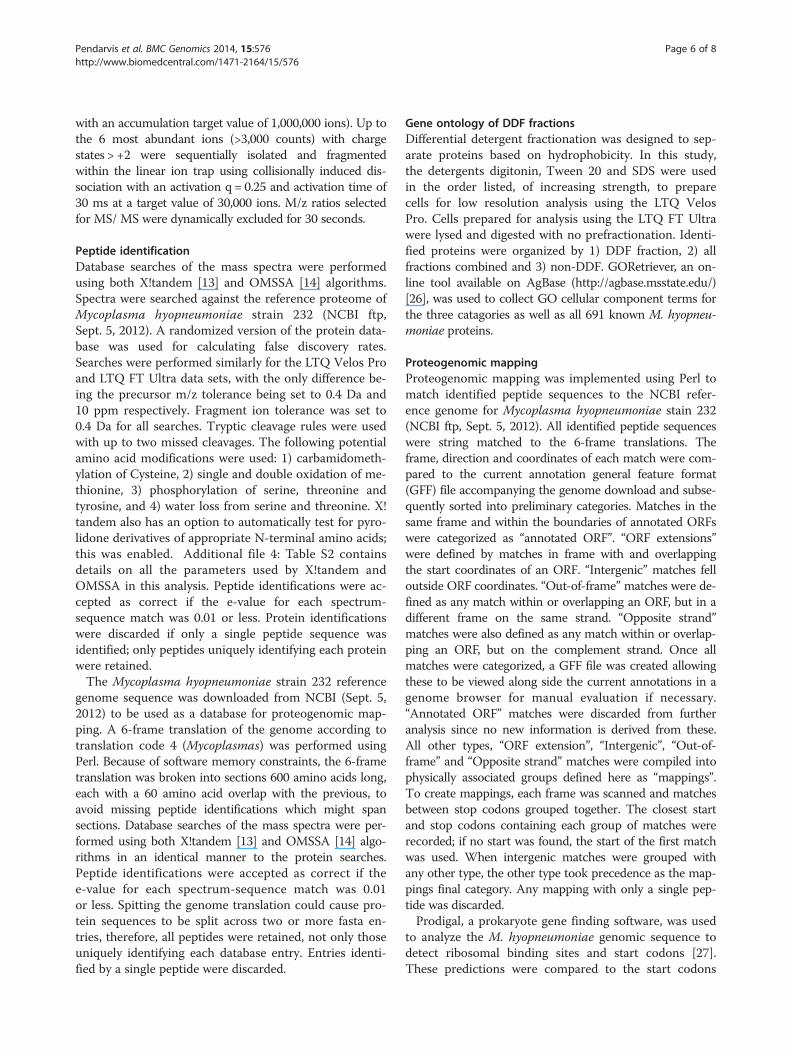
Proteogenomic mappingTo complement the identification of known and pre-dicted proteins in M. hyopneumoniae strain 232, andsubsequently identify possible unannotated open readingframes (ORFs) and errors in the current annotations, massspectra were searched using X!tandem and OMSSA againsta 6-frame genomic translation. The genomic searches re-sulted in 7,765 peptide sequences from 42,330 matchedspectra with a maximum false discovery rate of 0.73%.After combining both the protein and genome search re-sults, 9,039 unique peptide sequences were identified from47,674 positively matched spectra across all eight samples.Detailed peptide identifications with confidence scores areprovided in Supplemental Information Additional file 3:Genome search results.Peptide sequences were mapped to the M. hyop-
neumoniae strain 232 genome and categorized by lo-cation (Table 2). Proteogenomic mapping revealed twoareas of intergenic translation, annotated in other stains ofMycoplasma hyopneumoniae as genes gatC and rpmF.Five-prime extensions to annotated genes were identifiedin mhp063, mhp073, and mhp451; BLAST results indicatethese extensions are present in genes in other strains. TheProdigal prokaryotic gene predicting software also pre-dicted the previously unannotated genes and extensions inagreement with proteogenomic mapping (Table 3).
DiscussionIdentified proteinsOne other group has performed a recent global proteo-mics analysis of M. hyopneumoniae similar to our study;Pinto et al. reported identifying 35% of the proteins instrains J, 7422 and 7448 [15,16]. Jaffe et al. identified81% of the proteins of the related species M. pneumo-niae [17] and Yuan et al. identified 51% of the proteinsin M. suis [18]. By combining all of our samples, weidentified 70% of the proteins in M. hyopneumoniaestrain 232. The increase in proteome coverage from 35%to 70% achieved by our study is likely due to the largenumber of replicates (eight total) compared to a max-imum of three stated in the other studies, and the dualinstrument, dual sample preparation approach used inour analysis.Many of the proteins identified in this study are
only computationally predicted and, as such, given the“hypothetical” annotation. Our high throughput experi-mental annotation confirms that 171 (52%) of these genesare translated. From our previous transcriptome studies[4-8], evidence shows that 627 of the 691 protein codinggenes are transcribed under the growing conditions in
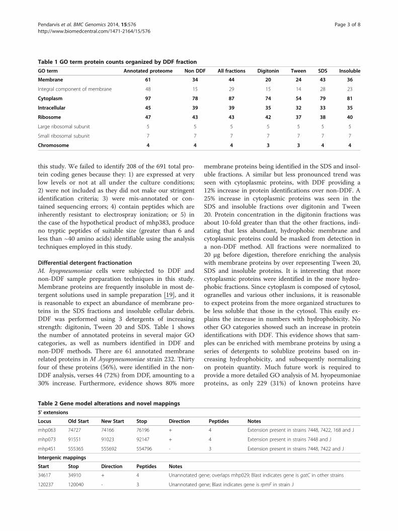
Table 1 GO term protein counts organized by DDF fraction
GO term Annotated proteome Non DDF All fractions Digitonin Tween SDS Insoluble
Membrane 61 34 44 20 24 43 36
Integral component of membrane 48 15 29 15 14 28 23
Cytoplasm 97 78 87 74 54 79 81
Intracellular 45 39 39 35 32 33 35
Ribosome 47 43 43 42 37 38 40
Large ribosomal subunit 5 5 5 5 5 5 5
Small ribosomal subunit 7 7 7 7 7 7 7
Chromosome 4 4 4 3 3 4 4
Pendarvis et al. BMC Genomics 2014, 15:576 Page 3 of 8http://www.biomedcentral.com/1471-2164/15/576
this study. We failed to identify 208 of the 691 total pro-tein coding genes because they: 1) are expressed at verylow levels or not at all under the culture conditions;2) were not included as they did not make our stringentidentification criteria; 3) were mis-annotated or con-tained sequencing errors; 4) contain peptides which areinherently resistant to electrospray ionization; or 5) inthe case of the hypothetical product of mhp383, produceno tryptic peptides of suitable size (greater than 6 andless than ~40 amino acids) identifiable using the analysistechniques employed in this study.
Differential detergent fractionationM. hyopneumoniae cells were subjected to DDF andnon-DDF sample preparation techniques in this study.Membrane proteins are frequently insoluble in most de-tergent solutions used in sample preparation [19], and itis reasonable to expect an abundance of membrane pro-teins in the SDS fractions and insoluble cellular debris.DDF was performed using 3 detergents of increasingstrength: digitonin, Tween 20 and SDS. Table 1 showsthe number of annotated proteins in several major GOcategories, as well as numbers identified in DDF andnon-DDF methods. There are 61 annotated membranerelated proteins in M .hyopyneumoniae strain 232. Thirtyfour of these proteins (56%), were identified in the non-DDF analysis, verses 44 (72%) from DDF, amounting to a30% increase. Furthermore, evidence shows 80% more
Table 2 Gene model alterations and novel mappings
5' extensions
Locus Old Start New Start Stop Direction
mhp063 74727 74166 76196 +
mhp073 91551 91023 92147 +
mhp451 555365 555692 554796 -
Intergenic mappings
Start Stop Direction Peptides Notes
34617 34910 + 4 Unannotated ge
120237 120040 - 3 Unannotated ge
membrane proteins being identified in the SDS and insol-uble fractions. A similar but less pronounced trend wasseen with cytoplasmic proteins, with DDF providing a12% increase in protein identifications over non-DDF. A25% increase in cytoplasmic proteins was seen in theSDS and insoluble fractions over digitonin and Tween20. Protein concentration in the digitonin fractions wasabout 10-fold greater than that the other fractions, indi-cating that less abundant, hydrophobic membrane andcytoplasmic proteins could be masked from detection ina non-DDF method. All fractions were normalized to20 μg before digestion, therefore enriching the analysiswith membrane proteins by over representing Tween 20,SDS and insoluble proteins. It is interesting that morecytoplasmic proteins were identified in the more hydro-phobic fractions. Since cytoplasm is composed of cytosol,ogranelles and various other inclusions, it is reasonableto expect proteins from the more organized structures tobe less soluble that those in the cytosol. This easily ex-plains the increase in numbers with hydrophobicity. Noother GO categories showed such an increase in proteinidentifications with DDF. This evidence shows that sam-ples can be enriched with membrane proteins by using aseries of detergents to solublize proteins based on in-creasing hydrophobicity, and subsequently normalizingon protein quantity. Much future work is required toprovide a more detailed GO analysis of M. hyopeumoniaeproteins, as only 229 (31%) of known proteins have
Peptides Notes
4 Extension present in strains 7448, 7422, 168 and J
4 Extension present in strains 7448 and J
3 Extension present in strains 7448, 7422 and J
ne; overlaps mhp029; Blast indicates gene is gatC in other strains
ne; Blast indicates gene is rpmF in strain J

Table 3 Comparison of Prodigal and proteomgenomicmapping coordinates
5' extensions
Locus PGMStart
ProdigalStart
Stop Direction RBS Motif*
mhp063 74166 74166 76196 + AGxAGG/AGGxGG
mhp073 91023 91023 92147 + GGA/GAG/AGG
mhp451 555692 555692 554796 - AGGAG
Intergenic mappings
Gene PGMStart
ProdigalStart
Stop Direction RBS Motif*
gatC 34617 34617 34910 + GGxGG
rpmF 120237 120237 120040 - AGGA
*An “x” in the motif indicates a mismatch is allowed.
Pendarvis et al. BMC Genomics 2014, 15:576 Page 4 of 8http://www.biomedcentral.com/1471-2164/15/576
cellular component annotation, most of which are verygeneralized. Better annotation would be helpful in categor-izing which cellular components are easily separated basedon DDF methods.
Proteogenomic mappingProteogenomic mapping indicates that the currentM. hyopneumoniae strain 232 genome is well annotatedwith only 1.8% of peptide mappings not belonging to cur-rently known genes. The identification of two unanno-tated genes, gatC and rpmF, is surprising considering thesmall genome size and high degree of genetic similaritybetween M. hyopneumoniae strains. The 5-prime exten-sions, present in annotated genes from other strains,were not predicted in strain 232 likely due to bias in theORF finding algorithm used. ORF finders typically scanthe 6 frames of a genomic sequence and predict ORFsbased on distance between start and stop codons. Prod-igal has an advantage over this type of ORF finder in thatit scans for ribosomal binding sites (RBS). As shown inTable 3, each gene predicted by Prodigal, has a differentRBS motif. All of these motifs are present in other pro-teins correctly predicted in the original annotations. Ifthe original prediction did not rely on RBS detection, fail-ure to determine the true start codons for certain ORFswould be more likely, explaining the 5' extensions de-tected by proteogenomic mapping. As for the unanno-tated genes, rpmF and gatC, it is unclear as to why thesewere missed. They are rather short genes, rpmF and gatCbeing 197 and 293 bases in length respectively, but 23annotated genes are shorter than both. gatC overlapsthe 5' end of gatA, but 172 other annotated genes overlapanother. These difficult to explain instances are good rea-sons to validate predictions with proteomic and transcrip-tomic data.Our previous study aimed at detecting intergenic tran-
scription in M. hyopneumoniae found evidence for 321instances of intergenic transcription [9]. We have evidence
of transcription in intergenic regions upstream frommhp073 and mhp451, which supports the 5-prime exten-sions of these genes detected in this study. No transcrip-tion evidence was found for the 5-prime extension ofmhp063. Both unannotated genes identified in this study,gatC and rpmF, also have corresponding areas of inter-genic transcription. In future studies, next generationtranscriptome sequencing would be a good choice tocomplement proteogenomic mapping and help confirmthe existence of unannotated and modified genes. Unlikeproteomics, transcriptomics allows gene boundaries to beclearly determined and errors in the genomic sequenceto be considered when mapping reads.Trypsin has been the enzyme of choice in proteomic
analyses for many years because of is high specificity,but protein primary structures rich in lysine (K) and ar-ginine (R) residues can result in peptides too small(<6 amino acids) to uniquely identify most proteins.Conversely, areas poor in K and R produce peptides toolarge (>40 amino acids) to be accurately identified by lowresolution mass spectrometers, such as the LTQ VelosPro used in this study. Secondary and tertiary proteinstructures resistant to denaturation can contain areasinaccessible to trypsin. Alternate protein fragmentationmethods can increase protein coverage, which is benefi-cial in proteogenomic mapping studies which rely onmaximizing coverage. Using multiple proteases whichtarget different residues, such as trypsin, elastase andthermolysin, can result in overlapping peptides averaging10 amino acids in length [20]. Proteinase K digestion car-ried out at high pH produces peptides of 6 to 20 aminoacids in length, ideal for MS/MS analysis [21]. A multipleprotease approach increases the likelihood accessingstructurally inaccessible cleavage sites and reduces theimpact of protein regions rich or poor in residues tar-geted by a single enzyme. A followup study employingthis approach would be beneficial by potentially increas-ing protein coverage and further confirming unannotatedareas of protein expression.
ConclusionsOur study has provided one of the deepest proteomeanalyses of M. hyopneumoniae to date. Seventy percentof strain 232 proteins were identified and 52% of hypo-thetical proteins have been confirmed. Previously unan-notated genes gatC and rpmF have been identified forthe first time strain 232. Five-prime extensions of genesmhp063, mhp073 and mhp451 were also detected. Theseadditions and modifications to the current annotationsare conserved in other strains of M. hyopneumoniae andall but one, mhp063, have evidence of transcription asdetermined by our previous studies [4-8]. These findingsillustrate how even the smallest annotated genomes arefar from perfect, and future work, both transcriptomic

Pendarvis et al. BMC Genomics 2014, 15:576 Page 5 of 8http://www.biomedcentral.com/1471-2164/15/576
and proteomic, is required to better understand theM. hyopneumoniae genome. Additionally, using a geneprediction program which detects ribosomal bindingsites ensures genes are less likely to be incorrectly de-fined or missed during analysis. Furthermore, the useof DDF effectively enriches samples with membraneproteins by allowing proteins to be separated based onincreasing hydrophobicity. Highly soluble, highly abun-dant proteins are concentrated in a relatively weak de-tergent while less soluble, less abundant membraneproteins are extracted in progressively stronger detergents.Normalizing fractions by quantity prior to trypsin diges-tion allows low abundance, hydrophobic proteins a greaterchance of being identified. The current GO annotationsfor Mycoplasma hyopneumoniae are lacking depth andcompletion; much work is required to annotate the prote-ome both physically and functionally. Better GO anno-tation would provide a more thorough breakdown ofprotein and cellular component affinity to DDF fraction.
MethodsSample preparationMycoplasma hyopneumoniae strain 232 was originallyisolated from a pig infected with strain 11 [22], is fullyvirulent in low passage, and has been commonly used inchallenge and pathogen studies in the United States.Four independent cultures (biological replicates) weregrown in Friis broth [23], each split into two flasks(technical replicates), until the media color change indi-cated mid to late log phase of growth had been achieved(pH ~ 6.5). The cells were then centrifuged at 10,000 × gfor 30 min, resuspended in phosphate buffered saline, andcentrifuged again. This was repeated three additional timesto remove medium contaminants. Of the eight replicates,six were reserved for shotgun proteomics analysis using anLTQ Velos Pro (Thermo Scientific) low resolution, high-throughput mass spectrometer, and the remaining two rep-licates were analyzed using an LTQ FT Ultra (ThermoScientific) high resolution mass spectrometer.No vertebrates subjects were involved in the culture
and sample preparation of the M. hyopneumoniae duringthe course of this study. All procedures were per-formed within the research guidelines of the Universityof Arizona, Iowa State University, and the University ofTechnology, Sydney and did not require approval of anethics committee.
Low resolution mass spectrometryFor the shotgun proteomics analysis, six cell pellets weresubject to differential detergent fractionation as de-scribed by McCarthy et al. using the detergents digito-nin, Tween 20 and SDS [24]. After each detergentapplication, samples were centrifuged to separate solu-blized proteins from cellular debris. The insoluble pellet
left after treatment was subject to trypsin digestion alongwith the soluble fractions, but could not be quantified.Fractions were normalized to 20 μg each and trypsin di-gestion as described by McCarthy et al. [24]. Followingdigestion, each fraction was desalted using a peptidemicrotrap (Michrom BioResources) according to themanufacturer's instructions. After desalting, each fractionwas further cleaned using a strong cation exchange (SCX)microtrap (Michrom BioResources) to remove any re-sidual detergent, which could interfere with the massspectrometry. Fractions were dried and resuspended in10 μL of 2% acetonitrile (ACN), 0.1% formic acid (FA) andtransferred to low retention vials in preparation for separ-ation using 1D-LC.The high performance liquid chromatography (HPLC)
equipment used for peptide separation was an Ultimate3000 (Dionex) operated in 1D-LC mode at a flow rate of333 nL per min and equipped with a 0.075 mm ×100 mm column packed with Halo C18 material (MichromBioResources) for reverse phase separation. Each samplewas separated using a 4 h gradient from 2% to 50% Aceto-nitrile with 0.1% formic acid as a proton source. The col-umn was located on the ion source and connected directlyto a nanospray emitter to minimize peak broadening. Scanparameters for the LTQ Velos Pro were one MS scanfollowed by 20 MS/MS scans of the 20 most intense peaksusing high energy collisional dissociation as the fragmenta-tion method. Dynamic exclusion was enabled with a massexclusion time of 3 min and a repeat count of 1 within30 sec of initial m/z measurement.
High resolution mass spectrometryThe two cell pellets reserved for high resolution analysiswere lysed and digested as described by Wilton et al.[25]. Digested peptides were dried, resuspended in 20 mMKH2PO4, 20% ACN, pH 3 (Buffer A) in 2.5 μL and trans-ferred to low retention vials in preparation for separationusing an Ultimate 3000 configured for 2D-LC. Eachsample was loaded at 15 μL/min onto an SCX micro-trap (Michrom BioResources) for the first dimension ofseparation, involving SCX steps of Buffer A plus 0, 5, 10,15, 20, 25, 30, 40, 50, 100, 250, 500, and 1000 mM KCl. Forthe second dimension of separation, each eluted salt stepwas desalted with an inline peptide microtrap (MichromBioResources) with 2% ACN, 0.1% FA at 5 μL/min. Oncedesalted, the microtrap was switched into line with a frit-less nano column (75 μm × ~10 cm) containing C18 media(5 μ, 200 Å Magic, Michrom) manufactured according toGatlin [26]. Peptides were eluted using a gradient of 2% to36% ACN, 0.1% FA at 350 nL/min over 60 min and elec-trospray ionized for analysis using an LTQ FT Ultra massspectrometer.A survey scan m/z 350–1750 was acquired in the FT ion
cyclotron resonance cell (Resolution = 100,000 at m/z 400,
Pendarvis et al. BMC Genomics 2014, 15:576 Page 6 of 8http://www.biomedcentral.com/1471-2164/15/576
with an accumulation target value of 1,000,000 ions). Up tothe 6 most abundant ions (>3,000 counts) with chargestates > +2 were sequentially isolated and fragmentedwithin the linear ion trap using collisionally induced dis-sociation with an activation q = 0.25 and activation time of30 ms at a target value of 30,000 ions. M/z ratios selectedfor MS/ MS were dynamically excluded for 30 seconds.
Peptide identificationDatabase searches of the mass spectra were performedusing both X!tandem [13] and OMSSA [14] algorithms.Spectra were searched against the reference proteome ofMycoplasma hyopneumoniae strain 232 (NCBI ftp,Sept. 5, 2012). A randomized version of the protein data-base was used for calculating false discovery rates.Searches were performed similarly for the LTQ Velos Proand LTQ FT Ultra data sets, with the only difference be-ing the precursor m/z tolerance being set to 0.4 Da and10 ppm respectively. Fragment ion tolerance was set to0.4 Da for all searches. Tryptic cleavage rules were usedwith up to two missed cleavages. The following potentialamino acid modifications were used: 1) carbamidometh-ylation of Cysteine, 2) single and double oxidation of me-thionine, 3) phosphorylation of serine, threonine andtyrosine, and 4) water loss from serine and threonine. X!tandem also has an option to automatically test for pyro-lidone derivatives of appropriate N-terminal amino acids;this was enabled. Additional file 4: Table S2 containsdetails on all the parameters used by X!tandem andOMSSA in this analysis. Peptide identifications were ac-cepted as correct if the e-value for each spectrum-sequence match was 0.01 or less. Protein identificationswere discarded if only a single peptide sequence wasidentified; only peptides uniquely identifying each proteinwere retained.The Mycoplasma hyopneumoniae strain 232 reference
genome sequence was downloaded from NCBI (Sept. 5,2012) to be used as a database for proteogenomic map-ping. A 6-frame translation of the genome according totranslation code 4 (Mycoplasmas) was performed usingPerl. Because of software memory constraints, the 6-frametranslation was broken into sections 600 amino acids long,each with a 60 amino acid overlap with the previous, toavoid missing peptide identifications which might spansections. Database searches of the mass spectra were per-formed using both X!tandem [13] and OMSSA [14] algo-rithms in an identical manner to the protein searches.Peptide identifications were accepted as correct if thee-value for each spectrum-sequence match was 0.01or less. Spitting the genome translation could cause pro-tein sequences to be split across two or more fasta en-tries, therefore, all peptides were retained, not only thoseuniquely identifying each database entry. Entries identi-fied by a single peptide were discarded.
Gene ontology of DDF fractionsDifferential detergent fractionation was designed to sep-arate proteins based on hydrophobicity. In this study,the detergents digitonin, Tween 20 and SDS were usedin the order listed, of increasing strength, to preparecells for low resolution analysis using the LTQ VelosPro. Cells prepared for analysis using the LTQ FT Ultrawere lysed and digested with no prefractionation. Identi-fied proteins were organized by 1) DDF fraction, 2) allfractions combined and 3) non-DDF. GORetriever, an on-line tool available on AgBase (http://agbase.msstate.edu/)[26], was used to collect GO cellular component terms forthe three catagories as well as all 691 known M. hyopneu-moniae proteins.
Proteogenomic mappingProteogenomic mapping was implemented using Perl tomatch identified peptide sequences to the NCBI refer-ence genome for Mycoplasma hyopneumoniae stain 232(NCBI ftp, Sept. 5, 2012). All identified peptide sequenceswere string matched to the 6-frame translations. Theframe, direction and coordinates of each match were com-pared to the current annotation general feature format(GFF) file accompanying the genome download and subse-quently sorted into preliminary categories. Matches in thesame frame and within the boundaries of annotated ORFswere categorized as “annotated ORF”. “ORF extensions”were defined by matches in frame with and overlappingthe start coordinates of an ORF. “Intergenic” matches felloutside ORF coordinates. “Out-of-frame” matches were de-fined as any match within or overlapping an ORF, but in adifferent frame on the same strand. “Opposite strand”matches were also defined as any match within or overlap-ping an ORF, but on the complement strand. Once allmatches were categorized, a GFF file was created allowingthese to be viewed along side the current annotations in agenome browser for manual evaluation if necessary.“Annotated ORF” matches were discarded from furtheranalysis since no new information is derived from these.All other types, “ORF extension”, “Intergenic”, “Out-of-frame” and “Opposite strand” matches were compiled intophysically associated groups defined here as “mappings”.To create mappings, each frame was scanned and matchesbetween stop codons grouped together. The closest startand stop codons containing each group of matches wererecorded; if no start was found, the start of the first matchwas used. When intergenic matches were grouped withany other type, the other type took precedence as the map-pings final category. Any mapping with only a single pep-tide was discarded.Prodigal, a prokaryote gene finding software, was used
to analyze the M. hyopneumoniae genomic sequence todetect ribosomal binding sites and start codons [27].These predictions were compared to the start codons

Pendarvis et al. BMC Genomics 2014, 15:576 Page 7 of 8http://www.biomedcentral.com/1471-2164/15/576
predicted through the proteogenomic mapping processusing Perl.
Availability of supporting dataMass spectra and protein identifications have been depos-ited to the ProteomeXchange Consortium (http://proteo-mecentral.proteomexchange.org) via the PRIDE partnerrepository [28] with the dataset identifier PXD000118 andDOI 10.6019/PXD000118. Results from protein and gen-omic translation searches, are included as supporting infor-mation in tab-delimited format.
Additional files
Additional file 1: Table S1. Mycoplasma hyopneumoniae strain 232proteins with mass spectrometry verification status and coverage metrics.
Additional file 2: Proteome search results.
Additional file 3: Genome search results.
Additional file 4: Table S2. Search parameters.
AbbreviationsORF: Open reading frame; GFF: General feature format; 1D: One dimensional;2D: Two dimensional; LC: Liquid chromatography; NSI: Nanospray ionization;MS/MS: Precursor and fragment mass spectrometry; Velos: LTQ Velos Promass spectrometer; FT: LTQ FT (Fourier Transform) Ultra mass spectrometer;SCX: Strong cation exchange; ACN: Acetonitrile; FA: Formic acid; HPLC: Highperformance liquid chromatography.
Competing interestsNone of the authors have a financial or commercial conflict of interest withthe study reported here.
Authors’ contributionsKP performed sample preparation, low resolution mass spectrometry datageneration, analysis of all mass spectrometry data, programming involved inpeptide mapping and wrote much of the manuscript. MPP, JLT and SPDcontributed to this work by supplying high resolution mass spectrometrydata and corresponding portions of the manuscript. ACP and FCM wereinvolved in sample preparation and study design. FCM and SCB alsocontributed to the manuscript and data interpretation. All authors read andapproved the final manuscript.
AcknowledgmentsThis work was partially supported by the National Pork Board (grant number08–062) to F. Chris Minion. Acknowledgment goes to the Bioanalytical MassSpectrometry Facility at the University of New South Wales for the LTQ FTsample analysis. An allocation of computer time from the UA ResearchComputing High Performance Computing (HTC) and High ThroughputComputing (HTC) at the University of Arizona is gratefully acknowledged.
Author details1Department of Veterinary Microbiology and Preventive Medicine, Iowa StateUniversity, Ames, IA, USA. 2School of Animal and Comparative BiomedicalSciences, University of Arizona, Tucson, AZ, USA. 3ithree institute, Universityof Technology, Sydney, Australia. 4Proteomics Core Facility, Faculty ofScience, University of Technology, Sydney, Australia. 5Bio5 Institute, School ofAnimal and Comparative Biomedical Sciences, University of Arizona, Tucson,AZ, USA.
Received: 31 March 2014 Accepted: 1 July 2014Published: 8 July 2014
References1. Ross R: Diseases of swine. In Dis Swine. Ames, Iowa, U.S.A: Iowa State
University Press; 1992:537–551.
2. Pointon AM, Byrt D, Heap P: Effect of enzootic pneumonia of pigs ongrowth performance. Aust Vet J 1985, 62:13–18.
3. Adams C, Pitzer J, Minion FC: In vivo expression analysis of the P97 andP102 paralog families of Mycoplasma hyopneumoniae. Infect Immun2005, 73:7784–7787.
4. Madsen ML, Nettleton D, Thacker EL, Edwards R, Minion FC: Transcriptionalprofiling of Mycoplasma hyopneumoniae during heat shock usingmicroarrays. Infect Immun 2006, 74:160–166.
5. Madsen ML, Nettleton D, Thacker EL, Minion FC: Transcriptional profiling ofMycoplasma hyopneumoniae during iron depletion using microarrays.Microbiology 2006, 152(Pt 4):937–944.
6. Schafer ER, Oneal MJ, Madsen ML, Minion FC: Global transcriptionalanalysis of Mycoplasma hyopneumoniae following exposure tohydrogen peroxide. Microbiology 2007, 153(Pt 11):3785–3790.
7. Madsen ML, Puttamreddy S, Thacker EL, Carruthers MD, Minion FC:Transcriptome changes in Mycoplasma hyopneumoniae duringinfection. Infect Immun 2008, 76:658–663.
8. Oneal MJ, Schafer ER, Madsen ML, Minion FC: Global transcriptionalanalysis of Mycoplasma hyopneumoniae following exposure tonorepinephrine. Microbiology 2008, 154(Pt 9):2581–2588.
9. Gardner SW, Minion FC: Detection and quantification of intergenictranscription in Mycoplasma hyopneumoniae. Microbiology 2010,156(Pt 8):2305–2315.
10. Minion FC, Lefkowitz EJ, Madsen ML, Cleary BJ, Swartzell SM, Mahairas GG:The genome sequence of Mycoplasma hyopneumoniae strain 232, theagent of swine mycoplasmosis. J Bacteriol 2004, 186:7123–7133.
11. Vasconcelos ATR, Ferreira HB, Bizarro CV, Bonatto SL, Carvalho MO, Pinto PM,Almeida DF, Almeida LGP, Almeida R, Alves-Filho L, Assunção EN, Azevedo VAC,Bogo MR, Brigido MM, Brocchi M, Burity HA, Camargo AA, Camargo SS,Carepo MS, Carraro DM, De Mattos Cascardo JC, Castro LA, Cavalcanti G,Chemale G, Collevatti RG, Cunha CW, Dallagiovanna B, Dambrós BP,Dellagostin OA, Falcão C, et al: Swine and poultry pathogens: thecomplete genome sequences of two strains of Mycoplasmahyopneumoniae and a strain of Mycoplasma synoviae. J Bacteriol2005, 187:5568–5577.
12. Liu W, Feng Z, Fang L, Zhou Z, Li Q, Li S, Luo R, Wang L, Chen H, Shao G,Xiao S: Complete genome sequence of Mycoplasma hyopneumoniaestrain 168. J Bacteriol 2011, 193:1016–1017.
13. Craig R, Beavis RC: TANDEM: matching proteins with tandem massspectra. Bioinformatics 2004, 20:1466–1467.
14. Geer LY, Markey SP, Kowalak JA, Wagner L, Xu M, Maynard DM, Yang X,Shi W, Bryant SH: Open mass spectrometry search algorithm. J Proteome Res2004, 3:958–964.
15. Pinto PM, Chemale G, De Castro LA, Costa APM, Kich JD, Vainstein MH,Zaha A, Ferreira HB: Proteomic survey of the pathogenic Mycoplasmahyopneumoniae strain 7448 and identification of novel post-translationally modified and antigenic proteins. Vet Microbiol 2007,121:83–93.
16. Pinto PM, Klein CS, Zaha A, Ferreira HB: Comparative proteomic analysis ofpathogenic and non-pathogenic strains from the swine pathogenMycoplasma hyopneumoniae. Proteome Sci 2009, 7:45.
17. Jaffe JD, Berg HC, Church GM: Proteogenomic mapping as a complementarymethod to perform genome annotation. Proteomics 2004, 4:59–77.
18. Yuan C, Yang X, Yang Z, Zhu N, Zheng S, Hou P, Gu X, Ye C, Yao C, Zhu J,Cui L, Hua X: Proteomic study of Mycoplasma suis using the gel-basedshotgun strategy. Vet Microbiol 2010, 142:303–308.
19. Trimpin S, Brizzard B: Analysis of insoluble proteins. Biotechniques 2009,46:321–326.
20. Schlosser A, Vanselow JT, Kramer A: Mapping of phosphorylationsites by a multi-protease approach with specific phosphopeptideenrichment and NanoLC-MS/MS analysis. Anal Chem 2005,77:5243–5250.
21. Wu CC, MacCoss MJ, Howell KE, Yates JR 3rd: A method for thecomprehensive proteomic analysis of membrane proteins. Nat Biotechnol2003, 21:532–538.
22. Mare CJ, Switzer WP: New Species: Mycoplasma hyopneumoniae; acausative agent of virus pig pneumonia. Vet Med Small Anim Clin 1965,60:841–846.
23. Friis NF: Some recommendations concerning primary isolation ofMycoplasma suipneumoniae and Mycoplasma flocculare a survey.Nord Vet Med 1975, 27:337–339.

Pendarvis et al. BMC Genomics 2014, 15:576 Page 8 of 8http://www.biomedcentral.com/1471-2164/15/576
24. McCarthy FM, Burgess SC, van den Berg BHJ, Koter MD, Pharr GT:Differential detergent fractionation for non-electrophoretic eukaryotecell proteomics. J Proteome Res 2005, 4:316–324.
25. Deutscher AT, Jenkins C, Minion FC, Seymour LM, Padula MP, Dixon NE,Walker MJ, Djordjevic SP: Repeat regions R1 and R2 in the P97 paralogueMhp271 of Mycoplasma hyopneumoniae bind heparin, fibronectin andporcine cilia. Mol Microbiol 2010, 78:444–458.
26. Gatlin CL, Kleemann GR, Hays LG, Link AJ, Yates JR 3rd: Protein identificationat the low femtomole level from silver-stained gels using a new fritlesselectrospray interface for liquid chromatography-microspray and nanospraymass spectrometry. Anal Biochem 1998, 263:93–101.
27. Hyatt D, Chen G-L, Locascio PF, Land ML, Larimer FW, Hauser LJ: Prodigal:prokaryotic gene recognition and translation initiation site identification.BMC Bioinformatics 2010, 11:119.
28. Vizcaíno JA, Côté RG, Csordas A, Dianes JA, Fabregat A, Foster JM, Griss J,Alpi E, Birim M, Contell J, O’Kelly G, Schoenegger A, Ovelleiro D, Pérez-Riverol Y,Reisinger F, Ríos D, Wang R, Hermjakob H: The PRoteomics IDEntifications(PRIDE) database and associated tools: status in 2013. Nucleic Acids Res 2013,41(Database issue):D1063–D1069.
doi:10.1186/1471-2164-15-576Cite this article as: Pendarvis et al.: Proteogenomic mapping ofMycoplasma hyopneumoniae virulent strain 232. BMC Genomics2014 15:576.
Submit your next manuscript to BioMed Centraland take full advantage of:
• Convenient online submission
• Thorough peer review
• No space constraints or color figure charges
• Immediate publication on acceptance
• Inclusion in PubMed, CAS, Scopus and Google Scholar
• Research which is freely available for redistribution
Submit your manuscript at www.biomedcentral.com/submit
Related Documents