HAL Id: hal-03216610 https://hal-amu.archives-ouvertes.fr/hal-03216610 Submitted on 15 Nov 2021 HAL is a multi-disciplinary open access archive for the deposit and dissemination of sci- entific research documents, whether they are pub- lished or not. The documents may come from teaching and research institutions in France or abroad, or from public or private research centers. L’archive ouverte pluridisciplinaire HAL, est destinée au dépôt et à la diffusion de documents scientifiques de niveau recherche, publiés ou non, émanant des établissements d’enseignement et de recherche français ou étrangers, des laboratoires publics ou privés. Distributed under a Creative Commons Attribution| 4.0 International License Novel Virulent Bacteriophages Infecting Mediterranean Isolates of the Plant Pest Xylella fastidiosa and Xanthomonas albilineans Fernando Clavijo-Coppens, Nicolas Ginet, Sophie Cesbron, Martial Briand, Mireille Ansaldi To cite this version: Fernando Clavijo-Coppens, Nicolas Ginet, Sophie Cesbron, Martial Briand, Mireille Ansaldi. Novel Virulent Bacteriophages Infecting Mediterranean Isolates of the Plant Pest Xylella fastidiosa and Xanthomonas albilineans. Viruses, MDPI, 2021, 13 (5), pp.725. 10.3390/v13050725. hal-03216610

Welcome message from author
This document is posted to help you gain knowledge. Please leave a comment to let me know what you think about it! Share it to your friends and learn new things together.
Transcript

HAL Id: hal-03216610https://hal-amu.archives-ouvertes.fr/hal-03216610
Submitted on 15 Nov 2021
HAL is a multi-disciplinary open accessarchive for the deposit and dissemination of sci-entific research documents, whether they are pub-lished or not. The documents may come fromteaching and research institutions in France orabroad, or from public or private research centers.
L’archive ouverte pluridisciplinaire HAL, estdestinée au dépôt et à la diffusion de documentsscientifiques de niveau recherche, publiés ou non,émanant des établissements d’enseignement et derecherche français ou étrangers, des laboratoirespublics ou privés.
Distributed under a Creative Commons Attribution| 4.0 International License
Novel Virulent Bacteriophages Infecting MediterraneanIsolates of the Plant Pest Xylella fastidiosa and
Xanthomonas albilineansFernando Clavijo-Coppens, Nicolas Ginet, Sophie Cesbron, Martial Briand,
Mireille Ansaldi
To cite this version:Fernando Clavijo-Coppens, Nicolas Ginet, Sophie Cesbron, Martial Briand, Mireille Ansaldi. NovelVirulent Bacteriophages Infecting Mediterranean Isolates of the Plant Pest Xylella fastidiosa andXanthomonas albilineans. Viruses, MDPI, 2021, 13 (5), pp.725. �10.3390/v13050725�. �hal-03216610�

viruses
Article
Novel Virulent Bacteriophages Infecting MediterraneanIsolates of the Plant Pest Xylella fastidiosa andXanthomonas albilineans
Fernando Clavijo-Coppens 1,2,3 , Nicolas Ginet 1 , Sophie Cesbron 2 , Martial Briand 2,Marie-Agnès Jacques 2 and Mireille Ansaldi 1,*
�����������������
Citation: Clavijo-Coppens, F.; Ginet,
N.; Cesbron, S.; Briand, M.; Jacques,
M.-A.; Ansaldi, M. Novel Virulent
Bacteriophages Infecting
Mediterranean Isolates of the Plant
Pest Xylella fastidiosa and Xanthomonas
albilineans. Viruses 2021, 13, 725.
https://doi.org/10.3390/v13050725
Academic Editors: Joana Azeredo
and Sílvio Santos
Received: 10 March 2021
Accepted: 17 April 2021
Published: 21 April 2021
Publisher’s Note: MDPI stays neutral
with regard to jurisdictional claims in
published maps and institutional affil-
iations.
Copyright: © 2021 by the authors.
Licensee MDPI, Basel, Switzerland.
This article is an open access article
distributed under the terms and
conditions of the Creative Commons
Attribution (CC BY) license (https://
creativecommons.org/licenses/by/
4.0/).
1 Laboratoire de Chimie Bactérienne, UMR7283, Centre National de la Recherche Scientifique, Aix-MarseilleUniversité, 13009 Marseille, France; [email protected] (F.C.-C.); [email protected] (N.G.)
2 Institute Agro, INRAE, IRHS, SFR QUASAV, University of Angers, 49000 Angers, France;[email protected] (S.C.); [email protected] (M.B.); [email protected] (M.-A.J.)
3 Bioline-Agrosciences, Equipe R&D–Innovation, 06560 Valbonne, France* Correspondence: [email protected]; Tel.: +33-491164585
Abstract: Xylella fastidiosa (Xf ) is a plant pathogen causing significant losses in agriculture worldwide.Originating from America, this bacterium caused recent epidemics in southern Europe and is thusconsidered an emerging pathogen. As the European regulations do not authorize antibiotic treatmentin plants, alternative treatments are urgently needed to control the spread of the pathogen andeventually to cure infected crops. One such alternative is the use of phage therapy, developed morethan 100 years ago to cure human dysentery and nowadays adapted to agriculture. The first steptowards phage therapy is the isolation of the appropriate bacteriophages. With this goal, we searchedfor phages able to infect Xf strains that are endemic in the Mediterranean area. However, as Xfis truly a fastidious organism, we chose the phylogenetically closest and relatively fast-growingorganism X. albineans as a surrogate host for the isolation step. Our results showed the isolation fromvarious sources and preliminary characterization of several phages active on different Xf strains,namely, from the fastidiosa (Xff ), multiplex (Xfm), and pauca (Xfp) subspecies, as well as on X. albilineans.We sequenced their genomes, described their genomic features, and provided a phylogeny analysisthat allowed us to propose new taxonomic elements. Among the 14 genomes sequenced, we couldidentify two new phage species, belonging to two new genera of the Caudoviricetes order, namely,Usmevirus (Podoviridae family) and Subavirus (Siphoviridae family). Interestingly, no specific phagescould be isolated from infected plant samples, whereas one was isolated from vector insects capturedin a contaminated area, and several from surface and sewage waters from the Marseille area.
Keywords: bacteriophage; Xylella fastidiosa; Xanthomonas albilineans; phage therapy; plant pathogens
1. Introduction
Xylella fastidiosa (Xf ) is a slow-growing Gram-negative plant-pathogenic bacteriumemerging in Asia and Europe, and is therefore listed as a quarantine organism [1]. Longknown in the Americas, this xylem-specialized bacterium is associated with numeroussocio-economically important plant diseases. The impact of diseases due to Xylella includedirect and indirect economic damages that were estimated to reach 150 million dollarsper year in California when including measures to control vectors [2,3]. In the Europeanolive oil production area, the projected impact of olive quick decline syndrome (OQDS)over 50 years ranges from two to five billion euros [4]. Nonetheless, such diseases havewider impacts; for instance, the impairment was estimated at 30% of the provision ofecosystem services provided by agroecological systems, including landscape value, culturaland natural heritage in the Puglia province of Italy [5]. Xf is transmitted by xylem sap-feeding insects and belongs to the Xanthomonadaceae (synonymous with the Lysobacteraceae)
Viruses 2021, 13, 725. https://doi.org/10.3390/v13050725 https://www.mdpi.com/journal/viruses
Viruses 2021, 13, 725 2 of 21
family. Xf genome has been the first plant pathogen genome sequenced, highlighting itsimportance as a plant pest [6]. Its genome of 2.6 Mb with a low GC content (52.7%) issmaller than that of other Xanthomonadaceae, even the ones of the phylogenetically closestrelatives Xanthomonas albilineans (Xa) and X. translucens (Xt) whose genomes are composedof 3.7 and 4.7 Mb, respectively [7,8]. It is tempting to link the Xf reduced genome to itsfastidious growth. Interestingly, a recent genome-scale metabolic network reconstructionanalysis suggested that the Xf metabolic network is pretty much complete, althoughminimalist and not particularly robust [9]. Indeed, alternative reactions seem to be missing;the metabolism of the bacteria thus relies on poorly efficient metabolic pathways and lackof flexibility. Moreover, the study also found that the production of some virulence factors,such as exopolysaccharides (EPS), is at a high cost to the plant and is thus detrimental tofast growth [9].
Xf has been identified as the causative agent of Pierce’s disease, which has been causingextensive damage to vineyards in California for almost 150 years [10]. However, it was onlyat the end of the 1970s that this fastidious bacterium could be isolated on solid medium [11].Since then, Xf has been associated with various forms of plant diseases on many different planthosts [12]. Today, this bacterium belongs in the top 10 of the plant pathogens that are moststudied worldwide [13]. Xf is endemic in the Americas and is persistently involved in largeepidemics, causing significant damage in vineyards and citrus orchards. Listed as a quarantinepest in Europe, the three main Xf subspecies (pauca (Xfp), multiplex (Xfm) and subsp. fastidiosa(Xff )) were recently detected in Europe, in particular in the Mediterranean area, probablyintroduced through commercial exchanges and causing a variety of diseases such as the leafscorch of olive trees in Italy [14]. Continuous monitoring using standardized real-time PCRprotocols is now performed in southern Europe [15,16]. However, in the absence of any efficientand authorized chemical method to control Xf, a major challenge is to develop environmentallyfriendly biotechnologies to control this plant disease. The current strategy implemented bythe European Commission to prevent Xf spread within the EU consists of eradication andcontainment measures involving intensive surveillance, vector control treatments, and differ-ential removal of infected plants and specified hosts in foci and buffer zones [17]. Additionalmeasures to these economically and socially devastating ones have been proposed, such asthe selection of naturally resistant host plants, the thermal treatment of young plants, or theuse of a combination of zinc, copper, manganese and citric acid [18–20]. Biocontrol strategieshave also been proposed using avirulent Xf strains or other avirulent plant colonizers [21,22].However, none of these later strategies has been recognized as fully efficient or totally safe bythe European authorities.
Bacteriophages, the viruses infecting bacteria, have been used to treat infectious diseasessince their discovery at the beginning of the twentieth century [23]. Their use in therapy haspros and cons, but the clear advantages are that they can infect antibiotic-resistant pathogensand can be adapted to any bacterial species as long as strictly virulent phages can be isolated,which can be difficult with some poorly growing species. Recently, the agricultural sciencescommunity put a great deal of emphasis on phage therapy to develop sustainable strategiesof biocontrol for plants and animals [24–26]. Currently, a number of phage products arebeing developed or are already commercially available for a variety of plant pathogenssuch as various Xanthomonas strains or Ralstonia solanacearum [27]. As an example, theOmniLytics™ company commercialized several phage products under the AgriPhage™ nameto control tomato and pepper bacterial spot and speck caused by Xanthomonas euvesicatoria pv.euvesicatoria and Pseudomonas syringae pv. tomato, as well as other plant diseases. In this context,a group from Texas A&M University proposed the use of bacteriophages as a treatment forPierce’s disease [28–30]. Following the same line of investigation, we attempted to isolateXf -specific phages and to focus on the three Xf subspecies present in the Mediterraneanbasin, since the strains currently spreading in Europe differ from the original Pierce’s diseasecausative strain Xff [14,16]. In order to develop phage cocktails active against specific strains ofinterest, i.e., Xf strains endemic in Europe, one should first constitute a collection of dedicatedand specific phages, determine their genome sequences and test them in vitro as well as

Viruses 2021, 13, 725 3 of 21
in vivo for their lytic activity. In this work, we started to build up a phage library that couldeventually be used as biocontrol products.
2. Materials and Methods2.1. Bacterial Strains and Media
The bacterial strains used in this study are listed in Table 1 and were provided by theFrench Collection of Plant-associated Bacteria (CIRM-CFBP, https://www6.inrae.fr/cirm_eng/CFBP-Plant-Associated-Bacteria, accessed on 10 March 2021). Xf strains were cul-tured at 28◦C on modified periwinkle wilt modified medium (PWG-M agar without phenol red) [31]for up to seven days. Then, the cultures were transferred to a new plate containing PWG-Magar or B-CYE medium [32] and incubated for up to seven days at 28 ◦C. When required, forliquid cultures, Xf strains were incubated at 28 ◦C under 160 rpm agitation in PD2 broth [31].Xanthomonas strains were cultured on YPG agar (yeast extract, 7 g l-1; peptone, 7 g l-1; glucose,7 g l-1; agar 15 g l-1 (pH 7.0 to 7.2)) [33] for up to 24 h at 28 ◦C, except for X. arboricola (Xaj)and X. albilineans (Xa) cultured for two to three days at 28 ◦C. For liquid cultures, Xanthomonasstrains were cultured in YPG broth medium (YPG without agar). The YPG soft agar medium(YPG broth with agar 7.5 g l-1) was used for X. albilineans overlays at 28 ◦C for up to three days.For all strains, suspensions made from fresh cultures were stored at −80 ◦C in YP glycerolmedium (yeast extract 5 g l-1; peptone 5 g l-1; glycerol 30% v/v), except for the X. albilineansstrain, which was stored directly in sterile distilled water.
Table 1. Bacterial strains used in this study.
Strain/Genotype Abbreviation Collection Code NCBI
Xylella fastidiosa subsp. fastidiosa Xff CFBP 7970 PRJNA417585Xylella fastidiosa subsp. pauca Xfp CFBP 8402 PRJNA383475
Xylella fastidiosa subsp. multiplex Xfm CFBP 8418 PRJNA314986Xanthomonas albilineans Xa CFBP 2523 PRJNA338244
Xanthomonas arboricola pv. juglandis Xaj CFBP 2528 PRJNA275685Xanthomonas citri pv. citri Xcci CFBP 3369 PRJNA254373Xanthomonas vesicatoria Xv CFBP 2537 PRJNA60019
Xanthomonas campestris pv. campestris Xcc CFBP 5241 PRJNA296Xanthomonas translucens pv. cerealis Xtc CFBP 2541 PRJNA268946
2.2. Environmental Samples for Phage Isolation
Several Xylella-associated and Xylella-non associated environments were assayedfor the presence of bacteriophages. For Xf-associated environments, plant samples werecollected in a Corsican public space (GPS coordinates 41.912444, 8.641105), and sap-feedinginsect samples (of the species Philaenus spumarius) were collected nearby (GPS coordi-nates 42.272662, 9.492568). Both positions are referenced as Xylella-infected locations inFrance [34]. Samples were sterilized on-site as follows: plant and insect extracts weremacerated in 5 mL of phage buffer (100 mM Tris-HCl (pH 7.6); 100 mM NaCl; 10 mMMgCl2; and 10 mM MgSO4) per gram of material. To remove large particles, the mixtureswere filtered through a 320 mm Whatman® filter paper, Grade 113V. Supernatants werefiltered through 0.45 µm then through 0.22 µm pore-size nylon Acrodisc® syringe filters,and then stored at 4 ◦C in the presence of a few drops of chloroform.
For Xylella-non-associated environments, one liter of sewage influent was collected fromthe wastewater treatment plant of Marseille Provence Metropole, and one liter of post-rainrunoff waters was collected from Marseille Vieux-Port, France (GPS coordinates 43.2941456,5.373712). Large particles were removed as above. Bacteriophage particles were then pelletedby high-speed centrifugation at 90,000× g for one hour at 4 ◦C. Pellets were suspended in 10mL phage buffer and stored at 4 ◦C. Since we noticed that any chloroform addition to thesamples led to a reduced number of plaques, chloroform was not added to the water samples.Runoff water sampling did not require specific permissions, whereas, for raw sewage samples,approval from Marseille Provence Metropole was obtained before sampling.

Viruses 2021, 13, 725 4 of 21
2.3. Phage Enrichment
Enrichments from the plant, insect, sewage and runoff water samples were conductedin two ways: on Xylella fastidiosa (Xf ) cultures, to select directly for Xf -specific phages, andon X. albilineans (Xa) cultures to maximize our chances of isolating Xanthomonaceae-infectingphages. Xf strains were grown at 28 ◦C on PWG-M agar medium [31] for up to 15 daysand transferred to 2 mL PD2 broth medium, to which 200 µL of pre-treated samples wereadded. The culture enrichments were incubated for 10 days at 28 ◦C without agitation.Plant and insect extracts were also enriched on the surrogate host Xa as follows: Xa strainCFBP 2523 was cultured overnight in 5 mL LPG broth at 28 ◦C under 160 rpm agitation.Two ml of Xa liquid culture in YPG broth was calibrated at an OD600 of 0.2 and incubatedwith 200 µL of pre-treated samples for two days at ~25 ◦C without agitation. Xa and Xfcells were then removed by centrifugation at 4000× g for 15 min, and the supernatantswere filtered through a 0.22 µm pore-size Acrodisc® filter. Enrichment products werestored in sterile conditions at 4 ◦C. To be noted, filtered wastewater and runoff samplesformed lysis plaques on a Xa overlay without any enrichment step. Phage enrichmentwas confirmed, and the phages were re-isolated using plaque assay on Xa overlays. Adescriptive workflow of our strategy is illustrated in Figure 1.
2.4. Phage Isolation, Propagation and Purification
Phage activity was tested using spot assay on double agar overlay plates, using thesurrogate host X. albilineans. Briefly, Xa was cultured for 8 to 12 h on YPG broth at 28 ◦Cunder 160 rpm agitation. Then, 200 µL of bacterial culture were mixed with 2.8 mL of YPGsoft agar at fusion temperatures close to 50 ◦C. The mixture was vortexed and plated onYPG agar plates. Lytic activity was revealed by exposing Xa soft agar overlays to serial10-fold dilutions of the enrichment products and incubated for two to three days at 28 ◦C.Phage-forming lysis plaques were selected and plugged off from the agar surface bottomusing a 1000 µL tip, and then suspended in 500 µL of phage buffer. Re-isolation wasperformed on a Xa overlay at least three times, repeating plugging and suspension steps toselect single plaques with a regular shape. At the third re-isolation step, phage particleswere propagated by adding 100 µL of phage suspension to a 6 mL Xa liquid culture in LPGbroth calibrated at an OD600 of 0.2. After two days of incubation at 28 ◦C under 160 rpmshaking, cells were removed by centrifugation at 5000× g for 15 min and supernatantswere filtered through a 0.22 µm pore size Acrodisc® syringe filter. For phage purification,phage particles were pelleted at least three times by centrifugation at 20,800× g for 1 h at4 ◦C, then suspended in phage buffer. Final suspensions were filtered through a 0.22 µmfilter and stored in sterile conditions at 4 ◦C.
2.5. Negative Staining and Transmission Electron Microscopy (TEM)
Phage preparations (2 mL) were pelleted by a mere centrifugation step at 20,800× g,4 ◦C for one hour. Pelleted phages were washed and pelleted again three times with2 mL of TEM-buffer (0.22 µm filtered 0.1 M NH4-acetate (pH 7)) and concentrated in afinal volume of 50 µL TEM-buffer. Then, five µL drops of diluted phage suspensions(~2 × 10⁸ PFU·mL−1) were spotted on glow discharged carbon-coated grids (EMS) and letstand for 3 min. The grids were then washed with two drops of 2% aqueous uranyl acetateand stained with a third drop for 2 min. Grids were dried on filter paper, and the sampleswere analyzed using a Tecnai 200 KV electron microscope (FEI). Digital acquisitions weremade with a numeric camera (Eagle, FEI). Electron micrograph images were analyzedusing the ImageJ software [35].
Viruses 2021, 13, 725 5 of 21Viruses 2021, 13, x 5 of 22
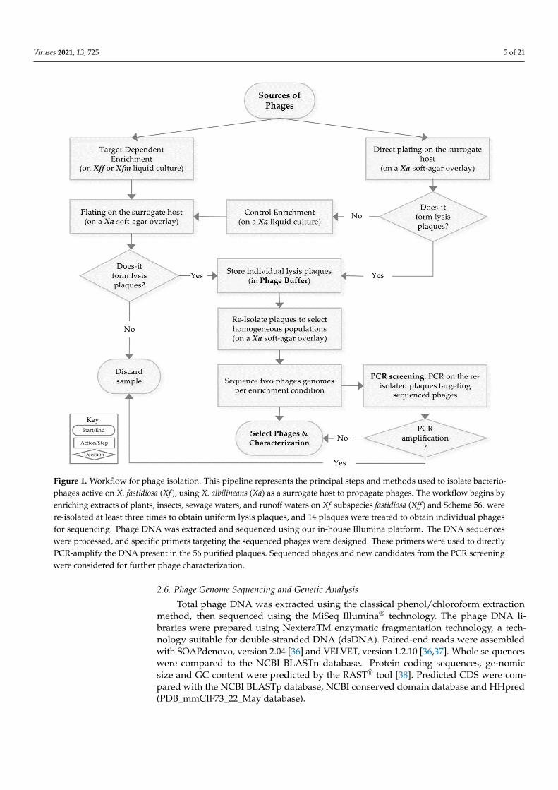
Figure 1. Workflow for phage isolation. This pipeline represents the principal steps and methods
used to isolate bacteriophages active on X. fastidiosa (Xf), using X. albilineans (Xa) as a surrogate
host to propagate phages. The workflow begins by enriching extracts of plants, insects, sewage
waters, and runoff waters on Xf subspecies fastidiosa (Xff) and Scheme 56. were re-isolated at least
three times to obtain uniform lysis plaques, and 14 plaques were treated to obtain individual
phages for sequencing. Phage DNA was extracted and sequenced using our in-house Illumina
platform. The DNA sequences were processed, and specific primers targeting the sequenced
phages were designed. These primers were used to directly PCR-amplify the DNA present in the
56 purified plaques. Sequenced phages and new candidates from the PCR screening were consid-
ered for further phage characterization.
2.4. Phage Isolation, Propagation and Purification
Phage activity was tested using spot assay on double agar overlay plates, using the
surrogate host X. albilineans. Briefly, Xa was cultured for 8 to 12 h on YPG broth at 28 °C
under 160 rpm agitation. Then, 200 μl of bacterial culture were mixed with 2.8 ml of YPG
soft agar at fusion temperatures close to 50 °C. The mixture was vortexed and plated on
YPG agar plates. Lytic activity was revealed by exposing Xa soft agar overlays to serial 10-
fold dilutions of the enrichment products and incubated for two to three days at 28 °C.
Phage-forming lysis plaques were selected and plugged off from the agar surface bottom
using a 1000 µl tip, and then suspended in 500 µl of phage buffer. Re-isolation was per-
formed on a Xa overlay at least three times, repeating plugging and suspension steps to
select single plaques with a regular shape. At the third re-isolation step, phage particles
Figure 1. Workflow for phage isolation. This pipeline represents the principal steps and methods used to isolate bacterio-phages active on X. fastidiosa (Xf ), using X. albilineans (Xa) as a surrogate host to propagate phages. The workflow begins byenriching extracts of plants, insects, sewage waters, and runoff waters on Xf subspecies fastidiosa (Xff ) and Scheme 56. werere-isolated at least three times to obtain uniform lysis plaques, and 14 plaques were treated to obtain individual phagesfor sequencing. Phage DNA was extracted and sequenced using our in-house Illumina platform. The DNA sequenceswere processed, and specific primers targeting the sequenced phages were designed. These primers were used to directlyPCR-amplify the DNA present in the 56 purified plaques. Sequenced phages and new candidates from the PCR screeningwere considered for further phage characterization.
2.6. Phage Genome Sequencing and Genetic Analysis
Total phage DNA was extracted using the classical phenol/chloroform extractionmethod, then sequenced using the MiSeq Illumina® technology. The phage DNA li-braries were prepared using NexteraTM enzymatic fragmentation technology, a tech-nology suitable for double-stranded DNA (dsDNA). Paired-end reads were assembledwith SOAPdenovo, version 2.04 [36] and VELVET, version 1.2.10 [36,37]. Whole se-quenceswere compared to the NCBI BLASTn database. Protein coding sequences, ge-nomicsize and GC content were predicted by the RAST® tool [38]. Predicted CDS were com-pared with the NCBI BLASTp database, NCBI conserved domain database and HHpred(PDB_mmCIF73_22_May database).

Viruses 2021, 13, 725 6 of 21
Genomic comparisons were performed with shared k-mer analysis using the KI-Sprotocol and the similarity matrix was represented using the KI-S circle packing tool [39]available at the CIRM-CFBP Galaxy platform (https://iris.angers.inra.fr/galaxypub-cfbp/,accessed on 28 March 2019).
Phylogenetic analyses were generated by the VICTOR online tool [40] using thecomplete genomic sequences of the Caudovirales bacteriophages infecting Xanthomonadalespresent in Genbank (Supplemental Table S1). All pairwise comparisons of the amino acidsequences were conducted using the Genome-BLAST Distance Phylogeny (GBDP) and treeswere inferred under settings recommended for prokaryotic viruses [40]. Taxon boundariesat the species, genus and family level were estimated with the OPTSIL program [41].
2.7. Phage Host Range
Host ranges of isolated phages were determined using the phage sensitivity spot testas follows: X. fastidiosa strains (Table 1) were cultured at 28 ◦C on PWG-M agar plates forup to seven days. Then, the cultures were suspended in sterile distilled water and OD600was calibrated at 0.1 for Xfp and Xfm and 0.01 for Xff strain. Drops of 20 µL of bacterialsuspensions were deposited on the surface of the B-CYE medium [31] and allowed to dry.Then, 10 µL of purified phage preparations (1010 PFU·mL−1) were deposited at the marginof the upper part of the bacterial drop. As a control for each strain, one bacterial drop didnot receive any treatment, and one bacterial drop was assayed with 10 µL of phage buffer.Spots were dried at room temperature and the plates cultured for up to 10 days at 28 ◦C.All experiments were performed in triplicate.
2.8. Phage Nomenclature and Nucleotide Sequence Accession Numbers
Phages isolated in this study were named according to the classification guide pro-posed by Adriaenssens and Brister [42]. Note that the names of the newly isolated phagesoriginate from neighborhood names in the city of Bogota, Colombia. The assembled genomesequences, as well as raw reads, were deposited at the European Nucleotide Archive (ENA)and the National Center for Biotechnology Information (NCBI) under the accession num-bers listed in Table 2.
Table 2. Phage genomes as sequenced.
Phage Name Full Nomenclature Accession Number Raw Sequence ID
FC03-UsmeFC08-Olaya
Xylella phage UsmeXanthomonas phage Olaya
LR743523MW802488
ERR5548206SRX10492030
FC12-BacataFC15-Bolivar
FC17-Usaquen
Xylella phage BacataXanthomonas phage Bolivar
Xanthomonas phage Usaquen
LR743524MW822535MW822536
ERR5548205SRX10492031SRX10492032
FC23-CotaFC25-Alcala
Xylella phage CotaXanthomonas phage Alcala
LR778216MW822537
ERR5548199SRX10492033
FC28-Sopo Xanthomonas phage Sopo LR743529 ERR5548204FC30-Tabio Xanthomonas phage Tabio LR743528 ERR5548203FC39-Tenjo Xanthomonas phage Tenjo LR743531 ERR5548202FC41-Suba Xanthomonas phage Suba LR743530 ERR5548201FC44-Bosa
FC47-FontebonFC57-Sumapaz
Xanthomonas phage BosaXanthomonas phage FontebonXanthomonas phage Sumapaz
LR743532MW822538MW822539
ERR5548200SRX10492034SRX10492035
3. Results3.1. Strategy to Isolate Xf-Specific Phages
To bypass the fastidious in vitro growth of Xf, we developed a strategy using Xa asa surrogate host to isolate phages. Using surrogate hosts for bacteriophage isolation andpropagation was proposed by several studies to isolate phages against pathogenic bacteriasuch as S. aureus [43,44], M. tuberculosis [45], and S. enterica [46], as well as for the plantpest X. fastidiosa [28]. To select bacteriophages with a lytic activity on Xf, samples from the
Viruses 2021, 13, 725 7 of 21
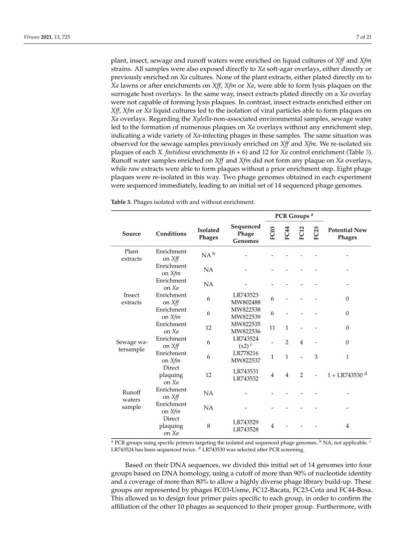
plant, insect, sewage and runoff waters were enriched on liquid cultures of Xff and Xfmstrains. All samples were also exposed directly to Xa soft-agar overlays, either directly orpreviously enriched on Xa cultures. None of the plant extracts, either plated directly on toXa lawns or after enrichments on Xff, Xfm or Xa, were able to form lysis plaques on thesurrogate host overlays. In the same way, insect extracts plated directly on a Xa overlaywere not capable of forming lysis plaques. In contrast, insect extracts enriched either onXff, Xfm or Xa liquid cultures led to the isolation of viral particles able to form plaques onXa overlays. Regarding the Xylella-non-associated environmental samples, sewage waterled to the formation of numerous plaques on Xa overlays without any enrichment step,indicating a wide variety of Xa-infecting phages in these samples. The same situation wasobserved for the sewage samples previously enriched on Xff and Xfm. We re-isolated sixplaques of each X. fastidiosa enrichments (6 + 6) and 12 for Xa control enrichment (Table 3).Runoff water samples enriched on Xff and Xfm did not form any plaque on Xa overlays,while raw extracts were able to form plaques without a prior enrichment step. Eight phageplaques were re-isolated in this way. Two phage genomes obtained in each experimentwere sequenced immediately, leading to an initial set of 14 sequenced phage genomes.
Table 3. Phages isolated with and without enrichment.
PCR Groups a
Source Conditions IsolatedPhages
SequencedPhage
Genomes FC03
FC44
FC12
FC23 Potential New
Phages
Plantextracts
Enrichmenton Xff NA b - - - - - -
Enrichmenton Xfm NA - - - - - -
Enrichmenton Xa NA - - - - - -
Insectextracts
Enrichmenton Xff 6 LR743523
MW802488 6 - - - 0
Enrichmenton Xfm 6 MW822538
MW822539 6 - - - 0
Enrichmenton Xa 12 MW822535
MW822536 11 1 - - 0
Sewage wa-tersample
Enrichmenton Xff 6 LR743524
(x2) c - 2 4 - 0
Enrichmenton Xfm 6 LR778216
MW822537 1 1 - 3 1
Directplaquing
on Xa12 LR743531
LR743532 4 4 2 - 1 + LR743530 d
Runoffwaterssample
Enrichmenton Xff NA - - - - - -
Enrichmenton Xfm NA - - - - - -
Directplaquing
on Xa8 LR743529
LR743528 4 - - - 4
a PCR groups using specific primers targeting the isolated and sequenced phage genomes. b NA, not applicable. c
LR743524 has been sequenced twice. d LR743530 was selected after PCR screening.
Based on their DNA sequences, we divided this initial set of 14 genomes into fourgroups based on DNA homology, using a cutoff of more than 90% of nucleotide identityand a coverage of more than 80% to allow a highly diverse phage library build-up. Thesegroups are represented by phages FC03-Usme, FC12-Bacata, FC23-Cota and FC44-Bosa.This allowed us to design four primer pairs specific to each group, in order to confirm theaffiliation of the other 10 phages as sequenced to their proper group. Furthermore, with
Viruses 2021, 13, 725 8 of 21
these four sets of primers we also tested all remaining plaques isolated in this study (56in total). This strategy aimed to rapidly eliminate phages that are already sequenced andidentify new phages to sequence.
Our PCR screening yielded the following results (Table 3). Among the 24 phagesisolated from insect extracts, 23 were FC03-Usme-like phages, while one belonged to theFC44-Bosa group. Among the 12 phages isolated using the sewage water samples, weobtained without enrichment four FC03-Usme-like phages, four FC44-Bosa-like phages,and two FC12-Bacata-like phages. Interestingly, among these 12 newly isolated phages,FC41-Suba and FC42 were not amplified by any of the specific primer pairs designedfor the four initial groups, suggesting new genome sequences; FC41-Suba DNA wasfurther extracted and sequenced. Similar sequence analysis led us to conclude that FC41-Suba represented a new group of phages. All six re-isolated phages obtained from thesewage water extracts after enrichment on Xff were identified as FC12-Bacata-like phages.However, two amplification profiles also revealed a positive amplification with FC44-Bosaprimers, suggesting that both phages were present on the re-isolated plaques, probably bycross-contamination. Interestingly, these results suggest that both FC12-Bacata-like andFC44-Bosa-like phages were able to multiply on Xylella strains during the enrichment step.Among the six phages isolated on sewage water samples enriched on Xfm, we identified byPCR screening three FC23-Cota-like, one FC03-Usme-like and one FC44-Bosa-like phages.Interestingly, more diversity was obtained on sewage water samples enriched on Xfm,and none of the primer pairs designed did amplify on phage FC24-Teja. Unfortunately,and despite extensive efforts, DNA from this phage was impossible to extract, suggestingthat its genome is composed either of highly modified DNA that cannot be extracted inthe same way as the other phage DNAs, or is composed of another type of nucleic acidmolecule. Of note, we have not yet tried to extract RNA from this phage preparation. Wewere thus not able to obtain the sequence of this particular phage genome. Finally, amongthe eight phages isolated from runoff water samples without enrichment, we found fourFC03-Usme-like phages and four phage isolates whose DNA could not be amplified withany primer pair, indicating either a different type of nucleic acid, or that they constituteone to four new groups of phages that were designated FC32-Sibate_a, FC32-Sibate_b,FC34-Cajica_a and FC34-Cajica_b (a and b indicate two different plaque morphologies onXa overlays).
At this stage, our PCR screening method enabled us to isolate at least five differentPCR groups of dsDNA bacteriophages represented by phages FC03-Usme, FC12-Bacata,FC23-Cota, FC41-Suba and FC44-Bosa. Three other bacteriophage isolates, representedby FC24-Teja, FC32-Sibate and FC34-Cajica, were potential new phages; their sequencesremain to be obtained to determine their nature.
3.2. Genomic Diversity of the Phages Isolated
According to our method, 10/14 sequenced phages and 32/56 phages screened byPCR belong to the FC03-Usme-like phage group, the most abundant group isolated in thisstudy. By comparing the complete DNA sequences (BLASTn analysis), we observed thatthe FC03-Usme genome shares 100% identity with 99% coverage with the FC08-Olaya,FC15-Bolivar, FC17-Usaquen, FC25-Alcala, FC47-Fontebon and FC57-Sumapaz genomes,indicating that these phages were different isolates of the same phage species using thespecies demarcation criteria defined by the International Committee on Taxonomy ofViruses (ICTV). The FC28-Sopo, FC30-Tabio and FC39-Tenjo genomes were similar to FC03-Usme, with 91–92% identity and a coverage of 89%. FC23-Cota, FC41-Suba and FC44-Bosawere phage singletons, and their DNA sequence could not be associated at the DNA levelwith any other phage DNA sequenced in this study.
In order to obtain more information on the diversity of the 14 phage genomes se-quenced thus far, we performed a comparative study based on the analysis of sharedoligonucleotides signatures (k-mers) using the KI-S tool [39]. Complete DNA sequenceswere compared, and the percentage of shared 22-mers was determined, allowing us to
Viruses 2021, 13, 725 9 of 21
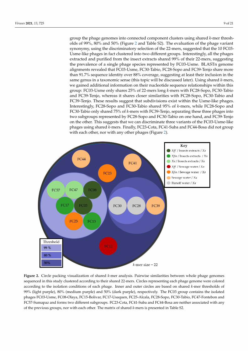
group the phage genomes into connected component clusters using shared k-mer thresh-olds of 99%, 80% and 50% (Figure 2 and Table S2). The evaluation of the phage variantsynonymy, using the discriminatory selection of the 22-mers, suggested that the 10 FC03-Usme-like phages in fact clustered into two different groups. Interestingly, all the phagesextracted and purified from the insect extracts shared 99% of their 22-mers, suggestingthe prevalence of a single phage species represented by FC03-Usme. BLASTn genomealignments revealed that FC03-Usme, FC30-Tabio, FC28-Sopo and FC39-Tenjo share morethan 91.7% sequence identity over 88% coverage, suggesting at least their inclusion in thesame genus in a taxonomic sense (this topic will be discussed later). Using shared k-mers,we gained additional information on their nucleotide sequence relationships within thisgroup: FC03-Usme only shares 25% of 22-mers long k-mers with FC28-Sopo, FC30-Tabioand FC39-Tenjo, whereas it shares closer similarities with FC28-Sopo, FC30-Tabio andFC39-Tenjo. These results suggest that subdivisions exist within the Usme-like phages.Interestingly, FC28-Sopo and FC30-Tabio shared 95% of k-mers, while FC28-Sopo andFC30-Tabio only shared 75% of k-mers with FC39-Tenjo, separating these three phages intotwo subgroups represented by FC28-Sopo and FC30-Tabio on one hand, and FC39-Tenjoon the other. This suggests that we can discriminate three variants of the FC03-Usme-likephages using shared k-mers. Finally, FC23-Cota, FC41-Suba and FC44-Bosa did not groupwith each other, nor with any other phages (Figure 2).
Viruses 2021, 13, x 10 of 22
Interestingly, FC28-Sopo and FC30-Tabio shared 95% of k-mers, while FC28-Sopo and FC30-
Tabio only shared 75% of k-mers with FC39-Tenjo, separating these three phages into two
subgroups represented by FC28-Sopo and FC30-Tabio on one hand, and FC39-Tenjo on
the other. This suggests that we can discriminate three variants of the FC03-Usme-like
phages using shared k-mers. Finally, FC23-Cota, FC41-Suba and FC44-Bosa did not group
with each other, nor with any other phages (Figure 2).
Figure 2. Circle packing visualization of shared k-mer analysis. Pairwise similarities between whole
phage genomes sequenced in this study clustered according to their shared 22-mers. Circles repre-
senting each phage genome were colored according to the isolation conditions of each phage. Inner
and outer circles are based on shared k-mer thresholds of 99% (light purple), 80% (medium purple)
and 50% (dark purple), respectively. The FC03 group contains the isolated phages FC03-Usme,
FC08-Olaya, FC15-Bolivar, FC17-Usaquen, FC25-Alcala, FC28-Sopo, FC30-Tabio, FC47-Fontebon
and FC57-Sumapaz and forms two different subgroups. FC23-Cota, FC41-Suba and FC44-Bosa are
neither associated with any of the previous groups, nor with each other. The matrix of shared k-
mers is presented in Table S2.
3.3. Genomic Characterization
To compare our isolated phage genomes with the diversity available in the Genebank
database, we performed a nucleotide BLASTn megablast search, optimized to retrieve
highly similar sequences using the complete genomic sequences of phages FC03-Usme,
FC12-Bacata, FC23-Cota, FC28-Sopo, FC30-Tabio, FC39-Tenjo, FC41-Suba and FC44-Bosa.
FC03-Usme, FC12-Bacata and FC44-Bosa shared homology with sequences available in
the Genbank database. FC03-Usme displayed high identity with the genome of phage CP2
of Xanthomonas axonopodis pv. citri [47] (92% identity over 83% coverage), as did FC44-Bosa
with phage DLP4 of Stenotrophomonas maltophilia [48] (97.4% identity over 99% coverage),
and FC12-Bacata with the Xff-infecting phage Salvo [28] (96.3% identity over 89%
coverage). Two groups represented by phages FC23-Cota and FC41-Suba did not get a hit
Figure 2. Circle packing visualization of shared k-mer analysis. Pairwise similarities between whole phage genomessequenced in this study clustered according to their shared 22-mers. Circles representing each phage genome were coloredaccording to the isolation conditions of each phage. Inner and outer circles are based on shared k-mer thresholds of99% (light purple), 80% (medium purple) and 50% (dark purple), respectively. The FC03 group contains the isolatedphages FC03-Usme, FC08-Olaya, FC15-Bolivar, FC17-Usaquen, FC25-Alcala, FC28-Sopo, FC30-Tabio, FC47-Fontebon andFC57-Sumapaz and forms two different subgroups. FC23-Cota, FC41-Suba and FC44-Bosa are neither associated with anyof the previous groups, nor with each other. The matrix of shared k-mers is presented in Table S2.

Viruses 2021, 13, 725 10 of 21
3.3. Genomic Characterization
To compare our isolated phage genomes with the diversity available in the Genebankdatabase, we performed a nucleotide BLASTn megablast search, optimized to retrievehighly similar sequences using the complete genomic sequences of phages FC03-Usme,FC12-Bacata, FC23-Cota, FC28-Sopo, FC30-Tabio, FC39-Tenjo, FC41-Suba and FC44-Bosa.FC03-Usme, FC12-Bacata and FC44-Bosa shared homology with sequences available inthe Genbank database. FC03-Usme displayed high identity with the genome of phageCP2 of Xanthomonas axonopodis pv. citri [47] (92% identity over 83% coverage), as didFC44-Bosa with phage DLP4 of Stenotrophomonas maltophilia [48] (97.4% identity over 99%coverage), and FC12-Bacata with the Xff -infecting phage Salvo [28] (96.3% identity over89% coverage). Two groups represented by phages FC23-Cota and FC41-Suba did not get ahit in any of the phage sequences in the Genbank database, indicating they were indeedtotally new.
The general characteristics of the 14 sequenced phage genomes are summarizedin Table 4. The genomic organization and the main functions encoded were commonto each of the PCR groups defined earlier and are described in Figure S1. FC03-Usme,FC28-Sopo, FC30-Tabio and FC39-Tenjo encoded 58 predicted CDS, with a genome sizeclose to 43 kb and a GC content around 67%. This group shared homology with theCP2 phage genome but to different extents (Figure S2). FC03-Usme shared 92% identityover 83% of the sequence length with the CP2 phage genomic sequence. At the DNAlevel, FC28-Sopo, FC30-Tabio, and FC39-Tenjo phage genomes showed more than 8%DNA sequence divergence compared to FC03-Usme, but less than 6% compared to theCP2 genome sequence (Figure S2). Interestingly, phages in this group were isolated fromdifferent environmental samples collected in different geographical areas such as Floridaand Argentina [49], whereas the original CP2 isolate originated from Yokohama, Japan [47].Moreover, at the DNA level, these phage genomes grouped together and displayed highidentity levels, around 93–95% over 83% coverage, confirming the k-mer analysis. Finally,FC12-Bacata displayed a genome size of 56 kb with 73 predicted CDS, and is homologousto phage Salvo, with 96% identity and 89% coverage. FC12-Bacata phage is an interestingcandidate due to its genomic proximity with a phage that was characterized as active onXff [12].
Table 4. General features of 14 sequenced phage genomes. In this table, we temporarily assign our newly sequenced phagesthe taxonomic affiliation of their closest homologs (when available).
Phage Size (bp) %GC CDS BLAST NearestOrganism
Coverage(%)
Identity(%) E-Value Order Family Genus Specie
FC03-Usme a 43721 66.6 58 X. axonopodisphage CP2 83 92.11% 0.0 Caudovirales Podovirus Unclassified
Podovirus Usmevirus
FC12-Bacata 56232 62.9 73 X. fastidiosa phageSalvo 89 96.29% 0.0 Caudovirales Siphovirus Sanovirus Bacatavirus
FC23-Cota 42307 58.8 52 none NA NA NA NA NA NA Tejovirus
FC28-Sopo 43861 66.9 58 X. axonopodisphage CP2 82 93.96% 0.0 Caudovirales Podovirus Unclassified
Podovirus Bacatavirus
FC30-Tabio 43458 66.9 58 X. axonopodisphage CP2 83 95.39% 0.0 Caudovirales Podovirus Unclassified
Podovirus Bacatavirus
FC39-Tenjo 43850 67.2 58 X. axonopodisphage CP2 83 94.70% 0.0 Caudovirales Podovirus Unclassified
Podovirus Bacatavirus
FC41-Suba 45510 52.3 71 none NA NA NA NA NA NA Subavirus
FC44-Bosa 63828 64.8 81 Stenotrophomonasphage DLP4 99% 97.40 % 0.0 Caudovirales Siphovirus Unclassified
Siphovirus Bosavirus
a FC08-Olaya, FC15-Bolivar, FC-17 Usaquen, FC25-Alcala, FC47-Fontebon and FC57-Sumapaz genomes have the same characteristics asFC03-Usme.
Surprisingly, the FC44-Bosa phage genome displayed a high identity with the phageDLP4 genome isolated from a soil sample and active on an unrelated species, Stenotrophomonasmaltophilia [14]. With a genome size of 63 kb and 81 predicted CDS by RAST [38], FC44-Bosa possessed the largest genome of the phages isolated during this study. FC44-Bosaphage was isolated from sewage extracts without any enrichment step, but it seems thatFC44-Bosa-like phages are also present in sewage extracts enriched on Xff and on insectextract enriched on Xa, according to PCR screening (Table 3). A putative integrase gene(CAA2409933.1) has been predicted in FC44-Bosa. A similar gene in the DLP4 genome was
Viruses 2021, 13, 725 11 of 21
confirmed to be involved in the lysogenic infection of S. maltophilia D1585 [48]. FC23-Cotaphage is the only isolate found in sewage water samples enriched on Xfm. Its genomesequence is 42 kb long, and 52 CDS were predicted. This phage genome displayed a low GCcontent of around 58%, and shared homology was restricted to only 2% of the genome se-quence with the X. citri phage Xc10 genome [15]. This 2% shared DNA region correspondedto region coding for a DNA polymerase A (ASZ72017.1) and a DNA-dependent RNA poly-merase (ASZ72029.1). Finally, the FC41-Suba genome was 45 kb long and 71 CDS werepredicted, with the lowest GC content of 52%. As for FC23-Cota, the FC41-Suba genome se-quence did not show any homology at the DNA level within the current Genebank database,sharing only a partial sequence of 253 nucleotides with the A. xylosoxidans phage phiAxp-1that corresponds to a region encoding for the Terminase large subunit (YP_009220341.1).The FC23-Cota and FC41-Suba phages were thus totally new and uncharacterized phages.
3.4. Morphological Characterization
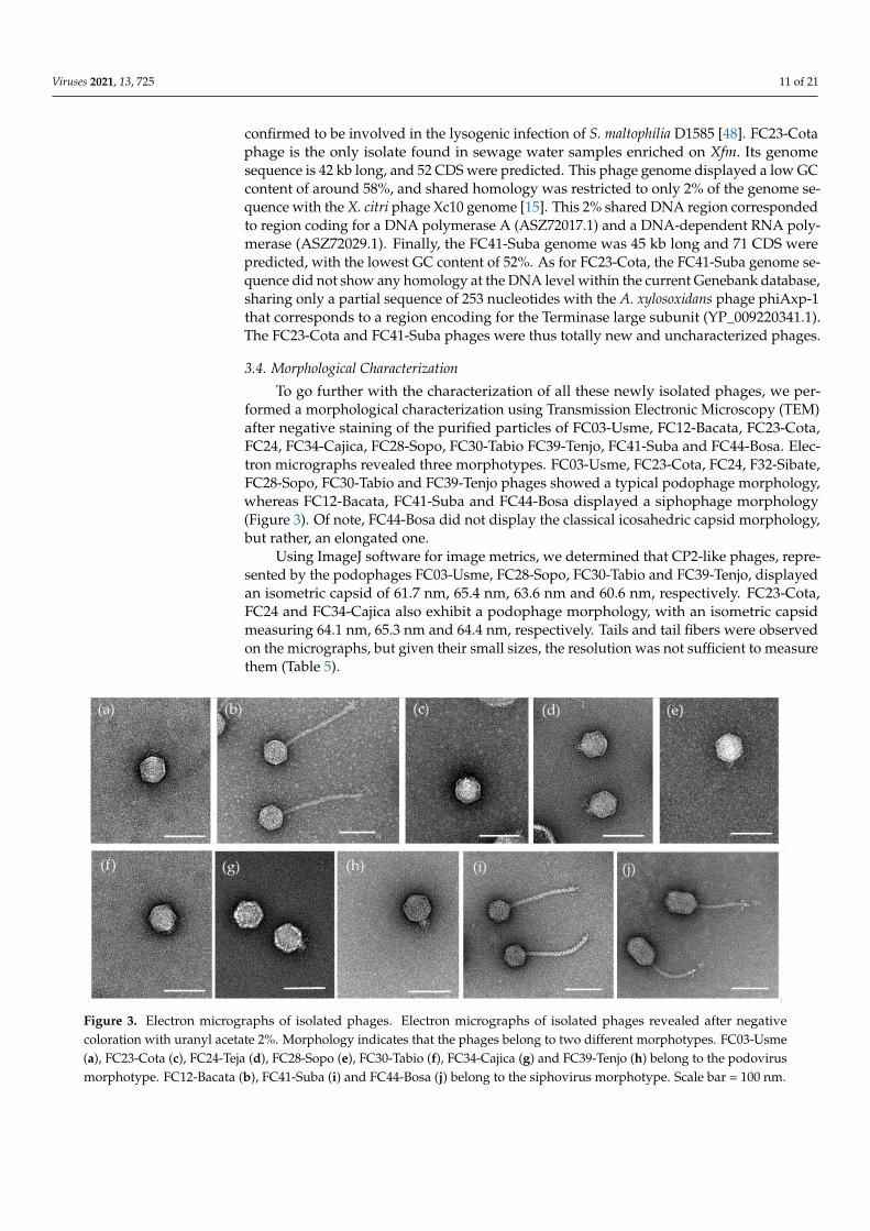
To go further with the characterization of all these newly isolated phages, we per-formed a morphological characterization using Transmission Electronic Microscopy (TEM)after negative staining of the purified particles of FC03-Usme, FC12-Bacata, FC23-Cota,FC24, FC34-Cajica, FC28-Sopo, FC30-Tabio FC39-Tenjo, FC41-Suba and FC44-Bosa. Elec-tron micrographs revealed three morphotypes. FC03-Usme, FC23-Cota, FC24, F32-Sibate,FC28-Sopo, FC30-Tabio and FC39-Tenjo phages showed a typical podophage morphology,whereas FC12-Bacata, FC41-Suba and FC44-Bosa displayed a siphophage morphology(Figure 3). Of note, FC44-Bosa did not display the classical icosahedric capsid morphology,but rather, an elongated one.
Using ImageJ software for image metrics, we determined that CP2-like phages, repre-sented by the podophages FC03-Usme, FC28-Sopo, FC30-Tabio and FC39-Tenjo, displayedan isometric capsid of 61.7 nm, 65.4 nm, 63.6 nm and 60.6 nm, respectively. FC23-Cota,FC24 and FC34-Cajica also exhibit a podophage morphology, with an isometric capsidmeasuring 64.1 nm, 65.3 nm and 64.4 nm, respectively. Tails and tail fibers were observedon the micrographs, but given their small sizes, the resolution was not sufficient to measurethem (Table 5).
Viruses 2021, 13, x 12 of 22
RNA polymerase (ASZ72029.1). Finally, the FC41-Suba genome was 45 kb long and 71
CDS were predicted, with the lowest GC content of 52%. As for FC23-Cota, the FC41-
Suba genome sequence did not show any homology at the DNA level within the current
Genebank database, sharing only a partial sequence of 253 nucleotides with the A. xylosox-
idans phage phiAxp-1 that corresponds to a region encoding for the Terminase large sub-
unit (YP_009220341.1). The FC23-Cota and FC41-Suba phages were thus totally new and
uncharacterized phages.
3.4. Morphological Characterization
To go further with the characterization of all these newly isolated phages, we
performed a morphological characterization using Transmission Electronic Microscopy
(TEM) after negative staining of the purified particles of FC03-Usme, FC12-Bacata, FC23-
Cota, FC24, FC34-Cajica, FC28-Sopo, FC30-Tabio FC39-Tenjo, FC41-Suba and FC44-Bosa.
Electron micrographs revealed three morphotypes. FC03-Usme, FC23-Cota, FC24, F32-
Sibate, FC28-Sopo, FC30-Tabio and FC39-Tenjo phages showed a typical podophage
morphology, whereas FC12-Bacata, FC41-Suba and FC44-Bosa displayed a siphophage
morphology (Figure 3). Of note, FC44-Bosa did not display the classical icosahedric capsid
morphology, but rather, an elongated one.
Using ImageJ software for image metrics, we determined that CP2-like phages,
represented by the podophages FC03-Usme, FC28-Sopo, FC30-Tabio and FC39-Tenjo,
displayed an isometric capsid of 61.7 nm, 65.4 nm, 63.6 nm and 60.6 nm, respectively.
FC23-Cota, FC24 and FC34-Cajica also exhibit a podophage morphology, with an
isometric capsid measuring 64.1 nm, 65.3 nm and 64.4 nm, respectively. Tails and tail fibers
were observed on the micrographs, but given their small sizes, the resolution was not
sufficient to measure them (Table 5).
In contrast, FC12-Bacata, FC41-Suba and FC44-Bosa showed a siphophage
morphology, with two different capsid organizations. FC12-Bacata was composed of an
isometric head, the diameter of which was 66.5 nm, and a long and flexible tail of 220.5
nm long. FC41-Suba also had a long and flexible tail of 199.5 nm long, with an isometric
capsid of 61.2 nm. In contrast, FC44-Bosa was composed of a heterometric head of 60.1 ×
88.8 nm long and a long flexible tail of 140.5 nm. All three siphophages in this study
displayed fibers observable at the distal extremity of the tails.
Figure 3. Electron micrographs of isolated phages. Electron micrographs of isolated phages re-
vealed after negative coloration with uranyl acetate 2%. Morphology indicates that the phages
belong to two different morphotypes. FC03-Usme (a), FC23-Cota (c), FC24-Teja (d), FC28-Sopo (e),
FC30-Tabio (f), FC34-Cajica (g) and FC39-Tenjo (h) belong to the podovirus morphotype. FC12-
Figure 3. Electron micrographs of isolated phages. Electron micrographs of isolated phages revealed after negativecoloration with uranyl acetate 2%. Morphology indicates that the phages belong to two different morphotypes. FC03-Usme(a), FC23-Cota (c), FC24-Teja (d), FC28-Sopo (e), FC30-Tabio (f), FC34-Cajica (g) and FC39-Tenjo (h) belong to the podovirusmorphotype. FC12-Bacata (b), FC41-Suba (i) and FC44-Bosa (j) belong to the siphovirus morphotype. Scale bar = 100 nm.

Viruses 2021, 13, 725 12 of 21
In contrast, FC12-Bacata, FC41-Suba and FC44-Bosa showed a siphophage morphol-ogy, with two different capsid organizations. FC12-Bacata was composed of an isometrichead, the diameter of which was 66.5 nm, and a long and flexible tail of 220.5 nm long.FC41-Suba also had a long and flexible tail of 199.5 nm long, with an isometric capsid of61.2 nm. In contrast, FC44-Bosa was composed of a heterometric head of 60.1 × 88.8 nmlong and a long flexible tail of 140.5 nm. All three siphophages in this study displayedfibers observable at the distal extremity of the tails.
Table 5. Morphological characteristics of the phages based on MET acquisitions.
Phage Head Shape &Diameter (nm)
Tail Shape & Length(nm) Morphotype a
FC03-Usme(n = 15) Isometric, 61.7 ± 2 *NA Podovirus C1
FC12-Bacata(n = 14) Isometric, 66.5 ± 2 Long, Flexible, 220.5 ± 5 Siphovirus B1
FC23-Cota(n = 14) Isometric, 64.1 ± 1 *NA Podovirus C1
FC24-Teja(n = 15) Isometric, 65.3 ± 2 *NA Podovirus C1
FC28-Sopo(n = 6) Isometric, 65.4 ± 2 *NA Podovirus C1
FC30-Tabio(n = 6) Isometric, 63.6 ± 2 *NA Podovirus C1
FC34-Cajica(n = 15) Isometric, 64.4 ± 2 *NA Podovirus C1
FC39-Tenjo(n = 6) Isometric, 60.6 ± 2 *NA Podovirus C1
FC41-Suba(n = 15) Isometric, 61.2 ± 2 Long, Flexible, 199.5 ± 4 Siphovirus B1
FC44-Bosa(n = 12)
Heterometric, 60.1 ± 1× 88.8 ± 2 Long, Flexible, 140.5 ± 7 Siphovirus B3
Phage physical dimensions are the means of measurements of at least n individual virions using ImageJ. *NA, notapplicable; a according to [50].
3.5. Phylogenetic Analysis of the Sequenced Phage Genomes
Among our PCR groups, we distinguished various degrees of homology with knownphage DNA sequences. Phages FC03-Usme, FC12-Bacata and FC44-Bosa could thus berelated to phage genomes present in the NCBIdatabase. In contrast, FC23-Cota and FC41-Suba genomes had no substantial sequence similarity throughout the NCBIdatabase (ac-cessed on January 2021).
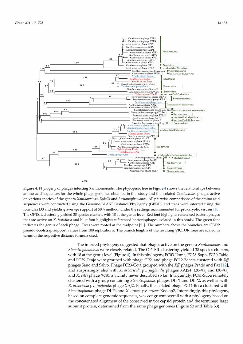
To analyze the phylogenetic relationships of the phages isolated in this study withother phages with lytic activity on Xanthomonas spp., Stenotrophomonas spp., and X. fastidiosa,a phylogenetic tree was generated using the VICTOR online tool [40] using completegenomic sequences of the Caudovirales bacteriophages infecting Xanthomonadales (Figure 4).
Viruses 2021, 13, 725 13 of 21Viruses 2021, 13, x 14 of 22
Figure 4. Phylogeny of phages infecting Xanthomonads. The phylogenic tree in Figure 4 shows the
relationships between amino acid sequences for the whole phage genomes obtained in this study
and the isolated Caudovirales phages active on various species of the genera Xanthomonas, Xylella
and Stenotrophomonas. All pairwise comparisons of the amino acid sequences were conducted using
the Genome-BLAST Distance Phylogeny (GBDP), and trees were inferred using the formulas D0
and yielding average support of 58% method, under the settings recommended for prokaryotic
viruses [40]. The OPTSIL clustering yielded 38 species clusters, with 18 at the genus level. Red font
highlights referenced bacteriophages that are active on X. fastidiosa and blue font highlights
referenced bacteriophages isolated in this study. The green font indicates the genus of each phage.
Trees were rooted at the midpoint [51]. The numbers above the branches are GBDP pseudo-
bootstrap support values from 100 replications. The branch lengths of the resulting VICTOR trees
are scaled in terms of the respective distance formula used.
The inferred phylogeny suggested that phages active on the genera Xanthomonas and
Stenotrophomonas were closely related. The OPTSIL clustering yielded 38 species clusters,
with 18 at the genus level (Figure 4). In this phylogeny, FC03-Usme, FC28-Sopo, FC30-
Tabio and FC39-Tenjo were grouped with phage CP2, and phage FC12-Bacata clustered
with Xff phages Sano and Salvo. Phage FC23-Cota grouped with the Xff phages Prado and
Paz [12], and surprisingly, also with X. arboricola pv. juglandis phages XAJ24, f20-Xaj and
f30-Xaj and X. citri phage Xc10, a vicinity never described so far. Intriguingly, FC41-Suba
remotely clustered with a group containing Stenotrophonas phages DLP1 and DLP2, as
well as with X. arboricola pv. juglandis phage XAJ2. Finally, the isolated phage FC44-Bosa
clustered with Stenotrophonas phage DLP4 and X. oryzae pv. oryzae Xoo-sp2. Interestingly,
this phylogeny, based on complete genomic sequences, was congruent overall with a
phylogeny based on the concatenated alignment of the conserved major capsid protein
Figure 4. Phylogeny of phages infecting Xanthomonads. The phylogenic tree in Figure 4 shows the relationships betweenamino acid sequences for the whole phage genomes obtained in this study and the isolated Caudovirales phages activeon various species of the genera Xanthomonas, Xylella and Stenotrophomonas. All pairwise comparisons of the amino acidsequences were conducted using the Genome-BLAST Distance Phylogeny (GBDP), and trees were inferred using theformulas D0 and yielding average support of 58% method, under the settings recommended for prokaryotic viruses [40].The OPTSIL clustering yielded 38 species clusters, with 18 at the genus level. Red font highlights referenced bacteriophagesthat are active on X. fastidiosa and blue font highlights referenced bacteriophages isolated in this study. The green fontindicates the genus of each phage. Trees were rooted at the midpoint [51]. The numbers above the branches are GBDPpseudo-bootstrap support values from 100 replications. The branch lengths of the resulting VICTOR trees are scaled interms of the respective distance formula used.
The inferred phylogeny suggested that phages active on the genera Xanthomonas andStenotrophomonas were closely related. The OPTSIL clustering yielded 38 species clusters,with 18 at the genus level (Figure 4). In this phylogeny, FC03-Usme, FC28-Sopo, FC30-Tabioand FC39-Tenjo were grouped with phage CP2, and phage FC12-Bacata clustered with Xffphages Sano and Salvo. Phage FC23-Cota grouped with the Xff phages Prado and Paz [12],and surprisingly, also with X. arboricola pv. juglandis phages XAJ24, f20-Xaj and f30-Xajand X. citri phage Xc10, a vicinity never described so far. Intriguingly, FC41-Suba remotelyclustered with a group containing Stenotrophonas phages DLP1 and DLP2, as well as withX. arboricola pv. juglandis phage XAJ2. Finally, the isolated phage FC44-Bosa clustered withStenotrophonas phage DLP4 and X. oryzae pv. oryzae Xoo-sp2. Interestingly, this phylogeny,based on complete genomic sequences, was congruent overall with a phylogeny based onthe concatenated alignment of the conserved major capsid protein and the terminase largesubunit protein, determined from the same phage genomes (Figure S3 and Table S3).

Viruses 2021, 13, 725 14 of 21
According to the genomic and phylogenic analysis, we propose the following taxo-nomic description of the viruses isolated in this study according to the latest recommen-dations [52]. All of them belong to the following lineage: Viruses, Duplodnaviria (realm),Heunggongvirae (kingdom), Uroviricota (phylum), Caudoviricetes (class), Caudovirales (order).Family, sub-family and genus affiliations for the newly isolated viruses are discussed below.
i: Xylella phage Cota (Autographiviridae family). According to the latest ICTV viraltaxonomy criteria (2019 release), the Xylella phage Cota belongs to the Autographiviridaefamily. Within this subfamily, the average genome size is 41 kb (42.3 kb for Cota), andthe overall genomic organization is conserved with genes on the same strand (predictedORFs are all situated on the same strand for Cota, Figure S1). Autographiviridae virionsdisplay a small (ca. 60 nm in diameter) icosahedral head attached to a short tail (podovirusmorphology) as is the case for Cota (Figure 3c). Finally, Autographiviridae encode theirown RNA polymerase, which is the case for Cota (Genbank protein ID CAB1282939.1).A genome-wide phylogenetic analysis based on protein-clustering indicates that Xylellaphage Cota is related to the Pradovirus genus (Figure 4) but classified by OPTSIL as an“Unclassified Autographiviridae”. A BLASTn analysis revealed that the two PradovirusesXylella phage Prado (type species of the Pradovirus genus) and Xylella phage Paz are almostas distantly related from the DNA point of view (73.08% identity, 27% cover) than Xylellaphage Prado and Xylella phage Cota (68.15% identity and 35% cover). Taking also intoaccount the low pseudo-bootstrap support values, we chose to classify Xylella phage Cotawithin the Pradovirus genus (Autographiviridae family) and not in a separated new genus assuggested by the phylogenetic tree in Figure 4.
ii: Usmevirus genus (Podoviridae family). In Figure 4, the genome-wide phylogeneticanalysis, based on protein clustering, indicates that Xylella phage Usme, Xanthomonasphage Sopo, Xanthomonas phage Tabio, Xanthomonas phage Tenjo (all 4 phages were isolatedduring this study) and the Xanthomonas citri phage CP2 form a new genus within the phagesinfecting Xanthomonads. Xylella phage Usme, Xanthomonas phage Sopo, Xanthomonasphage Tabio, and Xanthomonas phage Tenjo clearly belong to the same species according tothe BLASTn analysis (>98.8% identity, >94% cover in pairwise alignments). Xanthomonascitri phage CP2 is slightly more distant from the above-cited viruses (identity between 92 to95%, cover around 83%). Altogether, these results prompt us to classify these five phageswithin a new genus named “Usmevirus” in the Podoviridae family. The Xylella phage Usmeis defined as the type species of this new genus. A proposal will be submitted to the ICTVfor the creation of the Usmevirus genus. Six other Xanthomonas phages were isolated duringthis study, whose DNA sequences are highly identical to Xylella phage Usme according toour k-mer analysis (Figure 2). Thus, the Xanthomonas phages FC08-Olaya, FC15-Bolivar,FC17-Usaquen, FC25-Alcala, FC47-Fontebon and FC57-Sumapaz all belong to the newUsmevirus genus.
iii: Subavirus genus (Siphoviridae family). In Figure 4, Xanthomonas phage FC41-Subais too distantly related to the Stenotrophomonas phages DLP_1 and DLP_2 to be classifiedin the same Septimatrevirus genus (Siphoviridae family). We chose to define a new genusnamed “Subavirus” (Siphoviridae family) comprising only one Xanthomonas phage Suba as atype species for this new genus. A proposal will be submitted to the ICTV for the creationof the Subavirus genus.
iv: Xylella phage FC12-Bacata. According to the phylogenetic tree presented in Figure 4,the Xylella phage Bacata belongs to the Sanovirus genus (Siphoviridae family).
3.6. Host Range Determination
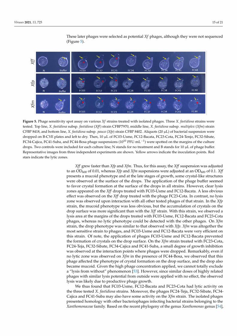
To determine the host range of the newly isolated phages on different Xf strains, weperformed a spot assay using three different bacterial strains representing the subspeciesfastidiosa (Xff ), pauca (Xfp) and multiplex (Xfm). This assay was performed for the sequencedphages FC03-Usme, FC12-Bacata, FC23-Cota, FC41-Suba and FC44-Bosa. We also includedFC24-Teja, isolated from sewage samples enriched on Xfm cultures, as well as FC32-Sibateand FC34-Cajica, isolated on Xa from runoff waters and without any enrichment on Xf.
Viruses 2021, 13, 725 15 of 21
These later phages were selected as potential Xf phages, although they were not sequenced(Figure 5).
Viruses 2021, 13, x 16 of 22
sequenced phages FC03-Usme, FC12-Bacata, FC23-Cota, FC41-Suba and FC44-Bosa. We
also included FC24-Teja, isolated from sewage samples enriched on Xfm cultures, as well as
FC32-Sibate and FC34-Cajica, isolated on Xa from runoff waters and without any
enrichment on Xf. These later phages were selected as potential Xf phages, although they
were not sequenced (Figure 5).
Figure 5. Phage sensitivity spot assay on various Xf strains treated with isolated phages. Three X. fastidiosa
strains were tested. Top line, X. fastidiosa subsp. fastidiosa (Xff) strain CFBP7970, middle line, X. fastidiosa subsp.
multiplex (Xfm) strain CFBP 8418, and bottom line, X. fastidiosa subsp. pauca (Xfp) strain CFBP 8402. Aliquots (20
μl) of bacterial suspension were dropped on B-CYE plates and left to dry. Then, 10 μl of FC03-Usme, FC12-
Bacata, FC23-Cota, FC24-Tenjo, FC32-Sibate, FC34-Cajica, FC41-Suba, and FC44-Bosa phage suspensions (1010
PFU.mL−1) were spotted on the margins of the culture drops. Two controls were included for each culture line;
N stands for no treatment and B stands for 10 μl of phage buffer. Representative images from three independent
experiments are shown. Yellow arrows indicate the inoculation points. Red stars indicate the lytic zones.
Xff grew faster than Xfp and Xfm. Thus, for this assay, the Xff suspension was
adjusted to an OD600 of 0.01, whereas Xfp and Xfm suspensions were adjusted at an OD600
of 0.1. Xff presents a mucoid phenotype and at the late stages of growth, some crystal-like
structures were observed at the surface of the drops. The application of the phage buffer
seemed to favor crystal formation at the surface of the drops in all strains. However, clear
lysis zones appeared on the Xff drops treated with FC03-Usme and FC12-Bacata. A less
obvious effect was observed on the Xff drop treated with the phage FC23-Cota. In contrast,
no lysis zone was observed upon interaction with all other tested phages of that strain. In
the Xfp strain, the mucoid phenotype was less obvious, but the accumulation of crystals
on the drop surface was more significant than with the Xff strain. With this strain, we
observed a lysis area at the margins of the drops treated with FC03-Usme, FC12-Bacata
and FC23-Cota phages, whereas no lytic phenotype could be detected with the other
phages. On Xfm strain, the drop phenotype was similar to that observed with Xfp. Xfm was
altogether the most sensitive strain to phages, and FC03-Usme and FC12-Bacata were very
efficient on this strain. Of note, the application of phages FC03-Usme and FC12-Bacata
prevented the formation of crystals on the drop surface. On the Xfm strain treated with
FC23-Cota, FC24-Teja, FC32-Sibate, FC34-Cajica and FC41-Suba, a small degree of growth
inhibition was observed at the interaction points where phages were dropped.
Remarkably, even if no lytic zone was observed on Xfm in the presence of FC44-Bosa, we
observed that this phage affected the phenotype of crystal formation on the drop surface,
and the drop also became mucoid. Given the high phage concentration applied, we cannot
totally exclude a “lysis from without” phenomenon [53]. However, since similar doses of
highly related phages with similar lysis potential from outside were applied with no
effect, the observed lysis was likely due to productive phage growth.
We thus found that FC03-Usme, FC12-Bacata and FC23-Cota had lytic activity on the
three tested X. fastidiosa strains. Moreover, the phages FC24-Teja, FC32-Sibate, FC34-
Cajica and FC41-Suba may also have some activity on the Xfm strain. The isolated phages
Figure 5. Phage sensitivity spot assay on various Xf strains treated with isolated phages. Three X. fastidiosa strains weretested. Top line, X. fastidiosa subsp. fastidiosa (Xff ) strain CFBP7970, middle line, X. fastidiosa subsp. multiplex (Xfm) strainCFBP 8418, and bottom line, X. fastidiosa subsp. pauca (Xfp) strain CFBP 8402. Aliquots (20 µL) of bacterial suspension weredropped on B-CYE plates and left to dry. Then, 10 µL of FC03-Usme, FC12-Bacata, FC23-Cota, FC24-Tenjo, FC32-Sibate,FC34-Cajica, FC41-Suba, and FC44-Bosa phage suspensions (1010 PFU·mL−1) were spotted on the margins of the culturedrops. Two controls were included for each culture line; N stands for no treatment and B stands for 10 µL of phage buffer.Representative images from three independent experiments are shown. Yellow arrows indicate the inoculation points. Redstars indicate the lytic zones.
Xff grew faster than Xfp and Xfm. Thus, for this assay, the Xff suspension was adjustedto an OD600 of 0.01, whereas Xfp and Xfm suspensions were adjusted at an OD600 of 0.1. Xffpresents a mucoid phenotype and at the late stages of growth, some crystal-like structureswere observed at the surface of the drops. The application of the phage buffer seemedto favor crystal formation at the surface of the drops in all strains. However, clear lysiszones appeared on the Xff drops treated with FC03-Usme and FC12-Bacata. A less obviouseffect was observed on the Xff drop treated with the phage FC23-Cota. In contrast, no lysiszone was observed upon interaction with all other tested phages of that strain. In the Xfpstrain, the mucoid phenotype was less obvious, but the accumulation of crystals on thedrop surface was more significant than with the Xff strain. With this strain, we observed alysis area at the margins of the drops treated with FC03-Usme, FC12-Bacata and FC23-Cotaphages, whereas no lytic phenotype could be detected with the other phages. On Xfmstrain, the drop phenotype was similar to that observed with Xfp. Xfm was altogether themost sensitive strain to phages, and FC03-Usme and FC12-Bacata were very efficient onthis strain. Of note, the application of phages FC03-Usme and FC12-Bacata preventedthe formation of crystals on the drop surface. On the Xfm strain treated with FC23-Cota,FC24-Teja, FC32-Sibate, FC34-Cajica and FC41-Suba, a small degree of growth inhibitionwas observed at the interaction points where phages were dropped. Remarkably, even ifno lytic zone was observed on Xfm in the presence of FC44-Bosa, we observed that thisphage affected the phenotype of crystal formation on the drop surface, and the drop alsobecame mucoid. Given the high phage concentration applied, we cannot totally excludea “lysis from without” phenomenon [53]. However, since similar doses of highly relatedphages with similar lysis potential from outside were applied with no effect, the observedlysis was likely due to productive phage growth.
We thus found that FC03-Usme, FC12-Bacata and FC23-Cota had lytic activity onthe three tested X. fastidiosa strains. Moreover, the phages FC24-Teja, FC32-Sibate, FC34-Cajica and FC41-Suba may also have some activity on the Xfm strain. The isolated phagespresented homology with other bacteriophages infecting bacterial strains belonging to theXanthomonaceae family. Based on the recent phylogeny of the genus Xanthomonas genus [54],
Viruses 2021, 13, 725 16 of 21
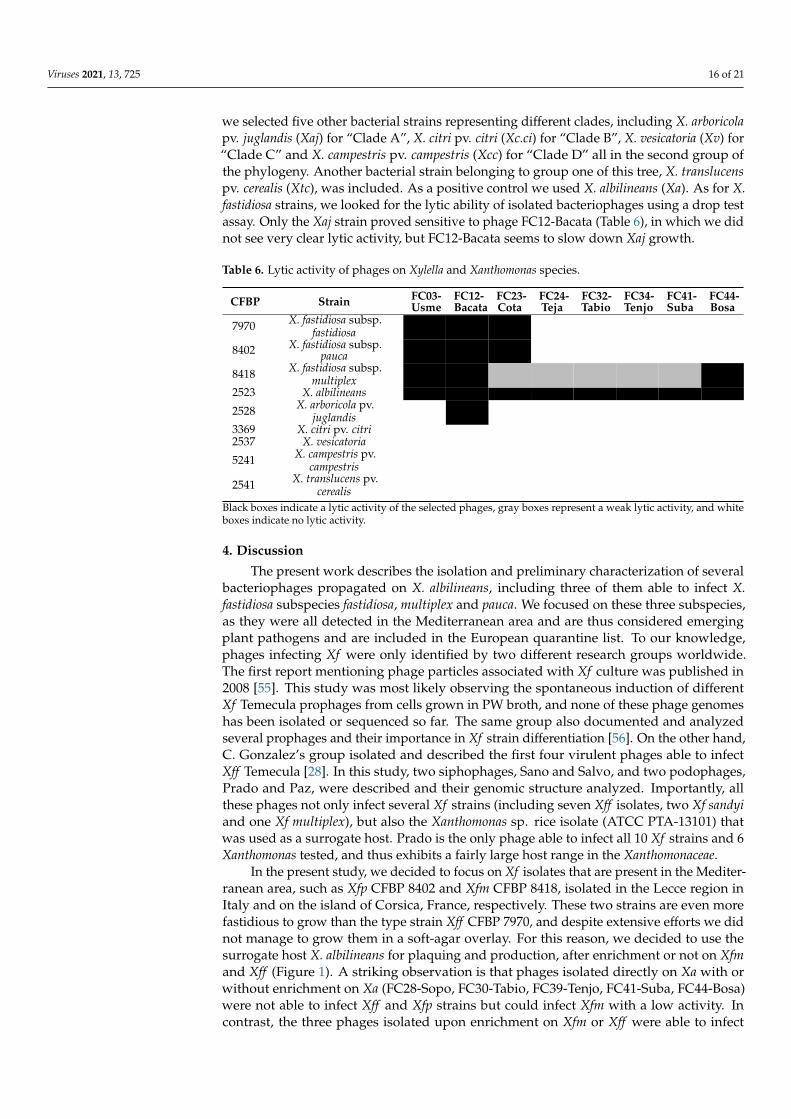
we selected five other bacterial strains representing different clades, including X. arboricolapv. juglandis (Xaj) for “Clade A”, X. citri pv. citri (Xc.ci) for “Clade B”, X. vesicatoria (Xv) for“Clade C” and X. campestris pv. campestris (Xcc) for “Clade D” all in the second group ofthe phylogeny. Another bacterial strain belonging to group one of this tree, X. translucenspv. cerealis (Xtc), was included. As a positive control we used X. albilineans (Xa). As for X.fastidiosa strains, we looked for the lytic ability of isolated bacteriophages using a drop testassay. Only the Xaj strain proved sensitive to phage FC12-Bacata (Table 6), in which we didnot see very clear lytic activity, but FC12-Bacata seems to slow down Xaj growth.
Table 6. Lytic activity of phages on Xylella and Xanthomonas species.
CFBP Strain FC03-Usme
FC12-Bacata
FC23-Cota
FC24-Teja
FC32-Tabio
FC34-Tenjo
FC41-Suba
FC44-Bosa
7970 X. fastidiosa subsp.fastidiosa
8402 X. fastidiosa subsp.pauca
8418 X. fastidiosa subsp.multiplex
2523 X. albilineans
2528 X. arboricola pv.juglandis
3369 X. citri pv. citri2537 X. vesicatoria
5241 X. campestris pv.campestris
2541 X. translucens pv.cerealis
Black boxes indicate a lytic activity of the selected phages, gray boxes represent a weak lytic activity, and whiteboxes indicate no lytic activity.
4. Discussion
The present work describes the isolation and preliminary characterization of severalbacteriophages propagated on X. albilineans, including three of them able to infect X.fastidiosa subspecies fastidiosa, multiplex and pauca. We focused on these three subspecies,as they were all detected in the Mediterranean area and are thus considered emergingplant pathogens and are included in the European quarantine list. To our knowledge,phages infecting Xf were only identified by two different research groups worldwide.The first report mentioning phage particles associated with Xf culture was published in2008 [55]. This study was most likely observing the spontaneous induction of differentXf Temecula prophages from cells grown in PW broth, and none of these phage genomeshas been isolated or sequenced so far. The same group also documented and analyzedseveral prophages and their importance in Xf strain differentiation [56]. On the other hand,C. Gonzalez’s group isolated and described the first four virulent phages able to infectXff Temecula [28]. In this study, two siphophages, Sano and Salvo, and two podophages,Prado and Paz, were described and their genomic structure analyzed. Importantly, allthese phages not only infect several Xf strains (including seven Xff isolates, two Xf sandyiand one Xf multiplex), but also the Xanthomonas sp. rice isolate (ATCC PTA-13101) thatwas used as a surrogate host. Prado is the only phage able to infect all 10 Xf strains and 6Xanthomonas tested, and thus exhibits a fairly large host range in the Xanthomonaceae.
In the present study, we decided to focus on Xf isolates that are present in the Mediter-ranean area, such as Xfp CFBP 8402 and Xfm CFBP 8418, isolated in the Lecce region inItaly and on the island of Corsica, France, respectively. These two strains are even morefastidious to grow than the type strain Xff CFBP 7970, and despite extensive efforts we didnot manage to grow them in a soft-agar overlay. For this reason, we decided to use thesurrogate host X. albilineans for plaquing and production, after enrichment or not on Xfmand Xff (Figure 1). A striking observation is that phages isolated directly on Xa with orwithout enrichment on Xa (FC28-Sopo, FC30-Tabio, FC39-Tenjo, FC41-Suba, FC44-Bosa)were not able to infect Xff and Xfp strains but could infect Xfm with a low activity. Incontrast, the three phages isolated upon enrichment on Xfm or Xff were able to infect
Viruses 2021, 13, 725 17 of 21
efficiently all three Xf strains included in our spot assay (Figure 5, Table 6). This highlightsthe absolute requirement of the enrichment step in order to isolate phages active against Xf.
Regarding the isolation sources and strategy, we could not isolate any phage from plantsamples displaying the typical symptoms of Xf -infected plants and collected in an infectedarea of Corsica. Of note, virulent phages able to infect Xff were isolated from plant extracts(rice and weeds) that contained Xanthomonas spp. but not Xf [28]. Different hypothesescould explain this result: (i) the processing of the plant samples (see Section 2.2) couldhave been detrimental to phage isolation, (ii) the symptoms observed on the collected plantsamples were not due to Xf infection, or (iii) the symptoms were indeed due to the presenceof Xf, but specific phages did not develop in these plants. In contrast, insect vector samplesharvested on the island of Corsica, close to the plant samples, were processed in a verysimilar way to the plant extracts and proved to be an actual phage reservoir for phage FC03-Usme and relatives belonging to the CP2-like group (Table 4). Although different phageisolates were obtained from this type of biological sample, they all proved to belong to thesame group. To our knowledge, this is the first example of a virulent phage, active againstXf, isolated from such a source. Interestingly, the xylem-feeding leafhopper vector, calledthe glassy-winged sharpshooter (GWSS, Homalodisca genus, Hemiptera: Cicadellidae),which is the main insect vector identified for Pierce’s disease transmission [57], has theability to uptake phages. In this experiment, the insect vector was allowed to feed onbeam stems immersed in a phage Paz solution, and showed efficient acquisition of thatphage up to 108 PFU gm−1 of GWSS tissue [58]. In the same work, however, the authorsmentioned that although phage Paz was efficiently acquired by GWSS, its transmission toplants (bean stems) through the same insect vector was much less efficient, probably due toa dilution effect. It is thus remarkable that several phages were successfully isolated frominsect extracts collected in the Xf -infected area. The environmental source that providedthe largest diversity in terms of phage isolates was the sewage water sample collected inthe Marseille wastewater plant. This is not surprising, as worldwide phage researcherssuccessfully isolate bacteriophages from sewage samples, targeting an immense variety ofhuman, plant, or animal pathogens [59]. The particularity of the water treatment plant wehad access to is that it collects water not only from Marseille, the largest city in the region,but also from many other cities and villages around the area with strong agriculturalactivity. This may explain the significant diversity of phages that could be isolated fromthis source.
The genomic analysis of the newly isolated phages led to interesting observations.First of all, we isolated and sequenced four different phages that all share a high genomicidentity with X. axonopodis phage CP2, among which only FC03-Usme was able to infect Xfstrains. This suggest that the CP2-like phages, whose first isolate came from Yokohama(Japan), are very abundant and are distributed all over the world, probably with theXanthomonaceae strains they infect. A similar conclusion applies to the phage FC12-Bacata,whose genome sequence is homologous to that of the phage Salvo isolated in Texas (USA).Then come phage genomes with very low identity to any genome present in the databases,such as FC23-Cota and FC41-Suba, which can be considered new phage species since theirgenomes only match a few conserved genes in the X. citri phage Xc10 and A. xylosoxidansphage phiAxp-1 genomes, respectively. Finally, the procedure we set up in this study ledmostly to the isolation of strictly virulent phages, as no specific signature of lysogenicdevelopment could be identified in their genomes, such as integrase or repressor genes;the BLAST search did not reveal any homology with Xanthomonaceae strains genomes soeffectively, as temperate phage genomes would do. The only temperate phage isolatedwas named FC44-Bosa, whose genome shared extensive identity with the Stenotrophomonasphage DLP4 (Table 4). Interestingly, this phage showed good lytic activity on Xfm.
5. Conclusions
This study allowed the isolation of several phages that constitute promising candi-dates for the biocontrol of Xylella species in planta. However, in order to reach this goal,
Viruses 2021, 13, 725 18 of 21
additional experimental work is needed. In particular, this project will require the settingup of plant assays to assess phage dissemination, as well as efficient Xf eradication inplanta. The application strategy also needs to be carefully designed, since Xf is restricted toplant xylem and application modes designed for other plant pathogens might be ineffective.Current application strategies encompass seed treatment, post-harvest spraying, soil-basedor watering system delivery, and plant puncturing [60]. The choice of the best deliverysystem should be thus performed in agreement with the lifestyle of the bacterium andtested experimentally.
Supplementary Materials: The following are available online at https://www.mdpi.com/article/10.3390/v13050725/s1, Figure S1: Genomic organization and main functions encoded by FC03-Usme, FC12-Bacata, FC23-Cota, FC41-Suba and FC44-Bosa phage genomes, the type phages of the5 PCR groups defined in this study, Figure S2: Visualization of BLAST-pairwise comparisons ofgenome sequences of FCO3-Usme-like phages and Xanthomonas ax-onopodis phage CP2, Figure S3:Phylogeny of phages infecting Xanthomonads, Table S1: Genomic information used for whole genomephylogeny in Figure 4, Table S2: Comparison matrix for similarities between genomes of the phagessequenced in this study, Table S3: Genomic information used for MCP and Terminase_lsu phylogeny.
Author Contributions: Conceptualization, M.A. and F.C.-C.; methodology, F.C.-C., M.A., N.G.,M.B., S.C. and M.-A.J.; software, M.B., F.C.-C. and N.G.; validation, F.C.-C., M.A., N.G. and M.-A.J.;formal analysis, F.C.-C., M.A., N.G. and M.B.; writing—original draft preparation, F.C.-C. and M.A.;writing—review and editing, F.C.-C., M.A., N.G., M.B., S.C. and M.-A.J.; supervision, M.A.; projectadministration, M.A. and M.-A.J.; funding acquisition, M.A. and M.-A.J. All authors have read andagreed to the published version of the manuscript.
Funding: This research was funded by the ANRT-CIFRE 2016/0923 through a CIFRE fellow-ship awarded to F.C.-C. Research in M.A.’s lab is funded by CNRS and Aix-Marseille University(UMR7283). Research in M.-A.J.’s lab is funded by INRAE. Bioline-Agrosciences partly covered thelab costs in both laboratories.
Institutional Review Board Statement: Not applicable.
Informed Consent Statement: Not applicable.
Data Availability Statement: The data presented in this study are openly available in EuropeanNucleotide Archive (project PRJEB35136) and NCBI (see Table 2 for detailed accession numbers).
Acknowledgments: This work has benefited from the expertise of Artemis Kosta and Hugo Le Guennoat the IMM Platform for Microscopy, Yann Denis from the IMM transcriptomic Platform and QuentinBeaurepère from the INRA-IRHS. Strains used in this study have been provided by the CIRM-CFBP:International Center for Microbial Resources—French Collection for Plant-associated Bacteria, IRHSUMR 1345 INRA-ACO-UA, 42 rue Georges Morel, 49070 Beaucouzé Cedex France (https://www6.inrae.fr/cirm_eng/CFBP-Plant-Associated-Bacteria, accessed on 10 March 2021). We thank all members ofour respective research groups for stimulating discussions regarding this work. The authors also wishto thank Jérôme Gouzy (CATI BBRIC) for the genome assembly pipeline and Khalid El Karkouri (LCB)for help in genome submission.
Conflicts of Interest: The project was supported by the Agence National de Recherche Technologique(ANRT) (CIFRE n◦2016/0923) which sponsored this collaborative project between the Laboratoire deChimie Bactérienne (UMR7283), the Institut de Recherche en Horticulture et Semences (UMR1345)and Bioline-Agrosciences. F. Clavijo was employed by Bioline-Agrosciences within the framework ofthe CIFRE PhD convention. The other authors in this study declare no conflict of interest.
References1. EFSA Panel on Plant Health (EFSA PLH Panel); Jeger, M.; Caffier, D.; Candresse, T.; Chatzivassiliou, E.; Dehnen-Schmutz, K.;
Gilioli, G.; Grégoire, J.-C.; Jaques Miret, J.A.; MacLeod, A.; et al. Updated Pest Categorisation of Xylella Fastidiosa. EFSA J. 2018,16, e05357. [CrossRef] [PubMed]
2. Alston, J.M.; Fuller, K.B.; Kaplan, J.D.; Tumber, K.P. Assessing the Returns to R&D on Perennial Crops: The Costs and Benefits ofPierce’s Disease Research in the California Winegrape Industry. Aust. J. Agric. Resour. Econ. 2015, 59, 95–115. [CrossRef]
3. Tumber, K.P.; Alston, J.M.; Fuller, K. Pierce’s Disease Costs California $104 Million per Year. Calif. Agric. 2014, 68. [CrossRef]
Viruses 2021, 13, 725 19 of 21
4. Schneider, K.; van der Werf, W.; Cendoya, M.; Mourits, M.; Navas-Cortés, J.A.; Vicent, A.; Lansink, A.O. Impact of XylellaFastidiosa Subspecies Pauca in European Olives. PNAS 2020, 117, 9250–9259. [CrossRef]
5. Ali, B.M.; van der Werf, W.; Oude Lansink, A. Assessment of the Environmental Impacts of Xylella Fastidiosa Subsp. Pauca inPuglia. Crop Prot. 2021, 142, 105519. [CrossRef]
6. Simpson, A.J.; Reinach, F.C.; Arruda, P.; Abreu, F.A.; Acencio, M.; Alvarenga, R.; Alves, L.M.; Araya, J.E.; Baia, G.S.; Baptista, C.S.;et al. The Genome Sequence of the Plant Pathogen Xylella Fastidiosa. The Xylella Fastidiosa Consortium of the Organization forNucleotide Sequencing and Analysis. Nature 2000, 406, 151–159. [CrossRef]
7. Falahi Charkhabi, N.; Booher, N.J.; Peng, Z.; Wang, L.; Rahimian, H.; Shams-Bakhsh, M.; Liu, Z.; Liu, S.; White, F.F.; Bogdanove,A.J. Complete Genome Sequencing and Targeted Mutagenesis Reveal Virulence Contributions of Tal2 and Tal4b of XanthomonasTranslucens Pv. Undulosa ICMP11055 in Bacterial Leaf Streak of Wheat. Front. Microbiol. 2017, 8. [CrossRef]
8. Pieretti, I.; Royer, M.; Barbe, V.; Carrere, S.; Koebnik, R.; Cociancich, S.; Couloux, A.; Darrasse, A.; Gouzy, J.; Jacques, M.-A.; et al.The Complete Genome Sequence of Xanthomonas Albilineans Provides New Insights into the Reductive Genome Evolution ofthe Xylem-Limited Xanthomonadaceae. BMC Genom. 2009, 10, 616. [CrossRef]
9. Gerlin, L.; Cottret, L.; Cesbron, S.; Taghouti, G.; Jacques, M.-A.; Genin, S.; Baroukh, C. Genome-Scale Investigation of theMetabolic Determinants Generating Bacterial Fastidious Growth. mSystems 2020, 5. [CrossRef]
10. Pierce, N.B.; Newton, B. The California Vine Disease: A Preliminary Report of Investigations; G.P.O.: Washington, DC, USA, 1892.11. Davis, M.J.; Purcell, A.H.; Thomson, S.V. Pierce’s Disease of Grapevines: Isolation of the Causal Bacterium. Science 1978, 199,
75–77. [CrossRef]12. Wells, J.M.; Raju, B.C.; Hung, H.-Y.; Weisburg, W.G.; Mandelco-Paul, L.; Brenner, D.J. Xylella Fastidiosa Gen. Nov., Sp. Nov:
Gram-Negative, Xylem-Limited, Fastidious Plant Bacteria Related to Xanthomonas Spp. Int. J. Syst. Evol. Microbiol. 1987, 37,136–143. [CrossRef]
13. Mansfield, J.; Genin, S.; Magori, S.; Citovsky, V.; Sriariyanum, M.; Ronald, P.; Dow, M.; Verdier, V.; Beer, S.V.; Machado, M.A.; et al.Top 10 Plant Pathogenic Bacteria in Molecular Plant Pathology: Top 10 Plant Pathogenic Bacteria. Mol. Plant Pathol. 2012, 13,614–629. [CrossRef]
14. Jacques, M.-A.; Arlat, M.; Boulanger, A.; Boureau, T.; Carrère, S.; Cesbron, S.; Chen, N.W.G.; Cociancich, S.; Darrasse, A.; Denancé,N.; et al. Using Ecology, Physiology, and Genomics to Understand Host Specificity in Xanthomonas. Annu. Rev. Phytopathol. 2016,54, 163–187. [CrossRef] [PubMed]
15. Bragard, C.; Dehnen-Schmutz, K.; Serio, F.D.; Gonthier, P.; Jacques, M.-A.; Miret, J.A.J.; Justesen, A.F.; MacLeod, A.; Magnusson,C.S.; Milonas, P.; et al. Effectiveness of in Planta Control Measures for Xylella Fastidiosa. EFSA J. 2019, 17, e05666. [CrossRef]
16. Dupas, E.; Briand, M.; Jacques, M.-A.; Cesbron, S. Novel Tetraplex QPCR Assays for Simultaneous Detection and Identification ofXylella Fastidiosa Subspecies in Plant Tissues. bioRxiv 2019, 699371. [CrossRef]
17. Directive européene 2006/7/EC Directive 2006/7/EC of the European Parliament and of the Council of 15 February 2006Concerning the Management of Bathing Water Quality and Repealing Directive 76/160/EEC. Off. J. Eur. Union 2006, L64, 37–51.
18. Andrea Luvisi; Francesca Nicolì; Luigi De Bellis Sustainable Management of Plant Quarantine Pests: The Case of Olive QuickDecline Syndrome. Sustainability 2017, 9, 659. [CrossRef]
19. EFSA Panel on Plant Health (PLH) Treatment Solutions to Cure Xylella Fastidiosa Diseased Plants. EFSA J. 2016, 14. [CrossRef]20. Scortichini, M.; Chen, J.; Caroli, M.D.; Dalessandro, G.; Pucci, N.; Modesti, V.; L’aurora, A.; Petriccione, M.; Zampella, L.;
Mastrobuoni, F.; et al. A Zinc, Copper and Citric Acid Biocomplex Shows Promise for Control of Xylella Fastidiosa Subsp. Paucain Olive Trees in Apulia Region (Southern Italy). Phytopathol. Mediterr. 2018, 57, 48–72. [CrossRef]
21. Hopkins, D.L. Biological Control of Pierce’s Disease in the Vineyard with Strains of Xylella Fastidiosa Benign to Grapevine. PlantDis. 2005, 89, 1348–1352. [CrossRef]
22. Baccari, C.; Antonova, E.; Lindow, S. Biological Control of Pierce’s Disease of Grape by an Endophytic Bacterium. Phytopathology2019, 109, 248–256. [CrossRef]
23. Vandamme, E.J.; Mortelmans, K. A Century of Bacteriophage Research and Applications: Impacts on Biotechnology, Health,Ecology and the Economy!: A Century of Bacteriophage Research and Applications. J. Chem. Technol. Biotechnol. 2019, 94, 323–342.[CrossRef]
24. Manyi-Loh, C.; Mamphweli, S.; Meyer, E.; Okoh, A. Antibiotic Use in Agriculture and Its Consequential Resistance in Environ-mental Sources: Potential Public Health Implications. Molecules 2018, 23, 795. [CrossRef]
25. Zaczek, M.; Weber-Dabrowska, B.; Górski, A. Phages in the Global Fruit and Vegetable Industry. J. Appl. Microbiol. 2015, 118,537–556. [CrossRef]
26. Sieiro, C.; Areal-Hermida, L.; Pichardo-Gallardo, Á.; Almuiña-González, R.; de Miguel, T.; Sánchez, S.; Sánchez-Pérez, Á.; Villa, T.G. AHundred Years of Bacteriophages: Can Phages Replace Antibiotics in Agriculture and Aquaculture? Antibiotics 2020, 9, 493. [CrossRef]
27. Buttimer, C.; McAuliffe, O.; Ross, R.P.; Hill, C.; O’Mahony, J.; Coffey, A. Bacteriophages and Bacterial Plant Diseases. Front.Microbiol. 2017, 8. [CrossRef] [PubMed]
28. Ahern, S.J.; Das, M.; Bhowmick, T.S.; Young, R.; Gonzalez, C.F. Characterization of Novel Virulent Broad-Host-Range Phages ofXylella Fastidiosa and Xanthomonas. J. Bacteriol. 2014, 196, 459–471. [CrossRef]
29. Das, M.; Bhowmick, T.S.; Ahern, S.J.; Young, R.; Gonzalez, C.F. Control of Pierce’s Disease by Phage. PLoS ONE 2015, 10, e0128902.[CrossRef] [PubMed]
Viruses 2021, 13, 725 20 of 21
30. Gonzalez, C.F.; Ahern, S.J.; Das, M.; Young, I.R.F.; Bhowmick, T.S. Methods and Compositions for Treatment and Control of PlantDisease. U.S. Patent No. 10,499,650, 10 December 2015.
31. PM 7/24 (2) Xylella Fastidiosa. EPPO Bull. 2016, 46, 463–500. [CrossRef]32. Lopes, S.A.; Torres, S.C.Z. An Effective and Low-Cost Culture Medium for Isolation and Growth of Xylella Fastidiosa from Citrus
and Coffee Plants. Curr. Microbiol. 2006, 53, 467–469. [CrossRef] [PubMed]33. Saux, M.F.-L.; Bonneau, S.; Essakhi, S.; Manceau, C.; Jacques, M.-A. Aggressive Emerging Pathovars of Xanthomonas Arboricola
Represent Widespread Epidemic Clones Distinct from Poorly Pathogenic Strains, as Revealed by Multilocus Sequence Typing.Appl. Environ. Microbiol. 2015, 81, 4651–4668. [CrossRef]
34. La Situation de Xylella en France et en Europe | Alim’agri. Available online: http://agriculture.gouv.fr/la-situation-de-xylella-en-france-et-en-europe (accessed on 27 February 2018).
35. Schneider, C.A.; Rasband, W.S.; Eliceiri, K.W. NIH Image to ImageJ: 25 Years of Image Analysis. Nat. Methods 2012, 9, 671–675.[CrossRef]
36. Li, Y.; Hu, Y.; Bolund, L.; Wang, J. State of the Art de Novo Assembly of Human Genomes from Massively Parallel SequencingData. Hum. Genom. 2010, 4, 271–277. [CrossRef]
37. Zerbino, D.R.; Birney, E. Velvet: Algorithms for de Novo Short Read Assembly Using de Bruijn Graphs. Genome Res. 2008, 18,821–829. [CrossRef] [PubMed]
38. Aziz, R.K.; Bartels, D.; Best, A.A.; DeJongh, M.; Disz, T.; Edwards, R.A.; Formsma, K.; Gerdes, S.; Glass, E.M.; Kubal, M.; et al. TheRAST Server: Rapid Annotations Using Subsystems Technology. BMC Genom. 2008, 9, 75. [CrossRef] [PubMed]
39. Briand, M.; Bouzid, M.; Hunault, G.; Legeay, M.; Saux, M.F.-L.; Barret, M. A Rapid and Simple Method for Assessing andRepresenting Genome Sequences Relatedness. bioRxiv 2019, 569640. [CrossRef]
40. Meier-Kolthoff, J.P.; Göker, M. VICTOR: Genome-Based Phylogeny and Classification of Prokaryotic Viruses. Bioinformatics 2017,33, 3396–3404. [CrossRef] [PubMed]
41. Göker, M.; García-Blázquez, G.; Voglmayr, H.; Tellería, M.T.; Martín, M.P. Molecular Taxonomy of Phytopathogenic Fungi: ACase Study in Peronospora. PLoS ONE 2009, 4, e6319. [CrossRef]
42. Adriaenssens, E.; Brister, J.R. How to Name and Classify Your Phage: An Informal Guide. Viruses 2017, 9, 70. [CrossRef]43. El Haddad, L.; Ben Abdallah, N.; Plante, P.-L.; Dumaresq, J.; Katsarava, R.; Labrie, S.; Corbeil, J.; St-Gelais, D.; Moineau, S.
Improving the Safety of Staphylococcus Aureus Polyvalent Phages by Their Production on a Staphylococcus Xylosus Strain. PLoSONE 2014, 9, e102600. [CrossRef]
44. González-Menéndez, E.; Arroyo-López, F.N.; Martínez, B.; García, P.; Garrido-Fernández, A.; Rodríguez, A. OptimizingPropagation of Staphylococcus Aureus Infecting Bacteriophage VB_SauM-PhiIPLA-RODI on Staphylococcus Xylosus UsingResponse Surface Methodology. Viruses 2018, 10, 153. [CrossRef] [PubMed]
45. David, H.L.; Clavel, S.; Clement, F. Adsorption and Growth of the Bacteriophage D29 in Selected Mycobacteria. Ann. L’institutPasteur Virol. 1980, 131, 167–184. [CrossRef]
46. Santos, S.B.; Fernandes, E.; Carvalho, C.M.; Sillankorva, S.; Krylov, V.N.; Pleteneva, E.A.; Shaburova, O.V.; Nicolau, A.; Ferreira,E.C.; Azeredo, J. Selection and Characterization of a Multivalent Salmonella Phage and Its Production in a NonpathogenicEscherichia Coli Strain. Appl. Environ. Microbiol. 2010, 76, 7338–7342. [CrossRef] [PubMed]
47. Ahmad, A.A.; Ogawa, M.; Kawasaki, T.; Fujie, M.; Yamada, T. Characterization of Bacteriophages Cp1 and Cp2, the Strain-TypingAgents for Xanthomonas Axonopodis Pv. Citri. Appl. Environ. Microbiol. 2014, 80, 77–85. [CrossRef] [PubMed]
48. Peters, D.; McCutcheon, J.; Stothard, P.; Dennis, J. Novel Stenotrophomonas Maltophilia Temperate Phage DLP4 Is Capable ofLysogenic Conversion. BMC Genom. 2019, 20, 300. [CrossRef]
49. Balogh, B. Characterization and Use of Bacteriophages Associated with Citrus Bacterial Pathogens for Disease Control.Ph.D. Thesis, University of Florida, Gainesville, FL, USA, 2006.
50. Ackermann, H.W. Frequency of Morphological Phage Descriptions in the Year 2000. Brief Review. Arch. Virol. 2001, 146, 843–857.[CrossRef]
51. Farris, J.S. Estimating Phylogenetic Trees from Distance Matrices. Am. Nat. 1972, 106, 645–668. [CrossRef]52. Turner, D.; Kropinski, A.M.; Adriaenssens, E.M. A Roadmap for Genome-Based Phage Taxonomy. Viruses 2021, 13, 506. [CrossRef]53. Abedon, S.T. Lysis from Without. Bacteriophage 2011, 1, 46–49. [CrossRef]54. Merda, D.; Bonneau, S.; Guimbaud, J.-F.; Durand, K.; Brin, C.; Boureau, T.; Lemaire, C.; Jacques, M.-A.; Fischer-Le Saux, M.
Recombination-Prone Bacterial Strains Form a Reservoir from Which Epidemic Clones Emerge in Agroecosystems. Environ.Microbiol. Rep. 2016, 8, 572–581. [CrossRef]
55. Chen, J.; Civerolo, E.L. Morphological Evidence for Phages in Xylella Fastidiosa. Virol. J. 2008, 5, 75. [CrossRef]56. de Mello Varani, A.; Souza, R.C.; Nakaya, H.I.; de Lima, W.C.; Paula de Almeida, L.G.; Kitajima, E.W.; Chen, J.; Civerolo, E.;
Vasconcelos, A.T.R.; Van Sluys, M.-A. Origins of the Xylella Fastidiosa Prophage-like Regions and Their Impact in GenomeDifferentiation. PLoS ONE 2008, 3, e4059. [CrossRef]
57. Almeida, R.P.P.; Purcell, A.H. Transmission of Xylella Fastidiosa to Grapevines by Homalodisca Coagulata (Hemiptera: Cicadelli-dae). J. Econ. Entomol. 2003, 96, 264–271. [CrossRef] [PubMed]
58. Bhowmick, T.S.; Das, M.; Heinz, K.M.; Krauter, P.C.; Gonzalez, C.F. Transmission of Phage by Glassy-Winged Sharpshooters, aVector of Xylella Fastidiosa. Bacteriophage 2016, 6, e1218411. [CrossRef] [PubMed]

Viruses 2021, 13, 725 21 of 21
59. Chanishvili, N. Phage Therapy–History from Twort and d’Herelle through Soviet Experience to Current Approaches. Adv. VirusRes. 2012, 83, 3–40. [CrossRef] [PubMed]
60. Holtappels, D.; Fortuna, K.; Lavigne, R.; Wagemans, J. The Future of Phage Biocontrol in Integrated Plant Protection forSustainable Crop Production. Curr. Opin. Biotechnol. 2021, 68, 60–71. [CrossRef]
Related Documents


