Protein targeting into complex diatom plastids: functional characterisation of a specific targeting motif Ansgar Gruber Sascha Vugrinec Franziska Hempel Sven B. Gould Uwe-G. Maier Peter G. Kroth Received: 7 December 2006 / Accepted: 30 March 2007 / Published online: 5 May 2007 ȑ Springer Science+Business Media B.V. 2007 Abstract Plastids of diatoms and related algae evolved by secondary endocytobiosis, the uptake of a eukaryotic alga into a eukaryotic host cell and its subsequent reduction into an organelle. As a result diatom plastids are sur- rounded by four membranes. Protein targeting of nucleus encoded plastid proteins across these membranes depends on N-terminal bipartite presequences consisting of a signal and a transit peptide-like domain. Diatoms and crypto- phytes share a conserved amino acid motif of unknown function at the cleavage site of the signal peptides (ASA- FAP), which is particularly important for successful plastid targeting. Screening genomic databases we found that in rare cases the very conserved phenylalanine within the motif may be replaced by tryptophan, tyrosine or leucine. To test such unusual presequences for functionality and to better understand the role of the motif and putative receptor proteins involved in targeting, we constructed prese- quence:GFP fusion proteins with or without modifications of the ‘‘ASAFAP’’-motif and expressed them in the diatom Phaeodactylum tricornutum. In this comprehensive muta- tional analysis we found that only the aromatic amino acids phenylalanine, tryptophan, tyrosine and the bulky amino acid leucine at the +1 position of the predicted signal peptidase cleavage site allow plastid import, as expected from the sequence comparison of native plastid targeting presequences of P. tricornutum and the cryptophyte Guil- lardia theta. Deletions within the signal peptide domains also impaired plastid import, showing that the presence of F at the N-terminus of the transit peptide together with a cleavable signal peptide is crucial for plastid import. Keywords Chloroplast Á Diatom Á Evolution Á Import Á Presequence Introduction According to the actual view, all plastids can be traced back to an endosymbiotic event in which a cyanobacterium was taken up by a eukaryotic cell, followed by the reduc- tion of the endosymbiont to an organelle. The resulting primary plastids are monophyletic and are found in glau- cophytes, rhodophytes, chlorophytes and land vascular plants (Martin et al. 1998; Moreira et al. 2000; Rodriguez- Ezpeleta et al. 2005). Diatoms and other groups of algae possess secondary plastids which originated from a sec- ondary endocytobiosis event: the uptake of a eukaryotic alga possessing primary plastids into a heterotrophic host cell. This endosymbiotic alga again was subsequently re- duced to a plastid. Secondary plastids are surrounded by either three or four membranes and thus are also known as complex plastids (Cavalier-Smith 1999, 2000; McFadden 2001). Secondary endocytobiosis was a key event during A. Gruber and S. Vugrinec contributed equally to this work. Electronic supplementary material The online version of this article (doi:10.1007/s11103-007-9171-x) contains supplementary material, which is available to authorized users. A. Gruber Á S. Vugrinec Á P. G. Kroth (&) Plant Ecophysiology, University of Konstanz, Universita ¨tsstraße 10, 78464 Konstanz, Germany e-mail: [email protected] F. Hempel Á S. B. Gould Á Uwe-G.Maier Cell Biology, Philipps-University Marburg, Karl-von-Frisch Straße 8, 35042 Marburg, Germany Present Address: S. B. Gould School of Botany, University of Melbourne, Melbourne, VIC 3010, Australia 123 Plant Mol Biol (2007) 64:519–530 DOI 10.1007/s11103-007-9171-x

Welcome message from author
This document is posted to help you gain knowledge. Please leave a comment to let me know what you think about it! Share it to your friends and learn new things together.
Transcript

Protein targeting into complex diatom plastids: functionalcharacterisation of a specific targeting motif
Ansgar Gruber Æ Sascha Vugrinec ÆFranziska Hempel Æ Sven B. Gould ÆUwe-G. Maier Æ Peter G. Kroth
Received: 7 December 2006 / Accepted: 30 March 2007 / Published online: 5 May 2007
� Springer Science+Business Media B.V. 2007
Abstract Plastids of diatoms and related algae evolved
by secondary endocytobiosis, the uptake of a eukaryotic
alga into a eukaryotic host cell and its subsequent reduction
into an organelle. As a result diatom plastids are sur-
rounded by four membranes. Protein targeting of nucleus
encoded plastid proteins across these membranes depends
on N-terminal bipartite presequences consisting of a signal
and a transit peptide-like domain. Diatoms and crypto-
phytes share a conserved amino acid motif of unknown
function at the cleavage site of the signal peptides (ASA-
FAP), which is particularly important for successful plastid
targeting. Screening genomic databases we found that in
rare cases the very conserved phenylalanine within the
motif may be replaced by tryptophan, tyrosine or leucine.
To test such unusual presequences for functionality and to
better understand the role of the motif and putative receptor
proteins involved in targeting, we constructed prese-
quence:GFP fusion proteins with or without modifications
of the ‘‘ASAFAP’’-motif and expressed them in the diatom
Phaeodactylum tricornutum. In this comprehensive muta-
tional analysis we found that only the aromatic amino acids
phenylalanine, tryptophan, tyrosine and the bulky amino
acid leucine at the +1 position of the predicted signal
peptidase cleavage site allow plastid import, as expected
from the sequence comparison of native plastid targeting
presequences of P. tricornutum and the cryptophyte Guil-
lardia theta. Deletions within the signal peptide domains
also impaired plastid import, showing that the presence of
F at the N-terminus of the transit peptide together with a
cleavable signal peptide is crucial for plastid import.
Keywords Chloroplast � Diatom � Evolution � Import �Presequence
Introduction
According to the actual view, all plastids can be traced
back to an endosymbiotic event in which a cyanobacterium
was taken up by a eukaryotic cell, followed by the reduc-
tion of the endosymbiont to an organelle. The resulting
primary plastids are monophyletic and are found in glau-
cophytes, rhodophytes, chlorophytes and land vascular
plants (Martin et al. 1998; Moreira et al. 2000; Rodriguez-
Ezpeleta et al. 2005). Diatoms and other groups of algae
possess secondary plastids which originated from a sec-
ondary endocytobiosis event: the uptake of a eukaryotic
alga possessing primary plastids into a heterotrophic host
cell. This endosymbiotic alga again was subsequently re-
duced to a plastid. Secondary plastids are surrounded by
either three or four membranes and thus are also known as
complex plastids (Cavalier-Smith 1999, 2000; McFadden
2001). Secondary endocytobiosis was a key event during
A. Gruber and S. Vugrinec contributed equally to this work.
Electronic supplementary material The online version of thisarticle (doi:10.1007/s11103-007-9171-x) contains supplementarymaterial, which is available to authorized users.
A. Gruber � S. Vugrinec � P. G. Kroth (&)
Plant Ecophysiology, University of Konstanz, Universitatsstraße
10, 78464 Konstanz, Germany
e-mail: [email protected]
F. Hempel � S. B. Gould � Uwe-G.Maier
Cell Biology, Philipps-University Marburg, Karl-von-Frisch
Straße 8, 35042 Marburg, Germany
Present Address:S. B. Gould
School of Botany, University of Melbourne, Melbourne, VIC
3010, Australia
123
Plant Mol Biol (2007) 64:519–530
DOI 10.1007/s11103-007-9171-x
the evolution of a variety of organisms and was found to
have occurred at least twice, as some complex plastids
have a green algal origin while others are related to red
algae (Cavalier-Smith 1999, 2000). There is increasing
evidence that the secondary plastids of the red algal lineage
originate from a single endosymbiotic event and that the
resulting chromalveolates (including heterokonts, crypto-
phytes, haptophytes, apicomplexa and dinoflagellates)
might be monophyletic (Cavalier-Smith 1999; Harper et al.
2005). While cryptophytes still possess a remnant nucleus
of the endosymbiont, the nucleomorph, which is located in
the periplastidic space between the second and third
envelope membrane, in heterokonts (including diatoms) the
reduction of the endosymbiont included the loss of the
endosymbiont’s nucleus, the mitochondria and all other
cytoplasmatic components (Keeling 2004). In apicom-
plexean parasites (like Plasmodium falciparum, the caus-
ative organism of malaria) also the plastid itself is highly
reduced (with respect to genome size and endomembranes)
down to the colourless and non-photosynthetic apicoplast
(Waller and McFadden 2004).
During the reduction of the primary and secondary
endosymbiotic cells, most of the genes of the endosymbi-
ont were either lost, replaced by genes of the host or
transferred to the nucleus of the host cell (Delwiche 1999;
Martin and Herrmann 1998; Timmis et al. 2005). There-
fore an efficient plastid protein import system had to be
established in order to provide the organelles with plastid
proteins now encoded in the nucleus (Ishida 2005; Kroth
2002). This must have been quite a challenge since at least
1240 plastid proteins were experimentally identified in the
higher plant Arabidopsis thaliana (Heazlewood et al.
2005), while the plastid proteome of A. thaliana in total
was estimated to consist of about 2,700 different proteins
(Millar et al. 2006). Protein targeting across the two
envelope membranes of the primary plastids of land plants
is well characterised and is mainly based on posttransla-
tional import by two protein translocator complexes called
translocator of the outer/inner chloroplast envelope mem-
brane (Toc and Tic) and a subsequent cleavage of the
N-terminal targeting signal called transit peptide (Soll and
Schleiff 2004).
In cryptophytes and diatoms there are two additional
membranes around the plastids, the outermost being studded
with ribosomes and continuous with the endoplasmic retic-
ulum (ER) (Gibbs 1981). The plastid genomes of the diatom
Phaeodactylum tricornutum and the cryptophyte Guillardia
theta contain only 162 and 177 genes (Douglas and Penny
1999; Oudot-Le Secq et al. 2007), however, a plastid pro-
teome size similar to that of higher plants must be assumed
because photosynthesis is a rather complex process. Plastid
protein import is therefore an important process for diatoms
and cryptophytes, but the mode of protein translocation into
these complex plastids derived from red algae is still mys-
terious (Kilian and Kroth 2003). Presequences of nucleus
encoded plastid proteins consist of a signal peptide followed
by a transit peptide-like domain (Pancic and Strotmann
1993). The functionality of both domains was proven indi-
vidually in vitro in heterologous import systems (Bhaya and
Grossman 1991; Chaal and Green 2005; Ishida et al. 2000;
Lang et al. 1998; Nassoury et al. 2003; Wastl and Maier
2000), and previous studies in the diatom Phaeodactylum
tricornutum demonstrated the in vivo functionality of native
plastid presequences:GFP fusion proteins (Apt et al. 2002).
Interestingly, also heterologous presequences from the dia-
tom Odontella sinensis (Kilian and Kroth 2005; Kroth et al.
2005) or from the dinoflagellate Symbiodinium sp. (Lang
2000) and the cryptophyte G. theta (Gould et al. 2006a) were
able to direct GFP into the plastid of P. tricornutum, indi-
cating similarities between the plastid protein import
machineries in cryptophytes, dinoflagellates and diatoms.
Another striking similarity of cryptophytes and diatoms is
the presence of a conserved amino acid motif at the signal
peptide’s predicted cleavage site (ASAFAP) in both algal
groups (Gould et al. 2006a; Kilian and Kroth 2005; Kroth
2002). Unlike most other import systems based on cleavable
presequences, here the presence of a single amino acid is
most crucial for plastid import. Also surprisingly large parts
of the C-terminus of the transit peptide-like domain can be
deleted without affecting protein transport into diatom
plastids in vivo (Apt et al. 2002), while the exchange of
phenylalanine within the ‘‘ASAFAP’’-motif may block
protein import completely (Kilian and Kroth 2005). Al-
though this phenylalanine is highly conserved, recent large
scale sequencing projects on diatoms and cryptophytes re-
vealed a few presequences that contain other aromatic amino
acids (tryptophan, tyrosine) or leucine instead.
To evaluate the necessity of individual amino acids
within the presequence and to collect information about
possible receptor proteins that recognise the presequences,
we tested these presequences by in vivo experiments and
modified existing presequences by site directed mutagen-
esis. We demonstrate that most modifications concerning
the phenylalanine within the ‘‘ASAFAP’’-motif block
plastid import of the respective fusion protein, while a few
other substitutions in the same position allow plastid
import.
Materials and methods
Sequence analysis
We screened sequences from a Guillardia theta EST pro-
ject (Gould et al. 2006a, b) and from the current US
Department of Energy Joint Genome Institute (JGI, http://
520 Plant Mol Biol (2007) 64:519–530
123
www.jgi.doe.gov/) diatom genome sequencing projects for
the diatoms Thalassiosira pseudonana (http://genome.jgi-
psf.org/Thaps3/Thaps3.home.html) (Armbrust et al. 2004)
and Phaeodactylum tricornutum (http://genome.jgi-psf.org/
Phatr2/Phatr2.home.html) (Bowler et al., in preparation) as
well as publicly available databases of sequences from
secondary algae for sequences with homology to plastid
proteins using the BLAST algorithm (Altschul et al. 1997).
Resulting hits were screened for the presence of signal
peptides using the program SignalP (http://
www.cbs.dtu.dk/services/SignalP/) (Bendtsen et al. 2004).
For cleavage site predictions the results of SignalP’s
Neuronal networks (NN) (Nielsen et al. 1997b) or Hidden
Markov Models (HMM) (Nielsen and Krogh 1998) were
used; for prediction of chloroplast transit peptide-like
domains, the programs ChloroP (http://www.cbs.dtu.dk/
services/ChloroP/) (Emanuelsson et al. 1999) and TargetP
(http://www.cbs.dtu.dk/services/TargetP/) (Emanuelsson
et al. 2000) were utilised. The transit peptide-like domains
of bipartite plastid targeting sequences often attain poor
prediction scores, so we used the NCBI (http://
www.ncbi.nlm.nih.gov/) Conserved Domain Search (http://
www.ncbi.nlm.nih.gov/Structure/cdd/wrpsb.cgi) (March-
ler-Bauer et al. 2005) to identify N-terminal extensions
from the conserved regions of the respective protein. If a
distance of at least 10 amino acids between the predicted
cleavage site of the signal peptide and the region of high
homology to respective proteins of other organisms was
found, also a weakly predicted transit peptide-like domain
was accepted. Sequence logos (Schneider and Stephens
1990) were prepared using the WebLogo server (http://
weblogo.berkeley.edu/) (Crooks et al. 2004) to illustrate
the predictions of the different algorithms with predictions
combining computation and manual correction.
Plasmid constructs
Standard cloning procedures were applied (Sambrook et al.
1989). Polymerase chain reaction (PCR) was performed with
a Master Cycler Gradient (Eppendorf, Hamburg, Germany)
using recombinant Pfu polymerase (Fermentas, GmbH, St.
Leon-Rot, Germany) according to the manufacturer’s
instructions. All presequences used in this work are based on
cDNAs derived from Phaeodactylum tricornutum or from
Guillardia theta. To produce the G. theta GtPGK:GFP
(GenBank AM413041) and the P. tricornutum PtF-
BAC1:GFP (GenBank AY191866) constructs, GFP fusions
were inserted into the EcoRI and HindIII restriction sites of
the Phaeodactylum tricornutum transformation vector
pPha-T1 (GenBank AF219942, Zaslavskaia et al. 2000).
Unmodified presequences were amplified by PCR, including
5–8 base pairs upstream of the start codon to facilitate ini-
tiation of translation (Kozak 1987). Homologous primer
pairs contained EcoRI and NcoI restriction sites within the
upstream or downstream primers, respectively. Fusions of
the plastid preprotein presequences to the gene encoding the
enhanced green fluorescent protein (GFP) were performed
by using an NcoI restriction site containing the start codon of
the GFP gene (BD Bioscience, Palo Alto, CA, USA). For the
PtOEE1:GFP (GenBank AY191862, Protein ID 20331,
annotated in the P. tricornutum genome database) fusion
protein the downstream primer for presequence amplifica-
tion contained the restriction sites XbaI and XhoI leading to
the derived artificial amino acid sequence ‘‘SRMLE’’
(indicated in Fig. 2). Here, the presequence was fused to the
GFP gene via an XhoI restriction site and the GFP fusion was
inserted into the EcoRI and HindIII restriction sites of pPha-
T1. Construction of the GFP fusion proteins PtOEE1:GFP
and PtFBAC1:GFP has been described in more detail pre-
viously (Apt et al. 2002; Kilian and Kroth 2004, 2005). For
the construction of the PtHLIP2:GFP (Protein ID: 55112),
PtFBPC4:GFP (Protein ID: 54279) and PtFSA:GFP (Protein
ID: 20779) fusion proteins GFP has been amplified in a first
step, adding the recognition site for StuI upstream of the start
codon ATG, which allowed in frame cutting. The shuttle
vector pPha-T1 was linearised using EcoRV and the modi-
fied GFP fragment was ligated into the plasmid in the ori-
entation of the fcpA promoter, resulting in the plasmid pPha-
T1-GFP. The presequences were amplified using unmodified
Primers. After digesting pPha-T1-GFP with StuI the prese-
quence amplification products were ligated into the plasmid
upstream of and in frame with GFP. All constructs were
sequenced from their 5¢ end, to ensure correct cloning.
Site directed point mutagenesis was performed with the
QickChange mutagenesis Kit (Stratagene, La Jolla, CA,
USA) according to the protocol supplied by the manufac-
turer. The artificial sequence information was modified or
inserted according to the codon usage in P. tricornutum
(Montsant et al. 2005), the most common codons for the
modified amino acids were used. Plasmids have been se-
quenced to control weather the introduced modifications
were incorporated properly.
Culture conditions
Phaeodactylum tricornutum Bohlin (University of Texas
Culture Collection, strain 646) was cultivated in Prova-
soli’s enriched seawater (Starr and Zeikus 1993) using
‘‘Tropic Marin’’ (Dr. Biener GmbH, Wartenberg, Ger-
many) salt (16.6 g l–1), 50% concentration compared to
natural seawater. Cells were grown in liquid culture in
flasks under rigorous shaking (120 rpm) at 22�C with
continuous illumination at 35 lmol�photons�m–2 s–1. Solid
media contained 1.2% (w/v) Bacto Agar (BD, Sparks, MD,
USA).
Plant Mol Biol (2007) 64:519–530 521
123
Nuclear transformation
Nuclear transformation of Phaeodactylum tricornutum has
been performed using a Bio-Rad Biolistic PDS-1000/He
Particle Delivery System (Bio-Rad, Hercules, CA, USA)
fitted with 1350 psi rupture disks as described previously
(Apt et al. 1996) and recently in more detail (Kroth 2007).
For the selection and cultivation of P. tricornutum trans-
formants 75 lg ml–1 Zeocin (Invitrogen, Carlsbad, CA,
USA) was added to the solid medium.
Microscopy
Cells were observed using an Olympus BX51 epifluores-
cence microscope equipped with a Nikon DXM1200 digital
camera system (Olympus Europe, Hamburg, Germany).
Nomarski’s differential interference contrast illumination
was used to view transmitted light images. Chlorophyll
autofluorescence and green GFP fluorescence of the
transformants have been dissected using the mirror unit U-
MWSG2 (Olympus) and the filter set 41020 (Chroma
Technology Corp, Rockingham, VT, USA), respectively.
Multichannel fluorescence pictures were taken and
assembled with the software LUCIA (Nikon GmbH,
Dusseldorf, Germany). The micrographs were size cali-
brated using a stage micrometer.
Results
Sequence analysis
In earlier work it was demonstrated that most nuclear en-
coded plastid preproteins of diatoms and cryptophytes
contain a phenylalanine in the region of the signal peptide
cleavage site (Armbrust et al. 2004; Gould et al. 2006a;
Kilian and Kroth 2005). We analysed genes of further
plastid preproteins by screening the whole genome data-
bases of Thalassiosira pseudonana and Phaeodactylum
tricornutum as well as EST sequences of Guillardia theta
and public databases for plastid preproteins of other related
algae. Although most gene products assigned as plastid
proteins contain the respective phenylalanine, in some
cases either a tryptophan, a leucine or a tyrosine are present
in the expected position instead. To access the frequency of
such unusual presequences and to evaluate alternative
prediction models we performed a genome wide compari-
son of plastid presequences from Phaeodactylum tricor-
nutum. Among 81 manually curated plastid gene models
within the first release of the genome v1.0 (http://geno-
me.jgi-psf.org/Phatr1/Phatr1.home.html) (Bowler et al., in
preparation), 72 contain a phenylalanine at the signal
peptide cleavage site. We found six sequences containing
tryptophan, two sequences containing leucine and one se-
quence containing tyrosine at the signal peptide cleavage
site (supplementary Fig. 6).
The predicted signal peptide cleavage sites may vary
depending on the calculation method. Two options are
available for SignalP (Bendtsen et al. 2004), prediction by
NN (Nielsen et al. 1997b) or by HMM (Nielsen and Krogh
1998). The prediction was identical in 68 of the 81 tested
sequences, and in 62 of these cases the predicted cleavage
site coincided with an ‘‘ASAFAP’’-motif. In 10 cases the
predictions differed between the models, but one of the
predicted cleavage sites coincided with an ‘‘ASAFAP’’-
motif (6 · NN, 4 · HMM), and in three cases both models
predicted different cleavage sites without ‘‘ASAFAP’’-
motif (supplementary Fig. 6). Manual analysis of these
sequences revealed an ‘‘ASAFAP’’-motif (often reduced to
‘‘AF’’) in proximity to the predicted cleavage site. Se-
quence logos (Schneider and Stephens 1990) created from
these data sets reveal a conserved motif flanking the pre-
dicted cleavage site (Fig. 1). Sequence conservation and
the resulting conserved sequences are displayed at the same
time when the sequence is printed as a stack of letters, with
the height of a stack representing the sequence conserva-
tion at that position. Sequence conservation is higher the
fewer the number of residues at one position is, resulting in
a higher information content measured as bits. The height
of an amino acid letter in the stack is proportional to its
frequency, with the most frequent residue printed on top of
the stack (Schneider and Stephens 1990). Different se-
quence logos have been prepared, relying on different
prediction algorithms (Fig. 1 and supplementary Fig. 6).
For the NN and HMM prediction sequence logos
(Fig. 1, upper left and upper right) the respective predic-
tions were used as indicated in supplementary Fig. 6. For
the ‘‘highest prediction’’ sequence logo (Fig. 1, lower left)
the model with the highest prediction score (Ymax for NN
versus Cmax from NN) was used, as printed in bold in
supplementary Fig. 6. A sequence logo combining auto-
mated prediction with manual corrections additionally
considering the presence of an ‘‘ASAFAP’’-motif (Fig. 1,
lower right) was prepared using the automated prediction if
it was identical between NN and HMM and coincided with
an ‘‘ASAFAP’’-motif (supplementary Fig. 6a). If the pre-
dictions differed between the models, predictions were
chosen if they coincided with an ‘‘ASAFAP’’-motif (sup-
plementary Fig. 6b), or an ‘‘ASAFAP’’-motif in proximity
to an automatically predicted cleavage site was chosen if
there was no exact coincidence of an automatically pre-
dicted cleavage site with an ‘‘ASAFAP’’-motif (supple-
mentary Fig. 6c). The cleavage site motifs used for the
‘‘manual prediction’’ sequence logo (Fig. 1, lower right)
are indicated in grey in supplementary Fig. 6. Sequence
conservation and proportion of phenylalanine is slightly
522 Plant Mol Biol (2007) 64:519–530
123

higher at the +1 position of the predicted cleavage site in
the HMM prediction compared to the NN prediction. When
combining both models, sequence conservation of the –1
and +1 position of the predicted cleavage site improves, but
the highest conservation is obtained when the automated
predictions of NN or HMM are corrected manually
depending on the presence of an ‘‘ASAFAP’’-motif close
to the automatically predicted cleavage site (Fig. 1 and
supplementary Fig. 6).
Native plastid targeting sequences
To test the functionality of different plastid protein se-
quences we fused the respective gene fragments encoding
presequences of interest to the GFP gene and expressed the
fusion proteins in Phaeodactylum tricornutum (Fig. 2). We
found that the fusion proteins were correctly imported into
the plastids and that the GFP fluorescence colocalised with
the chlorophyll autofluorescence of the plastid. The
P. tricornutum (Pt) PtOEE1 and the PtFBAC1 prese-
quences, both containing classical ‘‘ASAFAP’’-motifs,
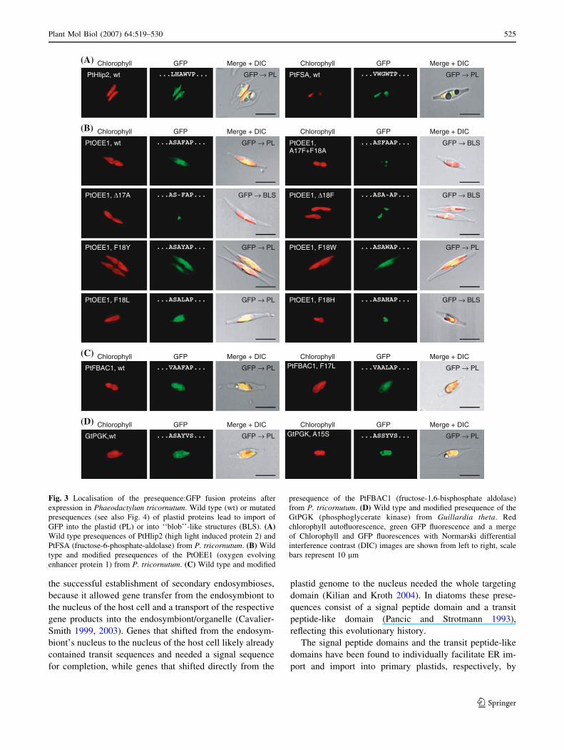
were imported into the plastids as expected (Fig. 3b, c).
Also the PtHLIP2 and the PtFSA presequences, containing
an ‘‘AW’’-cleavage site motif instead, led to GFP
fluorescence in the plastids of transformed cells (Fig. 3a).
Similarly, the presequence of the heterologous Guillardia
theta (Gt) GtPGK protein fused to GFP (tyrosine instead of
phenylalanine) was imported correctly into P. tricornutum
plastids (Fig. 3d). The PtFBPC4 presequence:GFP fusion
construct containing an ‘‘AW’’-cleavage site motif was the
only exception and gave ambiguous results. In some
transformant cell lines GFP was fluorescing inside the
plastids, while in others GFP fluorescence also appeared
outside of the plastids. The ‘‘blob’’-like structures de-
scribed below were never observed in these cell lines (data
not shown).
Mutations of the signal peptide’s cleavage site
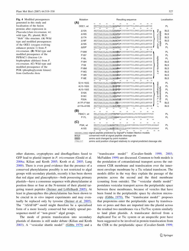
The presequence of the Phaeodactylum tricornutum Oxy-
gen evolving enhancer 1 (PtOEE1) protein has previously
been characterised intensively (Kilian and Kroth 2004,
2005). This protein is normally targeted into the thylakoids
(Ammon and Kroth, unpublished); for a better visualisation
of GFP fluorescence, in all of the following constructs the
third targeting domain responsible for thylakoid targeting
has been deleted (Kilian and Kroth 2005). To confine
crucial features of presequences for plastid import we
introduced various point mutations into the presequence of
the PtOEE1 presequence:GFP fusion protein (Fig. 4). In
P. tricornutum transformants expressing the wild type
presequence:GFP fusion protein PtOEE1:GFP, the GFP
accumulated as expected in the chloroplast stroma
(Fig. 3b). Deletion of the phenylalanine at the N-terminus
of the transit peptide-like domain in the fusion protein
PtOEE1D18F:GFP lead to a phenotype previously
described as ‘‘blob’’-like structure (BLS), (Kilian and
Kroth 2005), representing an accumulation of GFP in a
small reticular structure tightly associated to the plastid but
clearly outside the stroma (Fig. 3b). There are several
indications that these structures accumulate between the
plastid bounding membranes (Kilian and Kroth 2005).
By site directed mutagenesis we replaced the phenylal-
anine by other aromatic residues, like tyrosine, tryptophane
and histidine and expressed the constructs in P. tricornutum.
Substitution of phenylalanine by tyrosine and tryptophan
(PtOEE1F18Y:GFP, PtOEE1F18W:GFP) resulted in func-
tional targeting of GFP into the plastids (Fig. 3b), while a
replacement by histidine (PtOEE1F18H:GFP) led to the
BLS phenotype (Fig. 3b). Phenylalanine, tyrosine and
tryptophan are large and hydrophobic amino acids, so we
tested whether it would be sufficient to introduce other large
and hydrophobic residues instead of phenylalanine. We
inserted leucine, isoleucine and methionine and found that
only leucine (PtOEE1F18L:GFP) at this position is capable
of driving protein import (Fig. 3b), while PtOEE1F18I:GFP
and PtOEE1F18M:GFP again led to the BLS phenotype
NNprediction
HMMprediction
highestprediction
manualprediction
Fig. 1 Sequence logos constructed from 81 manually curated plastid
gene models (see also supplementary Fig. 6) within the Phaeodacty-lum tricornutum genome. Predictions from Signal P’s Neuronal
networks (NN, upper left) and Hidden Markov models (HMM, upper
right) can be compared. In addition a combined sequence logo using
the highest prediction score (Ymax for NN versus Cmax from NN,
lower left) has been prepared. A sequence logo using manual
predictions, where the automated outputs of NN and HMM have been
corrected with respect to the presence of an ‘‘ASAFAP’’-motif (see
supplementary Fig. 6 for the exact corrections applied) shows the
highest sequence conservation surrounding the signal peptide
cleavage site (lower right). Black: hydrophobic residues (AC-
FGILMPVWY), green: hydrophilic residues (NQST), blue: basic
residues (HKR) red: acidic residues (DE)
Plant Mol Biol (2007) 64:519–530 523
123
(data not shown). In P. tricornutum cells with a strong
expression of PtOEE1F18L:GFP we also found weak
labeling of the cytosol, which was never observed in wild
type PtOEE1:GFP or in the PtOEE1F18Y:GFP and
PtOEE1F18W:GFP transformants. This may indicate that
import of PtOEE1F18L:GFP into the chloroplast endo-
plasmic reticulum (CER) is less efficient than the import of
fusion proteins with the aromatic residues phenylalanine,
tyrosine or tryptophan at the N-terminus of the transit
peptide-like domain. More likely this phenotype is an
overexpression artefact, since several other transformed cell
lines showed fairly GFP labelled plastids. We repeated the
experiment by re-sequencing the PtOEE1F18L:GFP plas-
mid construct and re-transforming P. tricornutum with the
plasmid to ensure this particular result. We modified the
presequence of the P. tricornutum fructose bisphosphatase
(PtFBAC1) in a similar way, changing phenylalanine to
leucine, and obtained similar results: the resulting fusion
protein PtFBAC1F17L:GFP again was imported into the
plastid (Fig. 3c). A replacement of phenylalanine by
charged amino acids like arginine (PtOEE1F18R:GFP) and
glutamate (PtOEE1F18E:GFP) and by the small residue
glycine (PtOEE1F18G:GFP) did not result in plastid import
and transformants showed the BLS phenotype (data not
shown). To test the importance of the exact position of the
phenylalanine we exchanged the amino acids F and A
flanking the signal peptide’s cleavage site, the resulting
PtOEE1A17F+F18A:GFP construct led to the BLS
phenotype (Fig. 3b).
Mutations of the transit peptide-like domain
We inserted mutations in the transit peptide-like domain of
the motif to assess the importance of these residues for
successful plastid import. The deletion mutants PtOEE1-
D19A:GFP and PtOEE1D20P:GFP allowed plastid import
of the GFP (sequences listed in Fig. 4), furthermore we
were able to exchange proline by alanine (PtOEE1-
P20A:GFP) without affecting plastid import (data not
shown). Replacement of all alanine residues within the
‘‘ASAFAP’’-motif by serine and glycine did not affect the
plastid protein import, as the fusion proteins
PtOEE1A(15–19)S:GFP and PtOEE1A(15–19)G:GFP are
targeted into the plastids (data not shown).
Mutations of the signal peptide domain
In contrast, deletions of alanine and serine within the signal
peptide part of the ‘‘ASAFAP’’-motif blocked plastid pro-
tein targeting. The BLS phenotype was observed in trans-
formants expressing the fusion proteins PtOEE1D15A:GFP,
PtOEE1D16S:GFP, PtOEE1D17A:GFP and PtOEE1-
D17A+A19S:GFP (PtOEE1D17A:GFP shown as example
in Fig. 3). Exchange of the serine to alanine or cysteine led
to correct plastid import, transformants expressing the
PtOEE1S16A:GFP and PtOEE1S16C:GFP showed GFP
fluorescence within the plastids (data not shown). Also the
exchange of alanine to serine preceding the tyrosine in the
G. theta GtPGK presequence did not affect plastid import
of the fusion protein (Fig. 3d). The absence of serine in the
signal peptide of the wild type P. tricornutum PtFBAC1
presequence (Fig. 4b) shows that the presence of serine in
the signal peptide is not required for successful plastid
targeting, although serine is commonly found within the
‘‘ASAFAP’’-motif and within plastid protein signal
peptides (Figure 1 and supplementary Fig. 6).
Discussion
The development of protein targeting into the secondary
plastids of diatoms and cryptophytes was a prerequisite for
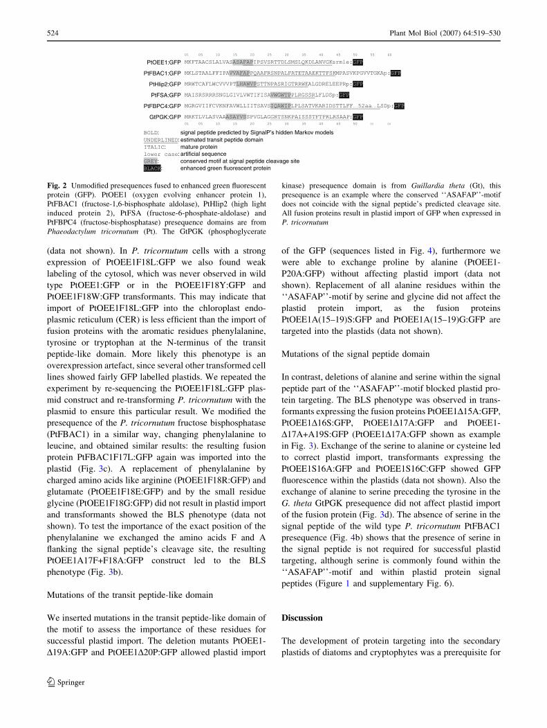
PtOEE1:GFP
PtFBAC1:GFP
PtHlip2:GFP
PtFSA:GFP
PtFBPC4:GFP
GtPGK:GFP
: signal peptide predicted by SignalP’s hidden Markov models: estimated transit peptide domain
: mature protein: artificial sequence
: conserved motif at signal peptide cleavage site: enhanced green fluorescent protein
Fig. 2 Unmodified presequences fused to enhanced green fluorescent
protein (GFP). PtOEE1 (oxygen evolving enhancer protein 1),
PtFBAC1 (fructose-1,6-bisphosphate aldolase), PtHlip2 (high light
induced protein 2), PtFSA (fructose-6-phosphate-aldolase) and
PtFBPC4 (fructose-bisphosphatase) presequence domains are from
Phaeodactylum tricornutum (Pt). The GtPGK (phosphoglycerate
kinase) presequence domain is from Guillardia theta (Gt), this
presequence is an example where the conserved ‘‘ASAFAP’’-motif
does not coincide with the signal peptide’s predicted cleavage site.
All fusion proteins result in plastid import of GFP when expressed in
P. tricornutum
524 Plant Mol Biol (2007) 64:519–530
123
the successful establishment of secondary endosymbioses,
because it allowed gene transfer from the endosymbiont to
the nucleus of the host cell and a transport of the respective
gene products into the endosymbiont/organelle (Cavalier-
Smith 1999, 2003). Genes that shifted from the endosym-
biont’s nucleus to the nucleus of the host cell likely already
contained transit sequences and needed a signal sequence
for completion, while genes that shifted directly from the
plastid genome to the nucleus needed the whole targeting
domain (Kilian and Kroth 2004). In diatoms these prese-
quences consist of a signal peptide domain and a transit
peptide-like domain (Pancic and Strotmann 1993),
reflecting this evolutionary history.
The signal peptide domains and the transit peptide-like
domains have been found to individually facilitate ER im-
port and import into primary plastids, respectively, by
GtPGK,wt ...ASAYVS... GFP PL→ GtPGK, A15S ...ASSYVS... GFP PL→
(D) Chlorophyll GFP Merge + DIC Chlorophyll GFP Merge + DIC
PtOEE1, F18Y ...ASAYAP... GFP PL→ PtOEE1, F18W ...ASAWAP... GFP PL→
PtOEE1, ∆17A ...AS-FAP... GFP BLS→ PtOEE1, ∆18F ...ASA-AP... GFP BLS→
PtOEE1, wt ...ASAFAP... GFP PL→ PtOEE1,A17F+F18A
...ASFAAP... GFP BLS→
PtOEE1, F18L ...ASALAP... GFP PL→ PtOEE1, F18H ...ASAHAP... GFP BLS→
(B) Chlorophyll GFP Merge + DIC Chlorophyll GFP Merge + DIC
PtFBAC1, F17L ...VAALAP... GFP PL→PtFBAC1, wt ...VAAFAP... GFP PL→
(C) Chlorophyll GFP Merge + DIC Chlorophyll GFP Merge + DIC
PtHlip2, wt ...LHAWVP... GFP PL→ PtFSA, wt ...VWGWTP... GFP PL→
(A) Chlorophyll GFP Merge + DIC Chlorophyll GFP Merge + DIC
Fig. 3 Localisation of the presequence:GFP fusion proteins after
expression in Phaeodactylum tricornutum. Wild type (wt) or mutated
presequences (see also Fig. 4) of plastid proteins lead to import of
GFP into the plastid (PL) or into ‘‘blob’’-like structures (BLS). (A)Wild type presequences of PtHlip2 (high light induced protein 2) and
PtFSA (fructose-6-phosphate-aldolase) from P. tricornutum. (B) Wild
type and modified presequences of the PtOEE1 (oxygen evolving
enhancer protein 1) from P. tricornutum. (C) Wild type and modified
presequence of the PtFBAC1 (fructose-1,6-bisphosphate aldolase)
from P. tricornutum. (D) Wild type and modified presequence of the
GtPGK (phosphoglycerate kinase) from Guillardia theta. Red
chlorophyll autofluorescence, green GFP fluorescence and a merge
of Chlorophyll and GFP fluorescences with Normarski differential
interference contrast (DIC) images are shown from left to right, scale
bars represent 10 lm
Plant Mol Biol (2007) 64:519–530 525
123
in vitro experiments (Bhaya and Grossman 1991; Lang
et al. 1998). In vivo experiments showed that these bipartite
presequences are sufficient for plastid import and that no
other targeting signals are needed (Apt et al. 2002; Kilian
and Kroth 2004, 2005). Interestingly although large parts of
the C-terminus of the transit peptide-like domain may be
deleted (Apt et al. 2002), plastid import is only possible if a
conserved ‘‘ASAFAP’’-motif is present between the signal
and the transit peptide-like domains (Kilian and Kroth
2005). Complete deletion of either the transit peptide-like
domain or the phenylalanine within the ‘‘ASAFAP’’-motif
lead to transport inhibition demonstrating that both ele-
ments are necessary. The very conserved phenylalanine
within the ‘‘ASAFAP’’-motif has already been shown to be
crucial for plastid targeting in a previous study (Kilian and
Kroth 2005). Here we demonstrate that only a few struc-
turally similar amino acids may replace this particular
amino acid, while in all other cases exchanges of phenyl-
alanine lead to blocked import. All other amino acids of the
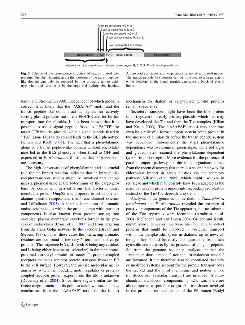
‘‘ASAFAP’’-motif may be replaced by glycine, alanine,
serine or cysteine without affecting import (Fig. 5). Inter-
estingly deletions in the signal-peptide part of the motif
could block plastid import, while exchanges at the same
positions allowed plastid import. Possibly due to the shorter
distance to the N-terminus in these cases the prediction of
the cleavage site shifted, which might explain why the
respective proteins are no longer imported. In these cases
the phenylalanine is predicted to be cleaved off together
with the signal peptide (Fig. 4, PtOEE1D15A,
PtOEE1D16S, PtOEE1D17A). However, in some cases the
phenylalanine (or the compensating tryptophan) is also
predicted to be cleaved off, but the mutated prese-
quence:GFP fusion proteins are imported into the plastid
(Fig. 4, PtOEE1F18W, PtOEE1D20P, PtOEE1P20A).
Probably because the overall length of the signal peptide is
not affected, cleavage in these cases takes place as usual,
regardless of the prediction.
The following requirements for preprotein import into
complex diatom plastids can be deduced from this and
from the former studies: (i) The presence of a cleavable
signal peptide. (ii) The presence of predominately phen-
ylalanine, sometimes tryptophan, rarely tyrosine or leucine
in the +1 position of the signal peptide cleavage site, often
followed by ‘‘AP’’ or a transit peptide domain. The
‘‘ASAFAP’’-motif fulfills these requirements, as the
pre-cleavage site part of the motif can be explained by the
‘‘(–3, –1) rule’’ (von Heijne 1983) for cleavable signal
peptides and the post-cleavage site part of the motif reflects
the second requirement (presence of phenylalanine, tryp-
tophan, tyrosine or leucine). The ‘‘(–3, –1) rule’’ is fol-
lowed to a lesser extent in eukaryotic signal peptides
compared to their prokaryotic counterparts (Nielsen et al.
1997a). Comparison of our sequence logos (Fig. 1) to
sequence logos of prokaryotic and eukaryotic signal pep-
tides (Nielsen et al. 1997a) shows that in P. tricornutum
the conservation of signal peptides in the –3, –1 positions is
higher than generally found in eukaryotes. This finding
might reflect the fact that signal peptide cleavage is crucial
in the process of plastid protein import into complex dia-
tom plastids.
Generally the predicted signal peptide cleavage sites
may vary depending on the calculation method. The pos-
sibility of miss-predictions complicates bioinformatic at-
tempts to recognise plastid proteins. A hand selected
sequence logo of plastid targeting signals revealed the
presence of the conserved cleavage site motif in all tested
sequences, but it was constructed from known plastid
proteins only (Kilian and Kroth 2005). The sequence logo
of a genome wide automated comparison of Thalassiosira
pseudonana transit peptides also showed other amino acids
than phenylalanine, tryptophan, tyrosine or leucine in the
first position of the transit peptide-like domain, predicting
alanine to be the second frequent amino acid in this posi-
tion (Armbrust et al. 2004). This is contradictory to our
finding that replacements of phenylalanine by structurally
dissimilar amino acids like alanine lead to blocked plastid
import. Here, only nine native plastid targeting sequences
contained an ‘‘ASAFAP’’-motif without phenylalanine at
the signal peptide cleavage site and until now we only
observed native plastid presequences containing the struc-
turally similar amino acids shown to functionally replace
phenylalanine (tryptophan, tyrosine or leucine) in this
position (supplementary Fig. 6).
We conclude that the occurrence of alanine as the second
frequent N-terminal amino acid in a bioinformatic approach
(Armbrust et al. 2004) is most probably explained by miss-
predictions of the signal peptide cleavage site, while in
some cases the phenylalanine within the ‘‘ASAFAP’’-motif
is replaced by tryptophan, phenylalanine or leucine, which
are shown to be functional in this study. Bioinformatic
approaches to determine plastid proteomes can be impeded
by such miss-predictions. Our results will facilitate future
bioinformatic analysis of plastid proteomes on a genomic
level, since the presence of the ‘‘ASAFAP’’-motifs in
proximity to a predicted cleavage site can be helpful to test
large numbers of proteins for the presence of plastid tar-
geting signals in diatoms and related algae.
The ‘‘ASAFAP’’-motif is very conserved in P. tricor-
nutum and similar motifs are found in other groups of algae
with secondary plastids like dinoflagellates and crypto-
phytes. Plastid preproteins in dinoflagellates possess a
conserved ‘‘FVAP’’ motif (Patron et al. 2005), while in
cryptophytes ‘‘AXAF’’ is found (Gould et al. 2006a).
Bipartite presequences containing an ‘‘ASAFAP’’-motif
apparently are functional across the species border, as
several heterologous plastid targeting presequences from
526 Plant Mol Biol (2007) 64:519–530
123
other diatoms, cryptophytes and dinoflagellates fused to
GFP lead to plastid import in P. tricornutum (Gould et al.
2006a; Kilian and Kroth 2005; Kroth et al. 2005; Lang
2000). There is even good evidence that the presence of a
conserved phenylalanine possibly is not restricted to algal
groups with secondary plastids, recently it has been shown
that red algae and glaucophytes—both possessing primary
plastids—have a consensus sequence with phenylalanine at
position three or four at the N-termini of their plastid tar-
geting transit peptides (Steiner and Loffelhardt 2005). At
least in glaucopyhtes this phenylalanine has been shown to
be crucial in in vitro import experiments and may even-
tually be replaced only by tyrosine (Steiner et al. 2005).
The ‘‘ASAFAP’’-motif might therefore be a specialised
form of a more loosely conserved but widely spread pre-
sequence-motif of ‘‘non-green’’ algal groups.
The mode of protein translocation into secondary
plastids of diatoms is still under debate (Kilian and Kroth
2003). A ‘‘vesicular shuttle model’’ (Gibbs 1979) and a
‘‘translocator model’’ (Cavalier-Smith 1999, 2003;
McFadden 1999) are discussed. Common to both models is
the postulation of cotranslational transport across the out-
ermost CER membrane and translocation over the inner-
most envelope membrane by a Tic related translocon. The
models differ in the way they explain the passage of the
proteins across the second and the third membrane
(counting from outside). The ‘‘vesicular shuttle model’’
postulates vesicular transport across the periplastidic space
between these membranes, because of vesicles that have
been found in the periplastidic space by electron micros-
copy (Gibbs 1979). The ‘‘translocator model’’ proposes
that preproteins enter the periplastidic space by transloca-
tors or pores and then are imported into the plastid across
the residual two membranes via a Tic/Toc system similarly
to land plant plastids. A translocator derived from a
duplicated Toc or Tic system or an unspecific pore have
been suggested to be involved in protein translocation from
the CER to the periplastidic space (Cavalier-Smith 1999;
Mutation Resulting sequence Localisation
(A) 01 05 10 15 20 25 30 35 40 43
OEE1, wt MKFTAACSLALVASASAFAPIPSVSRTTDLSMSLQKDLANVGK PL01 05 10 15 20 25 30 35 40 43
MKFTAACSLALVAS-SAFAPIPSVSRTTDLSMSLQKDLANVGK BLSMKFTAACSLALVASA-AFAPIPSVSRTTDLSMSLQKDLANVGK BLSMKFTAACSLALVASAS-FAPIPSVSRTTDLSMSLQKDLANVGK BLSMKFTAACSLALVASASA-APIPSVSRTTDLSMSLQKDLANVGK BLSMKFTAACSLALVASASAF-PIPSVSRTTDLSMSLQKDLANVGK PLMKFTAACSLALVASASAFA-IPSVSRTTDLSMSLQKDLANVGK PL01 05 10 15 20 25 30 35 40 43
F18W MKFTAACSLALVASASAWAPIPSVSRTTDLSMSLQKDLANVGK PL
F18Y MKFTAACSLALVASASAYAPIPSVSRTTDLSMSLQKDLANVGK PL
F18L MKFTAACSLALVASASALAPIPSVSRTTDLSMSLQKDLANVGK PL
F18H MKFTAACSLALVASASAHAPIPSVSRTTDLSMSLQKDLANVGK BLS
F18I MKFTAACSLALVASASAIAPIPSVSRTTDLSMSLQKDLANVGK BLS
F18M MKFTAACSLALVASASAMAPIPSVSRTTDLSMSLQKDLANVGK BLS
F18G MKFTAACSLALVASASAGAPIPSVSRTTDLSMSLQKDLANVGK BLS
F18R MKFTAACSLALVASASARAPIPSVSRTTDLSMSLQKDLANVGK BLS
F18E MKFTAACSLALVASASAEAPIPSVSRTTDLSMSLQKDLANVGK BLS01 05 10 15 20 25 30 35 40 43
S(14-16)G MKFTAACSLALVAGAGAFAPIPSVSRTTDLSMSLQKDLANVGK PL
A(15-19)G MKFTAACSLALVASGSGFGPIPSVSRTTDLSMSLQKDLANVGK PL
A(15-19)S MKFTAACSLALVASSSSFSPIPSVSRTTDLSMSLQKDLANVGK PL
S16A MKFTAACSLALVASAAAFAPIPSVSRTTDLSMSLQKDLANVGK PL
S16C MKFTAACSLALVASACAFAPIPSVSRTTDLSMSLQKDLANVGK PL
P20A MKFTAACSLALVASASAFAAIPSVSRTTDLSMSLQKDLANVGK PL
A17F+F18A MKFTAACSLALVASASFAAPIPSVSRTTDLSMSLQKDLANVGK BLSMKFTAACSLALVASAS-FSPIPSVSRTTDLSMSLQKDLANVGK BLS01 05 10 15 20 25 30 35 40 43
(B) 01 05 10 15 20 25 30 35 40 44
FBAC1, wt MKLSTAALFFIPAVVAFAPPQAAFRSNPALFATETAAEKTTFSK PL
F17L MKLSTAALFFIPAVVALAPPQAAFRSNPALFATETAAEKTTFSK PL01 05 10 15 20 25 30 35 40 44
(C) 01 05 10 15 20 25 30 35 40 45 48
PGK, wt MRKTLVLASVAAASAYVSSPVGLAGGRTSNKPAISSSTFTPRLRSAAP PL
A15S MRKTLVLASVAAASSYVSSPVGLAGGRTSNKPAISSSTFTPRLRSAAP PL01 05 10 15 20 25 30 35 40 45 48
UNDERLINED: signal peptide predicted by SignalP’s hidden Markov modelsBOLD: conserved motif at signal peptide cleavage siteGREY: amino acid changed by point mutationITALIC: amino acid position changed relatively to original predicted cleavage site
}
}}
deletionsw
ithinthe
“”-m
otifASAFAP
replacements
ofF
r eplacements
orinterchanges
of,
,,
ASPF
Fig. 4 Modified presequences
generated in this study and
localisation of the fusion
proteins after expression in
Phaeodactylum tricornutum. wt:
wild type, PL: plastid, BLS:
‘‘blob’’-like structure. (A) Wild
type and modified presequences
of the OEE1 (oxygen evolving
enhancer protein 1) from P.tricornutum. (B) Wild type and
modified presequence of the
PtFBAC1 (fructose-1,6-
bisphosphate aldolase) from P.tricornutum. (C) Wild type and
modified presequence of the
PGK (phosphoglycerate kinase)
from Guillardia theta
Plant Mol Biol (2007) 64:519–530 527
123
Kroth and Strotmann 1999). Independent of which model is
correct, it is likely that the ‘‘ASAFAP’’-motif and the
transit peptide-like domain act as signals for actively
sorting plastid proteins out of the ER/CER and for further
transport into the plastids. It has been shown that it is
possible to use a signal peptide fused to ‘‘FATTP’’ to
target GFP into the plastids, while a signal peptide fused to
‘‘FA’’ alone fails to do so and leads to the BLS phenotype
(Kilian and Kroth 2005). The fact that a phenylalanine
alone or a transit peptide-like domain without phenylala-
nine led to the BLS phenotype when fused to GFP and
expressed in P. tricornutum illustrates that both elements
are necessary.
The high conservation of phenylalanine and its crucial
role for the import reaction indicates that an intracellular
receptor/transport system might be involved that recog-
nises a phenylalanine at the N-terminus of the cargo pro-
tein. A component derived from the bacterial outer
membrane protein Omp85 was proposed to act as phenyl-
alanine specific receptor and membrane channel (Steiner
and Loffelhardt 2005). A specific interaction of aromatic
amino acid residues within the protein cargo with transport
components is also known from protein sorting into
caveolae, plasma membrane structures formed in the pro-
cess of endocytosis (Couet et al. 1997) and from targeting
from the trans Golgi network to the vacuole (Bryant and
Stevens 1998), but in these cases the interacting aromatic
residues are not found at the very N-termini of the cargo
proteins. The sequence F(X)6LL (with X being any residue,
and L being either leucine or isoleucine) in the membrane-
proximal carboxyl termini of many G protein-coupled
receptors mediates receptor protein transport from the ER
to the cell surface. However, the precise molecular mech-
anism by which the F(X)6LL motif regulates G protein-
coupled receptor protein export from the ER is unknown
(Duvernay et al. 2004). Since these vague similarities be-
tween cargo protein motifs point to unknown mechanisms,
conclusions from the ‘‘ASAFAP’’-motif on the import
mechanism for diatom or cryptophyte plastid proteins
remain speculative.
Secretory transport might have been the first protein
import system into early primary plastids, which first may
have developed the Tic and then the Toc complex (Kilian
and Kroth 2003). The ‘‘ASAFAP’’-motif may therefore
even be a relic of a former import system being present in
the ancestor of all plastids before the transit peptide system
was developed. Subsequently the strict phenylalanine
dependence was overcome in green algae, while red algae
and glaucophytes retained the phenylalanine dependent
type of import receptor. More evidence for the presence of
parallel import pathways in the same organisms comes
from the recent discovery that there is a second pathway for
chloroplast import in green plastids via the secretory
pathway (Villarejo et al. 2005), which might also exist in
red algae and which may possibly have been adapted as the
main pathway of protein import into secondary red plastids
instead of the Tic/Toc-dependent system.
Analyses of the genomes of the diatoms Thalassiosira
pseudonana and P. tricornutum revealed the presence of
putative components of the Tic apparatus, but no subunits
of the Toc apparatus were identified (Armbrust et al.
2004; McFadden and van Doren 2004; Gruber and Kroth,
unpublished). However, we were also not able to detect
proteins that might be involved in vesicular transport
within the periplastidic space in diatoms up to now, al-
though they should be easily distinguishable from their
cytosolic counterparts by the presence of a signal peptide.
So from the genome sequence analyses neither the
‘‘vesicular shuttle model’’ nor the ‘‘translocator model’’
are favoured. It can therefore also be speculated that new
or modified systems account for the protein transport over
the second and the third membrane and neither a Toc
translocon nor vesicular transport are involved. A mito-
chondrial translocon component, Tim23, was therefore
also proposed as possible origin of a translocon involved
in the protein translocation out of the ER lumen (Bodył
mature proteinsignal-peptide transit-peptideASA FAP
can be truncated or modified
F W Y Lcan be exchanged to , or
A G Scan be exchanged to or
S A Ccan be exchanged to or
A G Scan be exchanged to or
deletions can block plastid import deletion or exchange to , , , , or blocks plastid importH I M R E G
Fig. 5 Scheme of the presequence structure of diatom plastid pre-
proteins. The phenylalanine at the first position of the transit peptide-
like domain can only be replaced by the aromatic amino acids
tryptophan and tyrosine or by the large and hydrophobic leucine.
Amino acid exchanges at other positions do not affect plastid import.
The transit peptide-like domain can be truncated to a large extent,
while deletions in the signal peptide can cause a block of plastid
import
528 Plant Mol Biol (2007) 64:519–530
123

2004). Furthermore, genes for components of the ER-
associated degradation machinery (ERAD) were recently
found on the nucleomorph genome of G. theta. Respec-
tive genes are also duplicated in the genomes of P. tri-
cornutum and T. pseudonana. An altered ERAD-related
machinery involved in the regular transport of properly
folded proteins out of the ER and into the periplastidic
compartment was therefore suggested (Sommer et al.
2007). Meanwhile considerable knowledge about the
presequence structure of nucleus encoded plastid targeted
proteins from diatoms, cryptophytes and dinoflagellates
was gained (Apt et al. 2002; Gould et al. 2006a; Kilian
and Kroth 2005; Nassoury et al. 2003; Patron et al. 2005,
this study), remarkably, the detailed import process of
proteins targeted to the plastids via the ER remains
largely unknown.
Acknowledgements We thank D. Ballert for help with the trans-
formation and cultivation of Phaeodactylum tricornutum. This study
was supported by the University of Konstanz and grants of the
Deutsche Forschungsgemeinschaft (Project KR 1661/3) and the
European community (MARGENES, project QLRT-2001-01226) to
PGK.
References
Altschul SF, Madden TL, Schaffer AA, Zhang J, Zhang Z et al (1997)
Gapped BLAST and PSI-BLAST: a new generation of protein
database search programs. Nucleic Acids Res 25:3389–402
Apt KE, Kroth-Pancic PG, Grossman AR (1996) Stable nuclear
transformation of the diatom Phaeodactylum tricornutum. Mol
Gen Genet 252:572–579
Apt KE, Zaslavkaia L, Lippmeier JC, Lang M, Kilian O et al (2002)
In vivo characterization of diatom multipartite plastid targeting
signals. J Cell Sci 115:4061–4069
Armbrust EV, Berges JA, Bowler C, Green BR, Martinez D et al
(2004) The genome of the Diatom Thalassiosira pseudonana:
ecology, evolution, and metabolism. Science 306:79–86
Bendtsen JD, Nielsen H, von Heijne G, Brunak S (2004) Improved
prediction of signal peptides: SignalP 3.0. J Mol Biol 340:783–
795
Bhaya D, Grossman A (1991) Targeting proteins to diatom plastids
involves transport through an endoplasmic reticulum. Mol Gen
Genet 229:400–404
Bodył A (2004) Evolutionary origin of a preprotein translocase in the
periplastid membrane of complex plastids: a hypothesis. Plant
Biol 6:513–518
Bryant N, Stevens T (1998) Vacuole biogenesis in Saccharomycescerevisiae: protein transport pathways to the yeast vacuole.
Microbiol Mol Biol Rev 62:230–247
Cavalier-Smith T (1999) Principles of protein and lipid targeting in
secondary symbiogenesis: euglenoid, dinoflagellate and sporo-
zoan plastid origins and the eukaryotic family tree. J Eukary
Microbiol 46:347–366
Cavalier-Smith T (2000) Membrane hereditiy and early chloroplast
evolution. Trends Plant Sci 5:174–182
Cavalier-Smith T (2003) Genomic reduction and evolution of novel
genetic membranes and protein-targeting machinery in eukary-
ote-eukaryote chimaeras (meta-algae). Philos Trans R Soc Lond
B Biol Sci 358:109–134
Chaal BK, Green BR (2005) Protein import pathways in ‘complex’
chloroplasts derived from secondary endosymbiosis involving a
red algal ancestor. Plant Mol Biol 57:333–342
Couet J, Li S, Okamoto T, Ikezu T, Lisanti M (1997) Identification of
peptide and protein ligands for the caveolinscaffolding domain.
Implications for the interaction of caveolin with caveolae-
associated proteins. J Biol Chem 272:6525–6533
Crooks GE, Hon G, Chandonia JM, Brenner SE (2004) WebLogo: a
sequence logo generator. Genome Res 14:1188–1190
Delwiche CF (1999) Tracing the thread of plastid diversity through
the tapestry of life. Am Nat 154:S164–S177
Douglas SE, Penny SL (1999) The plastid genome of the cryptophyte
alga, Guillardia theta: complete sequence and conserved synteny
groups confirm its common ancestry with red algae. J Mol Evol
V48:236–244
Duvernay MT, Zhou F, Wu G (2004) A conserved motif for the
transport of G protein-coupled receptors from the endoplasmic
reticulum to the cell surface. J Biol Chem 279:30741–30750
Emanuelsson O, Nielsen H, Brunak S, von Heijne G (2000)
Predicting subcellular localization of proteins based on their
N-terminal amino acid sequence. J Mol Biol 300:1005–1016
Emanuelsson O, Nielsen H, von Heijne G (1999) ChloroP, a neural
network-based method for predicting chloroplast transit peptides
and their cleavage sites. Protein Sci 8:978–984
Gibbs SP (1979) The route of entry of cytoplasmically synthesized
proteins into chloroplasts of algae possessing chloroplast ER.
J Cell Sci 35:253–266
Gibbs SP (1981) The chloroplast endoplasmic reticulum: structure,
function and evolutionary significance. Int Rev Cytol 72:49–99
Gould SB, Sommer MS, Hadfi K, Zauner S, Kroth PG et al (2006a)
Protein targeting into the complex plastid of cryptophytes. J Mol
Evol V62:674–681
Gould SB, Sommer MS, Kroth PG, Gile GH, Keeling PJ et al (2006b)
Nucleus-to-nucleus gene transfer and protein retargeting into a
remnant cytoplasm of cryptophytes and diatoms. Mol Biol Evol
23:2413–2422
Harper JT, Waanders E, Keeling PJ (2005) On the monophyly of
chromalveolates using a six-protein phylogeny of eukaryotes. Int
J System Evol Microbiol 55:487–496
Heazlewood JL, Tonti-Filippini J, Verboom RE, Millar AH (2005)
Combining experimental and predicted datasets for determina-
tion of the subcellular location of proteins in Arabidopsis. Plant
Physiol 139:598–609
Ishida K (2005) Protein targeting into plastids: a key to understanding
the symbiogenetic acquisitions of plastids. J Plant Res 118:237–
245
Ishida K, Cavalier-Smith T, Green BR (2000) Endomembrane
structure and the chloroplast protein targeting pathway in
Heterosigma akashiwo (Raphidophyceae, Chromista). J Phycol
36:1135–1144
Keeling PJ (2004) Diversity and evolutionary history of plastids and
their hosts. Am J Bot 91:1481–1493
Kilian O, Kroth PG (2003) Evolution of protein targeting into
‘‘complex’’ plastids: the ‘‘secretory transport hypothesis’’. Plant
Biol 5:350–358
Kilian O, Kroth PG (2004) Presequence acquisition during secondary
endocytobiosis and the possible role of introns. J Mol Evol
58:712–721
Kilian O, Kroth PG (2005) Identification and characterization of a
new conserved motif within the presequence of proteins targeted
into complex diatom plastids. Plant J 41:175–183
Kozak M (1987) An analysis of 5¢-noncoding sequences from 699
vertebrate messenger RNAs. Nucleic Acids Res 15:8125–8148
Kroth PG (2002) Protein transport into secondary plastids and the
evolution of primary and secondary plastids. Int Rev Cytol
221:191–255
Plant Mol Biol (2007) 64:519–530 529
123
Kroth PG (2007) Genetic transformation; a tool to study protein
targeting in diatoms, chap. 17. In: Methods in molecular biology,
2nd edn., Totowa, NJ, USA: Humana Press
Kroth PG, Strotmann H (1999) Diatom plastids: secondary endocy-
tobiosis, plastid genome and protein import. Physiol Plant
107:136–141
Kroth PG, Schroers Y, Kilian O (2005) The peculiar distribution of
class I and class II aldolases in diatoms and in red algae. Curr
Genet 48:389–400
Lang M (2000) Untersuchungen zum Transport kernkodierter Plast-
iden-Proteine in Kieselalgen. Ph.D. thesis, Heinrich-Heine-
Universitat Dusseldorf, URL http://diss.ub.uni-duesseldorf.de/
home/etexte/diss/file?dissid=37
Lang M, Apt KE, Kroth PG (1998) Protein transport into ‘‘complex’’
diatom plastids utilizes two different targeting signals. J Biol
Chem 273:30973–30978
Marchler-Bauer A, Anderson JB, Cherukuri PF, DeWeese-Scott C,
Geer LY et al (2005) CDD: a conserved domain database for
protein classification. Nucl Acids Res 33:D192–D196
Martin W, Herrmann RG (1998) Gene transfer from organelles to the
nucleus: how much, what happens, and why? Plant Physiol
118:9–17
Martin W, Stoebe B, Goremykin V, Hansmann S, Hasegawa M et al
(1998) Gene transfer to the nucleus and the evolution of
chloroplasts. Nature 393:162–165
McFadden GI (1999) Plastids and protein targeting. J Eukaryot
Microbiol 46:339–346
McFadden GI (2001) Primary and secondary endosymbiosis and the
origin of plastids. J Phycol 37:1–9
McFadden GI, van Dooren GG (2004) Evolution: red algal genome
affirms a common origin of all plastids. Curr Biol 14:R514–
R516
Millar AH, Whelan J, Small I (2006) Recent surprises in protein
targeting to mitochondria and plastids. Curr Opin Plant Biol
9:610–615
Montsant A, Jabbari K, Maheswari U, Bowler C (2005) Comparative
genomics of the pennate diatom Phaeodactylum tricornutum.
Plant Physiol 137:500–513
Moreira D, Le Guyader H, Philippe H (2000) The origin of red algae
and the evolution of chloroplasts. Nature 405:69–72
Nassoury N, Cappadocia M, Morse D (2003) Plastid ultrastructure
defines the protein import pathway in dinoflagellates. J Cell Sci
116:2867–2874
Nielsen H, Krogh A (1998) Prediction of signal peptides and signal
anchors by a hidden Markov model. Proc Int Conf Intell Syst
Mol Biol 6:122–130
Nielsen H, Engelbrecht J, Brunak S, von Heijne G (1997a)
Identification of prokaryotic and eukaryotic signal peptides and
prediction of their cleavage sites. Protein Eng 10:1–6
Nielsen H, Engelbrecht J, Brunak S, von Heijne G (1997b) A neural
network method for identification of prokaryotic and eukaryotic
signal peptides and prediction of their cleavage sites. Int J Neural
Syst 8:581–599
Oudot-Le Secq MP, Grimwood J, Shapiro H, Armbrust EV, Bowler
C, et al. (2007) Chloroplast genomes of the diatoms Phaeo-dactylum tricornutum and Thalassiosira pseudonana: compari-
son with other plastid genomes of the red lineage. Mol Genet
Genom 277(4):427–439. PMID: 17252281
Pancic PG, Strotmann H (1993) Structure of the nuclear encoded csubunit of CF0CF1 of the diatom Odontella sinensis including its
presequence. FEBS Lett 320:61–66
Patron NJ, Waller RF, Archibald JM, Keeling PJ (2005) Complex
protein targeting to dinoflagellate plastids. J Mol Biol 348:1015–
1024
Rodriguez-Ezpeleta N, Brinkmann H, Burey SC, Roure B, Burger G
et al (2005) Monophyly of primary photosynthetic eukaryotes:
green plants, red algae, and glaucophytes. Curr Biol 15:1325–
1330
Sambrook J, Fritsch EF, Maniatis T (1989) Molecular cloning: a
laboratory manual, 2nd edn. Cold Spring Harbor Laboratory
Press, Cold Spring Harbor, New York
Schneider TD, Stephens RM (1990) Sequence logos: a new way to
display consensus sequences. Nucleic Acids Res 18:6097–6100
Soll J, Schleiff E (2004) Protein import into chloroplasts. Nat Rev
Mol Cell Biol 5:198–208
Sommer MS, Gould SB, Lehmann P, Gruber A, Przyborski JM et al
(2007) Der1-mediated pre-protein import into the periplastid
compartment of chromalveolates? Mol Biol Evol 24(4):918–928.
PMID: 17244602
Starr RC, Zeikus JA (1993) UTEX: the culture collection of algae at
the University of Texas at Austin, 1993 list of cultures. J Phycol
29:1–106
Steiner JM, Loffelhardt W (2005) Protein translocation into and
within cyanelles. Mol Membr Biol 22:123–132
Steiner JM, Yusa F, Pompe JA, Loffelhardt W (2005) Homologous
protein import machineries in chloroplasts and cyanelles. Plant J
44:646–652
Timmis JN, Ayliffe MA, Huang CY, Martin W (2005) Endosymbiotic
gene transfer: organelle genomes forge eukaryotic chromo-
somes. Nat Rev Genet 5:123–135
Villarejo A, Buren S, Larsson S, Dejardin A, Monne M et al (2005)
Evidence for a protein transported through the secretory pathway
en route to the higher plant chloroplast. Nat Cell Biol 7:1224–
1231
von Heijne G (1983) Patterns of amino acids near signal-sequence
cleavage sites. Eur J Biochem 133:17–21
Waller RF, McFadden GI (2004) The Apicoplast, chap. 11. Caister
Academic Press, Wymondham, UK, pp 291–338
Wastl J, Maier UG (2000) Transport of Proteins into cryptomonads
complex plastids. J Biol Chem 275:23194–23198
Zaslavskaia LA, Lippmeier JC, Kroth PG, Grossman AR, Apt KE
(2000) Transformation of the diatom Phaeodactylum tricornu-tum (Bacillariophyceae) with a variety of selectable marker and
reporter genes. J Phycol 36:379–386
530 Plant Mol Biol (2007) 64:519–530
123

Reproduced with permission of the copyright owner. Further reproduction prohibited without permission.
Related Documents


