International Journal of PharmTech Research CODEN (USA): IJPRIF ISSN : 0974-4304 Vol.6, No.1, pp 224-235, Jan-March 2014 Prolonged Culture in FBS and FBS-Substitute containing media: Spontaneous Chondrogenic Differentiation of Adipose tissue derived Mesenchymal Stem Cells Jeanne Adiwinata Pawitan, 1 * Des Suryani, 2 Dewi Wulandari, 3 Lia Damayanti, 1 Isabella Kurnia Liem, 4 Reza Yuridian Purwoko, 5,6 1 Department Histology, Faculty of Medicine, Universitas Indonesia, Jl. Salemba 6, Jakarta, Indonesia, 2 Biomedical Science Master Programme, Faculty of Medicine, Universitas Indonesia, Jl. Salemba 6, Jakarta, Indonesia, 3 Department of Clinical Pathology, Faculty of Medicine, Universitas Indonesia, Jl. Salemba 6, Jakarta, Indonesia 4 Department of Anatomy, Faculty of Medicine, Universitas Indonesia, Jl. Salemba 6, Jakarta, Indonesia 5 Erpour Skin Care, Jakarta, Indonesia 6 Biomedical Science Doctoral Programme,Faculty of Medicine, Universitas Indonesia, Jl., Salemba 6, Jakarta, Indonesia. *Corres. author: [email protected]; [email protected] Phone: 62 21 3146129 Abstract: Characterization of differentiation capacity of mesenchymal stem cells (MSCs) should be done before the MSCs can be used in regenerative medicine. Testing of differentiation capacity needs various substances containing medium, which are troublesome to prepare. Therefore, the objective of this study is to describe a simple and cheap method to check chondrogenic differentiation capacity of adipose tissue derived mesenchymal stem cells passage-5 by using fetal bovine serum (FBS) or platelet rich plasma (PRP) containing media in monolayer culture. In this study, adipose tissue derived mesenchymal stem cells (AT-MSCs) were isolated from lipoaspirate and cultured. To ensure the existence of AT-MSCs, flow cytometric analyses of passage-1 cells were done to count the CD34, CD73 and CD90 bearing cells. Prolonged cultures were done in various media, i.e. MesenCult®, 5% and 10% PRP containing high glucose Dulbeco’s minimal Eagle’s medium (HG-DMEM), 10% FBS containing α minimal essential medium (α MEM), and 10 ng/mL vascular endothelial growth factor (VEGF) and 10% human AB serum containing HG-DMEM. Ourf results showed that prolonged cultures in MesenCult® (one out of four), 5% PRP containing HG-DMEM (five out of six), 10% PRP containing HG-DMEM (all of nine), 10% FBS containing α MEM (twelve out of thirteen), and 10 ng/mL VEGF and 10% human AB serum containing HG-DMEM (seven out of eight) showed micromass formation. The micromasses were stained blue with alcian blue; therefore, micromass formation could be regarded as chondrogenic differentiation. In conclusion, prolonged culture in 5% PRP containing HG-DMEM, 10% PRP

Welcome message from author
This document is posted to help you gain knowledge. Please leave a comment to let me know what you think about it! Share it to your friends and learn new things together.
Transcript
International Journal of PharmTech Research CODEN (USA): IJPRIF ISSN : 0974-4304
Vol.6, No.1, pp 224-235, Jan-March 2014
Prolonged Culture in FBS and FBS-Substitute containing
media: Spontaneous Chondrogenic Differentiation of Adipose tissue derived Mesenchymal Stem Cells
Jeanne Adiwinata Pawitan,1* Des Suryani,2 Dewi Wulandari,3
Lia Damayanti,1 Isabella Kurnia Liem,4 Reza Yuridian Purwoko,5,6
1Department Histology, Faculty of Medicine, Universitas Indonesia, Jl. Salemba 6,
Jakarta, Indonesia, 2Biomedical Science Master Programme, Faculty of Medicine, Universitas Indonesia,
Jl. Salemba 6, Jakarta, Indonesia, 3Department of Clinical Pathology, Faculty of Medicine, Universitas Indonesia, Jl.
Salemba 6, Jakarta, Indonesia 4Department of Anatomy, Faculty of Medicine, Universitas Indonesia,
Jl. Salemba 6, Jakarta, Indonesia 5Erpour Skin Care, Jakarta, Indonesia
6Biomedical Science Doctoral Programme,Faculty of Medicine, Universitas Indonesia, Jl., Salemba 6, Jakarta, Indonesia.
*Corres. author: [email protected]; [email protected] Phone: 62 21 3146129
Abstract: Characterization of differentiation capacity of mesenchymal stem cells (MSCs) should be done before the MSCs can be used in regenerative medicine. Testing of differentiation capacity needs various substances containing medium, which are troublesome to prepare. Therefore, the objective of this study is to describe a simple and cheap method to check chondrogenic differentiation capacity of adipose tissue derived mesenchymal stem cells passage-5 by using fetal bovine serum (FBS) or platelet rich plasma (PRP) containing media in monolayer culture. In this study, adipose tissue derived mesenchymal stem cells (AT-MSCs) were isolated from lipoaspirate and cultured. To ensure the existence of AT-MSCs, flow cytometric analyses of passage-1 cells were done to count the CD34, CD73 and CD90 bearing cells. Prolonged cultures were done in various media, i.e. MesenCult®, 5% and 10% PRP containing high glucose Dulbeco’s minimal Eagle’s medium (HG-DMEM), 10% FBS containing α minimal essential medium (α MEM), and 10 ng/mL vascular endothelial growth factor (VEGF) and 10% human AB serum containing HG-DMEM. Ourf results showed that prolonged cultures in MesenCult® (one out of four), 5% PRP containing HG-DMEM (five out of six), 10% PRP containing HG-DMEM (all of nine), 10% FBS containing α MEM (twelve out of thirteen), and 10 ng/mL VEGF and 10% human AB serum containing HG-DMEM (seven out of eight) showed micromass formation. The micromasses were stained blue with alcian blue; therefore, micromass formation could be regarded as chondrogenic differentiation. In conclusion, prolonged culture in 5% PRP containing HG-DMEM, 10% PRP

Jeanne Adiwinata Pawitan et al /Int.J.PharmTech Res.2014,6(1),pp 224-235.
225
containing HG-DMEM, or 10% FBS containing α MEM can be used as an alternative cheap method to check the chondrogenic differentiation capacity of AT-MSCs. Key words: micromass, chondrogenic, prolonged culture, AT-MSCs.
Introduction
Adipose tissue derived mesenchymal stem cells (AT-MSCs) are very promising for regenerative medicine.1 They have similar characteristics to bone marrow derived MSCs, and can be differentiated into adipogenic, chondrogenic and osteogenic lineages.2 Characterization of differentiation capacity needs various substances containing medium, which are troublesome to prepare. Currently, various induction media are commercially available, but they are expensive. Therefore, additional cost is needed to buy them.
In case of chondrogenic differentiation, the most used protocol involves pellet culture in chondrogenic induction medium for 21 days. After the chondrogenic pellet is formed, it is processed histologically and stained by alcian blue to prove the chondrogenic matrix. This process takes a long time and very cumbersome. According to Zuk et al, chondrogenic differentiation does not occur in monolayer cultures of AT-MSCs although they are induced by chondrogenic medium.2 In contrast, Bosnakovski et al reported that pellet culture of bone marrow derived MSCs resulted in spontaneous chondrogenic differentiation without chondrogenic induction medium.3 Another study reported spontaneous osteogenic differentiation of a small proportion of human placenta derived MSCs after successive passages in growth medium (antibiotics and 10% fetal bovine serum [FBS] containing Dulbeco’s modified Eagle’s medium [DMEM]).4 Our experience in culturing AT-MSCs showed that prolonged monolayer cultures of passage 1 and 2 in platelet rich plasma (PRP) containing high glucose DMEM (HG-DMEM) caused retraction of the periphery of monolayer, which formed micromasses at the end. These micromasses were similar to the previously described VEGF induced chondrogenic micromasses.5
This article aims to describe a simple and cheap method to check chondrogenic differentiation capacity of AT-MSCs passage-5 using FBS or PRP containing media in monolayer culture.
Materials and Methods
This descriptive study was conducted in the Integrated Laboratory of the Faculty of Medicine Universitas Indonesia from August 2012 through January 2013, and was approved by the Ethical Committee of the Faculty of Medicine Universitas Indonesia.
Materials
Lipoaspirate samples were waste of liposuction procedure in Erpour Clinic in Jakarta, and were obtained after the patients signed the informed consent form. Materials that were used in processing, primary culture, subculture and prolonged culture were: phosphate buffered saline pH 7.4 (PBS [Sigma P3813]), collagenase type 1 (Gibco 17100-017), 100 µm cell strainer (Beckton Dickinson no352360), MesenCult® (STEMCELL Technologies), PRP (Indonesian Red Cross), HG-DMEM (Lonza 15-604D), TrypLE Select (GIBCO 12563-011), 12, 24, and 96 well plate (Biolite), FBS (Biowest S1810), α minimal essential medium (MEM [GIBCO 12000-022 1]), VEGF (Invitrogen PHC9394), human AB serum (Gibco 34005-100), penicillin/streptomycin (Gibco 15140-122), amphotericin B (JR Scientific 50701), and antibodies against various surface markers i.e. CD34-perCP (BD 340430), CD73-PE (BD 550257), and CD90-APC (BD 559869), and their respective isotypes, which were purchased from Beckton Dickinson.
Lipoaspirate processing and primary culture
Adipose tissue derived MSCs were obtained from lipoaspirate, which were thoroughly washed using a stainless steel filter (pore size 0.5 mm) soaked in PBS containing porcelain bowl, as described previously.6,7 Further, lipoaspirate was processed according to the method of Zuk et al to get the cells of the perivascular fraction.2 In brief, the lipoaspirate was subjected to 0.075% collagenase type 1 digestion at 37oC, for one hour. The MSC containing infranatant was filtered through a 100 µm cell strainer, and the cells were cultured in three kinds of media, i.e. commercial medium (MesenCult®, 5% and 10% PRP containing HG-DMEM. For every medium,
Jeanne Adiwinata Pawitan et al /Int.J.PharmTech Res.2014,6(1),pp 224-235.
226
seeding of viable cells was done in duplo, in a 12 well plate as describes previously7,8, and were incubated in 37oC, 5% CO2. After the cells were attached, the medium was changed every 2-3 days, and cell growth was observed every day under an inverted microscope.
Subcultures and prolonged cultures
After primary cultures reached 70% confluence, the cells in primary cultures were detached by TrypLE Select and passaged (subcultured). For every medium, subcultures were done by seeding viable cells in duplo, in a 12 well plate until passage-5 as described previously,8 and were incubated in 37oC, 5% CO2. Cells from passage-5 of 5% and 10% PRP containing HG-DMEM and MesenCult® were detached and variable numbers of viable cells were cultured in duplo in a 24 well plate (growth area 2 cm2), or in a 96 well plate (growth area 0.32 cm2), depending on the availability of cells. Seeding was done evenly or in spots.
Prolonged cultures were done in various media, i.e. MesenCult®, 5% and 10% PRP containing HG-DMEM, 10% FBS containing α MEM, and 10 ng/mL VEGF and 10% human AB serum containing HG-DMEM. All media contained penicillin/streptomycin (final 100U/mL), and amphotericin B (final 2500ng/mL) to prevent contamination. Medium changes were done every 2-3 days until 100% confluent, and was further cultured with medium changes every 2-3 days. Cell growth after confluence was observed and photographed.
After confluence, parts of the monolayer especially that were located at the periphery became retracted (Figure 1). At the retracted region, the cells formed multi layers, which further became micromasses. After a micromass/micromasses was/were formed, the cells were fixed by 10% formol saline, and stained by Alcian blue. Photographs were taken before and after staining.
Flowcytometric analysis of MSCs
When available, flow cytometric analyses of passage-1 cells were done to count the CD34, CD73 and CD90 bearing cells.
Data collection and analysis
Recorded data were the day when the culture became confluent and the day when retraction/multilayer and cartilage matrix (brown)/micromass appearance were noticed. The percentages of CD34, CD73, and CD90 bearing cells were also noted, and means and standard deviations (SD) were computed. All data were then presented in the form of tables.
Results and Discussion
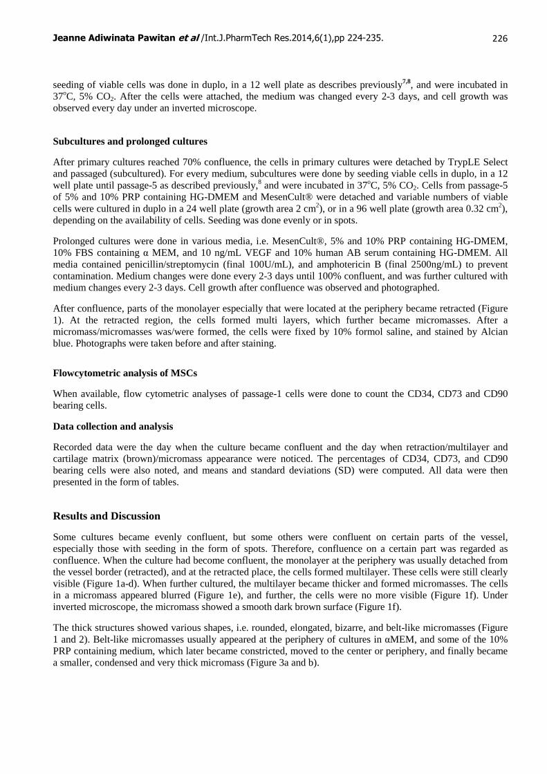
Some cultures became evenly confluent, but some others were confluent on certain parts of the vessel, especially those with seeding in the form of spots. Therefore, confluence on a certain part was regarded as confluence. When the culture had become confluent, the monolayer at the periphery was usually detached from the vessel border (retracted), and at the retracted place, the cells formed multilayer. These cells were still clearly visible (Figure 1a-d). When further cultured, the multilayer became thicker and formed micromasses. The cells in a micromass appeared blurred (Figure 1e), and further, the cells were no more visible (Figure 1f). Under inverted microscope, the micromass showed a smooth dark brown surface (Figure 1f).
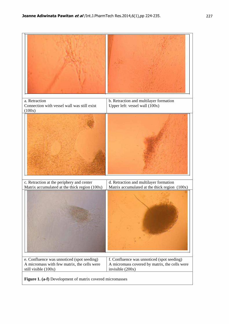
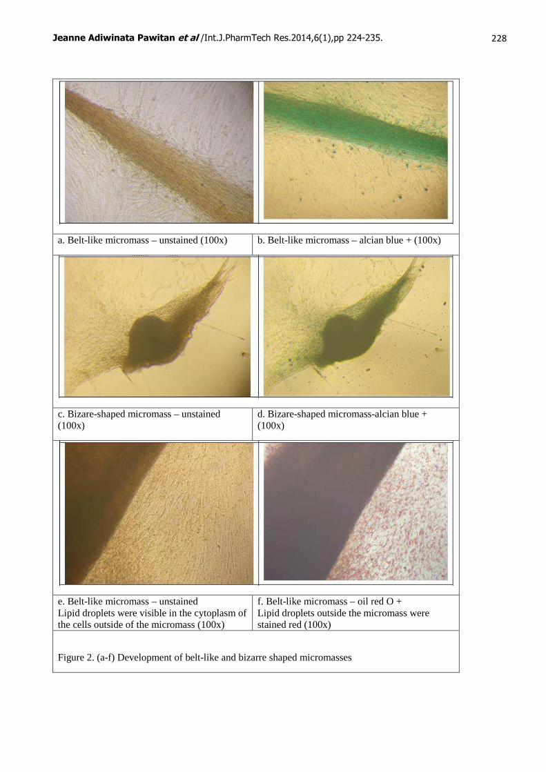
The thick structures showed various shapes, i.e. rounded, elongated, bizarre, and belt-like micromasses (Figure 1 and 2). Belt-like micromasses usually appeared at the periphery of cultures in αMEM, and some of the 10% PRP containing medium, which later became constricted, moved to the center or periphery, and finally became a smaller, condensed and very thick micromass (Figure 3a and b).
Jeanne Adiwinata Pawitan et al /Int.J.PharmTech Res.2014,6(1),pp 224-235.
227
a. Retraction Connection with vessel wall was still exist (100x)
b. Retraction and multilayer formation Upper left: vessel wall (100x)
c. Retraction at the periphery and center Matrix accumulated at the thick region (100x)
d. Retraction and multilayer formation Matrix accumulated at the thick region (100x)
e. Confluence was unnoticed (spot seeding) A micromass with few matrix, the cells were still visible (100x)
f. Confluence was unnoticed (spot seeding) A micromass covered by matrix, the cells were invisible (200x)
Figure 1. (a-f) Development of matrix covered micromasses
Jeanne Adiwinata Pawitan et al /Int.J.PharmTech Res.2014,6(1),pp 224-235.
228
a. Belt-like micromass – unstained (100x) b. Belt-like micromass – alcian blue + (100x)
c. Bizare-shaped micromass – unstained (100x)
d. Bizare-shaped micromass-alcian blue + (100x)
e. Belt-like micromass – unstained Lipid droplets were visible in the cytoplasm of the cells outside of the micromass (100x)
f. Belt-like micromass – oil red O + Lipid droplets outside the micromass were stained red (100x)
Figure 2. (a-f) Development of belt-like and bizarre shaped micromasses
Jeanne Adiwinata Pawitan et al /Int.J.PharmTech Res.2014,6(1),pp 224-235.
229
a. A condensed and very thick micromass – unstained (100x)
b. A condensed and very thick micromass – alcian blue staining (100x)
c. Macroscopic view of belt-like (left) and condensed (right) micromass – unstained
d. Macroscopic view of belt-like micromass – alcian blue staining
e. Spontaneous differentiation Fine lipid droplets were stained red (200x)
f. Induced differentiation Large lipid droplets were stained red (200x)
Figure 3. (a-f) Development of micromasses and lipid dropplets
Jeanne Adiwinata Pawitan et al /Int.J.PharmTech Res.2014,6(1),pp 224-235.
230
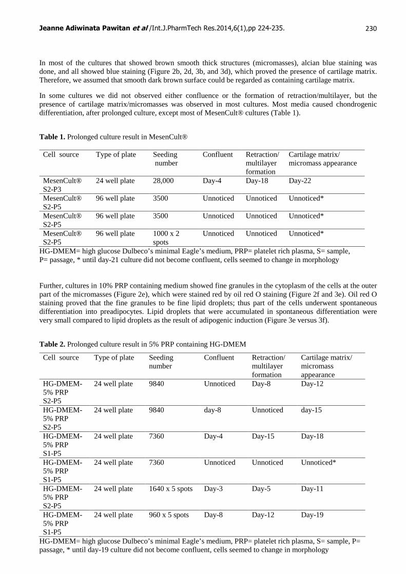
In most of the cultures that showed brown smooth thick structures (micromasses), alcian blue staining was done, and all showed blue staining (Figure 2b, 2d, 3b, and 3d), which proved the presence of cartilage matrix. Therefore, we assumed that smooth dark brown surface could be regarded as containing cartilage matrix.
In some cultures we did not observed either confluence or the formation of retraction/multilayer, but the presence of cartilage matrix/micromasses was observed in most cultures. Most media caused chondrogenic differentiation, after prolonged culture, except most of MesenCult® cultures (Table 1).
Table 1. Prolonged culture result in MesenCult® Cell source Type of plate Seeding
number Confluent Retraction/
multilayer formation
Cartilage matrix/ micromass appearance
MesenCult® S2-P3
24 well plate 28,000 Day-4 Day-18 Day-22
MesenCult® S2-P5
96 well plate 3500 Unnoticed Unnoticed Unnoticed*
MesenCult® S2-P5
96 well plate 3500 Unnoticed Unnoticed Unnoticed*
MesenCult® S2-P5
96 well plate 1000 x 2 spots
Unnoticed Unnoticed Unnoticed*
HG-DMEM= high glucose Dulbeco’s minimal Eagle’s medium, PRP= platelet rich plasma, S= sample, P= passage, * until day-21 culture did not become confluent, cells seemed to change in morphology Further, cultures in 10% PRP containing medium showed fine granules in the cytoplasm of the cells at the outer part of the micromasses (Figure 2e), which were stained red by oil red O staining (Figure 2f and 3e). Oil red O staining proved that the fine granules to be fine lipid droplets; thus part of the cells underwent spontaneous differentiation into preadipocytes. Lipid droplets that were accumulated in spontaneous differentiation were very small compared to lipid droplets as the result of adipogenic induction (Figure 3e versus 3f).
Table 2. Prolonged culture result in 5% PRP containing HG-DMEM
Cell source Type of plate Seeding number
Confluent Retraction/ multilayer formation
Cartilage matrix/ micromass appearance
HG-DMEM-5% PRP S2-P5
24 well plate 9840 Unnoticed Day-8 Day-12
HG-DMEM-5% PRP S2-P5
24 well plate 9840 day-8 Unnoticed day-15
HG-DMEM-5% PRP S1-P5
24 well plate 7360 Day-4 Day-15 Day-18
HG-DMEM-5% PRP S1-P5
24 well plate 7360 Unnoticed Unnoticed Unnoticed*
HG-DMEM-5% PRP S2-P5
24 well plate 1640 x 5 spots Day-3 Day-5 Day-11
HG-DMEM-5% PRP S1-P5
24 well plate 960 x 5 spots Day-8 Day-12 Day-19
HG-DMEM= high glucose Dulbeco’s minimal Eagle’s medium, PRP= platelet rich plasma, S= sample, P= passage, * until day-19 culture did not become confluent, cells seemed to change in morphology
Jeanne Adiwinata Pawitan et al /Int.J.PharmTech Res.2014,6(1),pp 224-235.
231
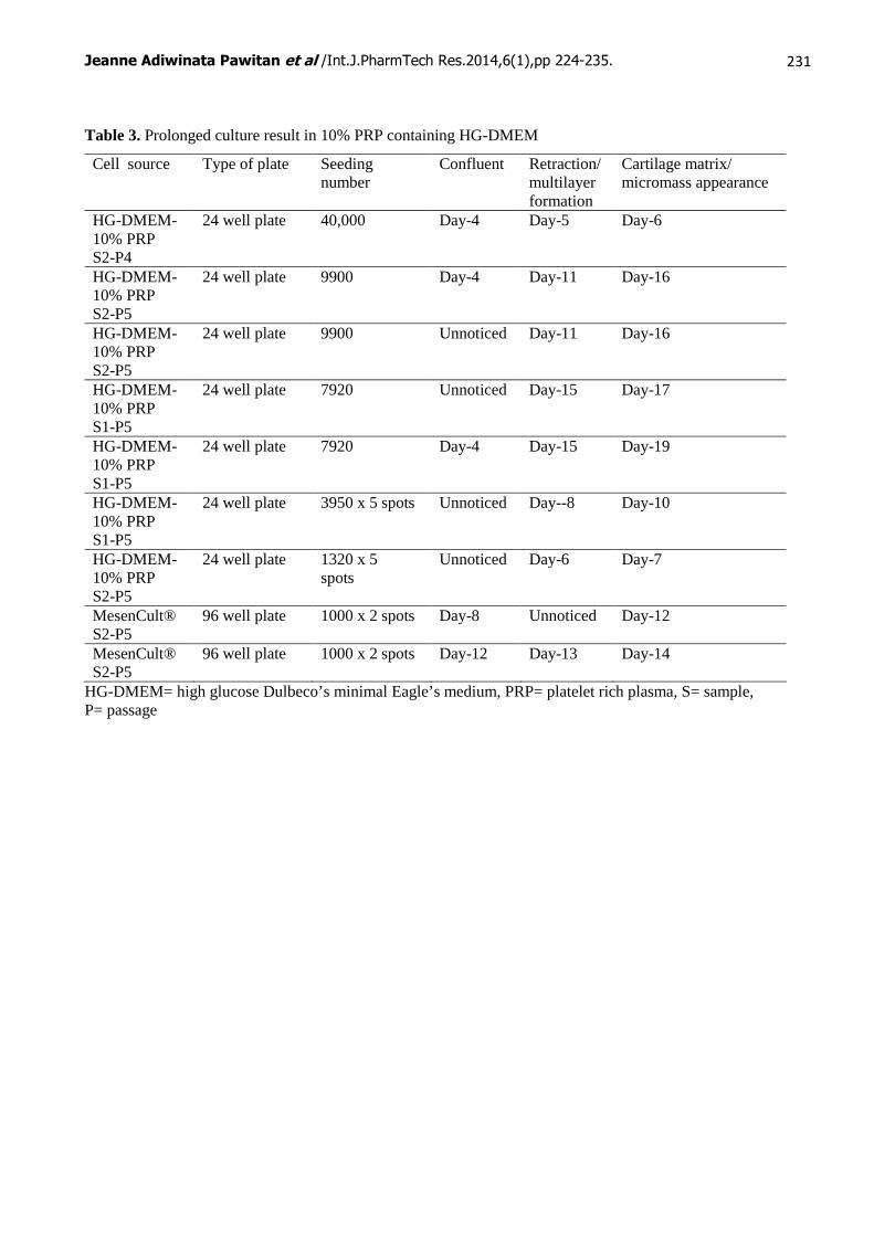
Table 3. Prolonged culture result in 10% PRP containing HG-DMEM
Cell source Type of plate Seeding number
Confluent Retraction/ multilayer formation
Cartilage matrix/ micromass appearance
HG-DMEM-10% PRP S2-P4
24 well plate 40,000 Day-4 Day-5 Day-6
HG-DMEM-10% PRP S2-P5
24 well plate 9900 Day-4 Day-11 Day-16
HG-DMEM-10% PRP S2-P5
24 well plate 9900 Unnoticed Day-11 Day-16
HG-DMEM-10% PRP S1-P5
24 well plate 7920 Unnoticed Day-15 Day-17
HG-DMEM-10% PRP S1-P5
24 well plate 7920 Day-4 Day-15 Day-19
HG-DMEM-10% PRP S1-P5
24 well plate 3950 x 5 spots Unnoticed Day--8 Day-10
HG-DMEM-10% PRP S2-P5
24 well plate 1320 x 5 spots
Unnoticed Day-6 Day-7
MesenCult® S2-P5
96 well plate 1000 x 2 spots Day-8 Unnoticed Day-12
MesenCult® S2-P5
96 well plate 1000 x 2 spots Day-12 Day-13 Day-14
HG-DMEM= high glucose Dulbeco’s minimal Eagle’s medium, PRP= platelet rich plasma, S= sample, P= passage
Jeanne Adiwinata Pawitan et al /Int.J.PharmTech Res.2014,6(1),pp 224-235.
232
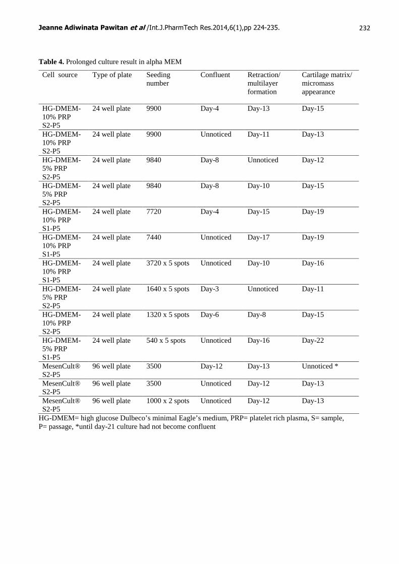
Table 4. Prolonged culture result in alpha MEM
Cell source Type of plate Seeding number
Confluent Retraction/ multilayer formation
Cartilage matrix/ micromass appearance
HG-DMEM-10% PRP S2-P5
24 well plate 9900 Day-4 Day-13 Day-15
HG-DMEM-10% PRP S2-P5
24 well plate 9900 Unnoticed Day-11 Day-13
HG-DMEM-5% PRP S2-P5
24 well plate 9840 Day-8 Unnoticed Day-12
HG-DMEM-5% PRP S2-P5
24 well plate 9840 Day-8 Day-10 Day-15
HG-DMEM-10% PRP S1-P5
24 well plate 7720 Day-4 Day-15 Day-19
HG-DMEM-10% PRP S1-P5
24 well plate 7440 Unnoticed Day-17 Day-19
HG-DMEM-10% PRP S1-P5
24 well plate 3720 x 5 spots Unnoticed Day-10 Day-16
HG-DMEM-5% PRP S2-P5
24 well plate 1640 x 5 spots Day-3 Unnoticed Day-11
HG-DMEM-10% PRP S2-P5
24 well plate 1320 x 5 spots Day-6 Day-8 Day-15
HG-DMEM-5% PRP S1-P5
24 well plate 540 x 5 spots Unnoticed Day-16 Day-22
MesenCult® S2-P5
96 well plate 3500 Day-12 Day-13 Unnoticed *
MesenCult® S2-P5
96 well plate 3500 Unnoticed Day-12 Day-13
MesenCult® S2-P5
96 well plate 1000 x 2 spots Unnoticed Day-12 Day-13
HG-DMEM= high glucose Dulbeco’s minimal Eagle’s medium, PRP= platelet rich plasma, S= sample, P= passage, *until day-21 culture had not become confluent
Jeanne Adiwinata Pawitan et al /Int.J.PharmTech Res.2014,6(1),pp 224-235.
233
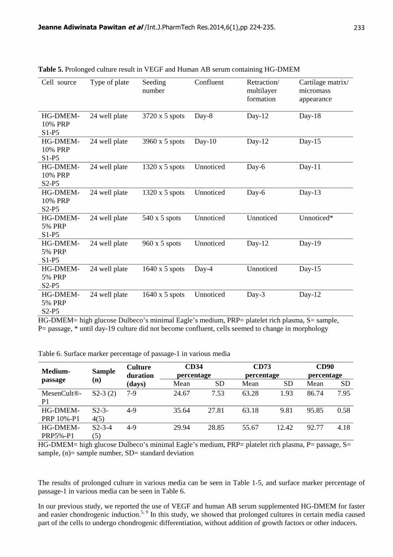
Table 5. Prolonged culture result in VEGF and Human AB serum containing HG-DMEM
Cell source Type of plate Seeding number
Confluent Retraction/ multilayer formation
Cartilage matrix/ micromass appearance
HG-DMEM-10% PRP S1-P5
24 well plate 3720 x 5 spots Day-8 Day-12 Day-18
HG-DMEM-10% PRP S1-P5
24 well plate 3960 x 5 spots Day-10 Day-12 Day-15
HG-DMEM-10% PRP S2-P5
24 well plate 1320 x 5 spots Unnoticed Day-6 Day-11
HG-DMEM-10% PRP S2-P5
24 well plate 1320 x 5 spots Unnoticed Day-6 Day-13
HG-DMEM-5% PRP S1-P5
24 well plate 540 x 5 spots Unnoticed Unnoticed Unnoticed*
HG-DMEM-5% PRP S1-P5
24 well plate 960 x 5 spots Unnoticed Day-12 Day-19
HG-DMEM-5% PRP S2-P5
24 well plate 1640 x 5 spots Day-4 Unnoticed Day-15
HG-DMEM-5% PRP S2-P5
24 well plate 1640 x 5 spots Unnoticed Day-3 Day-12
HG-DMEM= high glucose Dulbeco’s minimal Eagle’s medium, PRP= platelet rich plasma, S= sample, P= passage, * until day-19 culture did not become confluent, cells seemed to change in morphology Table 6. Surface marker percentage of passage-1 in various media
CD34 percentage
CD73 percentage
CD90 percentage Medium-
passage Sample (n)
Culture duration (days) Mean SD Mean SD Mean SD
MesenCult®- P1
S2-3 (2) 7-9 24.67 7.53 63.28 1.93 86.74 7.95
HG-DMEM- PRP 10%-P1
S2-3-4(5)
4-9 35.64 27.81 63.18 9.81 95.85 0.58
HG-DMEM-PRP5%-P1
S2-3-4 (5)
4-9 29.94 28.85 55.67 12.42 92.77 4.18
HG-DMEM= high glucose Dulbeco’s minimal Eagle’s medium, PRP= platelet rich plasma, P= passage, S= sample, (n)= sample number, SD= standard deviation
The results of prolonged culture in various media can be seen in Table 1-5, and surface marker percentage of passage-1 in various media can be seen in Table 6.
In our previous study, we reported the use of VEGF and human AB serum supplemented HG-DMEM for faster and easier chondrogenic induction.5, 9 In this study, we showed that prolonged cultures in certain media caused part of the cells to undergo chondrogenic differentiation, without addition of growth factors or other inducers.
Jeanne Adiwinata Pawitan et al /Int.J.PharmTech Res.2014,6(1),pp 224-235.
234
In this study, we used 24 and 96 well plates and different seeding numbers depending on the availability of the cells. Therefore, comparison of the result should consider seeding number per area into account. In MesenCult®, only one out of four cultures showed chondrogenic differentiation on day-22 for seeding number 14,000/cm2, while lower seeding number did not show chondrogenic differentiation until day-22. The MesenCult® cultures that failed to form micromasses were derived from passage-5 (Table 1). Moreover, one out of eight α MEM culture also failed to form micromasses, and the cells of the failed one were derived from passage-5 of MesenCult® culture (Table 1). Therefore, we supposed that MesenCult® passage-5 cells had lost their differentiation capacity. To check whether MesenCult® caused chondrogenic differentiation in prolonged culture, lower passages that still retain their differentiation capacity should be used.
Overall, the lower the seeding number per area, the longer the time needed to form micromasses (Table 1-5), with the lowest time occurred for 10% PRP containing HG-DMEM, which took only 5 days to form micromasses, when seeding number is 20,000/cm2 (Table 3). PRP was known to contain various kinds of growth factors, i.e. transforming growth factor beta-1 (TGF β1), platelet derived growth factors, epidermal growth factor, insulin-like growth factor-1, and VEGF. The highest content was TGF β1, which was known as inducer of chondrogenic differentiation.8
Spotted seeding showed inconsistent results, and seemed did not decrease the needed time for micromass formation, in 5% PRP containing HG-DMEM, 10% FBS containing α MEM, and VEGF and human AB serum containing HG-DMEM, as was supposed in our previous result.9 However, for 10% PRP containing HG-DMEM, spotted seeding tended to decrease the time for micromass formation (Table 3).
Flow cytometric analysis showed that adipose tissue derived passage-1 cells that were grown in 5% and 10% PRP containing medium contained high percentage of CD90 positive cells, similar to bone marrow-derived MSCs. However, CD90 positive cells in those that were grown in special medium for MSCs (MesenCult®) were lower. Moreover, CD73 and CD34 positive cell percentage did not meet the criteria of MSCs according to Dominici,10 though they were in line with the findings of Mitchell et al11 who found lower CD73, and higher CD34 in passage-1 of AT-MSCs. Therefore, the cells that were grown in MesenCult®, and 5%, and 10% containing medium can be regarded as AT-MSCs.
Therefore, high seeding number and prolonged culture in 5% or 10% PRP containing HG-DMEM, or 10% FBS containing α MEM can be used as a cheap method to check the chondrogenic differentiation capacity of AT-MSCs, without addition of any growth factor.
In addition, prolonged culture in 10% PRP containing HG-DMEM caused part of the cells to accumulate lipid droplets, a stage of adipogenic differentiation. However, as we observed the accumulation of lipid droplets by chance near the end of cultures, and did not intended to observe from the start, we did not know exactly, when did the lipid droplet began to accumulate. Therefore, further study need to be conducted, to know the exact time of appearance of these droplets.
Niemela et al concluded that fetal calf serum (FCS) supplemented medium tended to cause spontaneous differentiation into adipocyte, especially when the cells became confluent.12 This result was in line with our study, in term that cells in certain culture medium may undergo spontaneous differentiation into adipocytes, without the use of any induction medium.
Conclusion
Prolonged culture in 5% or 10% PRP containing HG-DMEM, or 10% FBS containing α MEM can be used as a an alternative cheap method to check the chondrogenic differentiation capacity of AT-MSCs.
Acknowledgements
This study was funded by the grant aid from Directorate of Research and Community Service of Universitas Indonesia 2012, contract no.1594/H2.R12/HKP.05.00/2012.
Jeanne Adiwinata Pawitan et al /Int.J.PharmTech Res.2014,6(1),pp 224-235.
235
References 1. Pawitan JA. Prospect of adipose tissue derived stem cells in regenerative medicine. Cell &tissue
transplantation &therapy 2009:2;7-9. 2. Zuk PA, Zhu M, Ashjian P, De Ugarte DA, Huang JI, Mizuno H, Alfonso ZC, Fraser JK, Benhaim P,
Hedrick MH. Human adipose tissue is a source of multipotent stem cells. Mol Biol Cell. 2002;13(12):4279-95.
3. Bosnakovski D, Mizuno M, Kim G, Takagi S, Okumur M, Fujinag T. Gene expression profile of bovine bone marrow mesenchymal stem cell during spontaneous chondrogenic differentiation in pellet culture system. Jpn J Vet Res. 2006;53(3-4):127-39.
4. Li Z, Liu C, Xie Z, Song P, Zhao RC, Guo L, Liu Z, Wu Y.Epigenetic dysregulation in mesenchymal stem cell aging and spontaneous differentiation. PLoS One. 2011;6(6):e20526. doi: 10.1371/journal.pone.0020526.
5. Pawitan JA, Suryani D, Bustami A, Liem IK, Purwoko RY. Effect of VEGF supplementation on lipoaspirate-derived plastic adherent cells in culture: A preliminary study. Proceedings of the 2nd IASCBC and 36th AAT Annual Conference. Chiang Mai, Thailand, 6-8 December 2012. Bangkok; the Anatomy Association of Thailand, 2012.
6. Liem IK, Pawitan JA, Suryani D, Bustami A, Purwoko RY. Simple lipoaspirate washing method: a preliminary study. 14th National Scientific Meeting of Indonesian Anatomist Association, Denpasar, 13 October 2012.
7. Pawitan JA, Liem IK, Suryani D, Bustami A, Purwoko RY. Simple lipoaspirate washing using a coffee filter. Asian Biomed. 2013;7(3):333-338.
8. Pawitan JA. Platelet Rich Plasma in Xeno-Free Stem Cell Culture: the Impact of Platelet Count and Processing Method. Curr SC Res Th 2012;7(5): 329-35.
9. Pawitan JA, Suryani D, Lilianty J, Purwoko RY, Liem IK. The use of VEGF supplemented media for chondrogenic differentiation of adipose derived mesenchymal stem cells. BioTechnology: An Indian Journal 2013; 7(5):169-173.
10. Dominici M, Le Blanc K, Mueller I, Slaper-Cortenbach I, Marini FC, Krause DS, Deans RJ, Keating A, Prockop DJ, Horwitz EM. Minimal criteria for defining multipotent mesenchymal stromal cells. The International Society for Cellular Therapy position statement. Cytotherapy 2006 8(4): 315-7.
11. Mitchell JB, McIntosh K, Zvonic S, Garrett S, Floyd ZE, Kloster A, Di Halvorsen Y, Storms RW, Goh B, Kilroy G, Wu X, Gimble JM. Immunophenotype of human adipose-derived cells: Temporal changes in stromal-associated and stem cell–associated markers. Stem Cells 2006; 24:376–85.
12. Niemelä S, Miettinen S, Sarkanen JR. and Ashammakhi N. Adipose Tissue and Adipocyte Differentiation: Molecular and Cellular Aspects and Tissue Engineering Applications. In: Ashammakhi N, Reis R, Chiellini F, eds. Topics in Tissue Engineering, Vol. 4. 2008. p. 1-26 (e-book). Available from: http://www.oulu.fi/spareparts/ebook_topics_in_t_e_vol4/
*****
Related Documents