Faculty of applied ecology and agriculture BRAGE Hedmark University College’s Open Research Archive http://brage.bibsys.no/hhe/ This is the author’s version of the article published in Stem Cell Research The article has been peer-reviewed, but does not include the publisher’s layout, page numbers and proof-corrections Citation for the published paper: Fink, T. Rasmussen, J.G., Emmersen, J., Fahlman, Å., Brunberg, S., Josefsson, J., Arnemo, J.M., Zachar, V., Swenson, J.E. & Fröbert, O. (2011). Adipose-derived stem cells from the brown bear (Ursus arctos) spontaneously undergo chondrogenic and osteogenic differentiation. Stem Cell Research 7(1), 89-95 DOI: 10.1016/j.scr.2011.03.003

Welcome message from author
This document is posted to help you gain knowledge. Please leave a comment to let me know what you think about it! Share it to your friends and learn new things together.
Transcript
Faculty of applied ecology and agriculture
BRAGE Hedmark University College’s Open Research Archive
http://brage.bibsys.no/hhe/
This is the author’s version of the article published in
Stem Cell Research
The article has been peer-reviewed, but does not include the publisher’s layout, page numbers and proof-corrections
Citation for the published paper:
Fink, T. Rasmussen, J.G., Emmersen, J., Fahlman, Å., Brunberg, S., Josefsson, J., Arnemo, J.M., Zachar, V., Swenson, J.E. & Fröbert, O. (2011). Adipose-derived stem cells from the brown bear (Ursus arctos) spontaneously undergo chondrogenic and osteogenic differentiation.
Stem Cell Research 7(1), 89-95
DOI: 10.1016/j.scr.2011.03.003
1
Adipose-derived Stem Cells from the Brown Bear (Ursus arctos)
Spontaneously Undergo Chondrogenic and Osteogenic Differentiation
Trine Fink, MS, Ph.D. a)*,
Jeppe G. Rasmussen, MD a,b),
Jeppe Emmersen, MS, Ph.D a)
, Åsa
Fahlman, DVM, Ph.D. c) d)
, Sven Brunberg e)
, Johan Josefsson, RS f), Jon M. Arnemo, DVM,
Ph.D. g), h)
, Vladimir Zachar, MD, Ph.D. a)
, Jon E. Swenson, Ph.D., Dr.habil. i)
, and Ole
Fröbert, MD, Ph.D. f)
a) Laboratory for Stem Cell Research, Aalborg University, Denmark.
b) Department of Pharmacology, Aarhus University, Denmark
c) Department of Clinical Sciences, Faculty of Veterinary Medicine and Animal Science,
Swedish University of Agricultural Sciences, Uppsala, Sweden.
d) Department of Clinical and Diagnostic Sciences, Faculty of Veterinary Medicine,
University of Calgary, Calgary, Canada
e) The Scandinavian Brown Bear Research Project, Tackåsen, Orsa, Sweden.
f) Department of Cardiology, Örebro University Hospital, Sweden
g) Department of Wildlife, Fish and Environmental Studies, Faculty of Forest Sciences,
Swedish University of Agricultural Sciences, Umeå, Sweden.
h) Faculty of Forestry and Wildlife Management, Hedmark University College, Campus
Evenstad, Norway
i) Department of Ecology and Natural Resources Management, Norwegian University of Life
Sciences, Norway and Norwegian Institute for Nature Research, Trondheim, Norway
*. Corresponding author: Trine Fink, MS, Ph.D, Laboratory for Stem Cell Research,
Aalborg University, Fredrik Bajers Vej 3B, 9220 Aalborg, Denmark. Phone: +45 9940 7550,
Fax: +45 9635 9816. E-mail: [email protected]
*ManuscriptClick here to view linked References
2
ABSTRACT
In the den, hibernating brown bears do not develop tissue atrophy or organ damage, despite
almost no physical activity. Mesenchymal stem cells could play an important role in tissue
repair and regeneration in brown bears. Our objective was to determine if adipose tissue-
derived stem cells (ASCs) can be recovered from adipose tissue of wild Scandinavian brown
bears and characterize osteogenic, chondrogenic, and adipogenic differentiation in the cells.
Following immobilization of 8 wild brown bears 7-10 days after leaving the den in mid-April,
adipose tissue biopsies (5-8 ml) were obtained subcutaneously from 7 bears. ASCs were
recovered and characterized. Adipose stem cell cultures were established from 6 of 7 bears.
Adipose tissue-derived stem cells from yearlings spontaneously formed bone-like nodules
surrounded by cartilaginous deposits, suggesting differentiation into osteogenic and
chondrogenic lineages. This ability appears to be lost gradually with age. This is the first
study to demonstrate stem cell recovery and growth from brown bears, and it is the first report
of ASCs spontaneously differentiating into osteocytes and chondrocytes. These findings could
have implications for the use of hibernating brown bears as a model to study osteoporosis.
Key words:
Adipose, osteogenesis, chondrogenesis, differentiation, brown bear

3
INTRODUCTION
Hibernating Scandinavian brown bears (Ursus arctos) have no physical activity for 5-7
months while inside their winter dens [1,2]. Despite this, hibernating bears do not develop
muscle atrophy, coagulopathies, decubitus ulcer (bedsore), or deterioration in heart function
and they are not prone to osteoporosis [3,4].
It is largely unknown how the brown bear tolerates the physiological extremes
related to hibernation, extremes that would cause tissue loss and injury in humans. Stem cells
are central components in tissue regeneration and repair and may play a role in protecting the
hibernating bears against disuse osteoporosis. Thus, the purpose of this study was twofold: 1)
to determine if adipose tissue-derived stem cells (ASCs) could be recovered from the adipose
tissue of wild brown bears, and 2) to compare the differentiation capacities of ASCs from
brown bears with those of human origin.
4
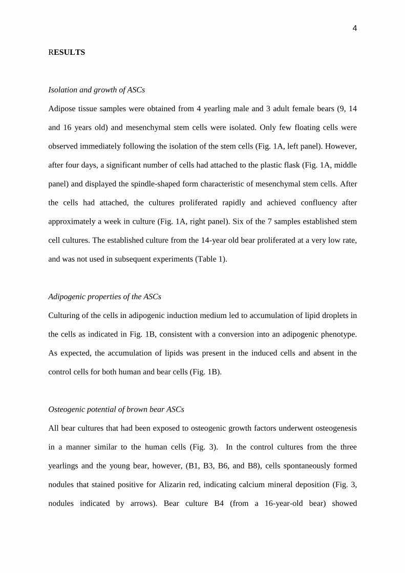
RESULTS
Isolation and growth of ASCs
Adipose tissue samples were obtained from 4 yearling male and 3 adult female bears (9, 14
and 16 years old) and mesenchymal stem cells were isolated. Only few floating cells were
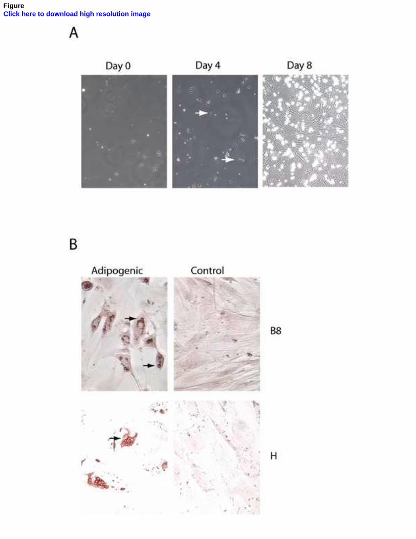
observed immediately following the isolation of the stem cells (Fig. 1A, left panel). However,
after four days, a significant number of cells had attached to the plastic flask (Fig. 1A, middle
panel) and displayed the spindle-shaped form characteristic of mesenchymal stem cells. After
the cells had attached, the cultures proliferated rapidly and achieved confluency after
approximately a week in culture (Fig. 1A, right panel). Six of the 7 samples established stem
cell cultures. The established culture from the 14-year old bear proliferated at a very low rate,
and was not used in subsequent experiments (Table 1).
Adipogenic properties of the ASCs
Culturing of the cells in adipogenic induction medium led to accumulation of lipid droplets in
the cells as indicated in Fig. 1B, consistent with a conversion into an adipogenic phenotype.
As expected, the accumulation of lipids was present in the induced cells and absent in the
control cells for both human and bear cells (Fig. 1B).
Osteogenic potential of brown bear ASCs
All bear cultures that had been exposed to osteogenic growth factors underwent osteogenesis
in a manner similar to the human cells (Fig. 3). In the control cultures from the three
yearlings and the young bear, however, (B1, B3, B6, and B8), cells spontaneously formed
nodules that stained positive for Alizarin red, indicating calcium mineral deposition (Fig. 3,
nodules indicated by arrows). Bear culture B4 (from a 16-year-old bear) showed
5
characteristics similar to the human control cell line, without nodule formation or positive
staining with Alizarin red (table 1).
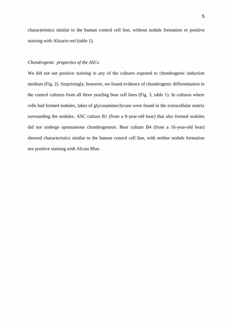
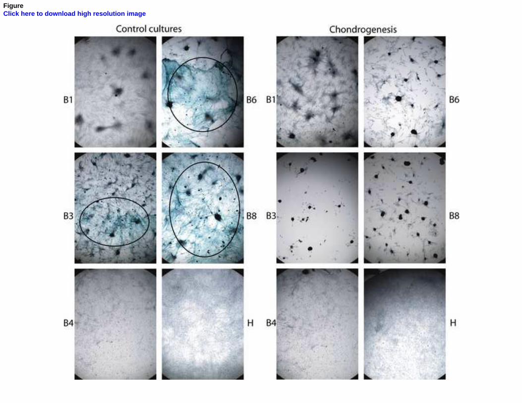
Chondrogenic properties of the ASCs
We did not see positive staining in any of the cultures exposed to chondrogenic induction
medium (Fig. 2). Surprisingly, however, we found evidence of chondrogenic differentiation in
the control cultures from all three yearling bear cell lines (Fig. 3, table 1). In cultures where
cells had formed nodules, lakes of glycosaminoclycans were found in the extracellular matrix
surrounding the nodules. ASC culture B1 (from a 9-year-old bear) that also formed nodules
did not undergo spontaneous chondrogenesis. Bear culture B4 (from a 16-year-old bear)
showed characteristics similar to the human control cell line, with neither nodule formation
nor positive staining with Alcian Blue.
6
DISCUSSION
This study represents the first documentation of stem cells from brown bears. ASCs were
recovered and cultured with a high success rate and the cells from yearlings showed
remarkable spontaneous cartilage and bone formation capacity. Interestingly, the spontaneous
bone and cartilage formation appears to occur in a concurrent manner in and around the
nodules, respectively, with mineralization characteristic of bone within the nodules and
cartilage formation in the periphery. To our knowledge, this is the first report of spontaneous
chondrogenic and osteogenic differentiation of ASCs. As the stem cells were recovered from
bears that recently were hibernating, it is possible that circulating factors that protect the bear
from bone degeneration during hibernation prime the stem cells. That we did not see any
chondrogenic differentiation in the cultures exposed to the chondrogenic induction medium
was not surprising, as it is notoriously difficult to achieve chondrogenic differentiation in
monolayer cultures, and most protocols call for either micromass pellet culture or culture in
alginate or similar scaffolds [5]. It is possible that the control medium allows for a higher
differentiation rate of the cells and optimum nodule formation.
The mineral content of femurs has been found to increase with age in black
bears (Ursus americanus) [4]. In this context, it would be interesting to determine if there is a
correlation between the propensity of stem cells from bears to spontaneously form bone cells
and the mineral content of bone. In conclusion, the spontaneous osteogenesis and
chondrogenesis of the ASCs, highlight the potential use of hibernating bears and ASCs
therefrom as model systems to study prevention of osteoporosis.
7
MATERIALS AND METHODS
Collection of adipose tissue samples from brown bears
All procedures involving the animals were in compliance with Swedish laws and regulations
and approved by the Uppsala animal ethics committee (Uppsala Djurförsöksetiska Nämnd, nr
C47/9, 2009-03-27). In mid-April 2009, approximately 7-10 days after leaving the den, wild
brown bears were immobilized from a helicopter by darting with a mixture of tiletamine-
zolazepam and medetomidine [6]. Adipose tissue biopsies (1.5-3 ml) were obtained
subcutaneously during intra abdominal implantation of tracking devices [7]. Each biopsy was
placed into a 15 ml v-bottomed centrifuge tube with phosphate buffered saline (PBS) with 10
IU/ml of penicillin, and 10 g/ml of streptomycin. All samples were kept at room temperature
and transported by courier to be processed within 48 hours of harvest.
Isolation of adipose tissue-derived stem cells
The tissue samples were minced finely and digested by incubation in a 0.14 Wünsch units/mL
Liberase Blendzyme 2 (Roche Applied Science, Hvidovre, Denmark) solution at 37°C for two
hours. The digests were centrifuged at 400g for 10 min. The pellet was briefly resuspended in
sterile water to lyse contaminating erythrocytes, after which the salt concentration was
adjusted through addition of 10x PBS. The cells were filtered through a 100 μm cell strainer,
centrifuged and resuspended in 5 ml growth medium, consisting of minimum essential
medium alpha (-MEM) (GIBCO/Invitrogen, Carlsbad, CA, USA) supplemented with 10%
fetal calf serum (FCS), and penicillin (10 IU/ml), streptomycin (10 g/ml) and gentamicin (5
g/ml) (all from GIBCO/Invitrogen). The cells were seeded in a T25 flask and transferred to a
CO2 incubator overnight, after which non-adherent cells were removed. The media was
changed twice a week during expansion of the cells. When cells were 90% confluent, they
8
were detached from the culture flasks using 0.125% trypsin/0.01% EDTA and transferred to
new flasks. When cells were in passage three, they were frozen in aliquots of approximately
0.5 x 10⁶ cells. All subsequent experiments were performed on cells in passage 4 in duplicates
in two independent experiments.
Human ASCs for use as control samples were isolated and propagated as previously described
after informed written consent [8]. The regional Committee on Biomedical Research Ethics of
Northern Jutland, Denmark approved analysis of human stem cells from persons undergoing
elective liposuction (project no. 2005054).
Induction of adipogenic, osteogenic, and chondrogenic differentiation
Cells from 5 bears and one human were used for the induction experiments. The induction of
the cells into the different lineages was carried out as previously described [8]. In brief, to
induce adipogenesis, cells were incubated for two weeks in adipogenic induction medium
-MEM supplemented with FCS, isobutylmethylxanthine (IBMX) insulin, and
indomethacin, after which the adipogenic differentiation was visualised through staining of
intracellular lipid accumulation with Oil Red O.
To induce ostogenesis, cells were maintained for three weeks in osteogenic
-MEM supplemented with FCS, dexamethasone, L-ascorbic
acid 2-phosphate, calcitriol, and glycerol 2-phosphate. After three weeks the degree of
osteogenesis was evaluated by staining of calcium deposits with Alizarin red.
To induce chondrogenesis, the cells were incubated in media consisting of high-
glucose (4.5 g/l) Dulbecco's modified Eagle's medium supplemented with 10 transforming
growth factor β3 (TGFβ3), dexamethazone, L-ascorbic acid 2-phosphate, L-proline, and 1×
ITS+
Premix. After three weeks in culture, the chondrogenic differentiation was assessed by
staining extracellular deposition of glycosaminoglycans with Alcian blue.

9
For all differentiation experiments, control cells were plated and incubated in growth medium
until completion of the experiment. All experiments were carried out twice, each in duplicate.

10
ACKNOWLEDGEMENTS
We thank the research personnel in the Scandinavian Brown Bear Research Project for their
assistance in the field. We thank Helle Skjødt for competent laboratory assistance.
11
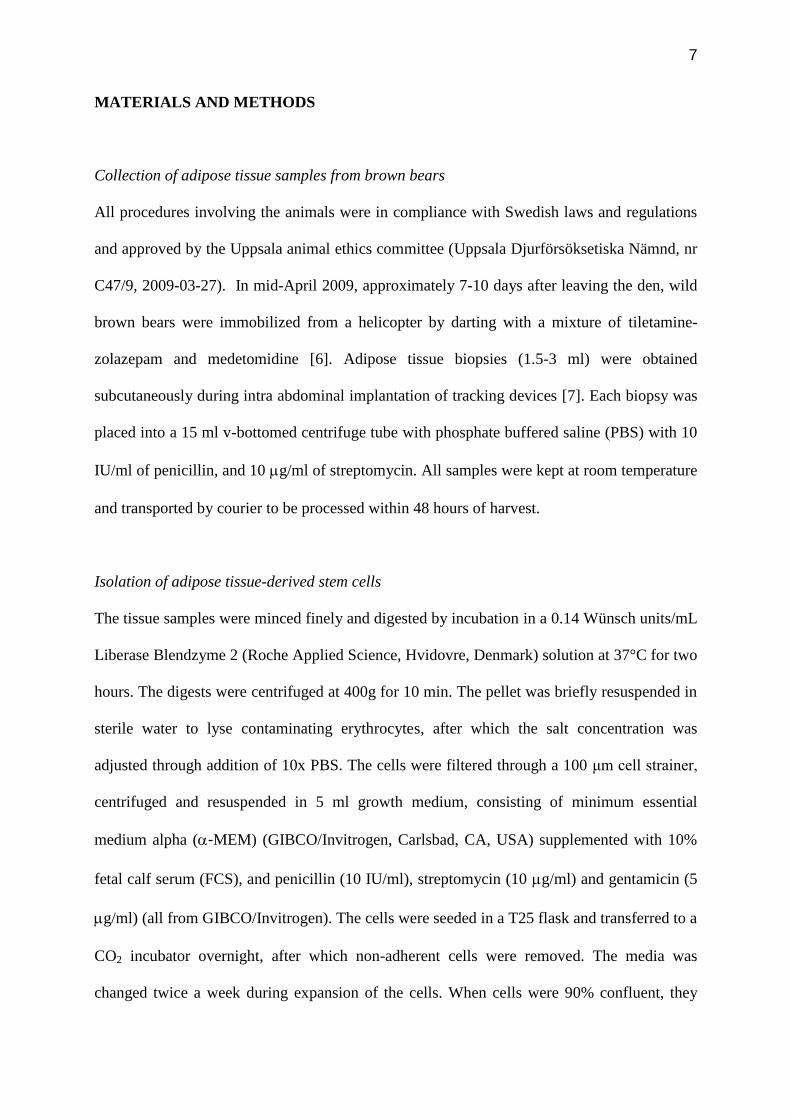
REFERENCES
[1] Manchi S, Swenson JE, Denning behavior of Scandinavian brown bears Ursus Arctos,
Wildlife Biology 11 (2005) 123-132.
[2] Friebe A, Swenson JE, Sandegren F, Denning chronology of female brown bears in
central Sweden, Ursus 12 (2001) 37-46.
[3] McGee-Lawrence ME, Wojda SJ, Barlow LN, Drummer TD, Bunnell K, et al., Six
months of disuse during hibernation does not increase intracortical porosity or
decrease cortical bone geometry, strength, or mineralization in black bear (Ursus
americanus) femurs, J Biomech 42 (2009) 1378-1383.
[4] McGee-Lawrence ME, Wojda SJ, Barlow LN, Drummer TD, Castillo AB, et al., Grizzly
bears (Ursus arctos horribilis) and black bears (Ursus americanus) prevent trabecular
bone loss during disuse (hibernation), Bone 45 (2009) 1186-1191.
[5] Bobick BE, Chen FH, Le AM, Tuan RS, Regulation of the chondrogenic phenotype in
culture, Birth Defects Res C Embryo Today 87 (2009) 351-371.
[6] Kreeger TJ Handbook of Wildlife Chemical Immobilization. Laramie, Wyoming, USA:
International Wildlife Veterinay Services, 2007.
[7] Arnemo J, Fahlman Å Biomedical protocols for free-ranging brown bears, gray wolves,
wolverines and lynx. Evenstad, Norway: Hedmark University College, 2008.
[8] Fink T, Lund P, Pilgaard L, Rasmussen JG, Duroux M, et al., Instability of standard PCR
reference genes in adipose-derived stem cells during propagation, differentiation and
hypoxic exposure, BMC Mol Biol 9 (2008) 98.
12
FIGURE LEGENDS
Figure 1. Cell growth and adipogenic differentiation of primary adipose tissue-derived stem
cells from wild brown bears. A: Photomicrographs of cells from one representative donor
grown for 1, 4, and 8 days after isolation. Arrows indicate spindle shaped cells. B: Cells
grown for two weeks in standard growth medium or adipogenic induction medium, after
which intracellular lipids were stained with Oil red O. Arrows indicate lipid inclusions.
Magnification, x 100.
Figure 2. Osteogenic differentiation of ASCs from bears and one human. Bear (B1-B8) and
human (H) stem cells were cultured in standard growth medium (control cultures) or
ostogenic induction medium for three weeks, after which the cultures were stained with
Alizarin red. Arrows indicate nodules. Magnification, x 40.
Figure 3. Chondrogenic differentiation of ASCs from bears and one human. Bear (B1-B8)
and human (H) stem cells were cultured in standard growth medium (control cultrues) or
chondrogenic induction medium for three weeks, after which the cultures were stained with
Alcian blue. The cyan-colored glycosaminoglycan deposits are indicated by circles.
Magnification, x 40.
FigureClick here to download high resolution image

FigureClick here to download high resolution image
FigureClick here to download high resolution image
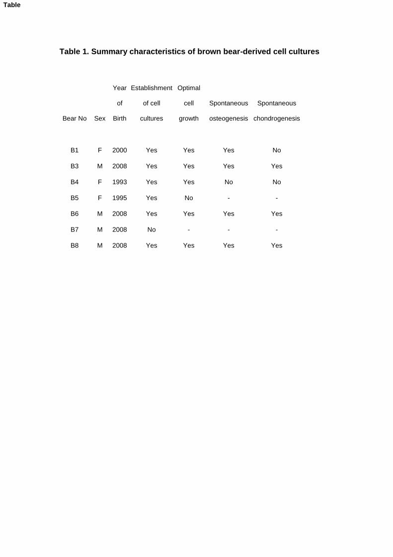
Table 1. Summary characteristics of brown bear-derived cell cultures
Bear No Sex
Year
of
Birth
Establishment
of cell
cultures
Optimal
cell
growth
Spontaneous
osteogenesis
Spontaneous
chondrogenesis
B1 F 2000 Yes Yes Yes No
B3 M 2008 Yes Yes Yes Yes
B4 F 1993 Yes Yes No No
B5 F 1995 Yes No - -
B6 M 2008 Yes Yes Yes Yes
B7 M 2008 No - - -
B8 M 2008 Yes Yes Yes Yes
Table
Related Documents