RIGHT: URL: CITATION: AUTHOR(S): ISSUE DATE: TITLE: Project 8 Analyzing Tumor Microenvironment and Exploiting Its Characteristics in Search of Optimizing Cancer Therapy Including Neutron Capture Therapy (30P8) Masunaga, S. Masunaga, S.. Project 8 Analyzing Tumor Microenvironment and Exploiting Its Characteristics in Search of Optimizing Cancer Therapy Including Neutron Capture Therapy (30P8). KURNS Progress Report 2019, 2018: 80-92 2019-08 http://hdl.handle.net/2433/244361

Welcome message from author
This document is posted to help you gain knowledge. Please leave a comment to let me know what you think about it! Share it to your friends and learn new things together.
Transcript
RIGHT:
URL:
CITATION:
AUTHOR(S):
ISSUE DATE:
TITLE:
Project 8 Analyzing TumorMicroenvironment and Exploiting ItsCharacteristics in Search of OptimizingCancer Therapy Including Neutron CaptureTherapy (30P8)
Masunaga, S.
Masunaga, S.. Project 8 Analyzing Tumor Microenvironment and Exploiting ItsCharacteristics in Search of Optimizing Cancer Therapy Including Neutron CaptureTherapy (30P8). KURNS Progress Report 2019, 2018: 80-92
2019-08
http://hdl.handle.net/2433/244361

I-1. PROJECT RESEARCHES
Project 8
80

Analyzing Tumor Microenvironment and Exploiting its Characteristics in Search of Optimizing Cancer Therapy Including Neutron Capture Therapy
Shin-ichiro Masunaga, M.D., Ph.D.
Particle Radiation Biology, Division of Radiation Life Sci-ence, Institute for Integrated Radiation and Nuclear Sci-ence, Kyoto University
BACKGROUNDS AND PURPOSES: Human solid tu-mors contain moderately large fractions of quiescent (Q) tumor cells that are out of the cell cycle and stop cell divi-sion, but are viable compared with established experi-mental animal tumor cell lines. The presence of Q cells is probably due, in part, to hypoxia and the depletion of nu-trition in the tumor core, which is another consequence of poor vascular supply. As a result, Q cells are viable and clonogenic, but stop cell division. In general, radiation and many DNA-damaging chemotherapeutic agents kill prolif-erating (P) tumor cells more efficiently than Q tumor cells, resulting in many clonogenic Q cells remaining following radiotherapy or chemotherapy. Therefore, it is harder to control Q tumor cells than to control P tumor cells, and many post-radiotherapy recurrent tumors result partly from the regrowth of Q tumor cells that could not be killed by radiotherapy. Similarly, sufficient doses of drugs cannot be distributed into Q tumor cells mainly due to heteroge-neous and poor vascularity within solid tumors. Thus, one of the major causes of post-chemotherapy recurrent tumors is an insufficient dose distribution into the Q cell fractions.
With regard to boron neutron capture therapy (BNCT), with 10B-compounds, boronophenylalanine-10B (BPA) increased the sensitivity of the total cells to a greater extent than mercaptoundecahydrododecaborate- 10B (BSH). However, the sensitivity of Q cells treated with BPA was lower than that in BSH-treated Q cells. The dif-ference in the sensitivity between the total and Q cells was greater with 10B-compounds, especially with BPA. These findings concerning the difference in sensitivity, including other recovery and reoxygenation following neutron irra-diation after 10B-compound administration were mainly based on the fact that it is difficult to deliver a therapeutic amount of 10B from 10B-carriers throughout the target tu-mors, especially into intratumor hypoxic cells with low up-take capacities.
Hypoxia is suggested to enhance metastasis by in-creasing genetic instability. Acute, but not chronic, hy-poxia was reported to increase the number of macroscopic metastases in mouse lungs. We recently reported the sig-nificance of the injection of an acute hypoxia-releasing agent, nicotinamide, into tumor- bearing mice as a com-bined treatment with -ray irradiation in terms of repress-ing lung metastasis. As the delivered total dose increased with irradiation, the number of macroscopic lung metasta-ses decreased reflecting the decrease in the number of clonogenically viable tumor cells in the primary tumor. The metastasis-repressing effect achieved through a reduc-tion in the number of clonogenic tumor cells by irradiation is much greater than that achieved by releasing tumor cells from acute hypoxia. On the other hand, more 10B from BPA than from BSH could be distributed into the acute hy-poxia-rich total tumor cell population, resulting in a greater decrease in the number of highly clonogenic P tu-mor cells with BPA-BNCT than with BSH-BNCT and with neutron beam irradiation only. BPA-BNCT rather than BSH-BNCT has some potential to decrease the num-ber of lung metastases, and an acute hypoxia- releasing treatment such as the administration of nicotinamide,
bevacizumab, wortmannin or thalidomide may be promis-ing for reducing numbers of lung metastases. Conse-quently, BPA-BNCT in combination with the treatment us-ing these agents may show a little more potential to reduce the number of metastases. Now, it has been elucidated that control of the chronic hypoxia-rich Q cell population in the primary solid tumor has the potential to impact the control of local tumors as a whole, and that control of the acute hypoxia-rich total tumor cell population in the primary solid tumor has the potential to impact the control of lung metastases.
The aim of this research project is focused on clari-fying and analyzing the characteristics of intratumor mi-croenvironment including hypoxia within malignant solid tumors and optimizing cancer therapeutic modalities, es-pecially radiotherapy including BNCT in the use of newly-developed 10B-carriers based on the revealed findings on intratumor microenvironmental characteristics.
RESEARCH SUBJECTS: The collaborators and allotted research subjects (ARS) were organized as follows;
ARS-1 (30P8-1): Optimization of Radiation Therapy In-cluding BNCT in terms of the Effect on a Specific Cell Fraction within a Solid Tumor and the Suppressing Effect of Distant Metastasis. (S. Masunaga,et al.)
ARS-2 (30P8-2): Development of Hypoxic Microenviron-ment-Oriented 10B-Carriers. (H. Nagasawa, et al.)
ARS-3 (30P8-3)*: Search and Functional Analysis of Novel Genes that Activate HIF-1, and Development into Local Tumor Control. (H. Harada, et al.)
ARS-4 (30P8-4)*: Radiochemical Analysis of Cell Le-thality Mechanism in Neutron Capture Reaction. (R. Hirayama, et al.)
ARS-5 (30P8-5): Development of Neutron Capture Ther-apy Using Cell-Membrane Fluidity Recognition Type Novel Boron Hybrid Liposome. (S. Kasaoka, et al.)
ARS-6 (30P8-6): Drug Delivery System Aimed at Adap-tation to Neutron Capture Therapy for Melanoma. (T. Nagasaki, et al.)
ARS-7 (30P8-7): Molecular Design, Synthesis and Func-tional Evaluation of Hypoxic Cytotoxin Including Boron. (Y. Uto, et al.)
ARS-8 (30P8-8): Bystander Effect on Malignant Trait of Tumor Cells by Irradiation. (H. Yasui, et al.)
ARS-9 (30P8-9): Analysis of the Response of Malignant Tumor to BNCT. (M. Masutani, et al.)
ARS-10 (30P8-10): Cell Survival Test by Neutron Capture Reaction Using Boron Compound and Inhibitory Effect on Tumor Growth. (K. Nakai, et al.)
ARS-11 (30P8-11): Multilateral Approach Toward Realization of Next Generation Boron Neutron Capture Therapy. (Y. Matsumoto, et al.)
ARS-12 (30P8-12): Analysis of Radiosensitization Effect through Targeting Intratumoral Environmental. (Y. Sanada, et al.)
ARS-13 (30P8-13)*: Exploratory Research on the Opti-mal Administration of 10B Compound Aiming at New En-forcement Method of Neutron Capture Therapy (S. Masunaga et al.)
ARS-14(30P8-14) *: Examination of Cancer Cell Accu-mulation Property of New Boron Agent (CbaP14). (J. Hiratsuka, et al.)
(*There was no allocated time for experiments using reactor facilities during their operation periods of FY 2018.)
PR8
30P8
81

Estimation of Therapeutic Efficacy of BCNTBased on the Intra- and Intercellular Heterogeneity in 10B-Distribution
T. Sato1 and S. Masunaga2
1Nuclear Science and Engineering Center, Japan Atomic Energy Agency
2Institute for Integrated Radiation and Nuclear Science, Kyoto University
INTRODUCTION: Two types of dose were proposed for use in the treatment planning of boron neutron capture therapy (BNCT) for expressing its high relative biologi-cal effectiveness (RBE). On one hand, the RBE -weighted dose is the sum of the absorbed doses weightedby fixed RBE for four dose components of BNCT, whichare attributable to 10B(n,α)7Li, 14N(n,p)14C, and 1H(n,n)preactions as well as photons, respectively. On the otherhand, photon-isoeffective dose is the photon dose to givethe same biological effect calculated considering the dosedependence of RBE and the synergetic effect betweendifferent types of radiation [1]. In this study, the depthdistributions of the two types of dose in a phantom placedat an accelerator-based BNCT field were calculated usingParticle and Heavy Ion Transport code System, PHITS[2], coupled with an extended stochastic microdosimetrickinetic (SMK) model [3].
MATERIALS AND METHODS: The basic flow of the calculation procedures is shown in Fig. 1. In the PHITS simulation, a cylindrical phantom with the radius of 10 cm and the depth of 20 cm composed of ICRU soft tissue was placed at the beam port of the Linac-based BNCT facility in University of Tsukuba [4]. The absorbed doses were determined from the calculated neutron and photon fluxes multiplied with their corresponding kerma factors for each dose component. The calculated absorbed doses were converted to RBE-weighted dose and pho-ton-isoeffective dose, using the extended SMK model. The parameters used in the model were evaluated by the least-square fitting of the surviving fractions of SCC VII murine squamous cell carcinomas obtained from in vi-vo/in vitro experiments of tumor-bearing mice treated by BNCT [5].
RESULTS AND DISCUSSION: Fig. 2 shows the RBE-weighted dose and photon-isoeffective dose in the phantom administrated with BPA or BSH, or without 10B. The absorbed doses at the entrance of the phantom with-out 10B were normalized to 2 and 10 Gy in panels (A) and (B), respectively. It is evident from Fig. 2 that the pho-ton-isoeffective doses are larger than the corresponding RBE-weighted doses in panel (A), while the reverse is true in panel (B) except at the locations deeper than 7 g/cm2. The depth dependence was more apparent in the RBE-weighted doses, which decreased more dramatically with increasing depth. Such tendency is mainly due to the consideration of the dose dependence of RBE in the cal-culation of the photon-isoeffective dose because RBE decreases with an increase in the absorbed dose. The
consideration of the synergetic effect also causes the dif-ference, which raises the photon-isoeffective dose by approximately 10-20%. More detailed discussions can be found in our recently published paper [6].
REFERENCES:
[1] S.J. Gonzalez, et al., Phys Med Biol 62 (2017)7938-7958.
[2] T. Sato et al., J Nucl Sci Technol 55, (2018) 684-690.[3] T. Sato et al., Sci Rep 8 (2018) 988.[4] A. Masuda et al., Appl Radiat Isot 127 (2017), 47-51.[5] S. Masunaga et al., Springerplus 3 (2014), 128.[6] T. Sato et al., Radiat Prot Dosim (2018) DOI:
10.1093/rpd/ncy235
Fig. 1. Basic flow of the calculation carried out in this
study.
Fig. 2. Calculated RBE-weighted dose and photon
-isoeffective dose in the phantom administrated
with BPA or BSH, or without 10B.
PR8-1
30P8-1
82
30P8-2
Design, synthesis and biological evaluation of lipopeptide conjugates of BSH for BNCT
A. Isono1, M. Tsuji1, Y. Sanada2, T. Hirayama1,
S. Masunaga2 and H. Nagasawa1
1 Laboratory of Medicinal & Pharmaceutical Chemistry, Gifu Pharmaceutical University
2 KURNS, Kyoto University
INTRODUCTION: Tumor selective delivery of suffi-
cient quantity of 10B is essential for the success of boron
neutron capture therapy (BNCT). The clinically used
boron carrier, sodium mercaptoundecahydro-closo-
dodecaborate (BSH: Na2B12H11SH) does not penetrate
the cell membrane directly, due to its highly hydrophilic
and anionic properties. Here, we developed a new mem-
brane-permeable boron cluster using the cell-penetrating
lipopeptide pepducin as the vehicle for intracellular de-
livery of boron clusters. Pepducins are mem-
brane-permeable, lipidated peptides, developed as allo-
steric modulators of the G-protein-coupled receptor
(GPCR). We have previously developed fluorescence
resonance energy transfer (FRET)-based pepducin probes
to demonstrate the direct membrane penetration of a
pepducin by fluorescence imaging in living cells. So, we
designed and synthesized pepducin-BSH conjugates and
performed structural optimization to improve cellular
uptake. (Fig.1)
Fig. 1. Molecular design (A) and putative mechanism
of intracellular delivery of pepdicin-BSH conjugates.
EXPERIMENTS: All compounds were synthesized
based on sol-id-phase synthesis. (Table 1) T98G, cells
were treated with the boron carriers (10 or 20 μM) at
37 °C for various times, then, washed with PBS three
times, and dissolved in 200 μL HNO3 for 1 h. The boron
concentrations of these extracts were measured by
ICP-AES. To evaluate neutron sensitizing ability of the
compounds, T98G cells were treated with 20 μM boron
carriers for 24 h. Then the cells were washed with PBS,
suspended in serum containing medium and aliquoted
into Teflon tubes for irradiation. Cells were irradiated
using the neutron beam at the Heavy Water Facility of the
Kyoto University Research Reactor (KUR) operated at 1
MW power output. The survival rates of the irradiated
cells were determined using conventional colony assays.
Table 1. Structures of new boron carriers.
RESULTS: Among the pepducin-BSH conjugates, the
most effectively incorporated and accumulated in the
cells was compound 1a, which has a peptide of 13 resi-
dues derived from the intracellular third loop of PAR1
and a palmitoyl group. Compound 1a and compound 4
both showed a stronger radio-sensitizing effect than BSH
on T98G cells under mixed-neutron beam irradiation.
(Table 2)
Table 2. Plating efficiencies without irradiation, D10 val-
ues and the enhancing effects of boron carriers. PE without irradiation (%) D10 value (Gy) The enhancing effect
Control 19.0±4.4 4.17 -
BSH 32.9±3.6 4.32 0.97
1a 51.2±5.6 0.55 7.69
4 20.0±4.5 0.72 5.79
The results demonstrate that lipopeptide conjugation is
apparently effective for enhancing intracellular delivery
and accumulation of BSH and improving the cytotoxic
effect of BNCT [1].
REFERENCES: [1] A. Isono et al., ChemMedChem., 14 (2019) 823-832.
PR8-2
83
30P8-3
Molecular mechanism underlying radioresistance of hypoxic tumor cells
M. Kobayashi1,2, S. Masunaga3, A. Morinibu1,2 and H.Harada1,2
1Laboratory of Cancer Cell Biology, Graduate School of Biostudies, Kyoto University. 2Department of Genome Dynamics, Radiation Biology Center, Graduate School of Biostudies, Kyoto University. 3 Institute for Integrated Radiation and Nuclear Science, Kyoto University.
INTRODUCTION: Cancer cells are 2-3 times more radioresistant under hypoxic than normoxic conditions, which causes poor prognosis of patients with more hy-poxic regions in their tumor tissues [1]. Although the radioresistance is known to be induced by biological mechanisms, at least in part, in a hypoxia-inducible factor 1 (HIF-1)-dependent manner, further studies are needed for better understanding of the nature of hypoxic cell ra-dioresistance [2, 3].
EXPERIMENTS & RESULTS: Here we found that hypoxia-inducible secretory protein 2, HISP2, which we recently identified as a novel hypox-ia-responsive gene through our own DNA microarray analysis, causes radioresistance of hypoxic cancer cells in a HIF-1-independent manner (in preparation). qRT-PCR experiments confirmed that HISP2 mRNA levels were significantly upregulated under hypoxic conditions. ELISA experiments for culture media showed that the amount of secreted HISP2 protein levels increases under hypoxic conditions. The hypoxia-dependent increase in the HISP2 mRNA levels was abrogated by a transcription inhibitor, actinomycin D, suggesting that HISP2 were upregulated under hypoxia at transcription initiation lev-els. A loss-of-function study found that the hypox-ia-dependent increase in the HISP-2 mRNA levels was significantly abrogated by silencing of HIF-1β, but not by that of HIF-1α and HIF-2α, suggested that the hypox-ia-mediated expression is dependent on HIF-1β. A re-porter gene assay with the use of an expression vector for EGFP-53BP1M fusion protein demonstrated that HISP2-overexpression decreased the number of DNA double strand breaks after irradiation. Clonogenic cell survival assay showed that cancer cells acquired radiore-sistance when transfected with HISP2-overexpression vector. All of these data suggested that HISP2 proteins are secreted under hypoxic conditions in a HIF-1-independent but HIF-1β-dependent manner and decrease the number of double-strand break after irradia-tion, leading to radioresistance of cancer cells in an auto-crine fashion.
REFERENCES: [1] *Harada H. Hypoxia-inducible factor 1-mediated
characteristic features of cancer cellsfor tumorradi-aresistance. J Radiat Res., 57 (2016) 99-105.
[2] Koyasu S, Kobayashi M, Goto Y, Hiraoka M,*Harada H. Regulatory mechanisms of hypox-ia-inducible factor 1 activity: Two decadesofknowledge. Cancer Sci. 109:560-571. 2018.
[3] Nagao A, Kobayashi M, Koyasu S, Chow CCT,*Harada H. HIF-1-dependent reprogramming ofglucose metabolic pathway of cancer cellsand itstherapeutic significance. Int J Mol Sci. 20:238.2019.
PR8-3
84

30P8-5
Membrane-Targeted Boron Neutron Capture Therapy with Bo-
ron Liposomes Composed of Dimyristoylphosphatidylcholine
S. Kasaoka1, Y. Okishima1, Y. Tanaka1, H. Yoshikawa1, Y.
Sanada2, Y. Sakurai2, H. Tanaka2 and S. Masunaga2
1Department of Pharmaceutical Science, Hiroshima In-ternational University
2KURNS, Kyoto University
INTRODUCTION: There are many reports that
membranes in cancer cells are relatively more fluid com-
pared to healthy cells. Higher membrane fluidity in can-
cer cells closely relates to their invasive potential, prolif-
eration, and metastatic ability [1]. Liposomes composed
of dimyristoylphosphatidylcholine (DMPC) and polyox-
yethylenedodecylether were found to inhibit the growth
of human promyelocytic leukemia cells without using
any drugs [2]. In this study, we have developed a novel
boron delivery system using the membrane-fluidity sensi-
tive boron liposomes (MFSBLs) composed of DMPC and
borocaptate (BSH)-conjugated chemical compounds for
boron neutron capture therapy.
EXPERIMENTS: Octadecylamine and
1,2-dimyristoyl-sn-glycero-3-phosphorylethanola
mine were conjugated with BSH using the opti-
mal hetero-crosslinking agents for boron com-
pounds. MFSBLs composed of DMPC, polyoxyeth-
ylenedodecylether and boron compounds at mole rati-
os of 8:0.9:1.1 were prepared by sonication method
in 5% glucose solution at 45°C. The diameter of
MFSBLs was measured with a light scattering spectrometer.
The boron concentration was measured by inductively cou-
pled plasma atomic emission spectrometry. B16F10 mu-
rine melanoma cells, COLO679 human melanoma
cells and human fibroblast cells were incubated
with 2-6 ppm of 10B at 37°C for 14 hours before
neutron irradiation. The cells were rinsed twice
in PBS and suspended in fresh medium. After
neutron irradiation the cells were plated into
plastic Petri dishes 60 mm in diameter at 200
cells per dish. They were incubated for an addi-
tional 7 days to allow colony formation.
RESULTS: MFSBLs had a mean diameter of 59.6 nm.
High encapsulation efficiency value from 55% to 89% of
10B in MFSBLs were obtained. MFSBLs had high stabil-ity (95-99%) in the retention of 10B during storage at 4°C
for 4 weeks. All borocaptate-loaded formulations had low
cytotoxic effects in human fibroblast cells. MFSBLs were
efficiently fused to melanoma cells, but were inefficiently
fused to human fibroblast cells. Thus, it is essential to
elevate the 10B concentration in melanoma cells, while
maintain low levels of 10B in normal fibroblast cells. The
tumor/normal ratio (T/N ratio) was 2.1-3.0. As shown in
Fig. 1, MFSBLs showed higher suppression of growth of
murine and human melanoma cells than BSH solution.
This result suggested novel MFSBLs is useful for 10B
carrier on BNCT for melanoma.
Fig. 1. Suppression of the colony formation of B16F10
cells, COLO679 melanoma cells and fibroblast cells after
in vitro BNCT.
REFERENCES: [1] Sherbet GV. Magalit, Exp Cell Biol., 57 (1989).[2] Y. Matsumoto et al. Int. J. Cancer, 115 (2005).
PR8-4
85

30P8-6
GLUT1-mediated endocytosis could be a major pathway for internalisation of kojic acid-appended carborane conjugate into melanoma cells
S. Dowaki, K. Matsuura, R. Kawasaki, M. Kirihata1, Y. Hattori1, Y. Sakurai 2 and S. Masunaga 2
Graduate School of Engineering, Osaka City University 1 BNCT Research Center, Osaka Prefecture University, 2 Institute for Integrated Radiation and Nuclear Science, Kyoto University
INTRODUCTION: Receptor-mediated endocytosis is potent pathways for the internalization of drugs into target cells. It is well known that the expression level of glucose transporter and uptake of glucose are increased in cancer cells [1]. We have previously reported the inclusion com-plex of kojic acid-appended carborane (CKA) with hy-droxypropyl-β-cyclodextrin (HP-β-CD) as a novel boron agent is potent for melanoma targeting BNCT. CKA/HP-β-CD complex showed melanoma cells selectivity, unique nuclear localization, and high tumor-suppression effect on BNCT toward melanoma-bearing mice. Herein, mecha-nism of high efficient internalization of CKA/HP-β-CD complex into melanoma cells is evaluated.
EXPERIMENTS: B16BL6 cells were maintained in RPMI containing 10% FBS and 1% penicillin/streptomy-cin. B16BL6 cells seeded on the 6 well plate at the con-centration of 2.0×10⁵ cells/well. The cells were exposed to CKA/HP-β-CD complex (10 ppm) and the cells were cor-rected at each time point (12 and 24 hr). The involvement of GLUT1 on the uptake of CKA is confirmed with not only a small-molecule inhibitor of GLUT1 [2] ,but also RNA interference-mediated reduction in GLUT1 [3]. The isolated cells were digested by using aqua regia with heating (95 oC, 30 min; 115 oC, 60 min). After centrifuga-tion, the cellular uptake amounts were quantified by ICP-AES.
RESULTS AND DISCUSSION: As shown in Fig. 1 and 2, clear suppression of CKA uptake by melanoma cells were observed in the presence of WZB117 inhibitor and siRNA for GLUT1. Furthermore, CKA uptake obviously reduced when melanoma cells were incubated in the nor-mal medium with glucose. These results suggested that the internalization of CKA/HP-β-CD complex into melanoma cells is enhanced with GLUT1-mediated endocytosis.
Fig.1 Suppression of CKA uptake by the inhibition of GLUT1 by WZB117. B16BL6 cells were incubated in the presence or absence of WZB117 (30 µM) for 12 and 24 hr. * denotes P < 0.02 between indicated treatments.
Fig.2 Suppression of CKA uptake by RNA interference-mediated reduction in GLUT1. B16BL6 cells were incu-bated in the presence or absence of siRNA (100 nM) for 12 and 24 hr. * denotes P < 0.02 between indicated treat-ments.
REFERENCES: [1] T. Yamamoto et al., Biochem. Biophys. Res. Commun.,
170 (1990) 223-30.[2] Y. Liu et al., Mol. Cancer Ther., 11 (2012) 1672-82.[3] L. Al-Khalili et al., Biochem. Biophys. Res. Commun.,
307 (2003) 127-32.
Structure of CKA/HP-b-CD
PR8-5
86

30P8-7
Development of novel BPA-Tirapazamine hybrid BNCT agent targeting hypoxic tumor cells
Y. Uto, Y. Tanaka, S. Masunaga1, Y. Sanada
1
Graduate School of Technology, Industrial and Social
Sciences, Tokushima University 1Radiation and Nuclear Science, Kyoto University
INTRODUCTION: Hypoxia is a ubiquitous environ-
ment of cancer, it has a resistance to anticancer drugs and
X-ray, and accelerated infiltration / metastasis is a prob-
lem. Boron neutron capture therapy (BNCT), a kind of
radiotherapy, is a treatment method utilizing neutron
capture reaction in tumor cells by accumulating 10
B in
tumor tissue. Hybrid molecules of Tirapazamine (TPZ),
as a hypoxic cytotoxin and the neutron scavenger
p-borono-L-phenylalanine (BPA) are useful as selective
BNCT agent for hypoxic tumors. Since therapeutic effi-
ciency of BNCT depends on 10
B concentration, it is ex-
pected to provide an effective therapeutic effect for hy-
poxic cancer.
In this study, UTX-117 with amide linkage of TPZ and
BPA and UTX-118 with ester linkage of TPZ and BPA
were designed and synthesized, and antitumor activity
and neutron sensitizing activity were evaluated. UTX-117
and UTX-118 exhibit hypoxia selective cytotoxicity and
UTX-118 has improved uptake rate compared to BPA
fructose complex. Further, UTX-117 and UTX-118
shows neutron sensitizing activity.
EXPERIMENTS: Tirapazamine derivatives and
L-BPA were condensed to synthesize UTX-117 and
UTX-118 (Fig.1). Using melanoma cells B16-F10 cells,
the IC50 values of UTX-117 and UTX-118 were evaluated
by WST-1 assay. The cell uptake of UTX-117 and
UTX-118 was measured by ICP-AES, and the intracellu-
lar boron concentration was calculated. Neutron sensitiz-
ing activity of UTX-117, UTX-118 and BPA-F to
B16-F10 cells were evaluated by WST-1 assay.
UTX-117 UTX-118 Fig.1
RESULTS: Cytotoxicity tests were performed by
WST-1 assay using B16-F10 cells. As shown in Table 1,
UTX-117 andUTX-118 exhibited hypoxia-selective cy-
totoxicity similar to that of tirapazamine.
Table. 1. Cytotoxicity of UTX-117 and UTX-118.
Normoxia Hypoxia N/H ratio
UTX-117 >1000 µM 641.6 µM >1.56
UTX-118 96.0 µM 33.8 µM 2.84
TPZ 123.2 µM 35.5 µM 3.74
N/H ratio=the IC50 value of Normoxia / Hypoxia
The cell uptake test of UTX-117 and UTX-118 was per-
formed using B16-F10 cells (Fig. 2). The maximum up-
take of BPA-F, a conventional BNCT agent, was ap-
proximately 1.3 × 10⁹ atoms / cell [1]. On the other hand,
the maximum uptake of UTX-118 was approximately 7.4
× 109 atoms / cell, it showed higher uptake efficiency
than BPA-F. Since the boron concentration required for
BNCT is at least 1.0 × 10⁹ atoms / cell, a neutron sensiti-
zation effect can be expected for UTX-118.
Fig. 2. Intracellular uptake of UTX-117 and UTX-118.
Neutron sensitizing activities of UTX-117, UTX-118
and BPA-F were evaluated using B16-F10 cells (Fig. 3).
UTX-117 and UTX-118 showed the neutron sensitizing
effect similar to BPA-F.
Fig. 3. Neutron sensitizing activity of UTX-117 and
UTX-118. +: Irradiation, -: Non-irradiation.
REFERENCES:
[1] F. F. Flores et al. Radiat. Environ. Biophys., 51(2012) 319-329.
PR8-6
87

X-irradiation-induced bystander effect enhances invasion in tumor cells
H. Yasui1, M. Eitaki, S1. Masunaga2, and O. Inanami1
1Laboratory of Radiation Biology, Graduate School of Veterinary Medicine, Hokkaido University 2Particle Radiation Biology, Division of Radiation Life Science, KURNS, Kyoto University
INTRODUCTION: In cancer studies, it has been sug-gested that irradiation sometimes enhances invasion of tumor cells. Wild-Bode et al. reported that sublethal dose of γ-ray irradiation enhanced invasiveness of human gli-oblastoma, and the enhancement was accompanied by the activation and/or up-regulation of integrin and matrix metalloproteinase (MMP) contributed to cellular adhe-sion and degradation of extracellular matrix (ECM). Fu-jita et al. also reported that X-ray irradiation enhanced cellular invasive ability via up-regulation of MMP in some human pancreatic tumors. Including these reports, a number of studies suggested that ECM degradation activ-ity was involved in the irradiation-enhanced invasiveness of tumor cells. However, although there are many studies that estimate effects of direct irradiation on tumor cell invasiveness, not so many reports did bystander effects of irradiated tumor cell conditioned medium (CM) on it. Furthermore, the same effect of high LET radiation ther-apy such as boron neutron capture reaction (BNCT) has not been reported. In this study, we conducted the ex-periments to examine effects of X-irradiated tumor cell CM on cellular invasiveness in breast cancer and lung cancer-derived cells, prior to BNCT challenge.
EXPERIMENTS: Human breast adenocarcinoma MDA-MB-231 cells and human lung adenocarcinoma A549 cells were seeded on 60 mm dishes (1 × 10⁶ cells/dish), and incubated with RPMI1640/10% FBS overnight. After washing by PBS, serum-free RPMI1640 medium was added to the dishes followed by 0, 2 or 4 Gy X-irradiation immediately. After 24 h incubation, the me-dia were centrifuged for 5 min at 3,000 rpm at 4°C, andsupernatants were collected as conditioned media (CM).Hereinafter, CM derived from 0, 2 and 4 Gy X-irradiatedcells are described as 0 Gy CM, 2 Gy CM and 4 Gy CM.
Cellular invasive ability was evaluated using a BD BioCoatTM MatrigelTM Invasion Chamber (8 μm pore size, 24-well plate, BD Biosciences, Billerica, MA). Cells (2 ×10⁴ MDA-MB-231 cells or 1 × 10⁵ A549 cells) were sus-pended in 500 μl of serum-free RPMI1640 medium withor without 500 nM AG1478, and they were loaded intothe upper chamber, followed by an addition of 750 μl ofCM containing 1% FBS (CM/1 % FBS) into the lowerchamber. After incubation for 6 h (MDA-MB-231 cells)or 12 h (A549 cells), non-invading cells were removedfrom the upper side of the membrane using cotton tips.Invading cells on the lower side of the membrane werefixed with 100% methanol and stained with 1% toluidineblue and 1% sodium borate. All the invading cells werecounted using a light microscope. Invasion index was
calculated as a relative value which set the number of 0 Gy CM-treated invaded cells to 1.
Cells (1 × 10⁴ cells) were suspended with 100 μl of RPMI1640/10% FBS, and loaded into each well of a 96-well plate. After overnight incubation, the mediumwas exchanged with CM/1% FBS, and the cells wereincubated for 6 h (MDA-MB-231 cells) or 12 h (A549cells). Ten μl of WST-1 solution (3.24 μg/μl WST-1, 70ng/μl PMS, 20 mM HEPES-NaOH [pH 7.4]) was addedinto each well, and 1 h incubation was performed. Ab-sorbance (450 nm) of the each well was measured withModel 680 Microplate Reader (Bio-Rad Laboratories).Cellular proliferation activity was described as relativevalues which set the absorbance of 0 Gy CM-treated cellsto 1.
RESULTS: In order to investigate the influence of CM on the infiltration ability of cells, an invasion assay was first performed. The lower chamber was filled with CM from MDA-MB-231 cells and A549 cells, and the upper chamber was filled with cells to evaluate the infiltration ability of each cell at 6 h and 12 h. As shown in Fig. 1, CM promoted the respective cell infiltration ability in a dose dependent manner of the irradiated X-rays. Next, a colorimetric test using WST-1 was performed to evaluate the effect of CM on cell proliferation activity. When MDA-MB-231 cells are treated with CM from X-irradiated cells for 6 h, 2 Gy CM and 4 Gy CM reducescell proliferation activity about 2% and 9% as comparedto 0 Gy CM. When A549 cells were treated with CMfrom X-irradiated cells for 12 h, 2 Gy CM and 4 Gy CMreduces cell proliferation activity about 3% and 24% ascompared to 0 Gy CM. From these results, it was sug-gested that the X-ray irradiated cell-derived CM pro-motes cell infiltration ability even if taken into considera-tion cell proliferation activity. In next fiscal year, we planto examine the effect of CM obtained after BNCT-treatedcells.
Fig. 1 The effect of X-irradiation CM on cellular inva-sion and proliferation.
PR8-7
88
30P8-8
30P8-9
Analysis of the response of tumor and normal tissues to BNCT
S. Imamichi1,2, L. Chen2,3, M. Ihara2,3, Y. Sasaki2,3, T.Onodera2,3, Y. Natori3, N. Toriya3, A. Takahira3, J.Itami1,4,5, S. Masunaga6 and M. Masutani1,2,3
1Division of Boron Neutron Capture Therapy, EPOC, National Cancer Center 2Lab of Collaborative Research, Division of Cellular Signaling, Research Institute, National Cancer Center 3Department of Frontier Life Sciences & 4Department of Comprehensive Oncology, Nagasaki University Graduate School of Biomedical Sciences 5Department of Radiation Oncology, National Cancer Center Hospital 6Institute for Integrated Radiation and Nuclear Sci-ence, Kyoto University
INTRODUCTION: Boron neutron capture therapy (BNCT) is one of the particle beam radiation therapy based on the boron neutron capture fission reaction, which causes α particle and lithium nuclei of high lin-ear-energy-transfer in a short range that is less than the diameter of typical cells. Boron compounds such as 10B-boronophenylalanine (BPA) are mainly clinically used with neutron sources of nuclear reactors or accel-erator-based BNCT systems. The cell death following DNA damage that is induced by BNCT1) is affected by various factors including the uptake of boron com-pounds by tumor cells and the thermal neutron flu-ence2). Gamma-H2AX, a marker for DNA double strand breaks and poly(ADP-ribose) became high lev-els 6 or 24 hrs after boron neutron capture reaction (BNCR) in the rat lymphosarcoma model3). The com-prehensive analysis of the transcriptome and proteome after BNCR in human squamous carcinoma SAS cells has been carried out4). As one of the factors induced after BNCR conditions, we focused on CSF2 gene product, granulocyte-macrophage colony stimulating factor (GM-CSF). We studied the dynamic properties of GM-CSF and its mRNA (CSF2) after BNCR in cells and also in xenograft models of mice.
EXPERIMENTS: We used human squamous cell carcinoma-derived SAS and malignant melano-ma-derived A375 cells. The neutron beam irradiation was operated at 1 MW at the Heavy Water Neutron Irradiation Facility of KUR reactor. Suspended cells were irradiated 2 hrs after incubation in the presence or absence of BPA 25 ppm (boron concentration) on Nov. 28th & Dec. 4th, 2018. For the xenograft models, SAS and A375 cells were transplanted into left hind legs of BALB/c nude male mice of 5 weeks old. Ten days later, neutron beam irradiation was carried out. As a boron compound, 10B-BPA fructose complex was prepared before irradiation. Mice were irradiated ap-proximately 30 min after intraperitoneal administration on Dec. 4th, 2018. Gold foil activation analysis was used for the measurement of thermal neutron fluences
and thermoluminescence dosimeter (TLD) was used for the measurement of the γ-ray dose including sec-ondary γ-ray. As previously described, total absorbed dose calculation was carried out using the flux-to-dose conversion factor by the sum of the absorbed doses resulting from 1H(n, γ)2D, 14N(n, p)14C, and 10B(n, α)7Li reactions. The cellular responses includ-ing factors present in culture supernatants were ana-lyzed 6 and 24 hrs after irradiation of therapeutic doses of BNCT and γ-ray. Cell survival was analyzed by colony formation assay. The cells were transfected with siRNA of CSF2 & CSF2 receptor using Lipofec-tamineTM 3000 Reagents (Thermo Fisher Scientific). GM-CSF levels in culture supernatant and mouse blood were measured using ELISA.
RESULTS: The irradiated beam and total radiation doses for cell experiments were exemplified in Table 1. SAS and A375 cells showed an increase in CSF2 mRNA expression after the therapeutic dose irradia-tion of neutron beam with BPA, respectively. Growth of tumors derived from SAS cells was decreased 3 and 8 days after neutron beam irradiation in the presence of BPA compared with non-irradiated controls. Three days after irradiation, the DNA double strand break marker was positive in the tumors but not in the sur-rounding normal tissues. Changes in GM-CSF and other factors in the blood of xenograft models and cell culture are being analyzed. The results suggest that CSF2 and CSF2 receptor pathway is limitedly in-volved in the cancer cell survival. The dynamic changes of GM-CSF levels in tumors suggest its in-volvement in the regulation of local tumor microenvi-ronment after BNCR and this point is being studied further.
Table 1. The irradiated thermal neutrons and doses. Irradiationtim e
(m in)P osition
Therm alneutron
fluence(/cm 2)
Totaldose
(G y-Eq)
A veragedose
(G y-Eq)
Sam plesurface 1.3E+ 12 2.9
B ackside 6.2E+ 11 1.5
Sam plesurface 7.1E+ 12 9.3
B ackside 3.6E+ 12 14.9
Sam plesurface 7.1E+ 11 1.6
B ackside 5.4E+ 11 1.3
Sam plesurface 1.2E+ 12 2.6
B ackside 8.9E+ 11 2.0
Sam plesurface 3.7E+ 12 8.3
B ackside 2.8E+ 12 6.4
10
60
7
12
36
2.2
12.1
1.4
2.3
7.4
REFERENCES: [1] S.G. Hussein et al., Proc. Montreal Int. Conf. EdsHarvey, Cusson, Geiger, Pearson (U. Mont Press)(1969) 91.[2] K. Okano et al., Nucl. Instr. and Meth, 186 (1981)115-120.[3] M. Masutani et al., Appl. Rad. Iso., 104 (2014)104-108.[4] A. Sato et al ., Appl. Rad. Iso., 106 (2015) 213-219.
PR8-8
89

30P8-10
Radiobiological effect of extratumoral boron distribution and Neutron irradiation
K. Nakai1, T. Tsurubuchi3, S. Masunaga2, Y. Sakurai2, H.Tanaka2, F. Yoshida3, M. Shirakawa4, A. Matsumura3 andH. Sakurai
Department of radiation oncology, Faculty of Medicine, University of Tsukuba1Department of Neuro-rehabilitation, Ibaraki Prefectural University Hospital 2KURNS, Kyoto University3Department of Neurosurgery, Faculty of Medicine, Uni-versity of Tsukuba 5Department of Pharmaceutical Sciences, University of Fukuyama
INTRODUCTION: Boron Neutron Capture Therapy (BNCT) is a particle radiation therapy for malignant dis-eases. However, Boron distribution of extra-cellular fluid or interstitial tumor tissue during the neutron irradiation and radiobiological effect of extracellular born neutron reaction is still unclear. In the previous studies, we have focused on intra-cellular boron concentration and tumor tissue boron concentrations. These were special average boron concentration and there have intrinsic heterogenei-ty. The goal of this study is, to clarify a role of ex-tra-cellular / peri-tumoral boron neutron reaction in BNCT. EXPERIMENTS: Materials Boron compounds were prepared using previously pub-lished method [1]. p-boronophenylalanine (BPA) were purchased from Interpharma Ltd. (Praha, Czech Repub-lic). Fructose-BSH final concentration was 2400μg/mL 10B. Cell Lines
CT-26 murine colon cancer cell lines were cultured in D-MEM supplemented with 10% fetal bovine serum and maintained at 37℃ in a humidified atmosphere with 5%CO2. After trypsinized and counted, cells were suspended in culture medium.Tumor models4w female bulb/c mice 1.0x10⁷cells were implanted to rt. thigh subcutaneously.Boron Neutron ReactionBPA was administrated via tail vain or i.p. injection 2hrs before neutron irradiation. Thermal neutron dose was 7.3x10¹² n/cm2.Tumor progression were measured by tumor size in each group.RESULTS AND DISCUSSIONS:As shown in Fig. 1, The group which administrated via i.p. has more inhibitory effect than i.v. group. These twogroups were administrated the same dose, some boronagent. It is presumed that the area under curve of BPAduring neutron irradiation is the difference. It is easilyconceivable that the peak boron concentration of i.v.group reached just after administration and blood boron
concentration was rapidly reduced by renal excretion. On the other hand, i.p. group reached peak boron concentra-tion of i.p. group reached after a certain period, and sup-plied from interstitial tissue to blood continuously, then reduced moderately. Neutron irradiation without boron administration group also showed inhibitory effect. With or without boron, these inhibitory effects were showed as a reducing growth curve rate. It means there are sublethal damaged cells despite high LET radiation, or damaged tumor sur-rounding normal tissue which supplying a proliferation factor or a nutrient factor to tumor. and such inhibitory effect prolonged after single boron neutron reaction. Ono [2] advocated that the tumor cell density connect withBNCT effectiveness.To clarify this inference, the detailed boron distribution
of intra and extra tumor cells, and interstitial peritumor-al tissue should be examined.
REFERENCES: [1] K.. Yoshino et al. , Strahlenther Onkol.,165(2-3) (1989) 127-129.[2] K. Ono et al., J Rad Res., 60 (2019) 29-36.
Fig. 1. BNCT treatment of subcutaneous model of colon cancer. Three groups of animals underwent the irradiation treatment at KUR. The first group (irradi-ated and i.v. treatment group, n=6) received f-BPA at a dose of 250mg10B/kg, i.v. via tail vein 2 hours be-fore neutron exposure. The second mice group (irra-diated and i.p. treatment group, n=5) received the same volume of f-BPA i.p. 2hrs before the irradia-tion. The third group (irradiated only, n=6) received neutron exposure only. Nine mice were followed up as non-treatment group.
PR8-9
90

30P8-11
Multilateral Approach toward Realization of Next Generation Boron Neutron Capture Therapy
Y. Matsumoto1,2, K. Hattori3,4, H. Arima3,4, K. Motoya-ma3,4, T. Higashi3,4, N. Fukumitsu1,2, T. Aihara1,2, H. Ku-mada1,2, K. Tsuboi1,2, Y. Sanada5, SI, Masunaga1,2, K.Tsuboi1,2 and H. Sakurai1,2
1Faculty of Medicine, University of Tsukuba 2Proton Medical Research Center, University of Tsukuba Hospital 3CyDing Co., Ltd 4Faculty of Life Sciences, Kumamoto University 5KURNS, Kyoto University
INTRODUCTION: Boron neutron capture therapy (BNCT) is a next-generation radiation therapy that irradi-ates thermal neutrons to boron compounds accumulated in tumor cells and selectively irradiates tumor cells with high LET radiation by the generated α-rays and Li nuclei. The therapeutic effect of BNCT greatly depends on the boron compound which collects boron at the tumor site, and existing boron compounds, L-p-Boronophenylalanine (L-BPA) and disodium mercaptoundecahydrododecabo-rate (BSH) have limitation with regard to the adaptive cancer type and its sufficient and specific accumulation to tumor. Therefore, new boron compounds and carrier with higher tumor cell accumulation without normal tissue accumulation are being searched. BSH don’t have the active accumulation to tumor cells, but it has 12 x 10B in one molecule, BSH induces a strong biological effect even with small accumulation. Folate receptor-α (FR-α) is highly expressed on the many tumor (ovarian, kidney, colorectal, et al.), and it is useful as a target for drug de-livery system (DDS) against cancer. It has been reported that the compound which is a cyclic oligosaccharide cy-clodextrin modified with folic acid, has improved tumor accumulation and therapeutic effects of paclitaxel (PTX) and doxorubicin (DOX), which are anticancer drugs. In this study, we aimed to construct BSH inclusions with folate-modified cyclodextrin (ND 201) and to realize active accumulation of BSH against folate-targeted tumor and its usefulness.
EXPERIMENTS: Colon-26 cells derived from murine colorectal cancer and A549 cells derived from human lung cancer were purchased from RIKEN BioResource Research Center. Colon-26 cells show the overexpression of FR and A549 cells show low expression level of FR. BALB/c nu/nu mice were used for in vivo kinetics ex-periments. BSH was purchased from Stella Pharma in powder form and dissolved in a phosphate buffer at the appropriate time before the experiment. ND201 was pur-chased from NanoDex corporation and dissolved in 0.1 mol/l carbonic acid/bicarbonate buffer (pH9-10). The solution was neutralized with a phosphate buffer (pH 6.8-7.2) and stocked at -30°C freezer. The interaction between BSH and ND201 was evaluated from stability constants and stoichiometric ratio. BSH and BSH con-
taining ND201 (BSH-ND201) were administered from the tail vein to mice at concentrations of 100 and 5 mg/kg, respectively. Boron concentration (ppm) in the tumor and blood was measured with ICPS-8100 (Shimadzu Corpo-ration) and the tumor/blood (T/B) ratio was calculated with each value.
RESULTS: The stability constants Kc was 1.4×10⁴ (/ M) in BSH and the value suggests that ND201 and BSHshows stable complex in serum-containing culture me-dium and human blood. The stoichiometry of a host-guestcomplex was determined by the continuous variation plotmethod. The plots made by monitoring the fluorescenceintensity change gave a maximum peak at 0.5, indicatingthat ND201 forms an inclusion complex with BSH at a1:1 molar ratio. Next, the boron concentration in tumorsand blood of BALB/c nu/nu mice was measured byICP-MS. The concentration in blood showed similar timecourse kinetics after BSH and BSH-ND201 without de-pending on the tumor type. On the other hand, the con-centration in Colon-26 tumor showed drastic decreaseimmediately after BSH administration, whereas it in-creased to 24 hours and showed high value at 72 hoursafter BSH-ND201 administration. The T/B ratio when theintratumoral boron concentration was peak was calculat-ed and BSH-ND201 showed high T/B ratio (10.6) forColon-26 tumor, and this value satisfied the T/B ratio >10 required for clinical safety in BNCT. On the otherhand, the ratio was too low (1.6) for A549 tumor.
REFERENCES: [1] 1. N Parker, et al., Folate receptor expression in car-cinomas and normal tissues determined by a quantitativeradioligand binding assay. Anal. Biochem., 338 (2005)284-293.[2] 2. A Okamatsu, et al., Folate-appended β-cyclodextrinas a promising tumor targeting carrier for antitumordrugs in vitro and in vivo. Bioconjugate Chem., 24 (2013)724-733.
Fig. 1. Comparison of the T/B ratio and anti-tumor effects using BSH and novel boron complex, BSH-ND201.
PR8-10
91
30P8-12
Attempts to sensitize tumor cells by exploiting the tumor microenvironment
Y. Sanada, T. Takata, Y. Sakurai, H. Tanaka and S.
Masunaga
Institute for Integrated Radiation and Nuclear Science,
Kyoto University
INTRODUCTION: Hypoxia and glucose deprivation
have been suggested to play important roles in resistance
to radiation [1]. Attempts to sensitize tumor cells by ex-
ploiting the tumor microenvironment have been studied.
A major mediator of the cellular hypoxic response, hy-
poxia inducible factor 1 (HIF-1), is a potential target for
cancer therapy, because it transcriptionally regulates a
number of genes, including those involved in glucose
metabolism, angiogenesis and resistance to chemotherapy
and radiation therapy [2]. We previously reported that the
disruption of Hif-1α enhanced the sensitivity of murine
squamous cell carcinoma (SCC-VII) cells to gamma-ray
[3]. In the present study, we investigated whether the
disruption of Hif-1α affects the radiosensitivity of
SCC-VII cells to the boron neutron capture reaction
(BNCR).
EXPERIMENTS: In vitro, SCC-VII and SCC-VII-
Hif-1α-deficient (ΔHif-1α) cell suspensions were incu-
bated with 10B-carrier (BPA or BSH) at 20 ppm 2h be-
fore neutron irradiation, and cell survival assay was per-
formed.
In order to examine the effect of hypoxia on cell survival,
the cells were cultured for 12 h under normoxic or hy-
poxic (1% O2) condition, and then incubated with
10B-carrier.
In order to examine the influence of the disruption of
Hif-1α in vivo, SCC-VII or SCC-VIIΔHif-1α cells were
inoculated subcutaneously into the hind legs of C57BL/6
mice. Tumor-bearing mice were irradiated with a reactor
neutron beam, and then clonogenic cell survival assay.
Irradiation was started from 60 min after the subcutane-
ous injection of 10B-carrier (BPA 250 mg/kg or BSH 125
mg/kg).
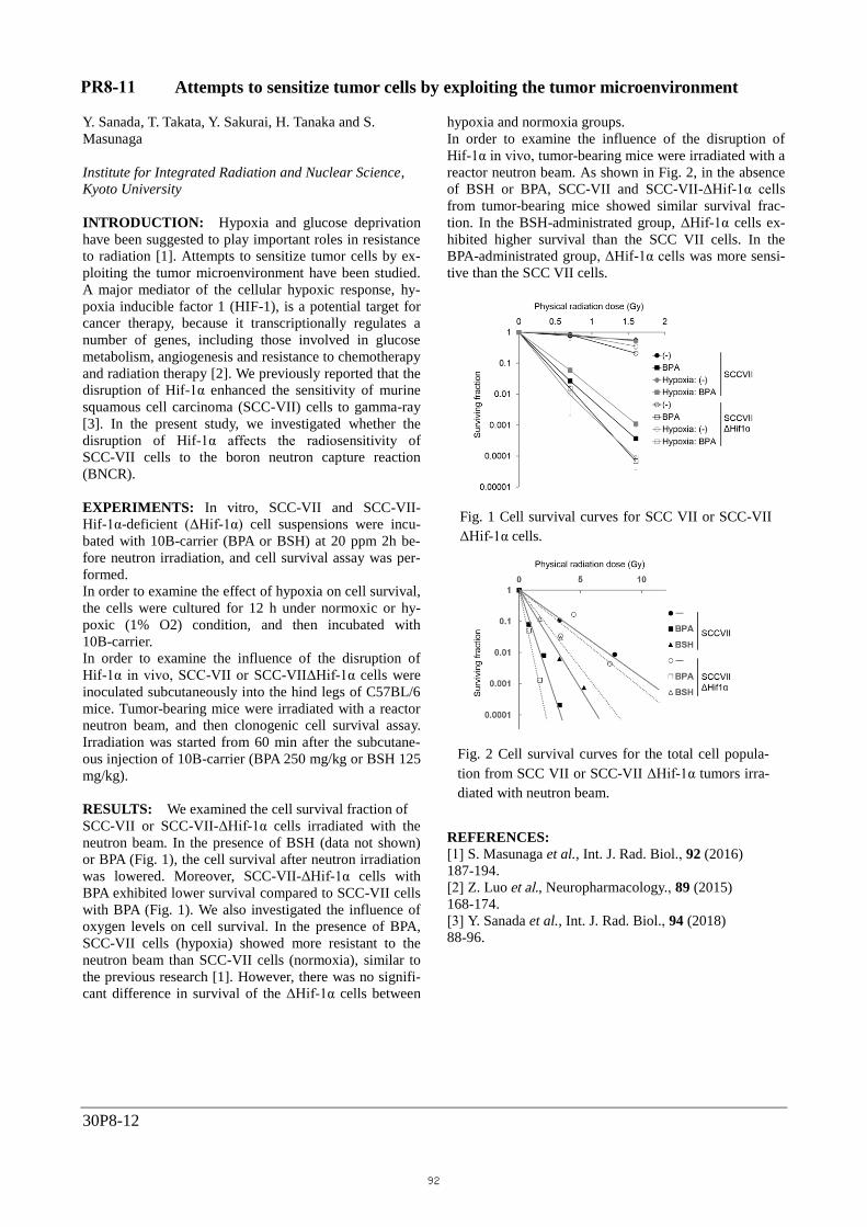
RESULTS: We examined the cell survival fraction of
SCC-VII or SCC-VII-ΔHif-1α cells irradiated with the
neutron beam. In the presence of BSH (data not shown)
or BPA (Fig. 1), the cell survival after neutron irradiation
was lowered. Moreover, SCC-VII-ΔHif-1α cells with
BPA exhibited lower survival compared to SCC-VII cells
with BPA (Fig. 1). We also investigated the influence of
oxygen levels on cell survival. In the presence of BPA,
SCC-VII cells (hypoxia) showed more resistant to the
neutron beam than SCC-VII cells (normoxia), similar to
the previous research [1]. However, there was no signifi-
cant difference in survival of the ΔHif-1α cells between
hypoxia and normoxia groups.
In order to examine the influence of the disruption of
Hif-1α in vivo, tumor-bearing mice were irradiated with a
reactor neutron beam. As shown in Fig. 2, in the absence
of BSH or BPA, SCC-VII and SCC-VII-ΔHif-1α cells
from tumor-bearing mice showed similar survival frac-
tion. In the BSH-administrated group, ΔHif-1α cells ex-
hibited higher survival than the SCC VII cells. In the
BPA-administrated group, ΔHif-1α cells was more sensi-
tive than the SCC VII cells.
REFERENCES:
[1] S. Masunaga et al., Int. J. Rad. Biol., 92 (2016)
187-194.
[2] Z. Luo et al., Neuropharmacology., 89 (2015)
168-174.
[3] Y. Sanada et al., Int. J. Rad. Biol., 94 (2018)
88-96.
Fig. 2 Cell survival curves for the total cell popula-
tion from SCC VII or SCC-VII ΔHif-1α tumors irra-
diated with neutron beam.
Fig. 1 Cell survival curves for SCC VII or SCC-VII
ΔHif-1α cells.
PR8-11
92
Related Documents


![Exploiting Asynchronous V2V Transmission for Sensing ... · waveform [e.g., frequency modulated continuous waveform (FMCW)] and analyzing its reflection by the object [19]. Particularly,](https://static.cupdf.com/doc/110x72/5b926aab09d3f206218b494f/exploiting-asynchronous-v2v-transmission-for-sensing-waveform-eg-frequency.jpg)


