BioMed Central Page 1 of 22 (page number not for citation purposes) BMC Neuroscience Open Access Research article Progranulin is expressed within motor neurons and promotes neuronal cell survival Cara L Ryan †1 , David C Baranowski †1,4 , Babykumari P Chitramuthu †1,4 , Suneil Malik 1 , Zhi Li 1 , Mingju Cao 1 , Sandra Minotti 2 , Heather D Durham 2 , Denis G Kay 4 , Christopher A Shaw 3 , Hugh PJ Bennett* 1 and Andrew Bateman 1 Address: 1 Endocrine Research Laboratory, Royal Victoria Hospital and Department of Medicine, McGill University Health Centre Research Institute, 687 Pine Avenue West, Montreal, Quebec, H3A 1A1, Canada, 2 Montreal Neurological Institute, McGill University, Montreal, Quebec, H3A 2B4, Canada, 3 University of British Columbia, Departments of Ophthalmology and Visual Sciences, and Experimental Medicine and Graduate Program in Neuroscience, Vancouver, British Columbia, V5Z 1L8, Canada and 4 Neurodyn Inc., Suite 508, NRC-INH, 550 University Avenue, Charlottetown, Prince Edward Island, C1A 4P3, Canada Email: Cara L Ryan - [email protected]; David C Baranowski - [email protected]; Babykumari P Chitramuthu - [email protected]; Suneil Malik - [email protected]; Zhi Li - [email protected]; Mingju Cao - [email protected]; Sandra Minotti - [email protected]; Heather D Durham - [email protected]; Denis G Kay - [email protected]; Christopher A Shaw - [email protected]; Hugh PJ Bennett* - [email protected]; Andrew Bateman - [email protected] * Corresponding author †Equal contributors Abstract Background: Progranulin is a secreted high molecular weight growth factor bearing seven and one half copies of the cysteine-rich granulin-epithelin motif. While inappropriate over-expression of the progranulin gene has been associated with many cancers, haploinsufficiency leads to atrophy of the frontotemporal lobes and development of a form of dementia (frontotemporal lobar degeneration with ubiquitin positive inclusions, FTLD-U) associated with the formation of ubiquitinated inclusions. Recent reports indicate that progranulin has neurotrophic effects, which, if confirmed would make progranulin the only neuroprotective growth factor that has been associated genetically with a neurological disease in humans. Preliminary studies indicated high progranulin gene expression in spinal cord motor neurons. However, it is uncertain what the role of Progranulin is in normal or diseased motor neuron function. We have investigated progranulin gene expression and subcellular localization in cultured mouse embryonic motor neurons and examined the effect of progranulin over-expression and knockdown in the NSC-34 immortalized motor neuron cell line upon proliferation and survival. Results: In situ hybridisation and immunohistochemical techniques revealed that the progranulin gene is highly expressed by motor neurons within the mouse spinal cord and in primary cultures of dissociated mouse embryonic spinal cord-dorsal root ganglia. Confocal microscopy coupled to immunocytochemistry together with the use of a progranulin-green fluorescent protein fusion construct revealed progranulin to be located within compartments of the secretory pathway including the Golgi apparatus. Stable transfection of the human progranulin gene into the NSC-34 motor neuron cell line stimulates the appearance of dendritic structures and provides sufficient Published: 27 October 2009 BMC Neuroscience 2009, 10:130 doi:10.1186/1471-2202-10-130 Received: 19 June 2009 Accepted: 27 October 2009 This article is available from: http://www.biomedcentral.com/1471-2202/10/130 © 2009 Ryan et al; licensee BioMed Central Ltd. This is an Open Access article distributed under the terms of the Creative Commons Attribution License (http://creativecommons.org/licenses/by/2.0 ), which permits unrestricted use, distribution, and reproduction in any medium, provided the original work is properly cited.

Welcome message from author
This document is posted to help you gain knowledge. Please leave a comment to let me know what you think about it! Share it to your friends and learn new things together.
Transcript
BioMed CentralBMC Neuroscience
ss
Open AcceResearch articleProgranulin is expressed within motor neurons and promotes neuronal cell survivalCara L Ryan†1, David C Baranowski†1,4, Babykumari P Chitramuthu†1,4, Suneil Malik1, Zhi Li1, Mingju Cao1, Sandra Minotti2, Heather D Durham2, Denis G Kay4, Christopher A Shaw3, Hugh PJ Bennett*1 and Andrew Bateman1Address: 1Endocrine Research Laboratory, Royal Victoria Hospital and Department of Medicine, McGill University Health Centre Research Institute, 687 Pine Avenue West, Montreal, Quebec, H3A 1A1, Canada, 2Montreal Neurological Institute, McGill University, Montreal, Quebec, H3A 2B4, Canada, 3University of British Columbia, Departments of Ophthalmology and Visual Sciences, and Experimental Medicine and Graduate Program in Neuroscience, Vancouver, British Columbia, V5Z 1L8, Canada and 4Neurodyn Inc., Suite 508, NRC-INH, 550 University Avenue, Charlottetown, Prince Edward Island, C1A 4P3, Canada
Email: Cara L Ryan - [email protected]; David C Baranowski - [email protected]; Babykumari P Chitramuthu - [email protected]; Suneil Malik - [email protected]; Zhi Li - [email protected]; Mingju Cao - [email protected]; Sandra Minotti - [email protected]; Heather D Durham - [email protected]; Denis G Kay - [email protected]; Christopher A Shaw - [email protected]; Hugh PJ Bennett* - [email protected]; Andrew Bateman - [email protected]
* Corresponding author †Equal contributors
AbstractBackground: Progranulin is a secreted high molecular weight growth factor bearing seven andone half copies of the cysteine-rich granulin-epithelin motif. While inappropriate over-expressionof the progranulin gene has been associated with many cancers, haploinsufficiency leads to atrophyof the frontotemporal lobes and development of a form of dementia (frontotemporal lobardegeneration with ubiquitin positive inclusions, FTLD-U) associated with the formation ofubiquitinated inclusions. Recent reports indicate that progranulin has neurotrophic effects, which,if confirmed would make progranulin the only neuroprotective growth factor that has beenassociated genetically with a neurological disease in humans. Preliminary studies indicated highprogranulin gene expression in spinal cord motor neurons. However, it is uncertain what the roleof Progranulin is in normal or diseased motor neuron function. We have investigated progranulingene expression and subcellular localization in cultured mouse embryonic motor neurons andexamined the effect of progranulin over-expression and knockdown in the NSC-34 immortalizedmotor neuron cell line upon proliferation and survival.
Results: In situ hybridisation and immunohistochemical techniques revealed that the progranulingene is highly expressed by motor neurons within the mouse spinal cord and in primary culturesof dissociated mouse embryonic spinal cord-dorsal root ganglia. Confocal microscopy coupled toimmunocytochemistry together with the use of a progranulin-green fluorescent protein fusionconstruct revealed progranulin to be located within compartments of the secretory pathwayincluding the Golgi apparatus. Stable transfection of the human progranulin gene into the NSC-34motor neuron cell line stimulates the appearance of dendritic structures and provides sufficient
Published: 27 October 2009
BMC Neuroscience 2009, 10:130 doi:10.1186/1471-2202-10-130
Received: 19 June 2009Accepted: 27 October 2009
This article is available from: http://www.biomedcentral.com/1471-2202/10/130
© 2009 Ryan et al; licensee BioMed Central Ltd. This is an Open Access article distributed under the terms of the Creative Commons Attribution License (http://creativecommons.org/licenses/by/2.0), which permits unrestricted use, distribution, and reproduction in any medium, provided the original work is properly cited.
Page 1 of 22(page number not for citation purposes)
BMC Neuroscience 2009, 10:130 http://www.biomedcentral.com/1471-2202/10/130
trophic stimulus to survive serum deprivation for long periods (up to two months). This is mediatedat least in part through an anti-apoptotic mechanism. Control cells, while expressing basal levels ofprogranulin do not survive in serum free conditions. Knockdown of progranulin expression usingshRNA technology further reduced cell survival.
Conclusion: Neurons are among the most long-lived cells in the body and are subject to low levelsof toxic challenges throughout life. We have demonstrated that progranulin is abundantlyexpressed in motor neurons and is cytoprotective over prolonged periods when over-expressedin a neuronal cell line. This work highlights the importance of progranulin as neuroprotectivegrowth factor and may represent a therapeutic target for neurodegenerative diseases includingmotor neuron disease.
BackgroundThe granulin-epithelin precursor, progranulin (PGRN)[1], also called proepithelin [2], PC-cell-derived growthfactor [3], or acrogranin [4], is a secreted glycoprotein thatpromotes mitosis, survival, and migration in many celltypes [5,6]. Recent work demonstrates that haploinsuffi-ciency of the PGRN gene causes a form of frontotemporallobar degeneration (FTLD) that is associated with the for-mation of ubiquitinated inclusions (FTLD-U) [7-9]. Sev-eral studies have shown that ubiquitinated Tar-DNABinding Protein 43 (TDP-43) is a component of inclusionbodies in both FTLD-U and Amyotrophic Lateral Sclerosis(ALS) [10-13] although other ubiquitinated proteins arealso present in these inclusion bodies in ALS [14]. TDP-43translocates from the nucleus to the cytoplasm in axot-omized motor neurons which is consistent with a role forTDP43 in the normal response of motor neurons to injury[15]. The depletion of PGRN in H4 gliomas results in theactivation of caspase-3 and the accumulation of cleavedTDP-43 [16]. This is suggestive of a functional relation-ship between the loss of PGRN and mobilization of TDP-43, although this conclusion has been challenged by otherinvestigators [17,18]. While PGRN is secreted by manycell types it has been suggested that in neurons its subcel-lular distribution resembles that of mitochondria or lyso-somal-endosomal markers [19].
PGRN is synthesised in neurons in many brain regionsincluding the cerebral cortex, in the Purkinje cells of thecerebellum, and in the hippocampus [20]. In addition, itis widely distributed in the developing central nervoussystem and the dorsal root and sympathetic gangliawithin the peripheral nervous system [21]. The roles ofPGRN in normal neuronal function and development, ineither the central or peripheral nervous systems are poorlyunderstood. It is known, however, that PGRN contributesto normal brain development since it regulates the male-specific differentiation of the neonatal hypothalamus[22,23]. Moreover, in culture, PGRN stimulates the prolif-eration of PC12 cells [20], as well as the estrogen-depend-ent growth of hippocampal neurospheres [24] and maybe neurotrophic for cortical and motor neurons [25].
The signalling pathways associated with PGRN in neuronsare unknown, but in non-neuronal cell types it activatesgrowth factor-related signal transduction pathwaysincluding the phosphorylation of shc, p44/42 mitogen-activated protein kinase, phosphatidylinositol 3-kinase,protein kinase B/AKT, and the p70S6 kinase [26-28], and,by so doing, contributes to carcinogenesis in numeroustumour types [29-39]. PGRN is involved in wound repairand inflammation [40-43], and plays an important role inearly embryonic development [44-46]. The ability ofPGRN to regulate critical proliferative, survival and motil-ity signals in a diverse range of non-neuronal cell typessuggests that it may support similar functions in nervecells.
In situ hybridization experiments (see below) revealedthat motor neurons express a high level of PGRN, suggest-ing a significant role for PGRN in the biology of the motorneuron. The significance of PGRN in healthy and diseasedmotor neurons is, however, unclear. PGRN expressionwas markedly up-regulated in spinal cord tissue frompatients who had ALS [47]. While this was probably dueto gliosis [47], increased immunoreactive staining forPGRN has been reported both in motor neurons and glialcells in spinal cord and brainstem tissue sections derivedfrom ALS patients relative to controls [48]. Furthermore,PGRN expression is increased in the lumbar cord motorneurons from murine models of familial ALS that iscaused by mutations in superoxide dismutase 1 (SOD1)[49,50]. PGRN was one of only 21 genes with dysregu-lated motor neuron expression in SOD1G37R andSOD1G85R mice at the onset of the disease, as judged byweight loss but before the detection of overt neurologicalsymptoms [49]. The PGRN gene was also up-regulated inSODG93A mice, but late in the disease progression [50]. Incontrast, a decrease in PGRN expression was noted in theNSC-34 neuronal cell line engineered to express SODG93A
[51]. It has been suggested that the PGRN gene may be amodulator of disease progression in ALS since a correla-tion between genetic variations in PGRN and the age ofonset or survival of patients with ALS has been reported[52]. This observation was not, however, reproduced by
Page 2 of 22(page number not for citation purposes)
BMC Neuroscience 2009, 10:130 http://www.biomedcentral.com/1471-2202/10/130
other investigators [53-55]. In some patients FTLD may beaccompanied by motor neuron disease [56,57], but muta-tions in PGRN are infrequent in patients with ALS-FTLDdefects [54,58-60], and for those that have been reportedit is uncertain whether or not they are pathogenic [53,61].
Growth factors, in particular vascular endothelial growthfactor [62] and insulin-like growth factor-1 [63], haveshown promise as neuroprotective agents in murine mod-els of familial ALS and if PGRN is neurotrophic for motorneurons [25] it could also have therapeutic potential inALS. Given the uncertainty of whether or not PGRN con-tributes to motor neuron function and survival, we soughtfirst to characterize the expression of PGRN in the healthybrain, spinal cord and dorsal root ganglia, and then toinvestigate its possible biological activities in an immor-talized motor neuron cell model.
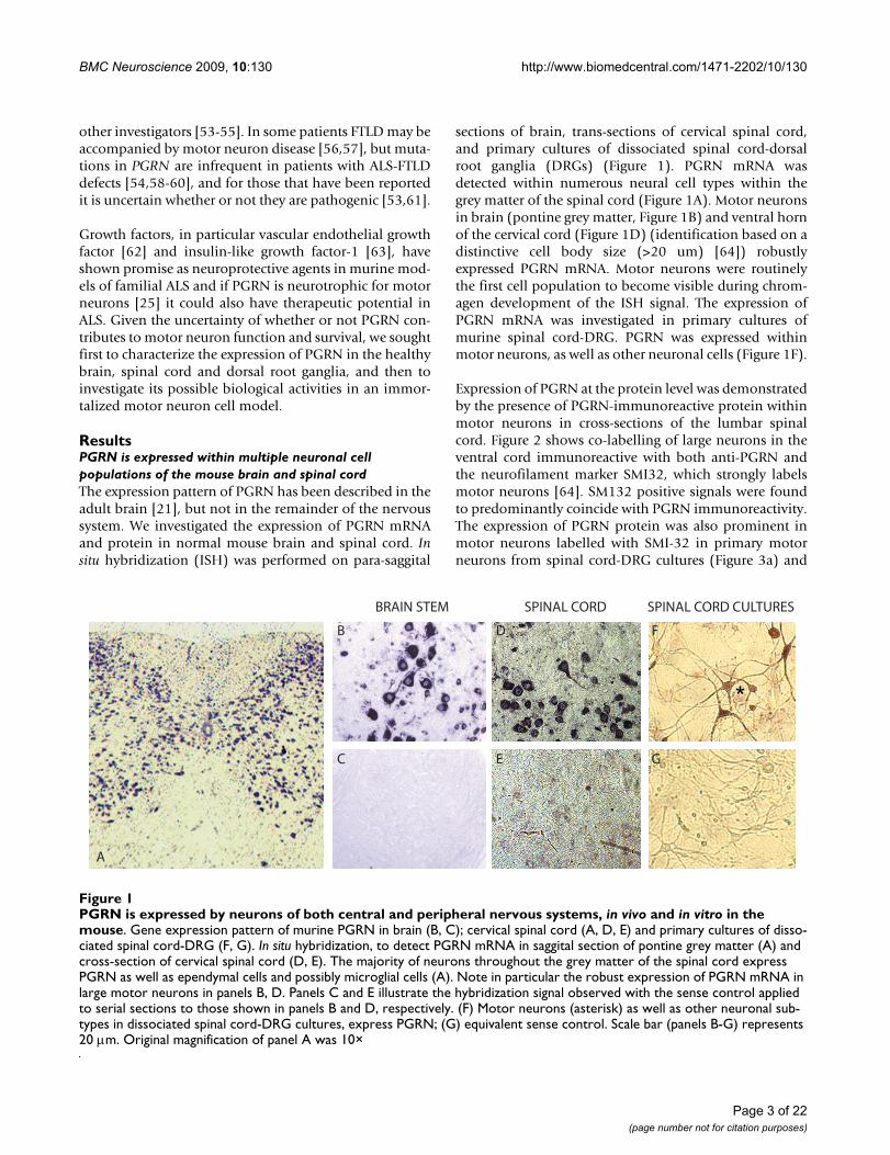
ResultsPGRN is expressed within multiple neuronal cell populations of the mouse brain and spinal cordThe expression pattern of PGRN has been described in theadult brain [21], but not in the remainder of the nervoussystem. We investigated the expression of PGRN mRNAand protein in normal mouse brain and spinal cord. Insitu hybridization (ISH) was performed on para-saggital
sections of brain, trans-sections of cervical spinal cord,and primary cultures of dissociated spinal cord-dorsalroot ganglia (DRGs) (Figure 1). PGRN mRNA wasdetected within numerous neural cell types within thegrey matter of the spinal cord (Figure 1A). Motor neuronsin brain (pontine grey matter, Figure 1B) and ventral hornof the cervical cord (Figure 1D) (identification based on adistinctive cell body size (>20 um) [64]) robustlyexpressed PGRN mRNA. Motor neurons were routinelythe first cell population to become visible during chrom-agen development of the ISH signal. The expression ofPGRN mRNA was investigated in primary cultures ofmurine spinal cord-DRG. PGRN was expressed withinmotor neurons, as well as other neuronal cells (Figure 1F).
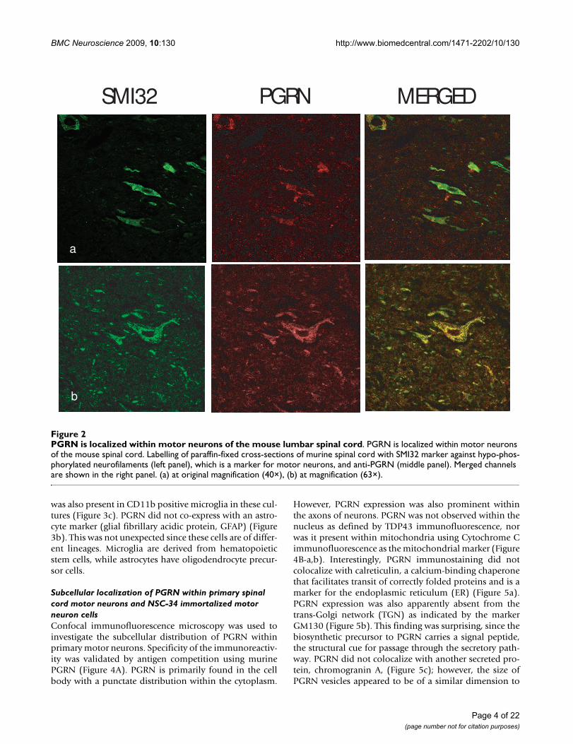
Expression of PGRN at the protein level was demonstratedby the presence of PGRN-immunoreactive protein withinmotor neurons in cross-sections of the lumbar spinalcord. Figure 2 shows co-labelling of large neurons in theventral cord immunoreactive with both anti-PGRN andthe neurofilament marker SMI32, which strongly labelsmotor neurons [64]. SM132 positive signals were foundto predominantly coincide with PGRN immunoreactivity.The expression of PGRN protein was also prominent inmotor neurons labelled with SMI-32 in primary motorneurons from spinal cord-DRG cultures (Figure 3a) and
PGRN is expressed by neurons of both central and peripheral nervous systems, in vivo and in vitro in the mouseFigure 1PGRN is expressed by neurons of both central and peripheral nervous systems, in vivo and in vitro in the mouse. Gene expression pattern of murine PGRN in brain (B, C); cervical spinal cord (A, D, E) and primary cultures of disso-ciated spinal cord-DRG (F, G). In situ hybridization, to detect PGRN mRNA in saggital section of pontine grey matter (A) and cross-section of cervical spinal cord (D, E). The majority of neurons throughout the grey matter of the spinal cord express PGRN as well as ependymal cells and possibly microglial cells (A). Note in particular the robust expression of PGRN mRNA in large motor neurons in panels B, D. Panels C and E illustrate the hybridization signal observed with the sense control applied to serial sections to those shown in panels B and D, respectively. (F) Motor neurons (asterisk) as well as other neuronal sub-types in dissociated spinal cord-DRG cultures, express PGRN; (G) equivalent sense control. Scale bar (panels B-G) represents 20 μm. Original magnification of panel A was 10×
A
B
C
D
E
F
G
BRAIN STEM SPINAL CORD SPINAL CORD CULTURES
Page 3 of 22(page number not for citation purposes)
BMC Neuroscience 2009, 10:130 http://www.biomedcentral.com/1471-2202/10/130
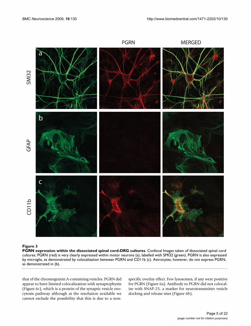
was also present in CD11b positive microglia in these cul-tures (Figure 3c). PGRN did not co-express with an astro-cyte marker (glial fibrillary acidic protein, GFAP) (Figure3b). This was not unexpected since these cells are of differ-ent lineages. Microglia are derived from hematopoieticstem cells, while astrocytes have oligodendrocyte precur-sor cells.
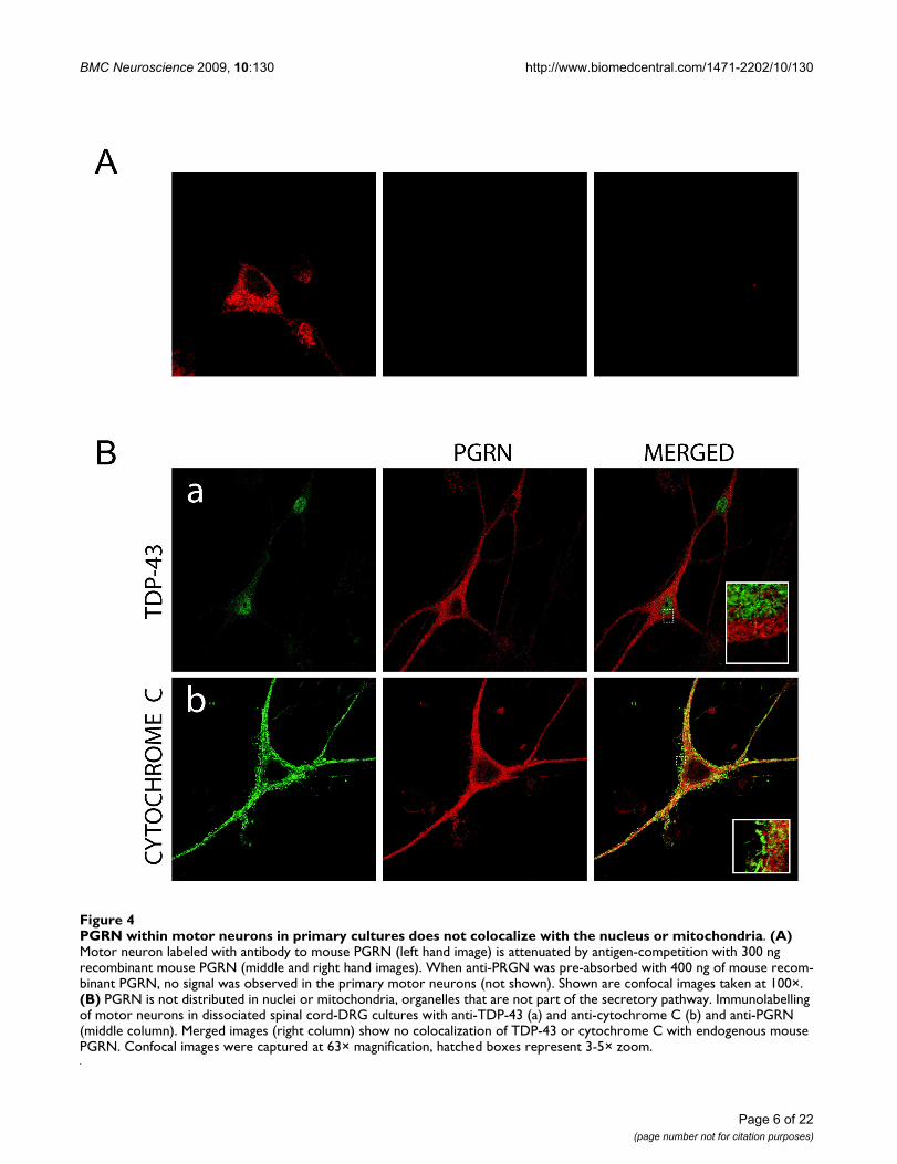
Subcellular localization of PGRN within primary spinal cord motor neurons and NSC-34 immortalized motor neuron cellsConfocal immunofluorescence microscopy was used toinvestigate the subcellular distribution of PGRN withinprimary motor neurons. Specificity of the immunoreactiv-ity was validated by antigen competition using murinePGRN (Figure 4A). PGRN is primarily found in the cellbody with a punctate distribution within the cytoplasm.
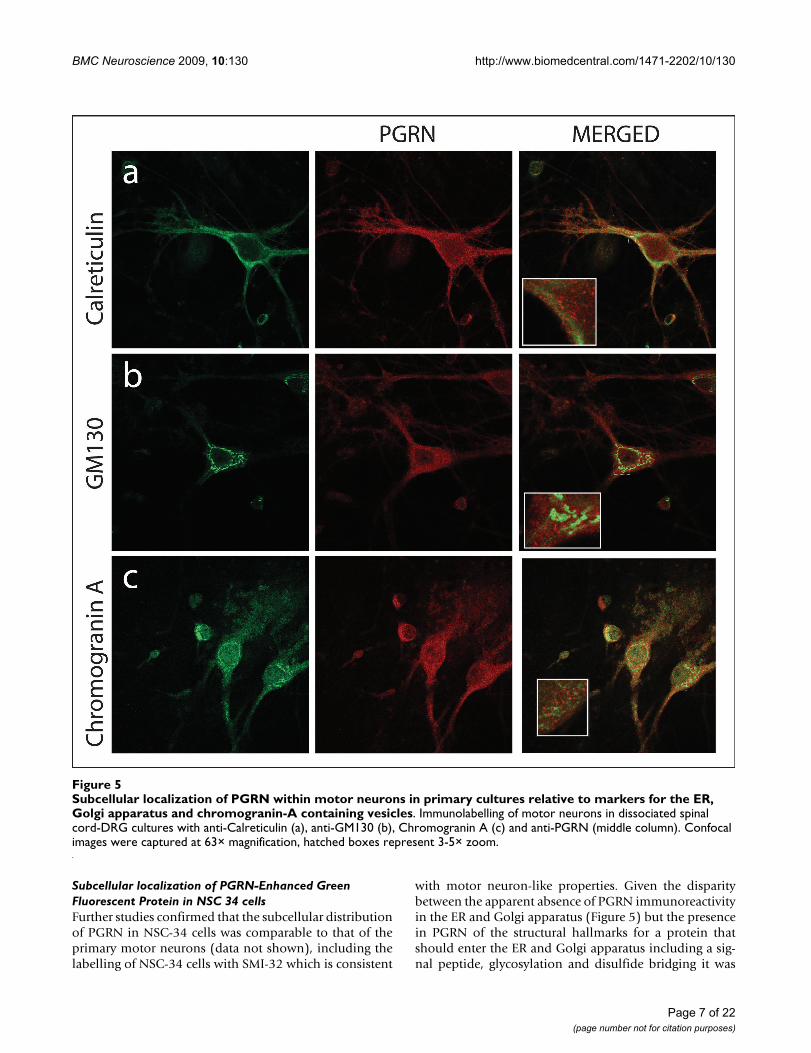
However, PGRN expression was also prominent withinthe axons of neurons. PGRN was not observed within thenucleus as defined by TDP43 immunofluorescence, norwas it present within mitochondria using Cytochrome Cimmunofluorescence as the mitochondrial marker (Figure4B-a,b). Interestingly, PGRN immunostaining did notcolocalize with calreticulin, a calcium-binding chaperonethat facilitates transit of correctly folded proteins and is amarker for the endoplasmic reticulum (ER) (Figure 5a).PGRN expression was also apparently absent from thetrans-Golgi network (TGN) as indicated by the markerGM130 (Figure 5b). This finding was surprising, since thebiosynthetic precursor to PGRN carries a signal peptide,the structural cue for passage through the secretory path-way. PGRN did not colocalize with another secreted pro-tein, chromogranin A, (Figure 5c); however, the size ofPGRN vesicles appeared to be of a similar dimension to
PGRN is localized within motor neurons of the mouse lumbar spinal cordFigure 2PGRN is localized within motor neurons of the mouse lumbar spinal cord. PGRN is localized within motor neurons of the mouse spinal cord. Labelling of paraffin-fixed cross-sections of murine spinal cord with SMI32 marker against hypo-phos-phorylated neurofilaments (left panel), which is a marker for motor neurons, and anti-PGRN (middle panel). Merged channels are shown in the right panel. (a) at original magnification (40×), (b) at magnification (63×).
SMI32 PGRN MERGED
a B
b
Page 4 of 22(page number not for citation purposes)
BMC Neuroscience 2009, 10:130 http://www.biomedcentral.com/1471-2202/10/130
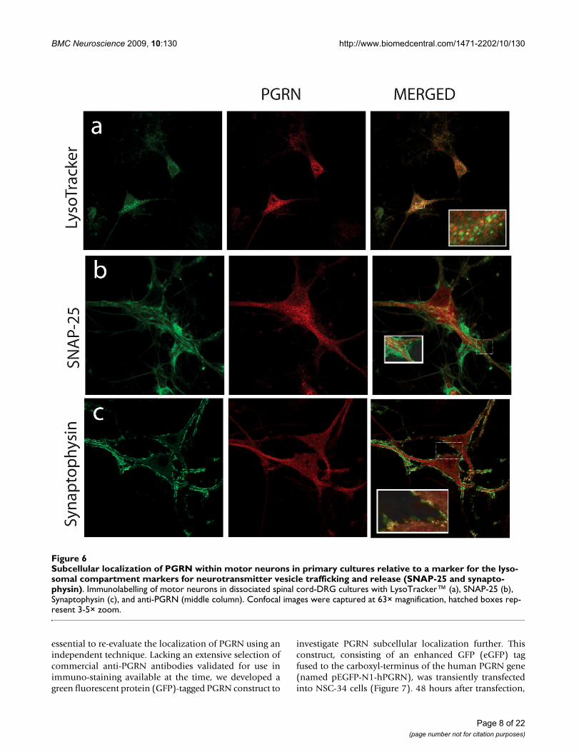
that of the chromogranin A-containing vesicles. PGRN didappear to have limited colocalization with synaptophysin(Figure 6c), which is a protein of the synaptic vesicle exo-cytosis pathway although at the resolution available wecannot exclude the possibility that this is due to a non-
specific overlay effect. Few lysosomes, if any were positivefor PGRN (Figure 6a). Antibody to PGRN did not colocal-ize with SNAP-25, a marker for neurotransmitter vesicledocking and release sites (Figure 6b).
PGRN expression within the dissociated spinal cord-DRG culturesFigure 3PGRN expression within the dissociated spinal cord-DRG cultures. Confocal Images taken of dissociated spinal cord cultures. PGRN (red) is very clearly expressed within motor neurons (a), labelled with SMI32 (green). PGRN is also expressed by microglia, as demonstrated by colocalization between PGRN and CD11b (c). Astrocytes, however, do not express PGRN, as demonstrated in (b).
Page 5 of 22(page number not for citation purposes)
BMC Neuroscience 2009, 10:130 http://www.biomedcentral.com/1471-2202/10/130
Page 6 of 22(page number not for citation purposes)
PGRN within motor neurons in primary cultures does not colocalize with the nucleus or mitochondriaFigure 4PGRN within motor neurons in primary cultures does not colocalize with the nucleus or mitochondria. (A) Motor neuron labeled with antibody to mouse PGRN (left hand image) is attenuated by antigen-competition with 300 ng recombinant mouse PGRN (middle and right hand images). When anti-PRGN was pre-absorbed with 400 ng of mouse recom-binant PGRN, no signal was observed in the primary motor neurons (not shown). Shown are confocal images taken at 100×. (B) PGRN is not distributed in nuclei or mitochondria, organelles that are not part of the secretory pathway. Immunolabelling of motor neurons in dissociated spinal cord-DRG cultures with anti-TDP-43 (a) and anti-cytochrome C (b) and anti-PGRN (middle column). Merged images (right column) show no colocalization of TDP-43 or cytochrome C with endogenous mouse PGRN. Confocal images were captured at 63× magnification, hatched boxes represent 3-5× zoom.
BMC Neuroscience 2009, 10:130 http://www.biomedcentral.com/1471-2202/10/130
Subcellular localization of PGRN-Enhanced Green Fluorescent Protein in NSC 34 cellsFurther studies confirmed that the subcellular distributionof PGRN in NSC-34 cells was comparable to that of theprimary motor neurons (data not shown), including thelabelling of NSC-34 cells with SMI-32 which is consistent
with motor neuron-like properties. Given the disparitybetween the apparent absence of PGRN immunoreactivityin the ER and Golgi apparatus (Figure 5) but the presencein PGRN of the structural hallmarks for a protein thatshould enter the ER and Golgi apparatus including a sig-nal peptide, glycosylation and disulfide bridging it was
Subcellular localization of PGRN within motor neurons in primary cultures relative to markers for the ER, Golgi apparatus and chromogranin-A containing vesiclesFigure 5Subcellular localization of PGRN within motor neurons in primary cultures relative to markers for the ER, Golgi apparatus and chromogranin-A containing vesicles. Immunolabelling of motor neurons in dissociated spinal cord-DRG cultures with anti-Calreticulin (a), anti-GM130 (b), Chromogranin A (c) and anti-PGRN (middle column). Confocal images were captured at 63× magnification, hatched boxes represent 3-5× zoom.
Page 7 of 22(page number not for citation purposes)
BMC Neuroscience 2009, 10:130 http://www.biomedcentral.com/1471-2202/10/130
essential to re-evaluate the localization of PGRN using anindependent technique. Lacking an extensive selection ofcommercial anti-PGRN antibodies validated for use inimmuno-staining available at the time, we developed agreen fluorescent protein (GFP)-tagged PGRN construct to
investigate PGRN subcellular localization further. Thisconstruct, consisting of an enhanced GFP (eGFP) tagfused to the carboxyl-terminus of the human PGRN gene(named pEGFP-N1-hPGRN), was transiently transfectedinto NSC-34 cells (Figure 7). 48 hours after transfection,
Subcellular localization of PGRN within motor neurons in primary cultures relative to a marker for the lysosomal compart-ment markers for neurotransmitter vesicle trafficking and release (SNAP-25 and synaptophysin)Figure 6Subcellular localization of PGRN within motor neurons in primary cultures relative to a marker for the lyso-somal compartment markers for neurotransmitter vesicle trafficking and release (SNAP-25 and synapto-physin). Immunolabelling of motor neurons in dissociated spinal cord-DRG cultures with LysoTracker™ (a), SNAP-25 (b), Synaptophysin (c), and anti-PGRN (middle column). Confocal images were captured at 63× magnification, hatched boxes rep-resent 3-5× zoom.
Page 8 of 22(page number not for citation purposes)
BMC Neuroscience 2009, 10:130 http://www.biomedcentral.com/1471-2202/10/130
confocal microscopy of the cells demonstrated successfultransfection, and a granule-like appearance for the GreenFluorescent Protein signal that was very different fromthat of control empty eGFP vector-transfected cells inwhich eGFP vector expression was more nuclear (Figure8c). The pEGFP-N1-hPGRN expressing cells demonstratedclear colocalization of EGFP-PGRN within the TGN (Fig-ure 8a). Furthermore, PGRN distribution was distinctfrom that of the mitochondria (Figure 8b). The presenceof the eGFP-tagged PGRN in the Golgi apparatus is notsecondary to PGRN over-expression since when we over-expressed untagged PGRN in NSC34 cells, as with the pri-mary motor neurons (Figure 5), no untagged-PGRNimmunoreactivity was detected in the Golgi apparatus(data not shown).
Over-expression of PGRN in NSC-34 cells promotes a neuron-like morphologyThe cell line NSC-34 was established by fusing embryonicspinal cord cells with neuroblastoma cells and in the dif-ferentiated state are reported to exhibit motor neuronalproperties [65,66]. They are frequently used to investigateneuroprotective processes and to model motor neurondegeneration in vitro [51,67-72]. In culture most NSC-34cells exhibit a rounded and undifferentiated morphology;however, serum-deprivation stimulates many of the cellsto undergo differentiation and to extend neurite-like pro-jections [66,73]. Serum-deprivation is also associatedwith significant apoptosis of the non-differentiated cells[73]. NSC-34 cells that express human PGRN were estab-lished by transfection and selected for stable incorpora-tion of the human PGRN gene. Previous work haddemonstrated that human PGRN is mitogenic for murinecells [28]. Untransfected NSC-34 cells, NSC-34 transfectedwith empty PCDNA3 vector (NSC-34/vector), and NSC-34 cells that were transfected with human PGRN cDNAsubcloned into pcDNA3 (NSC-34/PGRN) continued to
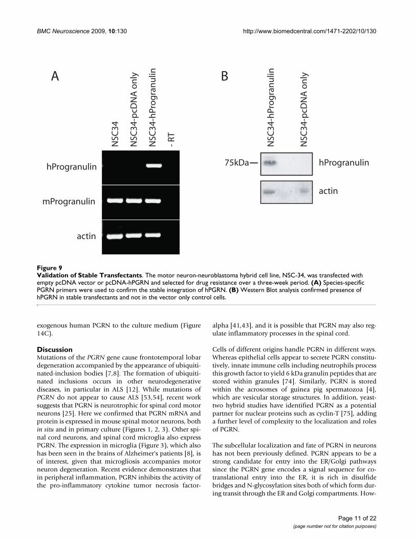
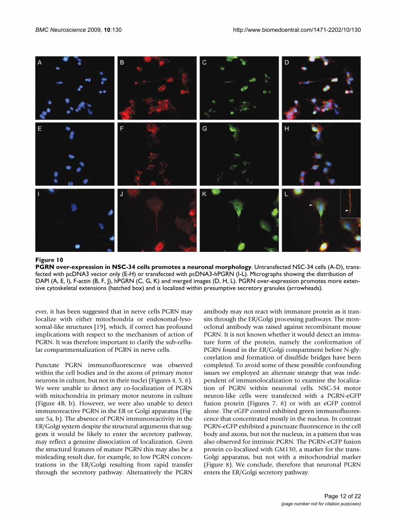
express equivalent levels of murine PGRN mRNA, whileonly the NSC-34/PGRN cells express the human PGRNmRNA (Figure 9A). Over-expression of PGRN protein wasconfirmed by Western blot analysis (Figure 9B). In NSC-34/PGRN cells, PGRN punctate immunoreactivity wasfound throughout the cell body and within the projec-tions (Figure 10). Over-expression of PGRN was associ-ated with morphological changes, such as flattening ofcell shape and more pronounced neurite-like extensions(Figure 10).
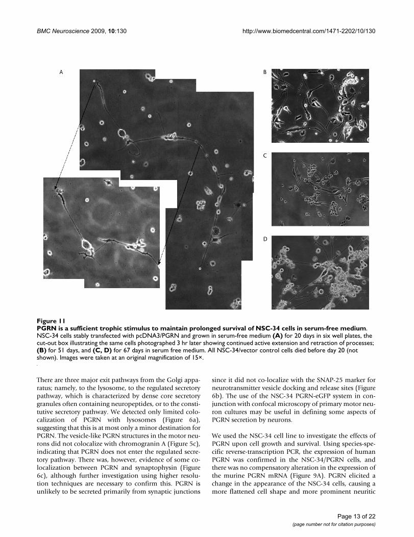
PGRN over-expression promotes cell survivalTo investigate the longevity of NSC-34/PGRN cells, 2independent cultures were propagated for 2 months in theabsence of serum. Within 20 days no NSC-34/vector cellssurvived (not shown), however many NSC-34/PGRN cellsremained alive, retaining a mixed morphology of undif-ferentiated cells and cells with complex neurite-like pro-jections [Figure 11A, B, C]. Cells with neuron-likemorphology remained viable at least for 2 months in theabsence of serum [Figure 11C, D]. At day 20, when com-pared over short intervals (3 hrs) the neuron-like NSC-34/PGRN cells were changing morphologically, continuingto elaborate and elongate neuritic processes [Figure 11A].
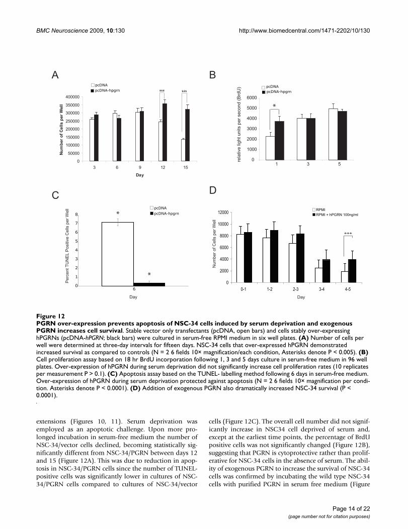
Between 12 and 15 days following the removal of serum,cell number significantly decreased in NSC-34/vector cul-tures, while cultures of NSC-34/PGRN cells showed nosuch change (Figure 12A). The absence of extensive prolif-eration in NSC34-PGRN cells cultured in serum freemedium was further confirmed using a BrdU incorpora-tion assay. No significant difference in BrdU incorpora-tion between the NSC-34/vector and NSC-34/PGRN cellswas observed on days 3 and 5. (Figure 12B). There was,however, a significant increase in TUNEL-positive (i.e.apoptosing) cells in the NSC-34/vector cultures versus theNSC-34/PGRN cultures (Figure 12C). The addition of
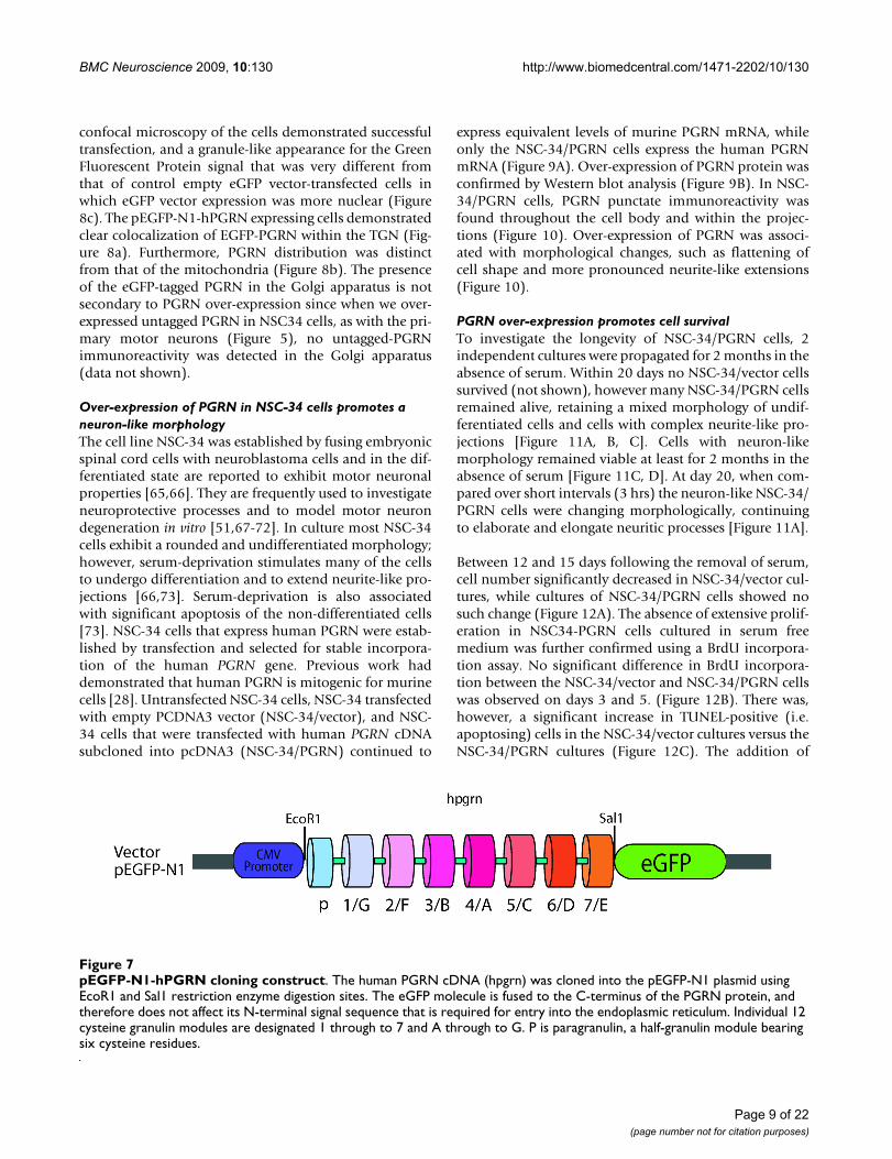
pEGFP-N1-hPGRN cloning constructFigure 7pEGFP-N1-hPGRN cloning construct. The human PGRN cDNA (hpgrn) was cloned into the pEGFP-N1 plasmid using EcoR1 and Sal1 restriction enzyme digestion sites. The eGFP molecule is fused to the C-terminus of the PGRN protein, and therefore does not affect its N-terminal signal sequence that is required for entry into the endoplasmic reticulum. Individual 12 cysteine granulin modules are designated 1 through to 7 and A through to G. P is paragranulin, a half-granulin module bearing six cysteine residues.
Page 9 of 22(page number not for citation purposes)

BMC Neuroscience 2009, 10:130 http://www.biomedcentral.com/1471-2202/10/130
exogenous recombinant human PGRN to the mediumalso resulted in an enhanced cell survival that became evi-dent at day 3 of the incubation (Figure 12D).
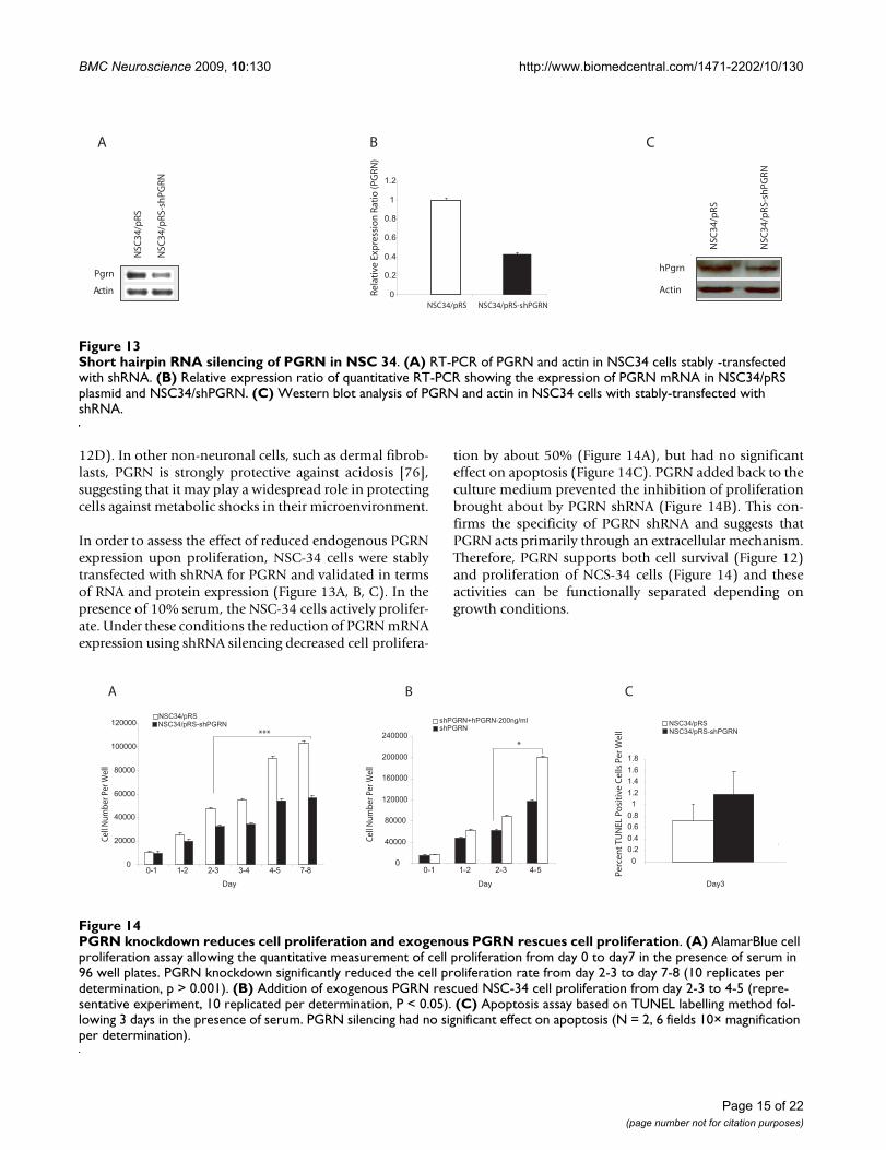
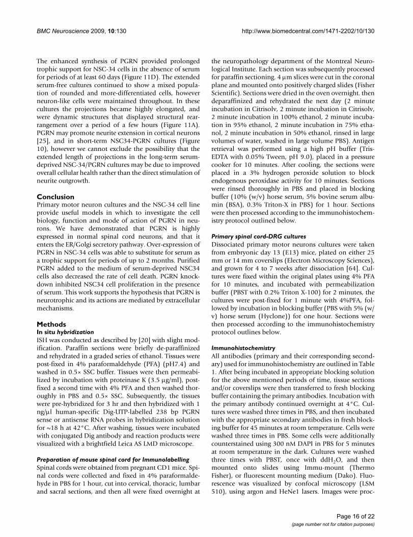
The stable expression of an shPGRN construct reducedPGRN mRNA levels by approximately 50% (Figure 13A,B). Consistent with the qRT-PCR data, significantdecreases in the level of PGRN protein was observed in the
shRNA expressing cells as confirmed by Western blot anal-ysis (Figure 13C). When the shPGRN cells were culturedin serum-containing medium, cell proliferation was sig-nificantly reduced relative to the vector control cells (Fig-ure 14A) but there was no statistical change in theproportion of apoptotic cells (Figure 14B). The reducedproliferation of shPGRN was reversed by the addition of
Transfection of pEGFP-N1-hPGRN plasmid shows granular morphology and particular PGRN localization in GolgiFigure 8Transfection of pEGFP-N1-hPGRN plasmid shows granular morphology and particular PGRN localization in Golgi. The overlap seen with PGRN and the Golgi marker GM130 (a) is very prominent, while there is no overlap between mitochondria (b) and PGRN. Panel (c) reveals the pEGFP-N1 vector only transfection, having primarily nuclear GFP signal, and a very different subcellular distribution of PGRN, which appears distinctly granular in panels (a and b). Confocal images were taken at 63×.
MERGEDpEGFP-N1-hPGRNp
EGFP
-N1
CO
NTR
OL
CY
TOC
HRO
ME
CG
M13
0
a
b
c
Page 10 of 22(page number not for citation purposes)
BMC Neuroscience 2009, 10:130 http://www.biomedcentral.com/1471-2202/10/130
exogenous human PGRN to the culture medium (Figure14C).
DiscussionMutations of the PGRN gene cause frontotemporal lobardegeneration accompanied by the appearance of ubiquiti-nated-inclusion bodies [7,8]. The formation of ubiquiti-nated inclusions occurs in other neurodegenerativediseases, in particular in ALS [12]. While mutations ofPGRN do not appear to cause ALS [53,54], recent worksuggests that PGRN is neurotrophic for spinal cord motorneurons [25]. Here we confirmed that PGRN mRNA andprotein is expressed in mouse spinal motor neurons, bothin situ and in primary culture (Figures 1, 2, 3). Other spi-nal cord neurons, and spinal cord microglia also expressPGRN. The expression in microglia (Figure 3), which alsohas been seen in the brains of Alzheimer's patients [8], isof interest, given that microgliosis accompanies motorneuron degeneration. Recent evidence demonstrates thatin peripheral inflammation, PGRN inhibits the activity ofthe pro-inflammatory cytokine tumor necrosis factor-
alpha [41,43], and it is possible that PGRN may also reg-ulate inflammatory processes in the spinal cord.
Cells of different origins handle PGRN in different ways.Whereas epithelial cells appear to secrete PGRN constitu-tively, innate immune cells including neutrophils processthis growth factor to yield 6 kDa granulin peptides that arestored within granules [74]. Similarly, PGRN is storedwithin the acrosomes of guinea pig spermatozoa [4],which are vesicular storage structures. In addition, yeast-two hybrid studies have identified PGRN as a potentialpartner for nuclear proteins such as cyclin-T [75], addinga further level of complexity to the localization and rolesof PGRN.
The subcellular localization and fate of PGRN in neuronshas not been previously defined. PGRN appears to be astrong candidate for entry into the ER/Golgi pathwayssince the PGRN gene encodes a signal sequence for co-translational entry into the ER, it is rich in disulfidebridges and N-glycosylation sites both of which form dur-ing transit through the ER and Golgi compartments. How-
Validation of Stable TransfectantsFigure 9Validation of Stable Transfectants. The motor neuron-neuroblastoma hybrid cell line, NSC-34, was transfected with empty pcDNA vector or pcDNA-hPGRN and selected for drug resistance over a three-week period. (A) Species-specific PGRN primers were used to confirm the stable integration of hPGRN. (B) Western Blot analysis confirmed presence of hPGRN in stable transfectants and not in the vector only control cells.
NSC
34-h
Pro
gra
nu
lin
NSC
34
NSC
34-p
cDN
A o
nly
- RT
hProgranulin
mProgranulin
actin
hProgranulin
actin
NSC
34-h
Pro
gra
nu
lin
NSC
34-p
cDN
A o
nly
75kDa
A B
Page 11 of 22(page number not for citation purposes)
BMC Neuroscience 2009, 10:130 http://www.biomedcentral.com/1471-2202/10/130
ever, it has been suggested that in nerve cells PGRN maylocalize with either mitochondria or endosomal-lyso-somal-like structures [19], which, if correct has profoundimplications with respect to the mechanism of action ofPGRN. It was therefore important to clarify the sub-cellu-lar compartmentalization of PGRN in nerve cells.
Punctate PGRN immunofluorescence was observedwithin the cell bodies and in the axons of primary motorneurons in culture, but not in their nuclei (Figures 4, 5, 6).We were unable to detect any co-localization of PGRNwith mitochondria in primary motor neurons in culture(Figure 4B, b). However, we were also unable to detectimmunoreactive PGRN in the ER or Golgi apparatus (Fig-ure 5a, b). The absence of PGRN immunoreactivity in theER/Golgi system despite the structural arguments that sug-gests it would be likely to enter the secretory pathway,may reflect a genuine dissociation of localization. Giventhe structural features of mature PGRN this may also be amisleading result due, for example, to low PGRN concen-trations in the ER/Golgi resulting from rapid transferthrough the secretory pathway. Alternatively the PGRN
antibody may not react with immature protein as it tran-sits through the ER/Golgi processing pathways. The mon-oclonal antibody was raised against recombinant mousePGRN. It is not known whether it would detect an imma-ture form of the protein, namely the conformation ofPGRN found in the ER/Golgi compartment before N-gly-cosylation and formation of disulfide bridges have beencompleted. To avoid some of these possible confoundingissues we employed an alternate strategy that was inde-pendent of immunolocalization to examine the localiza-tion of PGRN within neuronal cells. NSC-34 motorneuron-like cells were transfected with a PGRN-eGFPfusion protein (Figures 7, 8) or with an eGFP controlalone. The eGFP control exhibited green immunofluores-cence that concentrated mostly in the nucleus. In contrastPGRN-eGFP exhibited a punctuate fluorescence in the cellbody and axons, but not the nucleus, in a pattern that wasalso observed for intrinsic PGRN. The PGRN-eGFP fusionprotein co-localized with GM130, a marker for the trans-Golgi apparatus, but not with a mitochondrial marker(Figure 8). We conclude, therefore that neuronal PGRNenters the ER/Golgi secretory pathway.
PGRN over-expression in NSC-34 cells promotes a neuronal morphologyFigure 10PGRN over-expression in NSC-34 cells promotes a neuronal morphology. Untransfected NSC-34 cells (A-D), trans-fected with pcDNA3 vector only (E-H) or transfected with pcDNA3-hPGRN (I-L). Micrographs showing the distribution of DAPI (A, E, I), F-actin (B, F, J), hPGRN (C, G, K) and merged images (D, H, L). PGRN over-expression promotes more exten-sive cytoskeletal extensions (hatched box) and is localized within presumptive secretory granules (arrowheads).
Page 12 of 22(page number not for citation purposes)
BMC Neuroscience 2009, 10:130 http://www.biomedcentral.com/1471-2202/10/130
There are three major exit pathways from the Golgi appa-ratus; namely, to the lysosome, to the regulated secretorypathway, which is characterized by dense core secretorygranules often containing neuropeptides, or to the consti-tutive secretory pathway. We detected only limited colo-calization of PGRN with lysosomes (Figure 6a),suggesting that this is at most only a minor destination forPGRN. The vesicle-like PGRN structures in the motor neu-rons did not colocalize with chromogranin A (Figure 5c),indicating that PGRN does not enter the regulated secre-tory pathway. There was, however, evidence of some co-localization between PGRN and synaptophysin (Figure6c), although further investigation using higher resolu-tion techniques are necessary to confirm this. PGRN isunlikely to be secreted primarily from synaptic junctions
since it did not co-localize with the SNAP-25 marker forneurotransmitter vesicle docking and release sites (Figure6b). The use of the NSC-34 PGRN-eGFP system in con-junction with confocal microscopy of primary motor neu-ron cultures may be useful in defining some aspects ofPGRN secretion by neurons.
We used the NSC-34 cell line to investigate the effects ofPGRN upon cell growth and survival. Using species-spe-cific reverse-transcription PCR, the expression of humanPGRN was confirmed in the NSC-34/PGRN cells, andthere was no compensatory alteration in the expression ofthe murine PGRN mRNA (Figure 9A). PGRN elicited achange in the appearance of the NSC-34 cells, causing amore flattened cell shape and more prominent neuritic
PGRN is a sufficient trophic stimulus to maintain prolonged survival of NSC-34 cells in serum-free mediumFigure 11PGRN is a sufficient trophic stimulus to maintain prolonged survival of NSC-34 cells in serum-free medium. NSC-34 cells stably transfected with pcDNA3/PGRN and grown in serum-free medium (A) for 20 days in six well plates, the cut-out box illustrating the same cells photographed 3 hr later showing continued active extension and retraction of processes; (B) for 51 days, and (C, D) for 67 days in serum free medium. All NSC-34/vector control cells died before day 20 (not shown). Images were taken at an original magnification of 15×.
Page 13 of 22(page number not for citation purposes)
BMC Neuroscience 2009, 10:130 http://www.biomedcentral.com/1471-2202/10/130
extensions (Figures 10, 11). Serum deprivation wasemployed as an apoptotic challenge. Upon more pro-longed incubation in serum-free medium the number ofNSC-34/vector cells declined, becoming statistically sig-nificantly different from NSC-34/PGRN between days 12and 15 (Figure 12A). This was due to reduction in apop-tosis in NSC-34/PGRN cells since the number of TUNEL-positive cells was significantly lower in cultures of NSC-34/PGRN cells compared to cultures of NSC-34/vector
cells (Figure 12C). The overall cell number did not signif-icantly increase in NSC34 cell deprived of serum and,except at the earliest time points, the percentage of BrdUpositive cells was not significantly changed (Figure 12B),suggesting that PGRN is cytoprotective rather than prolif-erative for NSC-34 cells in the absence of serum. The abil-ity of exogenous PGRN to increase the survival of NSC-34cells was confirmed by incubating the wild type NSC-34cells with purified PGRN in serum free medium (Figure
PGRN over-expression prevents apoptosis of NSC-34 cells induced by serum deprivation and exogenous PGRN increases cell survivalFigure 12PGRN over-expression prevents apoptosis of NSC-34 cells induced by serum deprivation and exogenous PGRN increases cell survival. Stable vector only transfectants (pcDNA, open bars) and cells stably over-expressing hPGRNs (pcDNA-hPGRN; black bars) were cultured in serum-free RPMI medium in six well plates. (A) Number of cells per well were determined at three-day intervals for fifteen days. NSC-34 cells that over-expressed hPGRN demonstrated increased survival as compared to controls (N = 2 6 fields 10× magnification/each condition, Asterisks denote P < 0.005). (B) Cell proliferation assay based on 18 hr BrdU incorporation following 1, 3 and 5 days culture in serum-free medium in 96 well plates. Over-expression of hPGRN during serum deprivation did not significantly increase cell proliferation rates (10 replicates per measurement P > 0.1). (C) Apoptosis assay based on the TUNEL- labelling method following 6 days in serum-free medium. Over-expression of hPGRN during serum deprivation protected against apoptosis (N = 2 6 fields 10× magnification per condi-tion. Asterisks denote P < 0.0001). (D) Addition of exogenous PGRN also dramatically increased NSC-34 survival (P < 0.0001).
Per
cent
TU
NE
L P
ositi
ve C
ells
per
Wel
l
0
1
2
3
4
5
6
7
8
6
Day
*
*pcDNApcDNA-hpgrn
RPMIRPMI + hPGRN 100ng/ml
***
Num
ber o
f Cel
ls p
er W
ell
0
2000
4000
6000
8000
10000
12000
0-1 1-2 2-3 3-4 4-5
D
Day
pcDNApcDNA-hpgrn
0
1000
2000
3000
4000
5000
6000
1 3 5
*
pcDNApcDNA-hpgrn
A
0
50000
100000
150000
200000
250000
300000
350000
400000
3 6 9 12 15
Day
Num
ber o
f Cel
ls p
er W
ell
******
C
B
rela
tive
light
uni
ts p
er s
econ
d (B
rdU
)
Page 14 of 22(page number not for citation purposes)
BMC Neuroscience 2009, 10:130 http://www.biomedcentral.com/1471-2202/10/130
12D). In other non-neuronal cells, such as dermal fibrob-lasts, PGRN is strongly protective against acidosis [76],suggesting that it may play a widespread role in protectingcells against metabolic shocks in their microenvironment.
In order to assess the effect of reduced endogenous PGRNexpression upon proliferation, NSC-34 cells were stablytransfected with shRNA for PGRN and validated in termsof RNA and protein expression (Figure 13A, B, C). In thepresence of 10% serum, the NSC-34 cells actively prolifer-ate. Under these conditions the reduction of PGRN mRNAexpression using shRNA silencing decreased cell prolifera-
tion by about 50% (Figure 14A), but had no significanteffect on apoptosis (Figure 14C). PGRN added back to theculture medium prevented the inhibition of proliferationbrought about by PGRN shRNA (Figure 14B). This con-firms the specificity of PGRN shRNA and suggests thatPGRN acts primarily through an extracellular mechanism.Therefore, PGRN supports both cell survival (Figure 12)and proliferation of NCS-34 cells (Figure 14) and theseactivities can be functionally separated depending ongrowth conditions.
Short hairpin RNA silencing of PGRN in NSC 34Figure 13Short hairpin RNA silencing of PGRN in NSC 34. (A) RT-PCR of PGRN and actin in NSC34 cells stably -transfected with shRNA. (B) Relative expression ratio of quantitative RT-PCR showing the expression of PGRN mRNA in NSC34/pRS plasmid and NSC34/shPGRN. (C) Western blot analysis of PGRN and actin in NSC34 cells with stably-transfected with shRNA.
Actin
Pgrn
NSC
34/p
RS
NSC
34/p
RS-s
hPG
RN
0
0.2
0.4
0.6
0.8
1
1.2
NSC34/pRS NSC34/pRS-shPGRN
Rela
tive
Exp
ress
ion
Rat
io (P
GRN
)
Actin
hPgrn
NSC
34/p
RS
NSC
34/p
RS-s
hPG
RN
CBA
PGRN knockdown reduces cell proliferation and exogenous PGRN rescues cell proliferationFigure 14PGRN knockdown reduces cell proliferation and exogenous PGRN rescues cell proliferation. (A) AlamarBlue cell proliferation assay allowing the quantitative measurement of cell proliferation from day 0 to day7 in the presence of serum in 96 well plates. PGRN knockdown significantly reduced the cell proliferation rate from day 2-3 to day 7-8 (10 replicates per determination, p > 0.001). (B) Addition of exogenous PGRN rescued NSC-34 cell proliferation from day 2-3 to 4-5 (repre-sentative experiment, 10 replicated per determination, P < 0.05). (C) Apoptosis assay based on TUNEL labelling method fol-lowing 3 days in the presence of serum. PGRN silencing had no significant effect on apoptosis (N = 2, 6 fields 10× magnification per determination).
BA C
0
20000
40000
60000
80000
100000
120000
0-1 1-2 2-3 3-4 4-5 7-8
NSC34/pRSNSC34/pRS-shPGRN
Cell
Num
ber P
er W
ell
***
Day
00.20.40.60.81
1.21.41.61.8
NSC34/pRS-shPGRNNSC34/pRS
Perc
ent
TUN
EL P
osi
tive
Cel
ls P
er W
ell
Day3
Cell
Num
ber P
er W
ell
*
0
40000
80000
120000
160000
200000
240000
0-1 1-2 2-3 4-5
Day
shPGRNshPGRN+hPGRN-200ng/ml
Page 15 of 22(page number not for citation purposes)
BMC Neuroscience 2009, 10:130 http://www.biomedcentral.com/1471-2202/10/130
The enhanced synthesis of PGRN provided prolongedtrophic support for NSC-34 cells in the absence of serumfor periods of at least 60 days (Figure 11D). The extendedserum-free cultures continued to show a mixed popula-tion of rounded and more-differentiated cells, howeverneuron-like cells were maintained throughout. In thesecultures the projections became highly elongated, andwere dynamic structures that displayed structural rear-rangement over a period of a few hours (Figure 11A).PGRN may promote neurite extension in cortical neurons[25], and in short-term NSC34-PGRN cultures (Figure10), however we cannot exclude the possibility that theextended length of projections in the long-term serum-deprived NSC-34/PGRN cultures may be due to improvedoverall cellular health rather than the direct stimulation ofneurite outgrowth.
ConclusionPrimary motor neuron cultures and the NSC-34 cell lineprovide useful models in which to investigate the cellbiology, function and mode of action of PGRN in neu-rons. We have demonstrated that PGRN is highlyexpressed in normal spinal cord neurons, and that itenters the ER/Golgi secretory pathway. Over-expression ofPGRN in NSC-34 cells was able to substitute for serum asa trophic support for periods of up to 2 months. PurifiedPGRN added to the medium of serum-deprived NSC34cells also decreased the rate of cell death. PGRN knock-down inhibited NSC34 cell proliferation in the presenceof serum. This work supports the hypothesis that PGRN isneurotrophic and its actions are mediated by extracellularmechanisms.
MethodsIn situ hybridizationISH was conducted as described by [20] with slight mod-ification. Paraffin sections were briefly de-paraffinizedand rehydrated in a graded series of ethanol. Tissues werepost-fixed in 4% paraformaldehyde (PFA) (pH7.4) andwashed in 0.5× SSC buffer. Tissues were then permeabi-lized by incubation with proteinase K (3.5 μg/ml), post-fixed a second time with 4% PFA and then washed thor-oughly in PBS and 0.5× SSC. Subsequently, the tissueswere pre-hybridized for 3 hr and then hybridized with 1ng/μl human-specific Dig-UTP-labelled 238 bp PGRNsense or antisense RNA probes in hybridization solutionfor ~18 h at 42°C. After washing, tissues were incubatedwith conjugated Dig antibody and reaction products werevisualized with a brightfield Leica AS LMD microscope.
Preparation of mouse spinal cord for ImmunolabellingSpinal cords were obtained from pregnant CD1 mice. Spi-nal cords were collected and fixed in 4% paraformalde-hyde in PBS for 1 hour, cut into cervical, thoracic, lumbarand sacral sections, and then all were fixed overnight at
the neuropathology department of the Montreal Neuro-logical Institute. Each section was subsequently processedfor paraffin sectioning. 4 μm slices were cut in the coronalplane and mounted onto positively charged slides (FisherScientific). Sections were dried in the oven overnight. thendeparaffinized and rehydrated the next day (2 minuteincubation in Citrisolv, 2 minute incubation in Citrisolv,2 minute incubation in 100% ethanol, 2 minute incuba-tion in 95% ethanol, 2 minute incubation in 75% etha-nol, 2 minute incubation in 50% ethanol, rinsed in largevolumes of water, washed in large volume PBS). Antigenretrieval was performed using a high pH buffer (Tris-EDTA with 0.05% Tween, pH 9.0), placed in a pressurecooker for 10 minutes. After cooling, the sections wereplaced in a 3% hydrogen peroxide solution to blockendogenous peroxidase activity for 10 minutes. Sectionswere rinsed thoroughly in PBS and placed in blockingbuffer (10% (w/v) horse serum, 5% bovine serum albu-min (BSA), 0.3% Triton-X in PBS) for 1 hour. Sectionswere then processed according to the immunohistochem-istry protocol outlined below.
Primary spinal cord-DRG culturesDissociated primary motor neurons cultures were takenfrom embryonic day 13 (E13) mice, plated on either 25mm or 14 mm coverslips (Electron Microscopy Sciences),and grown for 4 to 7 weeks after dissociation [64]. Cul-tures were fixed within the original plates using 4% PFAfor 10 minutes, and incubated with permeabilizationbuffer (PBST with 0.2% Triton X-100) for 2 minutes, thecultures were post-fixed for 1 minute with 4%PFA, fol-lowed by incubation in blocking buffer (PBS with 5% (w/v) horse serum (Hyclone)) for one hour. Sections werethen processed according to the immunohistochemistryprotocol outlines below.
ImmunohistochemistryAll antibodies (primary and their corresponding second-ary) used for immunohistochemistry are outlined in Table1. After being incubated in appropriate blocking solutionfor the above mentioned periods of time, tissue sectionsand/or coverslips were then transferred to fresh blockingbuffer containing the primary antibodies. Incubation withthe primary antibody continued overnight at 4°C. Cul-tures were washed three times in PBS, and then incubatedwith the appropriate secondary antibodies in fresh block-ing buffer for 45 minutes at room temperature. Cells werewashed three times in PBS. Some cells were additionallycounterstained using 300 nM DAPI in PBS for 5 minutesat room temperature in the dark. Cultures were washedthree times with PBST, once with ddH2O, and thenmounted onto slides using Immu-mount (ThermoFisher), or fluorescent mounting medium (Dako). Fluo-rescence was visualized by confocal microscopy (LSM510), using argon and HeNe1 lasers. Images were proc-
Page 16 of 22(page number not for citation purposes)
BMC Neuroscience 2009, 10:130 http://www.biomedcentral.com/1471-2202/10/130
essed with Zen software. Identification of primary motorneurons within the heterogeneous culture was basedupon SMI32 immunoreactivity and cell body size [64].
To control for nonspecific binding of the mouse PGRNantibody, antigen-competition was carried out by pread-sorbing the antibody at 1:500 dilution with 300 ng/mLand 400 ng/mL recombinant mouse PGRN (Alexis Bio-chemicals). The mixture was incubated for 24 hr at 4°Cand following centrifugation, the supernatant was col-lected and used as a primary antibody, as described above.
Cloning and Transfection of pEGFP-N1-hPGRN vectorPCR primers were designed to include digestion sites forrestriction enzymes. For pEGFP-N1-hPGRN, forwardprimer 5' C GAA TTC GAA TTC ACC ATG TGG ACC CTGGTG AGC 3' and reverse primer 5' GAC GTC GAC CCCAGC TGT CTC 3' were designed for EcoR1 and Sal1 diges-tion respectively. PCR was carried out under the followingconditions: 94°C for 10 minutes, then 40 cycles of 94°C1 min, 62°C 1 min, 72°C 1 min, followed by 10 minutesat 72°C. When cycle was finished, PCR block was kept at4°C until samples were run on 1% agarose electrophore-sis gel.
1 ug pEGFP-N1 plasmid and 1 ug single stranded hpgrnDNA was used in the restriction digest with EcoR1 andSal1. Ligation mixtures were created as a 3:1 ratio, vectorsize to insert size respectively. 1 μl of T4 DNA Ligase(BioLabs) was added and tube was incubated at 20°C for20 minutes. Competent cells (E. Coli, aliquots of 50 μl,Invitrogen) incubated on ice for 30 minutes, then heatshocked at 37°C for 2 minutes, followed by chilling on icefor 5 minutes. 950 μl LB broth (at room temperature) wasadded to the cells, which were subsequently incubated at37°C for 1 hour. Cells were centrifuged at 12000 rpm for30 seconds. 100 μl of the pellet (plus media) was spreadonto a kanamycin coated LB plated Petri dishes andgrown overnight in a 37°C incubator. Individual colonies
were then amplified and confirmation of successful liga-tion was achieved through restriction enzyme digestion.
The NSC-34 cell line (generous gift from Dr. Neil Cash-man) was maintained in DMEM with 10% fetal bovineserum unless otherwise stated [65]. NSC-34 cells weregrown on 25 mm German glass coverslips in 6 well plates.At 70% confluency, cells were washed twice in serum-freeOPTIMEM in preparation for transfection. Serum-free,antibiotic-free OPTIMEM and Lipofectamine were com-bined according to the manufacturer's instructions. Foreach coverslip, 4 ug of pEGFP-N1-hPGRN, or controlempty vector pEGFP-N1, was added to the appropriatevolume of OPTIMEM-lipofectamine solution and mix-tures were incubated for 30 minutes prior to addition towell containing 1.5 ml of serum-free DMEM. Cells wereincubated with the transfection mixture for 4 hours, afterwhich the mixture was replaced with DMEM containing10% FBS and gentamycin.
NSC-34: Developing Stable Cell LinesFor stable transfections NSC-34 cells were transfected withhuman PGRN (pcDNA-PGRN) or empty vector (pcDNA)using Lipofectamine (Invitrogen) and selected with G418for one month according to manufacturer's instructions.
RT-PCR for NSC-34/HPGRN cell lineTo confirm the over-expression of human PGRN and toassess the expression of endogenous murine PGRN inNSC-34 cells, total RNA was isolated using Trizol reagent(Invitrogen) and 5 μg was used for cDNA synthesis usingAMV reverse transcriptase (Invitrogen) according to man-ufacturer's instructions. Species-specific primer setsdesigned to detect human and mouse PGRN mRNA were5'-GGACAGTACTGAAGACTCTG-3'/5'-GGAT-GGCAGCTTGTAATGTG-3' and 5'-GCTACAGACTTAAG-GAACTC-3'/5'-GAAATGGCAGTTTGATACGG-3',respectively. Beta-actin mRNA was used as a loading con-trol employing 5'-GAAGTGTGACGTGGACATCC-3' and5'-GAAATGGCAGTTTGATACGG-3' primers. Polymerase
Table 1: List of primary and secondary antibodies used for immunocytochemistry
ANTIBODY TITRE SOURCE SECONDARY
PGRN 1:500 R&D SYSTEMS DONKEY-ANTI-SHEEP (ALEXA FLUOR 488 or 594TDP-43 1:100 PROTEINTECH DONKEY-ANTI-RABBIT (ALEXA FLUOR 488)CYTOCHROME C 1:400 PHARMINGEN DONKEY-ANTI-MOUSE (ALEXA FLUOR 488 OR 594)CALRETICULIN 1:400 STRESSGEN DONKEY-ANTI-RABBIT (ALEXA FLUOR 488 OR 594)GM130 1:300 PHARMINGEN DONKEY-ANTI-MOUSE (ALEXA FLUOR 488 OR 594)CHROMOGRANIN A 1:100 RDI DONKEY-ANTI-RABBIT (ALEXA FLUOR 488)SNAP-25 1:1000 SIGMA DONKEY-ANTI-RABBIT (ALEXA FLUOR 488)SYNAPTOPHYSIN 1:200 SIGMA DONKEY-ANTI-RABBIT (ALEXA FLUOR 488)CD11b 1:100 BIOLEGENDS NONE -(PRIMARY LABELED WITH ALEXA-FLUOR 488)GFAP 1:400 PROTEINTECH DONKEY-ANTI-RABBIT (ALEXA FLUOR 488)SMI32 1:1000 STERNBERGER DONKEY-ANTI-MOUSE (ALEXA FLUOR 488)
Page 17 of 22(page number not for citation purposes)
BMC Neuroscience 2009, 10:130 http://www.biomedcentral.com/1471-2202/10/130
chain reaction was completed using Platinum Taq (Invit-rogen) with a denaturation of 2 min at 94°C; 35 cycles at94°C, 30 sec; 55°C, 30 sec; 72°C, 30 sec; and a finalextension of 5 min at 72°C.
Design and transfection of ShRNA -mPGRN into NSC-34 cellsThe 29 nucleotide stretches within the coding region ofmurine PGRN cDNA unique in the genome were identi-fied and an shRNA construct was designed by OriGeneTechnologies. NSC34 cells were cultured in DMEM with10% fetal bovine serum unless otherwise stated [65]. Forstable transfection, cells were seeded onto 6 well plates.When the cultures were 80% confluent, NSC-34 cells weretransfected with shRNA-m PGRN (pRS/shPGRN) or con-trol vector (pRS vector alone, Origene Technologies)using Fugene (Roche) and selected with puromycin (Inv-itrogen) for six weeks according to manufacturer's instruc-tions.
RT-PCR for shPGRN cell lineTo confirm the knockdown effect of murine PGRN inNSC-34 cells, total RNA was isolated using Trizol reagent(Invitrogen) and 5 μg was used for cDNA synthesis usingAMV reverse transcriptase (Invitrogen) according to man-ufacturer's instructions. Species-specific primer setdesigned to detect mouse PGRN mRNA was 5'-GCTACA-GACTTAAGGAACTC-3'/5'-GAAATGGCAGTTTGA-TACGG-3'. Beta-actin mRNA was used as a loadingcontrol employing 5'-GAAGTGTGACGTGGACATCC-3'and 5'-GAAATGGCAGTTTGATACGG-3' primers.Polymerase chain reaction was completed using Taq DNApolymerase (Invitrogen) with a denaturation of 2 min at94°C; 35 cycles at 94°C, 30 sec; 55°C, 30 sec; 72°C, 30sec; and a final extension of 5 min at 72°C.
Quantitative Real Time PCR (QRT-PCR)QRT-PCR was performed as described elsewhere [41]using a Light Cycler FastStart DNA Master SYBR Green I kit(Roche). Melting curve analysis confirmed the presence ofa single product for every PCR primer used, and genomiccontamination was excluded by amplification of a controlsample without reverse transcription. Quantitation wascalculated using the formula:
where RER is relative expression ratio, E is the efficiency ofthe PCR reaction, ΔCP is the difference in crossing pointsand ref is the corresponding value for a reference gene(Actin) [77]. PCR primers (as described for conventionalRT-PCR) were synthesized by Invitrogen. Each RT-PCRexperiment was performed using 3 independent RNAextracts in duplicate.
Western blot analysisNSC34 cells were lysed and proteins were solubilized inRIPA buffer (Sigma) containing Complete Protease Inhib-itor Cocktail (Roche Applied Science). Lysates were incu-bated on ice for 5 min, briefly sonicated and centrifugedat 13,000 rpm for 15 min at 4°C. Supernatants weremixed with equal amount of in 2× sample buffer boiledfor 5 min, and resolved on a 10% SDS-PAGE gel. Proteinswere transferred onto a nitrocellulose membrane andblocked over-night with membrane blocking agent (GEHealthcare) at 4°C. The blots were incubated in PBSTwith 1:250 anti-human PGRN polyclonal antibody (rab-bit anti-human PGRN generated in our laboratory) for 1hour followed by extensive washing. After incubating withan anti-rabbit or -mouse IgG-horseradish peroxidase(HRP)-conjugated secondary antibody (diluted 1:4,000)at room temperature for 1 hour and blots were visualizedusing enhanced chemiluminescence (GE Healthcare)according to the manufacturer's instructions. The sameblot was stained with mouse monoclonal β-actin anti-body (AC-40; Sigma) at a dilution of 1:1000 as a controlfor total protein loading.
Survival AssaysSerum deprivation assays were carried out in 6-well platesusing 200,000 cells/well and cultured in 4 ml of RPMI(with glutamine) for 3, 6, 9, 12 and 15 days without theaddition or exchange of fresh medium. For each timepoint the average cell number was determined over 6 vis-ual fields per well at 10× magnification using an Olympusphase-contrast microscope. For long term cultures NSC-34 cells were plated at a density of 200,000/well in 6-wellplates and maintained in serum free RPMI medium. Freshserum-free medium was provided every 10 days and 10×magnification photos taken at intervals for 2 monthsusing an Olympus phase-contrast microscope.
For stimulation with exogenous PGRN, NSC-34 cells wereplated in 96 well plates at 6000 cells per well. The follow-ing day the cultures were treated with or without PGRN at100 ng/ml under serum-free conditions in RPMI for 5days. The PGRN was prepared by transient transfection inCOS7 cells as previously described, and purified to homo-geneity by reversed-phase high pressure liquid chroma-tography [78]. The purity and identity of the protein wasconfirmed by SDS-gel electrophoresis followed by silverstaining and Western blotting. Cell viability was assessedusing the AlamarBlue (Biosource, Camarillo, CA) colori-metric assay. The AlamarBlue assay allows quantitativeanalysis of cell viability due to metabolic activity thatresults in a chemical reduction of AlamarBlue from theoxidized (blue) form to the reduced (pink) form. Plateswere read in an ELISA plate reader at 2 different wave-lengths, 570 nm and 630 nm 24 hrs after addition ofAlamarBlue.
RER of target E EtargetCPtarget control sample
refCP= −( ) /( )( )Δ Δ ref control sample( )−
Page 18 of 22(page number not for citation purposes)
BMC Neuroscience 2009, 10:130 http://www.biomedcentral.com/1471-2202/10/130
Apoptosis TUNEL assayNSC-34 cells were plated on photo-etched German glasscoverslips (Electron Microscopy sciences) in 6-well platesat 200,000/well and cultured in 4 ml of RPMI (withglutamine) for six days. At time of fixation, cells werewashed twice in PBS, and then fixed using 4% PFA/PBS for20 min. After being rinsed three times in PBST, cells wereincubated in permeabilization buffer (0.2% Triton X-100in PBST) for 20 min. Cells were subsequently post-fixedfor 10 min with 4% PFA/PBS. After being washed exten-sively with PBST, they were stored at 4°C in sterile PBS. Attime of processing, cells were rinsed once with PBS, fol-lowed by overlay with reaction solution from the Fluores-cein In Situ Death Detection Kit (Roche Applied Science),as directed by manufacturer's instructions. Cells wereincubated at 37°C for 1 hr, and then rinsed twice withPBST at room temperature in the dark. After rinsing threetimes in PBST, cells were counterstained with 300 nM 4',6-diamino-2-phenylindole (DAPI) for 5 min in the dark.Cells were then rinsed twice with PBST, once with ddH2Oand then mounted onto slides using Immu-mount(Thermo Fisher). Fluorescence was visualized with anAxioskop2 microscope equipped with appropriate filtersand total cells (DAPI) versus apoptotic cells (FITC) werecounted manually by visual inspection.
Chemiluminescent BrdU proliferation assayNSC-34 cells were plated in 96-well plates at 6000/welland cultured in RPMI (with glutamine) for 5 days. Eight-een hours prior to fixation/processing, BrdU labelingsolution (Roche Applied Sciences) was added to each wellat a concentration of 10 μM. At the time of fixation, theexcess unincorporated BrdU labeling medium wasremoved and cells were dried and then fixed using FixDe-nat solution (Roche Applied Sciences) for 30 min. FixDe-nat solution was decanted and the cells were incubatedwith anti-BrdU-POD (1:100) for 120 min. Cells wererinsed three times with washing solution for five mineach. Cells were then placed in substrate solution for 3min at room temperature on a sample shaker. The lightemission from the samples was measured using micro-plate a luminometer (LMAX II-384, Molecular Devices)with photomultiplier technology and expressed as relativelight units per second (rlu/s).
AlamarBlue Cell Proliferation AssayNSC 34 cells were plated in 96 well plates and cultured inDMEM with 10% fetal bovine serum. For rescue experi-ments the cells were cultured with or without 200 ng/mlof human PGRN at day 1. Cell number was assessed usingthe AlamarBlue (Biosource, Camarillo, CA) colorimetricassay from day0 to day7. Plates were read in an ELISAplate reader at 2 different wavelengths, 570 nm and 630nm 24 hrs after addition of AlamarBlue. Cell based Stand-ard curve was prepared using 2000, 6000, 10000, 20000
and 50000 cells per well and corresponding absorbancevalues. Cell numbers of unknowns were calculated usingStandard curve.
AbbreviationsALS: Amyotrophic Lateral Sclerosis; DRG: Dorsal rootganglia; FTLD: frontotemporal lobar degeneration; FTLD-U: frontotemporal lobar degeneration with ubiquitin pos-itive inclusions; GFP: Green fluorescent protein; GRN:granulin domain; ISH: In situ hybridization; PGRN: pro-granulin; SOD: Superoxide dismutase; TDP-43: TAR-DNAbinding protein 43; TGN: Trans-Golgi network.
Authors' contributionsDavid Baranowski and Cara Ryan undertook the NSC-34cell survival and TUNEL analyses, and localization ofPGRN in spinal cord sections and NSC-34 cells. Cara Ryanperformed the subcellular localization of PGRN in pri-mary spinal cord motor neurons by confocal microscopy,as well as the cloning, preparation and transfection of thehPGRN-eGFP-N1 construct. Babykumari Chitramuthuperformed the NSC-34 cell survival assay in response toexogenous PGRN and BrdU analysis, established and val-idated the shPGRN-NSC34 stable cell line by RT-PCR,qRT-PCR and western blot analysis, and carried out cellproliferation, rescue and TUNEL assays on PGRN defi-cient NSC34 cells. Zhi Li prepared and characterised therecombinant human PGRN. Suneil Malik established theNSC-34/PGRN cells. Ming Cao performed the in situhybridizations. Sandra Minotti and Heather D. Durhamprovided the primary spinal cord-DRG cultures andadvised on several aspects of the work. Denis G. Kayassisted with interpretation of in situ hybridization andcontributed to the design of the study. Christopher A.Shaw provided the spinal cord sections and contributed tothe design of the study. Andrew Bateman undertook thelong-term proliferation experiments with NSC-34/PGRN.Hugh P.J. Bennett and Andrew Bateman designed thestudy, coordinated its execution and finalized the manu-script. All co-authors read and approved the final manu-script.
AcknowledgementsThe authors acknowledge the financial support of Neurodyn Inc. and the Canadian Institutes for Health Research and the assistance of Dr. John Rich-ardson (Director, Department of Neuropathology) of the Montreal Neu-rological Institute in generating the mouse spinal cord sections.
References1. Bhandari V, Palfree RG, Bateman A: Isolation and sequence of the
granulin precursor cDNA from human bone marrow revealstandem cysteine-rich granulin domains. Proc Natl Acad Sci USA1992, 89(5):1715-1719.
2. Plowman GD, Green JM, Neubauer MG, Buckley SD, McDonald VL,Todaro GJ, Shoyab M: The epithelin precursor encodes twoproteins with opposing activities on epithelial cell growth.The Journal of Biological Chemistry 1992, 267:13073-13078.
Page 19 of 22(page number not for citation purposes)
http://www.ncbi.nlm.nih.gov/entrez/query.fcgi?cmd=Retrieve&db=PubMed&dopt=Abstract&list_uids=1542665
http://www.ncbi.nlm.nih.gov/entrez/query.fcgi?cmd=Retrieve&db=PubMed&dopt=Abstract&list_uids=1542665
http://www.ncbi.nlm.nih.gov/entrez/query.fcgi?cmd=Retrieve&db=PubMed&dopt=Abstract&list_uids=1542665
BMC Neuroscience 2009, 10:130 http://www.biomedcentral.com/1471-2202/10/130
3. Zhou J, Gao G, Crabb JW, Serrero G: Purification of an autocrinegrowth factor homologous with mouse epithelin presursorfrom a highly tumorigenic cell line. The Journal of Biological Chem-istry 1993, 268:10863-10869.
4. Anakwe OO, Gerton GL: Acrosome biogenesis begins duringmeiosis: evidence from the synthesis and distribution of anacrosomal glycoprotein, acrogranin, during guinea pig sper-matogenesis. Biology of Reproduction 1990, 42:317-328.
5. Ong CH, Bateman A: Progranulin (granulin-epithelin precur-sor, PC-cell derived growth factor, acrogranin) in prolifera-tion and tumorigenesis. Histology and Histopathology 2003,18:1275-1288.
6. Bateman A, Bennett HPJ: The granulin gene family: from cancerto dementia. BioEssays 2009, 31:1245-1254.
7. Cruts M, Zee GJvd, Engelborghs S, Wils H, Pirici D, Rademakers R,Vandenberghe R, Dermaut B, Martin J, Duijn Cv, et al.: Null muta-tions in progranulin cause ubiquitin-positive frontotemporaldementia linked to chromosome 17q21. Nature 2006,442:920-924.
8. Baker M, Mackenzie IR, Pickering-Brown SM, Gass J, Rademackers R,Lindholm C, Snowden J, Adamson J, Sadovnick A, Rollinson S, et al.:Mutations in progranulin cause tau-negative frontotemporaldementia linked to chromosome 17. Nature 2006, 442:916-921.
9. Gijselinck I, Zee Jvd, Engelborgs S, Goossens D, Peters K, Mattheijs-sens M, Corsmit E, Del-Favero J, DeDeyn PP, Broeckhoven CV, et al.:Progranulin locus deletion in frontotemporal dementia.Hum Mutat 2008, 29(1):53-58.
10. Tan CF, Eguchi H, Tagawa A, Onodera O, Iwasaki T, Tsujino A,Nishizawa M, Kakita A, Takahashi H: TDP-43 immunoreactivityin neuronal inclusions in familial amyotrophic lateral sclero-sis with or without SOD1 gene mutation. Acta Neuropathol2007, 113(5):535-542.
11. Arai T, Hasegawa M, Akiyama H, Ikeda K, Nonaka T, Mori H, MannD, Tsuchiya K, Yoshida M, Hashizume Y, et al.: TDP-43 is a compo-nent of ubiquitin-positive tau-negative inclusions in fronto-temporal lobar degeneration and amyotrophic lateralsclerosis. Biochem Biophys Res Commun 2006, 351(3):602-611.
12. Neumann M, Sampathu DM, Kwong LK, Truax AC, Micsenyi MC,Chou TT, Bruce J, Schuck T, Grossman M, Clark CM, et al.: Ubiqui-tinated TDP-43 in frontotemporal lobar degeneration andamyotrophic lateral sclerosis. Science 2006, 314(5796):130-133.
13. Mackenzie IR, Bigio EH, Ince PG, Geser F, Neumann M, Cairns NJ,Kwong LK, Forman MS, Ravits J, Stewart H, et al.: PathologicalTDP-43 distinguishes sporadic amyotrophic lateral sclerosisfrom amytrophic lateral sclerosis with SOD1 mutations. AnnNeurol 2007, 61(5):427-434.
14. Sanelli T, Xiao S, Horne P, Bilbao J, Zinman L, Robertson J: Evidencethat TDP-43 is not the major ubiquitinated target within thepathological inclusions of amyotrophic lateral sclerosis. JNeuropathol Exp Neurol 2007, 66(12):1147-1153.
15. Moisse K, Volkening K, Leystra-Lantz C, Welch I, Hill T, Strong MJ:Divergent patterns of cytosolic TDP-43 and neuronal pro-granulin expression following axotomy: implications forTDP-43 in the physiological response to neuronal injury.Brain Res 2009, 1249:202-211.
16. Zhang YJ, Xu YF, Dickey CA, Buratti E, Baralle PF, Bailey R, Pickering-Brown S, Dickson D, Petrucelli L: Progranulin mediates caspase-dependent cleavage of TAR DNA binding protein-43. J Neu-rosci 2007, 27(39):10530-10534.
17. Shankaran SS, Capell A, Hruscha AT, Fellerer K, Neumann M, SchmidB, Haass C: Missense mutations in the progranulin gene linkedto frontotemporal lobar degeneration with ubiquitin-immu-noreactive inclusions reduce progranulin production andsecretion. J Biol Chem 2008, 283:1744-1753.
18. Dormann D, Capell A, Carlson AM, Shankaran SS, Rodde R, Neu-mann M, Kremmer E, Matsuwaki T, Yamanouchi K, Nishihara M, et al.:Proteolytic processing of TAR DNA binding protein-43 bycaspases produces C-terminal fragments with disease defin-ing properties independent of progranulin. J Neurochem 2009,110(3):1082-1094.
19. NIH Symposium on Progranulin and the TDP-43 Protein-opathies. NINDS Workshop and Conference Proceedings2008 [http://www.ninds.nih.gov/news_and_events/proceedings/NIH_Symposium_on_Progranulin_and_the_TDP-43_Proteinopathies.htm]
20. Daniel R, He Z, Carmichael KP, Halper J, Bateman A: Cellular loca-tion of gene expression for progranulin. J Histochem Cytochem2000, 48(7):999-1009.
21. Daniel R, Daniels E, He Z, Bateman A: Progranulin (acrogranin/PC cell-derived growth factor/granulin-epithelin precursor)is expressed in the placenta, epidermis, microvasculature,and brain during murine development. Dev Dyn 2003,227(4):593-599.
22. Suzuki M, Yoshida S, Nishihara M, Takahashi M: Identification of asex steroid-inducible gene in the neonatal rat hypothalamus.Neuroscience Letters 1998, 242:127-130.
23. Suzuki M, Bannai M, Matsumuro M, Furuhata Y, Ikemura R, KuranagaE, Kaneda Y, Nishihara M, Takahashi M: Suppression of copulatorybehavior by intracerebroventricular infusion of antisense oli-godeoxynucleotide of granulin in neonatal male rats. Physiol-ogy & Behavior 2000, 68:707-713.
24. Chiba S, Suzuki M, Yamanouchi K, Nishihara M: Involvement ofgranulin in estrogen-induced neurogenesis in the adult rathippocampus. J Reprod Dev 2007, 53(2):297-307.
25. Van Damme P, Van Hoecke A, Lambrechts D, Vanacker P, Bogaert E,van Swieten J, Carmeliet P, Bosch L Van Den, Robberecht W: Pro-granulin functions as a neurotrophic factor to regulate neur-ite outgrowth and enhance neuronal survival. J Cell Biol 2008,181(1):37-41.
26. He Z, Ismail A, Kriazhev L, Sadvakassova G, Bateman A: Progranulin(PC-cell-derived growth factor/acrogranin) regulates inva-sion and cell survival. Cancer Research 2002, 62:5590-5596.
27. Lu R, Serrero G: Mediation of estrogen mitogenic effect inhuman breast cancer MCF-7 cells by PC-cell-derived growthfactor (PCDGF/granulin precursor). Proceedings of the NationalAcademy of Sciences of the United States of America 2001, 98:142-147.
28. Zanocco-Marani T, Bateman A, Romano G, Valentinis B, He ZH,Baserga R: Biological activities and signaling pathways of thegranulin/epithelin precursor. Cancer Research 1999,59:5331-5340.
29. Jones M, Michener C, Blanchette J, Kuznetsov V, Raffeld M, SerreroG, Emmert-Buck M, Petricoin E, Krizman D, Liotta L, et al.: The gran-ulin-epithelin precursor/PC-cell-derived growth factor is agrowth factor for epithlial ovarian cancer. Clinical CancerResearch 2003, 9:44-51.
30. Serrero G, Ioffe OB: Expression of PC-cell-derived growth fac-tor in benign and malignant human breast epithelium.Human Pathology 2003, 34:1148-1154.
31. Davidson B, Alejandro E, Florenes VA, Goderstad JM, Risberg B, Kris-tensen GB, Trope CG, Kohn EC: Granulin-epithelin precursor isa novel prognostic marker in epithelial ovarian carcinoma.Cancer 2004, 100:2139-2147.
32. Jones MB, Houwink AP, Freeman BK, Greenwood TM, Lafky JM, Lin-gle WL, Berchuck A, Maxwell GL, Podratz KC, Maihle NJ: The gran-ulin-epithelin precursor is a steroid-regulated growth factorin endometrial cancer. Journal of the Society for Gynecologic Investi-gation 2006, 13:304-311.
33. Donald CD, Laddu A, Chandham P, Lim SD, Choen C, Amin M, Ger-ton GL, MArshall FF, Petros JA: Expression of progranulin andthe epithelin/granulin precursor acrogranin correlates withneoplastic state in renal epithelium. Anticancer Research 2001,21:3739-3742.
34. Monami G, Gonzalez EM, Hellman M, Gomella LG, Baffa R, Iozzo RV,Morrione A: Proepithelin promotes migration and invasion of5637 bladder cancer cells through activation of ERK1/2 andthe formation of a paxillin/FAK/ERK complex. Cancer Research2006, 66:7103-7110.
35. Cheung ST, Wong SY, Leung KL, Chen X, So S, Ng IO, Fan ST: Gran-ulin-epithelin precursor overexpression promotes growthand invasion of hepatocellular carcinoma. Clinical CancerResearch 2004, 10:7629-7636.
36. Pan CX, Kinch MS, Kiener PA, Langermann S, Serrero G, Sun L, Cor-vera J, Sweeney CJ, Li L, Zhang S, et al.: PC cell-derived growthfactor expression in prostatic intraepithelial neoplasia andprostatic adenocarcinoma. Clinical Cancer Research 2004,10:1333-1337.
37. Liau LM, Lallone RL, Seitz RS, Buznikov A, Gregg JP, Kornblum HI,Nelson SF, Bronstein JM: Identification of a human glioma-asso-ciated growth factor gene, granulin, using differentialimmuno-absorption. Cancer Research 2000, 60:1353-1360.
Page 20 of 22(page number not for citation purposes)
http://www.ncbi.nlm.nih.gov/entrez/query.fcgi?cmd=Retrieve&db=PubMed&dopt=Abstract&list_uids=8496151
http://www.ncbi.nlm.nih.gov/entrez/query.fcgi?cmd=Retrieve&db=PubMed&dopt=Abstract&list_uids=8496151
http://www.ncbi.nlm.nih.gov/entrez/query.fcgi?cmd=Retrieve&db=PubMed&dopt=Abstract&list_uids=8496151
http://www.ncbi.nlm.nih.gov/entrez/query.fcgi?cmd=Retrieve&db=PubMed&dopt=Abstract&list_uids=1692485
http://www.ncbi.nlm.nih.gov/entrez/query.fcgi?cmd=Retrieve&db=PubMed&dopt=Abstract&list_uids=1692485
http://www.ncbi.nlm.nih.gov/entrez/query.fcgi?cmd=Retrieve&db=PubMed&dopt=Abstract&list_uids=1692485
http://www.ncbi.nlm.nih.gov/entrez/query.fcgi?cmd=Retrieve&db=PubMed&dopt=Abstract&list_uids=9530922
BMC Neuroscience 2009, 10:130 http://www.biomedcentral.com/1471-2202/10/130
38. Matsumura N, Mandai M, Miyanishi M, Fukuhara K, Baba T, Higuchi T,Kariya M, Takakura K, Fujii S: Oncogenic property of acrograninin human uterine leiomyosarcoma: direct evidence ofgenetic contribution in in vivo tumorigenesis. Clinical CancerResearch 2006, 12:1402-1411.
39. Wang W, Hayashi J, Kim WE, Serrero G: PC cell-derived growthfactor (granulin precursor) expression and action in humanmultiple myeloma. Clinical Cancer Research 2003, 9:2221-2228.
40. He Z, Ong CH, Halper J, Bateman A: Progranulin is a mediator ofthe wound response. Nature Medicine 2003, 9:225-229.
41. Ong CH, He Z, Kriazhev L, Shan X, Palfree RG, Bateman A: Regula-tion of progranulin expression in myeloid cells. Am J PhysiolRegul Integr Comp Physiol 2006, 291(6):R1602-1612.
42. Kessenbrock K, Frohlich L, Sixt M, Lammermann T, Pfister H, Bate-man A, Belaaouaj A, Ring J, Ollert M, Fassler R, et al.: Proteinase 3and neutrophil elastase enhance inflammation in mice byinactivating antiinflammatory progranulin. J Clin Invest 2008,118(7):2438-2447.
43. Zhu J, Nathan C, Jin W, Sim D, Ashcroft GS, Wahl SM, Lacomis L, Erd-jument-Bromage H, Tempst P, Wright CD, et al.: Conversion ofproepithelin to epithelins: roles of SLPI and elastase in hostdefense and wound repair. Cell 2002, 111(6):867-878.
44. Cadieux B, Chitramuthu BP, Baranowski D, Bennett HP: Thezebrafish progranulin gene family and antisense transcripts.BMC Genomics 2005, 6:156.
45. Diaz-Cueto L, Stein P, Jacobs A, Schultz RM, Gerton GL: Modulationof mouse preimplantation embryo development by acro-granin (epithelin/granulin precursor). Developmental Biology2000, 217:406-418.
46. Qin J, Diaz-Cueto L, Schwarze JE, Takahashi Y, Imai M, Isuzugawa K,Yakamoto S, Chang KT, Gerton GL, Imakawa K: Effects of progran-ulin on blastocyst hatching and subsequent adhesion andoutgrowth in the mouse. Biology of Reproduction 2005,73:434-442.
47. Malaspina A, Kaushik N, de Belleroche J: Differential expression of14 genes in amyotrophic lateral sclerosis spinal corddetected using gridded cDNA arrays. J Neurochem 2001,77(1):132-145.
48. Irwin D, Lippa CF, Rosso A: Progranulin (PGRN) expression inALS: An immunohistochemical study. J Neurol Sci 2008, 276(1-2):9-13.
49. Lobsinger CS, Boillee S, Cleveland DW: Toxicity from differentSOD1 mutants dysregulates the complement system andthe neuronal regenerative response in ALS motor neurons.PNAS 2007, 104(18):7319-7326.
50. Ferraiuolo L, Heath PR, Holden H, Kasher P, Kirby J, Shaw PJ: Micro-array analysis of the cellular pathways involved in the adap-tation to and progression of motor neuron injury in theSOD1 G93A mouse model of familial ALS. J Neurosci 2007,27(34):9201-9219.
51. Kirby J, Halligan E, Baptista MJ, Allen S, Heath PR, Holden H, BarberSC, Loynes CA, Wood-Allum CA, Lunec J, et al.: Mutant SOD1alters the motor neuronal transcriptome: implications forfamilial ALS. Brain 2005, 128(Pt 7):1686-1706.
52. Sleegers K, Brouwers N, Maurer-Stroh S, Es MAv, Damme PV, VughtPWv, Zee Jvd, Serneels S, Pooter TD, Broeck MVd, et al.: Progran-ulin genetic variability contributes to amyotrophic lateralsclerosis. Neurology 2008, 71(4):253-259.
53. Schymick JC, Yang Y, Andersen PM, Vonsattel JP, Greenway M,Momeni P, Elder J, Chio A, Restagno G, Robberecht W, et al.: Pro-granulin mutations and amyotrophic lateral sclerosis oramyotrophic lateral sclerosis-frontotemporal dementia phe-notypes. J Neurol Neurosurg Psychiatry 2007, 78(7):754-756.
54. Pickering-Brown SM, Rollinson S, Du Plessis D, Morrison KE, VarmaA, Richardson AM, Neary D, Snowden JS, Mann DM: Frequency andclinical characteristics of progranulin mutation carriers inthe Manchester frontotemporal lobar degeneration cohort:comparison with patients with MAPT and no known muta-tions. Brain 2008, 131(Pt 3):721-731.
55. Xiao S, Sato C, Kawarai T, Goodall EF, Pall HS, Zinman LH, RobertsonJ, Morrison K, Rogaeva E: Genetic studies of GRN and IFT74 inamyotrophic lateral sclerosis. Neurobiol Aging 2008,29(8):1279-1282.
56. Strong MJ: The evidence for ALS as a multisystems disorderof limited phenotypic expression. Can J Neurol Sci 2001,28:283-298.
57. Mackenzie IR: The neuropathology of FTD associated withALS. Alzheimer Dis Assoc Disord 2007, 21:S44-49.
58. Kelley BJ, Haidar W, Boeve BF, Baker M, Graff-Radford NR, Krefft T,Frank AR, Jack CRJ, Shiung M, Knopman DS, et al.: Prominent phe-notypic variability associated with mutations in Progranulin.Neurobiol Aging 2007, 30(5):739-51.
59. Ber IL, Zee Jvd, Hannequin D, Gijselinck I, Campion D, Puel M,Laquerriere A, Pooter TD, Camuzat A, Broeck MVd, et al.: Progran-ulin null mutations in both sporadic and familial frontotem-poral dementia. Hum Mutat 2007, 28(9):846-855.
60. Mackenzie IR: The neuropathology and clinical phenotype ofFTD with progranulin mutations. Acta Neuropathol 2007,114(1):49-54.
61. Spina S, Murrell JR, Huey ED, Wassermann EM, Pietrini P, BaraibarMA, Barbeito AG, Troncoso JC, Vidal R, Ghetti B, et al.: Clinico-pathologic features of frontotemporal dementia with pro-granulin sequence variation. Neurology 2007, 68(11):820-827.
62. Wang Y, Mao XO, Xie L, Banwait S, Marti HH, Greenberg DA, Jin K:Vascular endothelial growth factor overexpression delaysneurodegeneration and prolongs survival in amyotrophic lat-eral sclerosis mice. J Neurosci 2007, 27(2):304-307.
63. Kaspar BK, Llado J, Sherkat N, Rothstein JD, Gage FH: Retrogradeviral delivery of IGF-1 prolongs survival in a mouse ALSmodel. Science 2003, 301(5634):839-842.
64. Roy J, Minotti S, Dong L, Figlewicz DA, Durham HD: Glutamatepotentiates the toxicity of mutant Cu/Zn-superoxide dis-mutase in motor neurons by postsynaptic calcium-depend-ent mechanisms. J Neurosci 1998, 18(23):9673-9684.
65. Cashman NR, Durham HD, Blusztajn JK, Oda K, Tabira T, Shaw IT,Dahrouge S, Antel JP: Neuroblastoma × spinal cord (NSC)hybrid cell lines resemble developing motor neurons. DevDyn 1992, 194(3):209-221.
66. Durham HD, Dahrouge S, Cashman NR: Evaluation of the spinalcord neuron X neuroblastoma hybrid cell line NSC-34 as amodel for neurotoxicity testing. Neurotoxicology 1993,14(4):387-395.
67. Zou T, Ilangovan R, Yu F, Xu Z, Zhou J: SMN protects cellsagainst mutant SOD1 toxicity by increasing chaperone activ-ity. Biochem Biophys Res Commun 2007, 364(4):850-855.
68. Guo G, Bhat NR: p38alpha MAP kinase mediates hypoxia-induced motor neuron cell death: a potential target of mino-cycline's neuroprotective action. Neurochem Res 2007,32(12):2160-2166.
69. Maystadt I, Rezsohazy R, Barkats M, Duque S, Vannuffel P, Remacle S,Lambert B, Najimi M, Sokal E, Munnich A, et al.: The nuclear factorkappaB-activator gene PLEKHG5 is mutated in a form ofautosomal recessive lower motor neuron disease with child-hood onset. Am J Hum Genet 2007, 81(1):67-76.
70. Gal J, Strom AL, Kilty R, Zhang F, Zhu H: p62 accumulates andenhances aggregate formation in model systems of familialamyotrophic lateral sclerosis. J Biol Chem 2007,282(15):11068-11077.
71. Chi L, Ke Y, Luo C, Gozal D, Liu R: Depletion of reduced glutath-ione enhances motor neuron degeneration in vitro and invivo. Neuroscience 2007, 144(3):991-1003.
72. Kanekura K, Hashimoto Y, Kita Y, Sasabe J, Aiso S, Nishimoto I, Mat-suoka M: A Rac1/phosphatidylinositol 3-kinase/Akt3 anti-apoptotic pathway, triggered by AlsinLF, the product of theALS2 gene, antagonizes Cu/Zn-superoxide dismutase(SOD1) mutant-induced motoneuronal cell death. J Biol Chem2005, 280(6):4532-4543.
73. Eggett CJ, Crosier S, Manning P, Cookson MR, Menzies FM, McNeilCJ, Shaw PJ: Development and characterisation of a gluta-mate-sensitive motor neurone cell line. J Neurochem 2000,74(5):1895-1902.
74. Bateman A, Belcourt D, Bennett H, Lazure C, Solomon S: Granulins,a novel class of peptide from leukocytes. Biochem Biophys ResCommun 1990, 173(3):1161-1168.
75. Hoque M, Young TM, Lee CG, Serrero G, Mathews MB, Pe'ery T:The growth factor granulin interacts with cyclin T1 andmodulates P-TEFb-dependent transcription. Mol Cell Biol2003, 23:1688-1702.
76. Guerra RR, Kriazhev L, Hernandez-Blazquez FJ, Bateman A: Pro-granulin is a stress-response factor in fibroblasts subjected tohypoxia and acidosis. Growth Factors 2007, 25(4):280-285.
Page 21 of 22(page number not for citation purposes)
http://www.ncbi.nlm.nih.gov/entrez/query.fcgi?cmd=Retrieve&db=PubMed&dopt=Abstract&list_uids=9822728
http://www.ncbi.nlm.nih.gov/entrez/query.fcgi?cmd=Retrieve&db=PubMed&dopt=Abstract&list_uids=9822728
http://www.ncbi.nlm.nih.gov/entrez/query.fcgi?cmd=Retrieve&db=PubMed&dopt=Abstract&list_uids=9822728
http://www.ncbi.nlm.nih.gov/entrez/query.fcgi?cmd=Retrieve&db=PubMed&dopt=Abstract&list_uids=1467557
http://www.ncbi.nlm.nih.gov/entrez/query.fcgi?cmd=Retrieve&db=PubMed&dopt=Abstract&list_uids=1467557
http://www.ncbi.nlm.nih.gov/entrez/query.fcgi?cmd=Retrieve&db=PubMed&dopt=Abstract&list_uids=7909362
http://www.ncbi.nlm.nih.gov/entrez/query.fcgi?cmd=Retrieve&db=PubMed&dopt=Abstract&list_uids=7909362
http://www.ncbi.nlm.nih.gov/entrez/query.fcgi?cmd=Retrieve&db=PubMed&dopt=Abstract&list_uids=7909362
http://www.ncbi.nlm.nih.gov/entrez/query.fcgi?cmd=Retrieve&db=PubMed&dopt=Abstract&list_uids=2268320

BMC Neuroscience 2009, 10:130 http://www.biomedcentral.com/1471-2202/10/130
Publish with BioMed Central and every scientist can read your work free of charge
"BioMed Central will be the most significant development for disseminating the results of biomedical research in our lifetime."
Sir Paul Nurse, Cancer Research UK
Your research papers will be:
available free of charge to the entire biomedical community
peer reviewed and published immediately upon acceptance
cited in PubMed and archived on PubMed Central
yours — you keep the copyright
Submit your manuscript here:http://www.biomedcentral.com/info/publishing_adv.asp
BioMedcentral
77. Pfaffl MW: A new mathematical model for relative quantifica-tion in real-time RT-PCR. Nucleic Acids Res 2001, 29(9):e45.
78. He Z, Bateman A: Progranulin gene expression regulates epi-thelial cell growth and promotes tumor growth in vivo. Can-cer Res 1999, 59(13):3222-3229.
Page 22 of 22(page number not for citation purposes)
Related Documents