Probing the Role of the Histidine 759 Ligand in Cobalamin-Dependent Methionine Synthase † Matthew D. Liptak, ‡,§ Angela S. Fleischhacker, ‡,| Rowena G. Matthews, |,⊥ and Thomas C. Brunold* ,§ Department of Chemistry, UniVersity of WisconsinsMadison, Madison, Wisconsin 53706, and Department of Chemistry and Life Sciences Institute, Department of Biological Chemistry, and Biophysics Research DiVision, UniVersity of Michigan, Ann Arbor, Michigan 48109 ReceiVed February 17, 2007; ReVised Manuscript ReceiVed April 18, 2007 ABSTRACT: Cobalamin-dependent methionine synthase (MetH) of Escherichia coli is a 136 kDa, modular enzyme that undergoes large conformational changes as it uses a cobalamin cofactor as a donor or acceptor in three separate methyl transfer reactions. At different points during the reaction cycle, the coordination to the cobalt of the cobalamin changes; most notably, the imidazole side chain of His759 that coordinates to the cobalamin in the “His-on” state can dissociate to produce a “His-off” state. Here, two distinct species of the cob(II)alamin-bound His759Gly variant have been identified and separated. Limited proteolysis with trypsin was employed to demonstrate that the two species differ in protein conformation. Magnetic circular dichroism and electron paramagnetic resonance spectroscopies were used to show that the two species also differ with respect to the axial coordination to the central cobalt ion of the cobalamin cofactor. One form appears to be in a conformation poised for reductive methylation with adenosylme- thionine; this form was readily reduced to cob(I)alamin and subsequently methylated [albeit yielding a unique, five-coordinate methylcob(III)alamin species]. Our spectroscopic data revealed that this form contains a five-coordinate cob(II)alamin species, with a water molecule as an axial ligand to the cobalt. The other form appears to be in a catalytic conformation and could not be reduced to cob(I)alamin under any of the conditions tested, which precluded conversion to the methylcob(III)alamin state. This form was found to possess an effectively four-coordinate cob(II)alamin species that has neither water nor histidine coordinated to the cobalt center. The formation of this four-coordinate cob(II)alamin “dead-end” species in the His759Gly variant illustrates the importance of the His759 residue in governing the equilibria between the different conformations of MetH. Cobalamin-dependent methionine synthase (MetH) 1 from Escherichia coli catalyzes the conversion of homocysteine (Hcy) to methionine (Met) as the final step in the de novo biosynthesis of Met (Scheme 1). MetH is a 136 kDa enzyme composed of four functionally distinct modules that are arranged linearly with single interdomain linkers (1). The N-terminal module binds and activates Hcy, while the second module binds and activates methyltetrahydrofolate (CH 3 -H 4 - folate). The third module binds the cobalamin (Cbl) cofactor in such a way that the dimethylbenzimidazole base that acts as the R-ligand (i.e., “lower” axial ligand) to the cobalt in the free cofactor is replaced by the imidazole side chain of His759. Finally, the last module is necessary for the reductive methylation of MetH, as it binds and activates adenosylme- thionine (AdoMet) and contains surface residues implicated in the binding of flavodoxin (Fld) (2-4). Both catalytic substrate-binding modules interact with Cbl during the methyl transfer reactions (Figure 1). However, inspection of all available X-ray crystal structures of MetH reveals that, at any given time, the Cbl-binding module is in contact with only one other module, suggesting that MetH † This work was supported by the National Science Foundation (CAREER Grant MCB-0238530 to T.C.B.) and the National Institutes of Health (Grant GM29408 to R.G.M.). * To whom correspondence should be addressed: phone, (608) 265- 9056; fax, (608) 262-6143; e-mail, [email protected]. ‡ These two authors have contributed equally to this work. § Department of Chemistry, University of WisconsinsMadison. | Department of Chemistry, University of Michigan. ⊥ Life Sciences Institute, Department of Biological Chemistry, and Biophysics Research Division, University of Michigan. 1 Abbreviations: Abs, electronic absorption; AdoMet, adenosylme- thionine; Cbl, cobalamin; CH3-H4folate, methyltetrahydrofolate; Co 1+ - Cbl, cob(I)alamin; Co 1+ Cbl(act), Co 1+ Cbl-bound wild-type MetH in the reactivation conformation; Co 1+ Cbl(cat), Co 1+ Cbl-bound wild-type MetH in the catalytic conformations; Co 2+ Cbi + , cob(II)inamide; Co 2+ - Cbl, cob(II)alamin; DFT, density functional theory; EPR, electron paramagnetic resonance; Fld, flavodoxin; FPLC, fast protein liquid chromatography; H759G, His759Gly mutant; H759G(act), Co 2+ Cbl- bound H759G MetH in the reactivation conformation; H759G(cat), Co 2+ Cbl-bound H759G MetH in the catalytic conformations; H759G- (met), MeCbl-bound H759G MetH; HCl, hydrochloric acid; Hcy, homocysteine; MCD, magnetic circular dichroism; MeCbi + , methyl- cobinamide; MeCbl, methylcob(III)alamin; Met, methionine; MetH, cobalamin-dependent methionine synthase; MOs, molecular orbitals; SOMO, singly occupied molecular orbital; TLCK, tosyl lysyl chlo- romethyl ketone. Scheme 1 8024 Biochemistry 2007, 46, 8024-8035 10.1021/bi700341y CCC: $37.00 © 2007 American Chemical Society Published on Web 06/13/2007

Welcome message from author
This document is posted to help you gain knowledge. Please leave a comment to let me know what you think about it! Share it to your friends and learn new things together.
Transcript

Probing the Role of the Histidine 759 Ligand in Cobalamin-DependentMethionine Synthase†
Matthew D. Liptak,‡,§ Angela S. Fleischhacker,‡,| Rowena G. Matthews,|,⊥ and Thomas C. Brunold*,§
Department of Chemistry, UniVersity of WisconsinsMadison, Madison, Wisconsin 53706, and Department of Chemistry andLife Sciences Institute, Department of Biological Chemistry, and Biophysics Research DiVision, UniVersity of Michigan,
Ann Arbor, Michigan 48109
ReceiVed February 17, 2007; ReVised Manuscript ReceiVed April 18, 2007
ABSTRACT: Cobalamin-dependent methionine synthase (MetH) of Escherichia coli is a 136 kDa, modularenzyme that undergoes large conformational changes as it uses a cobalamin cofactor as a donor or acceptorin three separate methyl transfer reactions. At different points during the reaction cycle, the coordinationto the cobalt of the cobalamin changes; most notably, the imidazole side chain of His759 that coordinatesto the cobalamin in the “His-on” state can dissociate to produce a “His-off” state. Here, two distinctspecies of the cob(II)alamin-bound His759Gly variant have been identified and separated. Limitedproteolysis with trypsin was employed to demonstrate that the two species differ in protein conformation.Magnetic circular dichroism and electron paramagnetic resonance spectroscopies were used to show thatthe two species also differ with respect to the axial coordination to the central cobalt ion of the cobalamincofactor. One form appears to be in a conformation poised for reductive methylation with adenosylme-thionine; this form was readily reduced to cob(I)alamin and subsequently methylated [albeit yielding aunique, five-coordinate methylcob(III)alamin species]. Our spectroscopic data revealed that this formcontains a five-coordinate cob(II)alamin species, with a water molecule as an axial ligand to the cobalt.The other form appears to be in a catalytic conformation and could not be reduced to cob(I)alamin underany of the conditions tested, which precluded conversion to the methylcob(III)alamin state. This formwas found to possess an effectively four-coordinate cob(II)alamin species that has neither water nor histidinecoordinated to the cobalt center. The formation of this four-coordinate cob(II)alamin “dead-end” speciesin the His759Gly variant illustrates the importance of the His759 residue in governing the equilibriabetween the different conformations of MetH.
Cobalamin-dependent methionine synthase (MetH)1 fromEscherichia coli catalyzes the conversion of homocysteine(Hcy) to methionine (Met) as the final step in the de novobiosynthesis of Met (Scheme 1). MetH is a 136 kDa enzyme
composed of four functionally distinct modules that arearranged linearly with single interdomain linkers (1). TheN-terminal module binds and activates Hcy, while the secondmodule binds and activates methyltetrahydrofolate (CH3-H4-folate). The third module binds the cobalamin (Cbl) cofactorin such a way that the dimethylbenzimidazole base that actsas the R-ligand (i.e., “lower” axial ligand) to the cobalt inthe free cofactor is replaced by the imidazole side chain ofHis759. Finally, the last module is necessary for the reductivemethylation of MetH, as it binds and activates adenosylme-thionine (AdoMet) and contains surface residues implicatedin the binding of flavodoxin (Fld) (2-4).Both catalytic substrate-binding modules interact with Cbl
during the methyl transfer reactions (Figure 1). However,inspection of all available X-ray crystal structures of MetHreveals that, at any given time, the Cbl-binding module is incontact with only one other module, suggesting that MetH
† This work was supported by the National Science Foundation(CAREER Grant MCB-0238530 to T.C.B.) and the National Institutesof Health (Grant GM29408 to R.G.M.).* To whom correspondence should be addressed: phone, (608) 265-
9056; fax, (608) 262-6143; e-mail, [email protected].‡ These two authors have contributed equally to this work.§ Department of Chemistry, University of WisconsinsMadison.| Department of Chemistry, University of Michigan.⊥ Life Sciences Institute, Department of Biological Chemistry, and
Biophysics Research Division, University of Michigan.1 Abbreviations: Abs, electronic absorption; AdoMet, adenosylme-
thionine; Cbl, cobalamin; CH3-H4folate, methyltetrahydrofolate; Co1+-Cbl, cob(I)alamin; Co1+Cbl(act), Co1+Cbl-bound wild-type MetH inthe reactivation conformation; Co1+Cbl(cat), Co1+Cbl-bound wild-typeMetH in the catalytic conformations; Co2+Cbi+, cob(II)inamide; Co2+-Cbl, cob(II)alamin; DFT, density functional theory; EPR, electronparamagnetic resonance; Fld, flavodoxin; FPLC, fast protein liquidchromatography; H759G, His759Gly mutant; H759G(act), Co2+Cbl-bound H759G MetH in the reactivation conformation; H759G(cat),Co2+Cbl-bound H759G MetH in the catalytic conformations; H759G-(met), MeCbl-bound H759G MetH; HCl, hydrochloric acid; Hcy,homocysteine; MCD, magnetic circular dichroism; MeCbi+, methyl-cobinamide; MeCbl, methylcob(III)alamin; Met, methionine; MetH,cobalamin-dependent methionine synthase; MOs, molecular orbitals;SOMO, singly occupied molecular orbital; TLCK, tosyl lysyl chlo-romethyl ketone.
Scheme 1
8024 Biochemistry 2007, 46, 8024-8035
10.1021/bi700341y CCC: $37.00 © 2007 American Chemical SocietyPublished on Web 06/13/2007

undergoes large conformational changes during catalysis (5).In the MetH catalytic cycle shown in Figure 1, the methylgroup of methylcob(III)alamin (MeCbl) is transferred to Hcyto form Met and cob(I)alamin (Co1+Cbl); the latter form ofthe cofactor is subsequently remethylated with CH3-H4folate(6). During the catalytic cycle, Cbl is presumed to alternatebetween His-on MeCbl and His-off Co1+Cbl states as it issuccessively demethylated and remethylated. The Co1+Cblgenerated in this process is susceptible to oxidation to thecatalytically inactive cob(II)alamin (Co2+Cbl) state underaerobic conditions (2). The enzyme reactivates this Co2+-Cbl species to the catalytically active MeCbl state throughreductive methylation using reduced Fld as the electron donorand AdoMet as the source of the methyl group (7-9).Binding of Fld to MetH containing Co2+Cbl, which isinitially in a His-on conformation as judged by its electron
paramagnetic resonance (EPR) spectrum, leads to the dis-sociation of the His759 ligand to yield a His-off species (10,11).To obtain deeper insight into the catalytic and reactivation
cycles of MetH, the His759Gly (H759G) mutant, in whichthe cofactor is forced to adopt a His-off conformation, wasprepared and characterized. This variant has been shown tobe active in reductive methylation but not in catalyticturnover (12). The Co2+Cbl-bound state of H759GMetH alsoshowed a marked difference from the wild-type enzyme inits cleavage pattern upon limited proteolysis of the nativeenzyme with trypsin (13). These observations suggested thatthe H759G mutant, and by analogy the His-off form of Co2+-Cbl-bound wild-type MetH, is in a different conformationthan the His-on form of the native enzyme. The key structuralfeatures of this different conformation can be inferred fromthe X-ray crystal structure of a truncated H759G variant ofMetH (residues 649-1227) that contains only the Cbl- andAdoMet-binding modules (14). This structure revealed thatthe truncated variant protein adopts a conformation in whichthe AdoMet-binding module interacts with the Cbl-bindingmodule. This conformation of the protein is thus poised forreductive methylation with AdoMet and is therefore referredto as the reactivation conformation (Figure 2). A uniquefeature of this conformation is that a loop from the AdoMet-binding module is situated between the corrin ring and theCbl-binding module, suggesting that in wild-type MetH asimilar interaction serves to force the Cbl cofactor into theHis-off coordination mode. In order to avoid futile cycling,interconversion between the reactivation conformation andthe catalytic conformations is strictly regulated; in particular,enzyme containing the cofactor in the Co1+Cbl state is unableto interconvert between these two conformations. Hence,Co1+Cbl generated during reductive methylation is only ableto react with AdoMet, while Co1+Cbl generated by de-methylation with Hcy is only able to react with CH3-H4-folate (13).Since MetH-bound Co1+Cbl, which is always His-off,
cannot interconvert between the reactivation conformationand the catalytic conformations, it is of considerable interestto know the precise coordination mode of MetH-boundMeCbl and Co2+Cbl in their His-off forms. A series ofelectrochemical studies, summarized by Lexa and Saveant,have revealed that in aqueous solution the coordinationnumbers of the Co center in base-off MeCbl and Co2+Cblare six and five, respectively, with water bound to an axialcoordination site (15). However, prior to the present study,it was not clear whether His-off Cbl retained this axial watermolecule in the MetH active site. Furthermore, it remainedunknown whether the cofactor in the H759G mutant, whichis obligatorily His-off, would have the same axial ligationin the catalytic and reactivation conformations of the enzyme.Finally, since the H759G mutant no longer has the His759ligand that, by coordinating to the Co center, may aid indriving the catalytic cycle and/or the conformational switchbetween the reactivation and catalytic conformations, onemight expect these processes to be strongly perturbed fromwild-type behavior.Recently, we have employed magnetic circular dichroism
(MCD) and EPR spectroscopic techniques to probe the axialcoordination environment of the Co center in MeCbl andCo2+Cbl (16, 17). These studies have revealed that a
FIGURE 1: Schematic representation of the catalytic cycle of MetH.The four modules of MetH are colored green (Hcy-binding), gold(CH3-H4-folate-binding), red (Cbl-binding), and blue (AdoMet-binding; used for reactivation). The Hcy- and CH3-H4folate-bindingmodules are rigidly fixed with respect to one another, and onlyone of these two modules can interact with the Cbl at any giventime, requiring large conformational changes to provide the Cblaccess to the two different substrates during turnover. Substratebinding and product release can only occur on the module that isnot interacting with the Cbl-binding module; the dotted boxes showthe conformation formed after substrate binding and prior to theconformational change required for product release. Methyl transferfrom MeCbl to Hcy to form Met and Co1+Cbl is irreversible (6),while transfer from MeCbl to tetrahydrofolate (FH4) is reversible.
Role of the His759 Ligand in MetH Biochemistry, Vol. 46, No. 27, 2007 8025

perturbation of the axial coordination environment hasvirtually no effect on the relative energies of the corrin !-/!*-based molecular orbitals (MOs) but a rather large effecton the Co 3d-based MOs. In both MeCbl and Co2+Cbl, theperturbation of the Co 3d-based MOs causes a shift in thepeak position of the first observed electronic transition. Thispeak position is more precisely established via MCDspectroscopy than the more traditional electronic absorption(Abs) spectroscopy due to the increased resolution ofindividual electronic transitions inherent to a signed spectrum.Furthermore, in the case of Co2+Cbl the relative intensity ofthis transition is several orders of magnitude larger in theMCD spectrum than in the Abs spectrum since it involvesMOs with predominant Co 3d character, thus ensuring largespin-orbit mixing with ligand-field transitions (18).In this work, two distinct species of the Co2+Cbl-bound
H759G MetH mutant have been identified and separated byfast protein liquid chromatography (FPLC). The two speciesdiffer in their ability to catalyze the reductive methylationof the cofactor and are locked into different enzymeconformations, as revealed by limited proteolysis. Specifi-cally, one of the H759G mutant species is in the reactivationconformation (i.e., with the AdoMet-binding module in closecontact with the Cbl-binding module), while the other is ina conformation utilized for catalytic turnover. MCD and EPRspectra obtained for the H759G MetH mutant reveal that theaxial coordination environments of the Co2+Cbl cofactor arealso strikingly different in these two protein conformations.
Moreover, only the H759G mutant species in the reactivationconformation can be reductively methylated, and the resultingMeCbl species possesses a unique, five-coordinate ligandenvironment. The implications of these results with respectto the solvent accessibility of the active site in both proteinconformations and the possible role of the His759 residueof wild-type MetH in triggering a change between thereactivation conformation and the catalytic conformationsare explored.
MATERIALS AND METHODSReagents. Methyl viologen, AdoMet, sodium cyanide,
guanidine hydrochloride, hydroxocobalamin, MeCbl, trypsin,hydrochloric acid, 37% (HCl), tosyl lysyl chlorometh-yl ketone (TLCK), 1,3-dibromopropane, 2,2′-dipyridyl, ti-tanium(III) chloride, and Coomassie Brilliant Blue G wereobtained from Sigma-Aldrich and used without furtherpurification. 5-Deazaflavin-3-sulfonate was a gift from thelate Professor Vincent Massey (University of Michigan).Triquat (1,1′-trimethylene-2,2′-dipyridinium dibromide) wassynthesized as described previously from 1,3-dibromopro-pane and 2,2′-dipyridyl (19). E. coli Fld and ferrodoxin(flavodoxin):NADP+ oxidoreductase were purified accordingto published procedures (3, 4). Titanium(III) citrate wasprepared from titanium(III) chloride as described previously(20, 21).H759G MetH Expression and Purification. A protocol for
the expression and purification of the H759G variant of
FIGURE 2: Schematic representation of the catalytic and reactivation cycles of MetH. The scheme depicting catalytic turnover (lower left)has been simplified by omitting the substrate-binding and product-release steps, steps contained in the boxes in Figure 1. As indicated inthis figure by the dashed arrow, the Co1+Cbl cofactor becomes oxidized about once in every 2000 turnovers (26), yielding a Co2+Cblspecies that is in the His-on form. Addition of Fld results in a switch to His-off Co2+Cbl and conversion from a catalytic conformation(dotted box on the left) to the reactivation conformation (dotted box on the right). Following electron transfer from reduced Fld and methyltransfer from AdoMet, His-off MeCbl is formed. The subsequent conversion to His-on MeCbl with MetH in a catalytic conformation is therate-limiting step in the reductive methylation reaction (11). As indicated by the broken arrows, enzyme in the Co1+Cbl-bound form isunable to interconvert between catalytic and reactivation conformations (13).
8026 Biochemistry, Vol. 46, No. 27, 2007 Liptak et al.

MetH, isolated with the cofactor in the Co2+Cbl state, hasbeen developed previously and was used here with thefollowing minor modifications (22). After purification usingDEAE-Sepharose, the enzyme was dialyzed overnight against10 mM potassium phosphate buffer, pH 7.2, at 4 °C. Theprotein was then loaded onto a MonoQ 16/10 column on anAKTA FPLC (GE Healthcare) equilibrated with 10 mMpotassium phosphate buffer, pH 7.2. The column was washedwith 40 mL of 10 mM potassium phosphate buffer at 5 mL/min, and then the protein was eluted with a 240 mL lineargradient from 10 to 330 mM potassium phosphate buffer,pH 7.2. The fractions associated with the two major peakswere pooled separately and dialyzed overnight against 10mM potassium phosphate buffer, pH 7.2, at 4 °C. Each ofthe pooled fractions was further purified by again loadingonto a MonoQ 16/10 FPLC column equilibrated with 110mM potassium phosphate buffer, pH 7.2. The column waswashed with 40 mL of the same buffer at 5 mL/min, andthe protein was eluted with a 500 mL linear gradient from110 to 330 mM potassium phosphate buffer, pH 7.2. Thefractions were pooled on the basis of their Abs spectra,concentrated, and then exchanged into 10 mM potassiumphosphate buffer, pH 7.2, for storage at -80 °C.Partial Proteolysis of H759G MetH. The two different
forms of Co2+Cbl-bound H759G MetH were cleaved bypartial proteolysis as described previously with the followingminor modifications (2, 13). Trypsin (3% w/w) was addedto the enzyme (3.6 µM in 12 mL of 50 mM Tris, pH 7.2) toinitiate proteolysis. The reaction was allowed to proceed for30 min at room temperature before it was quenched withTLCK (0.015 mg/mL final concentration). The solution wasconcentrated to 2 mL and the buffer exchanged with 25 mMpotassium phosphate buffer, pH 7.2, in an Amicon Ultra-15concentrator at 4 °C. The solution was loaded onto a MonoQHR 5/5 column on an AKTA FPLC that was equilibratedwith 25 mM potassium phosphate buffer, pH 7.2. The columnwas washed at 1 mL/min with 20 mL of 25 mM potassiumphosphate buffer, pH 7.2. The fragments were then elutedwith a 25 mL linear gradient from 25 to 260 mM potassiumphosphate buffer, pH 7.2, while collecting 0.5 mL fractions.The fractions were analyzed by SDS-PAGE on 12%acrylamide gels and visualized by staining with CoomassieBrilliant Blue G.Determination of Cbl Extinction Coefficients and Con-
centrations When Bound to H759G MetH. Denaturing MetHin guanidine hydrochloride releases Cbl; hence, by collectingthe Abs spectrum of the liberated cofactor and usingpublished molar extinction coefficients, the concentration ofthe protein solution can be readily established. This approachalso allows for a straightforward determination of theextinction coefficients of the enzyme-bound cofactor species,as demonstrated previously for the MeCbl-bound state ofwild-type MetH (12). In the present case, this method wasused not only for determining extinction coefficients ofvarious forms of the cofactor bound to MetH but also as ameans for quantifying the fraction of Co2+Cbl-bound H759GMetH that was converted to MeCbl-containing protein viaelectrochemical or enzymatic methylation. As a mixture ofdifferent Cbl forms (aquocobalamin, Co2+Cbl, and MeCbl)was typically released from the protein in these experiments,the relative concentrations had to be determined via spectraldeconvolution, which was complicated by the fact that the
corresponding Abs spectra are strikingly similar. Therefore,sodium cyanide was added to convert the Co2+Cbl andaquocobalamin released from the protein to dicyanocobal-amin, as described below. The spectrum of this form of thecofactor could be easily distinguished from that of MeCbl,which does not react with cyanide (23, 24).In a typical experiment, a stock solution of the enzyme
(!100 µM in 10 mM potassium phosphate buffer, pH 7.2)was diluted 5-fold in guanidine hydrochloride (6 M in 80mM Tris, pH 8.0) containing 2 mM sodium cyanide at 37°C. The corresponding Abs spectrum was then recorded andcompared to the reference spectra of dicyanocobalamin andMeCbl in the same guanidine hydrochloride/sodium cyanidesolution. Varying amounts of the reference spectra werecombined to produce a trace that best reproduced theexperimental Abs spectrum of the released cofactor mixture.This fitting procedure was used to determine both the initialenzyme concentration in order to calculate the extinctioncoefficient of the bound cofactor and the fraction of MeCbl-containing enzyme after a methylation reaction.Reduction, Methylation, and Photolysis of Cbl Bound to
H759G MetH. H759G MetH is isolated in the Co2+Cbl-bound state (22). To convert it to the MeCbl-bound state,the as-isolated enzyme was reductively methylated byAdoMet in an electrochemical cell (21). A 1.5-2 mLsolution of 50-200 µM enzyme, 500 µM AdoMet, and 500µM methyl viologen in buffer (0.1 M potassium phosphatebuffer, pH 7.2, with 0.2 M KCl) was equilibrated with Ar(g)in an electrochemical cell with a gold working electrode (25).The cell was poised at -450 mV vs SHE at room temper-ature for 1-2 h. Following methylation in the electrochemi-cal cell, the sample was subjected to chromatography in orderto separate residual Co2+Cbl-bound enzyme from MeCbl-containing enzyme. The protein was purified by gel filtrationusing Sephadex G-50 (Amersham Biosciences), equilibratedwith 0.1 M potassium phosphate buffer, pH 7.2, and thenloaded onto a MonoQ 16/10 FPLC column equilibrated with110 mM potassium phosphate buffer, pH 7.2. The columnwas washed with 40 mL of the same buffer at 5 mL/min,and the protein was eluted with a 500 mL linear gradientfrom 110 to 330 mM potassium phosphate buffer, pH 7.2.The fractions were pooled on the basis of their Abs spectra,concentrated, and then exchanged into 10 mM potassiumphosphate buffer, pH 7.2. In a different set of methylationexperiments, triquat was used in place of methyl viologen,and the cell was poised at -600 mV vs SHE for up to 5 h.The Co2+Cbl-bound state of MetH can also be reductively
methylated using an enzymatic method, i.e., by employingthe in vivo methylating system consisting of Fld, ferrodoxin(flavodoxin):NADP+ oxidoreductase, and NADPH. Follow-ing a procedure published previously (21), a 1 mL solutionof 10-100 µM enzyme, 500 µM AdoMet, 2.5 µM fla-vodoxin, and 1 mM NADPH in buffer (100 mM potassiumphosphate, pH 7.2) was placed in an anaerobic glass cuvettewith a side arm containing ferrodoxin (flavodoxin):NADP+oxidoreductase (2.5 µM final concentration after mixing).The solution was equilibrated with Ar(g), and the methylationreaction was started by adding the contents of the side arm
Role of the His759 Ligand in MetH Biochemistry, Vol. 46, No. 27, 2007 8027
to the MetH solution. The progress of the reaction wasmonitored spectrophotometrically at 450 nm for 90 min.In some experiments MeCbl-containing MetH was con-
verted back to the Co2+Cbl-bound state by photolysis asfollows. An enzyme solution (10-50 µM in 100 mMpotassium phosphate buffer, pH 7.2) in an anaerobic cuvettewas equilibrated with Ar(g). The cuvette was placed in alarge beaker filled with ice water and irradiated repeatedlywith a 600 W tungsten/halogen lamp for 10 s. An Absspectrum was recorded after each irradiation until no furtherspectral changes were observed.The Co1+Cbl-bound state of MetH was generated by
chemical reduction of the as-isolated enzyme with titanium-(III) citrate (21). A 1 mL solution of 10-50 µM enzyme in100 mM potassium phosphate buffer, pH 7.2, was equili-brated with Ar(g) in an anaerobic glass cuvette with a septumand a screw cap. Titanium(III) citrate (250-500 µM finalconcentration) was added via a syringe through the septum.The Co2+Cbl-bound state of MetH was also subjected to
temperature-dependent spectroscopic experiments as well asa titration with AdoMet. For these experiments, MetH (10µM in 100 mM potassium phosphate buffer, pH 7.2) wasplaced in the sample cuvette, and an equal amount of bufferwas placed in the reference cuvette of a Cary Bio 300 double-beam UV-vis spectrophotometer. In the temperature-de-pendent experiments, the samples were allowed to equilibratefor 2 min after the desired temperature was reached beforea spectrum was recorded. In the AdoMet titration experiment,equal volumes of a 38 mM stock solution of AdoMet wereadded to both cuvettes and gently mixed with a stir bar at37 °C. Again, the contents were allowed to equilibrate for 2min prior to recording spectra.Spectrophotometric Determination of the Co2+Cbl/Co1+Cbl
Midpoint Potential of H759G MetH. The midpoint potentialwas measured as described previously (26). Briefly, a 1 mLsolution of 35 µM enzyme, 100 µM methyl viologen, and 5µM 5-deazaflavin-3-sulfonate in buffer (100 mM potassiumphosphate, 100 mM KCl, and 25 mM EDTA, pH 7.2) wasequilibrated with Ar(g) in an anaerobic glass cuvette. Theenzyme was reduced by irradiation with a 600 W tungsten/halogen lamp, and the slow oxidation at 37 °C was monitoredby Abs spectroscopy. The concentration of reduced methylviologen was calculated on the basis of the absorbance at600 nm (!600 ) 13600 M-1 cm-1) (27), taking into accountthat the enzyme also weakly absorbs at this wavelength.These values were used in the Nernst equation, along withthe midpoint potential for methyl viologen (-446 mV vsSHE), to calculate the system potential at each time point.Then, the absorbance at 468 nm was corrected for contribu-tions from methyl viologen and used to calculate theconcentrations of Co2+Cbl [!468 ) 10940 M-1 cm-1 (FPLCfraction 1), !468 ) 11740 M-1 cm-1 (FPLC fraction 2)] andCo1+Cbl enzyme [!468 ) 2000 M-1 cm-1 (FPLC fraction2)]. The midpoint potential was then determined graphicallyfrom a Nernst plot of the cell potential vs log [Co2+Cbl/Co1+Cbl].Spectroscopic Experiments. Low-temperature Abs and
MCD spectra were obtained using a Jasco J-715 spectropo-larimeter in conjunction with an Oxford Instruments SM4000-8T magnetocryostat. For these experiments, the two FPLCfractions containing the different forms of Co2+Cbl-boundH759G MetH, stored at -80 °C in 10 mM potassium
phosphate buffer, were thawed on ice and mixed withglycerol (60% v/v) to ensure glass formation at low tem-perature. Each fraction was then loaded into an MCD samplecell and immediately frozen in liquid N2. All samplepreparation steps took place under an atmosphere of N2(g)to prevent oxidation to the cob(III)alamin state. The finalconcentration of Co2+Cbl in each sample, which ranged from230 to 250 µM, was determined spectrophotometrically atroom temperature with a Varian Cary 5e spectrophotometer[!468 ) 10940 M-1 cm-1 (FPLC fraction 1), !468 ) 11740M-1 cm-1 (FPLC fraction 2)].Samples of base-on MeCbl and base-off MeCbl were
prepared in 10 mM potassium phosphate buffer (pH 7.2) and0.1 M aqueous HCl (pH 1.0), respectively. MeCbl-boundH759G MetH [H759G(met)], stored at -80 °C in 10 mMpotassium phosphate buffer, pH 7.2, was thawed on ice. Afterthe addition of glycerol (60% v/v), each sample was loadedinto an MCD sample cell and immediately frozen in liquidN2. All sample preparation steps were carried out underminimal ambient light to prevent photolytic cleavage of theCo-C bond. Abs and MCD spectra on fluid solutions ofthe MeCbl-containing samples were obtained at 280 K, underan atmosphere of He(g), using the same instrumentation asdescribed above for the 4.5 K experiments. The finalconcentration of MeCbl in each sample, which ranged from290 to 650 µM, was determined spectrophotometrically at280 K using published molar extinction coefficients (12).A Bruker ESP 300E spectrometer equipped with a Varian
EIP model 625A CW frequency counter, along with anOxford Instruments ESR 900 continuous flow liquid heliumcryostat regulated by an Oxford ITC4 temperature controller,was used to obtain X-band EPR spectra. All spectra werecollected with 2 mW of microwave power using a fieldmodulation of 5 G and 100 kHz and a time constant of 300ms. EPR samples of the Co2+Cbl-bound H759G MetHfractions were prepared anaerobically as described above forthe MCD experiments except that in this case the Co2+Cblconcentration was !200 µM and the glycerol content variedbetween 30% and 60% (v/v). After degassing on a vacuumline and purging with Ar(g), each sample was loaded into aquartz EPR tube under Ar(g) and immediately frozen inliquid N2. EPR spectral simulations were performed usingthe SIMPOW6 program, developed by Dr. Mark Nilges atthe University of Illinois based on the QPOW program (28),and assuming collinear g and A tensors. The completeparameter sets for the EPR simulations are included in theSupporting Information.
RESULTS AND ANALYSIS
Two Fractions Are Isolated during the Purification ofCo2+Cbl-Bound H759G MetH. During the purification of as-isolated (i.e., Co2+Cbl-bound) H759G MetH, two majorpeaks of approximately equal intensity were observed in theFPLC trace (Figure 3). The fractions associated with peak 1and peak 2 were pooled separately, and their Abs spectrawere recorded. While these Abs spectra confirmed that bothfractions contained Co2+Cbl-bound MetH, they also revealedslight differences between the two fractions that, togetherwith their different elution times on the MonoQ column,prompted further investigation. A second FPLC step wastherefore added to the purification procedure with a more
8028 Biochemistry, Vol. 46, No. 27, 2007 Liptak et al.

shallow gradient to more completely separate the MetHfractions associated with the two peaks. The two fractionswere then subjected to proteolysis and electrophoresis todetermine the specific protein conformation in each case.The Two Fractions of Co2+Cbl-Bound H759G MetH Are
in Different Conformations. On limited proteolysis withtrypsin, the two fractions of Co2+Cbl-bound H759G MetHdisplay the same cleavage pattern. Specifically, in each casethe enzyme was cleaved into three major fragments, aspreviously reported: the Cbl-containing module (28 kDa),the AdoMet-binding module (38 kDa), and the combinedHcy- and CH3-H4folate-binding modules (70 kDa) (11).However, when the cleaved enzyme was loaded onto aMonoQ FPLC column after proteolysis, a significant differ-ence was noted in the pattern of elution of the fragments(Figure 4). While the Cbl-containing module eluted togetherwith the combined Hcy- and CH3-H4folate-binding moduleswhen the fraction associated with peak 1 was cleaved, iteluted with the AdoMet-binding module upon proteolysisof the fraction associated with peak 2. In each case thefragments were completely cleaved, as revealed by gelelectrophoresis, yet they eluted together, suggesting nonco-valent interactions between them that likely already existedbefore cleavage. Collectively, these observations suggest thatthe two forms of Co2+Cbl-bound H759G MetH differ withrespect to the specific enzyme conformation; i.e., in fraction1, the dominant interaction involves the Cbl-binding andcatalytic substrate-binding modules (conformations used inthe catalytic cycle), while in fraction 2, the dominantinteraction appears to occur between the Cbl-binding andAdoMet-binding modules (corresponding to the conformationused in the reactivation cycle). Therefore, fractions 1 and 2of the Co2+Cbl-bound H759G MetH mutant are hereafterreferred to as H759G(cat) and H759G(act), respectively,based upon the predominant protein conformation in eachfraction.Abs and MCD Spectroscopic Studies of Co2+Cbl-Bound
H759G MetH. The peak positions of the dominant featuresin the visible region of the Abs spectrum of the two distinctfractions of Co2+Cbl-bound H759GMetH (Figure 5) are verysimilar [bands at 21280 and 21390 cm-1 for H759G(act) andH759G(cat), respectively, at 4.5 K]. On the basis of the peak
positions reported for aqueous Co2+Cbl (21230 cm-1) andcob(II)inamide (Co2+Cbi+, 21280 cm-1) (17), the latter ofwhich serves a model of base-off Co2+Cbl (Co2+Cbi+ lacksthe nucleotide loop and dimethylbenzimidazole and thusbinds a water molecule in the axial position), this resultsuggests a base-off coordination environment for the Co2+-Cbl cofactor in both fractions of Co2+Cbl-bound H759GMetH. In addition to the slight blue shift of the dominantvisible Abs feature from H759G(act) to H759G(cat), notice-able differences also exist in the overall shape of the Absenvelope. This suggests that a more dramatic difference inthe electronic structure of the Co2+Cbl cofactor in H759G-(act) and H759G(cat) is potentially masked in the Abs spectrawhere the individual electronic transitions combine toproduce a single dominant feature in the visible region.Compared to Abs spectroscopy, MCD spectroscopy offers
a far more sensitive probe of the electronic structure of Co2+-
FIGURE 3: FPLC trace of Co2+Cbl-bound H759G MetH obtainedusing a MonoQ 16/10 column. The two fractions labeled peak 1and peak 2 correspond to H759G(cat) and H759G(act), respectively.
FIGURE 4: SDS-PAGE gels of the products obtained by partialproteolysis of the two distinct FPLC fractions of Co2+Cbl-boundH759GMetH. In each case the enzyme was cleaved into three majorfragments: the Cbl-binding module (28 kDa, red), the AdoMet-binding module (38 kDa, blue), and the combined Hcy- and CH3-H4folate-binding modules (70 kDa, green and yellow). However,while in the case of fraction 1 (top panel) the fragments corre-sponding to the Cbl-binding module and the combined Hcy- andCH3-H4folate-binding modules eluted together, the Cbl- andAdoMet-binding modules eluted together when fraction 2 was used(bottom panel).
Role of the His759 Ligand in MetH Biochemistry, Vol. 46, No. 27, 2007 8029

Cbl due to the increased band resolution inherent to a signedspectrum and the dramatic increase in the relative intensitiesof electronic transitions involving MOs with predominantCo 3d orbital character. Indeed, a comparison of the MCDspectra of the aqueous Co2+Cbl and Co2+Cbi+ cofactors andthe two distinct fractions of Co2+Cbl-bound H759G MetHreveals significant differences in the spectral region below22000 cm-1 (Figure 6). This region of the Co2+Cbl MCDspectrum has previously been shown to be dominated byelectronic transitions that are primarily Co df d in character(17). These Co d f d transitions are particularly sensitive
to the nature of the axial ligand, as evidenced by thesignificant differences between the MCD spectra of aqueousCo2+Cbl and Co2+Cbi+ (Figure 6, traces a and b). Overall,the H759G(act) MCD spectrum is qualitatively similar tothat of aqueous Co2+Cbi+; hence, the Co2+Cbl species inthe H759G(act) fraction can be described as being five-coordinate with an axially bound water ligand (Figure 6,traces b and c).2 In contrast, since H759G(cat) exhibits anMCD feature at !13500 cm-1 that is absent in the spectraof aqueous Co2+Cbl and Co2+Cbi+ (Figure 6, trace d), it mustcontain a Co2+Cbl species that possesses a unique axialcoordination environment.Insight into the nature of the unique axial Co2+ coordina-
tion environment of the Co2+Cbl species in H759G(cat) canbe obtained from a qualitative analysis of the spectral featuresassociated with the Co d f d transitions. A combinedspectroscopic and density functional theory (DFT) compu-tational study of the Co2+Cbl and Co2+Cbi+ cofactors inaqueous solution has revealed that the singly occupiedmolecular orbital (SOMO) of each of these low-spin d7electronic systems is derived primarily from the Co 3dz2orbital. The remaining six “d electrons” occupy MOspossessing predominant Co 3dyz, 3dxz, and 3dx2-y2 character(17). Therefore, ignoring the effects of electron-electronrepulsion, the energies of the first few electronic transitionsin Co2+corrinoid species should roughly correspond to theenergy differences between the fully occupied Co 3d-derivedMOs and the Co 3dz2-based SOMO. Because the Co 3dz2orbital is oriented directly toward the axial coordination sites,the energy of the SOMO is considerably more perturbed thanthose of the remaining Co 3d-based MOs by changes in theaxial ligation. Consequently, the significant red shift of thefirst observed electronic transition from aqueous Co2+Cbi+to H759G(cat) implies that the weakly "-donating axial waterligand that is present in Co2+Cbi+ must partially dissociatefrom the Co2+ center or be replaced by an even more weakly"-donating ligand to generate an effectively four-coordinateCo2+Cbl species in H759G(cat) (Figure 6, traces b and d).A similar MCD spectral perturbation has previously beenobserved for two adenosyltransferases, where it was ascribedto the conversion of five-coordinate Co2+Cbl to a putativefour-coordinate species (29, 30).EPR Spectroscopic Data. The X-band EPR spectra of
aqueous Co2+Cbl and Co2+Cbi+ reveal that the g values andmetal hyperfine parameters [A(Co)] are extremely sensitiveto the axial ligation of the Co2+ center (Figure 7). Specifi-cally, the increase in g1 and g2 from Co2+Cbl to Co2+Cbi+can be attributed to a decrease in the energy differencebetween the Co 3dz2-based SOMO and the doubly occupiedCo 3dyz- and 3dxz-based MOs with a weakening of the Co-axial ligand bonding interaction, which simultaneously alsoleads to an increase in A(Co) (17). Additionally, the 14Nsuperhyperfine splittings evident in the g3 region of the Co2+-Cbl EPR spectrum vanish upon replacement of the axialnitrogen donor ligand 5,6-dimethylbenzimidazole with a
2 The feature at !13500 cm-1 in the H759G(act) MCD spectrum,which has no counterpart in the Co2+Cbi+ spectrum, is attributed to aminor contribution from H759G(cat). Although apparent base-lineseparation has been achieved using FPLC, as judged by the homogeneityof the samples taken across each peak, there is a small amount of cross-contamination of each fraction as judged on the basis of our MCD andEPR data (Figures S1 and S2, Supporting Information).
FIGURE 5: 4.5 K Abs spectra of (a) aqueous Co2+Cbl, (b) aqueousCo2+Cbi+, (c) Co2+Cbl-bound H759G MetH(act), and (d) Co2+-Cbl-bound H759G MetH(cat).
FIGURE 6: 7T, 4.5 K MCD spectra of (a) aqueous Co2+Cbl, (b)aqueous Co2+Cbi+, (c) Co2+Cbl bound to H759G MetH(act), and(d) Co2+Cbl bound to H759G MetH(cat).
8030 Biochemistry, Vol. 46, No. 27, 2007 Liptak et al.

water molecule as in aqueous Co2+Cbi+. For the H759G-(act) fraction, the g and A(Co) values extracted from theX-band EPR spectrum are very similar to those obtained foraqueous Co2+Cbi+, indicating that in the reactivation con-formation the Co2+Cbl cofactor favors a five-coordinateligand environmentwith a water molecule in the axialposition, consistent with our MCD data discussed above.3A qualitative analysis of the H759G(cat) EPR data (Figure
7) also fully supports the conclusions drawn from thecorresponding MCD data. In particular, our hypothesis thatin the catalytic cycle conformation the Co2+Cbl cofactorpossesses a unique axial ligand environment is entirelyconsistent with the fact that the corresponding g1, g2, andA(Co) values are significantly larger than those observed foreither Co2+Cbl or Co2+Cbi+ in aqueous solution.4 In analogyto the red shift of the first observed electronic transition,the dramatic increase in g1 and g2 from aqueous Co2+Cbi+to H759G(cat) can also be rationalized in terms of (partial)axial ligand dissociation and the consequent decrease inenergy splitting between the Co 3d-based SOMO and theoccupied Co 3d-derived MOs. Specifically, this reductionin Co 3d orbital splitting from aqueous Co2+Cbi+ to H759G-(cat) causes an increase in spin-orbit mixing of ligand-fieldexcited-state character into the ground state and, thus, anincrease in g1 and g2. The presence of an effectively four-coordinate Co2+Cbl species in H759G(cat) is further cor-
roborated by the dramatic increase in the A(Co) values fromaqueous Co2+Cbi+ to H759G(cat), since the loss of axialligation necessarily leads to an increase in the unpaired spindensity on the Co2+ center. Therefore, our EPR data alsostrongly support the assignment of the unique Co2+Cblspecies in the H759G(cat) fraction as an effectively four-coordinate Co2+Cbl species.ReductiVe Methylation of the H759G(cat) and H759G-
(act) Fractions. In attempts to reductively methylate Co2+-Cbl-bound H759G MetH with AdoMet, only about half ofthe protein was actually being converted to the MeCbl-boundform H759G(met). On the basis of the MCD spectrum ofthe corresponding products (Figure S3), the unreacted Co2+-Cbl-bound H759G MetH fraction was almost exclusivelyH759G(cat). To explore the origin of this puzzling result,each of the two fractions of as-isolated (i.e., Co2+Cbl-bound)H759G MetH was subjected to a separate electrochemicalmethylation experiment. When the H759G(act) fraction wasreductively methylated with AdoMet in the presence ofmethyl viologen in an electrochemical cell poised at -450mV vs SHE, almost complete conversion to H759G(met)was accomplished, as revealed by the blue shift of #max from468 to 450 nm (Figure 8). However, when the sameexperiment was carried out with the H759G(cat) fraction,no spectral changes were observed, indicating that in thiscase methylation of the corresponding Co2+Cbl species didnot occur. Even when the reaction was repeated with triquat(E°′ ) -540 mV) instead of methyl viologen (E°′ ) -446mV) and the cell was poised at -600 mV for up to 5 h, nomethylation was observed. Additionally, the H759G(cat)fraction could not be converted to the MeCbl form with thein vivo methylation system consisting of Fld, ferrodoxin(flavodoxin):NADP+ oxidoreductase, and NADPH, condi-tions under which the H759G(act) fraction was fully me-thylated.A key step in the conversion of MetH-bound Co2+Cbl to
MeCbl is the generation of the Co1+Cbl intermediate.Therefore, we sought to measure the reduction midpointpotentials of the Co2+Cbl species in both the H759G(cat)
3 Also consistent with our MCD data is the presence of an additionalweak signal in the low-field region of the H759G(act) EPR spectrumdue to the minor contribution from H759G(cat).
4 Similar to the case of H759G(act) discussed above, a smallcomponent of H759G(act) contributes to the H759G(cat) EPR spectrum.
FIGURE 7: Experimental (solid line) and simulated (dotted line)X-band (9.35 GHz) EPR spectra of (a) aqueous Co2+Cbl, (b)aqueous Co2+Cbi+, (c) Co2+Cbl bound to H759G MetH(act), and(d) Co2+Cbl bound to H759G MetH(cat). Note that the EPRspectrum of aqueous Co2+Cbl was simulated with an isotropic A(N)coupling of 55 MHz to account for the additional fine structure.Complete parameter sets are available in Tables S1-S4.
FIGURE 8: Abs spectra of different forms of H759G MetH. Whenthe as-isolated (i.e., Co2+Cbl-bound) H759G(act) form (solid trace)was reduced with titanium citrate, a Co1+Cbl-bound species (dottedtrace) was obtained. Alternatively, when Co2+Cbl-bound H759G-(act) was subjected to electrochemical methylation with AdoMet,a MeCbl-bound species was obtained (dashed trace). All spectrawere obtained in 50 mM potassium phosphate buffer, pH 7.2, at37 °C.
Role of the His759 Ligand in MetH Biochemistry, Vol. 46, No. 27, 2007 8031

and H759G(act) fractions. H759G(act) was photoreduced tothe Co1+Cbl form with 5-deazaflavin-3-sulfonate in thepresence of the indicator dye methyl viologen, as indicatedby a decrease in absorbance at 468 nm. The reoxidation tothe Co2+Cbl form was monitored as an increase in absor-bance at 468 nm (Figure 9A). From a Nernst plot, the Co2+-Cbl/Co1+Cbl midpoint potential was calculated to be -490mV at pH 7.2 (Figure 9B). This value is essentially identicalto that reported for wild-type MetH (-490 mV at pH 7.0)and is consistent with the successful methylation of theH759G(act) fraction under reducing conditions (31).In contrast, the H759G(cat) fraction could not be reduced
by following the procedure described above for the H759G-(act) fraction (Figure 9C). Moreover, attempts to reduceH759G(cat) using methyl viologen or triquat as mediatorswere similarly unsuccessful. Hence, the fact that H759G-(cat) cannot be converted to the MeCbl-bound state byelectrochemical methylation can be ascribed to the inabilityof this protein fraction to access the Co1+Cbl state. It shouldbe noted, however, that even if the Co1+Cbl state could beaccessed, methylation of H759G(cat) would presumably stillnot occur because this fraction is trapped in a conformationin which Cbl does not have access to AdoMet (vide supra).Co2+Cbl-Bound H759G MetH Cannot Switch between the
ReactiVation and Catalytic Conformations. Once separatedby FPLC, the H759G(cat) and H759G(act) fractions remaineddistinct over time, and any efforts to promote interconversionwere unsuccessful. Specifically, when H759G(act) wasconverted to the MeCbl form and subsequently subjected tophotoirradiation, Co2+Cbl-bound H759G(act) was regener-ated. Likewise, when H759G(act) was converted to the Co1+-Cbl form, it slowly reoxidized to Co2+Cbl-bound H759G-(act). Finally, neither H759G(cat) nor H759G(act) displayedany Abs spectral changes when the temperature was variedbetween 10 and 40 °C or upon titration with AdoMet at 37°C (Figures S4 and S5), indicating that conversion to anotherconformation did not occur.Abs and MCD Spectroscopic Studies of MeCbl-Bound
H759G MetH. The peak position of the so-called “R-band”in the 280 K Abs spectrum of H759G(met) (22400 cm-1) issignificantly blue shifted from those of both base-on MeCbl(19100 cm-1) and base-off MeCbl (21600 cm-1) in aqueoussolution (Figure 10). However, except for this difference inthe R-band position, the Abs spectra of H759G(met) andbase-off MeCbl are very similar, suggesting that the protein-bound cofactor is also in the base-off conformation.A more sensitive probe of the axial ligand environment
of the Co3+ center in H759G(met) is provided by MCDspectroscopy. Notably, our MCD data indicate that the firstobserved electronic transition is in fact even more dramati-cally blue shifted than suggested by the R-band peak positionin the corresponding Abs spectra, shifting from 18200 cm-1
for base-on MeCbl to 19200 cm-1 for base-off MeCbl to21000 cm-1 for H759G(met) (Figure 11). This transition haspreviously been assigned as a corrin-centered ! f !*excitation, corresponding to the HOMOf LUMO transitionin base-on MeCbl (16). Our MCD data therefore reveal thatthe energy splitting between these corrin !- and !*-basedMOs is significantly larger in H759G(met) than in both base-on and base-off MeCbl. Such an increase in energy splittingcould result from a stabilization of the HOMO [or theanalogous corrin !-based MO in H759G(met)], a destabiliza-tion of the LUMO, or a combination thereof.
A combined spectroscopic and DFT computational studyof MeCbl revealed that the formally unoccupied Co 3dz2orbital is a minor contributor to the corrin !-based HOMO
FIGURE 9: Spectrophotometric determination of the Co2+Cbl/Co1+-Cbl midpoint potential of H759G MetH. (A) H759G(act) was firstphotoreduced with 5-deazaflavin-3-sulfonate in the presence of theindicator dye methyl viologen to generate a Co1+Cbl-bound species(as evidenced by the decrease in absorbance at 468 nm), and spectrawere then recorded to monitor the increase in absorbance at 468nm associated with the spontaneous reoxidation to the Co2+Cbl-bound H759G(act) form. (B) Nernst plot for the Co2+Cbl/Co1+Cblreduction of H759G(act). From this plot, the midpoint potentialwas estimated to be -490 mV. (C) Attempts to photoreduceH759G(cat) with 5-deazaflavin-3-sulfonate in the presence ofmethyl viologen were unsuccessful, as evidenced by the lack of achange in absorbance at 468 nm right after photoreduction and overtime as the indicator dye methyl viologen oxidized.
8032 Biochemistry, Vol. 46, No. 27, 2007 Liptak et al.
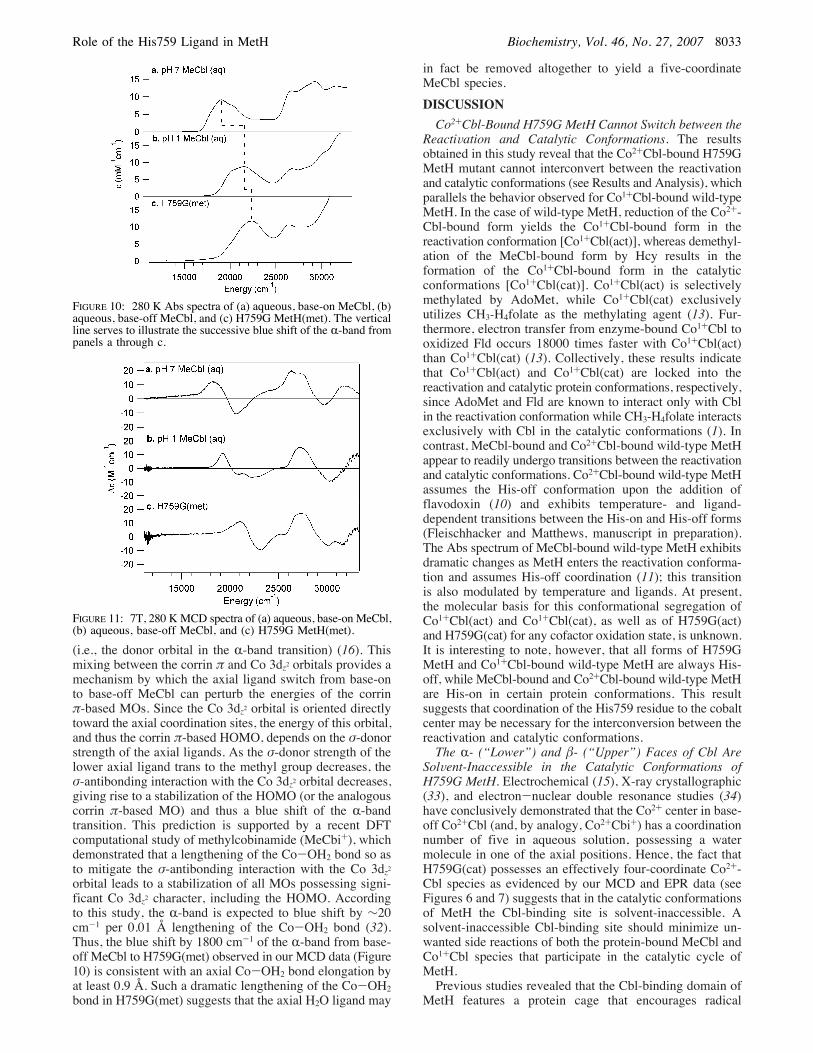
(i.e., the donor orbital in the R-band transition) (16). Thismixing between the corrin ! and Co 3dz2 orbitals provides amechanism by which the axial ligand switch from base-onto base-off MeCbl can perturb the energies of the corrin!-based MOs. Since the Co 3dz2 orbital is oriented directlytoward the axial coordination sites, the energy of this orbital,and thus the corrin !-based HOMO, depends on the "-donorstrength of the axial ligands. As the "-donor strength of thelower axial ligand trans to the methyl group decreases, the"-antibonding interaction with the Co 3dz2 orbital decreases,giving rise to a stabilization of the HOMO (or the analogouscorrin !-based MO) and thus a blue shift of the R-bandtransition. This prediction is supported by a recent DFTcomputational study of methylcobinamide (MeCbi+), whichdemonstrated that a lengthening of the Co-OH2 bond so asto mitigate the "-antibonding interaction with the Co 3dz2orbital leads to a stabilization of all MOs possessing signi-ficant Co 3dz2 character, including the HOMO. Accordingto this study, the R-band is expected to blue shift by !20cm-1 per 0.01 Å lengthening of the Co-OH2 bond (32).Thus, the blue shift by 1800 cm-1 of the R-band from base-off MeCbl to H759G(met) observed in our MCD data (Figure10) is consistent with an axial Co-OH2 bond elongation byat least 0.9 Å. Such a dramatic lengthening of the Co-OH2bond in H759G(met) suggests that the axial H2O ligand may
in fact be removed altogether to yield a five-coordinateMeCbl species.DISCUSSIONCo2+Cbl-Bound H759G MetH Cannot Switch between the
ReactiVation and Catalytic Conformations. The resultsobtained in this study reveal that the Co2+Cbl-bound H759GMetH mutant cannot interconvert between the reactivationand catalytic conformations (see Results and Analysis), whichparallels the behavior observed for Co1+Cbl-bound wild-typeMetH. In the case of wild-type MetH, reduction of the Co2+-Cbl-bound form yields the Co1+Cbl-bound form in thereactivation conformation [Co1+Cbl(act)], whereas demethyl-ation of the MeCbl-bound form by Hcy results in theformation of the Co1+Cbl-bound form in the catalyticconformations [Co1+Cbl(cat)]. Co1+Cbl(act) is selectivelymethylated by AdoMet, while Co1+Cbl(cat) exclusivelyutilizes CH3-H4folate as the methylating agent (13). Fur-thermore, electron transfer from enzyme-bound Co1+Cbl tooxidized Fld occurs 18000 times faster with Co1+Cbl(act)than Co1+Cbl(cat) (13). Collectively, these results indicatethat Co1+Cbl(act) and Co1+Cbl(cat) are locked into thereactivation and catalytic protein conformations, respectively,since AdoMet and Fld are known to interact only with Cblin the reactivation conformation while CH3-H4folate interactsexclusively with Cbl in the catalytic conformations (1). Incontrast, MeCbl-bound and Co2+Cbl-bound wild-type MetHappear to readily undergo transitions between the reactivationand catalytic conformations. Co2+Cbl-bound wild-type MetHassumes the His-off conformation upon the addition offlavodoxin (10) and exhibits temperature- and ligand-dependent transitions between the His-on and His-off forms(Fleischhacker and Matthews, manuscript in preparation).The Abs spectrum of MeCbl-bound wild-type MetH exhibitsdramatic changes as MetH enters the reactivation conforma-tion and assumes His-off coordination (11); this transitionis also modulated by temperature and ligands. At present,the molecular basis for this conformational segregation ofCo1+Cbl(act) and Co1+Cbl(cat), as well as of H759G(act)and H759G(cat) for any cofactor oxidation state, is unknown.It is interesting to note, however, that all forms of H759GMetH and Co1+Cbl-bound wild-type MetH are always His-off, while MeCbl-bound and Co2+Cbl-bound wild-type MetHare His-on in certain protein conformations. This resultsuggests that coordination of the His759 residue to the cobaltcenter may be necessary for the interconversion between thereactivation and catalytic conformations.The R- (“Lower”) and $- (“Upper”) Faces of Cbl Are
SolVent-Inaccessible in the Catalytic Conformations ofH759G MetH. Electrochemical (15), X-ray crystallographic(33), and electron-nuclear double resonance studies (34)have conclusively demonstrated that the Co2+ center in base-off Co2+Cbl (and, by analogy, Co2+Cbi+) has a coordinationnumber of five in aqueous solution, possessing a watermolecule in one of the axial positions. Hence, the fact thatH759G(cat) possesses an effectively four-coordinate Co2+-Cbl species as evidenced by our MCD and EPR data (seeFigures 6 and 7) suggests that in the catalytic conformationsof MetH the Cbl-binding site is solvent-inaccessible. Asolvent-inaccessible Cbl-binding site should minimize un-wanted side reactions of both the protein-bound MeCbl andCo1+Cbl species that participate in the catalytic cycle ofMetH.Previous studies revealed that the Cbl-binding domain of
MetH features a protein cage that encourages radical
FIGURE 10: 280 K Abs spectra of (a) aqueous, base-on MeCbl, (b)aqueous, base-off MeCbl, and (c) H759G MetH(met). The verticalline serves to illustrate the successive blue shift of the R-band frompanels a through c.
FIGURE 11: 7T, 280 KMCD spectra of (a) aqueous, base-on MeCbl,(b) aqueous, base-off MeCbl, and (c) H759G MetH(met).
Role of the His759 Ligand in MetH Biochemistry, Vol. 46, No. 27, 2007 8033

recombination following inadvertent homolytic cleavage ofthe Co-C bond of the enzyme-bound MeCbl cofactor (35).Solvent access would be expected to undermine the ef-fectiveness of this protein cage and thus to shorten thelifetime of the MeCbl-bound resting state of MetH byproviding alternative reaction pathways for the transientlyformed methyl radical. Additionally, because of its “super-nucleophilic” character, the Co1+Cbl species formed duringcatalytic turnover is poised for nucleophilic attack of a widerange of substrates (36, 37). Solvent access would likelypermit the enzyme-bound Co1+Cbl species to attack sub-strates other than CH3-H4folate, thereby precluding the re-formation of MetH-bound MeCbl. Recovery of the MetHresting state via the reactivation cycle following both theloss of the methyl radical and the inactivation of the Co1+-Cbl reaction intermediate requires AdoMet, a compoundderived from the Met product of MetH (38). This makesexcessive usage of the reactivation cycle disadvantageousto the cell by depleting the intracellular supply of AdoMet.Only the $- (“Upper”) Face of Cbl Is SolVent-Accessible
in the ReactiVation Conformation of H759G MetH. Becausethe MCD and EPR spectra of H759G(act) are very similarto those of aqueous Co2+Cbi+ (Figures 6 and 7), it followsthat H759G(act) contains a five-coordinate Co2+Cbl specieswith an axially bound water molecule. While this resultindicates that in the reactivation conformation of H759GMetH the Cbl-binding site is solvent-accessible, it is notpossible to determine whether the axial water ligand is boundto the R- or $-face of the cofactor on the basis of ourspectroscopic data alone. However, considering that the axialoxygen-donor ligands in cob(II)ester, a close mimic of base-off Co2+Cbl, and the Co2+Cbl species in the corrinoid:ironsulfur protein bind to the Co center on the sterically lesshindered $-face, as revealed by X-ray crystallographic data(33, 39), it is reasonable to assume that the Co2+Cbl speciesin H759G(act) also binds its water molecule on the $-face.Additional indirect support for this proposal is provided
by our H759G(met) spectroscopic data. Because photolysisof H759G(met), which is formed by reductive methylationof H759G(act), exclusively produces H759G(act), it can beconcluded that H759G(met) is locked into the reactivationconformation of MetH. Hence, the fact that our Abs andMCD spectra reveal that the water molecule that coordinateson the R-face of base-off MeCbl(aq) is no longer present inH759G(met) (Figures 10 and 11) suggests that in thereactivation conformation of MetH the R-face is solvent-inaccessible.Examination of the X-ray crystal structure of a truncated
H759G MetH variant (residues 649-1227), which is forcedto adopt the reactivation conformation, also supports thehypothesis that the $-face of the Cbl cofactor in H759G-(cat) is solvent-accessible (Figure 12) (14). Hence, in thisconformation the Co2+ center has much better access toexternal reductants, such as Fld or mediator dyes (11), thanin the catalytic conformations, in which the cofactor issolvent-inaccessible (vide supra). Moreover, as suggested bythe results obtained in cross-linking experiments with wild-type MetH and Fld as well as the X-ray crystal structure oftruncated H759G MetH that is trapped in the reactivationconformation (3, 14), the $-face of the Cbl cofactor maybecome solvent-inaccessible upon docking of Fld to MetH.Since the axial water molecule in H759G(act) binds exclu-sively to the $-face of Co2+Cbl, as demonstrated in this study,it may readily dissociate upon Fld docking so as to yield a
four-coordinate Co2+Cbl intermediate prior to, or concomi-tant with, the reduction of Co2+Cbl to Co1+Cbl in thereactivation cycle of MetH. A four-coordinate Co2+Cblintermediate fits the paradigm of enzymatic Co2+Cblf Co1+-Cbl reduction first suggested on the basis of spectroscopicstudies of two adenosyltransferases (29, 30) and providesan intuitively appealing explanation as to why this step isthermodynamically feasible despite the apparent mismatchof the Co2+Cbl and Fld reduction potentials (40).
ACKNOWLEDGMENT
We thank Prof. Tim Machonkin and Dr. Troy Stich foruseful discussions, especially with respect to the H759G-(cat) EPR spectrum. We also thank Prof. Mark Nilges andthe Illinois EPR Research Center for providing us with acopy of the SIMPOW6 program.
SUPPORTING INFORMATION AVAILABLE
Additional Abs and MCD data mentioned, but notdiscussed in text, as well as complete parameter sets for theSIMPOW6 EPR simulations. This material is available freeof charge via the Internet at http://pubs.acs.org.
REFERENCES1. Goulding, C. W., Postigo, D., and Matthews, R. G. (1997)Cobalamin-Dependent Methionine Synthase Is a Modular Proteinwith Distinct Regions for Binding Homocysteine, Methyltetrahy-drofolate, Cobalamin, and Adenosylmethionine, Biochemistry 36,8082-8091.
2. Drummond, J. T., Huang, S., Blumenthal, R. M., and Matthews,R. G. (1993) Assignment of Enzymatic Function to SpecificProtein Regions of Cobalamin-Dependent Methionine Synthasefrom Escherichia coli, Biochemistry 32, 9290-9295.
3. Hall, D. A., Jordan-Starck, T. C., Loo, R. O., Ludwig, M. L., andMatthews, R. G. (2000) Interaction of Flavodoxin with Cobalamin-Dependent Methionine Synthase, Biochemistry 39, 10711-10719.
4. Hall, D. A., Vander, Kooi, C. W., Stasik, C. N., Stevens, S. Y.,and Zuiderweg, E. R. P. (2001) Mapping the Interaction BetweenFlavodoxin and its Physiological Partners Flavodoxin Reductaseand Cobalamin-Dependent Methionine Synthase, Proc. Natl. Acad.Sci. U.S.A. 98, 9521-9526.
5. Evans, J. C., Huddler, D. P., Hilgers, M. T., Romanchuk, G.,Matthews, R. G., and Ludwig, M. L. (2004) Structures of the
FIGURE 12: Space-filling representation of the X-ray crystalstructure of the truncated H759G MetH variant (residues 649-1227) that only contains the Cbl- and AdoMet-binding modulesand is thus forced to adopt the same conformation as H759G MetH-(act) (14). Residues implicated in Fld docking are highlighted inwhite (3). Note that while the R-face of the Cbl cofactor is protectedby the Cbl-binding module (red), the $-face is left partially solvent-accessible by the AdoMet-binding module (blue).
8034 Biochemistry, Vol. 46, No. 27, 2007 Liptak et al.
N-terminal Modules Imply Large Domain Motions DuringCatalysis by Methionine Synthase, Proc. Natl. Acad. Sci. U.S.A.101, 3729-3736.
6. Banerjee, R. V., Frasca, V., Ballou, D. P., and Matthews, R. G.(1990) Participation of Cob(I)alamin in the Reaction Catalyzedby Methionine Synthase from Escherichia coli: A Steady-Stateand Rapid Reaction Kinetic Analysis, Biochemistry 29, 11101-11109.
7. Taylor, R. T., and Weissbach, H. (1967) N5-Methyltetrahydro-folate-Homocysteine Transmethylase, J. Biol. Chem. 242, 1502-1508.
8. Fujii, K., and Huennekens, F. M. (1974) Activation of MethionineSynthetase by a Reduced Triphosphopyridine Nucleotide-Depend-ent Flavoprotein System, J. Biol. Chem. 249, 6745-6753.
9. Mangum, J. H., and Scrimgeour, K. G. (1962) Cofactor Require-ments and Intermediates in Methionine Biosynthesis, Fed. Proc.21, 242.
10. Hoover, D. M., Jarrett, J. T., Sands, R. H., Dunham, W. R.,Ludwig, M. L., and Matthews, R. G. (1997) Interaction ofEscherichia coli Cobalamin-Dependent Methionine Synthase andits Physiological Partner Flavodoxin: Binding of FlavodoxinLeads to Axial Ligand Dissociation from the Cobalamin Cofactor,Biochemistry 36, 127-138.
11. Jarrett, J. T., Hoover, D. M., Ludwig, M. L., and Matthews, R.G. (1998) The Mechanism of Adenosylmethionine-DependentActivation of Methionine Synthase: A Rapid Kinetic Analysisof Intermediates in Reductive Methylation of Cob(II)alaminEnzyme, Biochemistry 37, 12649-12658.
12. Jarrett, J. T., Amaratunga, M., Drennan, C. L., Scholten, J. D.,Sands, R. H., Ludwig, M. L., and Matthews, R. G. (1996)Mutations in the B12-Binding Region of Methionine Synthase:How the Protein Controls Methylcobalamin Reactivity, Biochem-istry 35, 2464-2475.
13. Jarrett, J. T., Huang, S., and Matthews, R. G. (1998) MethionineSynthase Exists in Two Distinct Conformations That Differ inReactivity Toward Methyltetrahydrofolate, Adenosylmethionine,and Flavodoxin, Biochemistry 37, 5372-5382.
14. Bandarian, V., Pattridge, K. A., Lennon, B. W., Huddler, D. P.,Matthews, R. G., and Ludwig, M. L. (2002) Domain AlternationSwitches B12-Dependent Methionine Synthase to the ActivationConformation, Nat. Struct. Biol. 9, 53-56.
15. Lexa, D., and Saveant, J. M. (1983) The Electrochemistry ofVitamin-B12, Acc. Chem. Res. 16, 235-243.
16. Stich, T. A., Brooks, A. J., Buan, N. R., and Brunold, T. C. (2003)Spectroscopic and Computational Studies of Co3+-Corrinoids:Spectral and Electronic Properties of the B12 Cofactors andBiologically Relevant Precursors, J. Am. Chem. Soc. 125, 5897-5914.
17. Stich, T. A., Buan, N. R., and Brunold, T. C. (2004) Spectroscopicand Computational Studies of Co2+Corrinoids: Spectral andElectronic Properties of the Biologically Relevant Base-On andBase-Off Forms of Co2+Cobalamin, J. Am. Chem. Soc. 126,9735-9749.
18. Pavel, E. G., and Solomon, E. I. (1998) Recent Advances inMagnetic Circular Dichroism Spectroscopy, in SpectroscopicMethods in Bioinorganic Chemistry (Solomon, E. I., and Hodgson,K. O., Eds.) pp 119-135, American Chemical Society, Wash-ington, DC.
19. Salmon, R. T., and Hawkridge, F. M. (1980) The ElectrochemicalProperties of Three Dipyridinium Salts as Mediators, J. Electro-anal. Chem. 112, 253-264.
20. Zehnder, A. J. B., and Wuhrmann, K. (1976) Titanium(III) Citrateas a Nontoxic Oxidation-Reduction Buffering System for Cultureof Obligate Anaerobes, Science 194, 1165-1166.
21. Jarrett, J. T., Goulding, C. W., Fluhr, K., Huang, S., and Matthews,R. G. (1997) Purification and Assay of Cobalamin-DependentMethionine Synthase from Escherichia coli, Methods Enzymol.281, 196-213.
22. Amaratunga, M., Fluhr, K., Jarrett, J. T., Drennan, C. L., Ludwig,M. L., Matthews, R. G., and Scholten, J. D. (1996) A SyntheticModule for the metH Gene Permits Facile Mutagenesis of theCobalamin-Binding Region of Escherichia coli Methionine Syn-thase: Initial Characterization of Seven Mutant Proteins, Bio-chemistry 35, 2453-2463.
23. Pol, A., Gage, R. A., Neis, J. M., Reijnen, J. W. M., van der Drift,C., and Vogels, G. D. (1984) Corrinoids from Methanosar-cina barkerisThe $-Ligands, Biochim. Biophys. Acta 797, 83-93.
24. Grahame, D. A. (1989) Different Isozymes of Methylcobalamin:2-Mercaptoethanesulfonate Predominate in Methanol- VersusAcetate-grown Methanosarcina barkeri, J. Biol. Chem. 264,12890-12894.
25. Harder, S. R., Feinberg, B. A., and Ragsdale, S. W. (1989) ASpectroelectrochemical Cell Designed for Low TemperatureElectron Paramagnetic Resonance Titration of Oxygen-SensitiveProteins, Anal. Biochem. 181, 283-287.
26. Drummond, J. T., and Matthews, R. G. (1994) Nitrous OxideDegradation by Cobalamin-Dependent Methionine Synthase:Characterization of the Reactants and Products in the InactivationReaction, Biochemistry 33, 3732-3741.
27. Mayhew, S. (1978) The Redox Potential of Dithionite and SO2from Equilibrium Reactions with Flavodoxins, Methyl Viologenand Hydrogen plus Hydrogenase, Eur. J. Biochem. 85, 535-547.
28. Nilges, M. J. (1979) Electron paramagnetic resonance studies oflow symmetry nickel(I) and molybdenum(V) complexes, Ph.D.Thesis, University of Illinois.
29. Stich, T. A., Buan, N. R., Escalante-Semerena, J. C., and Brunold,T. C. (2005) Spectroscopic and Computational Studies of the ATP:Corrinoid Adenosyltransferase (CobA) from Salmonella en-terica: Insights into the Mechanism of AdenosylcobalaminBiosynthesis, J. Am. Chem. Soc. 127, 8710-8719.
30. Stich, T. A., Yamanishi, M., Banerjee, R., and Brunold, T. C.(2005) Spectroscopic Evidence for the Formation of a Four-Coordinate Co2+Cobalamin Species upon Binding to the HumanATP:Cobalamin Adenosyltransferase, J. Am. Chem. Soc. 127,7660-7661.
31. Jarrett, J. T., Choi, C. Y., and Matthews, R. G. (1997) Changesin Protonation Associated with Substrate Binding and Cob(I)-alamin Formation in Cobalamin-Dependent Methionine Synthase,Biochemistry 36, 15739-15748.
32. Stich, T. A., Seravalli, J., Venkateshrao, S., Spiro, T. G., Ragsdale,S. W., and Brunold, T. C. (2006) Spectroscopic Studies of theCorrinoid/Iron-Sulfur Protein fromMoorella thermoacetica, J. Am.Chem. Soc. 128, 5010-5020.
33. Krautler, B., Keller, W., Hughes, M., Caderas, C., and Kratky, C.(1987) A Crystalline Cobalt(II)corrinate Derived From VitaminB12: Preparation and X-Ray Crystal Structure, J. Chem. Soc.,Chem. Commun., 1678-1680.
34. Van Doorslaer, S., Jeschke, G., Epel, B., Goldfarb, D., Eichel,R.-A., Krautler, B., and Schweiger, A. (2003) Axial SolventCoordination in “Base-Off” Cob(II)alamin and Related Co(II)-Corrinates Revealed by 2D-EPR, J. Am. Chem. Soc. 125, 5915-5927.
35. Jarrett, J. T., Drennan, C. L., Amaratunga, M., Scholten, J. D.,Ludwig, M. L., and Matthews, R. G. (1996) A Protein RadicalCage Slows Photolysis of Methylcobalamin from Escherichia coli,Bioorg. Med. Chem. 4, 1237-1246.
36. Schrauzer, G. N., Deutsch, E., and Windgassen, R. J. (1968) TheNucleophilicity of Vitamin B12s, J. Am. Chem. Soc. 90, 2441-2442.
37. Schrauzer, G. N., and Deutsch, E. (1969) Reactions of Cobalt(I)Supernucleophiles. The Alkylation of Vitamin B12s, Cobaloxi-mes(I), and Related Compounds, J. Am. Chem. Soc. 91, 3341-3350.
38. Chiang, P. K., Gordon, R. K., Tal, J., Zeng, G. C., Doctor, B. P.,Pardhasaradhi, K., and McCann, P. P. (1996) S-Adenosylme-thionine and Methylation, FASEB J. 10, 471-480.
39. Svetlitchnaia, T., Svetlitchnyi, V., Meyer, O., and Dobbek, H.(2006) Structural Insights into Methyltransfer Reactions of aCorrinoid Iron-Sulfur Protein Involved in Acetyl-CoA Synthesis,Proc. Natl. Acad. Sci. U.S.A. 103, 14331-14336.
40. Liptak, M. D., and Brunold, T. C. (2006) Spectroscopic andComputational Studies of Co1+Cobalamin: Spectral and ElectronicProperties of the “Superreduced” B12 Cofactor, J. Am. Chem. Soc.128, 9144-9156.
BI700341Y
Role of the His759 Ligand in MetH Biochemistry, Vol. 46, No. 27, 2007 8035
Related Documents