Probing the Effect of Conformational Constraint on Phosphorylated Ligand Binding to an SH2 Domain using Polarizable Force Field Simulations Yue Shi 1 , Crystal Z. Zhu 1 , Stephen F. Martin 2 , and Pengyu Ren 1 1 Department of Biomedical Engineering, The University of Texas at Austin, TX 78712 2 Department of Chemistry and Biochemistry, The University of Texas at Austin, TX 78712 Abstract Preorganizing a ligand in the conformation it adopts upon binding to a protein has long been considered to be an effective way to improve affinity by making the binding entropy more favorable. However, recent thermodynamic studies of a series of complexes of the Grb2 SH2 domain with peptide analogs having constrained and flexible replacements for a phosphotyrosine residue revealed that less favorable binding entropies may result from constraining ligands in their biologically active conformations. Toward probing the origin of this unexpected finding, we examined the complexes of four phosphotyrosine-derived analogs with the Grb2 SH2 domain using molecular dynamics simulations with a polarizable force field. Significantly, the computed values for the relative binding free energies, entropies and enthalpies of two pairs of constrained and unconstrained ligands reproduced the trends that were determined experimentally, although the relative differences were overestimated. These calculations also revealed that a large fraction of the ligands lacking the constraining element exist in solution as compact, macrocyclic-like structures that are stabilized by interactions between the phosphate groups and the amide moieties of the C-terminal pY+2 residues. In contrast, the three-membered ring in the constrained ligands prevents the formation of such macrocyclic structures, leading instead to globally extended, less ordered conformations. Quasiharmonic analysis of these conformational ensembles suggests that the unconstrained ligands possess significantly lower entropies in solution, a finding that is consistent with the experimental observation that the binding entropies for the unconstrained Supporting Information Available: Temperatures in Replica Exchange Molecular Dynamics Simulations (in K); Figure S1. Potential energy distributions of configurations sampled at different temperatures (300 K, 600 K and 1000 K) and with different simulation length (10 ns and 30 ns). QM optimized structure and the structure taken from complex crystal structure are as the starting points for repeated simulations. The frames are sampled every 2 ps, and then minimized with a convergence of 0.1 kcal/mol/Å. Thus the conformational distributions we are examining are for 0 K, even though the simulations have been done at high temperatures to accelerate the sampling process. The dihedral angles are measured from 0~360 degree (instead of 0~180 degree) in order to capture all the different orientations. The QM optimized structure is selected as the reference to calculate torsional RMSDs for all snapshots; Figure S2. Atom types of model compounds used for parameterization; Figure S3. Autocorrelation of dihedral angle C407-C350-C352-C354 and C350-C352-C354- C428 of fpYVN in solvent from REMD simulation at 298 K; Figure S4. Conformational distribution of dihedral angle C407-C350- C352-C354 and C350-C352-C354-C428 of fpYVN in solvent from REMD simulation at 298 K; Figure S5. Overlap of potential energy differences calculated forward (blue) and backward (red) of perturbation from fpYVN to cpYVN in water. The X-axis represents the potential energy difference, and the Y-axis shows the probability; Figure S6. Overlap of potential energy differences calculated forward (blue) and backward (red) of perturbation from fpYVN to cpYVN in solvated complex. The X-axis represents the potential energy difference, and the Y-axis shows the probability; Figure S7. Statistical inefficiency of the free energy calculations for fpYVN->cpYVN in water (upper panel) and fpYVN in solvated complex (lower panel) at 298K; Table S1. The dependence of relative binding free energy (fpYVN->cpYVN) at 298K on the number of intermediate steps in the alchemical calculation (energy in kcal/ mol). Every 2nd (13 steps total), 4th (20 steps), 5th (21 steps), 6th (22 steps), 7th (23 steps), 10th (24 steps), 13th (23 steps with the middle step skipped) step is skipped respectively. Parameters for C428, O430, N426 and H429 are transferred from AMOEBA PRO; Force field parameters of the model compounds in Figure S2. This material is available free of charge via the Internet at http://pubs.acs.org. NIH Public Access Author Manuscript J Phys Chem B. Author manuscript; available in PMC 2013 February 9. Published in final edited form as: J Phys Chem B. 2012 February 9; 116(5): 1716–1727. doi:10.1021/jp210265d. NIH-PA Author Manuscript NIH-PA Author Manuscript NIH-PA Author Manuscript

Welcome message from author
This document is posted to help you gain knowledge. Please leave a comment to let me know what you think about it! Share it to your friends and learn new things together.
Transcript

Probing the Effect of Conformational Constraint onPhosphorylated Ligand Binding to an SH2 Domain usingPolarizable Force Field Simulations
Yue Shi1, Crystal Z. Zhu1, Stephen F. Martin2, and Pengyu Ren1
1Department of Biomedical Engineering, The University of Texas at Austin, TX 787122Department of Chemistry and Biochemistry, The University of Texas at Austin, TX 78712
AbstractPreorganizing a ligand in the conformation it adopts upon binding to a protein has long beenconsidered to be an effective way to improve affinity by making the binding entropy morefavorable. However, recent thermodynamic studies of a series of complexes of the Grb2 SH2domain with peptide analogs having constrained and flexible replacements for a phosphotyrosineresidue revealed that less favorable binding entropies may result from constraining ligands in theirbiologically active conformations. Toward probing the origin of this unexpected finding, weexamined the complexes of four phosphotyrosine-derived analogs with the Grb2 SH2 domainusing molecular dynamics simulations with a polarizable force field. Significantly, the computedvalues for the relative binding free energies, entropies and enthalpies of two pairs of constrainedand unconstrained ligands reproduced the trends that were determined experimentally, althoughthe relative differences were overestimated. These calculations also revealed that a large fractionof the ligands lacking the constraining element exist in solution as compact, macrocyclic-likestructures that are stabilized by interactions between the phosphate groups and the amide moietiesof the C-terminal pY+2 residues. In contrast, the three-membered ring in the constrained ligandsprevents the formation of such macrocyclic structures, leading instead to globally extended, lessordered conformations. Quasiharmonic analysis of these conformational ensembles suggests thatthe unconstrained ligands possess significantly lower entropies in solution, a finding that isconsistent with the experimental observation that the binding entropies for the unconstrained
Supporting Information Available:Temperatures in Replica Exchange Molecular Dynamics Simulations (in K); Figure S1. Potential energy distributions ofconfigurations sampled at different temperatures (300 K, 600 K and 1000 K) and with different simulation length (10 ns and 30 ns).QM optimized structure and the structure taken from complex crystal structure are as the starting points for repeated simulations. Theframes are sampled every 2 ps, and then minimized with a convergence of 0.1 kcal/mol/Å. Thus the conformational distributions weare examining are for 0 K, even though the simulations have been done at high temperatures to accelerate the sampling process. Thedihedral angles are measured from 0~360 degree (instead of 0~180 degree) in order to capture all the different orientations. The QMoptimized structure is selected as the reference to calculate torsional RMSDs for all snapshots; Figure S2. Atom types of modelcompounds used for parameterization; Figure S3. Autocorrelation of dihedral angle C407-C350-C352-C354 and C350-C352-C354-C428 of fpYVN in solvent from REMD simulation at 298 K; Figure S4. Conformational distribution of dihedral angle C407-C350-C352-C354 and C350-C352-C354-C428 of fpYVN in solvent from REMD simulation at 298 K; Figure S5. Overlap of potentialenergy differences calculated forward (blue) and backward (red) of perturbation from fpYVN to cpYVN in water. The X-axisrepresents the potential energy difference, and the Y-axis shows the probability; Figure S6. Overlap of potential energy differencescalculated forward (blue) and backward (red) of perturbation from fpYVN to cpYVN in solvated complex. The X-axis represents thepotential energy difference, and the Y-axis shows the probability; Figure S7. Statistical inefficiency of the free energy calculations forfpYVN->cpYVN in water (upper panel) and fpYVN in solvated complex (lower panel) at 298K; Table S1. The dependence of relativebinding free energy (fpYVN->cpYVN) at 298K on the number of intermediate steps in the alchemical calculation (energy in kcal/mol). Every 2nd (13 steps total), 4th (20 steps), 5th (21 steps), 6th (22 steps), 7th (23 steps), 10th (24 steps), 13th (23 steps with themiddle step skipped) step is skipped respectively. Parameters for C428, O430, N426 and H429 are transferred from AMOEBA PRO;Force field parameters of the model compounds in Figure S2. This material is available free of charge via the Internet athttp://pubs.acs.org.
NIH Public AccessAuthor ManuscriptJ Phys Chem B. Author manuscript; available in PMC 2013 February 9.
Published in final edited form as:J Phys Chem B. 2012 February 9; 116(5): 1716–1727. doi:10.1021/jp210265d.
NIH
-PA Author Manuscript
NIH
-PA Author Manuscript
NIH
-PA Author Manuscript
ligands are more favorable than for their constrained counterparts. This study suggests thatintroducing local constraints in flexible molecules may have unexpected consequences, and adetailed understanding of the conformational preferences of ligands in their unbound states is acritical prerequisite to correlating changes in their chemical structure with protein bindingentropies and enthalpies.
Keywordsligand preorganization; entropy paradox; AMOEBA polarizable force field
INTRODUCTIONUnderstanding the effects of making chemical modifications to ligands upon their relativebinding energetics is a critical step in structure-based drug design. Preorganizing a flexibleligand in the conformation it adopts upon binding has long been considered a useful strategyto achieve more a favorable binding entropy and thus an improved binding affinity.1 Indeed,the program CAVEAT was developed in part to facilitate the design of constrainedmolecules bearing substituents directed in predefined orientations.2 Awareness of thepotential energetic benefits of ligand preorganization dates back to work by Jencks in the1970s,3 and there are numerous reports of the increased affinities that may accompany theintroduction of conformational constraints into flexible ligands.3–9 However, it has recentlybeen reported that the binding entropy of a constrained ligand may actually be less favorablethan its flexible control.10–14 Even if ligand preorganization leads to a beneficial entropiccontribution to binding, the enhancement to binding affinity may be offset by acompensating enthalpic penalty.7,15–23 Entropy-enthalpy compensation has been widelystudied, but its origin is not well understood.5,24–29 While some consider this effect as anintrinsic physical phenomenon, others argue that the entropy-enthalpy compensation is astatistical artifact arising from obtaining entropy and enthalpy based on temperaturedependent data from both experiment and theoretical calculations.24,25,27,30–33
In order to understand the detailed energetic effects of ligand preorganization on protein-ligand interactions, it is essential to perform systematic experimental studies to determinethe contributions of entropy and enthalpy to binding free energy and to correlate these withstructural and dynamic analyses of the protein-ligand complexes using X-raycrystallographic and NMR spectroscopic methods.34 For example, DeLorbe et al.14 recentlyexamined the binding energetics and structures associated with complexes of a series ofconstrained and flexible phosphotyrosine-derived peptide analogs with the SH2 domain ofgrowth receptor binding protein 2 (Grb2); Grb2 is a 25 KDa cytosolic adapter protein that isinvolved in activation of the Ras signal transduction pathway.35 The constrained ligandscpYVN and cpYIN, which were preorganized by incorporating a cyclopropane ring at thepY replacement in the pseudopeptides fpYVN and fpYIN, respectively, (Figure 1), have thesame functional groups and the same number and type of heavy atoms as their flexiblecontrols. The thermodynamic binding parameters of these ligands for the Grb2 SH2 domainwere determined by isothermal titration calorimetry (ITC),36 and the binding entropies forthe constrained ligands were found to be less favorable than for their flexible analogs. Thisunexpected finding is contrary to the conventional wisdom that ligand preorganizationshould be accompanied by a more favorable binding entropy. Their less favorable bindingentropies notwithstanding, the constrained ligands bound with higher affinities than theirmore flexible counterparts because of significantly more favorable binding enthalpies. Thatthe measured heat capacity changes on binding of the flexible/constrained ligand pairs weresimilar suggests that these thermodynamic differences do not arise from desolvationeffects.37 Although these experimental studies clearly show that ligand preorganization does
Shi et al. Page 2
J Phys Chem B. Author manuscript; available in PMC 2013 February 9.
NIH
-PA Author Manuscript
NIH
-PA Author Manuscript
NIH
-PA Author Manuscript

not necessarily result in a more favorable binding entropy, the molecular origin of theunanticipated behavior was not examined.
Over the past decades, numerous efforts have been devoted to develop and applycomputational approaches to screen and design potent ligands for drug discovery.38–40
Computational methods such as docking and molecular dynamics utilizing continuum andexplicit solvent models have long been employed toward predicting protein-ligand bindingaffinities in silico.38,40–42 Among the various strategies that have been explored, detailedalchemical pathway simulations using explicit solvent show significant promise forproviding energetically accurate predictions of protein-ligand binding affinity.34,43,44 Theabsolute binding free energies calculated from such pathways correlate reasonably well withexperimental data, and root mean square (RMS) errors of less than 3 kcal/mol are oftenreported.41 On the other hand, the relative binding free energies can be calculated moreaccurately, if there is sufficient sampling of protein-ligand-water configurational space.45
Decomposition of binding free energy into entropic and enthalpic contributions offersimportant insights into the driving forces for protein-ligand recognition;34 however,quantitative estimation of the binding entropy remains a significant challenge. Commonapproaches to estimating the binding entropy include quasiharmonic analysis,46,47 normalmode analysis,48–50 and knowledge based scoring functions.51–53 These methods have beenapplied to several protein-ligand systems,54–60 but the contributions from solvation aretypically neglected or approximated due to the computational expense. More physicallyrigorous alchemical pathway approaches are also applicable to evaluating entropy, but fewinvestigations have reported using such methods in protein-ligand interactions. 61
In the present work, we use molecular modeling to explore how introducing conformationalconstraints into two closely related phosphotyrosine-derived peptides affects thethermodynamic parameters for their complexation with the Grb2 SH2 domain. Because thephosphate group in these ligands is charged, the AMOEBA polarizable force field,62–65
which accounts for multipole electrostatics and polarization effects, was used to modelprotein, ligand, and water components in each MD simulation. The AMOEBA force fieldhas been successfully applied to accurately model a number of highly polar molecularsystems, including water,63 monovalent and divalent ions,66–69 small molecules,70
peptides,71 and trypsin-benzamidinium binding.72–74 We report herein the calculations ofbinding free energies, enthalpies and entropies, as well as the results from simulations of thestructure and dynamics of the ligands in water and in their complexes with the Grb2 SH2domain. The results are compared with experimental observations, and a possible molecularorigin for the unexpected, unfavorable entropic effect resulting from preorganizing thesephosphotyrosine-derived ligands is presented.
METHODEntropy Calculation
The change in entropy (ΔS) is related to the change in free energy by75
(1)
In this study, we computed the entropy change numerically from the slope of a linear fit tothe temperature dependence of the free energy change near room temperature. This approachis essentially identical to the finite difference method that has been used previously.76–78
While other direct or perturbation methods exist for computing enthalpy or entropy, we havefound that the numerical approach is the most reliable for calculations of energetics. In
Shi et al. Page 3
J Phys Chem B. Author manuscript; available in PMC 2013 February 9.
NIH
-PA Author Manuscript
NIH
-PA Author Manuscript
NIH
-PA Author Manuscript

addition, because REMD is being used to simulate free ligands in solution, it is possible totake advantage of the temperature dependence of free energy data.
The relative change in enthalpy is then computed as
(2)
The Bennett Acceptance Ratio (BAR) method79 was utilized to calculate the relative bindingfree energy (ΔA). Not considering finite size effects, this Helmholtz free energy is equal tothe Gibbs free energy change at the same pressure. The free energy change betweenneighboring states (λi and λi+1) is given by the expression
(3)
where
(4)
and j is the iteration index. Here, Uλi is the potential energy of the system evaluated usingthe parameters from λi, and <> indicates the ensemble average. ΔA is solved iteratively untilthe value of (ΔA(j) − ΔA(j−1)) < 0.01 kcal/mol.
In order to avoid the end point singularity during the alchemical transformation, the soft-core buffered 14-7 potential was used for van der Waals (vdW) interactions
(5)
where ε is the well depth, λ is the scaling factor, ρij=Rij/Rij0 with Rij as the actual separation
between atoms i and j and Rij0 is the minimum energy distance parameter.
Quasiharmonic AnalysisQuasiharmonic analysis was performed to characterize the collective motions of moleculesat thermodynamic equilibrium.46,47 The quasiharmonic approximation assumes that thespatial fluctuations in the system follow a multivariate Gaussian distribution. In aquasiharmonic frequency analysis on an n-atom system, the eigenvalues λi (i=1,2,…,3n-6) ofthe mass weighted covariance matrix of atomic fluctuations are calculated to determine thequasiharmonic frequencies, , which are then used to estimate theconformational entropy, given by
(6)
where ħ is the Planck constant, and R is the gas constant. The quasiharmonic method hasbeen successfully applied to simple molecular systems with few energy minima. However,this method will overestimate the configurational entropy for more complex systems having
Shi et al. Page 4
J Phys Chem B. Author manuscript; available in PMC 2013 February 9.
NIH
-PA Author Manuscript
NIH
-PA Author Manuscript
NIH
-PA Author Manuscript
multiple occupied energy wells, particularly when using Cartesian coordinates rather thaninternal coordinates.80
Force Field and ParameterizationThe AMOEBA polarizable force field63,65 was applied to model the protein, watermolecules, and the amino acid residues in the peptide-analogs fpYVN, fpYIN, cpYVN andcpYIN (Figure 1). Additional parameterization was necessary for the phosphotyrosine (pY)replacements fpY and cpY. Missing valence parameters for the constrained andunconstrained pY segments were derived from quantum mechanics (QM) calculations byusing the “Valence” module in the TINKER software package.81 “Valence” sets theequilibrium bond lengths and angles based on the HF/6-31G* optimized structures of the pYreplacements; the force constants and vdW parameters were then transferred from theexisting AMOEBA force field parameters for the same atom types. Trimethyl and dimethylphosphate (TMP and DMP) were used to derive the vdW parameters of the phosphate groupby fitting to both the QM structure and energy of TMP/DMP-water dimers. The vdWparameters were further tuned to match the experimental liquid density and heat ofvaporization of TMP. The electrostatic parameters for the pY side chain were obtained fromQM at the MP2/6-311++G(2d,2p) level by the “original-fit” approach,70,82 wherein theatomic multipoles for the pY segment were initially derived from the MP2/6-311G**density matrix using the original distributed multipole analysis (DMA).83,84 The atomicdipole and quadrupole moments of the pY residue were then optimized to the electrostaticpotential (ESP) around the whole ligand computed at the MP2/6-311++G(2d,2p) level(electrostatic parameters of other residues were fixed). The ESP root mean squaredifferences between QM and that from the final atomic multipoles, evaluated over roughly35000 grid points around the peptide, were 1.55 and 0.92 kcal/mol per unit charge forcpYVN and fpYVN, respectively.
The model compounds of both unconstrained and constrained pY segments and the dihedralangles ah for which parameters were derived are illustrated in Figure 2. Dihedral angleparameters in the pY replacement that were missing were obtained by comparing the QMconformational energy profile to those computed from the corresponding MM (MolecularMechanics) using all energy terms except for the dihedral angle term. The difference inenergy was then fit to a three-term, Fourier series torsional function. The torsional energy inMM serves essentially as an “error” function. Typically, the torsional energy parameters(V1, V2, and V3) are less than 1–3 kcal/mol for rotation about a single bond, whereas theoverall conformational energy barriers could be much higher due to intramolecularelectrostatic and vdw interactions. (Figure 2)
Computational DetailsIn order to evaluate the thermodynamic driving forces for ligand binding to the SH2 domain,we calculated the relative binding free energy, enthalpy and entropy contributions forconstrained and unconstrained ligand pairs, according to the alchemical pathway shown inFigure 1. The relative binding free energies for each pair of constrained and unconstrainedligands were computed from the free energy differences between the ligands in water and inthe protein binding pocket. The fpYVN ligand was gradually transformed into cpYVN byperforming 26 simulation steps; 10 steps were performed in the alchemical transformationsof Val to Ile in the constrained and unconstrained ligands. For the ligand-water systems,replica exchange molecular dynamics (REMD)85–87 simulations were performed with 48replicas at temperatures between 260 and 620 K (detailed schedule can be found in theSupporting Information).88 For the transformation of fpYVN to cpYVN, the bond formingthe cyclopropane ring was introduced by gradually increasing the force constant from 0 to550 kcal/mol/Å2; while simultaneously transforming two hydrogen atoms into “dummy
Shi et al. Page 5
J Phys Chem B. Author manuscript; available in PMC 2013 February 9.
NIH
-PA Author Manuscript
NIH
-PA Author Manuscript
NIH
-PA Author Manuscript

atoms” by turning off their vdW and electrostatic interactions with all other atoms. Note thatthe valence contributions from the dummy atoms were canceled between perturbations ofthe ligand in water and in the solvated complex. Constant-volume and constant-temperature(NVT) simulations of 2.5 ns were performed at each step. Replicas were exchanged every 2ps, with exchange success rates for all replicas were in the range of 22% to 40%. All theREMD simulations were performed using the parallel SANDER module in AMBER10, andthe REMD implemented in AMBER1089 was modified for use with the AMOEBA forcefield. For the protein-ligand complexes in explicit water, relative free energies forconstrained and unconstrained ligands having Val and Ile at the pY+1 position werecalculated at 288 K, 298 K and 308 K. For each set of alchemical calculations at eachtemperature, a total of 65 ns of NVT molecular dynamics was performed over 26 steps,using the PMEMD module in AMBER10. For all the simulations, the vdW cutoff was set at12 Å and the long-range electrostatics were treated using Particle Mesh Ewald (PME)summation90–92 with a grid spacing of 0.8 Å and a real space cutoff of 7 Å. The induceddipoles, which were also computed with PME, were iterated until the root mean square(RMS) change was below 0.01 Debye. A tighter induced dipole convergence of 10−5 RMSDebye was used in the energy calculation for the post free energy analysis with the BennettAcceptance Ratio (BAR). By using a bootstrap procedure, the statistical uncertainty wasestimated as the standard deviation of the average free energy values that are computedusing 100 partial simulation trajectory blocks (1.0 ns). Given that the uncertainty in theentropy calculation was dominated by the free energies at the lowest and highesttemperatures (288 K and 308 K), the statistical error for − TΔS was estimated from theupper and lower bound of free energy changes at 288 K and 308 K; thus the statistical errorfor − TΔS is twice the statistical error of free energy.
The entropy was extracted from the temperature dependence of the free energies via linearregression (Eq. 1). For ligands in water, the relative free energy ΔAwat was obtained fromREMD at 18 temperatures between 260 K and 360 K. A linear fit was used to interpolateΔAwat at 288 K, 298 K and 308 K. These values were then subtracted from the free energychanges of the ligand in complex (ΔAcomp) at the same temperatures to obtain relativebinding free energy ΔΔAbind. The entropic contribution was computed from the slope of thefitted linear temperature dependence of the relative binding free energy. The enthalpy wasevaluated via ΔΔA + TΔΔS. The total simulation time for the combined MD simulations offree ligands and complexes at all temperatures was more than 6 μs.
RESULTS AND DISCUSSIONLigand conformational properties and sampling
One of the essential goals of this study is to obtain an accurate description of the distributionof the different conformational ensembles of the peptide-like ligands in solution. Theseligands present a significant challenge for molecular simulations as they each possess a totalof more than a dozen rotatable bonds, with four being in the constrained pY replacement(cpY) and six belonging to the unconstrained pY mimic (fpY). At the same time, strongintramolecular interactions between the charged phosphate moiety and other polar groups inthe molecule result in certain stable conformations with low potential energies. Theconformational energy profile for the pY replacement indicates the energy barrier forescaping these stable conformations can be tens of a kcal/mol (Figure 2). In solution, it islikely that the conformational population will be significantly different because ofinteractions with water molecules. In fact, it is known that the dipeptide Ala-Ala hasdistinctly different local minimum energy structures in gas and solution phases.64 Weexamined each of the main conformational degrees of freedom for the pY replacement thatwe parameterized. The AMOEBA conformational energy profiles depicted in Figure 2 are ingood agreement with high-level ab initio QM results. The average root-mean-square
Shi et al. Page 6
J Phys Chem B. Author manuscript; available in PMC 2013 February 9.
NIH
-PA Author Manuscript
NIH
-PA Author Manuscript
NIH
-PA Author Manuscript
deviation (RMSD) between QM and MM minimized structures for the entire ligand is about0.25 Å per atom.
Adequate sampling of the configurational space of the system, including the ligand, iscritical in order to obtain accurate thermodynamic information from the simulations. Whenthe peptide-like ligands are bound to SH2 domain, they are restricted within the proteinpocket, and their structures are well-defined and in good agreement with the X-raycrystallographic structures.14 Since the ensemble of structures of the free ligands in solventis unknown, we performed MD simulations of the unconstrained fpYVN ligand in adielectric continuum (ε = 80) at and above room temperature in order to explore theconformational space of these peptide analogs in water and to guide the more detailed,explicit-solvent simulations. The potential energy and torsional RMSD distributionsobtained from multiple independent simulation trajectories were compared to examine theconvergence of the MD sampling. We found that MD simulations at room temperature donot produce converged distributions after 30 ns. On the other hand, when these simulationsare performed at 600 K or above, the distributions quickly converge after a few nanoseconds(see Supporting Information). Based on the information obtained from the simple continuumsimulations, replica-exchange molecular dynamics (REMD) simulations in explicit solventat temperatures ranging from 260 K to 620 K were performed to compute the relative freeenergy and entropy between different ligands in the solvent environment. Using thesesimulations, we examined the distribution of the main torsions of fpYVN in solution at 298K. Overall, we observe a broad sampling in torsional space and a number of torsionaltransitions. The autocorrelation functions of these torsions exhibit fast decay of only a fewps to reach 1/e, perhaps because of the artificially fast kinetics of REMD; some examplesare included in the Supporting Information.
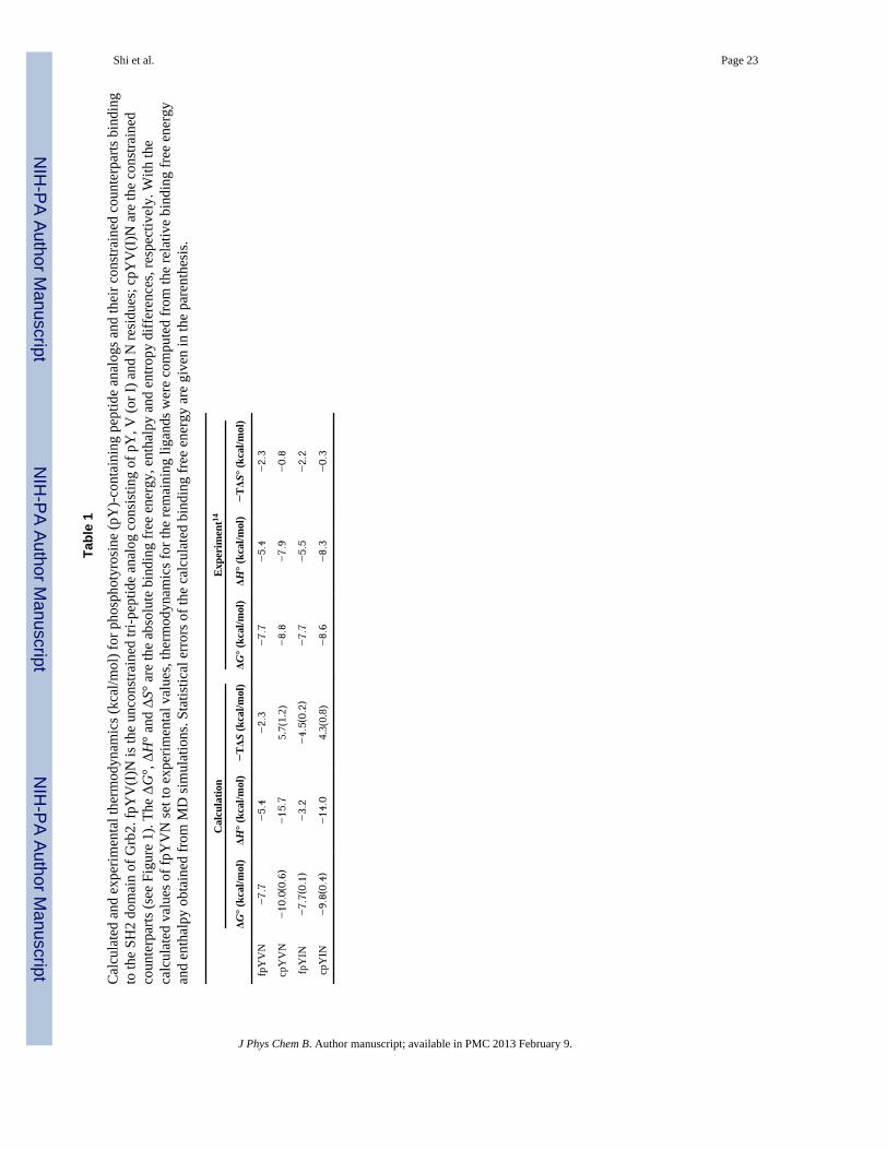
Calculated binding thermodynamics consistent with experimental measurementsWe evaluated the relative binding free energy and entropy for complex formation of theconstrained ligands cpYVN and cpYIN and their corresponding unconstrained analogsfpYVN and fpYIN with the Grb2 SH2 domain. The free energy and entropy were computedfrom molecular dynamics simulations using the AMOEBA polarizable force field, andresults are summarized and compared with the values that were determined experimentallyby ITC in Table 1.14 In order to facilitate the comparison of the calculated and experimentalvalues, we used the fpYVN ligand as the reference and set the calculated values for it tothose determined experimentally. The calculated thermodynamic parameters for the otherligands were then obtained by difference according to the relative binding free energies,entropies and enthalpies derived from the simulations.
The order of the calculated binding free energies correspond to those determinedexperimentally with fpYVN ≈ fpYIN < cpYIN < cpYVN. The calculated binding freeenergy difference between fpYVN and cpYVN was overestimated by about 1.2 kcal/molcompared to experiment, whereas the difference in free energies of the Val and Ile variantsmatch experimental results reasonably well. The transformation of fpYVN into cpYVNrequires that a covalent bond be created. A small change in the bond length corresponds tolarge energy fluctuations due to the stiff bond stretching and angle bending energy term.93,94
During the transformation of fpYVN into cpYVN in water or in their complexes with theGrb2 SH2 domain, 26 intermediate steps are necessary to obtain sufficient overlap betweenthe configurational spaces associated with neighboring steps. As illustrated in Figure 3, therelative free energies converge after 1.5 ns simulations for each intermediate step at differenttemperatures, with a deviation of less than 0.1 kcal/mol. REMD simulations for the ligandsin water at 48 different temperatures were performed to enhance the sampling near roomtemperature. Accordingly, the relative binding free energies for each pair of ligands areeffectively the result of 3 μs of simulation time.
Shi et al. Page 7
J Phys Chem B. Author manuscript; available in PMC 2013 February 9.
NIH
-PA Author Manuscript
NIH
-PA Author Manuscript
NIH
-PA Author Manuscript
Additional analyses were performed in order to scrutinize the convergence and effectivenessof the free energy calculations. To inspect the thermodynamic cycle closure, we reevaluatedthe relative binding free energy associated with the alchemical transformation fpYVN tocpYVN at 298 K after systematically skipping intermediate perturbation steps (skippingevery 4th, 5th … 13th steps). The results indicate that the relative binding free energyconverges within 0.3 kcal/mol after 22 to 26 perturbation steps. For neighboring steps duringthe perturbation, the histograms of potential energy differences from forward and backwardperturbation have been obtained and verified to overlap with each other. In addition,statistical inefficiency calculations95 show that ~22 ps and ~45 ps are needed for ligandsampling in water and in the complex to lose “memory” of their previous configurations,respectively. Further details of the thermodynamic cycle, energy overlap, and statisticalinefficiency tests are found in the Supporting Information.
The relative binding free energies were then decomposed into enthalpy and entropycontributions. Perturbation methods, including finite difference, single state perturbation, β-perturbation, modified β-perturbation, and the perturbation and correction method, provide aphysically rigorous evaluations of changes in entropy and enthalpy.78,96 However, comparedwith the free energy calculations, the entropy evaluated from computational methods hasmuch greater error. With the exception of the finite difference approach, all of the abovemethods suffer from numerical underflow problems for systems with large energyfluctuations such as solvated protein-ligand complexes. Recently, Wyczalkowski et al.97
calculated solvation entropy changes based on the analytical temperature derivative of theBennett Acceptance Ratio and Multistate Bennett Acceptance Ratio (mBAR) estimators.The BAR/mBAR approach to computing entropy is sensitive to energy fluctuations and thusrequires long simulation times to achieve convergence. Using the restraint release (RR)approach, Warshel et al.61 reported the binding entropy of three protein-ligand systems withencouraging agreement to experimental measurements. In the RR approach, multiplestructures of the ligands must be selected before free energy perturbation and quasiharmoniccalculations. Considering the potentially high flexibility of our peptide ligands, a largenumber of representative configurations are needed to account for the global minima in eachperturbation. In the current study, the REMD simulations that were performed on the freeligands in water already provided free energy data at different temperatures. Therefore, wetook advantage of the simulations at different temperatures and adopted the finite differencemethod that has been utilized to calculate entropy change for both small and relatively largesystems.60,75,98–100 We used a linear regression to fit the temperature dependence of therelative binding free energies at 288 K, 298 K and 308 K (see Computation Details). For allthe systems, the R2 values for the linear fit range from 0.81 to 0.97.
The calculated relative binding entropies and enthalpies for the two ligand pairs show thesame trends as experiment. Both calculation and experiment indicate that the constrainedligands bind to the Grb2 SH2 domain with less favorable binding entropy and morefavorable binding enthalpy than their more flexible controls (Table 1). Relative to theunconstrained ligands fpYVN and fpYIN, the increased binding affinity, or lower bindingfree energy, that is observed for the two constrained ligands cpYVN and cpYIN is attributedto an enthalpic advantage rather than an entropic one. Based upon the simulations,preorganization results in an unfavorable binding entropy change (higher −TΔS) of 8 kcal/mol, which is offset by an enthalpic gain of about −10 kcal/mol. Hence, althoughconstraining the ligands fpYVN and fpYIN increases their binding affinities, bothcomputational and experimental results show that this affinity enhancement does not arisefrom more favorable entropic factors as would normally be expected. The calculatedabsolute binding entropy −TΔS of the constrained cpYV(I)N ligands is unfavorable(positive), whereas slightly favorable (negative) values were obtained experimentally. Thisdifference is a reflection of our overestimation of the magnitude of the relative binding
Shi et al. Page 8
J Phys Chem B. Author manuscript; available in PMC 2013 February 9.
NIH
-PA Author Manuscript
NIH
-PA Author Manuscript
NIH
-PA Author Manuscript
entropy between constrained and unconstrained ligand pairs, even though the sign of therelative change was predicted correctly. The mutation of Val to Ile in both constrained andunconstrained ligands had an insignificant effect upon both the calculated and theexperimental values for binding enthalpies and entropies. Moreover, both the simulated andexperimental data seem to suggest that enthalpy/entropy compensation limits theenhancement to the binding affinity, as reflected by the linear relationship between entropyand enthalpy in Figure 4.
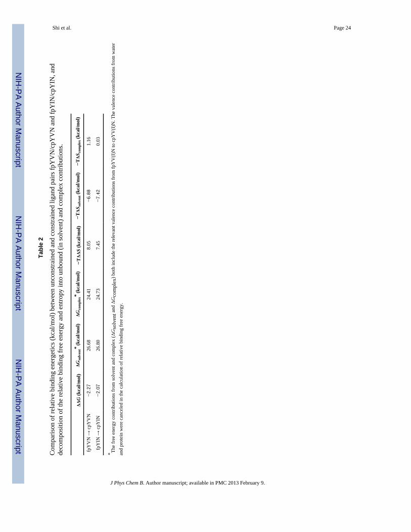
Binding is a process that involves ligand desolvation followed by formation of the protein-ligand complex, so the behavior of unbound ligands in water plays an important role in theoverall process. In the current study, the relative binding free energy and entropy wasevaluated as the difference between the free energy and entropy changes of the protein andthe ligands in solvent and complex environments. The entropy decomposition given in Table2 suggests that the majority of the unfavorable binding entropy observed for constrainingfpYVN and fpYIN arises from differences in the entropies of the free ligands in solventrather than in their complexes.
Effect of a chemical constraint on the organization of unbound ligandsThe concept underlying ligand preorganization is that constraining an unbound ligand in thethree-dimensional shape that corresponds to its bound structure will lead to a more favorableentropic term for binding. Both computational and structural studies show that thecyclopropane rings in cpYVN and cpYIN preorganize the atoms of the fpY replacement thatinteract with the protein in the bound conformations of fpYVN and fpYIN. Hence, in orderto probe why introducing a conformational constraint into the flexible ligands fpYVN andfpYIN did not result in the expected entropic advantage, we analyzed the simulatedstructures and dynamics of the bound and unbound forms of the pseudopeptides cpYVN andfpYVN.
The structures of the ligands extracted from molecular dynamics trajectories werehierarchically clustered based on the all-atom RMSD of the ligands using an average-linkage algorithm101 over 2 ns at 298 K. In an average-linkage algorithm, the distancebetween one cluster and another cluster is computed as the average distance from anymember of one cluster to any member of the other cluster. Recall that REMD simulationswith 48 replicas at various temperatures were performed to facilitate the sampling of theligand configurations at 298 K. This is equivalent to a total simulation time of 100 ns. Byusing 1.0 Å as the limit for the average distance to centroid, 22 and 20 clusters wereobtained for unconstrained and constrained ligand trajectories, respectively. Interestingly,the dominant configuration (35%) of fpYVN is a rather compact, macrocyclic-like structure(Figure 5) with a prominent intramolecular contact between the phosphate oxygen atomsand the amide N–H groups of the pY+2 residue. On the other hand, the cyclopropane ring inthe constrained ligands cpYVN and cpYIN prevents these functional groups from interactingwith each other, so these residues interact with water and are oriented in opposite directions.Superimposition of the dominant structure of cpYVN with the structure extracted from thecomplex14 reveals that the cyclopropane ring does preorganize the flexible pY replacementin its biologically active conformation. Thus, even though the cyclopropane ring locallypreorganizes the pY replacement, cpY, the simulations suggest that the constrained ligandspossess significantly more flexibility at the pY+1 and pY+2 positions relative to theirunconstrained counterparts.
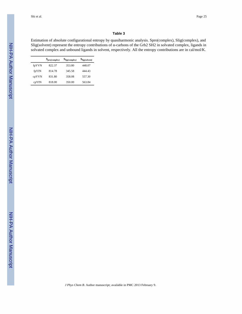
The entropy of both constrained and unconstrained ligands was estimated fromquasiharmonic analysis (Table 3). The absolute entropy calculated for solvated cpYVN is557 cal/mol/K, which is about 25% greater than the value of 448 cal mol/K−1 that wascalculated for fpYVN; this difference corresponds to about 30 kcal/mol in TΔS at room
Shi et al. Page 9
J Phys Chem B. Author manuscript; available in PMC 2013 February 9.
NIH
-PA Author Manuscript
NIH
-PA Author Manuscript
NIH
-PA Author Manuscript

temperature. The same trend is observed for fpYIN and cpYIN ligands, which haveessentially the same entropies as fpYVN and cpYVN, respectively. Vibration contributesabout 80% of the total entropy, while translation and rotation account for the remainder, sothe entropy difference between the constrained and unconstrained ligands arises primarilyfrom the vibrational components. We further decomposed the vibrational entropy intocontributions from individual atoms and found that the modes contributing most to theentropy difference arise from the pY+2 residue. This finding is consistent with the structuralanalysis above. Since the rigid cyclopropane ring separates the pY+2 residue and phosphategroup of the pY replacement in the constrained ligands, the pY+2 residue is well exposed insolvent and tends to be mobile. On the other hand, the unconstrained ligands lack this ring,so motion of the pY+2 residue is restrained by hydrogen bonding interactions with thephosphate moiety.
Water molecules also contribute to the entropy of the system and thus may affect bindingthermodynamics. The average number of intermolecular hydrogen-bonds between water andthe ligands were computed from the MD simulations at 298.79 K, and the results shown inTable 4 reveal noticeable differences between the constrained and unconstrained ligands.The constrained ligands are solvated by about two more water molecules on average thantheir unconstrained counterparts. The higher number of contacts with water molecules forconstrained ligands is consistent with the observation that constrained ligands are moreextended and better exposed to solvent (Table 4). The unconstrained ligands possess moreintramolecular hydrogen bonds than their constrained counterparts (2.6 vs. 1.4), so fewerwater molecules are associated with the unconstrained ligands. According to our calculation,the entropy contribution from ligand plays a dominant role in the binding energetics. Thesesimulations and quasiharmonic analyses suggest that the unconstrained ligands actually havelower absolute entropies than their constrained counterparts because they are able to formintramolecular hydrogen-bonding interactions between the phosphate group and the amidegroups of the terminal pY+2 residue. These computations thus offer a novel insight thataccounts for the observation that the constrained ligands bind with less favorable entropies.Namely, even though the cyclopropane ring in cpYVN and cpYIN orients the atoms of themore flexible pY replacements in fpYVN and fpYIN in their bound conformations, this ringprecludes the formation of the intramolecular interactions that lower the overall entropy offpYVN and fpYIN relative to cpYVN and cpYIN.
Flexibility of solvated complexes with unconstrained and constrained ligandsThere is the possibility that introducing a conformational constraint into a flexible moleculemight affect protein dynamics in the resultant complexes in a manner that could haveentropic consequences. In order to probe this question, structures of the four solvatedcomplexes were analyzed using the MD trajectories obtained from the free energycalculations. A comparison of the structures of the solvated complexes of the Grb2 SH2domain with each of fpYVN, cpYVN, fpYIN and cpYIN derived from simulations and X-ray crystallography yielded all-atom RMSDs of 1.6 Å, 1.6 Å, 1.1 Å and 1.5 Å, respectively,suggesting that the structures obtained computationally are generally in good agreementwith those determined by experiment. The RMSDs between the complexes of cpYVN andcpYIN and the complexes of fpYVN and fpYIN are both 1.4 Å. Representative structures ofthe complexes of fpYVN and cpYVN containing complexes from MD simulations arecompared with crystal structures in Figure 7.
The calculated B-factors about the mean coordinates of the simulations over 2 ns for all Cαatoms are shown in Figure 6. The four complexes share very similar fluctuation modes, andthe coordinates of most of the residues vary within only 1 Å, although residues in some ofthe loops fluctuate by as much as 2.1 Å. Structural variations in the BC loop (GluBC1–GluBC4) were observed upon comparing the crystal structures of complexes of the Grb2
Shi et al. Page 10
J Phys Chem B. Author manuscript; available in PMC 2013 February 9.
NIH
-PA Author Manuscript
NIH
-PA Author Manuscript
NIH
-PA Author Manuscript

SH2 domain with constrained and unconstrained ligands, although comparable variations inthe BC loops were also found in coexisting complexes in the asymmetric unit.11,14,102 In ourB-factor calculations, the fluctuations (1.5 to 2.0 Å) in the BC loops of the four complexesare the highest among all other loops. This observation suggests that the variations of the BCloop are more likely to result from the intrinsic flexibility of the loop rather than fromdifferences in the binding modes of the constrained and unconstrained ligands. Based on thesimulated B-factors in Figure 6, the B-factors for the protein backbone in the complexes ofthe constrained ligands are slightly greater than in the complexes of their unconstrainedcounterparts. Although the trends in the B-factors obtained computationally andexperimentally are the same, the differences are more prominent in the experimentalvalues.14
The entropies calculated from quasiharmonic analysis (Table 3) suggest that the Cα-atoms inthe Grb2 SH2 domain complexed with each of the four different ligands have similarentropies of about 820 cal/mol/K. The entropies of the domain in the complexes of theconstrained ligands cpYVN and cpYIN are slightly higher than in the complexes with theirunconstrained controls (about 1%). The higher entropies of the domain in the complexes ofthe constrained ligands suggest a more favorable contribution to binding free energy,however, the opposite trend is seen by both experiment and calculation. Based on theanalyses above, we are unable to find evidence that a change in dynamics of the SH2domain is responsible for differences in the experimentally observed binding entropies forthe various complexes.
The structures of fpYVN, cpYVN, fpYIN, and cpYIN in their respective complexes with theSH2 domain were also examined. The structures of the complexes were clustered based onRMSD of all atoms in the ligands from all MD snapshots using the same method describedabove for free ligands. For complexes containing fpYVN and cpYVN, there is one dominantconformation of the ligand that forms strong polar interactions with the protein, whereasthere are two dominant conformers for fpYIN and cpYIN because of the flexibility of the pY+1 (Ile) residue. Other portions of the pseudopeptides fpYIN and cpYIN are highly similarto each other as well as to fpYVN and cpYVN. The most representative structures offpYVN and cpYVN are depicted in Figure 7, while the corresponding structures for fpYINand cpYIN not shown due to their high similarity. The entropies calculated fromquasiharmonic analysis of the four ligands in the bound state range from 345.6 to 358.0 cal/mol/K, with the largest entropy difference being only 12 cal/mol/K, which is withinstatistical error, between cpYVN and fpYIN. Therefore, quasiharmonic analysis of theprotein and the ligands in the respective complexes suggests that the entropic advantage forbinding of the unconstrained ligands fpYVN and fpYIN does not arise from any differencesin the entropy of either the protein or the ligands in bound state.
SUMMARY AND CONCLUSIONSIn order to probe the origin of the unexpected entropic consequences observed for binding ofa series of constrained and flexible phosphotyrosine-derived peptide analogs to the Grb2SH2 domain,14 we performed a series of calculations involving alchemical transformationsat different temperatures on two sets of these analogs. Consistent with experimental results,these computations predicted that the binding affinity of the unconstrained peptidesfpYV(I)N for the Grb2 SH2 domain is lower than that of the corresponding constrainedpeptides cpYV(I)N and that the mutation of V to I is not accompanied by significantchanges in binding free energy. The experimental observation that unconstrained peptideanalogs bind with more favorable entropies but significantly less favorable enthalpies thantheir constrained counterparts is also well reproduced by our computations, but thedifferences in the relative binding entropy and enthalpy components are overestimated. Even
Shi et al. Page 11
J Phys Chem B. Author manuscript; available in PMC 2013 February 9.
NIH
-PA Author Manuscript
NIH
-PA Author Manuscript
NIH
-PA Author Manuscript
though these simulations nicely reproduce those trends observed experimentally, furtherrefinements to the method are needed in order to improve the accuracy of predictingdifferences in the relative binding thermodynamic parameters. Sampling of theconformations of the pseudopeptide ligands in solution is the most demanding calculation inthis study. Some 26 perturbation steps were applied to introduce a bond between carbonatoms that are separated by two bonds, and REMD simulations with 48 replicas wereperformed at each perturbation step. Hence, in order to evaluate the relative solvation freeenergy/entropy of the two pairs of peptide ligands, about 6 μs MD simulations wereperformed; however, the statistical uncertainties remain significant. More advancedsampling techniques beyond the first-order scheme adopted in this study are needed in orderto compute the solvation and binding free energy more efficiently and precisely.
We analyzed the structures and dynamics of ligands in solution and in their complexes withthe SH2 domain in order to probe the molecular origin of the effects of ligandpreorganization on binding thermodynamics in this system. Conformational clustering andquasiharmonic analysis of the free ligands in solution suggest that the unconstrained ligandspossess significantly lower entropy than their constrained counterparts. This unexpectedfinding is the consequence of intramolecular hydrogen bonding interactions between thephosphate group of the flexible pY replacement and the C-terminal amide moieties of thepY+2 residue that lead to a more compact and rigid, macrocyclic-like structure. Thepresence of the cyclopropane ring in the constrained pY replacement prevents thisinteraction, thereby resulting in more extended conformations in solution. MD simulationsof the protein-ligand complexes show that the unconstrained and constrained ligands sharesimilar binding modes; the distribution of conformations of the bound forms of theunconstrained and constrained ligands are comparable as are their nonbonded interactionswith the domain. These findings are consistent with the structures determinedexperimentally by X-ray crystallography.
These simulations reveal an important caveat that has not been previously acknowledgedregarding the use of ligand preorganization, which is widely presumed to have a favorableeffect upon binding entropy as a general design strategy. Namely, this work demonstratesthat introducing a conformational constraint into a flexible ligand does not necessarilylower its entropy in solution, because the flexibility of a ligand in solution is determined bya subtle balance between any intramolecular interactions and the intermolecular interactionsbetween the ligand and its aqueous environment. Comparing the dominant structures of theconstrained ligands in their bound and unbound states shows that the cyclopropane ring inthe constrained ligands, cpYVN and cpYIN, locally constrains and orients functionality onthe flexible phosphotyrosine replacement in fpYVN and fpYIN in the bound conformationas predicted from modeling studies. However, the macrocyclic-like structures of fpYVN andfpYIN in solution, which do not correspond to their bound conformations, reduce the globalflexibility of these ligands to an even greater degree than the cyclopropane ring. Because thebinding entropies for fpYVN and fpYIN are more favorable than for their constrainedderivatives cpYVN and cpYIN, it is now apparent that one cannot think simply in terms ofintroducing constraints to stabilize the biologically active conformation of a small moleculeas a strategy for enhancing ligand binding affinities. Rather, lowering the entropy of a ligandin any way that allows it to adopt its bound conformation can lead to more favorablebinding entropies. These studies also reveal that knowing the structures of small moleculesin their unbound states is a critical prerequisite to correlating changes in their structures withprotein binding entropies and enthalpies.
Supplementary MaterialRefer to Web version on PubMed Central for supplementary material.
Shi et al. Page 12
J Phys Chem B. Author manuscript; available in PMC 2013 February 9.
NIH
-PA Author Manuscript
NIH
-PA Author Manuscript
NIH
-PA Author Manuscript
AcknowledgmentsThis work was supported by grants from the National Institute of General Medical Sciences (R01GM079686) andTeragrid MCB100057. S. F. M. is grateful to the National Institutes of Health (GM 84965), the National ScienceFoundation (CHE 0750329), the Robert A. Welch Foundation (F-652), and the Norman Hackerman AdvancedResearch Program for support. The authors thank Prof. Ron Elber for the suggestions in research design and helpwith technical details. The authors thank Zhen Xia, Dr. Chunli Yan and Dr. John H. Clements for importantdiscussions.
References1. Mann, A. In The Practice of Medicinal Chemistry. 2. Academic Press; London: 2003.2. Lauri G, Bartlett PA. J Comput Aid Mol Des. 1994; 8:51–66.3. Page MI, Jencks WP. P Natl Acad Sci USA. 1971; 68:1678–1683.4. Khan AR, Parrish JC, Fraser ME, Smith WW, Bartlett PA, James MNG. Biochemistry. 1998;
37:16839–16845. [PubMed: 9836576]5. Widlanski T, Bender SL, Knowles JR. J Am Chem Soc. 1989; 111:2299–2300.6. Bartlett PA, Smith WW. J Am Chem Soc. 1998; 120:4622–4628.7. Davidson JP, Lubman O, Rose T, Waksman G, Martin SF. J Am Chem Soc. 2002; 124:205–215.
[PubMed: 11782172]8. Davidson JP, Martin SF. Tetrahedron Lett. 2000; 41:9459–9464.9. Verdine GL, Harrison BA, Gierasch TM, Neilan C, Pasternak GW. J Am Chem Soc. 2002;
124:13352–13353. [PubMed: 12418865]10. Udugamasooriya DG, Spaller MR. Biopolymers. 2008; 89:653–667. [PubMed: 18335423]11. DeLorbe JE, Clements JH, Whiddon BB, Martin SF. Acs Med Chem Lett. 2010; 1:448–452.
[PubMed: 21116482]12. Martin SF. Pure Appl Chem. 2007; 79:193–200.13. Martin SF, Benfield AP, Teresk MG, Plake HR, DeLorbe JE, Millspaugh LE. Angew Chem Int
Edit. 2006; 45:6830–6835.14. DeLorbe JE, Clements JH, Teresk MG, Benfield AP, Plake HR, Millspaugh LE, Martin SF. J Am
Chem Soc. 2009; 131:16758–16770. [PubMed: 19886660]15. Liu L, Guo QX. Chem Rev. 2001; 101:673–695. [PubMed: 11712500]16. Gilli P, Ferretti V, Gilli G, Borea PA. J Phys Chem. 1994; 98:1515–1518.17. Dunitz JD. Chem Biol. 1995; 2:709–712. [PubMed: 9383477]18. Cooper A, Johnson CM, Lakey JH, Nollmann M. Biophys Chem. 2001; 93:215–230. [PubMed:
11804727]19. Breslauer KJ, Remeta DP, Chou WY, Ferrante R, Curry J, Zaunczkowski D, Snyder JG, Marky
LA. P Natl Acad Sci USA. 1987; 84:8922–8926.20. Grunwald E, Steel C. J Am Chem Soc. 1995; 117:5687–5692.21. Rekharsky M, Inoue Y. J Am Chem Soc. 2000; 122:4418–4435.22. Chen W, Chang CE, Gilson MK. Biophys J. 2004; 87:3035–3049. [PubMed: 15339804]23. Al Omari MM, Zughul MB, Davies JED, Badwan AA. J Incl Phenom Macro. 2007; 57:379–384.24. Cornish-Bowden A. J Biosciences. 2002; 27:121–126.25. Krug RR, Hunter WG, Grieger RA. J Phys Chem. 1976; 80:2335–2341.26. Leung DH, Bergman RG, Raymond KN. J Am Chem Soc. 2008; 130:2798–2805. [PubMed:
18257565]27. Sharp K. Protein Sci. 2001; 10:661–667. [PubMed: 11344335]28. Reynolds CH, Holloway MK. Medicinal Chemistry Letters. 2011; 2:433.29. Williams DH, Stephens E, O’Brien DP, Zhou M. Angew Chem Int Edit. 2004; 43:6596–6616.30. Exner O. Collect Czech Chem C. 1975; 40:2762–2780.31. Exner O, Beranek V. Collect Czech Chem C. 1973; 38:781–798.32. Schowen RL. J Pharm Sci. 1967; 56:931. [PubMed: 5340702]
Shi et al. Page 13
J Phys Chem B. Author manuscript; available in PMC 2013 February 9.
NIH
-PA Author Manuscript
NIH
-PA Author Manuscript
NIH
-PA Author Manuscript

33. Linert W, Jameson RF. Chem Soc Rev. 1989; 18:477–505.34. Zhou HX, Gilson MK. Chem Rev. 2009; 109:4092–4107. [PubMed: 19588959]35. Bradshaw JM, Waksman G. Adv Protein Chem. 2002; 61:161–210. [PubMed: 12461824]36. Ladbury JE. Thermochim Acta. 2001; 380:209–215.37. Sturtevant JM. P Natl Acad Sci USA. 1977; 74:2236–2240.38. Jorgensen WL. Science. 2004; 303:1813–1818. [PubMed: 15031495]39. Gohlke H, Klebe G. Angew Chem Int Edit. 2002; 41:2645–2676.40. Brandsdal BO, Osterberg F, Almlof M, Feierberg I, Luzhkov VB, Aqvist J. Adv Protein Chem.
2003; 66:123. [PubMed: 14631818]41. Mobley DL, Dill KA. Structure. 2009; 17:489–498. [PubMed: 19368882]42. Lamb ML, Jorgensen WL. Curr Opin Chem Biol. 1997; 1:449–457. [PubMed: 9667895]43. Gilson MK, Zhou HX. Annu Rev Bioph Biom. 2007; 36:21–42.44. Pande VS, Chodera JD, Mobley DL, Shirts MR, Dixon RW, Branson K. Curr Opin Struc Biol.
2011; 21:150–160.45. Shirts MR, Mobley DL, Chodera JD. Annu Rep Comput Chem. 2007; 3:41–57.46. Andricioaei I, Karplus M. J Chem Phys. 2001; 115:6289–6292.47. Brooks BR, Janezic D, Karplus M. J Comput Chem. 1995; 16:1522–1542.48. Gohlke H, Case DA. J Comput Chem. 2004; 25:238–250. [PubMed: 14648622]49. Tidor B, Karplus M. J Mol Biol. 1994; 238:405–414. [PubMed: 8176732]50. Case DA. Curr Opin Struc Biol. 1994; 4:285–290.51. Bohm HJ. J Comput Aid Mol Des. 1998; 12:309–323.52. Jackson RN, Kulharia M, Goody RS. J Chem Inf Model. 2008; 48:1990–1998. [PubMed:
18767831]53. Muegge I, Martin YC. J Med Chem. 1999; 42:791–804. [PubMed: 10072678]54. Carlsson J, Aqvist J. Abstr Pap Am Chem S. 2005; 229:U783–U784.55. Hermans J, Wang L. J Am Chem Soc. 1997; 119:2707–2714.56. Luo HB, Sharp K. P Natl Acad Sci USA. 2002; 99:10399–10404.57. Luo R, Gilson MK. J Am Chem Soc. 2000; 122:2934–2937.58. Swanson JMJ, Henchman RH, McCammon JA. Biophys J. 2004; 86:67–74. [PubMed: 14695250]59. Baginski M, Fogolari F, Briggs JM. J Mol Biol. 1997; 274:253–267. [PubMed: 9398531]60. Olano LR, Rick SW. J Am Chem Soc. 2004; 126:7991–8000. [PubMed: 15212549]61. Warshel A, Singh N. Proteins. 2010; 78:1724–1735. [PubMed: 20186973]62. Ren PY, Ponder JW. J Comput Chem. 2002; 23:1497–1506. [PubMed: 12395419]63. Ren PY, Ponder JW. J Phys Chem B. 2003; 107:5933–5947.64. Ponder, JW.; Case, DA. Adv Protein Chem. Vol. 66. Academic Press Inc; San Diego: 2003. Force
fields for protein simulations; p. 2765. Ponder JW, Wu CJ, Ren PY, Pande VS, Chodera JD, Schnieders MJ, Haque I, Mobley DL,
Lambrecht DS, DiStasio RA, Head-Gordon M, Clark GNI, Johnson ME, Head-Gordon T. J PhysChem B. 2010; 114:2549–2564. [PubMed - in process]. [PubMed: 20136072]
66. Grossfield A, Ren PY, Ponder JW. J Am Chem Soc. 2003; 125:15671–15682. [PubMed:14664617]
67. Grossfield A, Ren PY, Ponder JW. Biophys J. 2003; 84:94A–94A.68. Jiao D, King C, Grossfield A, Darden TA, Ren PY. J Phys Chem B. 2006; 110:18553–18559.
[PubMed: 16970483]69. Wu JC, Piquemal J-P, Chaudret R, Reinhardt P, Ren P. J Chem Theory Comput. 2010; 6:2059–
2070. [PubMed: 21116445]70. Shi Y, Wu CJ, Ponder JW, Ren PY. J Comput Chem. 2011; 32:967–977. [PubMed: 20925089]71. Pappu RV, Drozdov AN, Grossfield A. J Am Chem Soc. 2004; 126:2574–2581. [PubMed:
14982467]72. Jiao D, Golubkov PA, Darden TA, Ren P. P Natl Acad Sci USA. 2008; 105:6290–6295.
Shi et al. Page 14
J Phys Chem B. Author manuscript; available in PMC 2013 February 9.
NIH
-PA Author Manuscript
NIH
-PA Author Manuscript
NIH
-PA Author Manuscript
73. Jiao D, Zhang JJ, Duke RE, Li GH, Schnieders MJ, Ren PY. J Comput Chem. 2009; 30:1701–1711. [PubMed: 19399779]
74. Shi, Y.; Jiao, D.; Schnieders, MJ.; Ren, P. Engineering in Medicine and Biology Society, 2009.EMBC 2009; Annual International Conference of the IEEE; 2009. p. 2328-2331.
75. Kubo MM, Gallicchio E, Levy RM. J Phys Chem B. 1997; 101:10527–10534.76. Smith DE, Haymet ADJ. J Chem Phys. 1993; 98:6445–6454.77. Tsunekawa N, Miyagawa H, Kitamura K, Hiwatari Y. J Chem Phys. 2002; 116:6725–6730.78. Lu N, Kofke DA, Woolf TB. J Phys Chem B. 2003; 107:5598–5611.79. Bennett CH. Journal of Computational Physics. 1976; 22:245–268.80. Chang CE, Chen W, Gilson MK. J Chem Theory Comput. 2005; 1:1017–1028.81. Ponder, JW. Washington University Medical School. 2010. http://dasher.wustl.edu/tinker/82. Ren P, Wu C, Ponder JW. J Chem Theory Comput. 2011; 7:3027–3034.83. Stone AJ, Alderton M. Molecular Physics. 1985; 56:1047–1064.84. Stone AJ, Alderton M. Molecular Physics. 2002; 100:221–233.85. Sugita Y, Okamoto Y. Chem Phys Lett. 1999; 314:141–151.86. Yoda T, Sugita Y, Okamoto Y. Chem Phys Lett. 2004; 386:460–467.87. Zhou RH. P Natl Acad Sci USA. 2003; 100:13280–13285.88. Patriksson A, van der Spoel D. Phys Chem Chem Phys. 2008; 10:2073–2077. [PubMed:
18688361]89. Pearlman DA, Case DA, Caldwell JW, Ross WS, Cheatham TE, Debolt S, erguson D, Seibel G,
Kollman P. Comput Phys Commun. 1995; 91:1–41.90. Essmann U, Perera L, Berkowitz ML, Darden T, Lee H, Pedersen LG. J Chem Phys. 1995;
103:8577–8593.91. Darden T, York D, Pedersen L. J Chem Phys. 1993; 98:10089–10092.92. Sagui C, Darden TA. Annu Rev Bioph Biom. 1999; 28:155–179.93. Boresch S, Karplus M. J Phys Chem A. 1999; 103:103–118.94. Boresch S, Karplus M. J Phys Chem A. 1999; 103:119–136.95. Allen, MP.; Tildesley, DJ. Computer simulation of liquids. Clarendon Press; Oxford: 1989.96. Chipot, C.; Pohorille, A. Theory and Applications in Chemistry and Biology. Springer; 2007. Free
Energy Calculations.97. Wyczalkowski MA, Vitalis A, Pappu RV. J Phys Chem B. 2010; 114:8166–8180. [PubMed:
20503993]98. Syme NR, Dennis C, Bronowska A, Paesen GC, Homans SW. J Am Chem Soc. 2010; 132:8682–
8689. [PubMed: 20524663]99. Peter C, Oostenbrink C, van Dorp A, van Gunsteren WF. J Chem Phys. 2004; 120:2652–2661.
[PubMed: 15268408]100. Aqvist J, Carlsson J. J Phys Chem B. 2009; 113:10255–10260. [PubMed: 19580304]101. Lee, JK. Statistical bioinformatics: a guide for life and biomedical science researchers. John
Wiley & Sons, Inc; Hoboken, New Jersey: 2010.102. Martin SF, Clements JH, DeLorbe JE, Benfield AP. Acta Crystallogr D. 2010; 66:1101–1115.
[PubMed: 20944243]103. DeLano, WL. DeLano Scientific. San Carlos, CA, USA: 2002. http://www.pymol.org
Shi et al. Page 15
J Phys Chem B. Author manuscript; available in PMC 2013 February 9.
NIH
-PA Author Manuscript
NIH
-PA Author Manuscript
NIH
-PA Author Manuscript

Figure 1.Chemical structures of the ligands studied and the perturbation scheme. A. fpYVN; B.cpYVN; C. fpYIN; D. cpYIN.
Shi et al. Page 16
J Phys Chem B. Author manuscript; available in PMC 2013 February 9.
NIH
-PA Author Manuscript
NIH
-PA Author Manuscript
NIH
-PA Author Manuscript
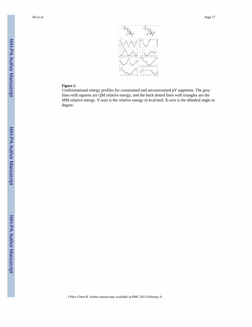
Figure 2.Conformational energy profiles for constrained and unconstrained pY segments. The greylines with squares are QM relative energy, and the back dotted lines with triangles are theMM relative energy. Y-axis is the relative energy in kcal/mol. X-axis is the dihedral angle indegree.
Shi et al. Page 17
J Phys Chem B. Author manuscript; available in PMC 2013 February 9.
NIH
-PA Author Manuscript
NIH
-PA Author Manuscript
NIH
-PA Author Manuscript
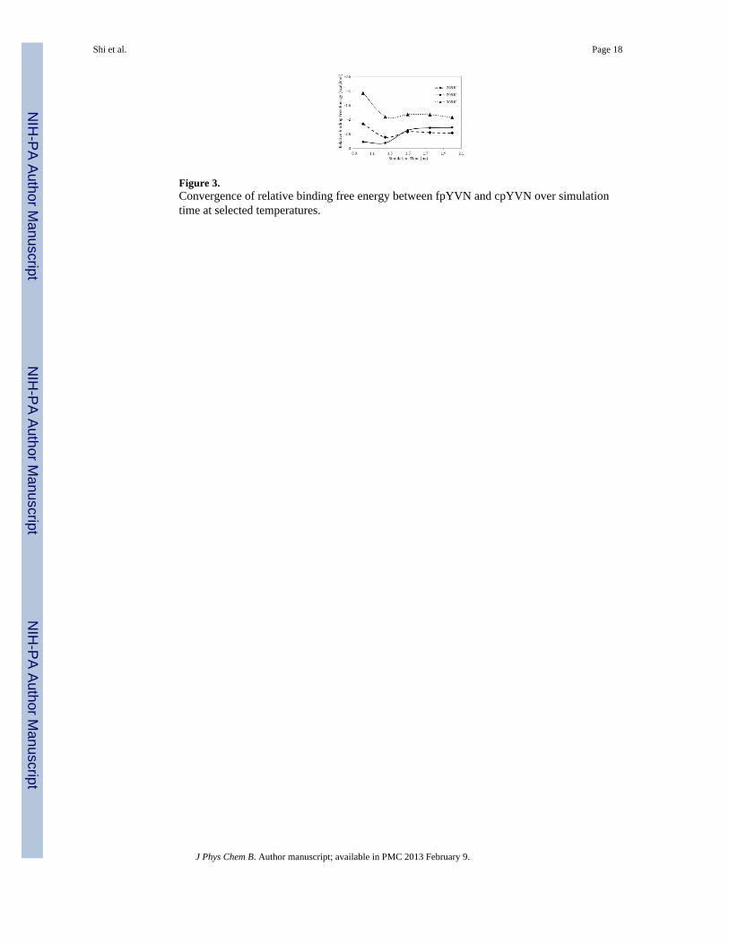
Figure 3.Convergence of relative binding free energy between fpYVN and cpYVN over simulationtime at selected temperatures.
Shi et al. Page 18
J Phys Chem B. Author manuscript; available in PMC 2013 February 9.
NIH
-PA Author Manuscript
NIH
-PA Author Manuscript
NIH
-PA Author Manuscript

Figure 4.Correlation between binding enthalpy and binding entropy of fpYVN, fpYIN, cpYVN andcpYIN. Black diamonds are calculated values; Grey squares are experimental data. Bothcalculated and experimental binding enthalpy and entropy of fpYVN are shifted to zero forcomparison purpose.
Shi et al. Page 19
J Phys Chem B. Author manuscript; available in PMC 2013 February 9.
NIH
-PA Author Manuscript
NIH
-PA Author Manuscript
NIH
-PA Author Manuscript

Figure 5.Clustering of the solvated ligand structures for fpYVN (pink) and cpYVN (blue). The mostrepresentative structures are plotted for clusters higher than 5%. The ligands structures fromcrystal (in transparency) are superposed onto the most dominant configuration.
Shi et al. Page 20
J Phys Chem B. Author manuscript; available in PMC 2013 February 9.
NIH
-PA Author Manuscript
NIH
-PA Author Manuscript
NIH
-PA Author Manuscript
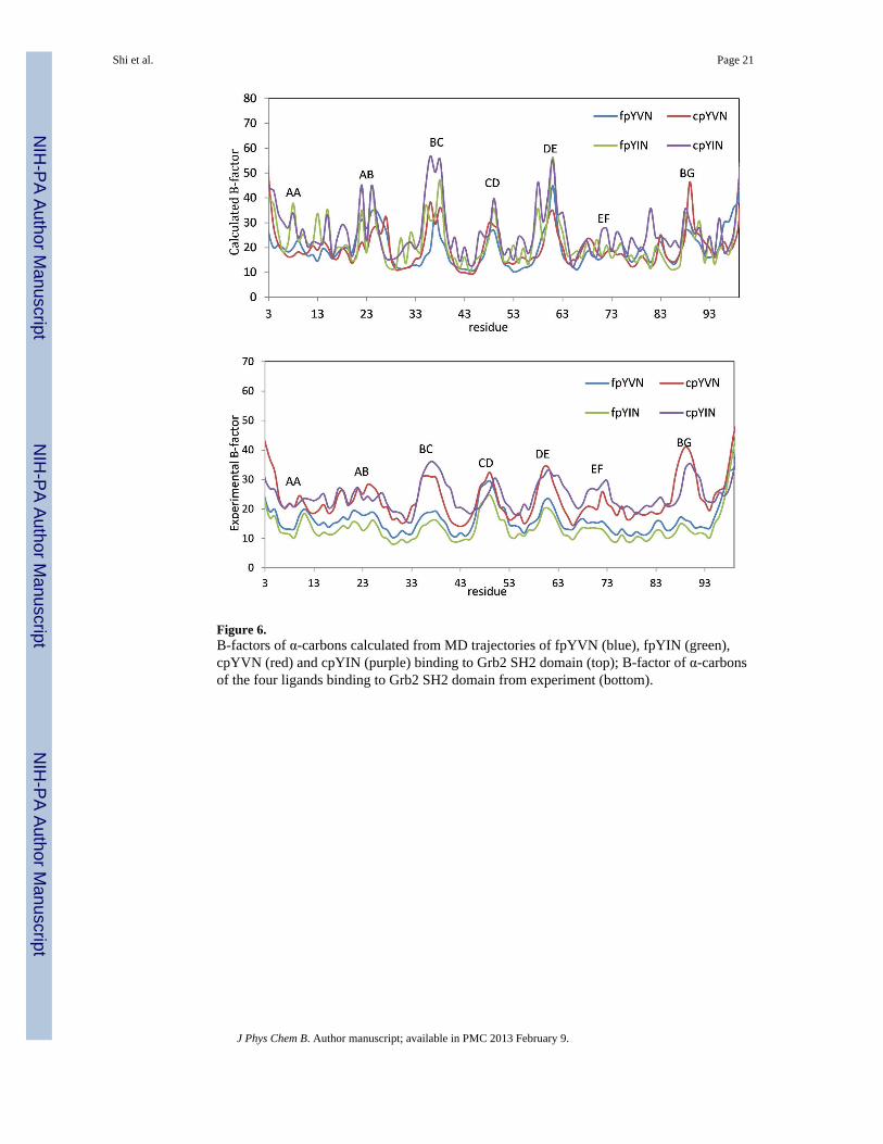
Figure 6.B-factors of α-carbons calculated from MD trajectories of fpYVN (blue), fpYIN (green),cpYVN (red) and cpYIN (purple) binding to Grb2 SH2 domain (top); B-factor of α-carbonsof the four ligands binding to Grb2 SH2 domain from experiment (bottom).
Shi et al. Page 21
J Phys Chem B. Author manuscript; available in PMC 2013 February 9.
NIH
-PA Author Manuscript
NIH
-PA Author Manuscript
NIH
-PA Author Manuscript
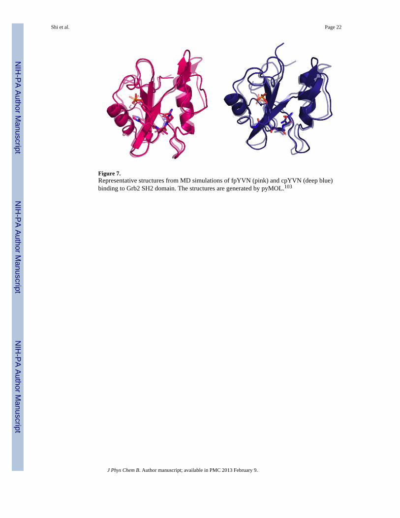
Figure 7.Representative structures from MD simulations of fpYVN (pink) and cpYVN (deep blue)binding to Grb2 SH2 domain. The structures are generated by pyMOL.103
Shi et al. Page 22
J Phys Chem B. Author manuscript; available in PMC 2013 February 9.
NIH
-PA Author Manuscript
NIH
-PA Author Manuscript
NIH
-PA Author Manuscript
NIH
-PA Author Manuscript
NIH
-PA Author Manuscript
NIH
-PA Author Manuscript
Shi et al. Page 23
Tabl
e 1
Cal
cula
ted
and
expe
rimen
tal t
herm
odyn
amic
s (kc
al/m
ol) f
or p
hosp
hoty
rosi
ne (p
Y)-
cont
aini
ng p
eptid
e an
alog
s and
thei
r con
stra
ined
cou
nter
parts
bin
ding
to th
e SH
2 do
mai
n of
Grb
2. fp
YV
(I)N
is th
e un
cons
train
ed tr
i-pep
tide
anal
og c
onsi
stin
g of
pY
, V (o
r I) a
nd N
resi
dues
; cpY
V(I
)N a
re th
e co
nstra
ined
coun
terp
arts
(see
Fig
ure
1). T
he Δ
G°, ΔH
° and
ΔS°
are
the
abso
lute
bin
ding
free
ene
rgy,
ent
halp
y an
d en
tropy
diff
eren
ces,
resp
ectiv
ely.
With
the
calc
ulat
ed v
alue
s of f
pYV
N se
t to
expe
rimen
tal v
alue
s, th
erm
odyn
amic
s for
the
rem
aini
ng li
gand
s wer
e co
mpu
ted
from
the
rela
tive
bind
ing
free
ene
rgy
and
enth
alpy
obt
aine
d fr
om M
D si
mul
atio
ns. S
tatis
tical
err
ors o
f the
cal
cula
ted
bind
ing
free
ene
rgy
are
give
n in
the
pare
nthe
sis.
Cal
cula
tion
Exp
erim
ent14
ΔG° (
kcal
/mol
)ΔH
° (kc
al/m
ol)
−TΔS
(kca
l/mol
)ΔG
° (kc
al/m
ol)
ΔH° (
kcal
/mol
)−
TΔS
° (kc
al/m
ol)
fpY
VN
−7.7
−5.4
−2.3
−7.7
−5.4
−2.3
cpY
VN
−10.0(0.6)
−15.7
5.7(
1.2)
−8.8
−7.9
−0.8
fpY
IN−7.7(0.1)
−3.2
−4.5(0.2)
−7.7
−5.5
−2.2
cpY
IN−9.8(0.4)
−14.0
4.3(
0.8)
−8.6
−8.3
−0.3
J Phys Chem B. Author manuscript; available in PMC 2013 February 9.
NIH
-PA Author Manuscript
NIH
-PA Author Manuscript
NIH
-PA Author Manuscript
Shi et al. Page 24
Tabl
e 2
Com
paris
on o
f rel
ativ
e bi
ndin
g en
erge
tics (
kcal
/mol
) bet
wee
n un
cons
train
ed a
nd c
onst
rain
ed li
gand
pai
rs fp
YV
N/c
pYV
N a
nd fp
YIN
/cpY
IN, a
ndde
com
posi
tion
of th
e re
lativ
e bi
ndin
g fr
ee e
nerg
y an
d en
tropy
into
unb
ound
(in
solv
ent)
and
com
plex
con
tribu
tions
.
ΔΔG
(kca
l/mol
)ΔG
solv
ent*
(kca
l/mol
)ΔG
com
plex
* (k
cal/m
ol)
−TΔΔ
S (k
cal/m
ol)
−TΔS
solv
ent (
kcal
/mol
)−
TΔS
com
plex
(kca
l/mol
)
fpY
VN
→ c
pYV
N−2.27
26.6
824
.41
8.05
−6.88
1.16
fpY
IN →
cpY
IN−2.07
26.8
024
.73
7.45
−7.42
0.03
* The
free
ene
rgy
cont
ribut
ions
from
solv
ent a
nd c
ompl
ex (Δ
Gso
lven
t and
ΔG
com
plex
) bot
h in
clud
e th
e re
leva
nt v
alen
ce c
ontri
butio
ns fr
om fp
YV
(I)N
to c
pYV
(I)N
. The
val
ence
con
tribu
tions
from
wat
eran
d pr
otei
n w
ere
canc
eled
in th
e ca
lcul
atio
n of
rela
tive
bind
ing
free
ene
rgy.
J Phys Chem B. Author manuscript; available in PMC 2013 February 9.
NIH
-PA Author Manuscript
NIH
-PA Author Manuscript
NIH
-PA Author Manuscript
Shi et al. Page 25
Table 3
Estimation of absolute configurational entropy by quasiharmonic analysis. Spro(complex), Slig(complex), andSlig(solvent) represent the entropy contributions of α-carbons of the Grb2 SH2 in solvated complex, ligands insolvated complex and unbound ligands in solvent, respectively. All the entropy contributions are in cal/mol/K.
Spro(complex) Slig(complex) Slig(solvent)
fpYVN 822.37 353.80 448.07
fpYIN 814.78 345.58 444.43
cpYVN 831.80 358.08 557.30
cpYIN 818.00 350.00 563.84
J Phys Chem B. Author manuscript; available in PMC 2013 February 9.

NIH
-PA Author Manuscript
NIH
-PA Author Manuscript
NIH
-PA Author Manuscript
Shi et al. Page 26
Table 4
The average numbers of intermolecular water-ligand hydrogen-bonds around the four solvated ligands, and theaverage numbers of intramolecular hydrogen bonds within the ligands in solution at 298.79 K.
fpYVN fpYIN cpYVN cpYIN
Intermolecular H-bond 33.5 33.2 35.7 35.2
Intramolecular H-bond 2.7 2.4 1.4 1.4
J Phys Chem B. Author manuscript; available in PMC 2013 February 9.
Related Documents