Neuron, Vol. 14, 217-228, February, 1995, Copyright © 1995 by Cell Press Principles of Human Brain Organization Derived from Split-Brain Studies Review Michael S. Gazzaniga Center for Neuroscience University of California, Davis Davis, California 95616 Studies of damaged human brains have always intrigued neuroscientists. The rich symptomatology that can result is riveting to all who experience examination of such pa- tients. Yet, one of the concerns of basic scientists is that structure-function correlates are difficult to make in the damaged brain, since the lesions are naturally occurring and usually quite diffuse. In addition to the problem of quantitating the lesion, there has always been concern whether the information gained about the brain in the pres- ence of lesions is all that useful in understanding normal brain mechanisms. Are observed effects due to the dam- age of specific areas or to distant effects? These and other issues have limited the general acceptance of the findings from clinical studies. One of the immediate appeals of the study of patients with surgical division of the forebrain is that the separate functions of the two cerebral hemispheres can be studied readily in the absence of focal damage. Also, the callosal surgery, while producing damage to the brain, was dis- crete damage to a fiber system and not to nuclear areas. In 1961, R. W. Sperry and his colleagues commenced a series of studies on patients who had undergone surgical section of the cerebral commissures in an effort to control their otherwise intractable epilepsy (Bogen et al., 1965). Studies continue on the original patients as well as others and still shed light on the nature of both cortical and sub- cortical neural networks. In what follows, I review some aspects of this work to illustrate how this initial work has progressed from the early sixties until the time of Sperry's death in the spring of 1994. Patients who undergo so-called split-brain surgery all suffer from intractable epilepsy. Prior to their surgery, ex- tensive attempts are made to control their seizures medi- cally. Failure to do so, along with other clinical criteria, finds them candidates for surgical division of the corpus callosum, and in some cases the anterior commissure as well. Although these patients are not normal, the onset of their epilepsy varies and has different etiologies. It has not been possible to correlate any of these variations in their neurologic history with the pattern of results obtained from the cognitive studies. It is known from other data that, in order for there to be significant changes in the normal patterns of cerebral lateralization, a large lesion must oc- cur to one side of the brain in early childhood (Rasmussen and Milner, 1977). In the original studies on the California series of patients, it was necessary to rely on surgical notes for determination of the completeness of the surgical sections. In more re- cent years, both magnetic resonance imaging (MRI) and other electrical brain mapping techniques have provided a more accurate representation of the extent of the surgical sections. Accu rate documentation of the extent of callosal section becomes crucial for understanding the organiza- tion of the cerebral commissure. The main methods of testing the perceptual and cogni- tive functions of each hemisphere have not changed in principle over the years. However, there have been signifi- cant advances in the technologies used to present stimuli to the surgically separated hemispheres. In the early years, information was visually lateralized to one or the other hemispheres by quickly flashing stimuli to one or the other visual field using electronic shutters attached to slide projectors (see Figure 1). As the patient fixates on a point in space, information flashed to the left of fixation is presented exclusively to the right hemisphere. The quick flashing is necessary to control for unwanted eye move- ments, which would redirect the information into the un- wanted hemisphere. This method has been replaced by using computer-based stimuli presentation arrangements. More importantly, the development of an image stabilizing system, used in concert with a Purkinje eye tracker, now permits sustained presentation of information to either vi- sual field, and therefore, either hemisphere. Accordingly, if a subject moves his or her eyes away from fixation, the stabilizing system moves the stimulus with the eyes and thereby prevents the information from being presented to the wrong hemisphere. This technological development has allowed for new findings on both the neurological and psychological aspects of hemisphere disconnection. Fundamental Principles Arising from Initial Studies The original reports on the California series of patients dealt with a number of fundamental issues concerning the basic psychological properties of the separated cerebral hemispheres, as well as basic issues of neurological orga- nization (Sperry et al., 1969; Gazzaniga, 1970). In many respects, the issues raised in the original studies still drive current research efforts. In the following, the neurologic consequences will be reviewed first, followed by the stud- ies on the separate psychological properties of the two cerebral hemispheres. Sensory and Motor Studies The human studies were carried out in the context of strong new animal evidence, obtained by Myers and Sperry (1958), that dividing the cerebral commissures pro- duced a profound deficit in the interhemispheric transfer of sensory and motor information. In the cat, monkey, and chimpanzee, Myers (1956) had determined that, following midline section of the corpus callosum and anterior com- missure, visual and tactile information lateralized to one hemisphere did not transfer to the opposite hemisphere. This resulted in the so-called "split-brain" animal. This star- tling discovery was completely contrary to earlier reports on the effects of human commissure section reported by Akelaitis (1941, 1944). Akelaitis reported no significant neurological or psychological effects following section of the callosum.

Welcome message from author
This document is posted to help you gain knowledge. Please leave a comment to let me know what you think about it! Share it to your friends and learn new things together.
Transcript
Neuron, Vol. 14, 217-228, February, 1995, Copyright © 1995 by Cell Press
Principles of Human Brain Organization Derived from Split-Brain Studies
Review
Michael S. Gazzaniga Center for Neuroscience University of California, Davis Davis, California 95616
Studies of damaged human brains have always intrigued neuroscientists. The rich symptomatology that can result is riveting to all who experience examination of such pa- tients. Yet, one of the concerns of basic scientists is that structure-function correlates are difficult to make in the damaged brain, since the lesions are naturally occurring and usually quite diffuse. In addition to the problem of quantitating the lesion, there has always been concern whether the information gained about the brain in the pres- ence of lesions is all that useful in understanding normal brain mechanisms. Are observed effects due to the dam- age of specific areas or to distant effects? These and other issues have limited the general acceptance of the findings from clinical studies.
One of the immediate appeals of the study of patients with surgical division of the forebrain is that the separate functions of the two cerebral hemispheres can be studied readily in the absence of focal damage. Also, the callosal surgery, while producing damage to the brain, was dis- crete damage to a fiber system and not to nuclear areas. In 1961, R. W. Sperry and his colleagues commenced a series of studies on patients who had undergone surgical section of the cerebral commissures in an effort to control their otherwise intractable epilepsy (Bogen et al., 1965). Studies continue on the original patients as well as others and still shed light on the nature of both cortical and sub- cortical neural networks. In what follows, I review some aspects of this work to illustrate how this initial work has progressed from the early sixties until the time of Sperry's death in the spring of 1994.
Patients who undergo so-called split-brain surgery all suffer from intractable epilepsy. Prior to their surgery, ex- tensive attempts are made to control their seizures medi- cally. Failure to do so, along with other clinical criteria, finds them candidates for surgical division of the corpus callosum, and in some cases the anterior commissure as well. Although these patients are not normal, the onset of their epilepsy varies and has different etiologies. It has not been possible to correlate any of these variations in their neurologic history with the pattern of results obtained from the cognitive studies. It is known from other data that, in order for there to be significant changes in the normal patterns of cerebral lateralization, a large lesion must oc- cur to one side of the brain in early childhood (Rasmussen and Milner, 1977).
In the original studies on the California series of patients, it was necessary to rely on surgical notes for determination of the completeness of the surgical sections. In more re- cent years, both magnetic resonance imaging (MRI) and other electrical brain mapping techniques have provided a more accurate representation of the extent of the surgical
sections. Accu rate documentation of the extent of callosal section becomes crucial for understanding the organiza- tion of the cerebral commissure.
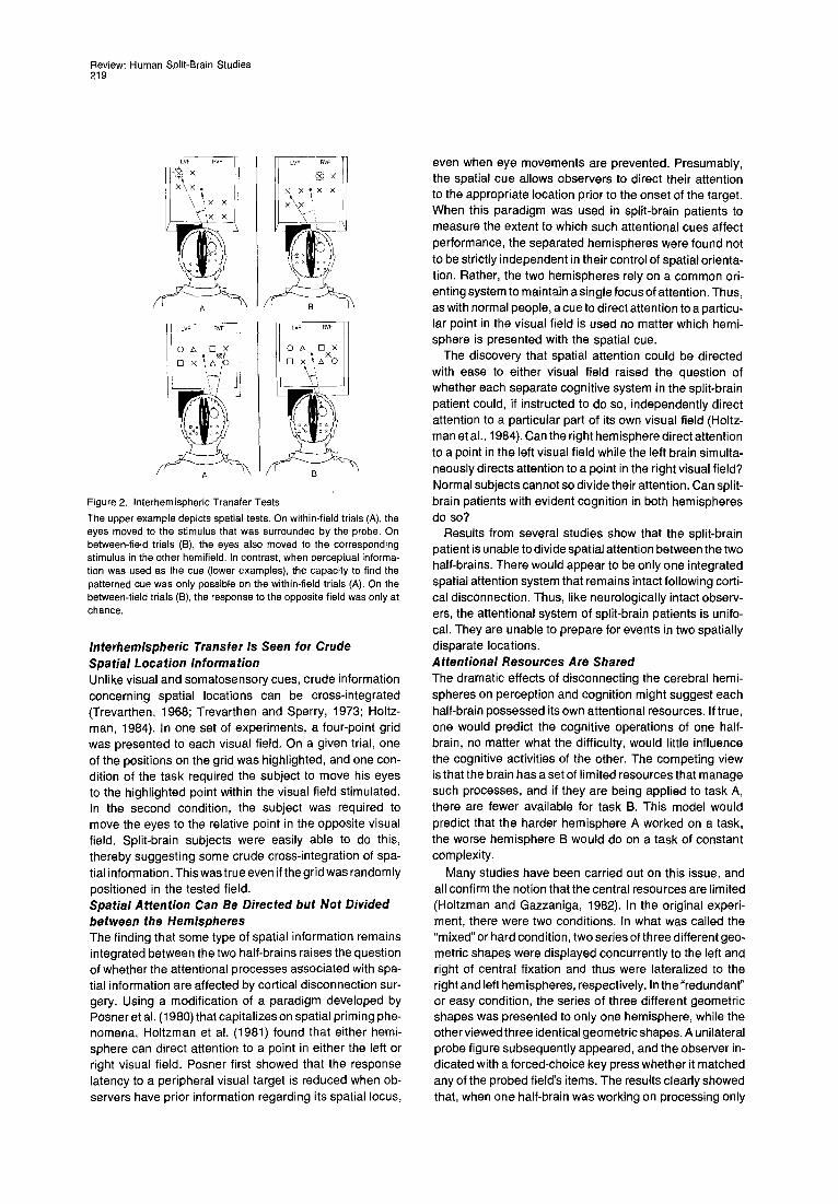
The main methods of testing the perceptual and cogni- tive functions of each hemisphere have not changed in principle over the years. However, there have been signifi- cant advances in the technologies used to present stimuli to the surgically separated hemispheres. In the early years, information was visually lateralized to one or the other hemispheres by quickly flashing stimuli to one or the other visual field using electronic shutters attached to slide projectors (see Figure 1). As the patient fixates on a point in space, information flashed to the left of fixation is presented exclusively to the right hemisphere. The quick flashing is necessary to control for unwanted eye move- ments, which would redirect the information into the un- wanted hemisphere. This method has been replaced by using computer-based stimuli presentation arrangements. More importantly, the development of an image stabilizing system, used in concert with a Purkinje eye tracker, now permits sustained presentation of information to either vi- sual field, and therefore, either hemisphere. Accordingly, if a subject moves his or her eyes away from fixation, the stabilizing system moves the stimulus with the eyes and thereby prevents the information from being presented to the wrong hemisphere. This technological development has allowed for new findings on both the neurological and psychological aspects of hemisphere disconnection.
Fundamental Principles Arising from Initial Studies The original reports on the California series of patients dealt with a number of fundamental issues concerning the basic psychological properties of the separated cerebral hemispheres, as well as basic issues of neurological orga- nization (Sperry et al., 1969; Gazzaniga, 1970). In many respects, the issues raised in the original studies still drive current research efforts. In the following, the neurologic consequences will be reviewed first, followed by the stud- ies on the separate psychological properties of the two cerebral hemispheres.
Sensory and Motor Studies The human studies were carried out in the context of strong new animal evidence, obtained by Myers and Sperry (1958), that dividing the cerebral commissures pro- duced a profound deficit in the interhemispheric transfer of sensory and motor information. In the cat, monkey, and chimpanzee, Myers (1956) had determined that, following midline section of the corpus callosum and anterior com- missure, visual and tactile information lateralized to one hemisphere did not transfer to the opposite hemisphere. This resulted in the so-called "split-brain" animal. This star- tling discovery was completely contrary to earlier reports on the effects of human commissure section reported by Akelaitis (1941, 1944). Akelaitis reported no significant neurological or psychological effects following section of the callosum.

Neuron 218
. . . . . .
DEFLECTOR
Horizontal
~ e~ . ya
Figure 1. Stimulus Lateralization Technique To examine differentially hemispheric processing differences, it is nec- essary to lateralize stimuli within the left and right visual fields (upper left). However, if the subject makes an eye movement during the stimu- lus presentation, proper lateralization is no longer maintained (upper middle). Thus, retinal stabilization is useful to counteract the effects of such eye movements (upper right). A dual Purkinje image eyetracker coupled with a mirror stimulus deflector allows such retinal stabilization (lower left). As eye movements occur, horizontal and vertical deflection mirrors move to counteract such movements, maintaining proper later- alization (lower right).
The surgeries on the original California cases (W J, NG, and LB) were able to show that humans responded to forebrain commissurotomy in essentially the same way as the monkey and chimpanzee. Visual information pre- sented to one half-brain was not available to the other half-brain for analysis. A similar principle applies to touch. Objects placed in the right hand were normally named and described, but objects presented in the left hand were not. Similarly, sensory information presented to one hemi- sphere was useful in guiding the contralateral hand but very ineffective in controlling the ipsilateral hand.
From a cognitive point of view, the first studies confirmed long-standing neurologic knowledge about the nature of the two cerebral hemispheres. The left brain was dominant for language, speech, and major problem solving, while the right appeared specialized for visuospatial tasks such as drawing cubes and other three-dimensional patterns. Of course, this meant that visual and tactile stimuli pre- sented to the right hemisphere could not be named or described, since the sensory information was discon- nected from the dominant left speech hemisphere. This dramatic result stood in contrast to the right hemisphere's ability to acknowledge the presence of these stimuli by allowing it to respond in a nonverbal manner, such as pointing to matching objects and the like.
In the subsequent 30 years, these studies have been followed up by a nu mber of investigators. While the original studies set the framework for subsequent research, the huge effort to characterize fully these unique human be- ings has continued to yield major insights into the organi- zation of the human brain.
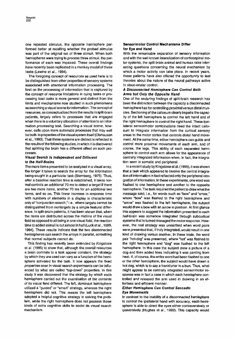
Perceptual and Attentional Studies Following Cerebral Disconnection The attentional and perceptual abilities of split-brain pa- tients have been extensively explored. Visual perception is the easiest to study. Overall, following cortical discon- nection, perceptual information does not interact between the two cerebral hemispheres, whereas the supporting cognitive processes of attentional mechanisms do some- times interact. Simple Perceptual Interactions Are Not Seen Split-brain patients are not able to cross-integrate visual information between their two visual half-fields. When vi- sual information is lateralized to either the left or right dis- connected hemisphere, the unstimulated hemisphere cannot use the information for perceptual analysis (see Figure 2). This is also true for stereognostic information presented to each hand. While the presence or absence of touch stimulation is noted to any part of the body by either hemisphere, patterned somatosensory information is lateralized. Thus, an object held in the left hand cannot help the right hand find an identical object. Although there have been reports arguing that some higher order percep- tual information is integrated at some level via subcortical structures (Cronin-Golomb, 1986; Sergent, 1990), these results have not been replicated by others (Seymour et al., 1994; Corballis et al., 1993; Corballis, 1994; McKeever et al., 1981). Humans Show Visual Midline Overlap Phenomenon There is some nasotemporal overlap at the retinal vertical meridian in cat and monkey (Stone, t966; Stone et al., 1973). In a stripe 1-2 degrees of visual angle wide that straddles the two visual half-fields, visual information is sent to both the left and right visual cortices. Whether or not the anatomical projections have any functional signifi- cance has never been examined in the animal. Fendrich et al. (1989) have examined this issue in split-brain patients. Using an image stabilizer combined with a Purkinje eye tracker, careful assessment of the visual midline of two split-brain patients has revealed an area of no more than 2 degrees in width at the vertical midline where visual information appears available to each half-brain. This con- trasts with the findings of Sugishita et al. (1994), who found no overlap but who were disadvantaged by not having the image stabilizer to carry out their midline studies. Still, within this strip of overlap, the signals conveyed to each hemisphere from the contralateral hemiretina appear to be weak or degraded. Stimuli could not be compared across the vertical meridian if these comparison required detailed shape information, or if the stimuli were presented for only 200 ms.
Review: Human Split-Brain Studies 219
I I LVF RVF -~ × x \ ×.
N / / B
:\
Figure 2. Interhemispheric Transfer Tests The upper example depicts spatial tests. On within-field trials (A), the eyes moved to the stimulus that was surrounded by the probe. On between-field trials (B), the eyes also moved to the corresponding stimulus in the other hemifield. In contrast, when perceptual informa- tion was used as the cue (lower examples), the capacity to find the patterned cue was only possible on the within-field trials (A). On the between-field trials (B), the response to the opposite field was only at chance.
Interhemispheric Transfer Is Seen for Crude Spatial Location Information Unlike visual and somatosensory cues, crude information concerning spatial locations can be cross-integrated (Trevarthen, 1968; Trevarthen and Sperry, 1973; Holtz- man, 1984). In one set of experiments, a four-point grid was presented to each visual field. On a given trial, one of the positions on the grid was highlighted, and one con- dition of the task required the subject to move his eyes to the highlighted point within the visual field stimulated. In the second condition, the subject was required to move the eyes to the relative point in the opposite visual field. Split-brain subjects were easily able to do this, thereby suggesting some crude cross-integration of spa- tial information. This was true even if the grid was randomly positioned in the tested field. Spatial Attention Can Be Directed but Not Divided between the Hemispheres The finding that some type of spatial information remains integrated between the two half-brains raises the question of whether the attentional processes associated with spa- tial information are affected by cortical disconnection sur- gery. Using a modification of a paradigm developed by Posner et al. (1980) that capitalizes on spatial priming phe- nomena, Holtzman et al. (1981) found that either hemi- sphere can direct attention to a point in either the left or right visual field. Posner first showed that the response latency to a peripheral visual target is reduced when ob- servers have prior information regarding its spatial locus,
even when eye movements are prevented. Presumably, the spatial cue allows observers to direct their attention to the appropriate location prior to the onset of the target. When this paradigm was used in split-brain patients to measure the extent to which such attentional cues affect performance, the separated hemispheres were found not to be strictly independent in their control of spatial orienta- tion. Rather, the two hemispheres rely on a common ori- enting system to maintain a single focus of attention. Thus, as with normal people, a cue to direct attention to a particu- lar point in the visual field is used no matter which hemi- sphere is presented with the spatial cue.
The discovery that spatial attention could be directed with ease to either visual field raised the question of whether each separate cognitive system in the split-brain patient could, if instructed to do so, independently direct attention to a particular part of its own visual field (Holtz- man et al., 1984). Can the right hemisphere direct attention to a point in the left visual field while the left brain simulta- neously directs attention to a point in the right visual field? Normal subjects cannot so divide their attention. Can split- brain patients with evident cognition in both hemispheres do so?
Results from several studies show that the split-brain patient is unable to divide spatial attention between the two half-brains. There would appear to be only one integrated spatial attention system that remains intact following corti- cal disconnection. Thus, like neurologically intact observ- ers, the attentional system of split-brain patients is unifo- cal. They are unable to prepare for events in two spatially disparate locations. Attentional Resources Are Shared The dramatic effects of disconnecting the cerebral hemi- spheres on perception and cognition might suggest each half-brain possessed its own attentional resources. If true, one would predict the cognitive operations of one half- brain, no matter what the difficulty, would little influence the cognitive activities of the other. The competing view is that the brain has a set of limited resources that manage such processes, and if they are being applied to task A, there are fewer available for task B. This model would predict that the harder hemisphere A worked on a task, the worse hemisphere B would do on a task of constant complexity.
Many studies have been carried out on this issue, and all confirm the notion that the central resources are limited (Holtzman and Gazzaniga, 1982). In the original experi- ment, there were two conditions. In what was called the "mixed" or hard condition, two series of three different geo- metric shapes were displayed concurrently to the left and right of central fixation and thus were lateralized to the right and left hem ispheres, respectively. In the"redundant" or easy condition, the series of three different geometric shapes was presented to only one hemisphere, while the other viewed three identical geometric shapes. A unilateral probe figure subsequently appeared, and the observer in- dicated with a forced-choice key press whether it matched any of the probed field's items. The results clearly showed that, when one half-brain was working on processing only
Neuron 220
one repeated stimulus, the opposite hemisphere per- formed better at recalling whether the probed stimulus was part of the original set of three stimuli. When both hemispheres were trying to process three stimuli, the per- formance of each was impaired. These overall findings have recently been replicated in a monkey model of these tasks (Lewine et al., 1994).
The foregoing concept of resources as used here is to be distinguished from other properties of sensory systems associated with attentional information processing. The limit on the processing of information that is captured by the concept of resource limitations in cuing tasks or pro- cessing load tasks is more general and distinct from the limits and mechanisms now studied in such phenomena as searching a visual scene for information. The concept of resources, as conceptualized from the results in split-brain patients, largely refers to processes that are engaged when there is a voluntary allocation of attention to an infor- mation processing task. Searching a visual scene, how- ever, calls upon more automatic processes that may well be built- in properties of the visual system itself (Olshausen et al., 1993). That these systems are distinct is reflected in the results of the following studies, in which it is discovered that splitting the brain has a different effect on such pro- cesses. Visual Search Is Independent and Different in the Half.Brains The more items presented to be analyzed in a visual array, the longer it takes to search the array for the information being sought in a particular task (Sternberg, 1975). Thus, after a baseline reaction time is established, it takes nor- mal controls an additional 70 ms to detect a target if there are two more items, another 70 ms for an additional two items, and so on. This linear increase in response time with numbers of elements in a display is characteristic only of "conjunction search," i.e., where targets cannot be distinguished from nontargets by a simple featural differ- ence. In split-brain patients, it has been shown that, when the items are distributed across the midline of the visual field as opposed to all being in one visual field, the reaction time to added stimuli is cut almost in half (Luck et al., 1989, 1994). These results indicate that the two disconnected hemispheres can search the arrays in parallel, something that normal subjects cannot do.
This finding has recently been extended by Kingstone et al. (1995) to show that, although the overall resources a brain commits to a task appear constant, the strategy by which they are used can vary as a function of the hemi- sphere activated for the task. It now appears the fixed properties seen in visual search experiments can be influ- enced by what are called "top-down" properties. In this study it was discovered that the strategy by which each hemisphere carried out the examination of the contents of its visual field differed. The left, dominant hemisphere utilized a "guided" or "smart" strategy, whereas the right hemisphere did not. This means the left hemisphere adopted a helpful cognitive strategy in solving the prob- lem, while the right hemisphere does not possess those kinds of extra cognitive skills to assist its visual search mechanism.
Sensorimotor Control Mechanisms Differ for Eye and Hand With the remarkable separation of sensory information and with the well known lateralization of corticospinal mo- tor systems, the split-brain animal and human raise inter- esting questions concerning the neural mechanism by which a motor activity can take place. In recent years, these patients have also offered the opportunity to test theories about the nature of the neural pathways active in visuo-ocular control. A Disconnected Hemisphere Can Control Both Arms but Only the Opposite Hand One of the enduring findings of split-brain research has been the distinction between the capacity a disconnected hemisphere has for controlling proximal versus distal mus- cles. Sectioning of the callosum clearly impairs the capac- ity of the left hemisphere to control the left hand and of the right hemisphere to control the right hand. These ipsi- lateral sensorimotor combinations need the intact callo- sum to integrate information from the cortical sensory areas to the motor cortex that controls distal hand move- ment. At the same time, either hemisphere can guide and control more proximal movements of each arm, and of course, the legs. This ability of each separated hemi- sphere to control each arm allows for the appearance of centrally integrated information when, in fact, the integra- tion seen is somatic and peripheral.
In a recent study by Kingstone et al. (1995), it was shown that a task which appeared to involve the central integra- tion of information in fact reflected only the peripheral inte- gration of information. In these experiments, one word was flashed to one hemisphere and another to the opposite hemisphere. The task required the patient to draw what the message said; i.e., for words such as "bow" and "arrow," where "bow" was flashed to the right hemisphere and "arrow" was flashed to the left hemisphere, the subject would draw a bow with an arrow in position. At first glance this appears to suggest the information presented to each half-brain was somehow integrated through subcortical systems that in turned managed the motor response. How- ever, the real strategy was unearthed when word pairs were presented that, if truly integrated, would result in one kind of drawing versus another. In these trials, the word pair "hot-dog" was presented, where "hot" was flashed to the right hemisphere and "dog" was flashed to the left hemisphere. In this case the subject drew a picture of a dog and then added lines indicating it was panting from heat. If, of course, the entire word had been flashed to one or the other hemisphere, the subject would have drawn a hot dog, which is to say a frankfurter in a bun. Thus, what might appear to be centrally integrated sensorimotor re- sponse was in fact a case in which each hemisphere con- trolled and released the arm used in drawing in an ef- fortless and efficient manner. Either Hemisphere Can Control Saccadic Eye Movements In contrast to the inability of a disconnected hemisphere to control the ipsilateral hand with accuracy, each hemi- sphere is able to direct the eyes either contraversively or ipsiversively (Hughes et al., 1992). This capacity would

Review: Human Split-Brain Studies 221
Figure 3. Postoperative Mid-Saggital MR Images Magnetic resonance images (MRIs) of four patients with varying degrees of callosal section. (A) The entire corpus callosum has been sectioned, resulting in no transfer of information between the cerebral hemispheres. (B) The posterior or splenia! regions have some sparing in a particular area, with the result that visual information could be transferred between the cerebral hemispheres. (C) The posterior callosum was sectioned, while the anterior half remained intact. This patient had difficulty with sensorimotor integration of one hand but not the other. (D) Two remnants were inadvertently left intact, one in the splenial region and one in anterior genu region (see Gazzaniga, 1989).
not be predicted by dozens of studies showing that in each hemisphere the frontal eye fields control contraversive eye movements (Bruce and Goldberg, 1984; Wurtz and AI- bane, 1980). This particular result reveals how psycho- physical studies on human patients with discrete lesions suggest that alternate neural pathways are involved, path- ways different from those that might otherwise be evident from the animal studies.
The foregoing group of studies reveals how discon- necting the cerebral hemispheres at the cortical level inter- rupts some kinds of attentional, perceptual, and sensori- motor processes, but not others. Although there is little evidence that any perceptual information can be trans- ferred between the hemispheres, there are some kinds of attentional and sensorimotor processes that remain un- changed with cortical disconnection. Just as formal genet- ics helps formulate the concept of a gene that later be- comes understood at the molecular level, identifying these differences in brain organization will help neuroscientists understand the systems basis of these mental activities.
Partial Callosal Sections Reveal Specificity of Commissure Function In animal studies, it was quickly determined that sectioning the entire corpus callosum and anterior commissure pre- vented the interhemispheric transfer of a wide range of modal and motor information. It was also shown that partial sectioning of the commissures could block specific func- tions from transferring across the callosum (Black and My- ers, 1966; Sullivan and Hamilton, 1973; Hamilton and Ver- meire, 1986). In humans, it was only after other cases appeared who had not undergone full callosal section that comparable studies could be done, and it became appar- ent that specific regions of the callosum were responsible for the transfer of specific modalities. This work was en- hanced when MRI allowed for the accurate description of cut and uncut fiber systems. MRI.Verified Lesions of Partial Sections Reveal Specific Modal Functions When the corpus callosum is fully sectioned, there is little or no perceptual or cognitive interaction between the hemi-
Neuron 222
SPLIT BRAIN
Right Hemisphere STIMULUS <>
COMPARISONS
Left Hemisphere VERBAL RESPONSE
LW t Knight RVF
Normal Brain
"Knight"
LVF t RVF Knight
Partial Split "1 have o picture in mind bul can't soy it.,.. Two fighters in a ring..,, Ancient .... wearing uniforms ond helmets..., on horses, trying to knock each other off... Knights?"
I LVF t RVF Knight
Complete Split
"\; \ l l
"1 didn't see anything"
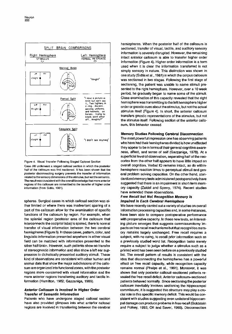
Figure 4. Visual Transfer Fallowing Staged Callosal Section Case JW underwent a staged callosal section in which the posterior half of the callosum was first sectioned. It has been shown that the posterior disconnecting surgery prevents the transfer of information related to the sensory dimensions of the stimulus, but not the semantic. The result was consistent with the overall knowledge that more anterior regions of the callosum are committed to the transfer of higher order information (from Sidtis, 1981).
spheres. Surgical cases in which callosal section was ei- ther limited or where there was inadvertent sparing of a part of the callosum allow for the examination of specific functions of the caUosum by region. For example, when the splenial region (posterior area of the callosum that interconnects the occipital lobe) is spared, there is normal transfer of visual information between the two cerebral hemispheres (Figure 3). In these cases, pattern, color, and linguistic information presented anywhere in either visual field can be matched with information presented to the other half-brain. However, such patients show no transfer of stereognostic information and also show a left ear sup- pression to dichotically presented auditory stimuli. These kind of observations are consistent with other human and animal data that show the major subdivisions of the calla- sum are organized into functional zones, with the posterior regions more concerned with visual information and the more anterior regions transferring auditory and tactile in- formation (Hamilton, 1982; Gaz.zaniga, 1989).
Anterior Callosum Is Involved in Higher Order Transfer of Semantic Information Patients who have undergone staged callosal section have also provided glimpses into what anterior callosal regions are involved in transferring between the cerebral
hemispheres. When the posterior half of the callosum is sectioned, transfer of visual, tactile, and auditory sensory information is severely disrupted. However, the remaining intact anterior callosum is able to transfer higher order information (Figure 4). Higher order information is a term used when it is clear the information transferred is not simply sensory in nature. This distinction was shown in one study (Sidtis et al., 1981) in which the corpus callosum was sectioned in two stages. Following the first stage of sectioning, the patient was unable to name stimuli pre- sented to the right hemisphere. However, over a 10 week period, he gradually began to name some of the stimuli. Close examination of this capacity revealed that the right hemisphere was transmitting to the left hemisphere higher order or gnostic cues about the stimulus, but not the actual stimulus itself (Figure 4). In short, the anterior callosum transfers gnostic representations of the stimulus, but not the stimulus itself. Following section of the anterior calla- sum, this behavior ceased.
Memory Studies Following Cerebral Disconnection The most powerful impression one has observing patients who have had their hemispheres divided is how unaffected they appear to be in terms of their general cognitive aware- ness, affect, and sense of self (Gazzaniga, 1970). At a superficial level of observation, separating half of the neo- cortex from the other half appears to have little impact on overall cognition. Verbal IQ remains intact, as do within- hemisphere reaction times to perceptual stimuli and gen- eral problem solving capacities. On the other hand, stan- dardized memory tests administered postoperatively have suggested that there is an impairment in short-term mem- ory capacity (Zaidel and Sperry, 1974). Recent studies have extended these observations. Free Recall but Not Recognition Memory Is Impaired in Each Cerebral Hemisphere We have recently carried out a variety of studies on overall information processing capacities and, in some instances, have been able to compare postoperative performance with preoperative capacity. In these new tests, an interest- ing picture emerges that suggests commissurotomy im- pacts on free recall mechanisms but that recognition mem- ory remains largely unchanged. Free recall requires a subject, with no cuing, to recall prior information such as a previously studied word list. Recognition tasks merely require a subject to judge whether a stimulus such as a printed word has been seen before on a previously studied list. The overall pattern of results is consistent with the idea that disconnecting the hemispheres has a powerful effect on free recall capacity, while recognition capacity remains normal (Phelps et al., 1991). Moreover, it was shown that only posterior callosal-sectioned patients re- vealed the free recall deficit. Anterior callosum-sectioned patients behaved normally. Since sectioning the posterior callosum inevitably involves sectioning the hippocampal commissure, it is suggested this structure may play a cru- cial role in this specific memory deficit. This would be con- sistent with studies suggesting even unilateral hippocam- pal damage can produce probems in free recall (Goldstein and Polkey, 1992; Ott and Saver, 1993). Disconnection

Review: Human Split-Brain Studies 223
RI~DUNIItJ(I" CONDfIION MIX[D CONOITION
T 5 T robe 'T 5 T F~obe
san~ d;~. ~ d~4
i I ~i ~
/
I 1 ~
LVF RVF L,A: RV~
Re~J,~an~ Mixed
Figure 5. Memory Performance Following Callosal Section On each "redundant" trial (top left), a bilaterally displayed target (X) moved among four homologous cells in the two matrices. A tone sounded and a unilateral probe sequence was presented. The ob- server indicated whether the probe sequence matched the probed field's target sequence. In this example, the bilateral target sequences began at time T~ in the upper left corners of the two matrices and concluded at -14 in the upper right corners, and the unilateral probe sequence was presented from Ts-Ts. All Xs appeared for 150 ms. The diagram on the right is an example of a mixed condition trial. Mixed conditions trials differed from redundant condition trials only in that different target sequences were presented in the two visual fields. Data are presented separately for commissurotomy patient JW and the average performance of the control observers (after Holtzman and Gazzaniga, 1985).
of the hemispheres in this commissural zone might well mimic the unilateral lesion effects. Overall, the data sug- gest that the intact neocortical systems that are intercon- nected through the posterior callosal system contribute to free recall mechanisms. It is as if the neural mechanisms required for richly encoding a stimulus, which contribute to free recall, are less available to each hemisphere following posterior cortical disconnection. Increase in Short-Term Memory Capacity Is More Apparent Than Real Although the data suggest there are difficulties in free re- call, the cortically disconnected patient can perform other memory tests in a manner superior to that of a normal. Consider the following.
As already reviewed, the callosum-sectioned patient has no measurable interactions between the two hemi- spheres in the processing of perceptual information. Iden- tical and simple visual patterns of all kinds can be pre- sented to each separate half-brain, and the patient is unable to say whether the stimuli are the same or different. This fact raises the possibility that, in a memory task involv- ing visual retention, a split-brain subject might perform at
a higher level than a normal intact control subject if the perceptual information were distributed between the two visual half-fields. In one study on this issue, a complex spatial memory task was administered to both a split-brain patient and normal controls in which critical information was presented in each visual half.field (Holtzman and Gaz- zaniga, 1985). For the controls, the visual information was automatically combined and perceived as one large prob- lem (Figure 5). For the split-brain patient, each hemisphere perceived a problem that remained separate from the per- ceptual information presented to the other half-brain; thus, each hemisphere perceived a much simpler task. The re- sults were clear. The split-brain patient out-performed the normal controls on critical test conditions. The callosum- sectioned patient benefited from the fact that the percep- tual array under one of the test conditions did not appear more difficult because the work was distributed to each separate hemisphere, even though the actual sensory array was identical to that experienced by the intact normal controls.
Disconnection of the cerebral hemispheres clearly allows for a unique cognitive state. In a sense it turns a serial, unified perceptual system into two simpler percep- tual systems that do not interact, and therefore do not interfere with each other. It allows for the breaking down of a large perceptual problem into smaller, more manageable problems that a half-brain is capable of solving. From the observer's point of view, however, it looks like the total information processing capacity of the patient has been increased and is superior to that of normal controls. Yet, as we saw in the studies on attention already described, close inspection of the problem indicates split-brain pa- tients have not increased the amount of resources they can call upon to solve problems. In short, it appears that the human brain has a set amount of resources it can allocate to cognitive tasks and that these resources remain constant following commissurotomy. How then do we ex- plain these two seemingly different sets of results? On the one hand, performance seems better than normal, while on the other hand we have seen there are resource limits for perceptual and cognitive tasks.
The conundrum forces the issue of where in a percep- tual motor task resource limitations are applied. Are they, for example, applied during the early phases of information processing that deal with the complexity of the visual stim- ulus itself? Or, are the resources applied at later loci of the information processing sequence that deal with the more cognitive aspects of the task? It would appear that interactions between the hemispheres on resource limits occur when the task is more cognitive and involves work- ing memory. Lewine et al. (1994) have proposed a similar scheme and suggest the site of subcortical interaction may be the brain stem.
Language and Speech Processes of the Left and Right Hemispheres A dichotomy that is useful when trying to understand lan- guage is the distinction between how the brain enables grammar and how it enables a lexicon. The grammar- lexicon distinction (Pinker, t 993) is different from the more
Neuron 224
traditional syntax-semantic distinction that is commonly invoked to understand the differential effects of brain le- sions on language processes. In general terms, grammar refers to the rule-based system humans have for ordering words to allow for effective communication. The lexicon, on the other hand, is the mind's dictionary, where specific words are associated with particular meanings. The rea- son for using the former distinction is that it takes into account such factors as memory, in that with memory such word strings as idioms can be learned by rote. While it is clearly the case that the lexicon (memory) cannot underlie most phrases and sentences, since there are endless unique sentences such as the one I am currently writing, memory does play a role in many short phrases. Thus, when uttered, such word strings do not reflect an underly- ing interaction of syntax and semantic systems. They are, instead, essentially an entry from the lexicon. In more gen- eral terms, a modern view would predict there ought to be brain areas wholly responsible for grammar, while evi- dence for localization of the lexicon ought to be more elu- sive, since it would reflect learned information and thus be part of the general memory/knowledge systems of the brain. The grammar system ought to be discrete and there- fore localizable. The lexicon should be distributed and therefore more difficult to damage completely. Language and Speech Processes Can Rarely Be Present in Both Hemispheres: Right Hemisphere Language Has a Different Organizational Structure Than Left While the separated left hemisphere is fully capable of normal comprehension of all aspects of language, the right hemisphere can possess language. When there was evi- dence of right hemisphere language, these disconnected right hemispheres were severely limited in the kinds of linguistic properties they possessed (Gazzaniga, 1970; Zaidel, 1990).
Over the last 30 years, very few cases have been added to the group that demonstrate some kind of language in the right hemisphere (reviewed in Baynes, 1990). In the early eighties, only five cases in all series of split-brain patients had proven to have a lexicon in the right hemi- sphere. Since that time we have been successful in finding only one more normally right-handed patient with a lexicon in both the left and the right hemispheres (Baynes et al., 1992).
The left and the right lexicons of these special patients can be nearly equal in their capacity, but they are orga- nized quite differently. For example, both hemispheres show a phenomenon called the "word superiority effect" (WSE). Normal readers are able to identify letters better when they occur in the context of real English words, rather than when the same letters occur in pseudo-word (korad) or nonword (twhx) strings. Since pseudo- and nonwords do not have lexical entries, letters occurring in such strings do not receive the additional processing benefit bestowed on words, and thus the WSE emerges. By this view, the WSE may be a useful measure of the integrity of the visual lexicon. When the appropriate studies assessing these effects were carried out with each hemisphere, it was ap-
parent each hemisphere in these special patients has a visual lexicon that provides an advantage for recognizing letters that occur in real words. At the same time, a closer analysis of the data revealed each hemisphere was using a different strategy in processing the words. Examination of the reaction time data, for example, suggested that the right hemisphere took more time to respond as the words became longer. The left hemisphere did not, just as is the case for normal readers. In addition, in the WSE task described above, the error data revealed a different pat- tern for each hemisphere. The left hemisphere made more errors on the middle letters of words, just as in controls, while the right hemisphere made more errors on the last letters of words. Finally, achieving similar levels of accu- racy in each hemisphere required exposure durations al- most 10 times as long in the nondominant hemispheres. This result suggests letter processing for the right hemi- sphere is serial and not parallel in nature, as is normally the case (Reuter-Lorenz and Baynes, 1992).
To locate where in the processing chain the hemisphere differences begin, we evaluated each hemisphere's per- formance on a letter priming task. The task was simply to indicate whether a briefly flashed upper case letter was an "H" or a "T." On each trial the upper case letter was preceded by a lower case letter that was either an "h" or a "t." Normally, subjects are significantly faster when an upper case"H" is preceded by a lower case "h," rather than a lower case "t." This is a version of the classic "priming" phenomenon frequently used in cognitive science to get at mechanisms of mental processes. When using letters instead of words in a priming task, it is assumed very early stages in the perceptual-cognitive continuum are being examined.
The difference between response latency on compatible (h-H) versus incompatible (t-H) trials is taken to be a mea- sure of letter priming. JW performed a lateralized version of this task in which the prime was exposed for 100 ms to either the right or left visual field, and 400 ms later, the target letter appeared in either the right or left visual field. His performance provides no evidence of letter priming for left visual field trials, but clear evidence of priming for right visual field trials. Thus, there are no priming phenom- ena in the disconnected right hemisphere. In summary, there can be two lexicons, one in each hemisphere, but this lexical organization is rare. When present, the right hemisphere lexicon seems to be organized differently than the lexicon of the left hemisphere. These observations would be consistent with the view that lexicons reflect learning processes and as such would have a wider distri- bution in the cerebral cortex. Still, it would be folly not to note that in the general population the lexicon seems to be in the left hemisphere. A right hemisphere lexicon is rarely present, and when it is, it stores information differently. Generative Syntax Is Present in Only One Hemisphere While the right hemispheres of some patients clearly have lexicons, these right hemispheres have shown erratic per- formance on other aspects of language such as under- standing verbs, pluralizations, the possessive, active/

Review: Human Split-Brain Studies 225
passive differences, etc. (Gazzaniga, 1970). The right hemisphere in the patients that possess some language also lacks a capacity to use word order to disambiguate stimuli for correct grammatical meaning. Nonetheless, these right hemispheres can indicate when a sentence ends with a semantically odd word (Kutas et al., 1988). Additionally, right hemispheres that reveal language ca- pacities are able to make judgments of grammaticality (Baynes and Gazzaniga, 1988; Baynes, 1990). Thus, even though they cannot use syntax to disambiguate stimuli or to guide comprehension judgments, they can recognize that one set of utterances is grammatical while another is not. This finding suggests that patterns of speech are learned by rote. Yet, recognizing the pattern of acceptable utterances does not mean a neural system can use this information to assist in the understanding of word strings• Some Right Hemispheres Can Develop Speech One of the hallmarks of most split-brain patients is that they speak out of the left hemisphere and not the right. This observation has been consistent with the neurologic literature and amytal studies which have shown that the left hemisphere is the dominant hemisphere for both lan- guage and speech (Lennenberg, 1967).
There are now three and possibly four split-brain pa- tients who seem able to speak out of each hemisphere. While there is always an initially dominant hemisphere following brain bisection, some patients have developed the capacity to make one word utterances from the discon- nected right hemisphere (Gazzaniga et al., 1979). In these experiments, it became apparent that stimuli presented to the left visual field (right hemisphere) began to be named. Ordinarily, one stimulus presented to the left hemisphere could be named. This rather startling development raised the question of whether the information was somehow transferring to the dominant hemisphere for speech out- put, or whether the right hemisphere itself had developed speech= After a series of tests, it was apparent that the latter option was correct. The patients, for example, while able to name an object presented in the left field, in the right field could not judge if they were the same objects or not. Or, if words like "father" were presented such that the fixation point fell between the "t" and the "h," the pa- tients would say either "fat" or "her," depending on which hemisphere spoke first. Thus, in these patients two of the three major systems in human language can be managed to some extent by either hemisphere. It also illustrates an extraordinary plasticity, occurring sometimes over 10 years after callosal surgery.
Hemisphere Specialization One of the cardinal features of behavioral studies on the brain, both human and animal, has been the realization that specific brain areas seem to be involved with rather specific perceptual and cognitive functions. The split-brain approach to this issue has been straightforward. By testing each disconnected hemisphere, one can assess the differ- ent capacities each might possess. While some of the claims in this regard have become exaggerated, there are
marked differences between the two half-brains (see Hel- lige, 1993). The Left Hemisphere Possesses a Unique Capacity to Interpret Behavior and Unconsciously Driven Emotional States A number of years ago we observed how the left, dominant speaking hemisphere dealt with the behaviors we knew we had elicited from the disconnected right hemisphere (Gazzaniga, 1995). We first revealed the phenomenon us- ing a simultaneous concept test. The patient is shown two pictures, one exclusively to the left hemisphere and one exclusively to the right, and is asked to choose from an array of pictures placed in full view in front of him the ones associated with the pictures lateralized to the left and right brain. In one example of this kind of test, a picture of a chicken claw was flashed to the left hemisphere, and a picture of a snow scene to the right hemisphere. Of the array of pictures placed in front of the subject, the obvi- ously correct association is a chicken for the chicken claw and a shovel for the snow scene. Case PS responded by choosing the shovel with the left hand and the chicken with the right. When asked why he chose these items, his left hemisphere replied, "Oh, that's simple. The chicken claw goes with the chicken, and you need a shovel to clean out the chicken shed." Here, the left brain, observing the left hand's response, interprets that response into a con- text consistent with its sphere of knowledge, one that does not include information about the left hemifield snow scene. We called this left hemisphere process the "inter- preter."
This same general idea has been observed when the left brain interpreter struggles to deal with mood shifts, produced in the experimental situation by manipulating the disconnected right hemisphere. A positive mood shift triggered by the right hemisphere finds the left interpreting its current experience in a positive way. Similarly, when the right triggers a negative mood state, the left interprets a previously neutral situation in negative terms.
In recent studies we have been able to show one of the many implications for having a left hemisphere interpreter. It has long been known that inference and interpretation are important aspects of normal memory functioning (Bart- lett, 1932), and one would predict the two hemispheres might respond differently in some mnemonic tasks. Phelps and Gazzaniga (1992) showed that, when split-brain pa- tients were shown pictures representing a common scene, the two hemispheres responded differently when tested 2 hr later. Their memory was tested with a lateralized yes- no recognition test in which the distracter pictures were either consistent or inconsistent with the original scene. The left hemisphere performed below chance on consis- tent distracter pictures, whereas the right hemisphere was above chance on these pictures and performed at the same level of accuracy as the pictures originally pre- sented. In short, the right hemisphere, which has no inter- pretive mechanism, rejected as seen before pictures that could have been part of the story. The left hemisphere, on the other hand, with its capacity for making inferences and interpretations, was more strongly influenced by the

Neuron 226
expectations for actions common to a scene and falsely recognized pictures consistent with the observed scene. Similar results have recently been reported by Metcalfe et al. (1995). Monitoring and Production of Facial Expressions Are Managed by Different Hemispheres In the perceptual domain, it appears that the right hemi- sphere has special processes devoted to the efficient de- tection of upright faces (Gazzaniga, 1989). Although the left hemisphere can also perceive and recognize faces as well as reveal superior capacities when the faces are familiar, the right hemisphere appears specialized for un- familiar facial stimuli (Levy et al., 1972; Gazzaniga and Smylie, 1983). This pattern of asymmetry has also been shown for the rhesus monkey (Hamilton and Vermiere, 1988).
Since the right hemisphere is superior for perception of faces, it would be reasonable to suppose it is also special- ized for the management of facial expressions. Recent studies, however, have shown that, while both hemi- spheres can generate spontaneous facial expressions, only the dominant left hemisphere can generate voluntary facial expressions (Gazzaniga and Smylie, 1990). In these studies, single word commands such as "sm ile" or "frown" were given to either the left or right hemisphere. Mixed in with these commands that would require control of lower facial muscles would be other commands that called upon the upper facial muscles, such as "blow," "wink," and "blink." While both hemispheres were able to respond to the latter commands, only the left hemisphere could re- spond to the commands of "smile" and "frown." The right hemisphere responded at chance, thereby suggesting the left was specialized for the production of voluntary facial expressions. Hemisphere Specialization Sensorimotor Tasks There are some tests that bring out hemisphere superiori- ties in some of the patients. The block design test from the Weschler Adult Intelligence Scale is one such test. Here, the simple task of arranging some red and white blocks to match those of a given pattern can find the left hemisphere performing poorly while the right triumphs (Bogen and Gazzaniga, 1965). However, in other patients both hemispheres when separated appear impaired, and in still others the left hemisphere, in addition to speaking and thinking, performs this task well.
The same pattern of results as seen with the block de- sign test is also seen for other tests such as the nonsense wire figure test (Milner and Taylor, 1972). Yet, for both tests the prevalent picture would be that the skill frequently seems localized to the right hemisphere. When the capac- ity happens to be lateralized in this way, it should be easier to analyze than when the processes involved are shared between the two half-brains. The question is, what is it?
The components of the block design task have not yet been identified. We do know that a patient who demon- strates a right hemisphere superiority for this kind of task can show no superiority on the perceptual aspects of the task. If a picture of the block design pattern is flashed to either hemisphere, each can easily find the match from a series of pictures. And, since each hand is demonstrably
dexterous, the right for writing and the left for this kind of task, the crucial link must be in the mapping of the sensory message onto the capable motor system. It remains for future research to understand this superiority in perfor- mance when it is seen in one hemisphere. The Left Hemisphere Is Specialized for Intelligent Behavior Following disconnection of the human cerebral hemi- spheres, the verbal IQ of the patient remains intact (Nass and Gazzaniga, 1987; Zaidel, 1990), and the problem solv- ing capacity, such as seen in hypothesis formation tasks, remains unchanged for the left hemisphere (LeDoux et al., 1977a, 1977b). While there can be deficits in free recall capacity (i.e., to recall either verbally or visually previously learned items without the benefit of any cues or recognition aids) and in some other performance measures, the over- all capacity for problem solving seems unaffected. In other words, isolating essentially half of the cortex from the dom- inant left hemisphere causes no major change in cognitive functions. The left remains unchanged from its preopera- tive capacity, while the largely disconnected, same-size right hemisphere is seriously impoverished on a variety of cognitive tasks. While the largely isolated right hemi- sphere remains superior to the isolated left hemisphere for some activities, such as the recognition of upright faces--as well as other functions not covered in this re- view, such as some attentional skills (Mangun et al., 1994) and perhaps also emotional processes (Gainotti et al., 1993)- i t is poor at problem solving and many other mental activities. A brain system (the right hemisphere) with roughly the same number of neurons as one that easily cogitates (the left hemisphere) is not capable of higher order cognition. This represents strong evidence that sim- ple cortical cell number by itself cannot fully explain human intelligence (Gazzaniga, 1994).
Conclusions Over 30 years of split-brain research has provided exten- sive insights into the organization of the human brain. The surgical disconnection of the cerebral hemispheres pro- duces an extraordinary opportunity to study which percep- tual and cognitive processes are cortical in nature and which are subcortical. Visual perceptual information, for example, remains strictly lateralized to one hemisphere following callosal section. Tactile patterned information remains lateralized. Attentional mechanisms, however, can involve subcortical systems. Taken together, cortical disconnection produces two independent sensory infor- mation processing systems that call upon a common atten- tional resource system in the carrying out of perceptual tasks.
Cortical disconnection also provides clues to the nature of human memory. Following posterior callosal surgery that involves sectioning the hippocampal commissure as well, split-brain patients retain normal recognition memory but are impaired on free recall tasks. This finding not only demonstrates that these two processes are dissociable, it also suggests that one role of the hippocampal commis- sures is to allow for the multiple representation of events that is known to be important in free recall memory.

Review: Human Split-Brain Studies 227
Split-brain studies have also revealed the complex mo-
saic of mental processes that go into human cognition.
On the one hand, each cerebral hemisphere has its own set of special ized capacities. The left hemisphere, for ex-
ample, is special ized for language and speech, and major problem solving capacit ies crucial for intell igent behavior.
It also possesses a uniquely human capacity to interpret behavior and to construct theories about the relationships between perceived events and feelings. The right hemi-
sphere, on the other hand, is special ized for specific tasks such as facial recognition, attentional monitoring, and pos- sibly other mental traits. It does not possess the overall cognit ive capacit ies of the left brain, which finds it reacting
more directly and simply to perceptual information. Finally, split-brain studies have demonstrated how spe-
cific perceptual and cognit ive skills can be isolated,
through disconnection surgery, to particular cerebral ar- eas. These skills then are not revealed by their absence as a result of a cortical lesion, which in turn makes structure-
function correlations difficult to make. The skills are pres- ent and can be demonstrated, but only when tested
through either the left or right hemisphere.
Acknowledgments
This work was aided by NIH grants NINDS 5 R01, NS22626-09, and NINDS 5 P01 NS17778-012 and by the James S. McDonnell Foun- dation.
January 12, 1995.
References
Akelaitis, A. J. (1941). Studies on corpus callosum: higher visual func- tions in each homonymous field following complete section of corpus callosum. Arch. Neurol. Psych. (Chicago) 45, 788. Akelaitis, A. J. (1944). Study on gnosis, praxis, and language following section of corpus callosum and anterior commissure. J. Neurosurg. 1, 94. Bartlett, F. C. (1932). Remembering: A Study in Experimental and Social Psychology (Cambridge, England: Cambridge University Press). Baynes, K. (1990). Language and reading in the right hemisphere: highways or byways of the brain? J. Cogn. Neurosci. 2, 159-179.
Baynes, K., and Gazzaniga, M. S. (1987). Right hemisphere language: insights into normal language mechanisms? In Language, Communi- cation, and the Brain, F. Plum, ed. (New York: Raven Press), pp. 117- 126. Baynes, K., Tramo, M. J., and Gazzaniga, M. S. (1992). Reading with a limited lexicon in the right hemisphere of a callosotomy patient. Neuropsychologia 30, 187-200. Black, P., and Myers, R. E. (1966). Visual function of the forebrain commissures in the chimpanzee. Science 146, 799-800. Bogen, J. E., and Gazzaniga, M. S. (1965). Cerebral commissurotomy in man: minor hemisphere dominance for certain visuospatial func- tions. J. Neurosurg. 23, 394-399. Bogen, J. E., Fisher, E. D., and Vogel, P. J. (1965). Cerebral commis- surotomy: a second case report. J. Am. Med. Assoc. 194, 1328-1329. Bruce, C. G., and Goldberg, M. E. (1984). Physiology of the frontal eye fields. Trends Neurosci. 7, 436-441. Corballis, M. C. (1994). Can commissurotomized subjects compare digits between the visual fields? Neuropsychologia 32, 1475-1486. Corballis, M. C., Trudel, C., and Carol, I. (1993). Role of the forebrain commissures in interhemispheric integration. Neuropsychology 7, 306-324. Cronin-Golomb, A. (1986). Subcorticat transfer of cognitive information
in subjects with complete forebrain commissurotomy. Cortex 22, 499- 519.
Fendrich, R., and Gazzaniga, M. S. (1989). Evidence of foveal splitting in a commissurotomy patient. Neuropsychologia 27, 273-281.
Gainotti, G., Caltagirone, C., and Zoccolotti, P. (1993). Left/right and cortical/subcortical dichotomies in the neuropsychological study of hu- man emotions. Cogn. Emotion 1, 71-93.
Gazzaniga, M. S. (1970). The Bisected Brain (New York: Appleton- Century-Crofts).
Gazzaniga, M S. (1989). Organization of the human brain. Science 245, 947-952.
Gazzaniga, M. S. (1994). On neural circuits and cognition. Neural Computat. 7, 1-12.
Gazzaniga, M. S. (1995). Consciousness and the cerebral hemi- spheres. In The Cognitive Neurosciences (Cambridge, Massachu- setts: MIT Press), pp. 1391-1400.
Gazzaniga, M. S., and Smylie, C. S. (1983). Facial recognition and brain asymmetries: clues to underlying mechanisms. Ann. Neurol. 13, 536-540.
Gazzaniga, M. S., and Smylie, C. S. (1990). Hemispheric mechanisms controlling voluntary and spontaneous facial expressions. J. Cogn. Neurosci. 2, 239-245.
Gazzaniga, M. S., LeDoux, J. E., Smylie, C. S., and Volpe, B. T. (1979). Plasticity in speech organization following commissurotomy. Brain 102, 805-815.
Goldstein, L. H., and Polkey, C. E. (1992). Everyday memory after unilateral temporal Iobectomy or amygdalo-hippocampectomy. Cortex 28, 189-201. Hamilton, C. R. (1982). Mechanisms of interocular equivalence. In Analysis of Visual Behavior, D. Ingle, M. Goodale, and R. Mansfield, eds. (Cambridge, Massachusetts: MIT Press), pp. 693-717.
Hamilton, C. R., and Vermeire, B. A. (1986). Localization of visual experience with partially split-brain monkeys. In Two Hemispheres- One Brain: Functions of the Corpus Callosum (New York: Alan R. Liss, Inc.), pp. 315-333.
Hamilton, C. R., and Vermeire, B. A. (1988). Complementary hemi- spheric specialization in monkeys. Science 242, 1691-1694. Hellige, J. B. (1993). Hemispheric Asymmetry: What's Right and What's Left (Cambridge, Massachusetts: Harvard University Press). Holtzman, J. D. (1984). Interactions between cortical and subcortical areas: evidence from human commissurotomy patients. Vis. Res. 24, 801-813.
Holtzman, J. D., and Gazzaniga, M. S. (1982). Dual task interactions due exclusively to limits in processing resources. Science 218, 1325- 1327.
Holtzman, J. D., and Gazzaniga, M. S. (1985). Enhanced dual task performance following callosal commissurotomy in humans. Neuro- psychologia 23, 315-321.
Holtzman, J. D., Sidtis, J. J., Volpe, B. T., Wilson, D. H., and Gazzan- iga, M. S. (1981). Dissociation of spatial information for stimulus local- ization and the control of attention. Brain 104, 861-872.
Holtzman, J. D., Volpe, B. T., and Gazzaniga, M. S. (1984). Spatial orientation following commissural section. In Varieties of Attention, R. Parasuraman and D. R. Davies, eds. (New York: Academic Press), pp. 375-394.
Hughes, H. C., Reuter-Lorenz, P. A., Fendrich, R., and Gazzaniga, M. S. (1992). Bidirectional control of saccadic eye movements by the disconnected cerebral hemispheres. Exp. Brain Res. 91,335-339.
Kingstone, A., Enns, J., Mangun, G. R., and Gazzaniga, M. S. (1995). Guided visual search is a left hemisphere process in split-brain pa- tients. Psychol. Sci., in press.
Kutae, M., Hillyard, S. A., and Gazzaniga, M. S. (1988). Processing of semantic anomaly by right and left hemispheres of commissurotomy patients: evidence from event-related potentials. Brain 111,553-576.
LeDoux, J. E., Wilson, D. H., and Gazzaniga, M. S. (1977a). Manipulo- spatial aspects of cerebral lateralization: clues to the origin of laterali- zation. Neuropsychologia 15, 743-750.
Neuron 228
LeDoux, J. E., Wilson, D. H., and Gazzaniga, M. S. (1977b). A divided mind: observations on the conscious properties of the separated hemi- spheres. Ann. Neurol. 2, 417-421. Lennenberg, E. H. (1967). Biological Foundations of Language (Lon- don: Wiley). Levy, J., Trevarthen, C. B., and Sperry, R. W. (1972). Perception of bilateral chimeric figures following hemispheric deconnection. Brain 95, 61-78. Lewine, J. D., Doty, R. W., Astur, R. S., and Provencal, S. L (1994). Role of the forebrain commissures in bihemispheric mnemonic integra- tion in macaques. J. Neurosci. 14, 2515-2530. Luck, S. J., Hillyard, S. A., Mangun, G. R., and Gazzaniga, M. S. (1989). Independent hemispheric attentional systems mediate visual search in split-brain patients. Nature 342, 543-545.
Luck, S. J, Hillyard, S. A., Mangun, G. R., and Gazzaniga, M. S. (1994). Independent hemispheric attentional systems mediate visual search in split-brain patients. J. Cogn. Neurosci. 6, 84-91. Mangun, G. R., Plager, R., Loftus, W., Hillyard, S. A., Luck, S. J., Clark, V., Handy, T., and Gazzaniga, M. S. (1994). Monitoring the visual world: hemispheric asymmetries and subcortical processes in attention. J. Cogn. Neurosci. 6, 265-273.
Metcalfe, J., Funnell, M., and Gazzaniga, M. S. (1995) Right hemi- sphere superiority: studies of a split-brain patient. Psychol. Sci., in press. McKeever, W. F., Sullivan, K. F., Ferguson, S. M., and Rayport, M. (1981). Typical cerebral hemisphere disconnection deficits following corpus callosum section despite sparing of the anterior commissure. Neuropsychologia 19, 745-755.
Milner, B., and Taylor, L. (1972). Right hemisphere superiority in tactile pattern recognition after cerebral commissurotomy. Evidence for non- verbal memory. Neuropsychologia 10, 1-15. Myers, R. E. (1956). Function of corpus callosum in interocular trans- fer. Brain 79, 358-363. Myers, R. E., and Sperry, R. W. (1958). Interhemispheric communica- tion through the corpus callosum. Mnemonic carry-over between the hemispheres. Arch. Neurol. Psychiat. 80, 298-303. Nass, R. D., and Gazzaniga, M. S. (1987). Lateralization and special- ization of the human central nervous system. In Handbook of Physiol- ogy, F. Plum, ed. (Bethesda, Maryland: The American Physiological Society), pp. 701-761. Olshausen, B. A., Anderson, C. H., and Van Essen, D. C. (1993). A neurobiological model of visual attention and invariant pattern recogni- tion based on dynamic routing of information. J. Neurosci. 13, 4700- 4719.
Ott, B. R., and Saver, J. L. (1993). Unilateral amnesic stroke. Six new cases and a review of the literature. Stroke 24, 1033-1042. Phelps, E. A., and Gazzaniga, M. S. (1992). Hemispheric differences in mnemonic processing: the effects of left hemisphere interpretation. Neuropsychologia 30, 293-297. Phelps, E. A., Hirst, W., and Gazzaniga, M. S. (1991). Deficits in recall following partial and complete commissurotomy. Cerebral Cortex 1, 492-498.
Pinker, S. (1993). The Language Instinct (New York: W. W. Morrow). Posner, M. I., Snyder, C. R., and Davidson, B. J. (1980). Attention and the detection of signals. J. Exp. Psychol. 2, 160-174. Rasmussen, T., and Milner, B. (1977). The role of early left brain dam- age in determining the lateralization of cerebral speech functions. In Evolution and Lateralization of the Brain, S. Dimond and D. Blizard, eds. (New York: New York Academy of Sciences), pp. 355-369.
Reuter-Lorenz, P. A., and Baynes, K. (1992). Modes of lexical access in the callosotomized brain. J. Cogn. Neurosci. 4, 155-164. Sergent, J. (1990). Furtive incursions into bicameral minds. Brain 113, 537-568. Seymour, S. A., Reuter-Lorenz, P. A., and Gazzaniga, M. S. (1994). The disconnection syndrome: basic findings reaffirmed. Brain 117, 105-115. Sidtis, J. J., Volpe, B. T., Holtzman, J. D., Wilson, D. H., and Gazzan- iga, M. S. (1981). Cognitive interaction after staged callosal section:
evidence for a transfer of semantic activation. Science 212, 344-346. Sperry, R. W., Gazzaniga, M. S., and Bogen, J. E. (1969). Interhemi- spheric relationships: the neocortical commissures; syndromes of hemisphere disconnection. In Handbook of Clinical Neurology, Vol. 4., P. J. Vinken and G. W. Bruyn, eds. (New York: John Wiley and Sons, Inc.), pp. 273-290.
Sternberg, S. (1975). Memory scanning: new findings and current con- troversies. Quart. J. Exp. Psychol. 27, 1-32. Stone, J. (1966). The naso-temporal division of the cat's retina. J. Comp. Neurol. 136, 585-600. Stone, J., Leicester, J., and Sherman, S. M. (1973). The naso-temporal division of the monkey retina. J. Comp. Neurol. 150, 333-348. Sugishita, M., Hamilton, C. R., Saku ma, I., and Hemmi, I. (1994). Hemi- spheric representation of the central retina of commissurotomized sub- jects. Neuropsychologia 32, 399-415.
Sullivan, M. V., and Hamilton, C. R. (1973). Interocular transfer of reversed and non-reversed discriminations via the anterior commis- sure in monkeys. Physiol. Behav. 10, 355-359. Trevarthen, C. B. (1968). Two mechanisms of vision in primates. Psy- chol. Forsch. 31,299-337. Trevarthen, C. B., and Sperry, R. W. (1973). Perceptual unity of the ambient visual field in human commissurotomy patients. Brain, 96, 547-570.
Wurtz, R. H., and Albane, J. E. (1980) Visuo-motor function of the primate superior colliculus, Annu. Rev. Neurosci. 3, 189-226.
Zaidel, E. (1990), Language functions in the two hemispheres following complete cerebral commissurotomy and hemispherectomy. In Hand- book of Neuropsychology, Vol. 4, R. Nebes and S. Corkin, eds. (Am- sterdam: Elsevier Science Publishing), pp. 115-150.
Zaidel, D., and Sperry, R. W. (1974). Memory impairment after commis- surotomy in man. Brain 97, 263-272,
Related Documents