RESEARCH PAPER Preparation of functional spherical polysilsesquioxane/gold nanoparticle composites and their applications in DNA assay Jung A. Jung • Young Baek Kim • Young A. Kim • Seung Bum Ryu • Veronica Kim Received: 29 January 2010 / Accepted: 8 June 2010 Ó Springer Science+Business Media B.V. 2010 Abstract Functional spherical solid and hollow particles of polysilsesquioxanes (PSQs) containing amine, thiol, and vinyl groups were prepared by polymerizing organotrialkoxysilanes (OTASs) con- taining corresponding chemical groups. Fluorescent PSQ particles were prepared by physically entrap- ping Rhodamine 6G, Coumarin 7, and Fluoresce- ine sodium salts. The intensity of fluorescent light increased initially with increasing amount of entrapped fluorophores and then leveled off or decreased slightly after reaching a maximum value. PSQ particles containing gold nanoparticles (GNPs), both inside and on the surface, were prepared by the in situ reduction of gold ions by the PSQ particles. When the reduction reaction was carried out for extended periods of time, the GNP that had formed inside the poly(3-mercaptopropyl)silsesquioxane (PMPSQ) and polyvinylsilsesequioxane (PVSQ) particles under- went interesting morphological changes. PSQ particles containing amine and thiol groups fixed the GNPs on the surface, which could be utilized further in binding amine-modified oligo-DNA strands. The aggregation of PSQ/GNP particles combined with complementary oligo-DNA strands was examined to demonstrate that these particles could be applied to DNA assays and isolation. The particles were characterized by scanning electron microscopy, transmission electron microscopy, solid state nuclear magnetic resonance spectroscopy, ultraviolet/visible spectroscopy, and fluorescence microscopy. Keywords Fluorescent particles Polysilsesquioxane particles Gold nanoparticles DNA assay DNA–amine interaction Synthesis Introduction Small particles with useful properties, such as fluores- cence, magnetism, distinctive colors, different densi- ties, and different sizes, have a range of potential applications in many fields including biology Electronic supplementary material The online version of this article (doi:10.1007/s11051-010-9997-z) contains supplementary material, which is available to authorized users. J. A. Jung Y. B. Kim (&) Department of Nanotechnology, PaiChai University, Daejeon 302-735, Korea e-mail: [email protected] Y. A. Kim PolyChrom Inc, 452-6 Moknaedong, Ahnsan, Kyungkido 425-100, Korea S. B. Ryu Alteogen, Inc, 461-8, Jeonmin-Dong, Yusung-Ku, Daejeon 305-811, Korea V. Kim Department of Animal Science, Cornell University, Ithaca, NY 14853, USA 123 DOI 10.1007/s11051-010-9997-z J Nanopart Res (2011) 13:2361–2374 / Published online: 20 June 2010

Welcome message from author
This document is posted to help you gain knowledge. Please leave a comment to let me know what you think about it! Share it to your friends and learn new things together.
Transcript
RESEARCH PAPER
Preparation of functional spherical polysilsesquioxane/goldnanoparticle composites and their applications in DNA assay
Jung A. Jung • Young Baek Kim • Young A. Kim •
Seung Bum Ryu • Veronica Kim
Received: 29 January 2010 / Accepted: 8 June 2010
� Springer Science+Business Media B.V. 2010
Abstract Functional spherical solid and hollow
particles of polysilsesquioxanes (PSQs) containing
amine, thiol, and vinyl groups were prepared by
polymerizing organotrialkoxysilanes (OTASs) con-
taining corresponding chemical groups. Fluorescent
PSQ particles were prepared by physically entrap-
ping Rhodamine 6G, Coumarin 7, and Fluoresce-
ine sodium salts. The intensity of fluorescent light
increased initially with increasing amount of entrapped
fluorophores and then leveled off or decreased slightly
after reaching a maximum value. PSQ particles
containing gold nanoparticles (GNPs), both inside
and on the surface, were prepared by the in situ
reduction of gold ions by the PSQ particles. When the
reduction reaction was carried out for extended
periods of time, the GNP that had formed inside the
poly(3-mercaptopropyl)silsesquioxane (PMPSQ) and
polyvinylsilsesequioxane (PVSQ) particles under-
went interesting morphological changes. PSQ particles
containing amine and thiol groups fixed the GNPs on
the surface, which could be utilized further in binding
amine-modified oligo-DNA strands. The aggregation
of PSQ/GNP particles combined with complementary
oligo-DNA strands was examined to demonstrate
that these particles could be applied to DNA assays
and isolation. The particles were characterized by
scanning electron microscopy, transmission electron
microscopy, solid state nuclear magnetic resonance
spectroscopy, ultraviolet/visible spectroscopy, and
fluorescence microscopy.
Keywords Fluorescent particles �Polysilsesquioxane particles � Gold nanoparticles �DNA assay � DNA–amine interaction �Synthesis
Introduction
Small particles with useful properties, such as fluores-
cence, magnetism, distinctive colors, different densi-
ties, and different sizes, have a range of potential
applications in many fields including biology
Electronic supplementary material The online version ofthis article (doi:10.1007/s11051-010-9997-z) containssupplementary material, which is available to authorized users.
J. A. Jung � Y. B. Kim (&)
Department of Nanotechnology, PaiChai University,
Daejeon 302-735, Korea
e-mail: [email protected]
Y. A. Kim
PolyChrom Inc, 452-6 Moknaedong, Ahnsan,
Kyungkido 425-100, Korea
S. B. Ryu
Alteogen, Inc, 461-8, Jeonmin-Dong, Yusung-Ku,
Daejeon 305-811, Korea
V. Kim
Department of Animal Science, Cornell University,
Ithaca, NY 14853, USA
123
DOI 10.1007/s11051-010-9997-z
J Nanopart Res (2011) 13:2361–2374
/ Published online: 20 June 2010
(Klostranec and Chan 2006; Parak et al. 2003; Pelleg-
rino et al. 2005). The particles would be even more
useful if they could be easily combined with useful
compounds, i.e., stealth compounds, biomarkers, and
probes. For example, gold nanoparticles (GNPs) are
used widely in a variety of applications on account of
their strong color and spontaneous reaction with SH
groups in different types of molecules including
proteins and modified DNA strands. The distinct color
changes caused by the aggregation of GNPs have also
been applied to DNA assays (Eaton et al. 2007;
Niemeyer and Simon 2005; Thaxton et al. 2006).
There are different strategies for preparing parti-
cles with chemical reactivity and useful physical
properties. One of them is to prepare composite
particles of more than one type (Corr et al. 2008;
Heitsch et al. 2008; Liong et al. 2008; Pasqua et al.
2009; Pellegrino et al. 2005; Ren et al. 2008;
Schottner 2001; Smith et al. 2008; Tan and Zhang
2005; Yang et al. 2009; Lin 2009). For example,
reactive fluorescent particles might be prepared by
entrapping fluorescent nanoparticles into other solid
and hollow particles containing reactive functional
groups.
Polysiloxane (PSO) and polysilsesquioxane (PSQ)
nanoparticles from organoalkoxysilanes attracted our
interest because of the excellent biocompatibility of
PSO. PSQ might be as biocompatible as PSO because
it contains identical chemical groups to those of PSO,
Si–R, and SiOH. It is also advantageous that organ-
oalkoxysilanes (OAS) containing different functional
groups are commercially available and many of them
can be polymerized in one step reaction to form
spherical particles. The direct preparation of func-
tionalized particles removes the additional chemical
reactions needed to introduce the functional groups to
non-functionalized particles (Kim et al. 2006; Lee
et al. 2008; Pasqua et al. 2009).
Our earlier results showed that the base-catalyzed
polymerization of pure diorganodialkoxysilane
(DODAS) gave only oligomeric oils and amorphous
monoliths (Lee et al. 2008). However, many organo-
trialkoxysilane (OTAS) formed monodisperse insol-
uble network particles in quantitative yield under
basic conditions. Furthermore, the PSQ particles with
functional groups reduced different metal ions to
produce the corresponding metal nanoparticles and
fixed them inside and on the surface (Kim et al.
2006).
This study examined the nature of PSQ spheres
further with an emphasis on the reduction of gold ions,
binding with GNP, and entrapping organic fluoro-
phores. The possibility of applying these particles to a
DNA assay that can be accomplished with great
convenience was also investigated.
Experimental section
Materials and reagents
Rhodamine 6G (Rh6G), Fluoresceine sodium salt (Fl),
Coumarin 7, methyltrimethoxysilane (95%, MTMS),
3-mercaptopropyltrimethoxysilane (95%, MPTMS),
3-aminopropyltrimethoxysilane (95%, APTMS), and
Tween 20 were purchased from Aldrich Chemicals,
and used as received. Two sets of complementary
oligo-DNA strands containing C7-amino groups on
the 30-ends, 50-TAGCTATGGAATTCCTCGTAGGC
A-amino(C7)-30 (DNA1), 50-TCGCTACGAGGAAT
TCCATAGCTA-amino(C7)-30 (DNA10), 50-TTTTTT-
aminor(C7)-30(DNA2), and 50-AAAAAA-amino(C7)-
30 (DNA20), were purchased from Xenotech (Daejeon,
Korea) and used after a purity check using 2% poly-
acrylamide gel electrophoresis.
Preparation of PSQ spheres
The production of PSQ particles was examined
by polymerizing OTAS in water using 3-aminopropyl-
trimethoxysilane (APTMS), 3-aminopropyltriethoxysi-
lane (APTES), triethylamine (TEA), 3-(2-aminoethyl)
aminopropyltrimethoxysilane (AEPTMS), and hexyl-
amine (HA) as catalysts. The polymerization mixtures
were heterogeneous because most OTAS were insoluble
in water. In a typical example of polymerizing MTMS
using TEA as catalyst, 1 mL of MTMS was mixed in
20 mL of distilled water. The mixture was hand shaken
until the MTMS layer disappeared, typically for approx-
imately 30 s. TEA (0.1 mL) was then added to the
reaction mixture, which was also hand shaken vigorously
until the reaction mixture turned milky, usually taking
approximately 1 min. The reaction mixture was shaken
every hour at room temperature. The mixtures were
observed by optical microscopy until the solid particles
formed, and were then allowed to stand overnight at room
temperature. The production of solid particles was
confirmed by observing the products recovered by
123
J Nanopart Res (2011) 13: –2361 23742362
centrifugation. The products recovered, if any, were
dispersed in ethanol and acetone to determine their
solubility in organic solvents.
Network PSQ spheres containing amine, vinyl, and
thiol groups were prepared by the polymerization of
MTMS, VTMS, and MPTMS with APTMS. The sizes
of the PSQ spheres were controlled using Tween 80 in
different amounts. A typical example for the prepara-
tion of 2 lm PSQ spheres with mercapto group was as
follows. Four grams (20.4 mmol) of 3-mercaptopro-
pyltrimethoxysilane (MPTMS) was added to a 70 mL
vial containing 50 mL of distilled water. The mixture
was mixed thoroughly by hand shaking for a few
minutes, and then 0.41 g (2.3 mmol) of APTMS was
added once. The reaction mixture was shaken gently
overnight. The particles were recovered by centrifu-
gation and washed thoroughly with water and ethanol.
The particles were finally washed with acetone and
dried at room temperature under vacuum until the
weight did not change after further drying for 24 h.
Small particles with 200 nm diameter were prepared
using the same method except that 10 mg of Tween 20
was added to the distilled water.
The hollow spheres were prepared by adding
100 mL of VTMS and 40 mL of APTMS to w/o
emulsions prepared from mixtures of distilled water
(1 mL), cyclohexane (10 mL), and Span 80 (20 mg).
The reaction mixture was allowed to stand at ambient
temperature for 12 h and then mixed with 20 mL of
1:1 mixture of water and ethanol. The precipitates
were recovered by centrifugation and washed with
ethanol and distilled water.
Reduction of gold ion by functional PSQ particles
Typically, 20 mg of functional PSQ particles were
dispersed in 20 mL of 0.2 mM aqueous solution of
HAuCl4 in a 50 mL centrifuge tube. The tubes were
tightly sealed and annealed in ovens at 70 and 50 �C.
When the color of supernatant HAuCl4 solution
turned noticeably thinner, PSQ particles were centri-
fuged and the old HAuCl4 solution was replaced with
flesh one. The reduction was continued until the
absorbance of supernatant HAuCl4 solution at
214 nm stopped decreasing for 24 h. The PSQ–
GNP particles were recovered by centrifugation and
washed with excess amounts of distilled water at least
three times.
The morphological changes of GNP formed in
PSQ particles were observed using PSQ–GNP parti-
cles that were annealed at 50 �C in 0.2 mM aqueous
solution of HAuCl4 after the absorbance of the
supernatant solution stopped decreasing.
Preparation and characterization fluorescent PSQ
particles
The following summarizes the typical procedure for
preparing fluorescent PSQ particles for PMSQA.
Fifty milliliters of distilled water, 1 mL of a 21 mM
Rhodamine 6G (Rh6G) solution in ethanol, 0.5 mL of
a 1 wt% aqueous solution of Tween 20, and 1 mL of
MTMS were mixed and stirred vigorously using a
magnetic stirring bar. 0.1 mL of APTMS was then
added at once and the resulting mixture was stirred at
room temperature for 24 h. The particles loaded with
Rh6G were recovered by centrifugation and washed
repeatedly with 50 mL of distilled water until the
absorbance of the supernatant solution was\0.001 at
530 nm. The fluorescent particles were redispersed in
50 mL of a pH 7.4 phosphate buffer s.
Preparation of 2.6 nm GNP colloidal solution
GNP colloidal solution was prepared following
method described elsewhere. Ninety milliliters of
deionized distilled water was placed in a 250 mL
beaker and heated to boiling point. One milliliter of a
0.025 M aqueous HAuCl4 solution, 0.034 M aqueous
sodium citrate solution, and 1 mL of a 0.075 wt%
NaBH4 solution in a 0.034 M aqueous sodium citrate
solution, which had been stored at 0 �C, were added
when the distilled water began to boil. The reaction
mixture was boiled for 10 min and allowed to cool to
room temperature to obtain the 2.6 nm GNP colloidal
suspension.
Coupling PSQ spheres with GNP. Approximately
10 mg of the dry PSQ particles was dispersed in
distilled water and recovered by centrifugation to
prepare the wet PSQ particles. Two milliliters of
aqueous colloidal 2.6 nm GNP solution was added to
the wet PSQ particles, and the resulting mixture was
stirred overnight at room temperature. The PSQ/GNP
composite particles were recovered by quick centri-
fugation and washed repeatedly with distilled water.
The recovered PSQ/GNP composite particles were
123
J Nanopart Res (2011) 13: –2361 2374 2363
redispersed in 10 mL of a pH 7.4 phosphate buffer
solution.
PSQ/GNP/DNA composites
One milliliter of a pH 7.4 phosphate buffer solution,
0.1 mL of 2 lm PMSQA/GNP and 200 nm PMSQA/
GNP fluorescent colloidal solution were mixed
separately in 1.5 mL Eppendorf tubes. Fifteen micro-
liters of the two complementary oligo-DNA strands
were added separately to each Eppendorf tube. The
tubes were agitated at room temperature for 1 h and
the particles were recovered by centrifugation. The
particles were dispersed in 1 mL of a pH 7.4
phosphate buffer solution.
Hybridization of PSQ/GNP/DNA composite
particles
0.1 mL of each of particles conjugated with comple-
mentary DNA were mixed in a 1.5 mL Eppendorf
tube and heated to 90 �C for 1 h. The particles
conjugated with DNA1 and DNA10 were annealed at
60 �C while the particles conjugated with DNA2 and
DNA20 were annealed at 40 �C for 2 h before they
were cooled to room temperature and observed with
an optical microscopy. The control samples were
prepared in the same manner using PSQ/GNP parti-
cles without the DNA strands, and PSQ/GNP parti-
cles conjugated with identical DNA strands.
Intensity of fluorescent light
The intensities of fluorescent light emitted from the
PSQ particles containing fluorophores were measured
from the same direction of the incident light beam
using a Y-shaped optical cable. The one branch of the
Y-optical cable was connected to incident light source
and the other branch was connected to the detector,
Ocean Optics USB 4000-00286. The particles were
packed in black Eppendorf tubes with a thickness of
1 cm and the incident light was then irradiated from
1 cm above the surface of the packed sample. The
spectrum of the wavelength of incident light was
adjusted so that the spectrum of fluorescent light did
not overlap (Supplementary data 1). To minimize
experimental error, the samples were prepared imme-
diately before the measurements. All measurements
were made with a minimal time gap between the
measurements of different samples. The consistency
of the reading values was checked by measuring the
fluorescent intensity of a working standard periodi-
cally during the measurements.
Miscellaneous
Transmission electron microscopy (TEM) and scan-
ning electron microscopy (SEM) images were taken
using a TECNAI G2 T-20S and Philips XL30 FEG,
respectively. The ultraviolet/visible (UV/Vis) spectra
were recorded using a Shimazu UV-2450 spectro-
photometer. An Olympus XI 7 microscope equipped
with fluorescent accessories, including a mercury arc
lamp and appropriate filters, was used to observe the
fluorescent particles. Solid state nuclear magnetic
resonance (NMR) spectroscopy was carried out at
KSBI Dadegu Center in Korea using a 400 MHz
Bruker Solid State NMR DSX.
Results and discussions
Table 1 shows the types of products obtained from the
attempted polymerization of the OTAS using different
amine compounds as catalysts. Only phenyltrimeth-
oxysilane (PMTS) produced insoluble spheres in
sizable yields (ca. 20%) when N-(2-aminoethyl)amino-
propyltrimethoxysilane (AEPTMS) was used as the
catalyst among OTAS containing polar and bulky
groups, glycidoxypropyltrimethoxysilane (GDPMS),
trimethoxysilylpropylisocyanate (TMSPC), trimeth-
oxysilylpropyl-p-methoxycinnamamide (TMSPMCA),
PTMS, and triethoxysilylpropyl-p-methoxycinnama-
mide (TESPMCA). The reactions of GDPMS and
TMSPC were attempted using only TEA because the
other amines would react with oxirane and isocyanate
groups in these compounds.
OTAS that contained small organic groups,
methyltrimethoxysilane (MTMS), vinyltrimethoxysi-
lane (VTMS), and 3-mercaptopropyltrimethoxysilane
(MPTMS), readily gave insoluble monodisperse PSQ
spheres regardless of the catalysts used.
TMSPMCA and TESPMCA produced significant
amount of insoluble spherical powders along with
monolith solid material when strong bases were used
as the catalyst. These results showed that it was not
possible to predict the nature of products based only
on either of the properties of OTAS or the catalyst.
123
J Nanopart Res (2011) 13: –2361 23742364
The type of products from a given OTAS differs
depending on the catalysts used and vice versa.
The formation of PSQ particles from MTMS
droplets dispersed in water was observed by an optical
microscopy. MTMS droplets that were initially stable
in water became unstable and moved around cata-
strophically, making the observation difficult, as soon
as a small amount of 3-aminopropyltrimethoxysilane
(APTMS) was added. The sizes of MTMS droplets
decreased and eventually no MTMS droplets were
observed when the reaction mixture became static.
Then numerous small particles appeared and grew
with time. The whole process took less than 5 min.
The catastrophic movement of the MTMS droplets
caused by the addition of APTMS was attributed to
the production of water soluble molecules from
MTMS. The disappearance of MTMS droplets and
sudden appearance of many particles indicated that
the particles were produced by the nucleation-growth
mechanism. It should be noted that nuclei can only
formed in the aqueous medium because MTMS has to
react with water molecules. Mixtures of water and
droplets of MPTMS and VTMS showed virtually
identical behaviors. These processes were compara-
ble to those in emulsion polymerization where the
nuclei are produced in the aqueous medium and grow
as monomers are provided from the monomer
droplets dispersed in the aqueous medium.
In these reaction systems, smaller particles were
produced when larger number of nuclei was formed.
The number of nuclei can be increased by increasing
the interfacial area between OTAS and water that
allows more OTAS molecules to diffuse into water.
Increasing the amount of catalyst is also expected to
increase the number of nuclei by enhancing the
reactions related to the formation of nuclei.
The interfacial area can be controlled by the
agitation rate, amounts of water and surfactants. Our
results showed that using surfactants was the most
efficient in production of small particles (Kim et al.
2006). For example, a mixture of 50 mL of water,
1 mL of MTMS, 0.1 mL of TEA, and 10 mg of
Tween 80 produced spheres with a mean diameter of
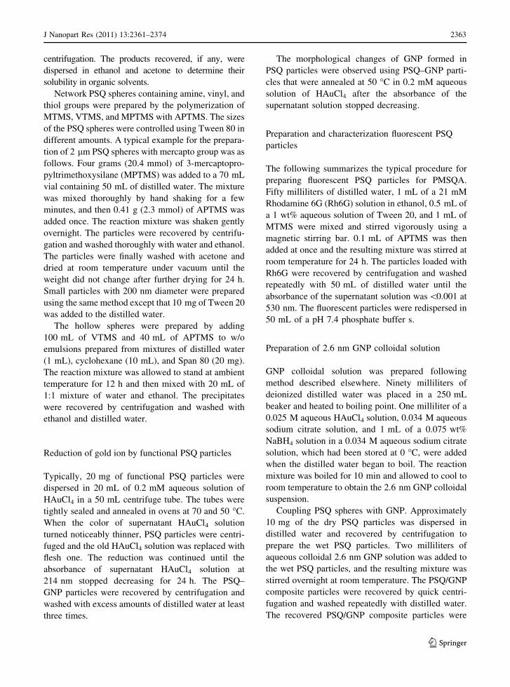
35 nm, as shown in Fig. 1a. The spheres prepared
under identical conditions in the absence of surfactant
typically had diameters approximately 1 lm as seen
in Fig. 1b.
Increasing the amounts of amine catalysts above
certain limits caused formation of irregularly sized
particles in low yields and no solid products. For
example, the SEM images of monodisperse parti-
cles and particles with very different sizes pro-
duced from MPTMS in the presence of smaller and
larger amounts of APTMS are seen in Fig. 1c and d,
which respectively show poly(3-mercaptopropyl)sils-
esquioxane (PMPSQ) spheres prepared from 994:9:1
and 994:9:10 molar mixtures of water, MPTMS, and
APTMS.
The initial decrease in size with the increase of
APTMS concentration was attributed to the faster and
Table 1 Types of products from attempted polymerization of the organotrialkoxysilane using different amine compounds
as catalysts
Organotrialkoxysilane Catalyst Product
Methyltrimethoxysilane,
vinyltrimethoxysilane
3-Aminopropyltri(m)ethoxysilane,
triethylamine, hexylamine, 3-(2-
aminoethyl)propyltrimethoxysilane
Insoluble monodisperse spheres
Glycidoxypropyltrimethoxysilane,
trimethoxysilylpropylisocyanate
Tiethylamine, hexylamine Oily product
Phenyltrimethoxysilane 3-(2-Aminoethyl)propyltrimethoxysilane Mixture of insoluble monodisperse spheres
and soluble spheres
3-Aminopropyltri(m)ethoxysilaen,
triethylamine, hexylamine
Mixture of soluble spheres and oil
Tri(m)ethoxysilylpropyl-p-
methoxycinnamamide
3-Aminopropyltri(m)ethoxysilane,
triethylamine, hexylamine, 3-(2-
aminoethyl)propyltrimethoxysilane
Mixtures of soluble amorphous solid and
insoluble monolith
3-Mercaptopropyltrimethoxysilane 3-Aminopropyltri(m)ethoxysilane,
triethylamine, hexylamine, 3-(2-
aminoethyl)propyltrimethoxysilane
Insoluble monodisperse spheres (agitated),
insoluble spheres with large size distribution
(non-agitated)
123
J Nanopart Res (2011) 13: –2361 2374 2365
more efficient formation of nuclei in an aqueous
medium. The formation of irregularly sized particles
in low yields and no solid products in high concen-
trations of amine catalysts was ascribed to the high
pH of the medium where the hydrolysis and conden-
sation of OTAS was faster. The faster polymerization
can lead to formation of solid shell on OTAS droplets
before OTAS molecules inside diffused into the
aqueous medium. The OTAS inside the shells that
could not diffuse out eventually polymerized to form
large solid particles. The irregularly sized particles
formed most frequently from MPTMS because
MPTMS was significantly more viscous than MTMS
and VTMS that caused slower diffusion into the
aqueous medium. Also, the reaction of MPTMS was
slower than MTMS and VTMS under the identical
reaction conditions. The mechanism for the formation
of large particles is comparable to suspension poly-
merization as the monomer droplets polymerize to
form solid particles in both reaction systems.
The effect of low pH was further elucidated by the
fact that a 9940:9:10 molar mixture of water,
MPTMS and APTMS gave monodisperse spheres.
The ratio between MPTMS and APTMS was the
same while the pH of this reaction system was lower
than that of the 994:9:10 molar mixture, which gave
irregularly sized particles, as described above.
One of the most interesting features of PSQ particles
containing amine, vinyl and mercapto groups was that
in the presence of different metal ions, they produced
metal nanoparticles that were fixed inside and on the
surface (Kim et al. 2006). The reduction of gold ions by
the PSQ particles was examined further using
PVSQT91, PVSQA91, PMPSQT91, PMPSQA91,
and PMSQA91. In these notations, the final letter,
T and A, respectively, indicates that the spheres were
prepared using TEA and APTMS as catalysts. The
number indicates the molar ratio of OTAS and the
catalyst. For example, PVSQA91 indicates PVSQ
spheres prepared from a 9:1 molar mixture of VTMS
and TEA. Quantitative solid state 13C NMR analysis
showed that the amount of 3-aminopropylsilsesquiox-
ane units in PVSQA91, PMPSQA91, and PMSQA91
was commonly approximately 10 mol% (Supplemen-
tary data 2).
Figure 2 shows the quantity of gold produced by a
unit mass of PVSQA91 particles heated in a 0.1 mM
aqueous gold ion solution at 50 and 70 �C as a
Fig. 1 SEM images of polymethylsilsesquioxane spheres
prepared from a 50 mL of water, 1 mL of methyltrimethoxy-
silane, 0.1 mL triethylamine, and 10 mg of Tween 80;
b 50 mL of water, 1 mL of methyltrimethoxysilane, and
0.1 mL triethylamine; SEM image of poly(3-mercaptopro-
pyl)silsesquioxane spheres prepared from c 994:9:1 molar
mixture of water, MPTMs, and APTMS; d 994:9:10 molar
mixtures of water, MPTMS, and APTMS
123
J Nanopart Res (2011) 13: –2361 23742366
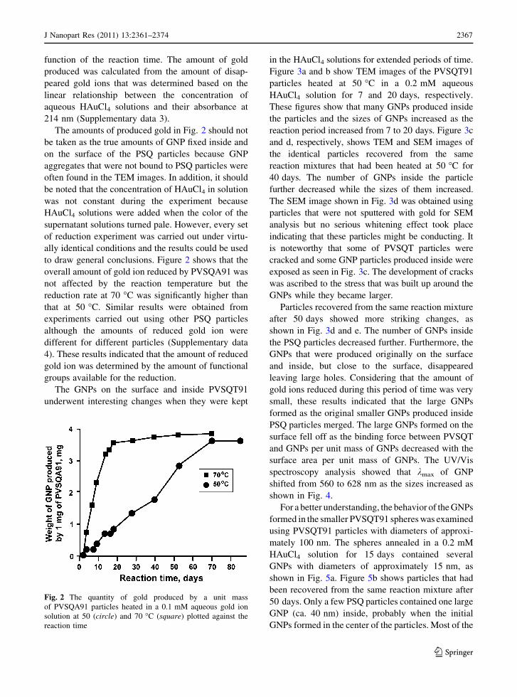
function of the reaction time. The amount of gold
produced was calculated from the amount of disap-
peared gold ions that was determined based on the
linear relationship between the concentration of
aqueous HAuCl4 solutions and their absorbance at
214 nm (Supplementary data 3).
The amounts of produced gold in Fig. 2 should not
be taken as the true amounts of GNP fixed inside and
on the surface of the PSQ particles because GNP
aggregates that were not bound to PSQ particles were
often found in the TEM images. In addition, it should
be noted that the concentration of HAuCl4 in solution
was not constant during the experiment because
HAuCl4 solutions were added when the color of the
supernatant solutions turned pale. However, every set
of reduction experiment was carried out under virtu-
ally identical conditions and the results could be used
to draw general conclusions. Figure 2 shows that the
overall amount of gold ion reduced by PVSQA91 was
not affected by the reaction temperature but the
reduction rate at 70 �C was significantly higher than
that at 50 �C. Similar results were obtained from
experiments carried out using other PSQ particles
although the amounts of reduced gold ion were
different for different particles (Supplementary data
4). These results indicated that the amount of reduced
gold ion was determined by the amount of functional
groups available for the reduction.
The GNPs on the surface and inside PVSQT91
underwent interesting changes when they were kept
in the HAuCl4 solutions for extended periods of time.
Figure 3a and b show TEM images of the PVSQT91
particles heated at 50 �C in a 0.2 mM aqueous
HAuCl4 solution for 7 and 20 days, respectively.
These figures show that many GNPs produced inside
the particles and the sizes of GNPs increased as the
reaction period increased from 7 to 20 days. Figure 3c
and d, respectively, shows TEM and SEM images of
the identical particles recovered from the same
reaction mixtures that had been heated at 50 �C for
40 days. The number of GNPs inside the particle
further decreased while the sizes of them increased.
The SEM image shown in Fig. 3d was obtained using
particles that were not sputtered with gold for SEM
analysis but no serious whitening effect took place
indicating that these particles might be conducting. It
is noteworthy that some of PVSQT particles were
cracked and some GNP particles produced inside were
exposed as seen in Fig. 3c. The development of cracks
was ascribed to the stress that was built up around the
GNPs while they became larger.
Particles recovered from the same reaction mixture
after 50 days showed more striking changes, as
shown in Fig. 3d and e. The number of GNPs inside
the PSQ particles decreased further. Furthermore, the
GNPs that were produced originally on the surface
and inside, but close to the surface, disappeared
leaving large holes. Considering that the amount of
gold ions reduced during this period of time was very
small, these results indicated that the large GNPs
formed as the original smaller GNPs produced inside
PSQ particles merged. The large GNPs formed on the
surface fell off as the binding force between PVSQT
and GNPs per unit mass of GNPs decreased with the
surface area per unit mass of GNPs. The UV/Vis
spectroscopy analysis showed that kmax of GNP
shifted from 560 to 628 nm as the sizes increased as
shown in Fig. 4.
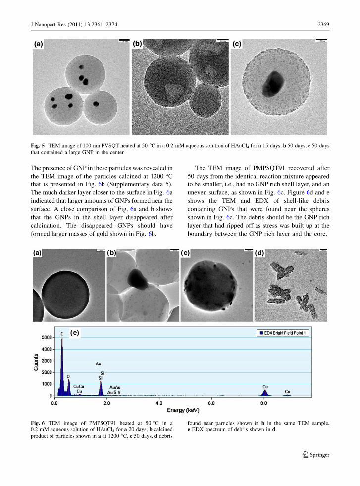
For a better understanding, the behavior of the GNPs
formed in the smaller PVSQT91 spheres was examined
using PVSQT91 particles with diameters of approxi-
mately 100 nm. The spheres annealed in a 0.2 mM
HAuCl4 solution for 15 days contained several
GNPs with diameters of approximately 15 nm, as
shown in Fig. 5a. Figure 5b shows particles that had
been recovered from the same reaction mixture after
50 days. Only a few PSQ particles contained one large
GNP (ca. 40 nm) inside, probably when the initial
GNPs formed in the center of the particles. Most of the
Fig. 2 The quantity of gold produced by a unit mass
of PVSQA91 particles heated in a 0.1 mM aqueous gold ion
solution at 50 (circle) and 70 �C (square) plotted against the
reaction time
123
J Nanopart Res (2011) 13: –2361 2374 2367
particles contained no GNPs on the surface or inside,
but had large holes on the surface, indicating that GNPs
had merged together and then fell off. These results
showed that the GNPs underwent identical morpho-
logical changes regardless of the PSQ particle size.
Figure 3d and e showed that pores with tens of
nanometers were clearly observed by TEM. Since
there were no PSQ particles that originally contained
such large pores as observed with TEM, the pores
where the large GNPs formed should have been
produced during the formation of large GNPs. Exclud-
ing the possibility of cleavage and reformation of
Si–O–Si bonds, these results suggest that PVSQT
particles were soft and porous enough for smaller
GNPs to move around and fuse together. The porous
nature of these particles was proved by efficient
entrapment of small fluorescent molecules as
described below.
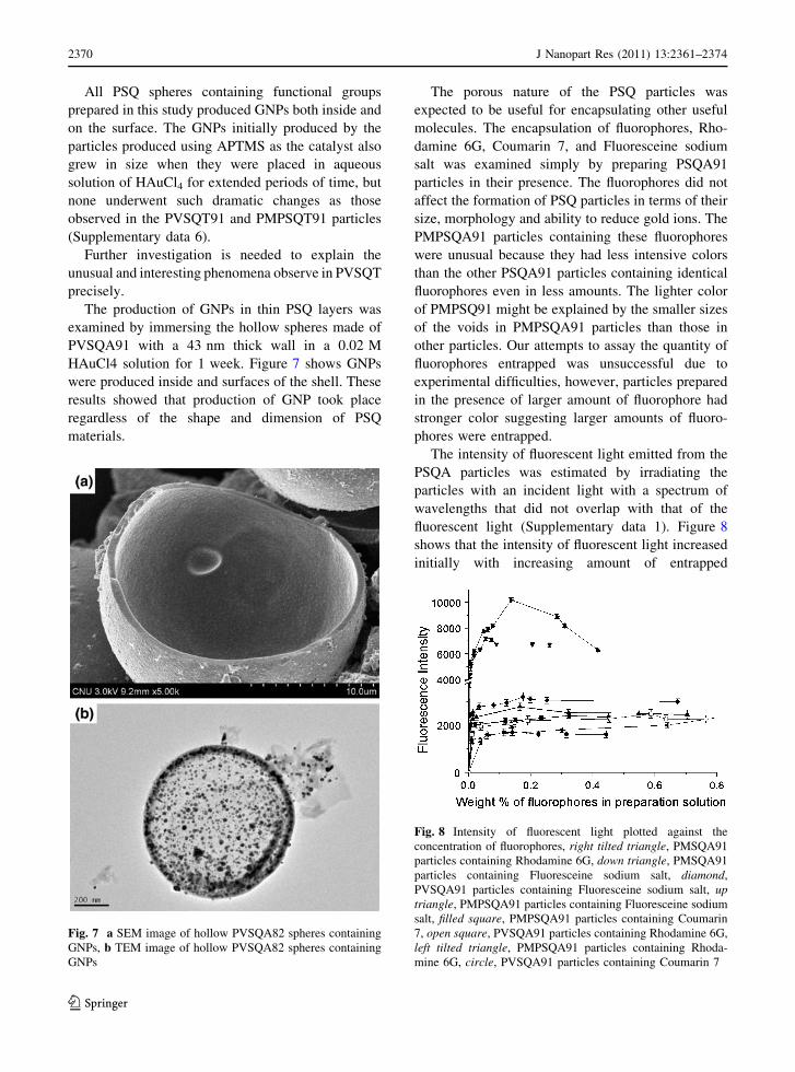
The change of the GNPs produced inside the
PMPSQT91 particles was also unusual. The GNPs
produced by PMPSQT91 appeared to be very small as
seen in Fig. 6a for PMPSQT91 particles that had been
placed in a 0.2 mM HAuCl4 solution for 20 days.
Fig. 3 TEM images of particles obtained by heating PVSQT91 at 50 �C in a 0.2 mM aqueous solution of HAuCl4 for a 7 days,
b 20 days, c 40 days, d SEM image of particles shown in c, e 50 days, f SEM image of particles shown in e
Fig. 4 UV/Vis spectra of colloidal solutions of PVSQ91
particles heated at 50 �C in a 0.2 mM aqueous solution of
HAuCl4 for a 7 days, b 20 days, c 40 days, d 50 days
123
J Nanopart Res (2011) 13: –2361 23742368
The presence of GNP in these particles was revealed in
the TEM image of the particles calcined at 1200 �C
that is presented in Fig. 6b (Supplementary data 5).
The much darker layer closer to the surface in Fig. 6a
indicated that larger amounts of GNPs formed near the
surface. A close comparison of Fig. 6a and b shows
that the GNPs in the shell layer disappeared after
calcination. The disappeared GNPs should have
formed larger masses of gold shown in Fig. 6b.
The TEM image of PMPSQT91 recovered after
50 days from the identical reaction mixture appeared
to be smaller, i.e., had no GNP rich shell layer, and an
uneven surface, as shown in Fig. 6c. Figure 6d and e
shows the TEM and EDX of shell-like debris
containing GNPs that were found near the spheres
shown in Fig. 6c. The debris should be the GNP rich
layer that had ripped off as stress was built up at the
boundary between the GNP rich layer and the core.
Fig. 5 TEM image of 100 nm PVSQT heated at 50 �C in a 0.2 mM aqueous solution of HAuCl4 for a 15 days, b 50 days, c 50 days
that contained a large GNP in the center
Fig. 6 TEM image of PMPSQT91 heated at 50 �C in a
0.2 mM aqueous solution of HAuCl4 for a 20 days, b calcined
product of particles shown in a at 1200 �C, c 50 days, d debris
found near particles shown in b in the same TEM sample,
e EDX spectrum of debris shown in d
123
J Nanopart Res (2011) 13: –2361 2374 2369
All PSQ spheres containing functional groups
prepared in this study produced GNPs both inside and
on the surface. The GNPs initially produced by the
particles produced using APTMS as the catalyst also
grew in size when they were placed in aqueous
solution of HAuCl4 for extended periods of time, but
none underwent such dramatic changes as those
observed in the PVSQT91 and PMPSQT91 particles
(Supplementary data 6).
Further investigation is needed to explain the
unusual and interesting phenomena observe in PVSQT
precisely.
The production of GNPs in thin PSQ layers was
examined by immersing the hollow spheres made of
PVSQA91 with a 43 nm thick wall in a 0.02 M
HAuCl4 solution for 1 week. Figure 7 shows GNPs
were produced inside and surfaces of the shell. These
results showed that production of GNP took place
regardless of the shape and dimension of PSQ
materials.
The porous nature of the PSQ particles was
expected to be useful for encapsulating other useful
molecules. The encapsulation of fluorophores, Rho-
damine 6G, Coumarin 7, and Fluoresceine sodium
salt was examined simply by preparing PSQA91
particles in their presence. The fluorophores did not
affect the formation of PSQ particles in terms of their
size, morphology and ability to reduce gold ions. The
PMPSQA91 particles containing these fluorophores
were unusual because they had less intensive colors
than the other PSQA91 particles containing identical
fluorophores even in less amounts. The lighter color
of PMPSQ91 might be explained by the smaller sizes
of the voids in PMPSQA91 particles than those in
other particles. Our attempts to assay the quantity of
fluorophores entrapped was unsuccessful due to
experimental difficulties, however, particles prepared
in the presence of larger amount of fluorophore had
stronger color suggesting larger amounts of fluoro-
phores were entrapped.
The intensity of fluorescent light emitted from the
PSQA particles was estimated by irradiating the
particles with an incident light with a spectrum of
wavelengths that did not overlap with that of the
fluorescent light (Supplementary data 1). Figure 8
shows that the intensity of fluorescent light increased
initially with increasing amount of entrapped
Fig. 7 a SEM image of hollow PVSQA82 spheres containing
GNPs, b TEM image of hollow PVSQA82 spheres containing
GNPs
Fig. 8 Intensity of fluorescent light plotted against the
concentration of fluorophores, right tilted triangle, PMSQA91
particles containing Rhodamine 6G, down triangle, PMSQA91
particles containing Fluoresceine sodium salt, diamond,
PVSQA91 particles containing Fluoresceine sodium salt, uptriangle, PMPSQA91 particles containing Fluoresceine sodium
salt, filled square, PMPSQA91 particles containing Coumarin
7, open square, PVSQA91 particles containing Rhodamine 6G,
left tilted triangle, PMPSQA91 particles containing Rhoda-
mine 6G, circle, PVSQA91 particles containing Coumarin 7
123
J Nanopart Res (2011) 13: –2361 23742370
fluorophores and then leveled off or decreased slightly
after reaching a maximum value. These results corre-
spond to the well-known fact that the fluorophores emit
weaker fluorescent light when they are concentrated
due to self-quenching. The intensity of fluorescent light
was the strongest when the fluorophores were
entrapped in PMSQA than PVSQA and PMPSQA.
Hollow PVSQA82 prepared in the presence of fluoro-
phores also gave particles with strong fluorescence
(Supplementary data 7).
It is known that amines bind to gold surface very
weakly and amine groups have been rarely used to fix
biological molecules to gold. The color of PSQ
particles prepared in this study commonly turned dark
purple when they were stored in colloidal solutions of
GNPs of different sizes. TEM images of the colored
particles revealed that significant amounts of GNPs
were bound. PSQA/GNP (2.8 nm diameter) particles
that were subjected to heating in 95 �C water for 6 h,
vortexing (5 min at maximum speed), and ultrasonic
treatment (10 min, 5 W) were virtually identical to the
original ones as observed with TEM. The supernatant
water was colorless as observed with naked eyes when
the PSQA/GNP particles described above were cen-
trifuged down. These results corresponded to those
reported earlier in that GNP bind quite strongly to
amine compounds in contrast to bulk gold (Pong et al.
2005).
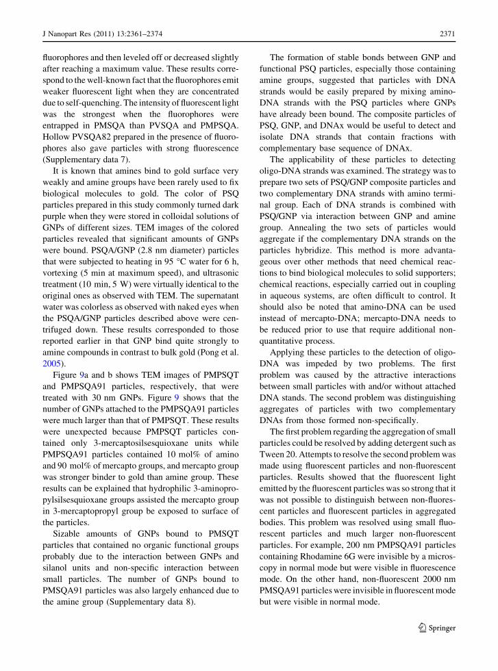
Figure 9a and b shows TEM images of PMPSQT
and PMPSQA91 particles, respectively, that were
treated with 30 nm GNPs. Figure 9 shows that the
number of GNPs attached to the PMPSQA91 particles
were much larger than that of PMPSQT. These results
were unexpected because PMPSQT particles con-
tained only 3-mercaptosilsesquioxane units while
PMPSQA91 particles contained 10 mol% of amino
and 90 mol% of mercapto groups, and mercapto group
was stronger binder to gold than amine group. These
results can be explained that hydrophilic 3-aminopro-
pylsilsesquioxane groups assisted the mercapto group
in 3-mercaptopropyl group be exposed to surface of
the particles.
Sizable amounts of GNPs bound to PMSQT
particles that contained no organic functional groups
probably due to the interaction between GNPs and
silanol units and non-specific interaction between
small particles. The number of GNPs bound to
PMSQA91 particles was also largely enhanced due to
the amine group (Supplementary data 8).
The formation of stable bonds between GNP and
functional PSQ particles, especially those containing
amine groups, suggested that particles with DNA
strands would be easily prepared by mixing amino-
DNA strands with the PSQ particles where GNPs
have already been bound. The composite particles of
PSQ, GNP, and DNAx would be useful to detect and
isolate DNA strands that contain fractions with
complementary base sequence of DNAx.
The applicability of these particles to detecting
oligo-DNA strands was examined. The strategy was to
prepare two sets of PSQ/GNP composite particles and
two complementary DNA strands with amino termi-
nal group. Each of DNA strands is combined with
PSQ/GNP via interaction between GNP and amine
group. Annealing the two sets of particles would
aggregate if the complementary DNA strands on the
particles hybridize. This method is more advanta-
geous over other methods that need chemical reac-
tions to bind biological molecules to solid supporters;
chemical reactions, especially carried out in coupling
in aqueous systems, are often difficult to control. It
should also be noted that amino-DNA can be used
instead of mercapto-DNA; mercapto-DNA needs to
be reduced prior to use that require additional non-
quantitative process.
Applying these particles to the detection of oligo-
DNA was impeded by two problems. The first
problem was caused by the attractive interactions
between small particles with and/or without attached
DNA stands. The second problem was distinguishing
aggregates of particles with two complementary
DNAs from those formed non-specifically.
The first problem regarding the aggregation of small
particles could be resolved by adding detergent such as
Tween 20. Attempts to resolve the second problem was
made using fluorescent particles and non-fluorescent
particles. Results showed that the fluorescent light
emitted by the fluorescent particles was so strong that it
was not possible to distinguish between non-fluores-
cent particles and fluorescent particles in aggregated
bodies. This problem was resolved using small fluo-
rescent particles and much larger non-fluorescent
particles. For example, 200 nm PMPSQA91 particles
containing Rhodamine 6G were invisible by a micros-
copy in normal mode but were visible in fluorescence
mode. On the other hand, non-fluorescent 2000 nm
PMSQA91 particles were invisible in fluorescent mode
but were visible in normal mode.
123
J Nanopart Res (2011) 13: –2361 2374 2371
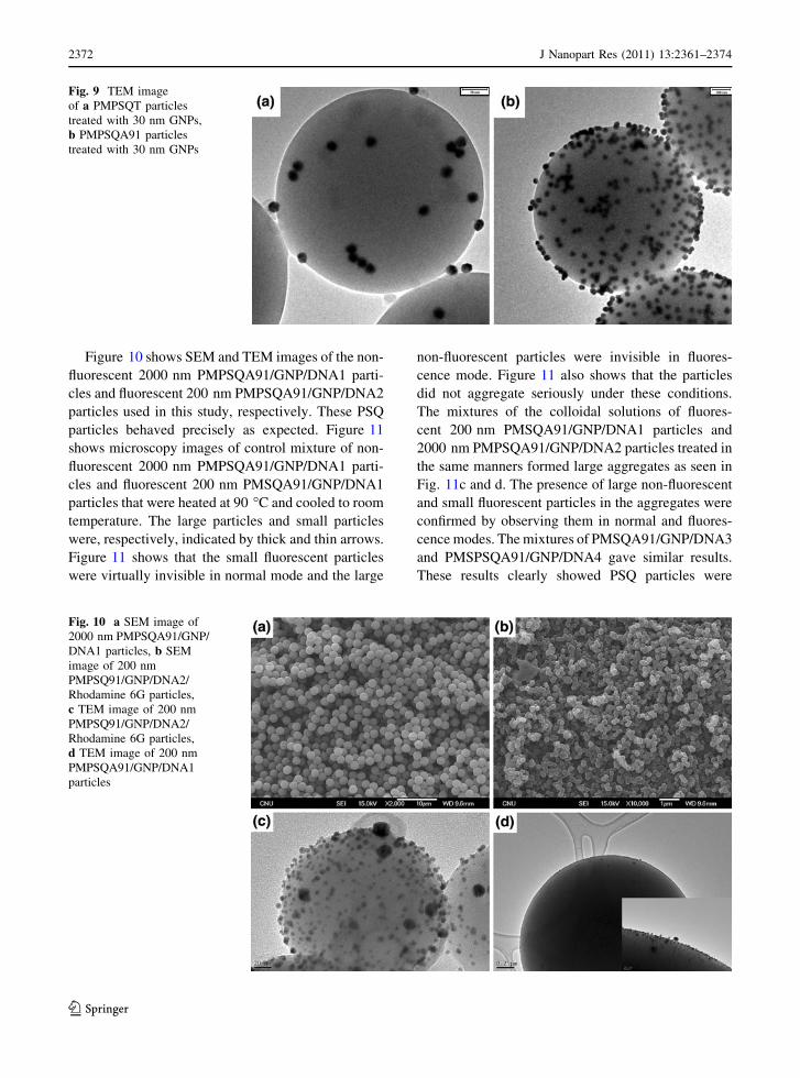
Figure 10 shows SEM and TEM images of the non-
fluorescent 2000 nm PMPSQA91/GNP/DNA1 parti-
cles and fluorescent 200 nm PMPSQA91/GNP/DNA2
particles used in this study, respectively. These PSQ
particles behaved precisely as expected. Figure 11
shows microscopy images of control mixture of non-
fluorescent 2000 nm PMPSQA91/GNP/DNA1 parti-
cles and fluorescent 200 nm PMSQA91/GNP/DNA1
particles that were heated at 90 �C and cooled to room
temperature. The large particles and small particles
were, respectively, indicated by thick and thin arrows.
Figure 11 shows that the small fluorescent particles
were virtually invisible in normal mode and the large
non-fluorescent particles were invisible in fluores-
cence mode. Figure 11 also shows that the particles
did not aggregate seriously under these conditions.
The mixtures of the colloidal solutions of fluores-
cent 200 nm PMSQA91/GNP/DNA1 particles and
2000 nm PMPSQA91/GNP/DNA2 particles treated in
the same manners formed large aggregates as seen in
Fig. 11c and d. The presence of large non-fluorescent
and small fluorescent particles in the aggregates were
confirmed by observing them in normal and fluores-
cence modes. The mixtures of PMSQA91/GNP/DNA3
and PMSPSQA91/GNP/DNA4 gave similar results.
These results clearly showed PSQ particles were
Fig. 9 TEM image
of a PMPSQT particles
treated with 30 nm GNPs,
b PMPSQA91 particles
treated with 30 nm GNPs
Fig. 10 a SEM image of
2000 nm PMPSQA91/GNP/
DNA1 particles, b SEM
image of 200 nm
PMPSQ91/GNP/DNA2/
Rhodamine 6G particles,
c TEM image of 200 nm
PMPSQ91/GNP/DNA2/
Rhodamine 6G particles,
d TEM image of 200 nm
PMPSQA91/GNP/DNA1
particles
123
J Nanopart Res (2011) 13: –2361 23742372
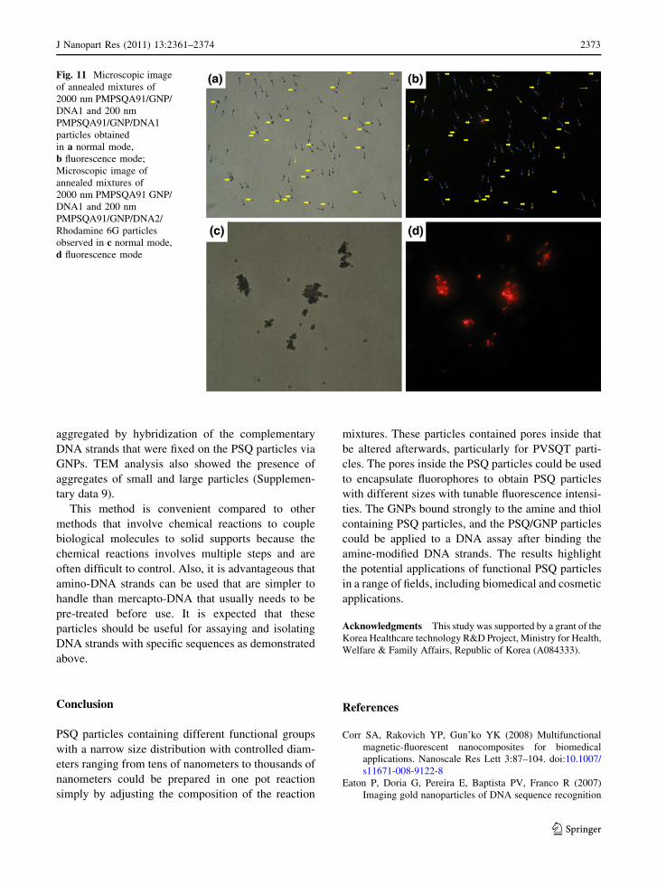
aggregated by hybridization of the complementary
DNA strands that were fixed on the PSQ particles via
GNPs. TEM analysis also showed the presence of
aggregates of small and large particles (Supplemen-
tary data 9).
This method is convenient compared to other
methods that involve chemical reactions to couple
biological molecules to solid supports because the
chemical reactions involves multiple steps and are
often difficult to control. Also, it is advantageous that
amino-DNA strands can be used that are simpler to
handle than mercapto-DNA that usually needs to be
pre-treated before use. It is expected that these
particles should be useful for assaying and isolating
DNA strands with specific sequences as demonstrated
above.
Conclusion
PSQ particles containing different functional groups
with a narrow size distribution with controlled diam-
eters ranging from tens of nanometers to thousands of
nanometers could be prepared in one pot reaction
simply by adjusting the composition of the reaction
mixtures. These particles contained pores inside that
be altered afterwards, particularly for PVSQT parti-
cles. The pores inside the PSQ particles could be used
to encapsulate fluorophores to obtain PSQ particles
with different sizes with tunable fluorescence intensi-
ties. The GNPs bound strongly to the amine and thiol
containing PSQ particles, and the PSQ/GNP particles
could be applied to a DNA assay after binding the
amine-modified DNA strands. The results highlight
the potential applications of functional PSQ particles
in a range of fields, including biomedical and cosmetic
applications.
Acknowledgments This study was supported by a grant of the
Korea Healthcare technology R&D Project, Ministry for Health,
Welfare & Family Affairs, Republic of Korea (A084333).
References
Corr SA, Rakovich YP, Gun’ko YK (2008) Multifunctional
magnetic-fluorescent nanocomposites for biomedical
applications. Nanoscale Res Lett 3:87–104. doi:10.1007/
s11671-008-9122-8
Eaton P, Doria G, Pereira E, Baptista PV, Franco R (2007)
Imaging gold nanoparticles of DNA sequence recognition
Fig. 11 Microscopic image
of annealed mixtures of
2000 nm PMPSQA91/GNP/
DNA1 and 200 nm
PMPSQA91/GNP/DNA1
particles obtained
in a normal mode,
b fluorescence mode;
Microscopic image of
annealed mixtures of
2000 nm PMPSQA91 GNP/
DNA1 and 200 nm
PMPSQA91/GNP/DNA2/
Rhodamine 6G particles
observed in c normal mode,
d fluorescence mode
123
J Nanopart Res (2011) 13: –2361 2374 2373
in biomedical application. IEEE Trans Nanobiosci 6:282–
288. doi:10.1109/TNB.2007.908985
Heitsch AT, Smith DK, Patel RN, Ress D, Korgel BA (2008)
Multifunctional particles: magnetic nanocrystals and gold
nanorods coated with fluorescent dye-doped silica shells.
J Solid State Chem 181:1590–1599. doi:10.1016/j.jssc.
2008.05.002
Kim Y, Kim YB, Yoon K (2006) Preparation of functionalized
polysilsesquioxane and polysilsesquioxane-metal nano-
particle composite spheres. Macromol Rapid Commun
27:1247–1253. doi:10.1002/(ISSN)1521-3927
Klostranec JM, Chan WCW (2006) Quantum dots in biological
and biomedical research: recent progress and present
challenges. Adv Mater 18:1953–1964. doi:10.1002/adma.
200500786
Lee D, Kim Y, Kim YB, Kim J, Han Y (2008) One step
preparation of spherical silicon resins from immiscible
reaction. Mixtures Macromol Res 16:353
Lin YS, Wu SH, Tseng CT, Hung Y, Chang C, Mou CY (2009)
Synthesis of hollow silica nanospheres with a micro-
emulsion as the template. Chem Commun 3542–3544.
doi:10.1039/b902681a
Liong M, Lu J, Kovochich M, Xia T, Ruehm SG, Nel AE,
Tamanoi F, Zink JI (2008) Multifunctional inorganic
nanoparticles for imaging, targeting, and drug delivery.
ACS Nano 2:889–896. doi:10.1021/nn800072t
Niemeyer CM, Simon U (2005) DNA-based assembly of metal
nanoparticles. Eur J Inorg Chem 3641–3655. doi:10.1002/
ejic.200500425
Parak WJ, Gerion D, Pellegrino T, Zanchet D, Micheel C,
Williams SC, Boudreau R, Le Gros MA, Larabell CA,
Alivisatos AP (2003) Biological applications of colloidal
nanocrystals. Nanotechnology 14:R15–R27. doi:10.1088/
0957-4484/14/7/201
Pasqua L, Cundari S, Ceresa C, Cavaletti G (2009) Recent
development, applications, and perspectives of mesoporous
silica particles in medicine and biotechnology. Curr Med
Chem 16:3054–3063. doi:10.2174/092986709788803079
Pellegrino T, Kudera S, Liedl T, Javier AM, Manna L, Parak
WJ (2005) On the development of colloidal nanoparticles
towards multifunctional structures and their possible use
for biological applications. Small 1:48–63. doi:10.1002/
smll.200400071
Pong BK, Lee JY, Trout BL (2005) First principles computa-
tional study for understanding the interactions between
ssDNA and gold nanoparticles: adsorption of methylamine
on gold nanoparticulate. Langmuir 21:11599–11603. doi:
10.1021/la052116k
Ren CL, Li JH, Liu Q, Ren J, Chen XG, Hu ZD, Xue DS (2008)
Synthesis of organic dye-impregnated silica shell-coated
iron oxide nanoparticles by a new method. Nanoscale Res
Lett 3:496–501. doi:10.1007/s11671-008-9186-5
Schottner G (2001) Hybrid sol–gel-derived polymers: applica-
tions of multifunctional materials. Chem Mater 13:3422–
3435. doi:10.1021/cm011060m
Smith AM, Duan HW, Mohs AM, Nie SM (2008) Bioconjugated
quantum dots for in vivo molecular and cellular imaging.
Adv Drug Deliv Rev 60:1226–1240. doi:10.1016/j.addr.
2008.03.015
Tan WB, Zhang Y (2005) Multifunctional quantum-dot-based
magnetic chitosan nanobeads. Adv Mater 17:2375–2380.
doi:10.1002/adma.200401650
Thaxton CS, Geroganopoulou DG, Mirkin CA (2006) Gold
nanoparticle probes for the detection of nucleic acid tar-
gets. Clin Chim Acta 363:120–126. doi:10.1016/j.cccn.
2005.05.042
Yang S, Liu HR, Zhang ZC (2009) Design and fabrication of
hollow, magnetic and fluorescent CdS-magnetite-poly
(styrene-co-methyl methacrylate) microspheres. New J
Chem 33:620–625. doi:10.1039/b811418h
123
J Nanopart Res (2011) 13: –2361 23742374
Related Documents