The Scientific World Journal Volume 2012, Article ID 680108, 8 pages doi:10.1100/2012/680108 The cientificWorldJOURNAL Research Article Preparation and Characterization of Alginate and Psyllium Beads Containing Lactobacillus acidophilus Farzaneh Lotfipour, 1, 2 Shahla Mirzaeei, 1, 3 and Maryam Maghsoodi 1, 4 1 Faculty of Pharmacy, Tabriz University of Medical Sciences, Tabriz 51664, Iran 2 Gastrointestinal and Liver Disease Research Center, Tabriz University of Medical Sciences, Tabriz, Iran 3 Faculty of Pharmacy, Kermanshah University of Medical Sciences, Kermanshah, Iran 4 Biotechnology Research Center, Tabriz University of Medical Sciences, Tabriz, Iran Correspondence should be addressed to Farzaneh Lotfipour, farzaneh.lotfi[email protected] and Maryam Maghsoodi, [email protected] Received 31 October 2011; Accepted 14 December 2011 Academic Editors: J. Ali, A. Concheiro, and P. Danckwerts Copyright © 2012 Farzaneh Lotfipour et al. This is an open access article distributed under the Creative Commons Attribution License, which permits unrestricted use, distribution, and reproduction in any medium, provided the original work is properly cited. This paper describes preparation and characterization of beads of alginate and psyllium containing probiotic bacteria of Lactobacillus acidophilus DMSZ20079. Twelve different formulations containing alginate (ALG) and alginate-psyllium (ALG- PSL) were prepared using extrusion technique. The prepared beads were characterized in terms of size, morphology and surface properties, encapsulation efficiency, viabilities in acid (pH 1.8, 2 hours) and bile (0.5% w/v, 2 hours) conditions, and release in simulated colon pH conditions. The results showed that spherical beads with narrow size distribution ranging from 1.59 ± 0.04 to 1.67 ± 0.09 mm for ALG and from 1.61 ± 0.06 to 1.80 ± 0.07 mm for ALG-PSL with encapsulation efficiency higher than 98% were achieved. Furthermore, addition of PSL into ALG enhanced the integrity of prepared beads in comparison with ALG formulations. The results indicated that incorporation of PSL into alginate beads improved viability of the bacteria in acidic conditions as well as bile conditions. Also, stimulating effect of PSL on the probiotic bacteria was observed through 20-hour incubation in simulated colonic pH solution. According to our in vitro studies, PSL can be a suitable polymer candidate for partial substitution with ALG for probiotic coating. 1. Introduction “Probiotics are live microorganisms (bacteria or yeasts), which when ingested or locally applied in sufficient numbers confer one or more specified demonstrated health benefits for the host” [1]. These benefits include maintenance of normal intestinal microflora, defense against enteropathogen infections, controlling serum cholesterol levels, improving lactose utilization in persons who are lactose maldigesters by production of β-galactosidase, and possessing anticarcino- genic and antimutagenic activities [2–4]. Probiotics can be bacteria, moulds, and yeasts. How- ever, most of probiotics are bacteria; among them lactic acid bacteria (LAB), typically associated with the human gastrointestinal tract, are the most widely used probiotic microorganisms. In order to exhibit their potential benefits, probiotics need to pass the harsh conditions of gastric tract and colonize and grow on the epithelium of colon in appropriate population [5]. It is suggested that probiotics should be formulated in products in a minimum count of 10 6-7 CFU/g or mL of viable probiotic bacteria [1]. To improve viability and stability of probiotics and efficient delivery of the cells to their active sites, various techniques have been utilized so far. In this regard, encapsulation of probiotics in wide variety of polymers is the most frequently applied method that is cited in numerous studies [6]. Alginate, a commonly used material to encapsulate probiotics, is a naturally occurring biocompatible and biodegradable linear anionic polysaccharide. Preparation of alginate bead, with well retained bacteria in their matrix, can be easily achieved by simple techniques like extrusion or emulsion methods. In spite of the wide application of alginate microcapsules in this area, some problems related to protection efficiency of them have been reported including

Welcome message from author
This document is posted to help you gain knowledge. Please leave a comment to let me know what you think about it! Share it to your friends and learn new things together.
Transcript

The Scientific World JournalVolume 2012, Article ID 680108, 8 pagesdoi:10.1100/2012/680108
The cientificWorldJOURNAL
Research Article
Preparation and Characterization of Alginate andPsyllium Beads Containing Lactobacillus acidophilus
Farzaneh Lotfipour,1, 2 Shahla Mirzaeei,1, 3 and Maryam Maghsoodi1, 4
1 Faculty of Pharmacy, Tabriz University of Medical Sciences, Tabriz 51664, Iran2 Gastrointestinal and Liver Disease Research Center, Tabriz University of Medical Sciences, Tabriz, Iran3 Faculty of Pharmacy, Kermanshah University of Medical Sciences, Kermanshah, Iran4 Biotechnology Research Center, Tabriz University of Medical Sciences, Tabriz, Iran
Correspondence should be addressed to Farzaneh Lotfipour, [email protected] Maryam Maghsoodi, [email protected]
Received 31 October 2011; Accepted 14 December 2011
Academic Editors: J. Ali, A. Concheiro, and P. Danckwerts
Copyright © 2012 Farzaneh Lotfipour et al. This is an open access article distributed under the Creative Commons AttributionLicense, which permits unrestricted use, distribution, and reproduction in any medium, provided the original work is properlycited.
This paper describes preparation and characterization of beads of alginate and psyllium containing probiotic bacteria ofLactobacillus acidophilus DMSZ20079. Twelve different formulations containing alginate (ALG) and alginate-psyllium (ALG-PSL) were prepared using extrusion technique. The prepared beads were characterized in terms of size, morphology and surfaceproperties, encapsulation efficiency, viabilities in acid (pH 1.8, 2 hours) and bile (0.5% w/v, 2 hours) conditions, and release insimulated colon pH conditions. The results showed that spherical beads with narrow size distribution ranging from 1.59± 0.04 to1.67±0.09 mm for ALG and from 1.61±0.06 to 1.80±0.07 mm for ALG-PSL with encapsulation efficiency higher than 98% wereachieved. Furthermore, addition of PSL into ALG enhanced the integrity of prepared beads in comparison with ALG formulations.The results indicated that incorporation of PSL into alginate beads improved viability of the bacteria in acidic conditions as wellas bile conditions. Also, stimulating effect of PSL on the probiotic bacteria was observed through 20-hour incubation in simulatedcolonic pH solution. According to our in vitro studies, PSL can be a suitable polymer candidate for partial substitution with ALGfor probiotic coating.
1. Introduction
“Probiotics are live microorganisms (bacteria or yeasts),which when ingested or locally applied in sufficient numbersconfer one or more specified demonstrated health benefitsfor the host” [1]. These benefits include maintenance ofnormal intestinal microflora, defense against enteropathogeninfections, controlling serum cholesterol levels, improvinglactose utilization in persons who are lactose maldigesters byproduction of β-galactosidase, and possessing anticarcino-genic and antimutagenic activities [2–4].
Probiotics can be bacteria, moulds, and yeasts. How-ever, most of probiotics are bacteria; among them lacticacid bacteria (LAB), typically associated with the humangastrointestinal tract, are the most widely used probioticmicroorganisms. In order to exhibit their potential benefits,probiotics need to pass the harsh conditions of gastric
tract and colonize and grow on the epithelium of colon inappropriate population [5]. It is suggested that probioticsshould be formulated in products in a minimum countof 106-7 CFU/g or mL of viable probiotic bacteria [1]. Toimprove viability and stability of probiotics and efficientdelivery of the cells to their active sites, various techniqueshave been utilized so far. In this regard, encapsulation ofprobiotics in wide variety of polymers is the most frequentlyapplied method that is cited in numerous studies [6].
Alginate, a commonly used material to encapsulateprobiotics, is a naturally occurring biocompatible andbiodegradable linear anionic polysaccharide. Preparation ofalginate bead, with well retained bacteria in their matrix,can be easily achieved by simple techniques like extrusionor emulsion methods. In spite of the wide application ofalginate microcapsules in this area, some problems related toprotection efficiency of them have been reported including

2 The Scientific World Journal
susceptibility to disintegration in the presence of excessmonovalent ions, Ca2+ chelating agents, and harsh chemicalenvironments [4].
Psyllium, the common name used for several members ofthe plant genus Plantago, is gel-forming mucilage composedof a highly branched arabinoxylan. The backbone consistsof xylose units, while arabinose and xylose form the sidechains [7, 8]. Psyllium has been reported as a medicinallyactive natural polysaccharide for the treatment of consti-pation, diarrhea, irritable bowel syndrome, inflammatorybowel disease ulcerative colitis, colon cancer, diabetes, andhypercholesterolemia [9]. Moreover, psyllium as a solublefiber has a potential to stimulate bacterial growth in digestivesystem, and, in some reports, it has been used as prebiotic[10–13]. Prebiotics is defined by Gibson and Roberfroid [14]as “non-digestible food ingredients that beneficially affectsthe host by selectively stimulating the growth and/or activityof one or a limited number of bacteria in the colon, and thusimproves host health.”
Having in mind the pharmacological benefits of psylliumin digestive system as well as its potential to stimulateprobiotic growth in the colon, here we aimed to incorporatepsyllium in alginate beads containing probiotic bacteria L.acidophilus DMSZ20079.
To this end, different formulations containing ALGand/or ALG-PSL were prepared using extrusion techniqueand characterized in terms of size, morphology and surfaceproperties, encapsulation efficiency (EE), viabilities in acid(pH 1.8, 2 hours) and bile (0.5% w/v, 2 hours) conditions,and release in simulated colon pH conditions.
2. Materials and Methods
2.1. Materials. L. acidophilus DSMZ20079 was obtainedfrom DSMZ (Germany), pepsin, pancreatin, sodium algi-nate, oxgall from Sigma-Aldrich (Germany), MRS broth andMRS agar, sodium hydrogen phosphate, calcium chloride,sodium hydroxide and hydrochloric acid from Merck (Ger-many), and psyllium seed husk was supplied from Sidpur SatIsabgol (India).
2.2. Methods
2.2.1. Preparation of Inoculum. L. acidophilus was culturedin MRS broth at 37◦C for 18 hours. Culture was harvested bycentrifugation at 700 RCF at 4◦C for 7 min and washed twicewith saline and collected by centrifugation as above. Thewashed bacterial cells were resuspended in 7 mL saline, andthe cell count was determined using pour plate technique inMRS agar in triplicate. The cell suspension divided in someequal parts and consequently was used to prepare differentformulations.
2.2.2. Extraction of Psyllium. Psyllium husk was extractedby a method described by Guo et al. [8] with some mod-ifications. First, 5 g psyllium husk was dispersed in 100 mLwater at 80◦C over night under constant stirring at 50 rpm,after 18–20 hours the dispersion became a homogenous
gel. Consequently, the obtained gel was centrifuged (HettichRotofix 32 A, Germany) at 18000 RCF for 90 min, to separatethe gel and the solution. The gel phase was dissolved in 2 MNaOH solution at room temperature for 2 hours; alkalinesolution was separated from the residue by centrifugation(18000 RCF for 90 min) and accordingly neutralized with2 M HCl. During the neutralization, a large amount ofgel-like yellow precipitate was produced and separated bycentrifugation (18000 RCF for 90 min) from the solublefraction and washed three times with distilled water. The gelprecipitate was dried at 40◦C for 48 hours.
2.2.3. Preparation of Beads. The extrusion technique wasused to prepare ALG and ALG-PSL beads [15]. Sodiumalginate and psyllium solutions were sterilized at 121◦C for15 min. The cooled ALG or ALG-PSL solutions (20 mL) weremixed with bacterial inoculum and gently stirred for 30 minto obtain a homogeneous suspension. The suspensions wereextruded dropwise through a 27 gage nozzle into sterilehardening solution (CaCl2). The beads were shaken at150 rpm for 40 min, isolated by aseptic filtration (WhatmanNo.1), washed twice with sterile water, and kept in 0.1%w/v peptone solution at 4◦C. The prepared formulations areshown in Table 1.
2.2.4. Size and Morphological Analysis. The particle sizeof beads was assessed using optical microscopy (Dino-lite, Taiwan) by Scion image analyzer software. Data werecollected from 60 beads in each sample, and mean particlesize was reported.
The topographical properties of prepared beads wereinvestigated by scanning electron microscopy (SEM)(Philipse XL30, Holland) at an accelerating voltage of 20 KV.Prior to examination, samples were prepared on aluminumstubs and coated with gold under argon atmosphere bymeans of a sputter coater.
2.2.5. Encapsulation Efficiency (EE). To determine the encap-sulation efficiency, firstly prepared beads were mechanicallydisintegrated in phosphate buffer (pH = 6.8), then thenumber of entrapped cells after adequate dilution weremeasured by pour plate method, and counts were expressedas number of colony forming units (CFU), and calculated as
EE =(
Log10N
Log10N0
)× 100, (1)
where N is the number of viable entrapped cells releasedfrom the beads and N0 is the number of free cells added tothe biopolymer mixture immediately before the productionprocedure.
2.2.6. Viability of Encapsulated and Free L. acidophilus atLow pH Condition. Low pH conditions were produced using9 g/L sodium chloride and 3.0 g/L of pepsin and pH adjustedto 1.8 with hydrochloric acid [16]. 100 mg beads withentrapped bacteria or 0.1 mL of cell suspension were mixedin 20 mL of acid solution and incubated for 120 min at 37◦C

The Scientific World Journal 3
with constant agitation at 50 rpm. After incubation, beadswere disintegrated in phosphate buffer (pH = 6.8), then1.0 mL aliquot of the mixture removed and assayed usingpour plate method.
The survival (%) of the bacteria was calculated as follows:
% Survival
= (log CFU/g beads after 2 hours exposure to
acidic condition/ log CFU/g beads initial count)× 100.
(2)
2.2.7. Viability of Encapsulated and Free L. acidophilus atHigh Bile Salt Concentration. Prepared beads after 2-houracid exposure were washed with distilled water, removed, andincubated in 50 mL of high bile condition, containing 6.8 g ofmonobasic potassium phosphate, and 10 g/L of pancreatinwith pH adjusted to 6.8 ± 0.1 using sodium hydroxide and0.5% w/v oxgall for 2 hours at 37◦C with constant agitation at50 rpm [17]. Samples were then taken, and bacterial growthwas assayed using pour plate method.
2.2.8. Release of Encapsulated Cells and Free L. acidophilus inSimulated Colonic pH Solution. The release of the preparedbeads was examined at simulated colonic pH solution asdescribed by Mandal et al. [18]. The beads were mixed with50 mL of simulated colonic pH solution containing 0.1 Mmonobasic potassium phosphate with pH adjusted to 7.4 ±0.1 with sodium hydroxide and incubated for 20 h at 37◦Cwith constant agitation at 50 rpm. Samples were taken atdifferent time intervals, and bacterial growth was assayedusing pour plate method as described in Section 2.2.5.
2.2.9. Statistical Analyses. Statistical testing was carried outusing SPSS19. All of the experiments were performed intriplicates. Data are presented as mean ± SD. The One-WayANOVA test was performed to assess the difference betweendifferent beads and control groups and P < 0.05 consideredas a statistically significant difference.
3. Results and Discussion
3.1. Characterization of Prepared Beads: Size, Morphology,Encapsulation Efficiency, and Surface Characteristic. In thepreliminary experiments, different concentrations of ALG(0.75 to 3% w/v) and CaCl2 as hardening solution (1 to6% w/v) were examined. According to the results of this step,it was found that uniform and spherical bead preparation byALG concentrations less than 1% (w/v) was quite difficultbecause of decreased viscosity and less ion sites for cross-linkage [19]. Also, ALG concentrations more than 2% (w/v)were too viscose to be extruded from the syringe. Hence,the ALG concentrations between 1 to 2% w/v were selected.Moreover, according to our tests, 4% w/v CaCl2 produced thebest result and chosen as optimum hardening solution.
In the second step of preliminary tests, the concen-trations of PSL to be incorporated in ALG beads wereoptimized. Addition of PSL into ALG gel results in anincrease in the viscosity and adherence of resultant gel.
Table 1: Compositions of the studied formulation.
Formulation Alginate (% w/v) Psyllium (% w/v)
F1 2 —
F2 2 0.1
F3 2 0.2
F4 2 0.3
F5 1.5 —
F6 1.5 0.3
F7 1.5 0.4
F8 1.5 0.5
F9 1 —
F10 1 0.4
F11 1 0.5
F12 1 0.6
Indeed, incorporation of PSL in the concentrations morethan 0.3, 0.5, and 0.6% w/v to ALG in the concentrations of2, 1.5, and 1% w/v, respectively, yields too adherent mixturesto easily fabricate the beads. Consequently, the compositionsin Table 1 were selected as the formulations to be furtheranalyzed.
Table 2 shows results for diameters and encapsulationefficiencies of different ALG and ALG-PSL beads. As it canbe seen, beads ranging from 1.59 to 1.67 mm for ALGand from 1.61 to 1.80 mm for ALG-PSL formulations wereachieved. The mean diameters of beads containing PSL weresignificantly higher than those without PSL (P < 0.05)that can be attributed to the viscosity of the resultant gel.According to the studies in this regard, an increase in theviscosity of the starter gel leads to the preparation of biggerbeads by the extrusion method [5]. Furthermore, narrowrange of size distribution was observed for all preparedbeads and no significant differences in size (P > 0.05)were observed between beads contained or not L. acidophilusloads.
Scanning electron microscopy images of our formula-tions also show that the resultant beads are in spherical shape(Figures 1(c) and 1(d)) with groups of entrapped bacteriaevident in the surfaces of the matrix (Figures 1(a) and 1(b)).The beads prepared using ALG concentrations of 2% w/v(F1) exhibited a smooth surface (Figure 1(a)) and relativesmall pores. On the other hand, optimum beads made using1% w/v ALG have a rough surface and markedly open andlarge pores (Figure 1(e)), that is in good agreement withvast majority of other studies regarding the optimizationof alginate concentration for probiotic microencapsulation[20]. On the other hand, as depicted in Figure 1(f), inclusionof PSL into ALG obviously lowered the surface roughnessand pores of prepared beads and interestingly in the presenceof PSL, even lower concentrations of ALG could also producesmoother surface with reduced pores (Figure 1(f)).
The initial cell count of L. acidophilus before beadpreparation was 9.81 ± 0.02 log CFU/mL. High bacterialcell entrapping in the range of 9.6 ± 0.06 to 9.78 ±0.06 (log CFU/g beads) was achieved in resultant beads(Table 2). The results pertaining to EE indicated that there

4 The Scientific World Journal
Table 2: Size, encapsulation efficiency, and % survival in acid condition of prepared formulations.
Formulation Diameter (mm) (n = 60) Count (CFU/g) after preparation Encapsulation efficiency (%) % Survival
F1 1.67± 0.08 9.8± 0.03 99.8± 0.3 81.1± 1.1
F2 1.66± 0.04 9.79± 0.07 99.8± 0.9 82.3± 0.2
F3 1.65± 0.05 9.79± 0.06 99.7± 0.7 83.2± 0.7
F4 1.64± 0.03 9.76± 0.05 99.4± 0.6 86.6± 0.3
F5 1.64± 0.04 9.78± 0.06 99.7± 0.8 74.5± 2.5
F6 1.61± 0.06 9.78± 0.06 99.6± 0.7 80.9± 1.1
F7 1.65± 0.05 9.7± 0.09 98.6± 1.2 82.1± 0.4
F8 1.74± 0.05 9.75± 0.02 99.3± 0.3 83.4± 1.6
F9 1.59± 0.09 9.7± 0.07 98.6± 0.9 64.4± 1.5
F10 1.80± 0.07 9.6± 0.06 98.4± 0.8 70.7 ± 0. 7
F11 1.71± 0.04 9.61± 0.07 98.4± 0.8 74.7± 0.2
F12 1.77± 0.09 9.65± 0.04 98.0± 0.5 76.0 ± 0. 3
Untreated cellsa — — — 39.1± 0.8aInoculum count: 9.81± 0.08 CFU/mL.
(a) (b) (c)
(d) (e) (f)
Figure 1: SEM pictures of F1 (a, c); F4 (b, d); F9 (e); F12 (f) beads at a magnification of 2000x (a, b, e, f), of 97x (c) and of 83x (d).
was no considerable loss of viability for all prepared beadsand more than 98.9% cells for all beads were successfullyentrapped that can be due to the gentle method applied [4].Furthermore, no significant differences (P > 0.05) regardingthe EE were observed between all formulations.
3.2. Viability of Free and Encapsulated Bacteria in AcidConditions. The protective effects of different coats of ALGand ALG-PSL after 2-hour exposure to acid conditions (pH =1.8) are compared to untreated cells, and results areexpressed as log CFU/g in Figure 2 and % survival in Table 2.

The Scientific World Journal 5
0
2
4
6
8
10
12
F1 F2 F3 F4 F5 F6 F7 F8 F9 F10 F11 F12 U.T.
Cel
l nu
mbe
r (l
og C
FU/g
)
Formulations
ba
Figure 2: The viability of L. acidophilus (CFU/g) encapsulated indifferent ALG or ALG-PSL beads (F1–F12) and untreated cells. (a)Counts of the bacteria after 2 h acid exposure, (b) Initial counts ofprepared beads and untreated cell count.
As it can be seen from bar graphs in Figure 2, theinitial inoculum count of untreated L. acidophiluswas 9.81± 0.08 log CFU/g which declined to 5.06 ±0.06 log CFU/g after acid exposure for 2 hours (around39% survival). On the other hand, in our prepared beadswith the initial cell numbers ranged between 9.6 ± 0.06 to9.8 ± 0.03 log CFU/g, after 2 h acid exposure, the countswere 7.03 ± 0.1 to 8.43 ± 0.04 log CFU/g indicating morethan 70% survival in all formulations. Overall, it is clear thatsurvived bacteria after acid exposure, in all prepared beadswere significantly (P < 0.05) higher than those of untreatedcells. In fact, around 5 log reduction in bacterial count inthe case of untreated L. acidophilus decreased to 1–3 logreduction among our obtained beads after 2 h acid exposure,and it can be concluded that coating of the bacteria as ALGor ALG-PSL beads can improve the viability of L. acidophilusin that conditions. There are numerous studies in thisregard to protect probiotics by encapsulation in alginatesbeads using different techniques [21]. However, obtainedresults are controversial. In some cases, the investigationssupport our finding about the ability of ALG coat inprotection of bacteria in acid conditions [15, 19, 22, 23].For instance, Sohail et al. reported that encapsulation ofprobiotic bacteria in cross-linked alginate beads is of majorinterest for improving the survivability in harsh acid and bileenvironment [2]. Furthermore, Mokarram and colloguesshowed the efficiency of multistage alginate coating onsurvivability of probiotic bacteria in simulated gastric andintestinal juices [4]. However, Sultana and coworkers foundthat encapsulation of bacteria in alginate beads did noteffectively protect the organisms from high acidity [24].
On the other hand, incorporation of PSL into alginatebeads resulted in a rise in the viability of L. acidophilus inthose beads in acid conditions and this effect is more obviousin higher concentrations of PSL. For instance, incorporationof 0.1 and 0.6% w/v PSL into 2% w/v (F2) and 1% w/v(F12) ALG solutions increased the survival around 1% and12%, respectively. The increase in viability of the bacteria by
0
1
2
3
4
5
6
7
8
9
10
1 2 3 4 5 6 7 8 9 10 11 12 13
Cel
l nu
mbe
r (l
og C
FU/g
)
Formulationsb
a
Figure 3: The viability of L. acidophilus (CFU/g) encapsulated indifferent ALG or ALG-PSL formulations (F1–F12) and untreatedcells. (a) The counts after 2 h acid exposure, (b) The counts after 2 hacid exposure followed by 2 h bile exposure.
addition of PSL is in line with our expectations, and it can beattributed to the total concentration of polymers blend used,as the survival of L. acidophilus in the beads with the sametotal amount of polymers showed no significant differences(P > 0.05) (F1 and F8 or F5 and F11). Moreover, the risingtrend in the viability values by increase in the proportionof PSL can be attributed to the alginate concentration.As in the lower concentrations of alginate (1% w/v), thePSL effect is more dominant probably due to the factthat ALG concentration is insufficient for protection andaddition of PSL increases the total polymer concentrationand brings it to the appropriate point to remarkably increasethe protection of the cells against acid conditions. Polymerblending is a simple yet attractive method to providecombined properties of polymers to a system and overcometheir limitations [25]. This kind of compositions is widelyused in encapsulation of probiotics. For instance, Albertiniand coworkers, in good agreement with our results, reportedthat the incorporation of XG or CAP within the 3% w/vof alginate solution increased the survival of the probioticbacteria in acid conditions [3]. Moreover, encapsulation ofprobiotic in alginate-starch blend [26] also showed improvedlevel of protection against acidic condition.
3.3. Viability of Encapsulated and Free L. acidophilus atHigh Bile Salt Concentration. The effect of 2 h exposure to0.5% w/v oxgall on the survival of L. acidophilus (passedthrough acidic conditions) in prepared beads and untreatedcells is demonstrated in Figure 3.
It is clear that L. acidophilus encapsulated in eitherALG or ALG-PSL beads showed better survivability (lessthan 2.5 log reduction) after 2 h bile exposure compared tothose of untreated (P < 0.05) that is dropped by around4.5 log CFU/mL after 2 h bile exposure. According to theprevious studies, survivability of encapsulated probioticsagainst harsh environmental conditions especially bile toler-ance is highly dependent on the strain type. Our finding also
6 The Scientific World Journal
3
4
5
6
7
8
9
10
0 2 4 6 8 10 12 14 16 18 20 22
Cou
nt
(log
CFU
/g)
Time (hours)
F1F2
F3F4
(a)
3
4
5
6
7
8
9
10
0 2 4 6 8 10 12 14 16 18 20 22
Cou
nt
(log
CFU
/g)
Time (hours)
F5F6
F7F8
(b)
3
4
5
6
7
8
9
10
11
0 2 4 6 8 10 12 14 16 18 20 22
Cou
nt
(log
CFU
/g)
Time (hours)
F9F10
F11F12
(c)
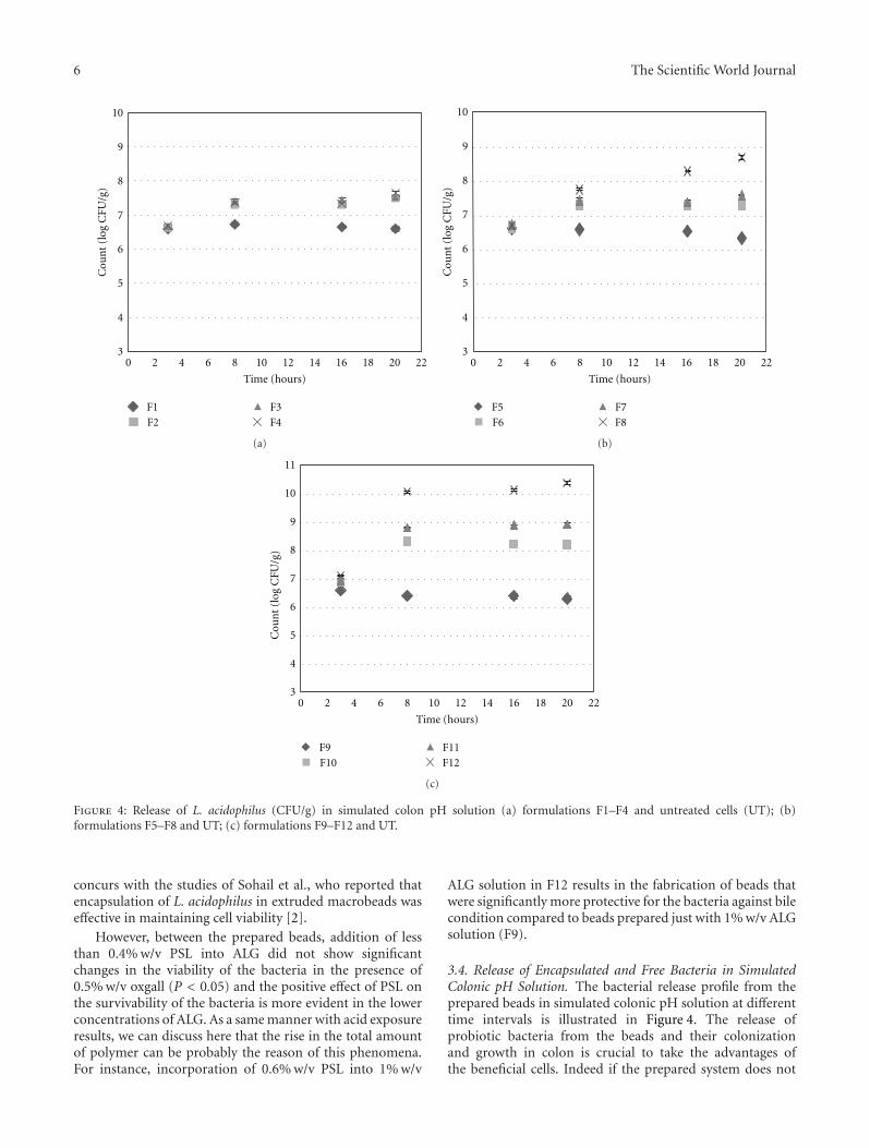
Figure 4: Release of L. acidophilus (CFU/g) in simulated colon pH solution (a) formulations F1–F4 and untreated cells (UT); (b)formulations F5–F8 and UT; (c) formulations F9–F12 and UT.
concurs with the studies of Sohail et al., who reported thatencapsulation of L. acidophilus in extruded macrobeads waseffective in maintaining cell viability [2].
However, between the prepared beads, addition of lessthan 0.4% w/v PSL into ALG did not show significantchanges in the viability of the bacteria in the presence of0.5% w/v oxgall (P < 0.05) and the positive effect of PSL onthe survivability of the bacteria is more evident in the lowerconcentrations of ALG. As a same manner with acid exposureresults, we can discuss here that the rise in the total amountof polymer can be probably the reason of this phenomena.For instance, incorporation of 0.6% w/v PSL into 1% w/v
ALG solution in F12 results in the fabrication of beads thatwere significantly more protective for the bacteria against bilecondition compared to beads prepared just with 1% w/v ALGsolution (F9).
3.4. Release of Encapsulated and Free Bacteria in SimulatedColonic pH Solution. The bacterial release profile from theprepared beads in simulated colonic pH solution at differenttime intervals is illustrated in Figure 4. The release ofprobiotic bacteria from the beads and their colonizationand growth in colon is crucial to take the advantages ofthe beneficial cells. Indeed if the prepared system does not
The Scientific World Journal 7
disintegrate and release its payloads in adequate time, itleaves the body without producing any claimed benefits [18].According to our results, during the first 3 h incubation insimulated colon pH solution, the cells increasingly releasedfrom all of the prepared beads to reach the level ranged6-7 CFU/g, where the situation started to be remarkablydifferent for coats of ALG-PSL in comparison with those ofALG.
Among beads prepared with ALG (F1, F5, and F9), in3 h, the counts hit their maximum level and beyond that nosignificant changes were observed in cell numbers (P > 0.05).It can be concluded that the ALG concentrations had noconsiderable effect on the release of L. acidophilus fromthe prepared beads. This finding also is in good agreementwith Mandal et al. [18]. They reported that the count ofL. casei in simulated colonic pH solution rose to its highestpoint in 60 min and after that remained constant. Moreover,according to Picot and Lacroix, a progressive release of coatedcells in whey protein in simulated intestinal conditionsoccurred [27].
In sharp contrast, in the case of beads prepared usingALG-PSL, not only the bacteria completely released fromthe beads but also different rates of bacterial growth after3 h was observed indicating a stimulating effect of PSL onthe bacteria. Our data revealed that higher concentrationsof PSL produced the greater stimulation effect on bacteria,as 0.6% w/v PSL in F12 showed more than 4 log rise inbacterial count. However, in F2 with minimum amount ofPSL (0.1% w/v), lowest amount of growth was observed(under 2 log). The stimulation effect of PSL on L. acidophiluscan be probably attributed to its structure as a soluble fiber.Based on prebiotic definition, nondigestible food ingredientssuch as carbohydrates in the form of soluble fiber canstimulate the growth and/or activity of bacteria. Indeed,in some cases, PSL has been used as prebiotic in differentsituations [10–13]. For instance, in a randomized controlledtrial for treatment of patients with ulcerative colitis usingsynbiotic versus probiotic or prebiotic, PSL has been usedas prebiotic and the results have shown that the quality oflife in patients has been improved [10]. However, PSL hasnot been officially designated as a prebiotic so far and in vivostudies in this regards are in progress. In a study, the prebioticpotential of PSL fiber in healthy women on bifidobacteriawas evaluated by Elli and colleagues [28], and they reportedthat PSL seed husk can be metabolized by bifidobacteria onlyafter partial hydrolysis.
In summary, whatever the prebiotic activity officiallyaccepted or not for PSL, it is important to note that basedon our data, it is assumable that PSL can potentially act asa prebiotic and preparation of PSL-ALG beads containing L.acidophilus improved its delivery to the active site.
4. Conclusion
ALG-PSL beads encapsulating probiotic L. acidophilus inthe size range of 1.61 ± 0.06 to 1.80 ± 0.07 mm with EEhigher than 98% were prepared using extrusion method.Inclusion of PSL into ALG beads maintained the survival
rate of L. acidophilus in acid and bile conditions as well asconsiderable stimulation effect on the bacteria in simulatedcolon pH solution. Psyllium as a pharmacologically activeingredient for gastrointestinal disease and a potential pre-biotic can be a suitable candidate to partially replace withalginate for encapsulation of probiotic bacteria.
Acknowledgment
The present study was financially supported by a grant fromTabriz University of Medical Sciences and BiotechnologyResearch Center, Tabriz, Iran.
References
[1] FAO/WHO, “Guidelines for the evaluation of probiotics infood,” Food and Agriculture Organization of United Nationsand World Health Organization Working Group Report,World Health Organization, London, UK, 2002.
[2] A. Sohail, M. S. Turner, A. Coombes, T. Bostrom, and B.Bhandari, “Survivability of probiotics encapsulated in alginategel microbeads using a novel impinging aerosols method,”International Journal of Food Microbiology, vol. 145, no. 1, pp.162–168, 2011.
[3] B. Albertini, B. Vitali, N. Passerini et al., “Development ofmicroparticulate systems for intestinal delivery of Lactobacillusacidophilus and Bifidobacterium lactis,” European Journal ofPharmaceutical Sciences, vol. 40, no. 4, pp. 359–366, 2010.
[4] R. R. Mokarram, S. A. Mortazavi, M. B. H. Najafi, andF. Shahidi, “The influence of multi stage alginate coatingon survivability of potential probiotic bacteria in simulatedgastric and intestinal juice,” Food Research International, vol.42, no. 8, pp. 1040–1045, 2009.
[5] K. Kailasapathy, “Microencapsulation of probiotic bacteria:technology and potential applications,” Current Issues inIntestinal Microbiology, vol. 3, no. 2, pp. 39–48, 2002.
[6] W. Ouyang, H. Chen, M. L. Jones et al., “Artificial cellmicrocapsule for oral delivery of live bacterial cells for therapy:design, preparation, and in-vitro characterization,” Journal ofPharmacy and Pharmaceutical Sciences, vol. 7, no. 3, pp. 315–324, 2004.
[7] M. H. Fischer, N. Yu, G. R. Gray, J. Ralph, L. Anderson, and J.A. Marlett, “The gel-forming polysaccharide of psyllium husk(Plantago ovata Forsk),” Carbohydrate Research, vol. 339, no.11, pp. 2009–2017, 2004.
[8] Q. Guo, S. W. Cui, Q. Wang, and J. Christopher Young, “Frac-tionation and physicochemical characterization of psylliumgum,” Carbohydrate Polymers, vol. 73, no. 1, pp. 35–43, 2008.
[9] B. Singh, “Psyllium as therapeutic and drug delivery agent,”International Journal of Pharmaceutics, vol. 334, no. 1-2, pp.1–14, 2007.
[10] S. Fujimori, K. Gudis, K. Mitsui et al., “A randomizedcontrolled trial on the efficacy of synbiotic versus probiotic orprebiotic treatment to improve the quality of life in patientswith ulcerative colitis,” Nutrition, vol. 25, no. 5, pp. 520–525,2009.
[11] D. Damaskos and G. Kolios, “Probiotics and prebiotics ininflammatory bowel disease: microflora ‘on the scope’,” BritishJournal of Clinical Pharmacology, vol. 65, no. 4, pp. 453–467,2008.
[12] S. Fujimori, A. Tatsuguchi, K. Gudis et al., “High dose probi-otic and prebiotic cotherapy for remission induction of active

8 The Scientific World Journal
Crohn’s disease,” Journal of Gastroenterology and Hepatology,vol. 22, no. 8, pp. 1199–1204, 2007.
[13] M. Rishniw and S. G. Wynn, “Azodyl, a synbiotic, fails to alterazotemia in cats with chronic kidney disease when sprinkledonto food,” Journal of Feline Medicine and Surgery, vol. 13, no.6, pp. 405–409, 2011.
[14] G. R. Gibson and M. B. Roberfroid, “Dietary modulation ofthe human colonic microbiota: introducing the concept ofprebiotics,” Journal of Nutrition, vol. 125, no. 6, pp. 1401–1412,1995.
[15] W. Krasaekoopt, B. Bhandari, and H. Deeth, “The influenceof coating materials on some properties of alginate beadsand survivability of microencapsulated probiotic bacteria,”International Dairy Journal, vol. 14, no. 8, pp. 737–743, 2004.
[16] M. Chavarri, I. Maranon, R. Ares, F. C. Ibanez, F. Marzo,and M. D.C. Villaran, “Microencapsulation of a probiotic andprebiotic in alginate-chitosan capsules improves survival insimulated gastro-intestinal conditions,” International Journalof Food Microbiology, vol. 142, no. 1-2, pp. 185–189, 2010.
[17] W. K. Ding and N. P. Shah, “An improved method ofmicroencapsulation of probiotic bacteria for their stability inacidic and bile conditions during storage,” Journal of FoodScience, vol. 74, no. 2, pp. M53–M61, 2009.
[18] S. Mandal, A. K. Puniya, and K. Singh, “Effect of alginateconcentrations on survival of microencapsulated Lactobacil-lus casei NCDC-298,” International Dairy Journal, vol. 16, no.10, pp. 1190–1195, 2006.
[19] V. Chandramouli, K. Kailasapathy, P. Peiris, and M. Jones, “Animproved method of microencapsulation and its evaluationto protect Lactobacillus spp. in simulated gastric conditions,”Journal of Microbiological Methods, vol. 56, no. 1, pp. 27–35,2004.
[20] T. Y. Sheu, R. T. Marshall, and H. Heymann, “Improvingsurvival of culture bacteria in frozen desserts by microentrap-ment,” Journal of Dairy Science, vol. 76, no. 7, pp. 1902–1907,1993.
[21] Y. Doleyres and C. Lacroix, “Technologies with free andimmobilised cells for probiotic bifidobacteria production andprotection,” International Dairy Journal, vol. 15, no. 10, pp.973–988, 2005.
[22] S. J. Kim, S. Y. Cho, S. H. Kim et al., “Effect of microencap-sulation on viability and other characteristics in Lactobacillusacidophilus ATCC 43121,” LWT—Food Science and Technology,vol. 41, no. 3, pp. 493–500, 2008.
[23] C. S. Favaro-Trindade and C. R. F. Grosso, “Microencapsu-lation of L. acidophilus (La-05) and B. lactis (Bb-12) andevaluation of their survival at the pH values of the stomachand in bile,” Journal of Microencapsulation, vol. 19, no. 4, pp.485–494, 2002.
[24] K. Sultana, G. Godward, N. Reynolds, R. Arumugaswamy,P. Peiris, and K. Kailasapathy, “Encapsulation of probioticbacteria with alginate-starch and evaluation of survival insimulated gastrointestinal conditions and in yoghurt,” Inter-national Journal of Food Microbiology, vol. 62, no. 1-2, pp. 47–55, 2000.
[25] B. Singh and V. Sharma, “Design of psyllium-PVA-acrylic acidbased novel hydrogels for use in antibiotic drug delivery,”International Journal of Pharmaceutics, vol. 389, no. 1-2, pp.94–106, 2010.
[26] P. Muthukumarasamy, P. Allan-Wojtas, and R. A. Holley,“Stability of Lactobacillus microcapsules and survival in simu-lated gastrointestinal conditions and in yoghurt,” InternationalDairy Journal, vol. 14, pp. 505–515, 2006.
[27] A. Picot and C. Lacroix, “Encapsulation of bifidobacteria inwhey protein-based reuteri in different types of microcap-sules,” Journal of Food Science, vol. 71, no. 1, pp. M20–M24,2004.
[28] M. Elli, D. Cattivelli, S. Soldi, M. Bonatti, and L. Morelli,“Evaluation of prebiotic potential of refined psyllium (Plan-tago ovata) fiber in healthy women,” Journal of ClinicalGastroenterology, vol. 42, pp. S174–176, 2008.

Submit your manuscripts athttp://www.hindawi.com
Hindawi Publishing Corporationhttp://www.hindawi.com Volume 2014
Anatomy Research International
PeptidesInternational Journal of
Hindawi Publishing Corporationhttp://www.hindawi.com Volume 2014
Hindawi Publishing Corporation http://www.hindawi.com
International Journal of
Volume 2014
Zoology
Hindawi Publishing Corporationhttp://www.hindawi.com Volume 2014
Molecular Biology International
GenomicsInternational Journal of
Hindawi Publishing Corporationhttp://www.hindawi.com Volume 2014
The Scientific World JournalHindawi Publishing Corporation http://www.hindawi.com Volume 2014
Hindawi Publishing Corporationhttp://www.hindawi.com Volume 2014
BioinformaticsAdvances in
Marine BiologyJournal of
Hindawi Publishing Corporationhttp://www.hindawi.com Volume 2014
Hindawi Publishing Corporationhttp://www.hindawi.com Volume 2014
Signal TransductionJournal of
Hindawi Publishing Corporationhttp://www.hindawi.com Volume 2014
BioMed Research International
Evolutionary BiologyInternational Journal of
Hindawi Publishing Corporationhttp://www.hindawi.com Volume 2014
Hindawi Publishing Corporationhttp://www.hindawi.com Volume 2014
Biochemistry Research International
ArchaeaHindawi Publishing Corporationhttp://www.hindawi.com Volume 2014
Hindawi Publishing Corporationhttp://www.hindawi.com Volume 2014
Genetics Research International
Hindawi Publishing Corporationhttp://www.hindawi.com Volume 2014
Advances in
Virolog y
Hindawi Publishing Corporationhttp://www.hindawi.com
Nucleic AcidsJournal of
Volume 2014
Stem CellsInternational
Hindawi Publishing Corporationhttp://www.hindawi.com Volume 2014
Hindawi Publishing Corporationhttp://www.hindawi.com Volume 2014
Enzyme Research
Hindawi Publishing Corporationhttp://www.hindawi.com Volume 2014
International Journal of
Microbiology
Related Documents